mpi-artifacts
TRANSCRIPT

Peeyush Bhargava MD MBA, Zhiyun Yang MD,
Scott Adams MD
Artifacts in Myocardial Perfusion Imaging
Louisiana State University Health Science Center, Shreveport, LA
ARRS, April 2016, Los Angeles, CA

Teaching Points
• Recognize common artifact patterns on SPECT
(Single Photon Emission Computed Tomography)
MPI (Myocardial Perfusion Imaging)
• Understand the causes of artifacts and how they
can be corrected or avoided
• Target Audience: Residents, Radiologists,
Cardiologists, Nuclear Medicine Physicians
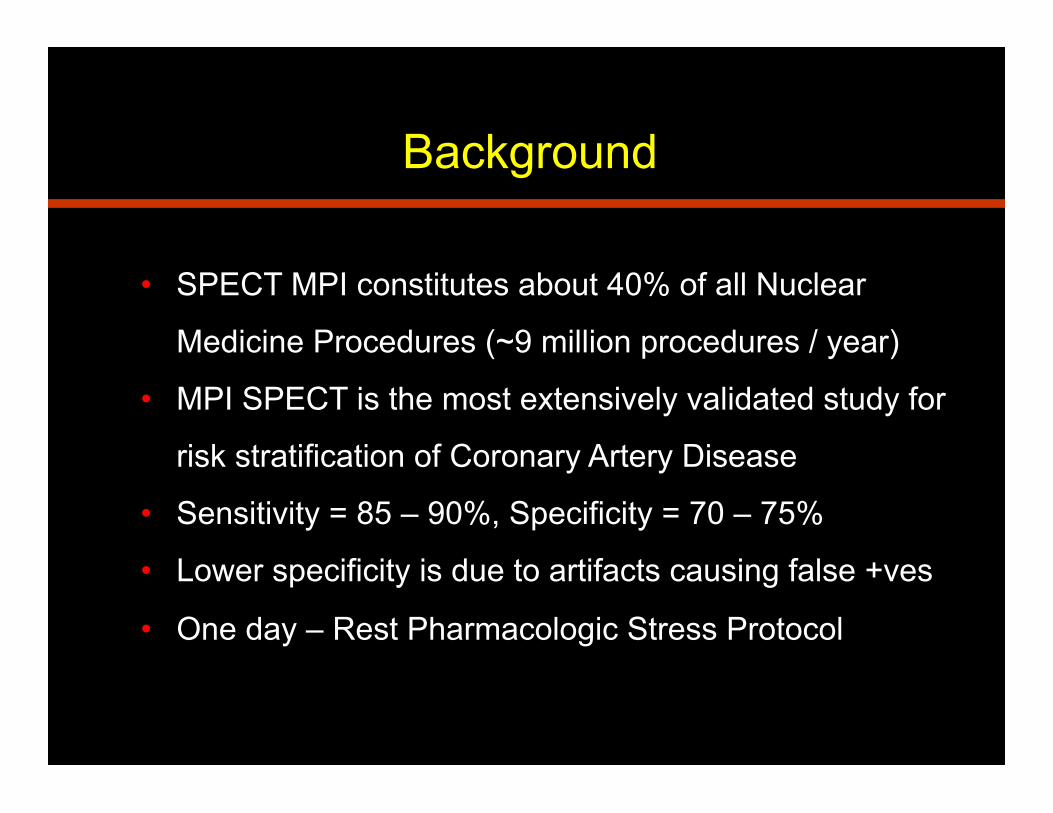
Background
• SPECT MPI constitutes about 40% of all Nuclear
Medicine Procedures (~9 million procedures / year)
• MPI SPECT is the most extensively validated study for
risk stratification of Coronary Artery Disease
• Sensitivity = 85 – 90%, Specificity = 70 – 75%
• Lower specificity is due to artifacts causing false +ves
• One day – Rest Pharmacologic Stress Protocol

• Dual head gamma camera with High Res Collimators
• 20% window set around 140 keV
• Circular orbit (vs elliptical/body contouring)
• 180 degree SPECT: LPO to RAO
• Step and shoot (20-30 sec); Frame mode (vs List)
• Resting SPECT and post stress Gated SPECT (16 fr)
• Filtering (lower cut off = more filtering / smoothening)
• Processing (FBP vs OSEM)
Acquisition and Processing

Normal Study

Normal Study - Gated

• Uniform uptake at rest and stress
• No fixed or reversible myocardial perfusion defects
• Normal myocardial wall motion and thickening
• Normal LVEF (>50%), EDV (<110), ESV (<50)
• Normal polar plots and scores (SRS, SSS, SDS)
• No significant non cardiac activity
• No patient motion or soft tissue attenuation
MPI SPECT – Normal Study

• Soft Tissue Attenuation
• Patient Motion
• Non Cardiac Activity
• Non Coronary Disease
• Image Normalization
Categories of MPI SPECT Artifacts
• Prep. / Injection / Stress test
• Processing Related
• Flood Field Non Uniformity
• COR Misalignment
• Camera Head Misalignment

Soft Tissue Attenuation
• Causes: diaphragm, breasts, obese body habitus
• Imaging Characteristics: decreased counts, fixed
defects, worse at rest, normal wall motion, ischemia
• Recognition: review raw data, inferior wall in males,
anterior/lateral/apical in women, worse with Thallium
• Solution: reimaging, prone imaging, breast / arm
positioning, attenuation correction (CT)

Diaphragmatic Attenuation

Diaphragmatic Attenuation

Breast Shadow

Breast Attenuation

Patient Motion
• Causes: vertical (or horizontal) motion (≥ 2 pixels)
• Imaging Characteristics: opposed defects and
streaks from edges, anterior / inferior (hurricane
sign) vs septal / lateral, cardiac creep – exercise
and thallium
• Recognition: review raw data in cine mode
• Solution: reimaging, preparation and positioning,
motion correction software

Before and after Motion Correction

Non Cardiac Activity
• Causes: liver, stomach, bowel or tumor uptake
• Imaging Characteristics: scattering ! increased
counts, ramp filter artifact ! decreased counts
• Recognition: review raw data in cine mode
• Solution: drink water / milk, walk / low level
exercise, and reimage, optimize injection to
imaging time

Gastric Uptake

Gastric Uptake
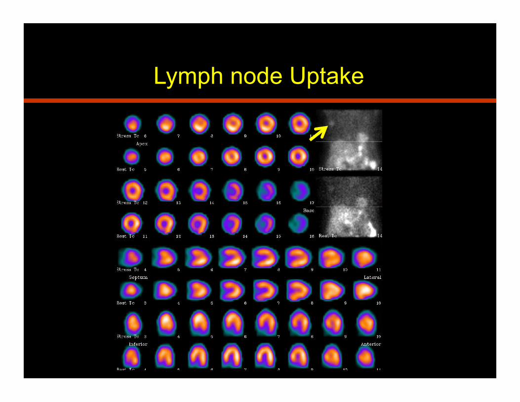
Lymph node Uptake

Thymoma

Ascites

Non Coronary Disease
• Causes: LBBB, hypertrophic cardiomyopathy, short
septum, apical thinning, balanced ischemia, dextrocardia
• Imaging Characteristics / Recognition: perfusion defect
at increased heart rate (pharmacologic stress test),
septum > lateral wall, septal defect, apical defect, false –
ve / stunning, right sided heart / processing
• Solution: review patient history, EKG, Echo, prior studies

LV Hypertrophy

LV Hypertrophy

RV Hypertrophy

Dextroxardia
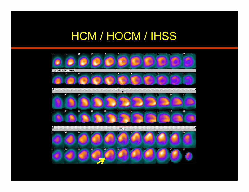
HCM / HOCM / IHSS

LBBB

Normalization Artifacts
• Causes: images normalized to the hottest pixel
(cardiac or non cardiac)
• Imaging Characteristics: focal hot spot (papillary
muscle), decreased counts in myocardium
• Solution: reprocess / reimaging, increase intensity

Normalization Artifact

Prep. / Injection / Stress test
• Causes / Examples: inadequate preparation
(coffee / NPO, attenuators / uncomfortable) /
suboptimal IV access, suboptimal stress test
• Recognition: low count images, artifacts from
infiltration / contamination, false negative studies
• Solution: attention to detail, teamwork, effective
communication, ALARA
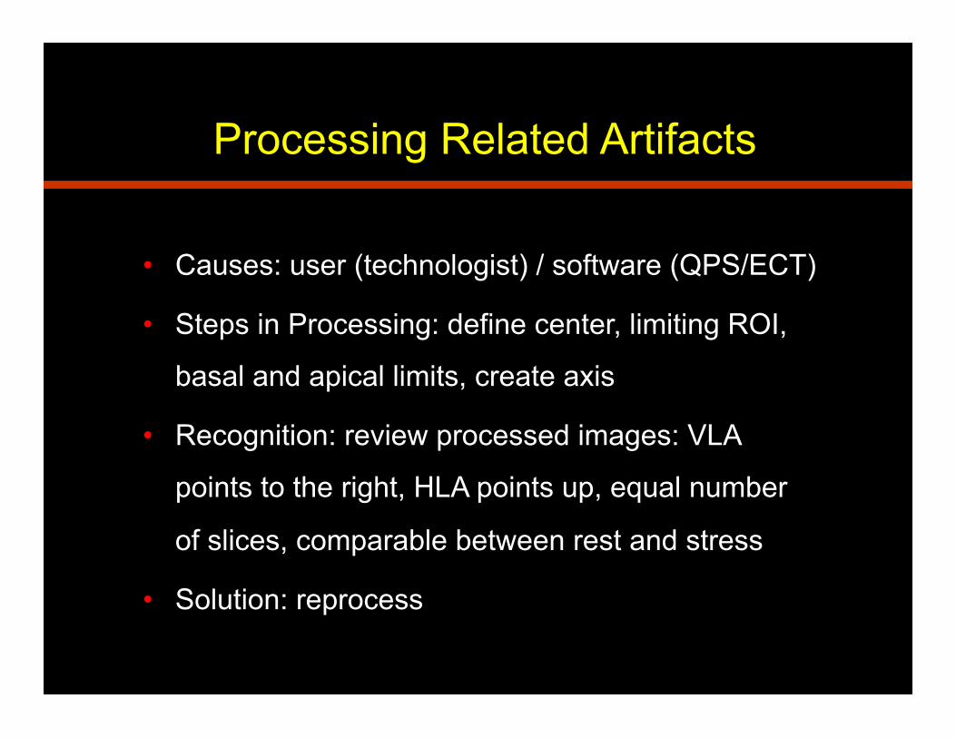
Processing Related Artifacts
• Causes: user (technologist) / software (QPS/ECT)
• Steps in Processing: define center, limiting ROI,
basal and apical limits, create axis
• Recognition: review processed images: VLA
points to the right, HLA points up, equal number
of slices, comparable between rest and stress
• Solution: reprocess

Non Uniformity Artifacts
• Causes: flood field non uniformity
• Imaging Characteristics: ring artifact, fixed
or reversible defect
• Recognition: review uniformity flood
• Solution: reimage after camera repair, Q/C

Normal PMT malfunction/uncoupling
Poor mixing
Cracked crystal
Poor gain alignment (tuning) of PMTs
Collimator defect
Flood Non Uniformities

COR Misalignment
• Causes: center of rotation misalignment
• Imaging Characteristics: oblong cavity,
decreased activity, streaks, blurred images
• Solution: COR correction, Q/C

Camera Head Misalignment
• Imaging Characteristics: decreased counts,
defects similar to patient motion, blurred images
• Recognition: review raw data
• Solution: Camera Q/C

Camera Quality Control

1. Artifacts and pitfalls in myocardial perfusion imaging.
Burrell S, MacDonald A. J Nucl Med Technol. 2006 Dec;34(4):
193-211
2. Artifacts in planar and SPECT myocardial perfusion imaging.
Wackers FJ. Am J Card Imaging. 1992 Mar;6(1):42-57
3. How to detect and avoid myocardial perfusion SPECT
artifacts. DePuey EG 3rd. J Nucl Med. 1994 Apr;35(4):699-702
References

Contact Information
Peeyush Bhargava MD MBA
PGY 3 Radiology Resident
Phone: 832-374-2110
Louisiana State University Health Science Center, Shreveport, LA