mouse mammary epithelial histamine system - semantic … filemouse mammary epithelial histamine...
TRANSCRIPT

W. WAGNER, A. ICHIKAWA*, S. TANAKA*, P. PANULA**, W.A. FOGEL
MOUSE MAMMARY EPITHELIAL HISTAMINE SYSTEM
Institute of Biogenic Amines, Polish Academy of Sciences, Lodz, Poland, *Department ofPhysiological Chemistry, Graduate School of Pharmaceutical Sciences, Kyoto University, Kyoto,
Japan, ** Department of Biology, Äbo Akademi University, Turku, Finland
Histamine is suggested to play a role in mammary gland growth regulation,differentiation and functioning during pregnancy and lactation. Two pools ofhistamine are thought to be involved in these processes: mastocyte- and epithelialcell related histamine. In the present study we focused on epithelial cells.Immunohistochemistry has shown that the epithelial cells positive for histamine andL-histidine decarboxylase (HDC), the primary enzyme regulating histaminebiosynthesis, were mainly found in cells forming alveolar structures in the mammarygland. Cultured primary mouse mammary epithelial cells (MMEC) expressed strongHDC immunoreactivity, especially dividing cells and non-differentiated ones.Histidine decarboxylase activity undergoes significant changes during pregnancyand lactation. Pregnancy associated intensive growth of the mammary glandcoincided with an increase and the first days of lactation with a decrease of HDCprotein expression. Binding studies with mammary tissue membranes and epithelialcell membranes revealed the presence of H1 and H3 but not H2 receptors.Summarizing, our data have shown that mammary epithelial cells are capable ofsynthesizing and excreting histamine and they bear histamine receptors. Thesefindings further substantiate the role of histamine in mammary gland physiology.
K e y w o r d s : mouse mammary gland, epithelial cells, L- histidine decarboxylase, histamine,histamine receptors
INTRODUCTION
The original observation of a high level of histamine in mouse mammaryglands (1), suggested a possible role for this amine in mammary gland functionand development. Histamine is known to exert various effects in physiological
JOURNAL OF PHYSIOLOGY AND PHARMACOLOGY 2003, 54, 2, 211�223
www.jpp.krakow.pl

and pathological reactions such as: smooth muscle contraction, gastric acidsecretion, cell growth, neurotransmission and inflammation (2). Histamine is animinoamine synthesized locally from L-histidine by a specific enzyme L-histidinedecarboxylase (HDC, EC 41.1.22). The histamine is inactivated by two mainenzymes: histamine N-methyltransferase (HMT, EC 2.1.1.8) via methylation(brain and periphery) and diamine oxidase (DAO, EC 1.4.3.6) via oxidativedeamination (peripheral tissues only) (3).
The actions of histamine are mediated through the activation of membrane-associated G protein coupled receptors: H1, H2, H3 and H4. Histamine receptors inmice have been characterised pharmacologically and murine H1 and H2 receptorshave been cloned (4, 5). In addition to well known histamine containing cells, i.e.mast cells, enterochromaffin-like cells and neurons, studies have demonstratedthe presence of histamine systems in some other tissues and cells, such as bloodvessel endothelial cells (6, 7) or mammary gland epithelial cells (8-10). The latterobservation and the effects of HDC inhibitors and H2 receptor antagonists on agrowth of mammary cancer cells in vitro, has led to the development of clinicalresearch in this area (11, 12).
In the first study on the metabolism of histamine in mammary glands (13), it wasdemonstrated that the mammary histamine system changes markedly during theestrous cycle as well as during pregnancy and lactation. Potent physiological effectsof histamine on mammary glands were shown in vivo and in vitro (14). Histaminereceptors agonists on their own or in the presence of oxytocin increased milksecretion. The H3-receptor antagonist - FUB 181 had an additive effect on oxytocinstimulated milk secretion from goat mammary glands (15). Although the mainhistamine immuno-reactive sites in mammae were mast cells, some histamine-positive signals were also found in glandular epithelium and in the stroma (16).
In this paper we present the results of a study of the histamine system and HDCprotein expression profile in mouse mammary glands at various physiological states.
MATERIALS AND METHODS
Animals
Virgin female mice (Mus musculus) (BALB/C, 20 g body wt) were used. Animal procedurescomplied with the Polish legislation concerning experiments on animals and were approved by thelocal care committee.
Pregnancy and lactation
Female mice were housed with males of proven fertility for three days accompanied by dailymicroscopic examination of vaginal smears. The presence of spermatozoa and/ or vaginal plug wasregarded as the first day of pregnancy. Pregnancy day was further verified at autopsy by embryodevelopment stage. The parturition day was designated as the first lactation day.
212

Cell isolation procedure
Mammary epithelial cells (MMEC) were obtained from resting animals as well as pregnant andlactating animals and were isolated from the abdominal mammary glands. In brief, dissected tissueswere digested with collagenase (collagenase IV 2 mg/ml, hyaluronidase 100U/ml in DMEMsupplemented with antibiotic-antimycotic solution (all reagents from Sigma, St. Louis, MO, USA)for 2 h at 37°C. Finally, the cells were purified using differential sieving and repetitive DMEMwashing and sedimentation.
Primary cell culture
Isolated MMEC were cultured in DME/F12 medium supplemented with 10% horse serum,hydrocortisone (0.5 µg/ml), insulin (10 µg/ml) and antibiotic-antimycotic solution. They wereincubated at 37°C in 3% CO2 humidified environment.
For histamine synthesis assays, cells were cultured in complete medium based on DMEMinstead of DME/F12. HDC activity assays were performed after approximately 4-5 days of culturewhen cells reached confluence on 16 millimetres (24-well) (Corning, NY, USA) or 60 millimetresin diameter culture dishes (Nunc, Denmark).
For immunocytochemistry, cells were grown for 9 days on 8-well Lab-Tek Chamber GlassSlides (Nunc, Inc., IL, USA). For receptor binding studies cells were incubated up to 72 h on 60 or120 millimetres in diameter culture dishes (Nunc, Denmark).
Histamine synthesis and secretion assay
Twenty four h after cell plating and every 24 h thereafter during a 72 h experiment, the cells andmedium were harvested and centrifuged at 20 g. Postculture medium was removed and the cellswere purified by repeated washing in cold PBS and centrifugation. Finally, the cells wereresuspended in 0.3 ml of PBS and lysed by three cycles of freezing and thawing. Then, cellhomogenates and culture medium were deproteinized by boiling at 100°C for 10 minutes and spundown at 12,000 g for 10 minutes. Histamine levels in supernatant and postculture media weremeasured by RIA (Immunotech, France) and expressed per 106 cells.
Histidine decarboxylase activity assay
The epithelial cell histidine decarboxylase activity was estimated after 4-5 days cell culture(17). Harvested and purified cells (as described above) were suspended in enzyme-extraction buffer(0.1 M potassium buffer, pH 6.8, containing 1mM of dithiothreitol and 0.1 mM pyridoxal 5'-phosphate) and lysed by three cycles of freezing and thawing followed by sonication (5 minutes).The homogenates were centrifuged at 12,000g for 20 minutes and resulting supernatants dialysed.Dialysis was carried out against extraction buffer for 60 minutes at 4°C with a fresh buffer changeevery 20 minutes. HDC activity was assayed with 25 mM L-histidine. Incubation was for 1 hour at37°C. The reaction was stopped by heat enzyme inactivation (100°C, 10 minutes). Newlysynthesised histamine was used as a measure of the enzyme activity. It was estimated with aradioenzymatic assay employing histamine N-methyltransferase (HMT) (18) and adenosyl-L-methionine S-[methyl - 3H] (New Nuclear, Boston, MA, USA) as a donor of methyl group (19).
Histamine- , histidine decarboxylase - immunohistochemistry
Immunostaining for histamine was carried out on 20 µm thick cryostat sections as describedpreviously (20). Fixed samples were washed in 0.01 M PBS containing 0.125% Triton X-100 (PBS
213

- T) followed by incubation with a histamine antiserum diluted (1: 2,000) in PBS-T containing 1%normal goat serum overnight at 4°C in humidified chamber. After washing in PBS-T the sampleswere incubated for 1 hour at room temperature with fluorochrome conjugated goat anti-rabbit IgG(1:500, Alexa 488 GAR 2 mg/ml, Molecular Probes, Eugen, USA). Control samples were incubatedwith the same serum pre-adsorbed with histamine-BSA conjugate. After the final washes in PBS-Tand PBS the samples were mounted in 50% glycerol (in PBS) and analyzed with Leitz Aristoplanfluorescence microscope.
Histidine decarboxylase immunostaining was carried out on 3-4 µm cryostat sections. Sectionswere fixed in acetone for 10 minutes at room temperature. After pretreatment with 0. 05 M TBS(Tris buffer solution, pH 7.6) slides were incubated with rabbit anti-GST-HDC fusion proteinpolyclonal antibody (1:500) (21) for 1 hour at room temperature. TBS washed slides were followedeither by secondary horse anti-rabbit IgG and visualised with an ABC kit (Novocastra LaboratoriesLtd., UK) or secondary fluorescein conjugated swine anti-rabbit IgG (Dako, Denmark). Washedwith TBS samples were mounted in mounting mediums and examined with Olympus CX41microscope or Jena Lumar (Carl Zeiss Jena) fluorescence microscope.
Histidine decarboxylase - immunocytochemistry
Histidine decarboxylase immunostaining was examined using primary mammary epithelialcells culture grown on Lab-Tech chamber glass slides. Cultures were washed briefly in PBS (pH7.4)and fixed in acetone for 10 minutes at room temperature. Immunostaining was carried out asdescribed above with an ABC kit visualisation.
Saturation binding experiments
Saturation binding assays were essentially performed as described previously (22) with somechanges. Cell membranes (400 µl; gland: 0.5-1.6 mg protein/ml; cells: 0.4-1.2 mg protein/ml) wereincubated for 30 min with continuous shaking (30°C) in triplicate in a total volume of 500 µl withincreasing concentrations of the specific histamine receptors H1, H2 and H3 radioligands. For the H1
receptor assay isotopic ligand [3H]-Mepyramine (0.1-30 nM) (29.0-30.0 Ci/mmol, AmershamPharmacia Biotech, UK) and "cold" ligand Triprolidine dihydrochloride 2 µM (Sigma) were used.For the H2 receptor assay ligands were: [3H]-Tiotidine (0.5-30 nM) (89.6 Ci/mmol, New EnglandNuclear, Boston, MA, USA) and 5 µM Histamine dihydrochloride (Sigma) respectively. [3H]-R(-)-α-Methylhistamine (0.15-9 nM) (29.0-35.0 Ci/mmol, Amersham Pharmacia Biotech, UK) and 1µM Histamine were applied for the H3 receptor assay.
Data analysis
The data are presented as mean ± SEM for n experiments. Comparisons among groups werecarried out using analysis of variance (ANOVA) and post hoc Newman-Keuls test. Saturation datawas analysed using the non-linear curve fitting program (GraphPad Prism 3.0).
RESULTS
Epithelial cell HDC activity and histamine concentration changes duringpregnancy and lactation
The highest level of HDC activity was 409.29 ± 47.41 pmol/h/mg protein andwas measured in epithelial cell cultures derived from 10-12th day of pregnancy
214

(Fig.1A). It was almost 4 fold higher than in epithelial cells from restingmammary glands (109.18 ± 24.48 pmol/h/mg protein). A plateau of HDC activitywas seen at between 14th-16th and the 20th day of pregnancy: 146.78 ± 13.74 and104.63 ± 11.46 pmol/h/mg protein respectively. A marked reduction in enzyme
215
Fig. 1. (A) L- Histidinedecarboxylase (HDC) activityin cultured mammaryepithelial cells derived fromfemale mice mammary glandsof different physiologicalstages. Each point representsthe mean ± SEM of 4 - 6determinations. *** p<0.001compared with resting, 10-12th, 14-16th, 20th day ofpregnancy and 6th day oflactation. ** p<0.01compared with 6th day oflactation. (B) Intracellular andextracellular histamine (C) incultured mammary epithelialcells. Cultures were preparedfrom mammary glands ofpregnant (10th-12th, 14th-16th,19th-21st day) and lactating(4th-7th day) mice. Cell andmedium contents weremeasured after 24 hr, 48 hrand 72 hr of incubation. *p<0.05 and ** p<0.01compared with time -corresponding counterparts.
activity was found after parturition on the 6th day of lactation (6.71 ± 0.75). On12th day after parturition the HDC activity again reached a high level of 344.92 ±43.64 pmol/h/mg protein.
Throughout the 72 hour experiments, cell histamine contents in epithelial cellsfrom mammary gland at 14-16th day of pregnancy were significantly higher(39.21 ± 11.09 pmol/106 cells) than in corresponding cell cultures obtained fromthe gland at the 10-12th, the 19-21st days of pregnancy and the 4-7th day of lactation(7.09 ± 3.55; 11.52 ± 2.37; 8.38 ± 1.41 pmol/106 cells, respectively) (Fig.1B). Theepithelial cell cultures derived from mammary gland at the 10-12th and at the 19-21st days of pregnancy showed higher histamine release from the cells, as judgedby medium and cell histamine contents. The cells from the 14-16th day ofpregnancy showed balanced histamine synthesis and secretion during the wholeexperiment. The histamine concentrations in the medium tended to increase in atime-dependent manner in all cell cultures, and the maximum values wererecorded after 72 h culture (Fig.1C).
216
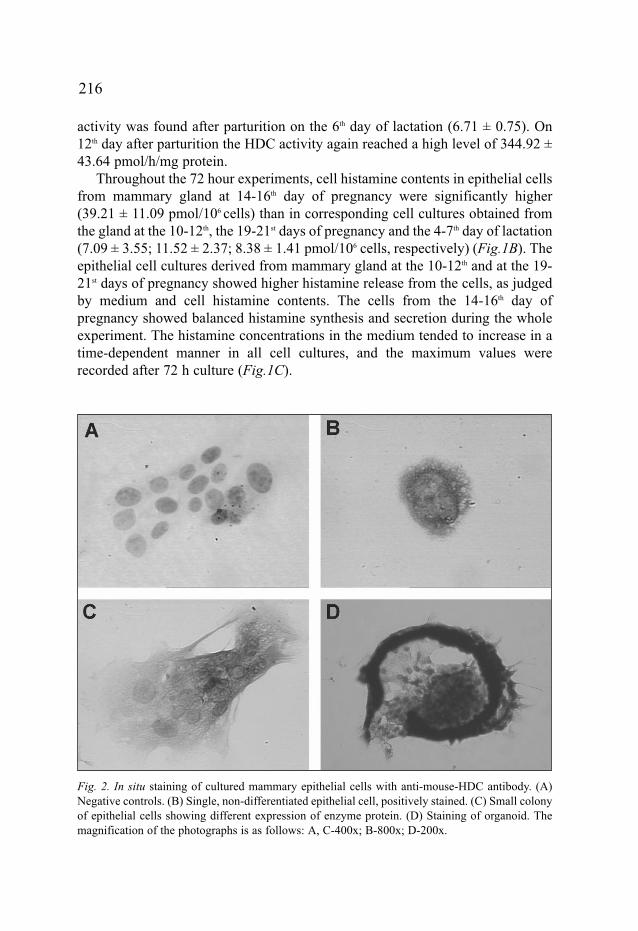
Fig. 2. In situ staining of cultured mammary epithelial cells with anti-mouse-HDC antibody. (A)Negative controls. (B) Single, non-differentiated epithelial cell, positively stained. (C) Small colonyof epithelial cells showing different expression of enzyme protein. (D) Staining of organoid. Themagnification of the photographs is as follows: A, C-400x; B-800x; D-200x.

HDC immunocytochemistry in primary epithelial cell culture
To visualise and localise HDC in mammary gland epithelial cells, primaryepithelial cell cultures were used. Cells isolated from mammary glands from
217
Fig. 3. Left panel shows the results of HDC immunohistochemistry of mouse resting, pregnancyand 5th day of lactation mammary glands. (A) Resting mammary gland, positively stained, singlemast cell in the centre. (B) 14-16th day of pregnancy mammary gland sections, positively stainedalveolar epithelium is seen. (C) HDC - immunopositive cells spread in the vicinity of the duct, 5th
day of lactation. Right panel presents the indirect histamine - immunofluorescent histochemistry of15th day pregnancy mammary gland. (D) Positive staining of few epithelial cells in alveolarepithelium. (E) Positive staining of alveolar structures (upper part). A few mast cells showingstrong signals of immunofluorescence are seen in intralobular stroma. (F) Positively stainedalveolus is seen in the lower part of the left side of the photograph. The magnification of thephotographs is as follows: A,D-400x; B-200x; C-300x; E-100x; F-40x.

animals in the 14-16th day of pregnancy, were cultured and stained as described inMaterials and Methods. Under the conditions employed dividing or single not-differentiated cells were rich in HDC molecules (Fig.2B, C). On the other hand,single flattened differentiated cells exhibited low or no HDC protein expression.
Histamine and HDC immunohistochemistry in mouse mammary gland sections
The study on histamine/HDC immuno-localization in mammary gland wasfocused on glandular tissue. Signals of histamine immunofluorescence werefound in alveolar structures of the mammary gland and they were mostlyassociated with epithelium (Fig.3D,F). Stronger histamine-immunoreactivity wasconfined to mast cells which were numerous in connective tissue and diffused inglandular tissue (Fig.3E,F)
HDC-immunopositive cells with a mast cell like granular pattern of stainingwere clearly visible in preparations visualised with the ABC system. They werefound in connective tissue of resting (Fig.3A) and lactating mammary glands(data not shown). Epithelial cells of alveolar formations displayed only a weaksignal, barely distinguishable from the background (Fig.3B). Strong HDCimmunoreactivity was also found in the vicinity of the ducts from lactating
218
Fig. 4. Stage - course HDC - immunofluorescent histochemistry of mouse mammary gland. (A) 10-12th day of pregnancy, strong immunoreactivity in the alveolar structures. (B) 19th day of pregnancy,diminished immunoreactivity in alveolar epithelium as compared to the 10-12th day of pregnancy.(C) 5th day of lactation, lack of specific immunofluorescence. (D) Immunopositive signals appearagain in alveolar epithelium on 10th day of lactation. Magnification: 150x.
mammary glands (Fig.3C). When the fluorescence technique was applied, theepithelial immunopositivity was found in cells of alveolar formations and liningduct cells in pregnancy and lactating mammary glands (Fig.4).
Histamine receptors in mammary gland tissue and primary epithelial cell culture
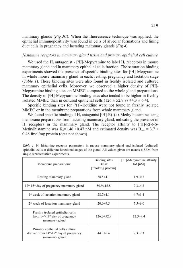
We used the H1 antagonist - [3H]-Mepyramine to label H1 receptors in mousemammary gland and in mammary epithelial cells fraction. The saturation bindingexperiments showed the presence of specific binding sites for [3H]-Mepyraminein whole mouse mammary gland in each: resting, pregnancy and lactation stage(Table 1). These binding sites were also found in freshly isolated and culturedmammary epithelial cells. Moreover, we observed a higher density of [3H]-Mepyramine binding sites on MMEC compared to the whole gland preparations.The density of [3H]-Mepyramine binding sites also tended to be higher in freshlyisolated MMEC than in cultured epithelial cells (126 ± 52.9 vs 44.3 ± 6.4).
Specific binding sites for [3H]-Tiotidine were not found in freshly isolatedMMEC or in the membrane preparations from whole mammary gland.
We found specific binding of H3 antagonist [3H]-R(-)-α-Methylhistamine usingmembrane preparations from lactating mammary gland, indicating the presence ofH3 receptors in the mammary gland. The receptor affinity to [3H]-R(-)-α-Methylhistamine was Kd=1.46 ±0.47 nM and estimated density was Bmax = 3.7 ±0.48 fmol/mg protein (data not shown).
219
Table 1. H1 histamine receptor parameters in mouse mammary gland and isolated (cultured)epithelial cells at different functional stages of the gland. All values given are means ± SEM fromsingle representative experiments.
Binding sites [3H]-Mepyramine affinityMembrane preparations: Bmax Kd [nM]
[fmol/mg protein]
Resting mammary gland 38.5±4.1 1.9±0.7
12th-15th day of pregnancy mammary gland 50.9±15.8 7.3±4.2
1st week of lactation mammary gland 28.7±4.1 4.7±1.4
2nd week of lactation mammary gland 20.0±9.5 7.5±6.0
Freshly isolated epithelial cellsfrom 14th-18th day of pregnancy 126.0±52.9 12.3±9.4
mammary gland
Primary epithelial cells culturederived from 14th-18th day of pregnancy 44.3±6.4 7.3±2.3
mammary gland
DISCUSSION
Previous biochemical studies showed active histidine decarboxylase and itsproduct - histamine in mammary glands and indicated major changes of thissystem during various physiological states (16). In this study we successfullytargeted the cells responsible for these phenomena as well as providing evidencefor histamine receptors involved. It is well known that mast cells are rich inhistamine (3). Both HDC and histamine positive mast cells were localisedimmunohistochemically in mammary glands. It is evident from the subcellularanalysis that HDC- positive immunoprecipitation was exhibited in granules. Thelatter observation agrees well with the recent findings of Japanese group (23).These authors studied the intracellular localization of the 74 and 53- kDa formsof HDC in RBL-2H3 cell line and demonstrated that posttranslationally processedHDC as 53 kDa form is originally localised in the endoplasmic reticulum andGolgi system and then moved and stored in the granules.
It should be mentioned that the mast cells found in lactating glands seem to havea different appearance than the cell population in glands from pregnant animals.The former cells are rather elongated and have an irregular shape, the latter have aregular and oval shape, are more compact giving stronger histamine signals.
Epithelial cells were considered as a second source of histamine. Indeed, bothbiochemical and immunohistochemical studies revealed the presence of histamineand histidine decarboxylase in primary epithelial cell cultures as well as thespecific histamine receptors H1 in the primary epithelial cell culture and H1 and H3
in mammary tissue. We found that epithelial cells isolated from resting, pregnancyand lactating mammary glands show different histamine synthesis profiles. Theactivity peak of HDC at the 12th day and plateau of enzyme activity recorded in thesecond and third decade of pregnancy was almost completely abolished afterparturition and throughout first week of lactation. The distinct pattern of HDCactivity changes in the cell culture model matches clearly the changes observedpreviously in mouse mammary gland during pregnancy and lactation (16). Thesedata are thus in line with the suggestion that the epithelial histamine stimulatesmammary gland growth and differentiation as well as function during lactationthrough a paracrine pathway and an autocrine loop (8, 9, 24).
To further characterize the mammary epithelial cells and HDC enzymeprotein expression the complex immunochemical study was performed. Theillustrations presented show HDC protein expression in cultured epithelial cells,especially in dividing and non differentiated cells. Moreover, it seems that theenzyme protein expression was relatively higher than in mammary gland tissue,in which the immunopositive epithelial structures were only found after the useof the more sensitive immunofluorescence method. It cannot be ruled out, thatthe positive effect on HDC regulation may have been due to the presence ofglucocorticoids in the culture medium. Dexamethasone treatment up-regulatesHDC expression in rat lungs (25) and in mastocytoma cells (26) via
220

glucocorticoid - responsive elements. However, the changes in HDC proteinexpression we found with the immunofluorescence method corresponded well todata obtained in biochemical study.
Saturation binding assays showed H1 receptor presence in resting, pregnancy andlactation stages of mammary gland. The Kd values calculated for [3H]-Mepyraminein our study are within a range of reported values (5-12 nM) obtained for rat andhuman mammary glands (10, 11). The affinity of [3H]-Mepyramine to the H1
receptor seems to be relatively higher in resting and 1 week of lactation mammaryand it could be associated with HDC activity decrease and lower availability ofhistamine. The distribution of H1 receptor in mammary gland is suggested to belargely due to epithelial cells as the density of H1 receptors was significantly higherin isolated MMC population than in the whole gland (Table 1). A decrease in H1
receptor expression under culture conditions was recorded. It is likely thatglucocorticoid treatment (medium supplementation) could down-regulate H1 and H2
receptors expression, as was observed in cerebral endothelial cell cultures (6).Alternatively, this may suggest lower degree of differentiation of outgrowing cells orphenomenon of receptor internalisation during cell preparations. We report also thepresence of H3 receptors in mammary gland which may be associated with bloodvessels and mast cells (27). In this context it is worth noting that the H3 antagonist -FUB181 added positively to oxytocine-stimulated milk secretion from dairy goatmammary gland (15). No evidence was found for the presence of H2 receptors inwhole gland preparations or in epithelial cell fractions. This is in contrast topreviously published work (10, 11) and the discrepancy may be explained by thedifferences in the assay conditions and the species studied (man, rat, mouse).
We propose that mouse mammary epithelial cells possessing specific HDC andbearing histamine receptors control the growth and differentiation of mammarygland in autocrine and paracrine fashion. Thus, histamine mostly of glandular originis suggested to be involved in pregnancy mammary outgrowth as well as lactation.
Acknowledgement: Supported by statutory activity and KBN grant No 6P05A 067 21.
REFERENCES
1. Maslinski C, Kierska D. Histamine in C3H/W mice carrying spontaneous tumors of themammary gland. Agents Actions 1991; 33: 192-194.
2. Coruzzi G, Morini G, Adami M, Grandi D. Role of histamine H3 receptors in the regulation ofgastric functions. J Physiol Pharmacol 2001; 52: 539-553.
3. Maslinski C. Histamine and its metabolism in mammals. Part I: Chemistry and formation ofhistamine. Agents Actions 1975; 5: 89-107; Part II: Catabolism of histamine and histamineliberation. Agents Actions 1975; 5: 182-225.
4. Inoue I, Taniuchi I, Kitamura D, Jenkins NA, Gilbert DJ, Copeland NG, Watanabe T.Characteristics of the mouse genomic histamine H1 receptor gene. Genomics 1996; 36: 178-181.
221

5. Kobayashi T, Inoue I, Jenkins NA, Gilbert DJ, Copeland NG, Watanabe T. Cloning, RNAexpression, and chromosomal location of a mouse histamine H2 receptor gene. Genomics 1996;37: 390-394.
6. Karlstedt K, Sallmen T, Eriksson KS, Lintunen M, Corrand PO, Joo F, Panula P. Lack ofhistamine synthesis and down-regulation of H1 and H2 receptors mRNA levels bydexamethasone in cerebral endothelial cells. J Cereb Blood Flow Metab 1999; 19: 321-330.
7. Yamakami J, Sakurai E, Kuramasy A, Sakurai E, Yanai K, Watanabe T, Tanaka Y. L - Histidinedecarboxylase protein and activity in rat brain microvascular endothelial cells. Inflamm Res2000; 49: 231-235.
8. Cricco GP, Davio CA, Martin G, Engel N, Fitzsimons CP, Bergoc R, Rivera ES. Histamine asan autocrine growth factor in experimental mammary carcinomas. Agents Actions 1994; 43: 17- 20.
9. Davio C, Martin G, Cricco G, Fitzsimons C, Bergoc R, Rivera E. Effect of histamine on growthand differentiation of the rat mammary gland. Agents Actions 1994; 41: C115-117.
10. Davio C, Baldi A, Mladovan A, Cricco G, Fitzsimons C, Bergoc R, Rivera E. Expression ofhistamine receptors in different cell lines derived from mammary gland and human breastcarcinomas. Inflamm Res 1995; 44 (Suppl. 1): S70-71.
11. Rivera ES, Davio CA, Venturino A, Caro RA, Bergoc RM. Histamine receptors in anexperimental mammary carcinoma. Biomed Pharmacother 1994; 48: 8-9.
12. Hegyesi H, Somlai B, Lia Varga V, Toth G, Kovacs P, Molnar EL, Laszlo V, Karpati S, RiveraE, Falus A, Darvas Z. Suppression of melanoma cell proliferation by histidine decarboxylasespecific antisense oligonucleotides. J Invest Dermatol 2001; 117: 151-153.
13. Maslinski C, Kierska D, Fogel WA, Kinnunen A, Panula P. Histamine: its metabolism andlocalization in mammary gland. Comp Biochem Physiol 1993; 105C: 269-273.
14. Maslinski C, Kierska D, Fogel WA. Histamine stimulates milk release from mammary glandslices. J Physiol Pharm 1997; 48: 22-26.
15. Eriksson L, Fogel WA, Eklund-Uusitalo S, Tuomisto L, Ma�liñski C. Is histamine involved inmilk ejection in goats?. Inflamm Res 1999; 48(Suppl. 1): S90-91.
16. Maslinski C, Kierska D, Fogel WA, Kinnunen A, Panula P. Histamine in mammary gland:pregnancy and lactation. Comp Biochem Physiol 1997; 116A: 57-64.
17. Taguchi Y,Watanabe T, Kubota H, Hayashi H, Wada H. Purification of histidine decarboxylasefrom the liver of fetal rats and its immunochemical and immunohistochemical characterisation.J Biol Chem 1984; 259: 5214-5221.
18. Verburg KM, Bowsher RR, Henry DP. A new radioenzymatic assay for histamine using purifiedhistamine N - methyltransferase. Life Sci 1983; 32: 2855-2867.
19. Beaven MA, Jacobsen S, Horakova Z. Modification of the enzymatic isotopic assay ofhistamine and its application to measurement of histamine in tissues, serum and urine. ClinChim Acta 1972; 37: 91-103.
20. Fogel WA, Michelsen KA, Panula P, Sasiak K, Andrzejewski W. Cerebral and gastric histaminesystem is altered after portocaval shunt. J Physiol Pharmacol 2001; 52: 657-670.
21. Asahara M, Mushiake S, Shimada S, Fukui H, Kinoshita Y, Kawanami C, Watanabe T, TanakaS, Ichikawa A, Uchiyama Y, Narushima Y, Takasawa S, Okamat H, Tohyama M, Chiba T. Reggene expression is increased in rat gastric enterochromaffin-like cells following waterimmersion stress. Gastroenterology 1996; 111: 45-55.
22. Lozeva V, Tuomisto L, Sola D, Plumed C, Hippeläinen M, Butterworth R. Increased density ofbrain histamine H1 receptors in rats with portacaval anastomosis and in cirrhotic patients withchronic hepatic encephalopathy. Hepatology 2001; 33: 1370-1376.
222

23. Tanaka S, Nemoto K,Yamamura E, Ichikawa A. Intracellular localization of the 74- and 53- kDaforms of L-histidine decarboxylase in a rat basophilic/mast cell line, RBL-2H3. J Biol Chem1998; 273: 8177-8182.
24. Rivera ES, Cricco GP, Engel NI, Fitzsimons CP, Martin GA, Bergoc RM. Histamine as anautocrine growth factor: an unusual role for a widespread mediator. Seminars in Cancer Biology2000; 10: 15-23.
25. Zahnow CA, Panula P, Yamatodani A, Millhorn DE. Glucocorticoid hormones downregulatehistidine decarboxylase mRNA and enzyme activity in rat lung. Am J Physiol 1998; 275: L407-413.
26. Ichikawa A, Fukui T, Yamamoto J, Ohgoh M, Tanaka S, Funakoshi S. De novo synthesis andposttranslational processing of L-histidine decarboxylase in mice. Methods Find Exp ClinPharmacol 1995; 17C: 5-9.
27. Rouleau A, Tuong MD, Newlands GF, Miller HR, Schwartz JC, Garbarg M. Fasting ordexamethasone treatment reduce protease content in rat lung mast cells and modulation ofhistamine synthesis by H3 receptors. Agents Actions 1994; 42: 7-12.
R e c e i v e d : March 14, 2003A c c e p t e d : April 24, 2003
Authors address: Waldemar Wagner, Institute of Biogenic Amines, Polish Academy of Sciences,3 Tylna Street, 90-364 Lodz, Poland, tel: +48 42 6813140, fax: +48 42 6815283E-mail: [email protected]
223