motor systems (bme 7014) - university of...
TRANSCRIPT

Motor Systems(BME 7014)
Dr. Hugo Bergen, Ph.D.
Dept. Human Anatomy & Cell Science
Faculty of Medicine
E-mail: [email protected]

Motor Control (and Pathways Involved)
• The CNS sites involved in the regulation of voluntary motor activity are:
1. Cerebral Cortex
2. Basal Nuclei
3. Cerebellum
4. Brainstem nuclei (e.g., Retic. Form., Vestib. N.)
5. ‘Lower Motor Neurons (LMNs)’ of spinal cord that project to muscles to produce movement (we’ll ignore brainstem cranial nerves for now)

Motor Control: Cerebral Cortex
• The cerebral cortex (includes primary motor cortex, premotor area, supplementary motor area) projects to the lower motor neurons of the spinal cord
• The axons of these neurons descend (as projection fibres) through the diencephalon, brainstem, and spinal cord to travel in descending tracts to LMN or interneurons
• Descending projections to the LMNs are referred to as ‘Upper Motor Neurons (UMNs)’
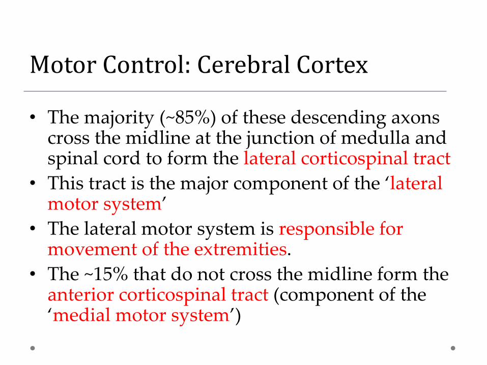
Motor Control: Cerebral Cortex
• The majority (~85%) of these descending axons cross the midline at the junction of medulla and spinal cord to form the lateral corticospinal tract
• This tract is the major component of the ‘lateral motor system’
• The lateral motor system is responsible for movement of the extremities.
• The ~15% that do not cross the midline form the anterior corticospinal tract (component of the ‘medial motor system’)

Lateral Motor System
• Located in lateral white matter of spinal cord
• Responsible for adjusting fine motor activities of the distal limbs
• Supplies contralateral LMN in the spinal cord
• Comprised mainly of 1 tract from the brain:
i. Lateral Corticospinal tract (motor ctx to spinal cord); projects to the contralateral spinal cord
o Rubrospinal (originates from red nucleus of midbrain) tract is sometimes mentioned, but of limited importance

Representation of the lower motor neurons
and upper motor neurons of the lateral
corticospinal tract
From: Neuroanatomy through Clinical Cases (2nd
Ed.) by Blumenfeld. © 2010, Sinauer. Fig. 6.8
Note: The lateral corticospinal tract (see below) is the largest component of the ‘lateral motor system’.

Lateral Motor System
• Also a corticonuclear tract; analogous to corticospinal tract
• Responsible for controlling voluntary motor activities associated the cranial nerves
• Supplies LMNs in the brainstem for:
1. CN V: muscles of mastication
2. CN VII (muscles of facial expression
3. CN IX and CN X: muscles of swallowing and speech
4. CN XII: muscles of tongue movement

From: Neuroanatomy: An Illustrated Colour Text (5th Ed.) by Crossman & Neary. © 2015, Elsevier Limited.
UMNs
UMNs
LMNs
LMNs
MotorHomunculus

From: Neuroanatomy through Clinical Cases (2nd
Ed.) by Blumenfeld. © 2010, Sinauer. Fig. 6.11
Spinal Cord
Cortex
Decussation(crossing of midline)
Brainstem
LMNs
UMNs
Anterior Corticospinal TractLateral Corticospinal Tract

Motor Control: Brainstem
• Brainstem neurons also project to the LMNs of the spinal cord as UMNs.
• Originate from vestibular nuclei (important for balance) and reticular formation (provides pattern generation for initiation of walking)
• Descend in the medial part of the cord as the medial motor system (movement of the postural muscles and core for balance and locomotion)

Medial Motor System
• Located in medial white matter of spinal cord
• Responsible for adjusting gross motor activities of the axial & girdle muscles that control posture and proximal movements
• Supplies LMNs of the ipsilateral and contralateral side
• Constantly operates to maintain upright body posture and equilibrium, as well as reflexive movement patterns such as coordinated eye/head movements

Medial Motor System
• Comprised of tracts originating from the cortex and the brainstem. The principal tracts are:
1. Anterior corticospinal tract: from cortex to spinal cord for bilateral innervation of axial and girdle muscles
2. Reticulospinal tract: from reticular formation to spinal cord for providing automatic adjustments for posture and gait related movements

Medial Motor System
3. Vestibulospinal tracts: from
i. vestibular n. to cervical spinal cord for head and neck movements in response to vestibular information
ii. vestibular n. to spinal cord for maintaining balance (esp. limb extensor muscles)
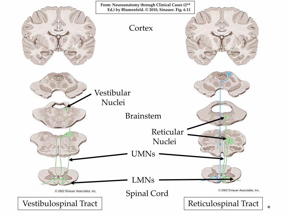
Vestibulospinal Tract Reticulospinal TractSpinal Cord
Cortex
Vestibular Nuclei
ReticularNuclei
From: Neuroanatomy through Clinical Cases (2nd
Ed.) by Blumenfeld. © 2010, Sinauer. Fig. 6.11
Brainstem
LMNs
UMNs

Medial motor pathways. The medial vestibulospinal tract is omitted for clarity. Black filled neurons are inhibitory, black open neurons excitatory. Red neurons are lower motor neurons.Lesion Effects: Unilateral lesions generally produce little in the way of obvious deficits. This is because most of the LMNs that they innervate are innervated bilaterally.
From: Medical Sciences by (2nd Ed.) by Pentland. © 2015, Elsevier Ltd.

Motor System so far:
• UMNs arise from cerebral cortex & brainstem and descend in the white matter of the spinal cord to innervate LMNs.
• Lateral motor system (control of the motor activity of the arms and legs) is located in the lateral part of the spinal cord (mainly lateral corticospinal tract).
• Medial motor system (control of the motor activity of the muscles for posture and locomotion) is located in the medial part of the spinal cord (includes anterior corticospinal, vestibulospinal, and reticulospinal tracts).

Corticospinal Tract (UMN)
• CS tract also referred to as “Pyramidal tract”• Important for: i) fine movements of limb and
face, and ii) control of neck, shoulder, and trunk muscles (posture)..
• Note: CS tract crosses the midline at the junction of the brainstem and spinal cord.
• Lesion above result in contralateral deficits and lesions below decussation produce ipsilateral deficits
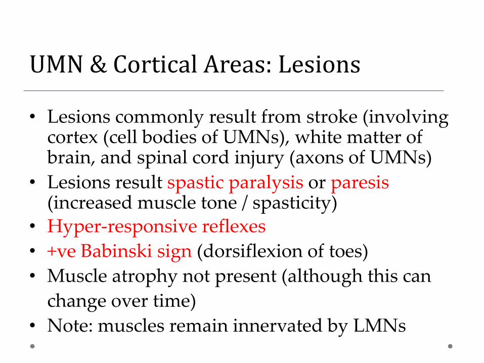
UMN & Cortical Areas: Lesions
• Lesions commonly result from stroke (involving cortex (cell bodies of UMNs), white matter of brain, and spinal cord injury (axons of UMNs)
• Lesions result spastic paralysis or paresis(increased muscle tone / spasticity)
• Hyper-responsive reflexes
• +ve Babinski sign (dorsiflexion of toes)
• Muscle atrophy not present (although this can
change over time)
• Note: muscles remain innervated by LMNs

Hyper-responsive Reflexes

Babinski Sign

Lower Motor Neurons (LMNs)
• Cell bodies located in spinal cord & brain stem, but axon synapses directly with skeletal muscle
• Exit spinal cord via ventral root
• Functional unit is the motor unit; i.e, a
motor neuron and all of the muscle
fibres it innervates
• Damage results in muscle specific
dysfunction
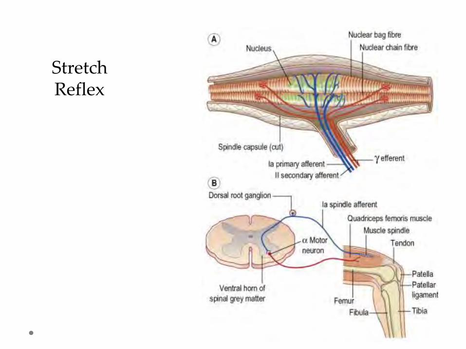
StretchReflex

Motor Unit
Wheater, 2000

Functions of Spinal Cord
• Organization of spinal nerves into sensory (dermatomes) and motor (myotomes) patterns
• Transmission of sensory & motor info. that is subject to modulation and processing
• Integration of circuitry to allow modulation of sensory info.
• Integration of circuitry to allow coordination of motor patterns
• Generates reflexes (integration of ascending sensory info with descending motor commands via interneurons)

From: Neuroanatomy: An Illustrated Colour Text (5th Ed.) by Crossman & Neary. © 2015, Elsevier Limited. Figure 8.15
Spinal Cord: Internal Anatomy

Lower Motor Neurons: Lesions
Cell bodies located in the:
• Brainstem - innervating muscles of the face
• Spinal cord - innervating muscles of the
body
If these neurons are lesioned:
1. Flaccid paralysis/paresis (weakness)
2. Loss of reflexes or hypoactive reflexes
3. Muscle atrophy (muscle wasting)

Motor Control Hierarchy
1. Motor Cortex (motor areas of frontal lobe): Planning and execution of movement. Source of descending fibres (UMNs) to LMNs of spinal cord (and brainstem cranial nerve nuclei). Lesions result in motor weakness or paralysis.
2. Basal Nuclei: initiation and regulation of learned movements. Regulates balance between promoting and suppressing movement. Lesions result in hyperkinetic or hypokinetic disorders.

Motor Control Hierarchy
3. Cerebellum: Timing, coordination and accuracy of movements (esp. fine motor skills). Lesions result in intention tremor, dysmetria, slurred speech and ataxic movements (poor muscle control).
4. Brainstem Nuclei (including vestib. nuclei and reticular formation): Primarily affect axial musculature important for posture and balance and also locomotion.

From: Neuroanatomy through Clinical Cases (2nd Ed.) by Blumenfeld. © 2010, Sinauer. Fig. 6.6
UMNs

Motor Control by Cortex (Ctx)
Motor ctx consists of 10
Motor ctx, Premotor, and Supplementary Motor ctx.
A. 10 Motor Ctx: Located on post-central gyrus of frontal lobe. Source of many UMNs for control of voluntary movement (corticospinal [CS] tract). Body is ‘mapped’ onto gyrus (homunculus). Input from cerebellum.
From: Medical Sciences by (2nd Ed.) by Pentland. © 2015, Elsevier Ltd.
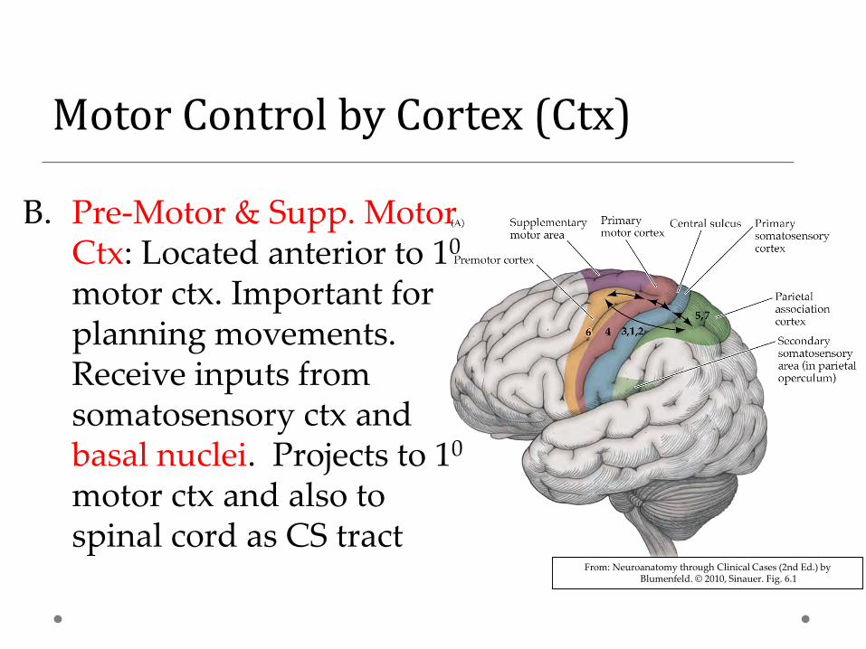
Motor Control by Cortex (Ctx)
B. Pre-Motor & Supp. Motor Ctx: Located anterior to 10
motor ctx. Important for planning movements. Receive inputs from somatosensory ctx and basal nuclei. Projects to 10
motor ctx and also to spinal cord as CS tract
From: Neuroanatomy through Clinical Cases (2nd Ed.) by Blumenfeld. © 2010, Sinauer. Fig. 6.1

Motor cortex (cont.)
• Controls contra-lateral voluntary movements of face, hand & feet.
• These areas take up a disproportionate amount of motor cortex due to the precision and control required.
• Muscle that act bi-laterally (ie. muscles of stomach & back) are controlled by the motor cortex on both sides.

Motor Cortex (cont.)
From: Fitzgerald’s Clinical Neuroanatomy and Neuroscience (7th Ed.) by Mtui, Gruener, & Dockery. © 2016, Elsevier Limited.

Motor Control by Cortex (Summary)
A. 10 Motor Ctx: Contains UMNs (CS tract) and receives input from Premotor and Supplementary Motor ctx (these house motor programs / the instructions for movement). Also receive input from cerebellum to ‘fine tune’ activation of UMNs.
B. Pre-Motor & Supp. Motor Ctx: Receive inputs from prefrontal cortex (behavior), SS ctx and basal nuclei. Projects to 10 motor ctx and also to spinal cord as CS tract.

Motor Control by Ctx (Summary)(cont.)
B. Pre-Motor & Supp. Motor Ctx:
• Also important for initiation of movement
• Planning of movement
• Programming of the sequencing of muscle activation (extensors/flexors)

Flow of Cortical Information

Motor Cortex: Lesions
Lesions of pre-motor cortex and
supplementary motor cortex may produce
apraxia; i.e., difficulty in performing ‘motor
programs’ such as throwing a ball or combing
hair

Motor ProgramsRandom
Planned & executed
Planned but not
executed
Motor Control by Subcortical Structures
A. Basal Nuclei: Receives input from cortex and projects back to cortex (via thalamus) to inhibit or enhance the activity of motor programs. Lesions result in dyskinesias (involuntary movements and tics; hyperkinesias) such as Huntington’s chorea. Also bradykinesia, e.g., Parkinson’s Disease (also characterized by muscle rigidity tremors). Contains info. on the regulation of stereotypical movements.

Hyperkinetic Gait: Demo

Hyperkinesis: Huntingtons Chorea

Hypokinetic Gait: Demo
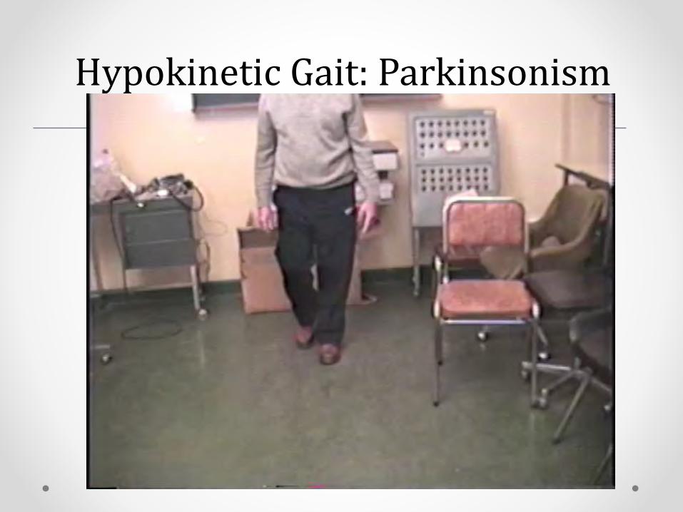
Hypokinetic Gait: Parkinsonism
Parkinsonian Walk / Cycle
• See NEJM Video
• http://www.nejm.org/action/showMediaPlayer?doi=10.1056%2FNEJMicm0810287&aid=NEJMicm0810287_attach_1&area=

Motor Control by Subcortical Structures
B. Cerebellum: Receives inputs from cortex, spinal cord (proprioception), and vestibular system.
• Projects to 10 motor ctx (via the thalamus) to regulate the timing, force, speed, and sequencing of motor activity as governed by the descending CS tract.
• Also acts on descending brainstem tract to aid in maintaining balance (vestibulospinal tract).
• Lesions result in deficits in fine motor skills (and balance).

Motor Control by Subcortical Structures
B. Cerebellum (cont.):
• Integrates (and compares) sensory information with motor information to produce intended movement
• Regulates movement patterns & posture by adjusting output of descending tracts
• Acts on lateral motor system: projects to cortex
• Acts on medial motor system: projects to reticular formation and vestibular nuclei

Cerebellar Lesion: Video #1
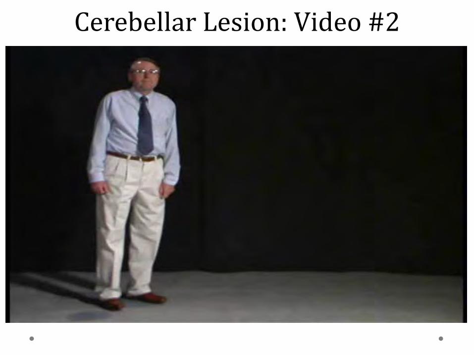
Cerebellar Lesion: Video #2

Cerebellar Lesion: Video #3

Motor Control: Lesions
Assoc. ctx, basal nuclei, and cerebellum do
not project to LMNs of spinal cord (act on
UMNs)
Therefore: lesions of assoc. ctx, basal n., and
cerebellum, paralysis or paresis is not present
However: paralysis or paresis is a key feature
with lesions of UMNs or LMNs

Motor System Responsibility
• Coordinating activities of agonist / antagonistic muscle groups (flexors vs. extensors)
• Conveying accurately timed commands to many one or many groups of muscles
• Making postural adjustments and coordinating balance
• Coordinating movements of segments, joints & muscles
• Normal function relies on feed forward / back systems and a hierarchy of control
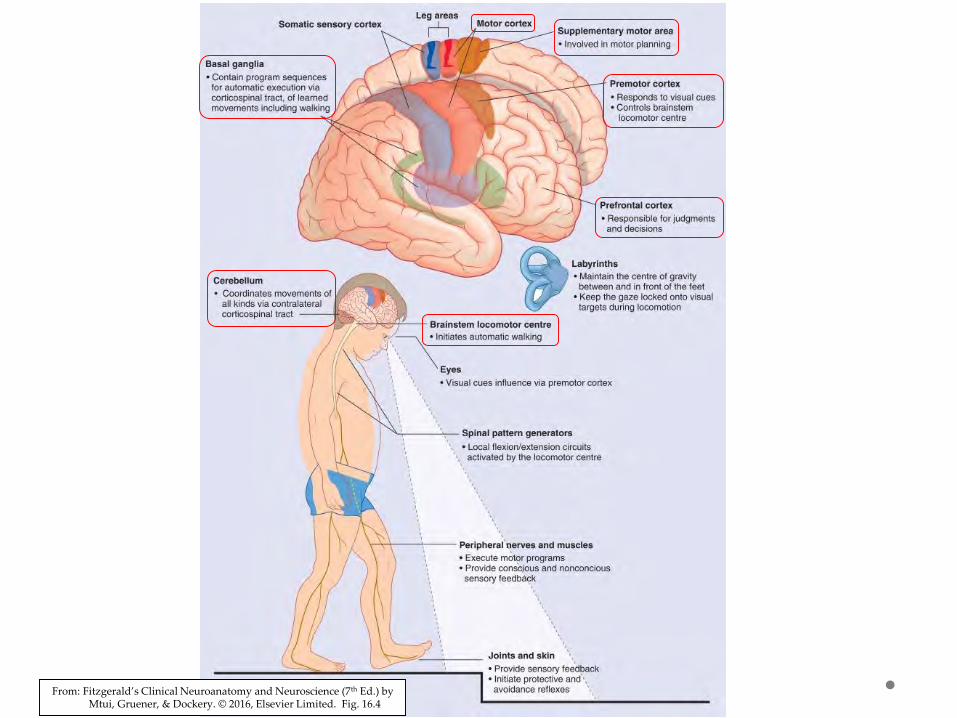
From: Fitzgerald’s Clinical Neuroanatomy and Neuroscience (7th Ed.) by Mtui, Gruener, & Dockery. © 2016, Elsevier Limited. Fig. 16.4

3 Types of Movement
1. Voluntary
• Has purpose
• Is goal directed
• Involves higher cortical centers
• Once learned, performance improves with practice
• Relies heavily on vision and somatosensory systems

3 Types of Movement (cont.)
2. Reflexes
• Occurs via central commands to LMN
• Involuntary and controlled via spinal cord and brainstem
• Important for balance and orientation of head and eyes
• Uses feed forward / back sensory loops and info from visual, vestibular, and somatosensory systems.
• E.g., withdrawal reflex

3 Types of Movement (cont.)
3. Rhythmic Movement Patterns• Generally only initiation and termination are
entirely voluntary• Involves input from all aspects of nervous system
(cortex and brainstem and spinal cord circuits)• Central pattern generators (CPGs) important for
stereotypic repetitious movements (e.g., walking or swimming)
• CPGs are localized neural networks that include the spinal cord, brain stem, and cerebellum.
• CPGs can trigger simple repetitive motor activities, in the lower brain stem or spinal cord.

From: Fundamental Neuroscience (3rd Ed.) by Squire, Bloom, & Spitzer. © 2008, Elsevier Limited.
Location of Central Pattern Generators in the CNS

Summary of Hierarchy
Level 1: Spinal Cord – location of the motor neurons innervating the muscle and circuits for rhythmic behaviors
Level 2: Brainstem – location of neurons descending in medial motor system
Level 3: Motor Cortex – location of motor programs and neurons descending in lateral motor system
Level 4: Association Cortex – location of higher order functioning (directing behavior)

Hierarchy of Motor Systems