motifs in protein sequencesgiri/teach/bioinf/s06/lec9.pdf · motifs in protein sequences examples:...
TRANSCRIPT

Motifs in Protein SequencesMotifs in Protein Sequences
Examples: Helix-Turn-Helix, Zinc-finger, Homeobox domain, Hairpin-beta motif, Calcium-binding motif, Beta-alpha-beta motif,Coiled-coil motifs.
Examples: Helix-Turn-Helix, Zinc-finger, Homeobox domain, Hairpin-beta motif, Calcium-binding motif, Beta-alpha-beta motif,Coiled-coil motifs.
Motifs are combinations of secondary structures in proteins with a specific structure and a specific function.They are also called super-secondary structures.
Motifs are combinations of secondary structures in proteins with a specific structure and a specific function.They are also called super-secondary structures.
Several motifs may combine to form domains. • Serine proteinase domain, Kringle domain, calcium-binding domain, homeobox domain.
Several motifs may combine to form domains. • Serine proteinase domain, Kringle domain, calcium-binding domain, homeobox domain.
CAP5510/CGS5166 12/23/06
CAP5510/CGS5166 22/23/06
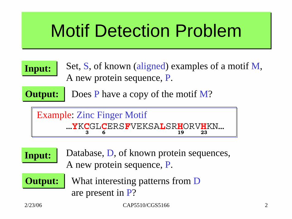
Motif Detection ProblemMotif Detection Problem
Input:Input: Set, S, of known (aligned) examples of a motif M,A new protein sequence, P.
Output:Output: Does P have a copy of the motif M?
Example: Zinc Finger Motif…YYKCCGLCCERSFFVEKSALLSRHHORVHHKN…
3 6 19 23
Input:Input: Database, D, of known protein sequences,A new protein sequence, P.
Output:Output: What interesting patterns from Dare present in P?

CAP5510/CGS5166 32/23/06
Motifs in DNA Sequences• Given a collection of DNA sequences of
promoter regions, locate the transcription factor binding sites (also called regulatory elements)– Example:
http://www.lecb.ncifcrf.gov/~toms/sequencelogo.html

Motifs in DNA Sequences
CAP5510/CGS5166 42/23/06

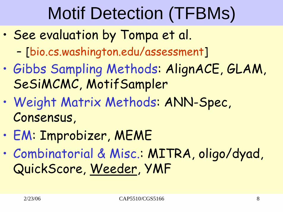
Motif Detection (TFBMs)• See evaluation by Tompa et al.
– [bio.cs.washington.edu/assessment]• Gibbs Sampling Methods: AlignACE, GLAM,
SeSiMCMC, MotifSampler• Weight Matrix Methods: ANN-Spec,
Consensus, • EM: Improbizer, MEME• Combinatorial & Misc.: MITRA, oligo/dyad,
QuickScore, Weeder, YMF
CAP5510/CGS5166 52/23/06

Motif Detection• Profile Method
– If many examples of the motif are known, then • Training: build a Profile and compute a threshold• Testing: score against profile
• Gibbs Sampling• Expectation Method• HMM• Combinatorial Pattern Discovery Methods
CAP5510/CGS5166 62/23/06

How to evaluate these methods?• Calculate TP, FP, TN, FN• Compute sensitivity fraction of known sites
predicted, specificity, and more.– Sensitivity = TP/(TP+FN)– Specificity = TN/(TN+FN)– Positive Predictive Value = TP/(TP+FP)– Performance Coefficient = TP/(TP+FN+FP)– Correlation Coefficient =
CAP5510/CGS5166 72/23/06
Motif Detection (TFBMs)• See evaluation by Tompa et al.
– [bio.cs.washington.edu/assessment]• Gibbs Sampling Methods: AlignACE, GLAM,
SeSiMCMC, MotifSampler• Weight Matrix Methods: ANN-Spec,
Consensus, • EM: Improbizer, MEME• Combinatorial & Misc.: MITRA, oligo/dyad,
QuickScore, Weeder, YMF
CAP5510/CGS5166 82/23/06

Gibbs Sampling for Motif Detection
CAP5510/CGS5166 92/23/06
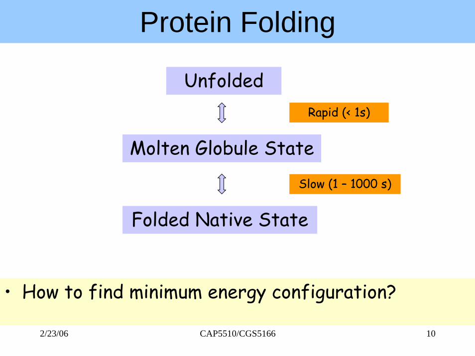
Protein Folding
• How to find minimum energy configuration?
Unfolded
Molten Globule State
Folded Native State
Rapid (< 1s)
Slow (1 – 1000 s)
CAP5510/CGS5166 102/23/06

Modular Nature of Protein Structures
Example: Diphtheria Toxin
CAP5510/CGS5166 112/23/06
Protein Structures• Most proteins have a hydrophobic core.• Within the core, specific interactions take
place between amino acid side chains. • Can an amino acid be replaced by some other
amino acid?– Limited by space and available contacts with
nearby amino acids• Outside the core, proteins are composed of
loops and structural elements in contact with water, solvent, other proteins and other structures.
CAP5510/CGS5166 122/23/06
Viewing Protein Structures• SPDBV• RASMOL• CHIME
CAP5510/CGS5166 132/23/06

Structural Classification of Proteins• Over 1000 protein families known
– Sequence alignment, motif finding, block finding, similarity search
• SCOP (Structural Classification of Proteins)– Based on structural & evolutionary relationships.– Contains ~ 40,000 domains– Classes (groups of folds), Folds (proteins sharing
folds), Families (proteins related by function/evolution), Superfamilies (distantly related proteins)
CAP5510/CGS5166 142/23/06

CAP5510/CGS5166 152/23/06
SCOP Family View

CATH: Protein Structure Classification• Semi-automatic classification; ~36K domains• 4 levels of classification:
– Class (C), depends on sec. Str. Content • α class, β class, α/β class, α+β class
– Architecture (A), orientation of sec. Str.– Topolgy (T), topological connections & – Homologous Superfamily (H), similar str and
functions.
CAP5510/CGS5166 162/23/06

DALI/FSSP Database• Completely automated; 3724 domains• Criteria of compactness & recurrence• Each domain is assigned a Domain
Classification number DC_l_m_n_p representing fold space attractor region (l), globular folding topology (m), functional family (n) and sequence family (p).
CAP5510/CGS5166 172/23/06

Structural Alignment• What is structural alignment of proteins?
– 3-d superimposition of the atoms as “best as possible”, i.e., to minimize RMSD (root mean square deviation).
– Can be done using VAST and SARF• Structural similarity is common, even among
proteins that do not share sequence similarity or evolutionary relationship.
CAP5510/CGS5166 182/23/06
Other databases & tools• MMDB contains groups of structurally
related proteins• SARF structurally similar proteins using
secondary structure elements• VAST Structure Neighbors• SSAP uses double dynamic programming to
structurally align proteins
CAP5510/CGS5166 192/23/06
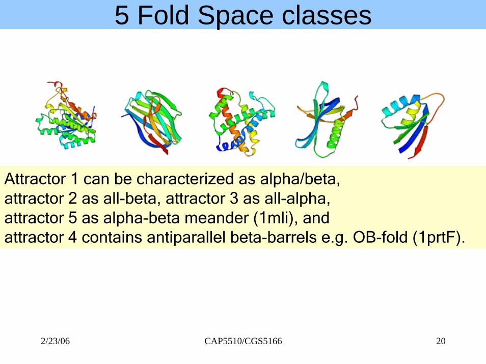
5 Fold Space classes
Attractor 1 can be characterized as alpha/beta, attractor 2 as all-beta, attractor 3 as all-alpha, attractor 5 as alpha-beta meander (1mli), and attractor 4 contains antiparallel beta-barrels e.g. OB-fold (1prtF).
CAP5510/CGS5166 202/23/06
CAP5510/CGS5166 212/23/06
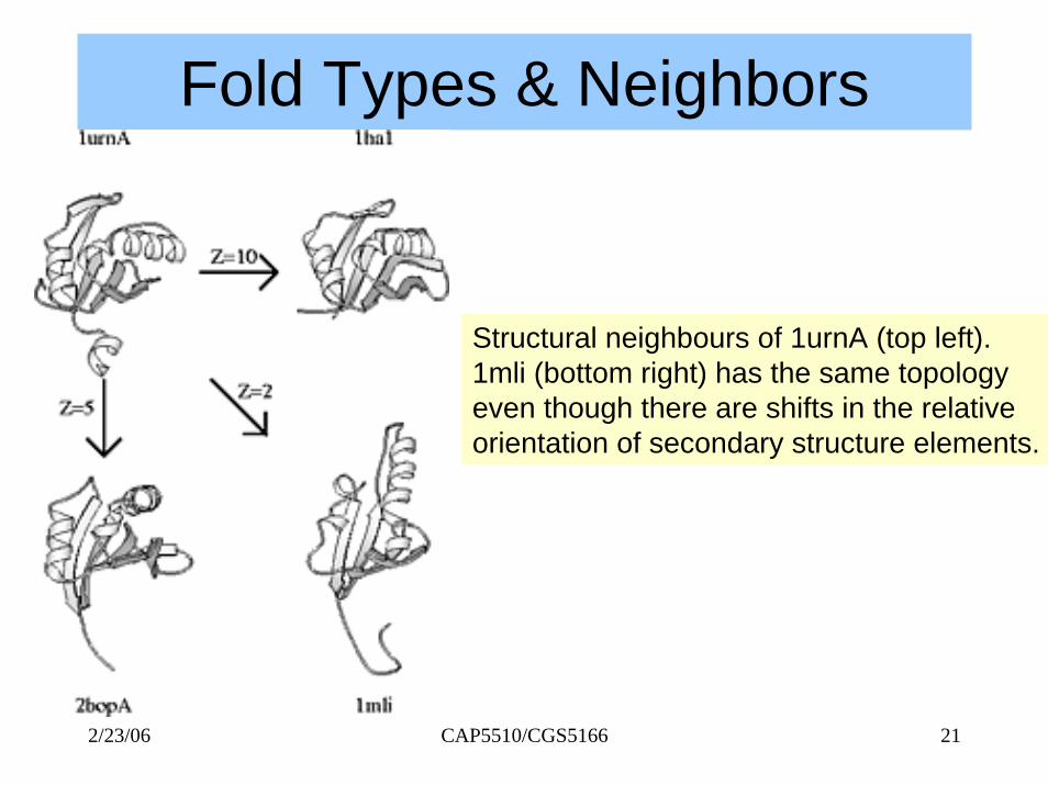
Fold Types & Neighbors
Structural neighbours of 1urnA (top left). 1mli (bottom right) has the same topology even though there are shifts in the relativeorientation of secondary structure elements.
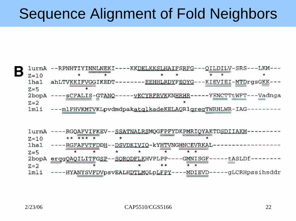
Sequence Alignment of Fold Neighbors
CAP5510/CGS5166 222/23/06
CAP5510/CGS5166 232/23/06
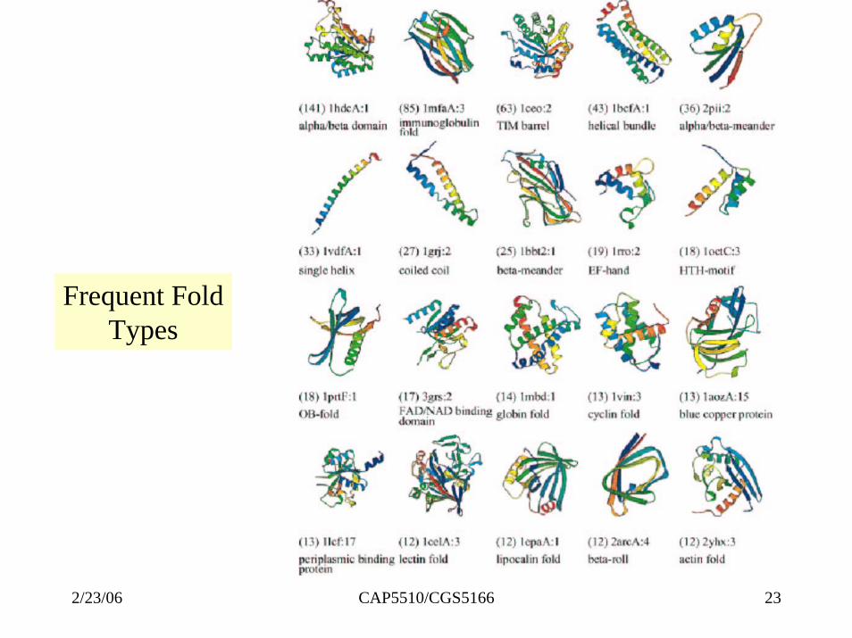
Frequent FoldTypes
Protein Structure Prediction• Holy Grail of bioinformatics • Protein Structure Initiative to determine a
set of protein structures that span protein structure space sufficiently well. WHY?– Number of folds in natural proteins is limited.
Thus a newly discovered proteins should be within modeling distance of some protein in set.
• CASP: Critical Assessment of techniques for structure prediction– To stimulate work in this difficult field
CAP5510/CGS5166 242/23/06

PSP Methods• homology-based modeling • methods based on fold recognition
– Threading methods• ab initio methods
– From first principles– With the help of databases
CAP5510/CGS5166 252/23/06

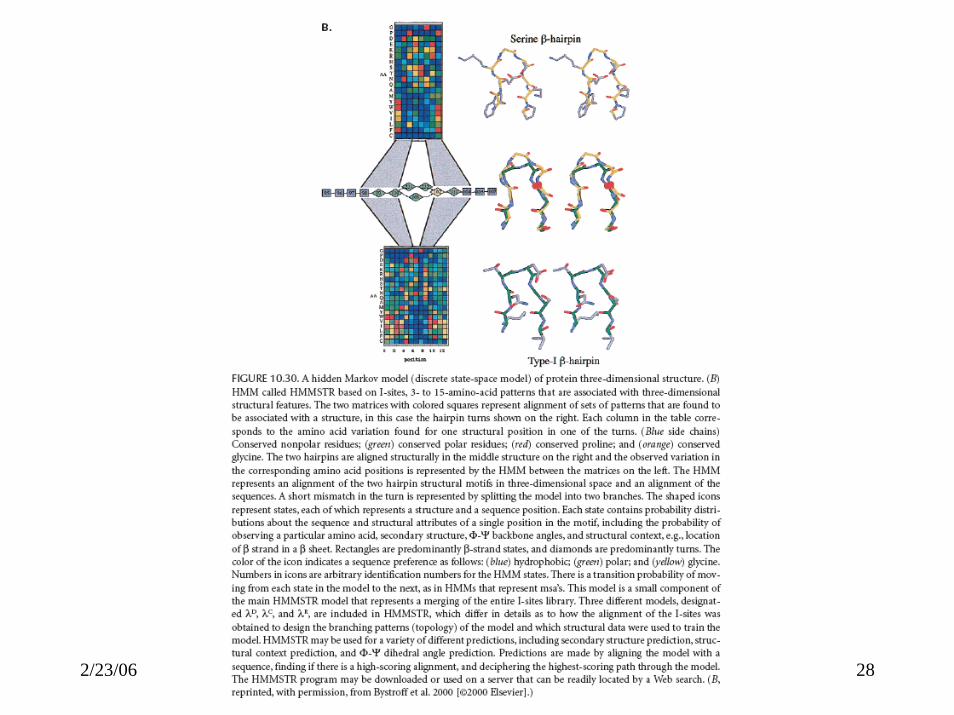
ROSETTA• Best method for PSP• As proteins fold, a large number of partially
folded, low-energy conformations are formed, and that local structures combine to form more global structures with minimum energy.
• Build a database of known structures (I-sites) of short sequences (3-15 residues).
• Monte Carlo simulation assembling possible substructures and computing energy
CAP5510/CGS5166 262/23/06
Threading Methods• See p471, Mount
– http://www.bioinformaticsonline.org/links/ch_10_t_7.html
CAP5510/CGS5166 272/23/06
CAP5510/CGS5166 282/23/06
CAP5510/CGS5166 292/23/06
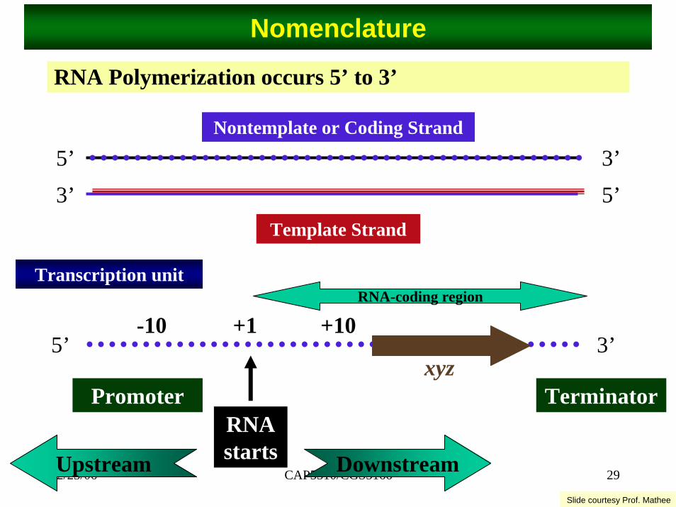
Nomenclature
RNA Polymerization occurs 5’ to 3’
5’ 3’5’3’
Template Strand
Nontemplate or Coding Strand
5’ 3’
RNA starts
xyz
+1
Promoter Terminator
DownstreamUpstream
-10 +10
Transcription unitRNA-coding region
Slide courtesy Prof. Mathee
CAP5510/CGS5166 302/23/06
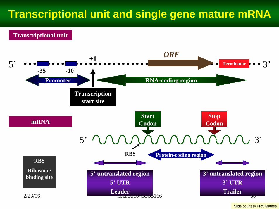
Transcriptional unit and single gene mature mRNA
Transcriptional unit
5’ 3’
Transcription start site
ORF+1Terminator
-10RNA-coding region
-35Promoter
StartCodon
3’5’Protein-coding region
StopCodon
5’ untranslated region5’ UTRLeader
3’ untranslated region3’ UTRTrailer
RBSRBS
Ribosome binding site
mRNA
Slide courtesy Prof. Mathee
CAP5510/CGS5166 312/23/06
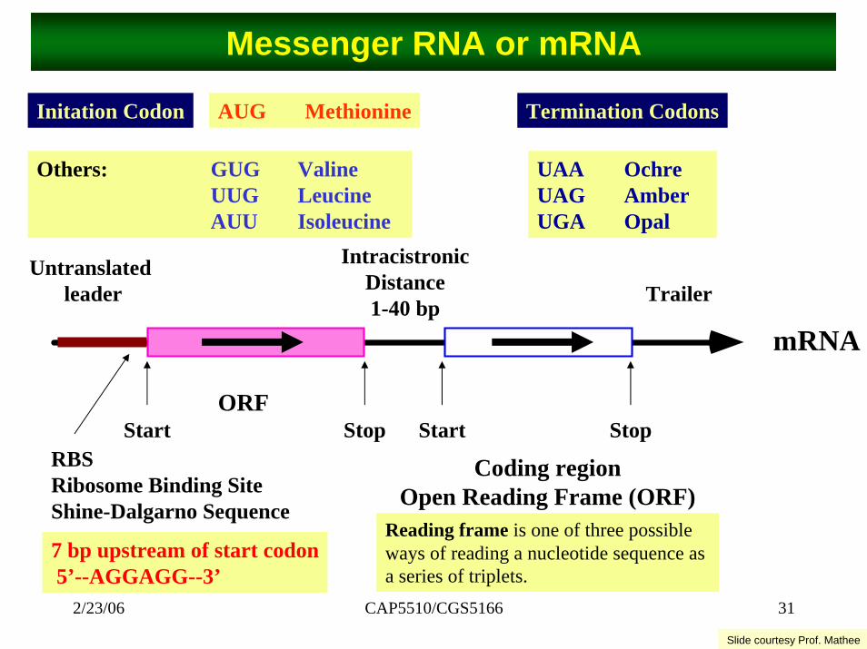
Messenger RNA or mRNA
Initation Codon AUG Methionine Termination Codons
Others: GUG ValineUUG LeucineAUU Isoleucine
UAA OchreUAG AmberUGA Opal
Untranslatedleader
IntracistronicDistance1-40 bp
Trailer
Coding regionOpen Reading Frame (ORF)
Start
mRNA
StopORF
RBSRibosome Binding SiteShine-Dalgarno Sequence
Start Stop
7 bp upstream of start codon5’--AGGAGG--3’
Reading frame is one of three possible ways of reading a nucleotide sequence as a series of triplets.
Slide courtesy Prof. Mathee

CAP5510/CGS5166 322/23/06
β '
α β
σ
αRNAP Holoenzyme
Transcription Starts
Spacer Region16-18 bp
TTGACA TATAATE. coli consensus for σ70
+20+1A/G
-10-20-30-40-60 -50
-35 -104-8 bp
Basal Promoter:ORF
+20+1A/G
-10-20-30-40-60 -50
-35 -10UP Element
AT-richα-CTD makes the contact
Transcription StartsStronger Promoter:
ORF
Transcriptional machinery: RNA Polymerase and DNA
Slide courtesy Prof. Mathee

CAP5510/CGS5166 332/23/06
Prokaryotic Gene Characteristics

Motif Detection (TFBMs)• See evaluation by Tompa et al.
– [bio.cs.washington.edu/assessment]• Gibbs Sampling Methods: AlignACE, GLAM,
SeSiMCMC, MotifSampler• Weight Matrix Methods: ANN-Spec,
Consensus, • EM: Improbizer, MEME• Combinatorial & Misc.: MITRA, oligo/dyad,
QuickScore, Weeder, YMF
CAP5510/CGS5166 342/23/06
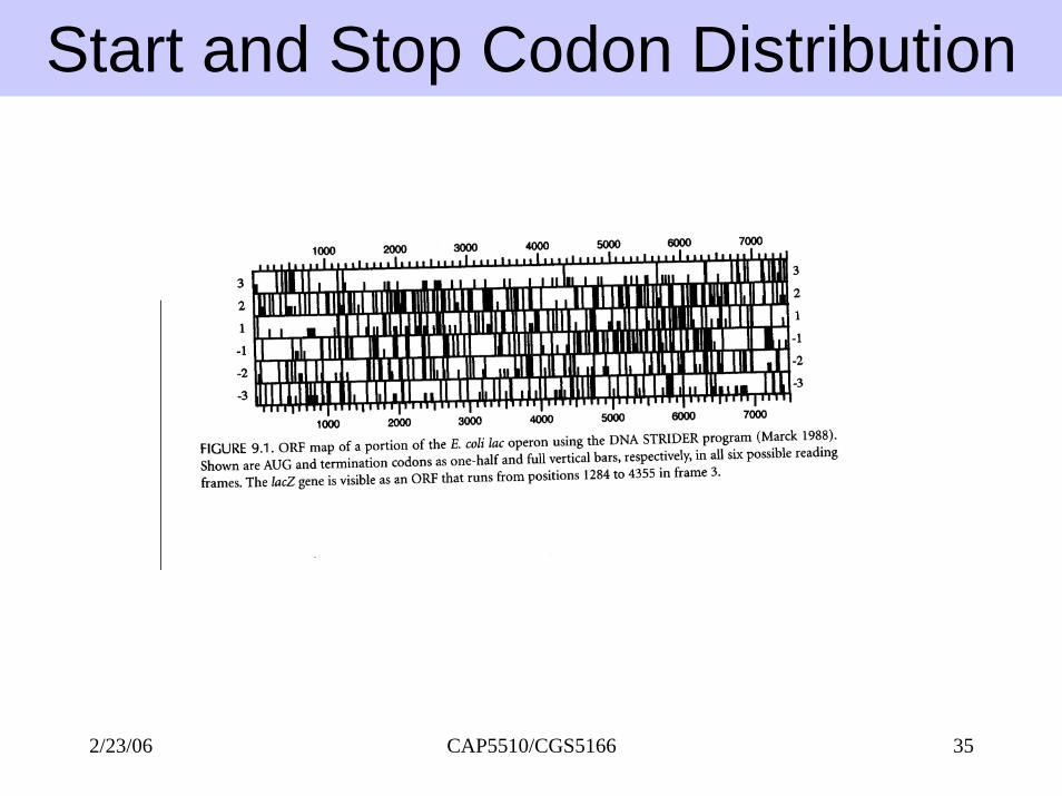
Start and Stop Codon Distribution
CAP5510/CGS5166 352/23/06
CAP5510/CGS5166 362/23/06
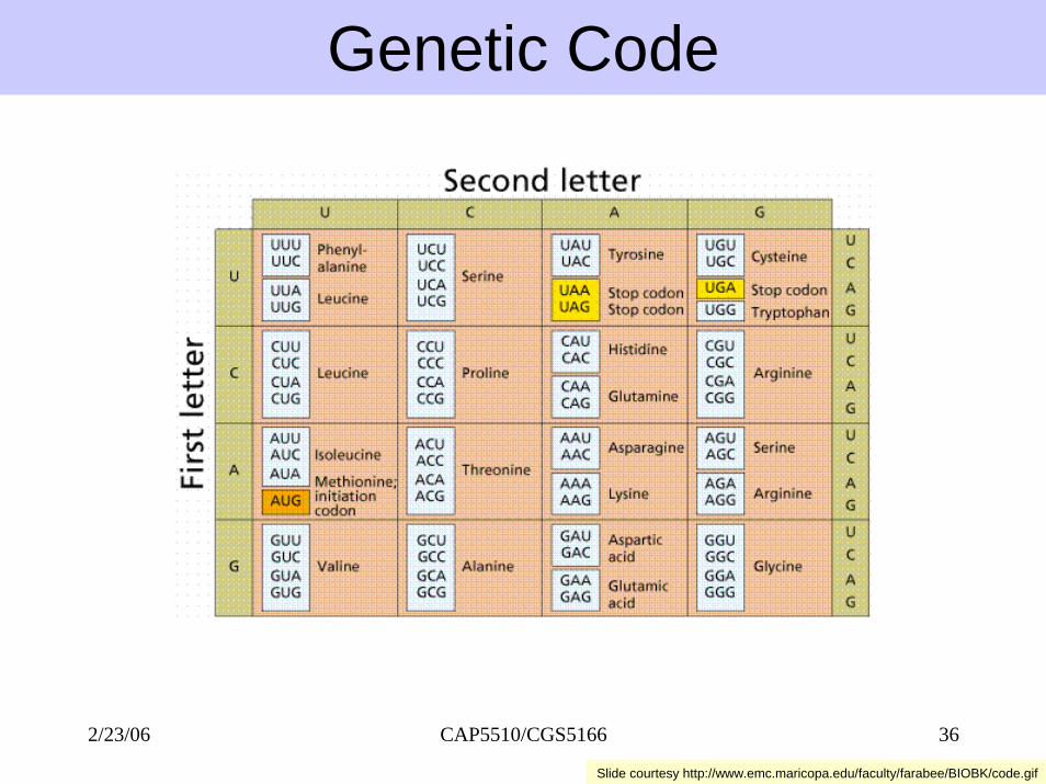
Genetic Code
Slide courtesy http://www.emc.maricopa.edu/faculty/farabee/BIOBK/code.gif
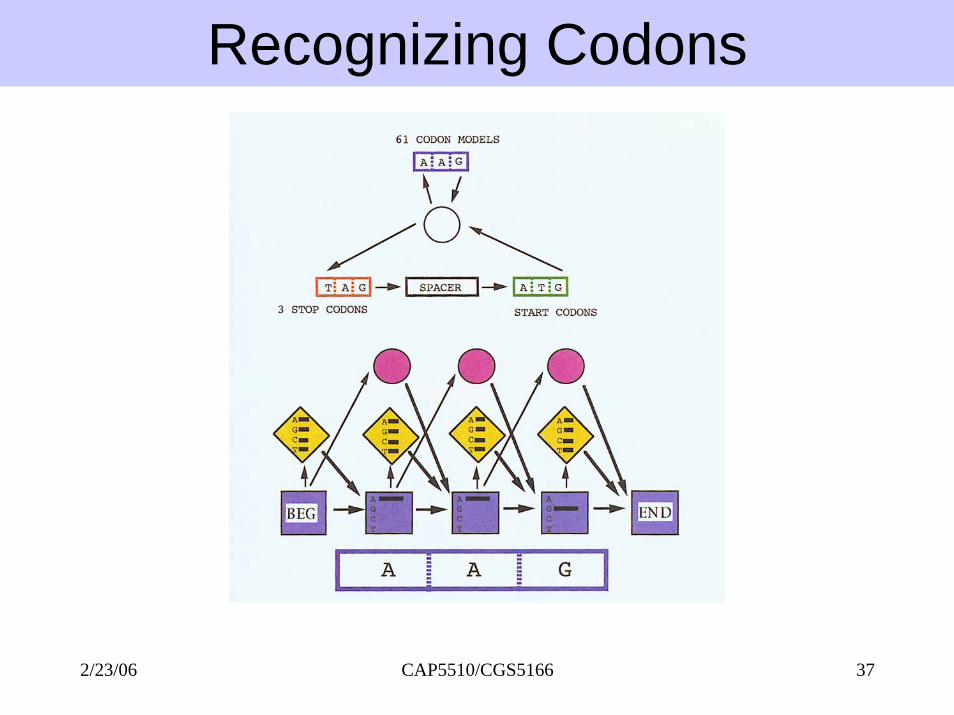
Recognizing Codons
CAP5510/CGS5166 372/23/06

CAP5510/CGS5166 382/23/06
Codon Bias• Some codons preferred over others. O = optimal
S = suboptimalR = rareU = unfavorable
Frame Shift 1
Frame Shift 2
CAP5510/CGS5166 392/23/06
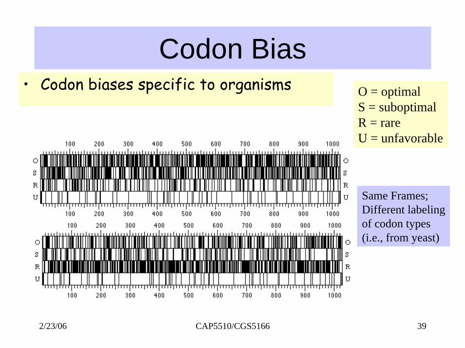
Codon Bias• Codon biases specific to organisms O = optimal
S = suboptimalR = rareU = unfavorable
Same Frames;Different labelingof codon types(i.e., from yeast)
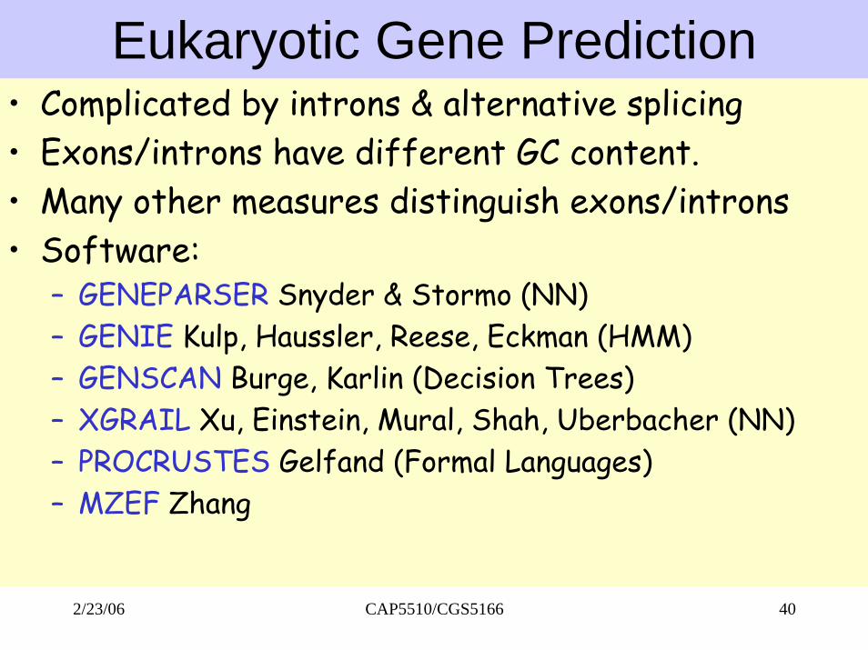
Eukaryotic Gene Prediction• Complicated by introns & alternative splicing • Exons/introns have different GC content.• Many other measures distinguish exons/introns• Software:
– GENEPARSER Snyder & Stormo (NN)– GENIE Kulp, Haussler, Reese, Eckman (HMM)– GENSCAN Burge, Karlin (Decision Trees)– XGRAIL Xu, Einstein, Mural, Shah, Uberbacher (NN)– PROCRUSTES Gelfand (Formal Languages)– MZEF Zhang
CAP5510/CGS5166 402/23/06
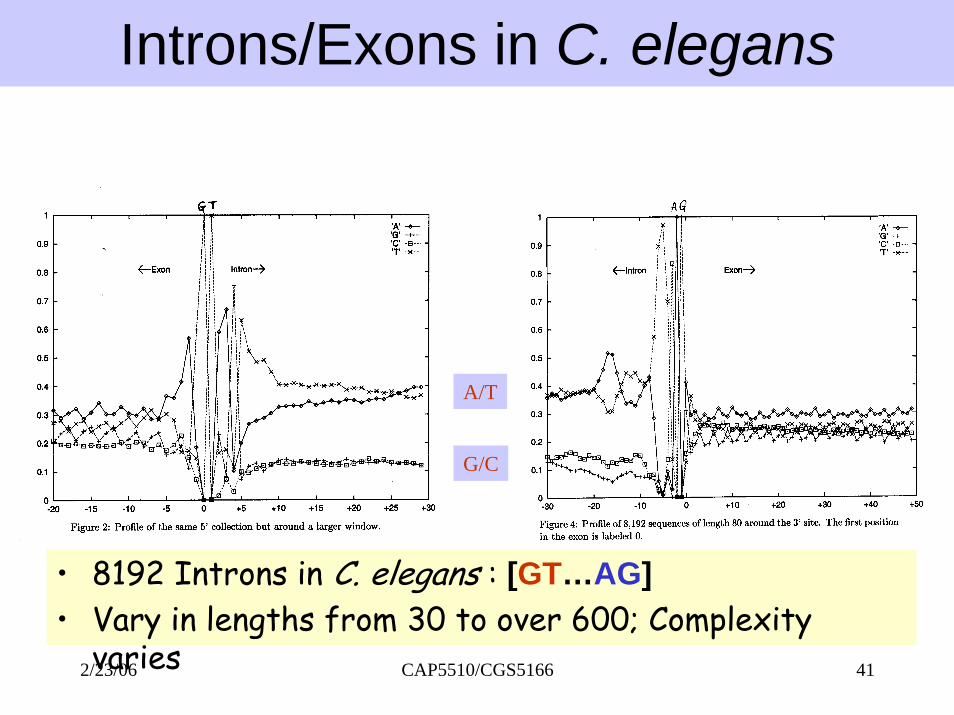
Introns/Exons in C. elegans
A/T
G/C
• 8192 Introns in C. elegans : [GT…AG]• Vary in lengths from 30 to over 600; Complexity
CAP5510/CGS5166 412/23/06varies
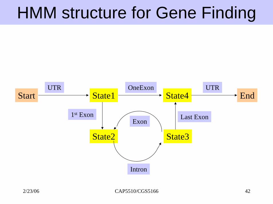
HMM structure for Gene Finding
Start EndState1 State4
State2 State3
UTR OneExon
1st ExonExon
Intron
Last Exon
UTR
CAP5510/CGS5166 422/23/06

Motifs in Protein SequencesMotifs in Protein Sequences
Examples: Helix-Turn-Helix, Zinc-finger, Homeobox domain, Hairpin-beta motif, Calcium-binding motif, Beta-alpha-beta motif,Coiled-coil motifs.
Examples: Helix-Turn-Helix, Zinc-finger, Homeobox domain, Hairpin-beta motif, Calcium-binding motif, Beta-alpha-beta motif,Coiled-coil motifs.
Motifs are combinations of secondary structures in proteins with a specific structure and a specific function.They are also called super-secondary structures.
Motifs are combinations of secondary structures in proteins with a specific structure and a specific function.They are also called super-secondary structures.
Several motifs may combine to form domains. • Serine proteinase domain, Kringle domain, calcium-binding domain, homeobox domain.
Several motifs may combine to form domains. • Serine proteinase domain, Kringle domain, calcium-binding domain, homeobox domain.
CAP5510/CGS5166 432/23/06

CAP5510/CGS5166 442/23/06
Helix-Turn-Helix MotifsHelix-Turn-Helix Motifs
• Structure• 3-helix complex• Length: 22 amino acids• Turn angle
• Function• Gene regulation by
binding to DNA
Branden & Tooze

CAP5510/CGS5166 452/23/06
DNA Binding at HTH MotifDNA Binding at HTH Motif
Branden & Tooze
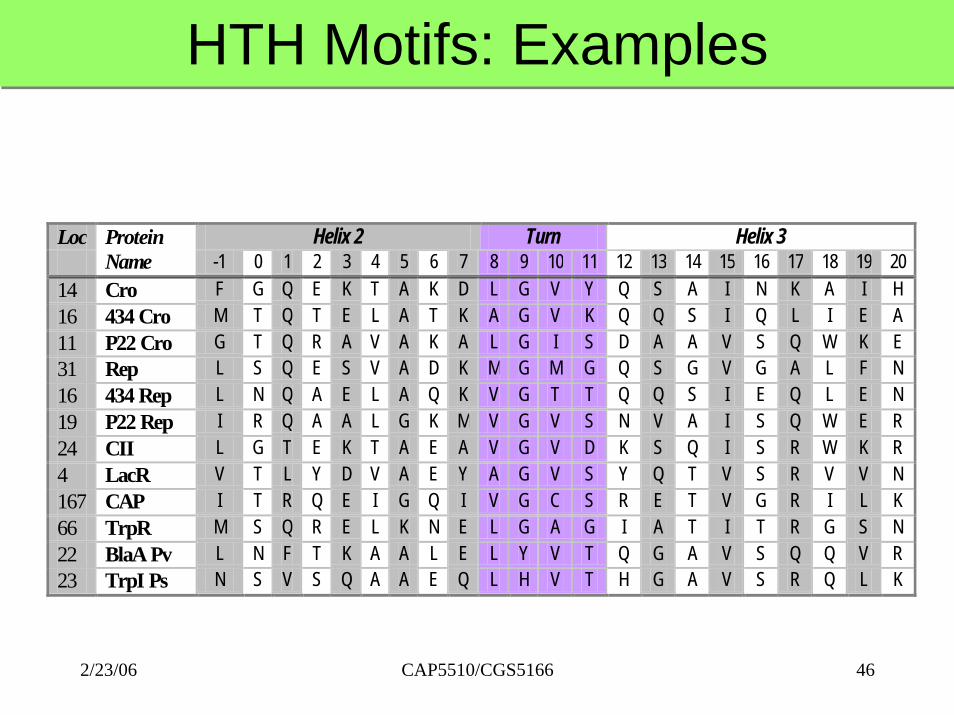
HTH Motifs: ExamplesHTH Motifs: Examples
Loc Helix 2 Turn Helix 3
Protein Name -1 0 1 2 3 4 5 6 7 8 9 10 11 12 13 14 15 16 17 18 19 20
14 Cro F G Q E K T A K D L G V Y Q S A I N K A I H 16 434 Cro M T Q T E L A T K A G V K Q Q S I Q L I E A 11 P22 Cro G T Q R A V A K A L G I S D A A V S Q W K E 31 Rep L S Q E S V A D K M G M G Q S G V G A L F N 16 434 Rep L N Q A E L A Q K V G T T Q Q S I E Q L E N 19 P22 Rep I R Q A A L G K M V G V S N V A I S Q W E R 24 CII L G T E K T A E A V G V D K S Q I S R W K R 4 LacR V T L Y D V A E Y A G V S Y Q T V S R V V N 167 CAP I T R Q E I G Q I V G C S R E T V G R I L K 66 TrpR M S Q R E L K N E L G A G I A T I T R G S N 22 BlaA Pv L N F T K A A L E L Y V T Q G A V S Q Q V R 23 TrpI Ps N S V S Q A A E Q L H V T H G A V S R Q L K
CAP5510/CGS5166 462/23/06
Basis for New AlgorithmBasis for New Algorithm• Combinations of residues in specific locations
(may not be contiguous) contribute towards stabilizing a structure.
• Some reinforcing combinations are relatively rare.
• Combinations of residues in specific locations (may not be contiguous) contribute towards stabilizing a structure.
• Some reinforcing combinations are relatively rare.
CAP5510/CGS5166 472/23/06
CAP5510/CGS5166 482/23/06
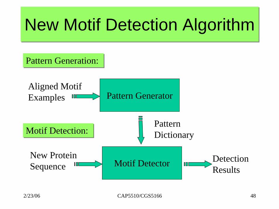
New Motif Detection AlgorithmNew Motif Detection Algorithm
Pattern Generation: Pattern Generation:
Pattern GeneratorAligned MotifExamples
Pattern DictionaryMotif Detection: Motif Detection:
Motif DetectorNew ProteinSequence
DetectionResults

CAP5510/CGS5166 492/23/06
PatternsPatternsLoc Helix 2 Turn Helix 3
Protein Name -1 0 1 2 3 4 5 6 7 8 9 10 11 12 13 14 15 16 17 18 19 20
14 Cro F G Q E K T A K D L G V Y Q S A I N K A I H 16 434 Cro M T Q T E L A T K A G V K Q Q S I Q L I E A 11 P22 Cro G T Q R A V A K A L G I S D A A V S Q W K E 31 Rep L S Q E S V A D K M G M G Q S G V G A L F N 16 434 Rep L N Q A E L A Q K V G T T Q Q S I E Q L E N 19 P22 Rep I R Q A A L G K M V G V S N V A I S Q W E R 24 CII L G T E K T A E A V G V D K S Q I S R W K R 4 LacR V T L Y D V A E Y A G V S Y Q T V S R V V N 167 CAP I T R Q E I G Q I V G C S R E T V G R I L K 66 TrpR M S Q R E L K N E L G A G I A T I T R G S N 22 BlaA Pv L N F T K A A L E L Y V T Q G A V S Q Q V R 23 TrpI Ps N S V S Q A A E Q L H V T H G A V S R Q L K
• Q1 G9 N20• A5 G9 V10 I15

Pattern Mining Algorithm Pattern Mining Algorithm Algorithm Pattern-MiningInput: Motif length m, support threshold T,
list of aligned motifs M.Output: Dictionary L of frequent patterns.
1. L1 := All frequent patterns of length 1 2. for i = 2 to m do3. Ci := Candidates(Li-1)4. Li := Frequent candidates from Ci5. if (|Li| <= 1) then6. return L as the union of all Lj , j <= i.
Algorithm PatternPattern--MiningMiningInput: Motif length m, support threshold T,
list of aligned motifs M.Output: Dictionary L of frequent patterns.
1. L1 := All frequent patterns of length 1 2. for i = 2 to m do3. Ci := Candidates(Li-1)4. Li := Frequent candidates from Ci5. if (|Li| <= 1) then6. return L as the union of all Lj , j <= i.
CAP5510/CGS5166 502/23/06

Candidates FunctionCandidates Function
G1, V2, S3 G1, V2, T6 G1, V2, I7G1, V2, E8G1, S3, T6G1, T6, I7V2, T6, I7V2, T6, E8
L3
G1, V2, S3, T6 G1, V2, S3, I7G1, V2, S3, E8G1, V2, T6, I7G1, V2, T6, E8G1, V2, I7, E8V2, T6, I7, E8
C4
G1, V2, S3, T6 G1, V2, S3, I7G1, V2, S3, E8
G1, V2, T6, E8
V2, T6, I7, E8
L4
CAP5510/CGS5166 512/23/06

Motif Detection AlgorithmMotif Detection AlgorithmAlgorithm Motif-Detection
Input : Motif length m, threshold score T, pattern dictionary L, and input protein sequence P[1..n].
Output : Information about motif(s) detected.
1. for each location i do2. S := MatchScore(P[i..i+m-1], L).3. if (S > T) then4. Report it as a possible motif
Algorithm MotifMotif--DetectionDetection
Input : Motif length m, threshold score T, pattern dictionary L, and input protein sequence P[1..n].
Output : Information about motif(s) detected.
1. for each location i do2. S := MatchScore(P[i..i+m-1], L).3. if (S > T) then4. Report it as a possible motif
CAP5510/CGS5166 522/23/06

Experimental Results: GYM 2.0Experimental Results: GYM 2.0
Motif Protein Family
Number Tested
GYM = DE Agree
Number Annotated
GYM = Annot.
Master 88 88 (100 %) 13 13 Sigma 314 284 + 23 (98 %) 96 82
Negates 93 86 (92 %) 0 0 LysR 130 127 (98 %) 95 93 AraC 68 57 (84 %) 41 34 Rreg 116 99 (85 %) 57 46
HTH Motif (22)
Total 675 653 + 23 (94 %) 289 255 (88 %)
CAP5510/CGS5166 532/23/06
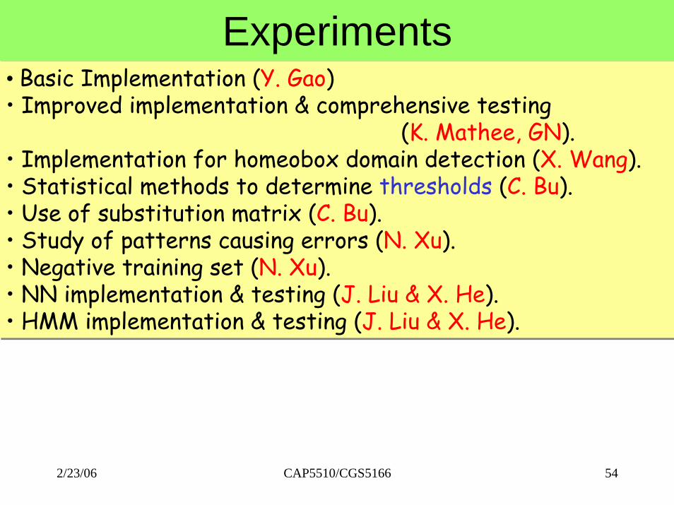
ExperimentsExperiments• Basic Implementation (Y. Gao)• Improved implementation & comprehensive testing
(K. Mathee, GN).• Implementation for homeobox domain detection (X. Wang). • Statistical methods to determine thresholds (C. Bu). • Use of substitution matrix (C. Bu). • Study of patterns causing errors (N. Xu). • Negative training set (N. Xu). • NN implementation & testing (J. Liu & X. He).• HMM implementation & testing (J. Liu & X. He).
• Basic Implementation (Y. Gao)• Improved implementation & comprehensive testing
(K. Mathee, GN).• Implementation for homeobox domain detection (X. Wang). • Statistical methods to determine thresholds (C. Bu). • Use of substitution matrix (C. Bu). • Study of patterns causing errors (N. Xu). • Negative training set (N. Xu). • NN implementation & testing (J. Liu & X. He).• HMM implementation & testing (J. Liu & X. He).
CAP5510/CGS5166 542/23/06