morphology of parasites - centurion university
TRANSCRIPT

MORPHOLOGY OF PROTOZOA
Parasite is defined as an animal or plant that
lives in or upon another organism (Host) and
draws its nutrient directly from it.
E.g include Bacteria, Viruses, Fungi,
Protozoas and helminths.
Study of parasite is known as Parasitology.
Medical Parasitology is the study of animal
parasite that infect and produce diseases in
humans

1. Ectoparasite
The parasites that live on the outer surfaceor
in the superficial tissues of the host are called
ectoparasites. Infection caused by these
parasites is called infestation. E.g Lice
2. Endoparasite
The parasites that live within the host are
called endoparasites. Invasion by such
parasites is called infection. Eg. Leishmania

a. Obligate ParasitesThe parasites that cannot exist without a host are called obligate parasites. Eg. Toxoplasma gonodii
b. Facultative ParasitesThe parasites that live a parasitic or free-living existence when an oppurtunity arises are called facultative parasites.Eg Naegleria fowleri
c. Accidental ParasitesThe parasites that attack an unusal host are called accidental parasites. Eg. Echinococcus granulosus
d. Aberrant ParasitesThe parasites that during migration in the host, reach a site where they cannot live or develop further are called aberrant parasites. Eg Toxocara types

A host is defined as an organism that harbours the parasites and provide nourishment and shelter.The hosts are relatively larger in size in comparison to their pasrsites.
Types of Hosti. Definitive Host
The host that harbours adult parasites (e.g. Taenia saginata) or highly developed forms of parasites (Trypanosoma cruzi) or where parasites replicate sexually( Paragonimus westermani).Definitive host may be human or non-human living things.
ii. Intermediate HostThe host that harbours the asexual forms of parasites are called intermediate host. They are needed for completion of life cycle.

iii. Reservoir HostThe animals that harbour parasites and serve as an important source of the infection to other susceptible hosts are known as reservoir host.E.g. dogs are the reservoir host for cystic echinococcus.
iv. Paratenic HostWhen the parasite lives in a host in which it cannot develop further, such a host is called paratenic host. E.g. freshwater prawn is the paratenic host for Angiostrongylus cantonensis.
v. Natural HostThe host that are naturally infected with certain species of parasitesare called natural host. E.g. pig is natural host for Balantidium coli.
vi. Accidental HostThe hosts in which parasites is not usually found are called accidental hosts. E.g. human is an accidental host for E. granulosus

a.Symbiosis- parasites and host cannot live without each other.
b.commensalism- it is an association in which only the parasite derives the benefit without causing any injury to the host. E.g. Entamoeba coli in the human intestine.
c. Mutualism- both the parasite and the host are benefitted. E.g, the flagellates in the gut of termite digest woody materials consumed by termites. These are converted into glycogenous substances used by the host. In absence of each other both perish
d. Parasitism- it is an association in which the parsitesderives benefits from the host and always causes injury to the host e.g. plasmodium spp. Causing malaria

The word protozoa is come from Greekprotozoon word meaning “FirstAnimal”.
Protozoa are unicellular (may be Multicellular) Eukaryotic microorganism.
Protozoa constitute a large group of about 65,000 species. Most of which are harmless free living and inhabits water and soil
A few species are pathogenic in nature parasitize human and other animals causing hundreds of million of infections in a year around the world
CHARACTERISTICS
• Mostly Unicellular organism with fully functional
cell
• Live freely, may be parasitic or symbiotic
• Protozoa are chemo-hetrotrops
• They are motile have locomotive organelles. E.g.
Flagella and Cilia for movement

MORPHOLOGY
• Protozoa are Eukaryotic resemble to animal cell, contain
major cell organelles (including Nucleus, Mitochondria)
• They are microscopic in size less than 50 µm.
• Their organelles are highly specialized for feeding,
reproduction and movement
• The cytoplasm of protozoa are divided into an outer layer
called Ectoplasm and an inner layer called Endoplasm
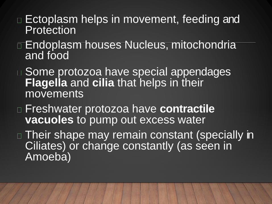
Ectoplasm helps in movement, feeding and Protection
Endoplasm houses Nucleus, mitochondriaand food
Some protozoa have special appendages Flagella and cilia that helps in their movements
Freshwater protozoa have contractile vacuoles to pump out excess water
Their shape may remain constant (specially in Ciliates) or change constantly (as seen in Amoeba)


CLASSIFICATION OF PROTOZOA
• Protozoa are classified on the basis of their
motility and method of reproduction
• They are classified into Four main types
• Flagellates
• Ciliates
• Sarcodina
• Sporozoates

FLAGELLATES
• Flagellates move by help of Flagella (a tail-like
structure ). The movement is whip like
• Example of Flagellates are
• Trypnosoma, Leishmenia (blood pathogen)
• Giardia (intestinal parasite)
• Trichomonas (reproductive tract pathogen)

CILIATES
• Ciliates protozoa have movement through cilia(
fine hair like structure attached with their body).
• Some protozoa have special kind of cilia for
feeding and attachment.
• Most are harmless. Only one species
Balantidium Coli is pathogenic for human
causes a rare and server form of Dysentery.

SARCODINA• Major loco-motor organelles in Sarcodina is
pseudopodia (Pseudo means false, podia
means Foot)
• Common example of Sarcodina is Amoeba
• Most species are harmless
• Enaemoba is a parasitic for human causes
intestinal disease
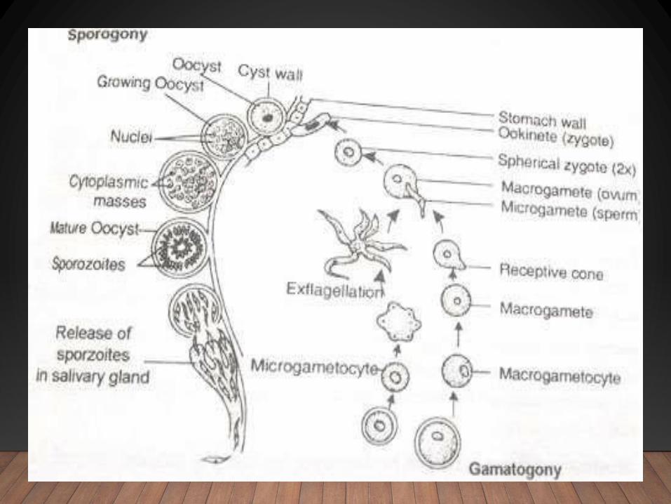
SPOROZOITES
• Sporozoites are the only non-motile form of
protozoa.
• Sporozoites have well developed sexual and
asexual stages
• Entire group is parasitic in nature and are harmful
• Some common examples of Sporozoites and their
infections are
• Plasmodium (causative agent of Malaria, causes
100 to 300 million infection world wide)
• Toxoplasma Gondii (causes Toxoplasmosis)

Cytoplasm
Homogeneous, show colours (green, brown,
blue , purple due to pigment
Has submicroscopic protein fibrils (myonemes,
microtubules)
Arranged in parallel
Divided in two portions- ectoplasm &
endoplasm
Ectoplasm- more gel like and endoplasm is
voluminous and fluid
Cell organelles are present (ER, ribosomes,
golgi complex, mitochondria, food vacuole,
contractile vacuole, kinetosome)


NUCLE
US
Eucaryotic nucleus- has chromosomes,
the nucleolar substance, the nuclear
membrane, nucleoplasm
one or many
Some have two
a.Macronucleus- large in size, controls
the metabolic activities and regeneration
processes
b.Micronucleus- small in size concerned
with reproductive activity


PLASMALEMMA & OTHER
COVERINGS
Cell membrane or plasmalemma
outermost layer, semi permeable
Functions
a. protection
b.site of perception of chemical and
mechanical stimuli
c. transport
Compound coverings
Combinations of membranes, also called
pellicle
Simplest is plasmalemma eg, Amoeba
Some have mucopolysaccharide on
plasmalemma plays an important role in
pinocytosis and adhesion
Can be thick, ridged and sculputured,
nodular thickening,

ADDITIONAL
COVERINGS
Diverse
Egs. thecae, shells, tests or loricae
Thecae- directly secreted by the
organism
Others are the loose coverings
Made up of organic and inorganic
materials (calcium carbonate and silica)


FEEDING
STRUCTURESPseudopodia- in AmoebaTentacular feeding tubes- in suctoriansMouth (cytosome)- in ciliatesa. simple round openingb. a slit like- remains open all the time in some and some have slit which can be opened and closed and always located anteriorlyOral groove- an indentation in the pellicle ,guides food toward the cytosome and act as a concentrating devicePeristome- this an oral groove with membranellesCytopharynx- it is a region through which thefood must pass and is enclosed in the food vacuole.

PseudopodiaTENTACULAR FEEDING
TUBES

Trophozoites
It is the replicative stage of most of the
protozoans
It is the active feeding stage of the
parasites and this stage is associated with
the pathogenesis of the disease.


CYS
TS
Resistant structureAble to survive an adverse conditions like dessication, low nutrient supply, lack of oxygen etc.Surrounded by a protective membraneor thick wall called cyst wallCyst stage is an infective stage for intestinal pathogensCyst is important mean of asexualreproduction


PROTOZOA CAN REPRODUCE THEIR OFF SPRING BY BOTH SEXUAL AND
ASEXUAL METHODS
Asexual methods of reproduction are:• Budding
• Binary Fission• Schizogony or Multiple Fission
Sexual Methods• Conjugation
• Gametogony


It is the method of multiple fission in which
first the nucleus undergoes multiple
division, form many nuclei that a small
portion of cytoplasm concentrate around
each nucleus and than protozoan cell is
divide into many daughter cells


Conjugation:
• Two protozoa meet together and
exchange their genetic material
Gametogony:
• Union of two sexually differentiated cells