morphology and distribution of microglial cells in the ... · the morphology and distribution of...
TRANSCRIPT

THE JOURNAL OF COMPARATIVE NEUROLOGY 361:602-616 (1995)
Morphology and Distribution of Microglial Cells in the Young and Adult
Mouse Cerebellum
JOSE MIGUEL VELA, ISHAR DALMAU, BERTA G O N Z ~ E Z , AND BERNARD0 CASTELLANO
Unit of Histology, Department of Cell Biology and Physiology, Faculty of Medicine, Autonomous University of Barcelona, Bellaterra 08193, Barcelona, Spain
ABSTRACT The morphology and distribution of microglial cells were studied in the normal cerebellum
of young and adult mice using the histochemical demonstration of nucleoside diphosphatase as a specific microglial marker. Our results showed that microglial cells were present in all cerebellar lobules of both young and adult mice, but their distribution and morphology were not homogeneous throughout the cerebellum. Heterogeneity in microglial cell distribution was exclusively related to their location in the different histological layers, and no significant differences were found either between the different cerebellar lobules or between young and adult mice. Microglial density was higher in the cerebellar nuclei than in the cortex; within the cortex, the molecular layer was less densely populated by microglial cells than the granular layer and the white matter. The morphological study revealed that microglial cells were ramified in all cerebellar lobules of both young and adult mice but showed different sizes and ramification patterns as a function of their specific location in the different histological layers. Several typologies of microglial cells were described on the basis of observations in both horizontal and coronal sections. The specific layer-related pattern of microglial distribution and morphology in mouse cerebellum strongly suggests a physical and functional adaptation of these cells to the characteristics of their microenvironment.
Indexing terms: neuroglia, purine nueleosides, nucleoside diphosphatase, enzyme histochemistry, CNS
o 1995 Wiley-Liss, Inc.
The mammalian cerebellum is one of the better known parts of the central nervous system (CNS) because of the relative simplicity of its basic cytoarchitectonic organiza- tion. The cerebellar cortex displays a geometrical and uniform three-layered pattern that has facilitated both morphological and topographic distribution studies of the different cell types (Cajal, 1911; Eccles et al., 1967; Sotelo, 1967; Palay and Chan-Palay, 1974; Ghandour et al., 1980; Wilkin and Levi, 1986). The histological organization of the cerebellar nuclei has also been well characterized (Cajal, 1911; Sotelo and Angaut, 1973; Chan-Palay, 1977; Beitz and Chan-Palay, 1979a,b).
Although cytological features of the different types of neurons and macroglial cells in the cerebellum are well established, very little is known about the characteristics of the microglial cell population. The general lack of knowl- edge of microglial cells in the cerebellum, like other loca- tions of the CNS, may be attributed to the absence of reliable techniques allowing specific visualization of this cell type. In the last two decades, the development of new and reliable specific markers for microglia has substantially contributed to the increase in interest in this glial cell type.
At present, it is mainly accepted that microglial cells are a resident, specialized macrophage population derived from hematogenous monocytes invading the developing CNS during the late embryonic and early postnatal period (for review, see Perry and Gordon, 1988, 1991). These precur- sors of microglial cells, the so-called ameboid microglial cells, have been reported in different areas of the CNS (Ling, 1979; Ferrer and Sarmiento, 1980; Ling et al., 1980; Murabe and Sano, 1982b; Innocenti et al., 19831, including the mouse cerebellum (Ashwell, 19901, and differentiate during the perinatal period into ramified microglial cells. Ramified microglial cells that are widely distributed through- out the white and gray matter of the adult apparently remain as “quiescent” cells in the nervous parenchyma until they are confronted with pathologically or experimen- tally induced changes in conditions that compromise the normal integrity of the CNS. In these circumstances,
Accepted March 17,1995 Address reprint requests to Jose Miguel Vela, Departamento de Biologfa
Celular y Fisiologfa, Histologfa, Facultad de Medicina, Torre M-5, Universi- dad Autonoma de Barcelona, Bellaterra 08193, Barcelona, Spain.
o 1995 WILEY-LISS, INC.

MICROGLIAL CELLS IN THE MOUSE CEREBELLUM 603
activation and reactivity of microglia may include changes in their distribution and morphology (Murabe et al., 1981; Streit and Kreutzberg, 1988; Jprrgensen et al., 1993; Jensen et al., 1994) as well as in their immunophenotype and metabolism (Finsen et al., 1991; Banati et al., 1993; McGeer et al., 1993).
Notwithstanding the fact that microglial response in different conditions is presently well documented, detailed studies focused on the normal morphology and distribution of ramified microglia are scarce, and the precise role of these cells in the normal mature CNS remains unknown. Other authors (Perry and Gordon, 1988) have pointed out that the normal microglial morphology and distribution should be systematically investigated in order to under- stand the cell relations and presumable functions of this cell population in the normal CNS parenchyma.
In this regard, the aim of the present study was to characterize the microglial cell population in the different regions of the normal mouse cerebellum by using the nucleoside diphosphatase (NDPase) technique as a specific marker (Murabe and Sano, 1982a; Castellano et al., 1991a). Detailed examination of microglial morphology has been made using coronal and horizontal sections from cerebella of young and adult mice. In addition, distribution and cell density in the different cerebellar areas have been esti- mated.
MATERIALS AND METHODS A total of ten young (25-30 days) and six adult (3-4
months) C57BLi6 male mice were used for this study. Animals were deeply anesthetized with sodium pentobarbi- tal (100 mg/Kg body weight) and fixed for 10 minutes by intracardiac perfusion with 4% paraformaldehyde in 0.1 M cacodylate, pH 7.4, with 5% sucrose. The cerebella were removed and postfixed by immersion for 4 hours in the same fixative at 4°C. After rinsing (three times for 10 minutes) in cacodylate rinse buffer (CRB; 0.1 M sodium cacodylate, pH 7.4, with 7.5% sucrose), the cerebella were horizontally or coronally cut on a Vibratome, and sections (40 km thick) were serially collected in CRB.
Histoenzymatic demonstration of NDPase For microglia demonstration, Vibratome sections were
stained with the NDPase method as described previously (Castellano et al., 1991a). In brief, histoenzymatic incuba- tion was carried out at 38°C for 25 minutes in the following medium: 80 mM Trizma maleate buffer (Sigma; pH 7.41, 3.62 mM lead nitrate, 5.05 mM manganese chloride, and 2.16 mM inosine 5’-diphosphate (Sigma). For a control of the histochemical reaction, some sections were incubated in substrate-free medium. After incubation, sections were rinsed (three times for 10 minutes) in CRB, treated with 2% ammonium sulfide for 2 minutes, and rinsed again (twice for 10 minutes) in CRB. Finally, sections were mounted on gelatin-coated slides and coverslipped with Glycergel (Dako). After ammonium sulfide treatment, some sections were rinsed in distilled water, immersed for 1 minute in 1% silver nitrate, rinsed again in distilled water, and counterstained with 0.1% toluidine blue in 0.2 M Walpole acetate buffer, pH 4.5. These sections were then mounted on gelatin- coated slides, dehydrated in alcohol and xylene, and cover- slipped with DPX synthetic resin.
Microglial morphology The morphological characterization of microglial cells
was achieved by microscopical examination of horizontal and coronal sections. Special attention was given in order to discern possible differences in the morphological features of microglial cells pertaining to the different ages, cerebellar lobules, and histological cerebellar areas (cortical layers, medullar white matter, and gray nuclei). Detailed drawings of individual microglial cells in the different areas were obtained with the aid of a drawing tube using a x l00 oil-immersion objective.
Distribution of microglial cells and cell density estimation
Microglial cell distribution in young and adult mice was analyzed on sections from three animals of each group. Three horizontal cerebellar sections of each animal belong- ing to three different anatomical levels were selected for the study (Fig. 1A). The specific study of the cerebellar nuclei and medullar white matter required two additional sec- tions.
Drawings of the entire cerebellar sections were made with the drawing tube using a ~ 4 0 objective. The bound- aries of the different histological cerebellar areas and lobules of the cortex were delimited (Fig. 1B). The relative position of each microglial cell was expressed by means of a dot on the drawings that indicated the location of the cell body. Estimation of microglial density was obtained by counting the dots on the different drawn areas. Cell counts were corrected for overcounting due to split perikarya using the method of Abercrombie (1946). In order to evaluate possible differences between ages, cerebellar lobules, and cerebellar areas, a multiple way analysis of variance was performed on estimated densities, and differences were contrasted at the 95% significance level.
RESULTS Microscopic examination of the cerebellar sections that
were histochemically reacted for NDPase revealed the presence of blood vessels and a population of ramified glial cells (see Fig. 2). These NDPase-positive glial cells were identified as microglial cells on the basis of their morphologi- cal features (Murabe and Sano, 1982a,b; Vorbrodt and Wisniewski, 1982; Schnitzer, 1989; Castellano et al., 1991a,b). In addition, we found the presence of a weak and diffuse NDPase staining in the neuropillum with no specific relation to any cellular type. This diffuse NDPase staining was particularly high in the cerebellar nuclei (Fig. 2A). The study of control sections incubated without substrate did not show any staining in relation to cells and neuropillum.
Microglial cells in the cerebellum appeared as ramified cells in both young and adult mice, and in no case did we observed the presence of ameboid cells in any location. We were not able to establish morphological differences be- tween microglial cells when comparing sections from young mice to sections from adult mice, although, in both cases, we observed that microglial morphology was not constant along the entire cerebellar section. Microglial cells showed distinctive features, displaying different size and ramifica- tion patterns as a function of their specific location in the different histological cerebellar areas. No differences in microglial morphology in relation to the different cerebellar lobules were noticed.

604 J.M. VELA ET AL.
A
B 1 rnrn
Fig. 1. Camera lucida drawings of horizontal cerebellar sections. A The anatomical levels considered in the distribution study: a-c were completely analyzed, and d and e were also used in the study of the gray nuclei and medullar white matter. B: The boundaries of the different histological areas and lobules of the cortex considered in the distribu- tion study. Dashed lines in the right hemisphere delimit the different
lobules. 1-10, Cerebellar lobules; Sim, simple lobule; C1 and C2, crus 1 and 2 of the ansiform lobule; PmS, paramedian sulcus; PpF, prepyrami- dal fissure; IcF, intercrural fissure; PsF, posterior superior fissure; PrF, primary fissure; Med, medial cerebellar nucleus; Int, interposed cerebel- lar nucleus; Lat, lateral cerebellar nucleus.
MICROGLIAL CELLS IN THE MOUSE CEREBELLUM 605
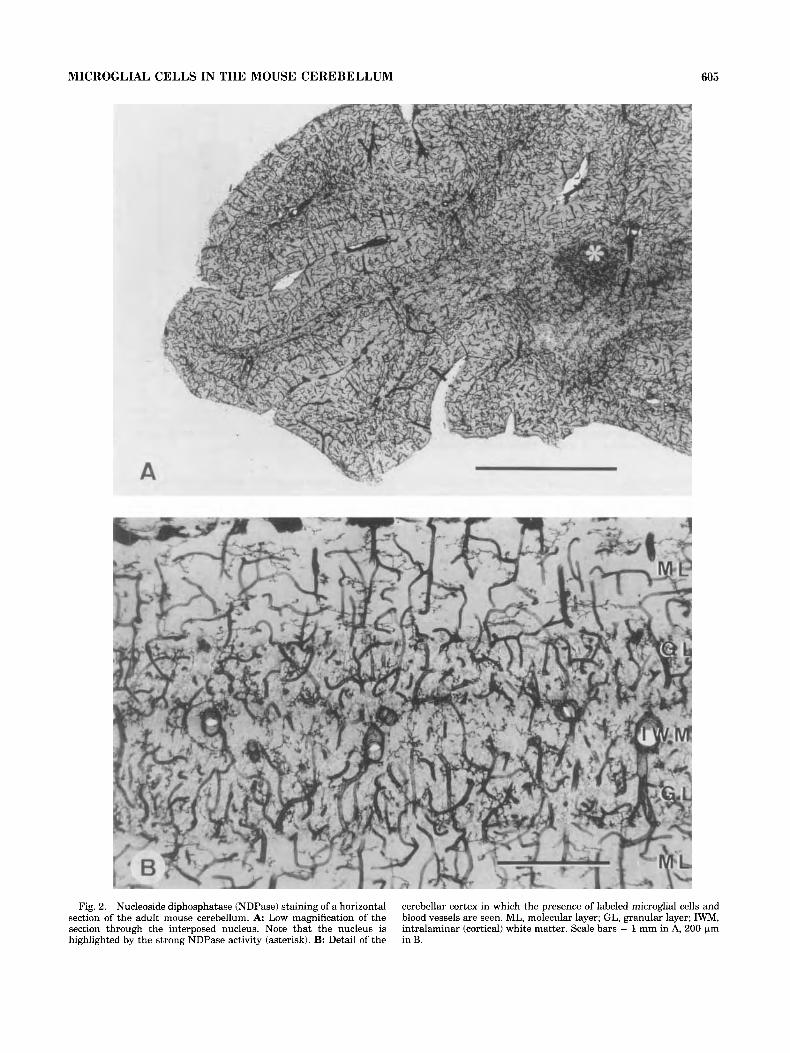
Fig. 2. Nucleoside diphosphatase (NDPase) staining of a horizontal section of the adult mouse cerebellum. A: Low magnification of the section through the interposed nucleus. Note that the nucleus is highlighted by the strong NDPase activity (asterisk). B: Detail of the
cerebellar cortex in which the presence of labeled microglial cells and blood vessels are seen. ML, molecular layer; GL, granular layer; IWM, intralaminar (cortical) white matter. Scale bars = 1 mm in A, 200 Fm in B.

606 J.M. VELA ET AL.
5000 ,.I IH YOUNG I 10 ADULT I
7 - r 4000 1 3000 - 2000 -
ML GL Fig. 3. Histogram showing the mean microglial density estimated in
the different histological cerebellar areas of adult and young mice. At both ages, the molecular layer (ML) showed the lowest density, the granular layer (GL) and white matter (WM) showed intermediate
The distribution study revealed that microglial cells were present in all cerebellar areas of both young and adult mice, but they were not uniformly distributed: the molecular layer showed the lowest microglial density, the granular layer and white matter showed intermediate values, and the cerebellar nuclei showed the highest density (Fig. 3). The statistical analysis indicated that differences in esti- mated microglial density were exclusively related to the above mentioned histological areas, and no significant differences were found between the different cerebellar lobules or between young and adult mice. Therefore, in the following paragraphs, a specific description of microglial morphology and distribution has been detailed that takes into consideration only the different histological cerebellar areas.
Molecular layer (ML) The estimated microglial density in ML was 1,425 f 106
cells/mm3 in young animals and 1,387 ? 108 cells/mm3 in adult mice. Microglial cells present in the ML shared common features and were always distinguished as big, isolated, ramified cells occupying a projection territory in which processes from adjacent microglia were rarely seen (Fig. 4). The cell body was often round or slightly elongated, measuring from 5.5 pm to 13 pm, and cell processes were fairly rectilinear but showed numerous constrictions and dilations along their way, leading to a constant variation in thickness. These microglial cells displayed two to four straight primary processes whose direction was nearly perpendicular or parallel to the pial surface direction. Primary cell processes were scarcely ramified, and the subsidiary secondary branches usually branched out at right angles. Processes of higher order were very scarce, except in terminal position, where tertiary processes could be noticed. Numerous, thin, thorn-like expansions measur- ing 1-8 pm in length were also seen in processes and cell
T T
CN values, and the cerebellar nuclei (CN) showed the greatest microglial density. Significant differences (P < 0.05) were found between the different histological layers but not between young and adult mice. Error bars indicate standard deviations.
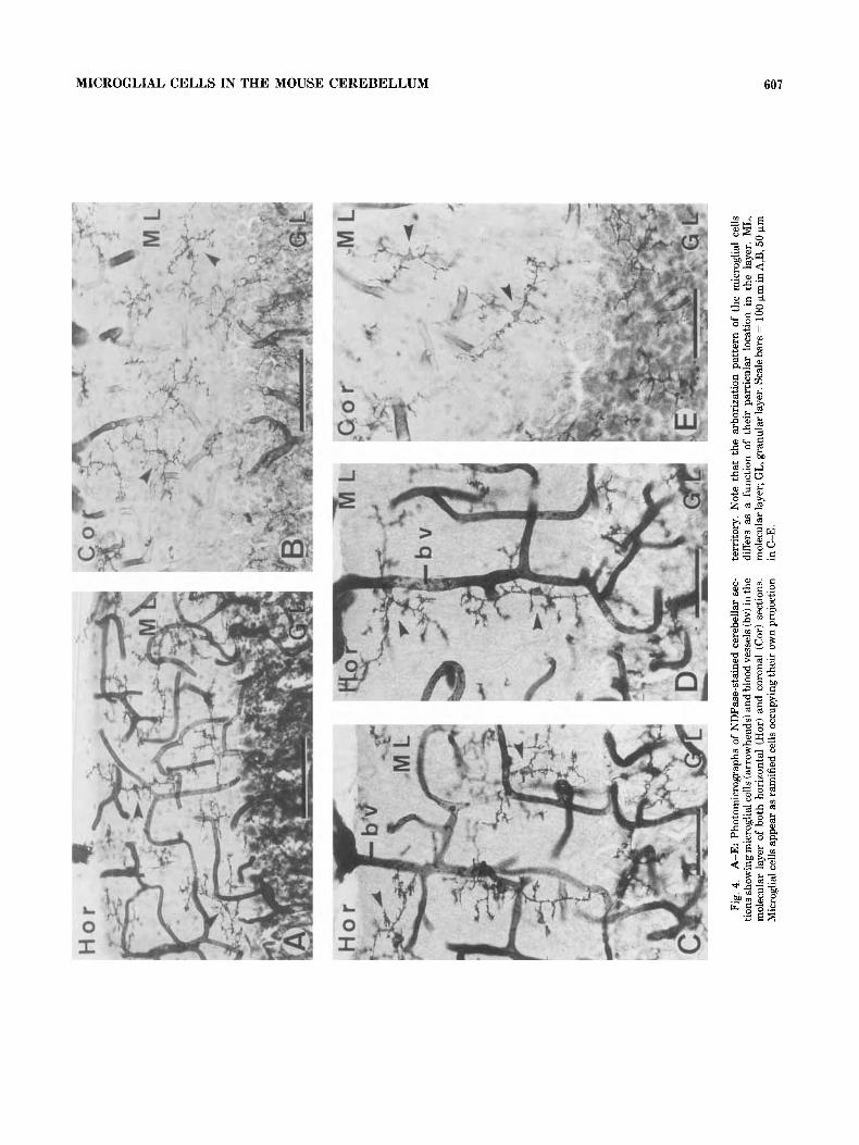
bodies and were especially abundant at branch endings (see detailed camera lucida drawings in Figs. 5,6) . The correla- tion between observations made on horizontal (Fig. 5) and coronal sections (Fig. 6) allowed us to establish different morphological microglial cell types in the ML according to their relative location and three-dimensional ramification pattern (Fig. 7).
In the external part of the ML, just under the meningeal covering, microglial cells usually displayed two long pri- mary processes running in opposite directions that consti- tute a major axis (100-150 pm length) oriented parallel to the pial surface. The projection territory occupied by these cells was semielliptic in both coronal and horizontal sec- tions, and they could be considered to be contained three- dimensionally within a dome (semisphere) with the flat surface oriented to the pial surface (Fig. 7Bd).
In the medial part of the ML, in horizontal sections, microglial cells displayed either a regular stellate shape with a circular projection territory (100-150 pm diameter) or an elliptic projection territory (100-175 pm length; main diameter) with the two longest primary processes perpen- dicularly oriented to the pial surface. In coronal sections, microglial cells displayed either a circular projection terri- tory with a mean diameter equal to or usually lower (50-120 pm) than that observed in horizontal sections or an elliptic projection territory with the major axis (100-150 pm length) parallel to the pial surface. The interpretation of these observations in both planes allowed us to establish that microglial cells located in the medial part of the ML displayed a three-dimensional ramification pattern that could be included within a sphere, a rugby ball (ellipsoid), or a lentil (biconvex disc) as illustrated in Figure 7Ba-c.
In the inner part of the ML, just over the Purkinje cell layer, microglial cells displayed a major axis parallel to the pial surface showing a morphology and size very similar to
MICROGLIAL CELLS IN THE MOUSE CEREBELLUM 607

608 J.M. VELA ET AL.
HORIZONTAL
ML
PCL
4
\ -- ---------- - -
/ - - ----
*- c 4-
/ c
25 ym Fig. 5. Composition of camera lucida drawings of microglial cells in the molecular layer obtained from
horizontal sections. PS, pial surface; ML, molecular layer; PCL, Purkinje cell layer.
those observed in the external part of ML. The arborization pattern of these cells could also be considered to be con- tained within a dome with the flat surface oriented to the
Purkinje cell monolayer (Fig. 7Be). It should be noted that these cells rarely sent processes crossing the Purkinje cell layer to penetrate into the granular layer. In addition, no

MICROGLIAL CELLS IN THE MOUSE CEREBELLUM 609
CORONAL --I
_--c---_ - /-- /
/
PS /
/ / .
/ -
Fig. 6. Composition of camera lucida drawings of microglial cells in the molecular layer obtained from coronal sections. PS, pial surface; ML, molecular layer; PCL, Purkinje cell layer.
microglial cell bodies were located among the Purkinje cell bodies.
Simultaneous visualization of the blood vessel network and microglial cells with NDPase staining allowed us to observe
that microglial cells found in the ML frequently had processes in close relationship with the capillary wall. At these points of contact with vasculature, we have noticed an increased degree of ramification and sinuosity of the microglial processes.

610 J.M. VEI ;A ET AL.
I I 1 1 I I I I 1 I 1 I I I
I I I I I I I I
I I
I
I I I I
I I I c I 1 1
WI .- 0 I I c,
I
d I
a b C I
. . : i ....................................................................................................................................................... . . . . . . i : '.. : . . . .
'< .......................................................................................................................................................
............................................................................................................................................................................... :: pcL ;
............................................................................................................................................................................. :
Fig. 7. Graphic representation of the projection territory occupied by the most characteristic microglial cells located in the different zones of the molecular layer. The correlation of the observations in both horizontal and coronal sections (A) allowed us the three-dimensional interpretation of the microglial typologies (B). The orientation of the dendritic tree of a Purkinje cell (PC) relative to the different microglial
phenotypes has been also illustrated in B. The medial zone of the molecular layer was occupied by microglial cells whose ramification was contained within a sphere (a), a lentil (biconvex disk b), or a rugby ball (ellipsoid; c). In the marginal zones of the molecular layer, microglial cells were contained within a dome (semisphere; d,e). PS, pial surface; ML, molecular layer; PCL, Purkinje cell layer.

MICROGLIAL CELLS IN THE MOUSE CEREBELLUM 611
Granular layer (GL) The estimated microglial density in GL was 3,179 2 422
cells/mm3 in young animals and 3,312 5 343 cells/mm3 in adult mice. Microglial cells in the GL were stellate in shape (Fig. 8A) and always showed a circular projection territory (70-110 pm diameter) in both horizontal and coronal sections. Therefore, three-dimensionally, microglial cells in GL could be considered to be included within a sphere in which the cell body was located in the center.
Microglial cells populating GL displayed three to five primary, radially oriented processes that branched into secondary and terminal branches of the third and fourth orders (Fig. 9A). The microglial cell body was generally round (7-13 pm diameter). Branches followed a sinuous way among the granular cell bodies. Irregularities in the diameter of the processes and the presence of thorny expansions were common. We frequently noticed overlap- ping between cell processes belonging to adjacent microglial cells. Many microglial branches and occasional microglial cell bodies were in contact with the well developed vascular network in this layer. Similar to the finding in ML, there was an increase in the ramification degree and sinuosity of microglial processes in contact with the blood vessel walls (Figs 8A, 9A).
Branches of microglial cells located immediately under the Purkinje cell layer cross this layer and slightly pen- etrate into the adjacent ML. In addition, branches of microglial cells located near the border with the white matter sent processes penetrating this area.
White matter (WM) The estimated microglial density in whole WM was
3,695 * 587 cells/mm3 in young animals and 3,981 * 780 cells/mm3 in adult mice. In WM, microglial cells appeared as ramified cells displaying a circular or often elliptic projection territory (80-140 pm diameter) in both coronal and horizontal sections. The correlation of horizontal and coronal observations indicated that microglial cells located in this layer could be considered to be contained within a sphere, sometimes a little flattened due to the presence of a major axis constituted by two long and opposite primary cell processes oriented in the same direction as nerve fibers (Figs. 8B, 9B).
The microglial cell body was generally round or slightly extended (5-12 pm in diameter) and was located in the center of the projection field. These cells showed three to six fairly straight and poorly ramified primary processes that branched out at an angle of approximately 90" until they produced second-order and, subsequently, short third- order branches. Constrictions, dilations, and long thorny expansions of up to 12 pm in length were observed. Microglial cells in this location, like those in other cerebel- lar areas, appeared isolated, and no cell processes coming from neighboring microglial cells penetrated the projection territory of others. Relationships between microglial cells and blood vessels were also observed.
In the thinnest WM layers located between the cortical lamellae, microglial cells showed a more reduced size, and their cell processes were more sinuous (Fig. 9B). Some- times, processes from these cells slightly penetrated the GL.
Cerebellar nuclei (CN) The estimated microglial density in CN was 5,910 + 867
cells/mm3 in young animals and 5,579 * 793 cells/mm3 in adult mice. We found no significant differences in microglial density between the three gray nuclei.
The three cerebellar nuclei (medial, interposed and lat- eral) were notable for their abundance of microglial cells. We did not observe noticeable morphological differences between microglial cells populating the three cerebellar nuclei and therefore they were considered all together.
Microglial cells in CN were observed in horizontal and coronal sections as ramified cells with a small and round projection territory (80-125 pm in diameter). Thus, they could be considered three-dimensionally when included within a sphere. They had a roundish or elongated cell bodies (8-15 pm in diameter) from which four to six highly ramified primary cell processes arose, leading to third- and fourth-order terminal branches that frequently overlapped with branches belonging to neighboring cells (Figs. 8C,D). Cell processes with irregular diameters and numerous thorny expansions followed a sinuous way in all directions (Figs. 9C). Generally, microglial cells located in the borders of the nuclei did not send cell processes to the medullar WM, although some cell processes coming from microglial cells located in WM penetrated the cerebellar nuclei. Occa- sionally, pairs of ramified microglial cells with cell bodies closely apposed were noticed in CN (Fig. 8E). Very often, microglial cell processes and cell bodies were observed in close relationship with the blood vessel network, which was particularly developed in CN.
DISCUSSION The present study employing the histochemical demon-
stration of NDPase as a specific microglial cell marker revealed the presence of this glial cell type in all cerebellar areas of adult and young mice. In addition, we demon- strated that the microglial population is not homogeneously distributed in the cerebellum but shows differing density values depending on the particular histological layer consid- ered. In the same way, microglial cells did not show a homogeneous morphology throughout all cerebellar areas but displayed specific morphological features as a function of their location.
Microglial cells in all cerebellar locations of both young and adult mice always appeared as ramified cells. No differences in microglial morphology were found when comparing young and adult mice. In no case did we observe the presence of ameboid microglial cells, in accordance with studies showing that ameboid microglia in the rat brain differentiate into ramified cells during the early postnatal period (Ling, 1979; Murabe and Sano, 1982b), an event that takes place in the mouse cerebellum before postnatal day 14 (Ashwell, 1990). The heterogeneity in microglial morphol- ogy in the cerebellum requires some comments. We have not found morphological differences when considering the different cerebellar lobules in both adult and young mice. However, differences in microglial morphology can be appre- ciated clearly in relation to the different histological layers: ML, GL, WM, and CN. Therefore, the precise morphology of a particular microglial cell depends on its specific location in one or another layer, and it will even depend on its relative position within the layer, as seen in the ML, indicating that microglial morphology is strongly related to

612 J.M. VELA ET AL.

MICROGLIAL CELLS IN THE MOUSE CEREBELLUM 613
A
5
GRANULAR LAYER
WHITE MATTER I ntralaminar Medullar
C CEREBELLAR N UCLE I
25 pm Fig. 9. Composition of camera lucida drawings of microglial cells of
different histological areas of the cerebellum. A: Microglial cells of the granular layer. Note that some cell processes were seen to reach the vascular wall. B: Microglial cells of the intralaminar and medullar white matter. Cells in the intralaminar white matter showed a more reduced size, and their cell processes were less rectilinear than in the
medullar white matter. Note that microglial cell processes and blood vessels of the medullar white matter were oriented in the same direction as nerve fibers (arrows). C: Microglial cells of the cerebellar nuclei showed very sinuous and ramified cell processes. A pair of microglial cells whose cell bodies were closely apposed can be seen.

614 J.M. VELA ET AL.
the histological characteristics of its local microenviron- ment.
Differences in microglial morphology between layers were also associated with differences in the distribution of microglial cells in these layers. In both young and adult mouse cerebellum, our study revealed that ML shows the lowest microglial density, GL and WM show intermediate values, and CN shows the highest density. In correspon- dence with these observations, high numbers of infected microglia have been reported in the dentate nucleus of brains from patients with acquired immunodeficiency syn- drome (Kure et al., 1990). However, studies on microglial cell distribution in normal nervous tissue are scarce, and the available data concerning the cerebellum do not corre- spond exactly with our observations. In early studies using the silver-carbonate technique, Del Rio-Hortega (1921) reported that rabbit cerebellar gray matter contained more microglia than WM. In human cerebellum, using RCA-1 lectin histochemistry, Mannoji et al. (1986) reported that microglial cells were observed more frequently in ML than in GL. Our findings in mouse cerebellum are in disagree- ment with the above-mentioned works but correspond substantially with data reported by Lawson et al. (1990), where a quick estimation of microglial distribution in different CNS areas of the adult mouse, including cerebel- lum, was made using the F4/80 antibody as a microglial marker. Methodological and species-specific differences could be the cause of variability in reported cell counts.
Our observations show that there are no significant differences in the estimated microglial densities when comparing young and adult values within each layer, indicating that the microglial cell population remains stable in number. However, we cannot discard the possibility that some microglial proliferation occurs during this period. In fact, an increase in the number of microglial cells during aging has been demonstrated in the cerebral cortex of rhesus monkey (Peters et al., 19911, and studies combining 3H-thymidine autoradiography and immunocytochemistry in normal mouse brain have demonstrated that microglial cells continue to divide in the adult, albeit at a very low level (Lawson et al., 1992). In this regard, the pairs of microglial cells we observed in CN, which is the more densely popu- lated cerebellar area, could correspond with the final stages of local division of microglia.
Microglia and blood vessels The simultaneous visualization of microglial cells and the
vascular network using the NDPase staining allowed us to analyze the possible association of microglial cells with blood vessels. Our observations clearly revealed the exis- tence of a close correspondence between microglial density and the degree of vascularization. The high microglial density in CN corresponds with a high density of vascular profiles in this area, whereas a low microglial density corresponds with a low degree of vascularization in ML. In addition, in all cerebellar layers, we observed that some microglial cell processes are in contact with the vessel wall. This physical relation has already been reported in the rat brain by ultrastructural studies showing that microglial cell processes were incorporated between the astrocytic foot processes and reached the microvascular basement mem- brane (Lassmann et al. 1991).
Although the role played by such microglial processes in relation to vasculature remains unclear, there is evidence suggesting that this physical interrelation could be impor-
tant for the reciprocal maintenance of the vascular network and the microglial characteristics in the normal adult tissue: 1) It is known that microglial cells produce angio- genic and vasoregulatory molecules (Giulian et al., 1986, 1988; Giulian and Corpuz, 1993) in the same way that macrophages produce angiogenic factors in other locations (Leibovich et al., 1987; Eisenstein, 1991); 2) extracellular matrix components, which are present in the basement membrane around blood vessels and the glia limitans, have regulatory effects on the in vitro differentiation of micro- glial cells (Chamak and Mallat, 1991); and 3) the integrity of the blood-brain barrier seems to be essential for the maintenance of the normal ramified microglial phenotype, as suggested by in vitro experiments showing that serum addition to culture medium induced morphological transfor- mation of microglia (Chamak and Mallat, 1991). Moreover, it should be noted that, in certain regions of the CNS, such as the median eminence, where the blood-brain barrier is absent and the cells are exposed to plasma proteins, micro- glial cells display a distinctive morphology in addition to an upregulation in their antigen expression (Perry and Gor- don, 1988; Lawson et al., 1990).
Possible factors influencing microglial distribution and morphology
Microglial characteristics in the mouse cerebellum are strongly related to the characteristics of their environment. This is also corroborated by other studies reporting distinc- tive distribution and/or morphological features of micro- glial cells in diverse species and CNS areas (Del Rio- Hortega, 1932; Cammermeyer, 1970; Schnitzer, 1989; Lawson et al., 1990; Castellano et al., 1991b). In a very simplistic view, microglial adaptation to the environment could be exclusively interpreted in the sense of a microglial filling of the space unoccupied by neurons and macroglial cells. However, we would like to emphasize here that microglial morphology and distribution must reflect pre- dominantly an adaptation of these cells to their specific function within the CNS. This leads to a controversial point, because the exact role played by microglia in the normal adult CNS remains to be clarified.
It has been suggested that normal, ramified microglial cells could participate in the control of extracellular me- dium and fluid cleansing by means of pinocytosis (Glenn et al., 1991; Ward et al., 1991). It has also been suggested that microglial cells in the adult may form a network of immune competent cells (Graeber and Streit, 1990). The ramified appearance of microglial cells, their regularly spaced distri- bution within each layer, their tendency to cover a larger territory when their density is lower, and their narrow relation with the vascular network, taken together, enable them to play both putative functions: control of the extracel- lular medium and immunovigilance.
Using enzyme histochemistry, it has been shown that microglial cells display nucleoside triphosphatase (Sjos- trand, 1966; Ibrahim et al., 1974), nucleoside diphospha- tase (Murabe and Sano, 1982a,b; Vorbrodt and Wisniewski, 1982; Castellano et al., 1991a,b), 5'-nucleotidase (Kreutzberg and Barron, 1978), and purine nucleoside phosphorylase (Castellano et al., 1990) enzymatic activities. Therefore, in our opinion, microglial cells clearly may be involved in the control of extracellular levels of purine nucleosides. Purine nucleotides and their dephosphory- lated nucleosides (i.e., adenosine, guanosine, and inosine) are released from neurons and act as neurotransmitters or

MICROGLIAL CELLS IN THE MOUSE CEREBELLUM
neuromodulators (Phillis and Wu, 1981; Williams, 1989; Edwards et al., 1992; Fredholm et al., 1993). In more concrete terms, there is now considerable evidence that ATP is actively transported into synaptic vesicles (Zimmer- man, 1988; Unsworth and Johnson, 1990) and may be costored in vesicles with other neurotransmitters (Zimmer- man, 1978; Winkler and Westhead, 1980). When neurons in a number of regions of the CNS are stimulated, ATP is released (White, 1984; White et al., 1985) and has excit- atory effects on the activity of neurons via extracellular P2-purinergic receptors (Stone, 1981; Edwards et al., 1992). Extracellular ATP is rapidly metabolized by ectonucleotid- ases, and the end products are AMP or adenosine, which are taken up via extracellular PI-purinergic receptors by astro- cytes and neurons (Wu and Phillis, 1984; Hosli and Hosli, 1988). Unlike ATP, extracellular adenosine has inhibitory effects on the activity of neurons throughout the CNS (Stone, 1981; Dunwiddie, 1985; Snyder, 1985; De Koninck and Henry, 1992). Adenosine taken up in the nerve termi- nals may be catabolized intracellularly (Wu and Phillis, 1984) or recycled to form ATP, which is repackaged into vesicles (Zimmerman, 1978) and is subsequently available for release.
Because microglial cells, on their plasma membrane, display the degradative enzymes needed for the dephos- phorylation in cascade of ATP, it is tempting to suggest that microglial cells are directly involved in the inactivation and turnover of these neuromodulators or neurotransmitters. In this regard, it should be added that, on the basis of ultrastructural studies, Murabe and Sano (1982a) reported a close relationship between microglial processes and syn- apses in the cerebral cortex of the normal rat. Blinzinger and Kreutzberg (1968) have also described the direct physical contact and microglial displacement of synaptic boutons from the surface of regenerating motor neurons.
Our observations in the lizard brain (Castellano et al., 1991b) strongly suggest the existence of a close correspon- dence between microglial cell distribution and the seroton- ergic immunoreactivity pattern. In addition, an increase in the number and complexity of microglial processes in response to selective changes in serotonergic innervation has been demonstrated under experimental conditions (Wil- son and Molliver, 1994). In this context, it should be pointed out that, in normal mouse cerebellum, serotonergic innervation in the CN is denser than in the cortex (Triar- hou and Ghetti, 1991), corresponding with a higher micro- glial density in CN. Likewise, the density of serotonin- immunoreactive fibers is higher in the GL than in the ML (Triarhou and Ghetti, 1991), which is in agreement with a higher microglial density in GL than in ML. Furthermore, CNS areas that were densely populated by microglial cells (i.e., olfactory bulb, hippocampal formation, basal ganglia, and substantia nigra; Lawson et al., 1990) also receive serotonergic projections (Steinbusch, 1984).
On the basis of our results and present studies, we cannot conclude that microglial cells are clearly involved in the synaptic function. However, their capacity to regulate extracellular purine nucleoside levels and their presumable relation with serotonergic terminals strongly suggest their participation in the regulation of neurotransmission. Fur- ther studies using double-labeling techniques and electron microscopy could help to elucidate the function or functions of microglial cells.
ACKNOWLEDGMENTS The authors thank Miguel Angel Martil
technical assistance.
615
for excellent
LITERATURE CITED Abercrombie, M. (1946) Estimation of nuclear population from microtome
sections. Anat. Rec. 94:239-247. Ashwell, K. (1990) Microglia and cell death in the developing mouse
cerebellum. Dev. Brain Res. 55:21:219-230. Banati, R.B., J. Gehrmann, P. Schubert, and G.W. Kreutzberg (1993)
Cytotoxicity of microglia. Glia 7:111-118. Beitz, A., and V. Chan-Palay (1979a) The medial cerebellar nucleus in the
rat: Nuclear volume, cell number, density and orientation. Neuroscience 4:3145.
Beitz, A., and V. Chan-Palay (1979b) A Golgi analysis of neuronal organiza- tion in the medial cerebellar nucleus in the rat. Neuroscience 4t47-63.
Blinzinger, K., and G.W. Kreutzberg (1968) Displacement of synaptic terminals from regenerating motoneurons by microglial cells. Zeitschr. Zellforsch. 85: 145-157.
Cajal, S. Ram6n y (1911) Histologie du sisteme nerveux de l’homme et des vertebr&s, Vols. I and 11. Paris: Maloine.
Cammermeyer, J. (1970) The life history of the microglial cell: A light microscopic study. Neurosci. Res. 3:4%129.
Castellano, B., B. Gonzaez, B.R. Finsen, and J. Zimmer (1990) Histochemi- cal demonstration of purine nucleoside phosphorylase (PNPase) in microglial and astroglial cells of adult rat brain. J. Histochem. Cytochem. 38:1535-1539.
Castellano, B., B. Gonzdez, M.B. Jensen, E.B. Pedersen, B.R. Finsen, and J. Zimmer (1991a) A double staining technique for simultaneous staining of astrocytes and microglial cells in vibratome brain sections and astroglial cell cultures. J. Histochem. Cytochem. 39:561-568.
Castellano, B., B. Gonzdez, I. Dalmau, and J.M. Vela (1991b) Identification and distribution of microglial cells in the cerebral cortex of the lizard: A histochemical study. J. Cornp. Neurol. 31 1:434444.
Chamak, B., and M. Mallat (1991) Fibronectin and laminin regulate the in vitro differentiation of microglial cells. Neuroscience 45:513-527.
Chan-Palay, V. (1977) Cerebellar dentate nucleus. Organization, cytology and transmitters. Berlin: Springer-Verlag.
De Koninck, Y., and J.L. Henry (1992) Peripheral vibration causes an adenosine-mediated postsynaptic inhibitory potential in dorsal horn neurones in the cat spinal cord. Neuroscience 50t435-443.
Del Rio-Hortega, P. (1921) Histoghesis y evoluci6n normal; Exodo y distribuci6n regional de la microglia. Mem. de la SOC. Esp. de Hist. Nat. 11:105-150.
Del Rio-Hortega, P. (1932) Microglia. In W. Penfield (ed.): Cytology and Cellular Pathology of the Nervous System, Vol. 11. New York: Paul B. Hoeber, pp. 481434.
Dunwiddie, T.V. (1985) The physiological role of adenosine in the central nervous system. Int. Rev. Neurobiol. 27:63-139.
Eccles, J.C., K. Sasaki, and P. Strata (1967) A comparison of the inhibitory actions of Golgi cells and ofbasquet cells. Exp. Brain Res. 3:81-94.
Edwards, F.A., A.J. Gibb, and D. Colquhoun (1992) ATP receptor-mediated synaptic current in the central nervous system. Nature 359:144-146.
Eisenstein, R. (1991) Angiogenesis in arteries: Review. Pharmacol. Ther. 49: 1-19.
Ferrer, I., and J. Sarmiento (1980) Nascent microglia in the developing brain. Acta Neuropathol. 50%-67.
Finsen, B.R., T. Sgrensen, B. Castellano, E.B. Pedersen, and J. Zimmer (1991) Leukocyte infiltration and glial reactions in xenografts of mouse brain tissue undergoing rejection in the adult rat brain. A light and electron microscopical immunocytochemical study. J. Neurol. Immunol. 32159-183.
Fredholm, B.B., B. Johansson, I. van der Ploeg, P.S. Hu, and S. Jin (1993) Neuromodulatory roles of purines. Drug Dev. Res. 28:349-353.
Ghandour, M.S., G. Vincendon, and G. Gombos (1980) Astrocyte and oligodendrocyte distribution in adult rat cerebellum: An immunohisto- logical study. J. Neurocytol. 9:637-646.
Giulian, D., and M. Corpuz (1993) Microglial secretion products and their impact on the nervous system. In F.J. Seil (ed.): Advances in Neurology, Vol. 59. New York: Raven Press, pp. 315-320.
616 J.M. VELA ET AL.
Giulian, D., T.J. Baker, L.N. Shih, and L.B. Lachman (1986) Interleukin-1 of the central nervous system is produced by ameboid microglia. J. Exp. Med. 164:594-604.
Giulian, D., J. Woodward, D. Young, J.F. Krebs, and L.B. Lachman (1988) Interleukin-1 injected into mammalian brain stimulates astrogliosis and neovascularization. J. Neurosci. 8.2485-2490.
Glenn, J.A., P.L. Booth, and W.E. Thomas (1991) Pinocytotic activity in ramified microglia. Neurosci. Lett. 123:27-31.
Graeber, M.B., and W.J. Streit (1990) Microglia: Immune network in the CNS. Brain Pathol. 1.2-5.
Hosli, E., and L. Hosli (1988) Autoradiographic studies on the uptake of adenosine and on binding of adenosine analogues in neurones and astrocytes of cultured rat cerebellum and spinal cord. Neuroscience 24:621-628.
Ibrahim, M.Z.M., Y. Khreis, and D.S. Koshayan (1974) The histochemical identification of microglia. J. Neurol. Sci. 22.21 1-233.
Innocenti, G.M., H. Koppel, and S. Clarke (1983) Transitorymacrophages in the white matter of the developing visual cortex. I. Light and electron microscopic characteristics and distribution. Dev. Brain Res. 11:39-53.
Jensen, M.B., B. Gonzalez, B. Castellano, and J. Zimmer (1994) Microglial and astroglial reactions to anterograde axonal degeneration: A histochemi- cal and immunocytochemical study of the adult rat fascia dentata after entorhinal perforant path lesions. Exp. Brain Res. 98:245-260.
Jdrgensen, M.B., B.R. Finsen, M.B. Jensen, B. Castellano, N.H. Diemer, and J. Zimmer (1993) Microglial and astroglial reactions to ischemic and kainic acid-induced lesions of the adult rat hippocampus. Exp. Neurol. 120:70-88.
Kreutzberg, G.W., and K.D. Barron (1978) 5‘-Nucleotidase of microglial cells in the facial nucleus during axonal reaction. J. Neurocytol. 7.601-610.
Kure, K., K.M. Weidenheim, W.D. Lyman, and D.W. Dickson (1990) Morphology and distribution of HIV-1 gp41-positive microglia in sub- acute AIDS encephalitis. Pattern of involvement resembling a multisys- tem degeneration. Acta Neuropathol. 80:393-400.
Lassmann, H., F. Zimprich, K. Vass, and W.F. Hickey (1991) Microglial cells are a component of the perivascular glia limitans. J. Neurosci. Res. 28.236-243.
Lawson, L.J., V.H. Perry, P. Dri, and S. Gordon (1990) Heterogeneity in the distribution and morphology of microglia in the normal adult mouse brain. Neuroscience 39:151-170.
Lawson, L.J., V.H. Perry, and S. Gordon (1992) Turnover of resident microglia in the normal adult mouse brain. Neuroscience 48:405415.
Leibovich, S.J., P.J. Polverini, H.M. Shepard, D.M. Wiseman, V. Shively, and N. Nuseir (1987) Macrophage-induced angiogenesis is mediated by tumor necrosis factor-a. Nature 329530432.
Ling, E.A. (1979) Transformation of monocytes into amoeboid microglia and into microglia in the corpus callosum in postnatal rats, as shown by labelling monocytes by carbon particles. J. Anat. 128:847458.
Ling, E.A., D. Penney, and C.P. Leblond (1980) Use of carbon labelling to demonstrate the role of blood monocytes as precursors of the “amoeboid cells” present in the corpus callosum of postnatal rats. J. Comp. Neurol. 193.63 1-657.
Mannoji, H., H. Yeger, and L.E. Becker (1986) A specific histochemical marker (lectin Ricinus communis agglutinin-1) for normal human microglia, and application to routine histopathology. Acta Neuropathol. 71:341-343.
McGeer, P.L., T. Kawamata, D.G. Walker, H. Akiyama, I. Tooyama, and E.G. McGeer (1993) Microglia in degenerative neurological disease. Glia 7:84-92.
Murabe, Y., and Y. Sano (1982a) Morphological studies on neuroglia. V. Microglial cells in the cerebral cortex of the rat, with special reference to their possible involvement in synaptic function. Cell Tissue Res. 223:493- 506.
Murabe, Y., and Y. Sano (1982b) Morphological studies on neuroglia. VI. Postnatal development of microglial cells. Cell Tissue Res. 225:469-485.
Murabe, Y., Y. Ibata, and Y. Sano (1981) Morphological studies on neuroglia. 111. Macrophage response and “microgliocytosis” in kainic acid-induced lesions. Cell Tissue Res. 218.75586.
Palay, S.L., and V. Chan-Palay (1974) Cerebellar Cortex. Cytology and Organization. Berlin: Springer-Verlag.
Perry, V.H., and S. Gordon (1988) Macrophages and microglia in the nervous system. Trends Neurosci. 11:273-277.
Perry, V.H., and S. Gordon (1991) Macrophages and the nervous system. Int. Rev. Cytol. 125:203-244.
Peters, A,, K. Josephson, and S.L. Vincent (1991) Effects of aging on the neuroglial cells and pericytes within area 17 of the Rhesus monkey cerebral cortex. Anat. Rec. 229.384-398.
Phillis, J., and P. Wu (1981) The role of adenosine and its nucleotides in central synaptic transmission. Progr. Neurobiol. 16:187-239.
Schnitzer, J. (1989) Enzyme-histochemical demonstration of microglial cells in the adult and postnatal rabbit retina. J. Comp. Neurol. 282249-263.
Sjostrand, J. (1966) Changes of nucleoside phosphatase activity in the hypoglossal nucleus during nerve regeneration. Acta Physiol. Scand. 672 19-228.
Snyder, S.H. (1985) Adenosine as a neuromodulator. Annu. Rev. Neurosci. 8: 103-124.
Sotelo, C. (1967) Cerebellar neuroglia: Morphological and histochemical aspects. In C.A. Fox and R.S. Snider (eds.): The Cerebellum. Progress In Brain Research, Vol. 25. Amsterdam: Elsevier, pp. 226-250.
Sotelo, C., and P. Angaut (1973) The fine structure of the cerebellar central nuclei in the cat. I. Neurons and neuroglial cells. Exp. Brain Res. 16:410-430.
Steinbusch, H.W.M. (1984) Serotonin-immunoreactive neurons and their projections in the CNS. In A. Bjorklund, T. Hokfelt, and M.J. Kuhar ieds.): Handbook of Chemical Neuroanatomy, Vol. 3, Part 11. Amster- dam: Elsevier, pp. 68-125.
Stone, T.W. (1981) Physiological roles for adenosine and adenosine 5‘- triphosphate in the nervous system. Neuroscience 6:523-555.
Streit, W.J., and G.W. Kreutzberg (1988) Response of endogenous glial cells to motor neuron degeneration induced by toxic ricin. J. Comp. Neurol. 268:248-263.
Triarhou, L.C., and B. Ghetti (1991) Serotonin-immunoreactivity in the cerebellum of two neurological mutant mice and the corresponding wild-type genetic stocks. J. Chem. Neuroanat. 4.421428.
Unsworth, C.D., and R.G. Johnson (1990) ATP compartmentation in neuroendocrine secretory vesicles. Ann. NY Acad. Sci. 603:353-365.
Vorbrodt, A.W., and H.M. Wisniewski (1982) Plasmalemma-bound nucleo- side diphosphatase as cytochemical marker of central nervous system (CNS) mesodermal cells. J. Histochem. Cytochem. 30:418-424.
Ward, S.A., P.A. Ranson, P.L. Booth, and W.E. Thomas (1991) Characteriza- tion of ramified microglia in tissue culture: Pinocytosis and motility. J. Neurosci. Res. 29.13-28.
White, T.D. (1984) Characteristics of neuronal ATP. Progr. Neurol. Psycho- pharmacol. Biol. Psychiatr. 8:487493.
White, T.D., J.W. Downie, and R.A. Leslie (1985) Characteristics of K+- and veratridine-induced release of ATP from synaptosomes prepared from dorsal and ventral spinal cord. Brain Res. 334.372-374.
Wilkin, G.P., and G. Levi (1986) Cerebellar astrocytes. In S. Fedoroff and A. Vernadakis (eds.): Astrocytes. Development, Morphology, and Regional Specialization of Astrocytes, Vol. I. London: Academic Press, pp. 245- 268.
Williams, M. (1989) Adenosine: The prototypic neuromodulator. Neuro- chem. Int. 14.249-264.
Wilson, M.A., and M.E. Molliver (1994) Microglial response to degeneration of serotonergic axon terminals. Glia 11:18-34.
Winkler, H., and E. Westhead (1980) The molecular organization of adrenal chromaffin granules. Neuroscience 5: 1803-1824.
Wu, P.H., and J.W. Phillis (1984) Uptake by central nervous tissues as a mechanism for the regulation of extracellular adenosine concentrations. Neurochem . Int .6: 6 13-632.
Zimmerman, H. (1978) Turnover of adenine nucleotides in cholinergic synaptic vesicles of the Torpedo electric organ. Neuroscience 3:827-836.
Zimmerman, H. (1988) Cholinergic synaptic vesicles. Hdbk. Exp. Pharma- col. 86:349-375.