morphological and phylogenetic studies of …tai2.ntu.edu.tw/taiwania/pdf/tai.2017.62.93.pdf ·...
TRANSCRIPT
Taiwania 62(1): 93‒98, 2017
DOI: 10.6165/tai.2017.62.93
93
Morphological and phylogenetic studies of Lycoperdon rupicola: first report for the Indian mycobiota Dyutiparna CHAKRABORTY1, Kanad DAS1*, Abhishek BAGHELA2, Nikita MEHTA2, Sanjay Kumar SINGH2, Sobhan Kumar MUKHERJEE3 and Rui Lin ZHAO4
1. Botanical Survey of India, Cryptogamic Unit, P.O. Botanic Garden, Howrah 711103, India. 2. MACS' Agharkar Research Institute, Biodiversity and Palaeobiology Group, National Fungal Culture Collection of India
(NFCCI), G.G. Agarkar Road, Pune 411004, India. 3. University of Kalyani, Department of Botany, Kalyani 741235, Nadia, India. 4. State Key Laboratory of Mycology, Institute of Microbiology, Chinese Academy of Sciences, West Beicheng Road, Beijing, China. *Corresponding author's email: [email protected] (Manuscript received 13 April 2016; accepted 26 January 2017; online published 22 February 2017) ABSTRACT: Lycoperdon rupicola Jeppson, E. Larss. & M.P. Martín growing on mossy bed attached to rocks or soil, is reported here for the first time from India with its morphological details, supporting illustrations and phylogenetic evidences. Similar looking or allied taxa are compared. The ITS- and LSU-rDNA based phylogenetic analysis of our collection also confirms its conspecificity with its European counterpart. KEY WORDS: Agaricales, India, Macrofungi, New record, Phylogeny, Taxonomy. INTRODUCTION
The genus Lycoperdon Pers. is characterized as: basidiomata subglobose with a plicating base or pyriform to turbinate with well-developed rhizomorphs; exoperidium may be spinose, verrucose, granular to furfuraceous or a combination of these features, becoming deciduous (at least in part) with maturity; endoperidium thin, papery, persistent, dehiscing by simple to stellately lobed apical ostiole; gleba compact and white initially, finally powdery, variously colored, consisting of warty basidiospores and capillitium, with or without distinct pseudocolumella; subgleba usually chambered, diaphragm absent (Kreisel, 1962; Demoulin 1968, 1973); capillitium usually dichotomously branched, aseptate, thick to thin-walled, mostly pitted; paracapillitium present or absent; basidiospores globose to oval, with various kinds of warts, pedicellate or apedicellate. For long time Lycoperdon was kept in the family Lycoperdaceae Chev. along with other puffballs (Miller and Miller, 1988; Pegler et al., 1995). While the taxonomic literature usually places Lycoperdon in its own family and order, the molecular evidence for a relationship to Agaricales may lead some authors to include them in a highly heterogeneous family Agaricaceae along with a number of fleshy-gilled mushrooms (Hibbett and Thorn, 2001; Monclavo et al., 2002; Kirk et al., 2008). About 173 species are hitherto reported from the world (www.speciesfungorum.org), although the Dictionary of Fungi (Kirk et al., 2008) maintained about the existence of about 50 species from the world. According to Bisht (2008), 20 species occur in India.
During macrofungal surveys to different parts of East district of Sikkim (a small Himalayan state in India) a few gasteroid mushrooms were collected. Among them one appeared as Lycoperdon rupicola Jeppson, E. Larss. & M.P. Martín after thorough morphological and phylogenetical studies. In the present communication, this species (from Sikkim) is described and illustrated for the first time from India. ITS- and LSU-rDNA based phylogenetic analyses were also performed.
MATERIALS AND METHODS
Morphology
Macromorphological or field characteristics were recorded in the field or basecamp from the fresh and dissected basidiomata. Images of the fresh basidiomata were captured with the help of Nikon D300s and Canon PowerShot SX 220 HS. Color codes and terms mentioned here are mostly after Methuen Handbook of Color (Kornerup and Wanscher, 1978). Samples were dried in a field drier. Micromorphological characteristics were recorded with the help of a compound microscope (Nikon Eclipse Ni-U) from the dry samples mounted in a mixture of 5% KOH, 1% Phloxin, Congo red and separately in Cotton Blue or distilled water. Micromorphological drawings were made with the drawing tube (dedicated to Nikon Eclipse Ni-U) at 400× and 1000× magnifications. Basidiospore-measurements exclude the height of ornamentations and were noted based on the observations of twenty randomly chosen basidiospores. Herbarium names are after Holmgren et al. (1990).

Taiwania Vol. 62, No. 1
94
DNA extraction, polymerase chain reaction (PCR) and sequencing
DNA extraction, PCR and sequencing were carried out at the NFCCI, MACS’ Agharkar Research Institute, Pune, India. Genomic DNA was isolated from the dried specimen following Buzina et al. (2001). The ITS & LSU regions of rDNA were amplified using standard primer pairs ITS4–ITS5 and LROR–LR7 respectively (White et al., 1990). The PCR products were purified with Axygen PCR cleanup kit (Axygen Scientific Inc, CA, USA). The cycle sequencing products were run on an ABI Avant 3100 automated DNA sequencer (Applied Biosystems, USA). The raw DNA sequencing files were edited and combined using ChromasLite v. 2.01 (http:/www.technelysium.com.au). The final sequences were deposited in the NCBI nucleotide sequence database (Accesssion Numbers: ITS - KU167031, LSU - KU167032). Phylogenetic analyses
Phylogenetic analysis based on ITS and LSU sequence data was carried out to establish the phylogenetic placement of our isolate. Reference sequences and out-group were selected from the relevant literature and GenBank. Alignment was performed using CLUSTAL W (http://www.ebi.ac.uk/clustalw/). The phylogenetic analysis was performed by using the Neighbor-Joining method (Saitou and Nei 1987). The optimal tree with the sum of branch length = 0.17422194 is shown. The percentage of replicate trees in which the associated taxa clustered together in the bootstrap test (1000 replicates) is shown next to the branches. The tree is drawn to scale, with branch lengths in the same units as those of the evolutionary distances used to infer the phylogenetic tree. The evolutionary distances were computed using the Kimura 2-parameter method (Kimura, 1980) and are in the units of the number of base substitutions per site. Evolutionary analyses were conducted in MEGA 6.0 (Tamura, 2013).
The evolutionary history was also inferred using the Maximum Likelihood and Maximum Parsimony methods so as to further substantiate our results of Neighbor-Joining analysis, however, the data is only shown for NJ analysis. (see Supplementary 1 & 2)
RESULTS ITS-LSU sequences and phylogeny
The multiple ITS-LSU sequences of 15 different species of Lycoperdon including the Indian isolate of L. rupicola were analyzed. Calvatia candida and Langermannia gigantea were chosen as out-group taxa. The phylogenetic tree based on the Neighbor-Joining method (Fig. 1) representing 32–35 combined sequences from ITS and LSU genes are shown here. Our Indian isolate (DC 14-024) was found be clustered amongst the sequences (NCBI sequences: DQ112580,
DQ112581, JN572900, JN572901, JN572902 and JN572903) derived from European materials of Lycoperdon rupicola (Jeppson et al., 2012) showing its wide range of distribution from Europe to Asia. Taxonomy
Lycoperdon rupicola Jeppson, E. Larss. & M.P. Martín, Mycol. Progr.11(4): 891 (2012) Figs. 2,3
Basidiomata scattered, pyriform with maturity, 15–25 mm high, 8–14 mm broad. Exoperidium pale yellow (4A3) at base and brownish orange (5C4) near apex when young, gradually dark brown (6F5) near base and becoming darker (6F7 to 6F8) towards apex on maturity. Exoperidial wart conical to pyramidal or spinoid in young basidiomata, mainly deciduous on maturity; warts 0.1–0.2 mm long on apex. Endoperidium papery, white, becoming grey brown with maturity. Dehiscence mostly by a central mammiform, protruding and stellately multi-lobed ostiole. Gleba chalky white (1A1) when young, yellowish brown (5E5) on maturity, with an indistict pseudocolumella. Subgleba small, up to 5 mm long, firm, alveolate (alveolae 3–4/mm), greyish brown (6D3, 7D3 or 8E3).
Basidiospores mostly globose, (3.0)4–5.2–6.5 × 3–5.12–6.2 µm (Q= 1–1.02–1.09), ornamented with isolated warts; under SEM, composed mainly of conical warts (0.3–0.4 µm) which occasionally connected by thin connectors. Capillitium 4–7 µm wide, ‘Lycoperdon-type’, composed of olive brown, branched, pitted, pits regular, thick walled (up to 0.7 µm), occasionally septate (septa joint like) hyphae. Paracapillitium absent. Outer exoperidium (warts) 100–200 µm high, 150–230 µm wide, composed mostly of globose to subglobose cells; cells 10–22 × 7–15.5 µm, thick-walled (up to 1.5 µm thick). Endoperidium cellular, composed mostly of angular to setose cells (near ostiole) and some subglobose to ellipsoid or irregular cells; setose cells 21–40 × 5–16.5 µm.
Habitat - growing in groups or gregariously on a mossy bed attached to a rock at the edge of a subalpine mixed forest that is located beside a stream.
Material examined:—INDIA. Sikkim: East district, surroundings of Memainchu Lake, elev. 3601 m, N27°21′0.6′′ E88°49′58.9′′, 2 August 2014, D. Chakraborty & K. Das, DC 14-024 DISCUSSION
The ITS-LSU phylogeny (Fig. 1) shows close proximity of the present Indian collection to Lycoperdon rupicola reported from the same habitat in Finland, Norway, Sweden and Spain (Martín and Jeppson, 2001; Jeppson et al., 2012) of Europe. The morphological features of materials from India were with conformity of European materials in terms of size and morphology of basidiomata, stellately lobed ostiole, indistinct pseudocolumella and ‘Lycoperdon-type’ capillitium hyphae with pits. One apparent difference

March 2017 Chakraborty et al.: Lycoperdon rupicola in India
95
Fig. 1 Phylogram generated from Neighbor-Joining method based on ITS- & LSU-rDNA sequences: Indian collection of Lycoperdon rupicola (DC 14-024) is shown in red and bold. The evolutionary history was inferred using the Neighbor-Joining method (Saitou N. and Nei M. 1987). The optimal tree with the sum of branch length = 0.17422194 is shown. The percentage of replicate trees in which the associated taxa clustered together in the bootstrap test (1000 replicates) is shown next to the branches. The tree was rooted with Calvatia candida and Langermannia gigantea. Evolutionary analyses were conducted in MEGA6 [2]. Bootstrap values lower than 50% are not shown. would be a bigger spore size, which had been reported by Martin and Jeppson (2001) as 4.0–4.5–(5.0) µm, while Jeppson, Larsson and Martin (2012) gave the same size p.892 and reduced it to 4.0–4.5 µm p.494.
V. Demoulin who is working on an monograph of the genus and has studied several collections of L. rupicola from Europe, including some used by Martin and Jeppson and also two from Asia, communicated us informations that make it completely convincing that the Sikkim collection belongs to that species. Concerning basidiospore size, his systematic measurements show the basidiospores are constantly large, 4.3–4.6–5.1–5.7 µm, where 4.3–5.7 µm are the
extremes and 4.6–5.1 µm the extreme means of individual specimens. Of the two specimens of the collection reported from Spain in 2001, only one is fully ripe and its basidiospores are 4.7–5.1–5.5 µm.
The following two collections attest the presence of L. rupicola in cold regions of Asia; Sibiria australis, Montes Sajaneneses orientales, Us-Beldir, 13/8/1972, A. Raitviir et B. Kullman (TAA 66 101), inter muscos in montibus clivosis (two basidiomata were unfortunately quite old but nonetheless typical); Bhutan, Thimphu, Chankaphug, 23/8/1981, B. M. Sharma (PAN 23 381) (On soil among mosses, alt. 8500ft; a good collection of five small (12–16 × 11–15 mm) basidiomata, fitting

Taiwania Vol. 62, No. 1
96
Fig. 2. Photographic illustrations of Lycoperdon rupicola (DC 14-024): A & B: Fresh basidiomata. C: Stellately multi-lobed ostiole. D: Gleba and subgleba. E & H: T.S. through exoperidium (under low and high magnifications). F & G: Capillitia with abundant pits. I & J: Basidiospores under light microscope and SEM. Scale bar: E = 100 µm; F, G&I = 10 µm; H = 50 µm; G = 10 µm.

March 2017 Chakraborty et al.: Lycoperdon rupicola in India
97
Fig. 3. Drawing illustrations of Lycoperdon rupicola (DC 14-024): A: T.S. through exoperidium (high magnifications) showing thick-walled sphaerocysts. B & C: Septate and aseptate capillitia. D: Setose cells at the opening of the endoperidium. Scale bar: A = 25 mm; B, C & D = 10 µm. well the Sikkim material; the pores in the capillitium are regular and the basidiospores moderately verrucose, 4.4–4.9–5.4 µm; spiny spherocysts are present in the dehiscence zone).
Given the misleading report of basidiospore size in the original description and other confirmed records in Asia, the identity of the Sikkim material with L. rupicola does not make doubts. Two problems remain to be studied that make further collecting in Asia and especially the Himalayas urgently needed.
One is the ITS variation for which geographical patterns would be interesting to uncover. The other is the morphological variability and discrimination toward the two somewhat similar looking species L. ericaeum Bonord. (including L. muscorum Morgan) and L. niveum Kreisel. Lycoperdon ericaeum is a well known species which is morphologically and in term of ITS
sequences rather distinct. The same cannot be said of L. niveum. This species (L. niveum) was described by Kreisel (1969) from very high elevation in Nepal and the small basidiomata of the type collection (J. Poelt G 13, M) mainly differ from L. rupicola in the longer whitish spines of the exoperidium. The possibility that L. niveum is a high elevation and L. rupicola a lower elevation form of the same species cannot be excluded. In Lycoperdaceae white exoperidium is often linked to open habitats and variations in color and development of the exoperidium linked to habitat has been described in Bovista aestivalis (Bonord.) Demoulin by Moyersoen and Demoulin (1996). It is quite possible that the often weather damaged collections reported from Europe by Demoulin (1971) and Jeppson (2006) of whose ITS sequences are slightly distinct from L. rupicola belong to a different taxon.
Taiwania Vol. 62, No. 1
98
ACKNOWLEDGEMENTS
The authors are grateful to the Director, Botanical Survey of India, Kolkata and to the Director, Agharkar Research Institute, Pune for providing facilities during this study. Two of us (DC & KD) are thankful to the forest department of Govt. of Sikkim, Gangtok for allowing them to undertake forays to restricted and nonrestricted areas of Sikkim. Dr. V. Demoulin (Belgium) was kind enough to share his invaluable comments vide several emails on different collections of Lycoperdon rupicola he examined through several decades and to revise this manuscript for the improvement. Dr. F.D. Calonge (Spain) offers valuable opinion on this species. KD is indebted to Drs. F.D. Calonge (Spain), I.G. Baseia (Brazil) and Mikael Jeppson (Sweden) for literature help. Mr. Subhash Pradhan (BSI, Gangtok) is duly thanked for assisting DC & KD in the field. The National Natural Science Foundation of China (Project ID: 31000013, 31360014 and 31470152 to RLZ) is also acknowledged for the support in this study. LITERATURE CITED Bisht, D. 2008. Gasteromycetes (Lycoperdales& related fungi)
of Uttarakhand (an unpublished report), Botanical Survey of India, NRC, Dehradun.
Buzina, W., D. Lang-Loidolt, H. Braun, K. Freudenschuss and H. Stammberger. 2001.Development of molecular methods for identification of Schizophyllum commune from clinical samples. J. Clin. Microbiol. 39(7): 2391–2396.
Demoulin, V. 1968. Gastéromycètes de Belgique: Sclerodermatales, Tulostomatales, Lycopedales, Bull. Jard. Bot. nat. Belgique 38(1): 1–101.
Demoulin, V. 1971. Le genre Lycoperdonen Europe et en Amérique du Nord, 284 pp. (Doctoral Thesis) University of Liège, 284 pp.
Demoulin, V. 1973. Definition and typification of the genus Lycoperdon Tourn. per Pers. (Gasteromycetes), Persoonia 7: 151–154.
Edgar, R.C. 2004a. MUSCLE: multiple sequence alignment with high accuracy and high throughput. Nucleic Acids Res. 32(5): 1792–1797.
Edgar, R.C. 2004b. MUSCLE: a multiple sequence alignment method with reduced time and space complexity. BMC Bioinformatics 5: 113.
Gascuel, O. 1997. BIONJ: an improved version of the NJ algorithm based on a simple model of sequence data. Mol. Biol. Evol. 14: 685–695.
Hibbett, D.S. and R.G. Thorn. 2001. Basidiomycota: Homobasidiomycetes. In The Mycota. Systematics and Evolution, vol. 7., Part B. Edited by D.J. McLaughlin, E.G. McLaughlin and P.A. Lemke. Springer-Verlag, New York, pp. 121–168.
Holmgren, P.K., N.H. Holmgren and L.C. Barnett 1990. Index Herbariorum. Part 1: Herbaria of the world, 86th Ed., Bronx: New York Botanical Garden, USA.
Jeppson, M. 2006. The genus Lycoperdon in Greenland and Svalbard. In Arctic and alpine mycology 6. Meddelelser om Grønland. BioScience.Edited by D. Boertmann and H. Knudsen, pp. 106–127.
Jeppson, M., E. Larsson, and M.P. Martín. 2012. Lycoperdon rupicola and L. subumbrinum: two new puffballs from Europe. Mycol.Prog. 11(4): 887–897.
Kirk, P.M., P.F. Cannon, D.W. Minter and J.A. Stalpers. 2008. Ainsworth & Bisby’s dictionary of the fungi. 10th Ed., CAB International, Wallingford.
Kimura, M. 1980. A simple method for estimating evolutionary rate of base substitutions through comparative studies of nucleotide sequences. J. Mol. Evol. 16(2):111–120.
Kornerup, A. and J.H. Wanscher. 1978. Methuen handbook of color, 3rd Ed., Eyre Methuen Ltd., London, UK.
Kreisel, H. 1962. Die Lycoperdaceae der Deutschen Demokratischen Republik, Feddes Repert. 64: 89–261.
Kreisel, H. 1969. Gasteromycetenaus Nepal. Khumbu Himal. 6(1): 25–35.
Martín, M.P. and M. Jeppson. 2001. An interesting Lycoperdon affin to L. ericaeum. Rev. Catalana Micol. 23: 47–50.
Miller, O.K.Jr. and H.H. Miller. 1988. Gasteromycetes: morphological and development features with keys to the orders, families and genera, Mad River Press, CA, USA.
Moncalvo, J.-M., R. Vilgalys, S.A. Redhead, J.E. Johnson, T.Y. James, M.C. Aime, V. Hofstetter, S. Verduin, E. Larsson, T.J. Baroni, R.G. Thorn, S. Jacobsson, H. Clemencon and O.K. Miller.2002. One hundred and seventeen clades of euagarics.Mol. Phylogenet. Evol. 23: 357–400.
Moyersoen, B. and V. Demoulin.1996. Les Gastéromycètes de Corse: taxonomie, écologie, chorologie. Lejeunia, n. s., 152, 128 pp.
Pegler, D. N., T. Lӕssøe, and B.M. Spooner. 1995. British puffballs, earthstars and stinkhorns: an account of the British gasteroid fungi, Royal Botanic Garden, Kew, UK.
Saitou N. and M. Nei. 1987. The neighbor-joining method: A new method for reconstructing phylogenetic trees. Mol. Biol. Evol. 4: 406-425.
Tamura, K., M. Nei and S. Kumar. 2004. Prospects for inferring very large phylogenies by using the neighbor-joining method. PNAS 101(30):11030–5.
Tamura, K., G. Stecher, D. Peterson, A. Filipski and S. Kumar. 2013. MEGA6: Molecular Evolutionary Genetics Analysis version 6.0. Mol. Biol. Evol. 30: 2725–2729.
White, T.J., T. Bruns, S. Lee, and J. Taylor.1990. Amplification and direct sequencing of fungal ribosomal RNA genes for phylogenetics. In: Innis, M.A., Gelfand, D.H., Sninsky, J.J. & White, T.J. (Eds.) PCR Protocols: a guide to method and applications. Academic Press, San Diego, pp. 315–322.
Taiwania 62(1): 93‒98, 2017
DOI: 10.6165/tai.2017.62.93
98 S-1
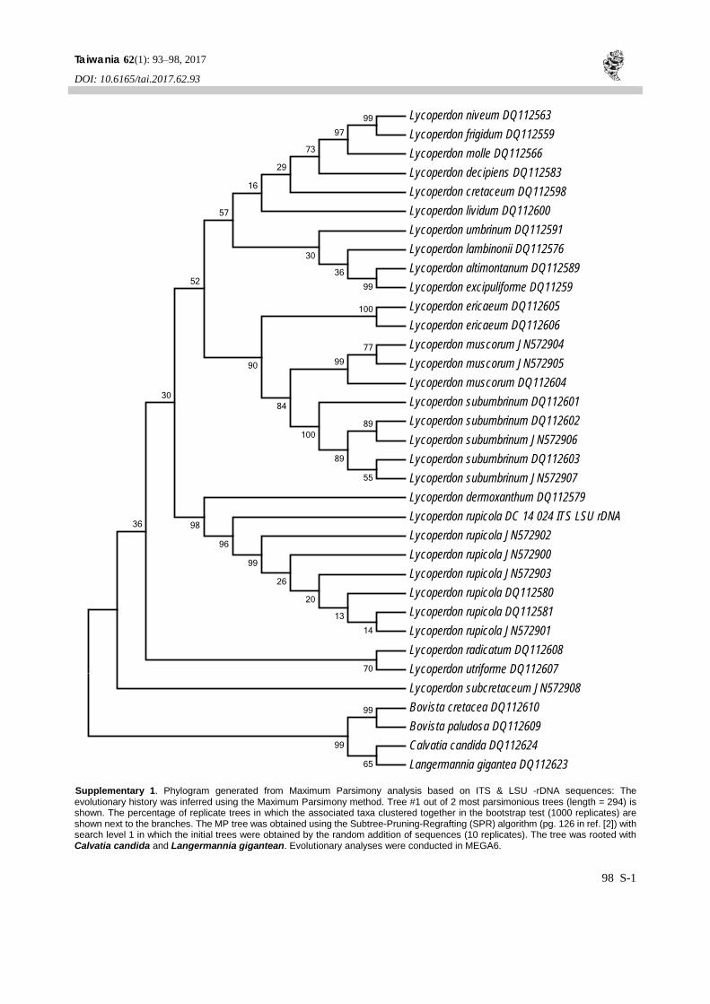
Supplementary 1. Phylogram generated from Maximum Parsimony analysis based on ITS & LSU -rDNA sequences: The evolutionary history was inferred using the Maximum Parsimony method. Tree #1 out of 2 most parsimonious trees (length = 294) is shown. The percentage of replicate trees in which the associated taxa clustered together in the bootstrap test (1000 replicates) are shown next to the branches. The MP tree was obtained using the Subtree-Pruning-Regrafting (SPR) algorithm (pg. 126 in ref. [2]) with search level 1 in which the initial trees were obtained by the random addition of sequences (10 replicates). The tree was rooted with Calvatia candida and Langermannia gigantean. Evolutionary analyses were conducted in MEGA6.
Lycoperdon niveum DQ112563 Lycoperdon frigidum DQ112559 Lycoperdon molle DQ112566 Lycoperdon decipiens DQ112583 Lycoperdon cretaceum DQ112598 Lycoperdon lividum DQ112600 Lycoperdon umbrinum DQ112591 Lycoperdon lambinonii DQ112576 Lycoperdon altimontanum DQ112589 Lycoperdon excipuliforme DQ11259 Lycoperdon ericaeum DQ112605 Lycoperdon ericaeum DQ112606 Lycoperdon muscorum JN572904 Lycoperdon muscorum JN572905 Lycoperdon muscorum DQ112604 Lycoperdon subumbrinum DQ112601 Lycoperdon subumbrinum DQ112602 Lycoperdon subumbrinum JN572906 Lycoperdon subumbrinum DQ112603 Lycoperdon subumbrinum JN572907 Lycoperdon dermoxanthum DQ112579 Lycoperdon rupicola DC 14 024 ITS LSU rDNA Lycoperdon rupicola JN572902 Lycoperdon rupicola JN572900 Lycoperdon rupicola JN572903 Lycoperdon rupicola DQ112580 Lycoperdon rupicola DQ112581 Lycoperdon rupicola JN572901 Lycoperdon radicatum DQ112608 Lycoperdon utriforme DQ112607 Lycoperdon subcretaceum JN572908 Bovista cretacea DQ112610 Bovista paludosa DQ112609 Calvatia candida DQ112624 Langermannia gigantea DQ112623
99
65
99
70
89
99
97
73
29
16
100
36
30
57
77
99
14
13
20
26
99
96
98
99
36
30
52
90
84
100
55
89
Taiwania Vol. 62, No. 1
98 S-2
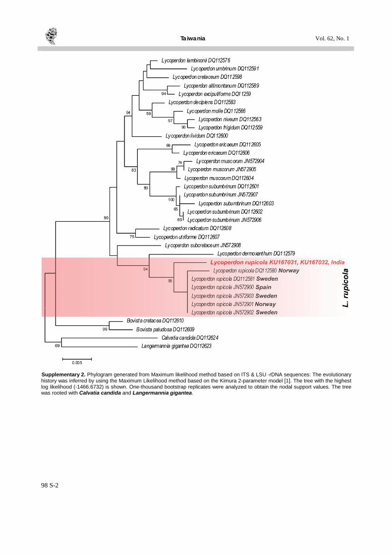
Supplementary 2. Phylogram generated from Maximum likelihood method based on ITS & LSU -rDNA sequences: The evolutionary history was inferred by using the Maximum Likelihood method based on the Kimura 2-parameter model [1]. The tree with the highest log likelihood (-1466.6732) is shown. One-thousand bootstrap replicates were analyzed to obtain the nodal support values. The tree was rooted with Calvatia candida and Langermannia gigantea.