morphogenesis in the yeast cell cycle: regulation by cdc28 and
TRANSCRIPT
Morphogenesis in the Yeast Cell Cycle: Regulation by Cdc28 and Cyclins Daniel J. Lew and Steven I. Reed Department of Molecular Biology, Scripps Research Institute, La Jolla, California 92037
Abstract. Analysis of cell cycle regulation in the bud- ding yeast Saccharomyces cerevisiae has shown that a central regulatory protein kinase, Cdc28, undergoes changes in activity through the cell cycle by associat- ing with distinct groups of cyclins that accumulate at different times. The various cyclin/Cdc28 complexes control different aspects of cell cycle progression, in- cluding the commitment step known as START and mitosis. We found that altering the activity of Cdc28 had profound effects on morphogenesis during the yeast cell cycle. Our results suggest that activation of Cdc28 by G1 cyclins (Clnl, Cln2, or Cln3) in unbudded G1 cells triggers polarization of the cortical actin cytoskel-
eton to a specialized pre-bud site at one end of the cell, while activation of Cdc28 by mitotic cyclins (Clbl or Clb2) in budded G2 cells causes depolariza- tion of the cortical actin cytoskeleton and secretory apparatus. Inactivation of Cdc28 following cyclin de- struction in mitosis triggers redistribution of cortical actin structures to the neck region for cytokinesis. In the case of pre-bud site assembly following START, we found that the actin rearrangement could be trig- gered by Cln/Cdc28 activation in the absence of de novo protein synthesis, suggesting that the kinase may directly phosphorylate substrates (such as actin-binding proteins) that regulate actin distribution in cells.
T HE generation of specialized and often elaborate cell shapes is crucial to the function of most cell types. Development of many of these shapes, be they neu-
ronal processes or yeast buds, involves polarization of secre- tion towards a defined subdomain of the plasma membrane. In yeast cells, which are surrounded by a cell wall, the force that drives growth is generated by an osmotic gradient across the plasma membrane (Harold, 1990). This force, or turgor pressure, is isotropic, but growth is channeled to specific sites by vectorial delivery of wall modifying enzymes and new cell wall constituents to these sites (Harold, 1990). To understand the generation of cell shape, we must find out how this delivery is targeted and how the sites of cell growth are chosen. The actin cytoskeleton has been implicated in these processes in the yeast Saccharomyces cerevisiae. Cor- tical structures containing actin filaments cluster at sites of cell growth during the cell cycle (Adams and Pringle, 1984; Kilmartin and Adams, 1984). Furthermore, mutations in the single actin gene (ACT/) (Novick and Botstein, 1985) or in genes encoding actin binding proteins (MY02, unconven- tional myosin [Johnston et al., 1991]; TPM/, tropomyosin [Liu and Bretscher, 1992]; SAC6, fimbrin [Adams et al., 1991]; CAP2, capping protein [Amatruda et al., 1990]; PFY/, profilin [Haarer et al., 1990]) disrupt vectorial secre- tion and lead to more uniform expansion. These observa- tions suggest that the actin cytoskeleton may play a role in specifying target sites on the plasma membrane and deliver-
Address correspondence to D. Lew at Department of Molecular Biology, MB7, Scripps Research Institute, 10666 North Torrey Pines Road, La Jolla, CA 92037.
ing secretory vesicles to these sites, but they do not address the question of how the growth sites are chosen during the cell cycle so as to produce a bud with the same shape as the mother cell.
Growth of S. cerevisiae cells by budding involves a number of dramatic rearrangements of the cytoskeleton and secre- tory apparatus of the cell during the cell cycle (see Fig. 1 A) (Chant and Pringle, 1991). First, a small region of the sur- face of an unbudded cell in G1 assembles into a "pre-bud" site. This involves a polarization of the actin cytoskeleton such that cortical actin-rich patches congregate in a ring at the prebud site (Kilmartin and Adams, 1984). Near the G1/S transition, secretion becomes polarized to the region demar- cated by the ring of cortical actin patches and the bud emerges. The cortical actin patches then redistribute into the bud. During bud growth in S phase and G2, secretion is directed into the bud and the cortical actin patches stay ex- clusively in the bud (Tkacz and Lampen, 1972; Farkas et al., 1974; Adams and Pringle, 1984; Kilmartin and Adams, 1984). The shape of the bud (and hence of the daughter cell) is determined by the balance between apical growth (directed towards the tip of the bud) and isotropic bud growth during this period. Apical growth predominates during the early budded phase, whereas isotropic bud growth predominates in the late budded phase (Farkas et al., 1974). During mito- sis, cortical actin patches transiently redistribute throughout the surface of both mother and bud portions of the cell, and then congregate on both sides of the neck region at the time of cytokinesis (Kilmartin and Adams, 1984). Coincident with this, secretion is redirected towards the neck region in order to form a specialized chitin-rich septum (Tkacz and
�9 The Rockefeller University Press, 0021-9525/93/03/1305/16 $2.00 The Journal of Cell Biology, Volume 120, Number 6, March 1993 1305-1320 1305
on March 16, 2018
jcb.rupress.orgD
ownloaded from

Lampen, 1972; Cabib et al., 1982). Cell separation ensues, and the newborn G1 cells retain a "post-cytokinesis" cluster- ing of cortical actin patches at the site of cytokinesis (Kil- martin and Adams, 1984). The patches are then randomized and/or redistributed to assemble a new pre-bud site.
How are these reorganizations triggered at the appropriate times in the cell cycle? In contrast to the extensive progress that has been made recently in elucidating the regulatory sig- nals that control the events of the nuclear cell cycle (see be- low), very little is known about how the cytoplasmic events that govern morphogenesis are controlled. Considerable variation has been observed in the relative timing of the events in the nuclear and morphogenetic cycles when com- paring different yeast strains (Brewer et al., 1984). Further- more, various mutations and drug treatments can apparently uncouple these two cycles (Pringle and Hartwell, 1981), per- haps suggesting that the detailed order of events in the cyto- plasmic and nuclear cell cycles are determined by different mechanisms. However, both cycles are dependent on com- mitment to a new round of cell division, which occurs at a regulatory point in late G1 called START (Pringle and Hart- well, 1981).
Cells in early G1, before START, can exit the mitotic cycle in order to conjugate (if exposed to mating pheromones) or to enter a metabolically quiescent"stationary phase" (if there are insufficient nutrients to support cell division) (Hartwell et al., 1974). However, once cells pass the START control point they are committed to complete the next cell cycle (Hartwell et al., 1974). START is a prerequisite for both DNA replication and bud emergence. Passage through START requires activation of a 34-kD protein kinase en- coded by the CDC28 gene (the S. cerevisiae homolog of the Schizosaccharomyces pombe cdc2 gene) (Hartwell et al., 1974; Reed, 1980; Nurse and Bissett, 1981; L6rincz and Reed, 1984; Reed et al., 1985). Monomeric Cdc28 is inac- tive (Wittenberg and Reed, 1988), but can be activated by as- sociation with positive regulatory subunits known as cyclins. Cyclins were discovered through their strikingly periodic patterns of accumulation and destruction during the cell cy- cle (Evans et al., 1983; Hunt, 1989). Subsequently, many classes of cyclins have been identified, which appear to acti- vate Cdc28 at different times in the cell cycle to regulate different processes (Xiong and Beach, 1991; Lew and Reed, 1992).
In S. cerevisiae, passage through START requires activa- tion of Cdc28 by one of three G1 cyclins, Clnl, Cln2, and Cln3 (Sudbery et al., 1980; Cross, 1988; Nash et al., 1988; Hadwiger et al., 1989; Richardson et al., 1989; Wittenberg et al., 1990). DNA replication and spindle assembly in S phase and early G2, as well as spindle elongation and chro- mosome separation in mitosis, require activation of Cdc28 by one of four B-type cyclins, Clbl, Clb2, Clb3, and Clb4 (Ghiara et al., 1991; Surana et al., 1991; Richardson et al., 1992). Cdc28 (and its homologs in other organisms) in con- junction with B-type cyclins constitutes the universal regu- lator of entry into mitosis, MPF (M-phase Promoting Fac- tor) (Arion et al., 1988; Dunphy et al., 1988; Draetta et al., 1989; Labbe et al., 1989; Meijer et al., 1989; Nurse, 1990). Clb3 and Clb4 activate Cdc28 earlier in the cell cycle than Clbl and Clb2, suggesting that they may be responsible for the DNA replication and early spindle assembly functions, while Clbl and Clb2 predominantly act to induce mitotic
events (Richardson et al., 1992). Thus, the sequential acti- vation of the Cdc28 regulator by distinct groups of cyclins, which presumably confer different functional properties on the kinase, results in the orderly progression of events in the nuclear cycle. Do these same regulators control the morpho- genetic events of the cytoplasmic cycle?
In this report we have examined the role played by Cdc28 and cyclins in the control of actin distribution and polarized secretion during the yeast cell cycle. Based on the effects of mutational inactivation or hyperactivation of different Cdc28/ cyclin complexes in cells at different stages of the cell cycle, we conclude that morphogenetic events in S. cerevisiae are indeed controlled by the same regulators that govern the nu- clear cycle. Changes in Cdc28 activity appear to trigger po- larization of cortical actin in G1, depolarization of cortical actin and secretion in G2, and redistribution of cortical actin to the neck region after mitosis. Cdc28 activation can trigger cytoskeletal reorganization in the absence of de novo protein synthesis, suggesting that the control of cell polarity occurs post-translationally. Our results also suggest that activation of Cdc28 by different cyclins can have opposing effects on polarization of both the cytoskeleton and the secretory ap- paratus, providing direct evidence for specialized roles for different cyclins.
Materials and Methods
Yeast Strains, Media and Growth Conditions
All yeast strains used in this study were derivatives of BF264-15DU: MATa are1 his2 1eu2-3,112 trpl-1 a ura3Dns (Richardson et al., 1989). The rele- vant genotypes of strains used in this study are shown in Table I. Standard genetic procedures for yeast were used to construct the strains (Sherman et al., 1982). Null cln alleles were as described by (Cross and Tinkelenberg, 1991), GALI:CLN constructs as described by (Cross and Tinkelenberg, 1991; Lew et ai., 1991), null clb alleles as described by (Richardson et al., 1992), and GALI:CLB constructs as described by C. S. Stueland, D. J. Lew, and S. I. Reed (submitted for publication).
Yeast cultures were grown at 30~ except for experiments involving tem- perature shift of ts mutants, where cells were grown at 25"C and shifted to 37~ as indicated. Cells were grown in YEP (1% yeast extract, 2% bac- topeptone, 0.005 % adenine, 0.005 % uracil) supplemented with glucose, su- crose, or galactose (all at 2 %). For induction of GAL/regulated genes, cells were grown in sucrose and either galactose was added to 2 % final concen- tration or the cells were harvested and resuspended in YEPgalactose (no differences were observed between these procedures). For repression of GAL/regulated genes, cells were grown in galactose and either glucose was added to 2 % final concentration or the cells were harvested and resuspended in YEPglucose (no differences were observed between these procedures). Temperature shifts and nutritional shifts were performed at a cell density of "~2 x 10⁶ cells/nil.
Centrifugal Elutriation and Flow Cytometry
1-2 liters of cells were grown at 25 or 30~ to a density of 107 cells/mi, then chilled to 40C and sonicated to disperse clumps. The cells were loaded into an elutriator rotor (Beckman Instruments, Inc., Fnilerton, CA) at 4,000 rpm, 4~ The pump speed was adjusted so that the smallest cells flowed through the elutriator and were collected in bottles on ice, whereas larger cells remained in the rotor. When enough ceils were collected ("~5 % of the population, collected over 10-15 min) they were concentrated by cen- trifugation, resuspended in fresh media at 2 x 10⁶ cells/ml, and warmed to the appropriate temperature. Elutriations comparing cells of different genotypes (as in the experiments of Figs. 2 and 10) were performed on the same day: both cultures were chilled at the same time and one culture stayed on ice while the other was elutriated, so that both sets of cells spent the same amount of time on ice (usually •1 h for such double elutriations).
Cells were fixed, stained with propidium iodide, and analyzed by flow
The Journal of Cell Biology, Volume 120, 1993 1306
on March 16, 2018
jcb.rupress.orgD
ownloaded from
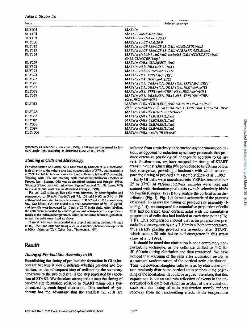
Table L S tra ins l ist
Strain Relevant genotype
DLY005 DLY104 DLY105 DLY106 DLYll2 DLY113 DLY224
DLY257 DLY372 DLY373 DLY374 DLY375 DLY376 DLY377 DLY378 DLY379
DLY384
DLY328 DLY333 DLY385 DLY338 DLY389 DLY1006
MATa/~ MATa/ct cdc28-4/cdc28-4 MATa/ot cdc28-13/cdc28-13 MATa/t~ cdc28-9/cdc28-9 MATa/c~ cdc28-13/cdc28-13 GALI:CLN2(LEU2)/Ieu2 MATa/ct cdc28-13/cdc28-13 GALl :CLB1 At 52 (LEU2)/leu2 MATa/cc clnl/clnl ctn2/cln2 cln3/cln3 GALI:CLN3(LEU2)/Ieu2 GALI :CLN3(TRP1)/trp l MATa/t~ GALI:CLN2(LEU2)/Ieu2 MATa/ct clb l : : URA3 /clb l : : URA3 MATa/c~ clb2: :LEU2/clb2: :LEU2 MATa/t~ clb3::TRP1/clb3::TRP1 MATa/u clb4::HIS2/clb4::HIS2 MATa/u clb l : : URA3 /clb l : : URA3 clb 3 : : TRP1/clb 3 : : TRP1 MATa/a ctb l : :URA3/clbt : :URA3 ctb4: :HIS2/clb4: :HIS2 MATa/ot clb3::TRPl/clb3::TRP1 clb4::HIS2/clb4::HIS2 MATa/u clb l : : URA3 / clb l : : URA3 clb 3 : : TRP1/clb 3 : : TRP1 clb 4 : : HIS2 / clb4 : : HIS2 MATa/u GALI:CLBI(LEU2)/Ieu2 clbl::URA3/clbl::URA3 clb2:: LE U2/ctb 2: :LE U2 clb 3:: TRP t/clb 3:: TRP1 clb4:: HIS2/clb4:: HIS2 MATa/c~ GALl :CLB1A152(LEU2)/Ieu2 MATa/ot GALI:CLB1 (LEU2)/leu2 MATa/ot GALI:CLB2(LEU2)/leu2 MATa/ot GALI :CLB3(LEU2)/Ieu2 MATa/ot GALI :CLB4(LEU2)/Ieu2 MATa/a GALl :weel+ (URA3)/ura3
cytometry as described (Lew et al., 1992). Cell size was measured by for- ward angle light scattering as described (Lew et al., 1992).
S t a i n i n g o f C e l l s a n d M i c r o s c o p y
For visualization of F-actin, cells were fixed by addition of 37% formalde- hyde directly to the culture to a final concentration of 3.7 %, and incubation at 25"C for 1-2 h. In some cases the fixed cells were left at 4"C overnight. Washing with PBS and staining with rhodamine-phalloidin (Molecular Probes, Inc., Eugene, OR) was as described (Adams and Pringle, 1991). Staining of fixed cells with calcofluor (Sigma Chemical Co., St. Louis, MO) to visualize bud scars was as described (Pringle, 1991).
For cell wall staining, live cells were harvested by centrifugation and resuspended in 50 mM Tris-HCl pH ZS, 150 mM NaC1 at 5-10 x 107 cdls/ml and sonicated to disperse clumps. FITC-ConA (E-Y Laboratories, Inc., San Mateo, CA) was added to a final concentration of 50-100 #g/ml, and the cells were incubated for 10 rain at 25~ in the dark. After labeling, the cells were harvested by centrifugation and resuspended in appropriate media at the indicated temperature. After the indicated return-to-growth in- terval, the cells ~ re fixed as above.
Stained cells were resuspended in a drop of mounting medium (Pringle et ai., 1991) and observed using a Zeiss Axiophot photomicroscope with a 100x objective (Carl Zeiss, Inc., Thornwood, NY).
Results
l i m i n g o f P r e - b u d S i t e A s s e m b l y i n G I
Establishing the timing ofpre-bud site formation in G1 is im- portant because it would indicate whether pre-bud site for- mation, or the subsequent step of redirecting the secretory apparatus to the pre-bud site, is the step regulated by execu- tion of START. We therefore attempted to map the timing of pre-bud site formation relative to START using cells syn- chronized by centrifugal elutriation. This method of syn- chrony has the advantage that the smallest G1 cells are
selected from a relatively unperturbed asynchronous popula- tion, as opposed to induction synchrony protocols that pro- duce extensive physiological changes in addition to G1 ar- rest. Furthermore, we have mapped the timing of START transit in our strains using this procedure to be 20 min before bud emergence, providing a landmark with which to com- pare the timing of pre-bud site assembly (Lew et al., 1992).
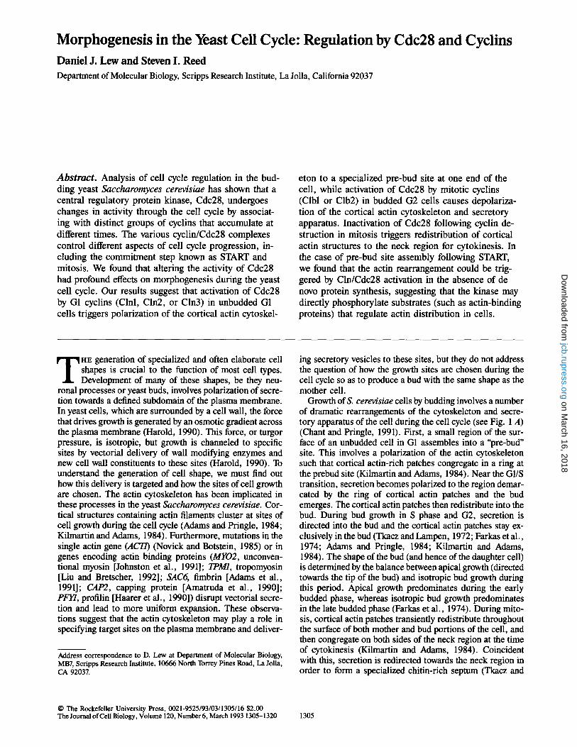
Early G1 cells were inoculated into YEPdextrose at either 25 or 37~ At various intervals, samples were fixed and stained with rhodamine-phalloidin (which selectively binds to F-actin [Cooper, 1987]) to visualize the cortical actin dis- tribution (Fig. 1). Fig. 1 A shows a schematic of the patterns observed. To assess the timing of pre-bud site assembly (b in Fig. 1 A), we compared the cumulative proportion of cells that had polarized their cortical actin with the cumulative proportion of cells that had budded at each time point (Fig. 1 B). This comparison showed that actin polarization pre- ceded bud emergence by only 7-10 min at both temperatures, thus clearly placing pre-bud site assembly after START, which occurs 20 min before bud emergence in this strain (Lew et al., 1992).
It should be noted that elutriation is not a completely non- perturbing technique, as the cells are chilled to 4~ for 30-60 min during elutriation and then rewarmed. We have noticed that warming of the cells after elutriation results in a transient randomization of the cortical actin distribution. Thus, the newborn daughter cells isolated by elutriation con- tain randomly distributed cortical actin patches at the begin- ning of the incubation. It could be argued, therefore, that this experiment is not an accurate reflection of events in the un- perturbed cell cycle but rather an artifact of the elutriation, such that the timing of actin polarization merely reflects recovery from the randomizing effects of the temperature
Lew and Reed Cell Cycle Control of Morphogenesis in Yeast 1307
on March 16, 2018
jcb.rupress.orgD
ownloaded from
A
a b c d �9 f g
B loo
2,~ 80
o lJ'
'~ 4 0
20
60 120 180 Time (rain)
100
8 0 o b-g
�9 c-g 60,
4o.
60 120 180 Time (rain)
Figure 1. Timing of pre-bud site assembly in G1. (A) Schematic of the distributions of cortical actin patches (black dots) observed dur- ing the cell cycle. Actin cables are omitted for simplicity. (B) Timecourse of actin polarization and bud emergence. Wild-type (DLY005) cells were elutriated and incubated at 25~ (left) or 37~ (right) in YEPglucose. At the indicated times, samples were fixed for analysis of the actin distribution by rhodamine-phalloidin stain- ing and fluorescence microscopy. Percent of cells that had polarized the cortical actin patches (morphologies b-g in A) or budded (mor- phologies c-g in A) were quantitated by counting 400 cells at each time point.
shift. To circumvent this problem, we attempted to deter- mine the timing of actin polarization to the pre-bud site in unperturbed cycling cells.
The major ditticulty in this analysis is that the "post- cytokinesis" polarization of cortical actin in newborn cells is morphologically very similar to the polarization of cortical actin to the pre-bud site. In diploid daughter cells, however, it is possible to distinguish these patterns because the bud al- ways forms at the opposite pole of the cell from the site of cytokinesis (Chant and Pringle, 1991). G1 daughter cells with a polarized cortical actin distribution can be scored as newborn (actin patches at the site of cytokinesis) or pre- budding (actin patches at the opposite pole from the site of cytokinesis).
Upon cell abscission, a chitin-rich bud scar is left in the cell wall of the mother cell, while a less characterized birth scar marks the site of cytokinesis in the daughter cell wall (Barton, 1950; Beran, 1968). Bud scars can be stained with calcofluor (Pringle, 1991), and we report here that birth scars stain brightly with FITC-Con A (Fig. 2). The lectin Con A binds to mannose residues, which are abundant on the "man- nan" or glycoproteins which form a major component of the yeast cell wall (Ballou, 1981). Thus, the entire surface of the cell is stained upon exposure to FITC-Con A. However, the staining is not homogeneous, and a bright patch can be observed at one pole of every cell (Fig. 2). In addition, slightly increased staining is also apparent at the bud scars. The bright FITC-Con A labeled patch marks the birth scar,
Figure 2. Birth scars are stained by FITC-ConA. Wild-type cells (DLY005) growing in YEPglucose were fixed and stained with calcofluor to visualize bud scars (A), and with FITC-ConA to stain mannan (B). Examples of birth scars (stained with FITC-ConA but not calcofluor) are indicated by arrowheads; bud scars (stained by calcofluor and faintly by FITC-ConA) by arrows. Bar, 10 #m.
as shown by the fact that daughter ceils (cells without bud scars) always budded at the opposite pole of the cell from the patch (Fig. 2 and data not shown).
Wild-type diploid cells growing exponentially in rich medium were fixed and stained with calcofluor, FITC-Con A, and rhodamine-phalloidin. G1 daughter cells (unbudded without bud scars) were assigned to one of three categories based on the actin distribution. Of these, 48% were scored as "post-cytokinesis" (cortical actin patches clustered at the same pole as the FITC-Con A labeled birth scar), 26% were random (cortical actin patches distributed with no clear polarity), and 26% were prebudding (cortical actin patches clustered at the opposite pole from the birth scar). Assuming that all daughter cells were born with a post-cytokinesis actin distribution which was then randomized and reassembled into a pre-bud site, these data indicate that pre-bud site as- sembly occurred about three quarters of the way through G1 in an unperturbed rapidly proliferating population. This is fully consistent with the analysis of elutriated cells presented above, strongly suggesting that actin polarization to the pre- bud site occurs in late G1 after START.
Pre-bud Site Assembly Requires Activation o f the Cln/Cdc28 Kinase Because assembly of the pre-bud site followed START, it seemed plausible that activation of Cdc28 by G1 cyclins (Clns) could be the trigger for actin polarization. To test this hypothesis, homozygous cdc28-13 diploid cells were elutri- ated to obtain G1 daughter cells, which were inoculated into rich medium at 37~ (the restrictive temperature for this mu- tan0. These cells grew in size at the same rate as wild-type cells at 37~ (assayed by forward angle light scattering in a flow cytometer: data not shown), but did not pass START or
The Journal o f Cell Biology, Volume 120, 1993 1308
on March 16, 2018
jcb.rupress.orgD
ownloaded from
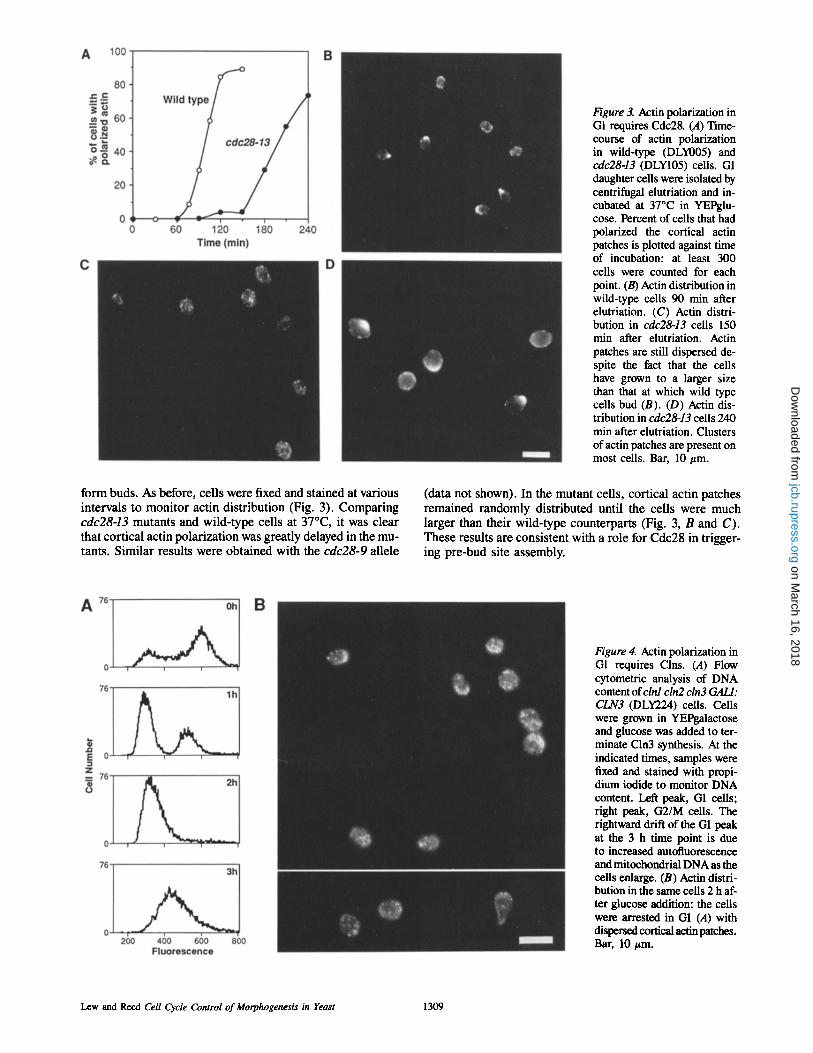
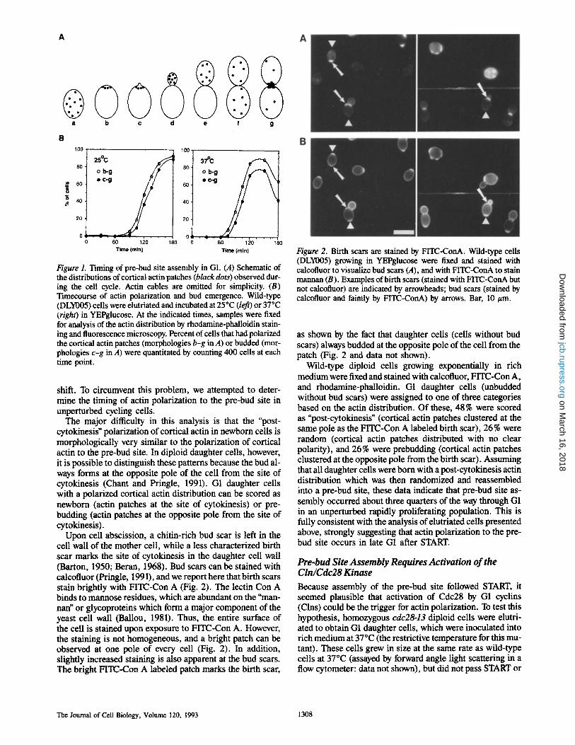
Figure 3. Actin polarization in G1 requires Cdc28. (A) Time- course of actin polarization in wild-type (DLY005) and cdc28-13 (DLY105) cells. G1 daughter cells were isolated by centrifugal elutriation and in- cubated at 37~ in YEPglu- cose. Percent of cells that had polarized the cortical actin patches is plotted against time of incubation: at least 300 cells were counted for each point. (B) Actin distribution in wild-type cells 90 min after elutriation. (C) Actin distri- bution in cdc28-13 cells 150 min after elutriation. Actin patches are still dispersed de- spite the fact that the cells have grown to a larger size than that at which wild type cells bud (B). (D) Actin dis- tribution in cdc28-13 cells 240 min after elutriation. Clusters of actin patches are present on most cells. Bar, 10 #m.
form buds. As before, cells were fixed and stained at various intervals to moni tor actin distribution (Fig. 3). Comparing cdc28-13 mutants and wild-type cells at 37~ it was clear that cortical actin polarization was greatly delayed in the mu- tants. Similar results were obtained with the cdc28-9 allele
(data not shown). In the mutant cells, cortical actin patches remained randomly distributed until the cells were much larger than their wild-type counterparts (Fig. 3, B and C). These results are consistent with a role for Cdc28 in trigger- ing pre-bud site assembly.
Figure 4. Actin polarization in G1 requires Clns. (A) Flow cytometric analysis of DNA content of clnl cln2 cln3 GALl: CLN3 (DLY224) cells. Cells were grown in YEPgalactose and glucose was added to ter- minate Cln3 synthesis. At the indicated times, samples were fixed and stained with propi- dium iodide to monitor DNA content. Left peak, G1 cells; right peak, G2/M cells. The rightward drift of the G1 peak at the 3 h time point is due to increased autofluorescence and mitochondrial DNA as the cells enlarge. (B) Actin distri- bution in the same cells 2 h af- ter glucose addition: the cells were arrested in G1 (A) with dispersed cortical actin patches. Bar, 10 #m.
Lew and Reed Cell Cycle Control of Morphogenesis in Yeast 1309
on March 16, 2018
jcb.rupress.orgD
ownloaded from
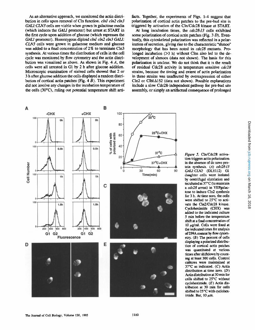
As an alternative approach, we monitored the actin distri- bution in cells upon removal of Cln function, clnl cln2 cln3 GALI:CLN3 cells are viable when grown in galactose media (which induces the GAL/promoter) but arrest at START in the first cycle upon addition of glucose (which represses the GAH promoter). Homozygous diploid clnl cln2 cln3 GALl: CLN3 ceils were grown in galactose medium and glucose was added to a final concentration of 2 % to terminate Cln3 synthesis. At various times the distribution of cells in the cell cycle was monitored by flow cytometry and the actin distri- bution was visualized as above. As shown in Fig. 4 A, the cells were all arrested in G1 by 2 h after glucose addition. Microscopic examination of stained cells showed that 2 or 3 h after glucose addition the cells displayed a random distri- bution of cortical actin patches (Fig. 4 B). This experiment did not involve any changes in the incubation temperature of the cells (30~ ruling out potential temperature shift arti-
facts. Together, the experiments of Figs. 1-4 suggest that polarization of cortical actin patches to the pre-bud site is triggered by activation of the Cln/Cdc28 kinase at START.
At long incubation times, the cdc28-13 cells exhibited some polarization of cortical actin patches (Fig. 3 D). Even- tually, this cytoskeletal polarization was reflected in a polar- ization of secretion, giving rise to the characteristic "shmoo" morphology that has been noted in cdc28 mutants. Pro- longed incubation (>3 h) without Clns also led to the de- velopment of shmoos (data not shown). The basis for this polarization is unclear. We do not think that it is the result of residual Cdc28 activity in temperature sensitive cdc28 strains, because the timing and extent of actin polarization in these strains was unaffected by overexpression of either Cln2 or ClblA152 (data not shown). Possible explanations include a slow Cdc28-independent pathway for pre-bud site assembly, or simply an artifactual consequence of prolonged
Figure 5. Cln/Cdc28 activa- tion triggers actin polarization in the absence of de novo pro- tein synthesis. (A) cdc28-13 GALI:CLN2 (DLYII2) G1 daughter ceils were isolated by centrifugal elutriation and incubated at 37~ (to maintain a cdc28 arrest) in YEPgalac- tose to induce Cln2 synthesis for 3 h. At time zero, the cells were shifted to 25~ to acti- vate the Cln2/Cdc28 kinase. Cycloheximide (CHX) was added to the indicated culture 5 rain before the temperature shift at a final concentration of 10/zg/ml. Cells were fixed at the indicated times for analysis of DNA content by flow cytom- etry. (B) The percent of cells displaying a poladr~ distribu- tion of cortical actin patches was quantitated at various times after shiftdown by count- ing at least 300 cells. Control cultures were maintained at 37~ as indicated. (C) Actin distribution at time zero. (D) Actin distribution at 30 rain for cells shifted to 25~ without cycloheximide. (E) Actin dis- tribution at 30 min for cells shifted to 25"C with cyclohex- imide. Bar, 10 pm.
The Journal of Cell Biology, Volume 120, 1993 1310
on March 16, 2018
jcb.rupress.orgD
ownloaded from

arrest without Cdc28 function that is unrelated to normal pre-bud site assembly. Another possibility is that this polar- ization is somehow related to the shmooing observed upon exposure of haploid cells to appropriate mating pheromones. However, we consider this unlikely because diploid MATa/ot cells (used in this study) do not express the genes encoding and Cln2; Cln3 is present in very low abundance and its peri-
Cln/Cdc28 Activation Triggers Actin Polarization in the Absence of De Novo Protein Synthesis
Regulation of actin polarization by the Cln/Cdc28 kinase could occur in various ways, from the most direct route (per- haps involving direct phosphorylation of actin binding pro- teins) to a very indirect route (perhaps involving induction of new transcripts whose products somehow affect the actin distribution). As a first step towards characterizing this regulatory pathway, we tested whether actin polarization could be triggered by kinase activation in the absence of de novo protein synthesis. To do this, we again employed the cdc28-13 allele, which is easily reversible upon return to 25~ after arrest at 37~ cdc28-13 cells arrested at 37~ ac- cumulate very low levels of Clnl and Cln2, but upon shift- down to 25~ there is a massive accumulation of these Clns through a positive feedback mechanism whereby CLN1 and CLN2 transcription is stimulated by the Cln/Cdc28 kinase (Cross and Tinkelenberg, 1991; Dirick and Nasmyth, 1991). This burst of Cln accumulation leads to a rapid activation of high levels of Cln/Cdc28 activity, and hence passage through START. We reasoned that if cdc28-13 cells were arrested and then shifted down to 25~ in the presence of cycloheximide, the positive feedback loop would be interrupted (because Cln synthesis would be blocked) and only very low levels of Cln/Cdc28 kinase would be activated. To circumvent this problem, we used a strain containing an integrated GALl: CLN2 construct. This allowed the cells to induce the GAL/ promoter and accumulate Cln2 during the cdc28 arrest at 37~ so that high levels of Cln/Cdc28 kinase activity could be attained upon shift-down to 25~ even in the presence of cycloheximide.
cdc28-13 GALI:CLN2 cells in early G1 were isolated by centrifugal elutriation and incubated for 3 h in galactose based medium at 37~ to allow time for Cln2 accumulation. At this time, the cells were still uniformly arrested in G1 (Fig. 5 A) and 93 % of the cells displayed a random distribu- tion of cortical actin patches (Fig. 5 C). Upon shift-down to 25~ the cells not treated with cycloheximide rapidly polar- ized their actin, budded, and replicated their DNA (Fig. 5, A, B, and D). In the presence of 10 #g/ml cycloheximide (a concentration sufficient to block 96-97 % of cellular protein synthesis [Marini and Reed, 1992]), the cells did not bud or replicate their DNA upon shift-down, but •50% of the cells polarized their actin after a short (15 min) lag (Fig. 5, B and E). By focusing up and down, it could be seen that the corti- cal actin patches formed a ring, as observed at a normal pre- bud site. Thus, activation of Cln/Cdc28 induced actin polar- ization in a rapid, post-translational manner.
The basis for the 15 min lag in the cycloheximide-treated cells is unclear, as is the reason why only half of these cells polarized their cortical actin patches. Possibly there is a par- aUel pathway for actin polarization in response to Cln/Cdc28 activation that does involve protein synthesis. Alternatively,
the pleiotropic effects of cycloheximide treatment may in- directly affect the actin distribution (this is supported by the observation that the cells kept at 37~ in the presence of cy- cloheximide all randomized their cortical actin patches, un- like the controls that gradually increased actin polarization: Fig. 5).
Activation of Cln/Cdc28 in G2 Results in Hyperpolarization of Cortical Actin and Secretion
Normally, Cln accumulation is periodic in the cell cycle, peaking in late G1 and decaying in S phase (at least for Clnl and Clrd; Cln3 is present in very low abundance and its peri- odicity is unclear) (Wittenberg et al., 1990; Tyers et al., 1992). We used a wild-type strain containing an integrated GALI:CLN2 construct to test whether deregulated expres- sion of Cln2 would have any effects on the actin distribution of cells in G2. Wild-type cells in G2 contain cortical actin patches spread over the entire bud surface (Fig. 1 A, Fig. 6, top). As shown in Fig. 6, budded cells in an asynchronous culture induced to express Cln2 by addition of galactose dis- played a remarkable hyperpolarization of the cortical actin to the bud tip. Furthermore, the buds became very elon- gated, suggesting that secretion had also become hyperpolar- ized towards the tip of the bud in these cells (see below). The degree of hyperpolarization was dependent on the level of Cln2 expression. Similar effects were observed with deregulated Clnl expression, but the effects of deregulated Cln3 expression were much less severe (data not shown).
Figure 6. Ectopic Cln2 expression in budded ceUs causes hyper- polarization of the cortical actin patches. Wild-type (DLY005) or GALl:CLN2 (DLY257) cells were grown in YEPsucmse and galac- tose was added for 2 h. (Left) Rhodamine-phalloidin staining. (Right) Phase contrast of same cells. Bar, 10 #m.
Lew and Reed Cell Cycle Control of Morphogenesis in Yeast 1311
on March 16, 2018
jcb.rupress.orgD
ownloaded from
Thus, activation of the Cln/Cdc28 kinase apparently results in polarization of cortical actin and secretion in G2 as well as G1.
Activation of the Clb/Cdc28 Kinase is Required for Depolarization of Cortical Actin in G2
Once the bud has formed, the cortical actin patches are redis- tributed into the bud. As the bud grows, the cortical actin patches spread out over the bud surface, so that the patch-to- patch distance increases. In mitosis, the cortical actin patches redistribute randomly throughout both the mother and bud, and then cluster on either side of the neck region (Adams and Pringle, 1984; Kilmartin and Adams, 1984).
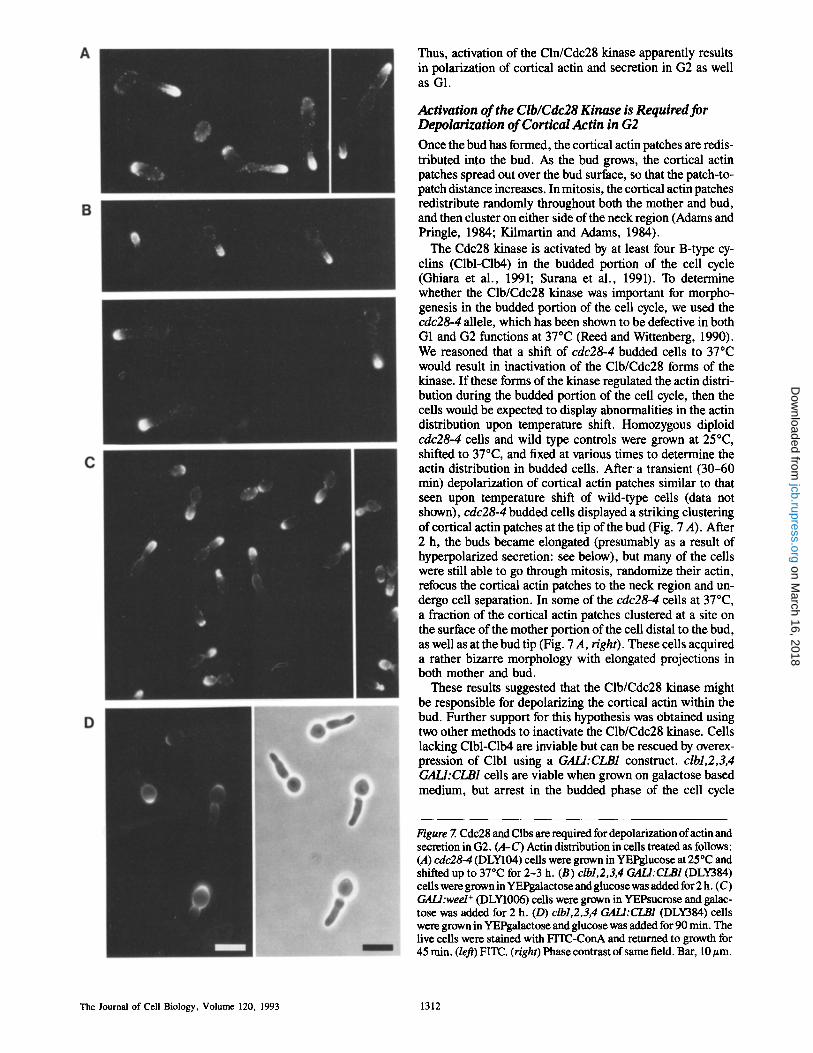
The Cdc28 kinase is activated by at least four B-type cy- clins (Clbl-Clb4) in the budded portion of the cell cycle (Ghiara et al., 1991; Surana et al., 1991). To determine whether the Clb/Cdc28 kinase was important for morpho- genesis in the budded portion of the cell cycle, we used the cdc28o4 allele, which has been shown to be defective in both G1 and G2 functions at 37~ (Reed and Wittenberg, 1990). We reasoned that a shift of cdc28-4 budded cells to 37~ would result in inactivation of the Clb/Cdc28 forms of the kinase. If these forms of the kinase regulated the actin distri- bution during the budded portion of the cell cycle, then the cells would be expected to display abnormalities in the actin distribution upon temperature shift. Homozygous diploid cdc28-4 cells and wild type controls were grown at 25~ shifted to 37~ and fixed at various times to determine the actin distribution in budded cells. After a transient (30-60 min) depolarization of cortical actin patches similar to that seen upon temperature shift of wild-type ceils (data not shown), cdc28-4 budded cells displayed a striking clustering of cortical actin patches at the tip of the bud (Fig. 7 A). After 2 h, the buds became elongated (presumably as a result of hyperpolarized secretion: see below), but many of the cells were still able to go through mitosis, randomize their actin, refocus the cortical actin patches to the neck region and un- dergo cell separation. In some of the cdc28-4 cells at 37~ a fraction of the cortical actin patches clustered at a site on the surface of the mother portion of the cell distal to the bud, as well as at the bud tip (Fig. 7 A, right). These cells acquired a rather bizarre morphology with elongated projections in both mother and bud.
These results suggested that the Clb/Cdc28 kinase might be responsible for depolarizing the cortical actin within the bud. Further support for this hypothesis was obtained using two other methods to inactivate the Clb/Cdc28 kinase. Cells lacking Clbl-Clb4 are inviable but can be rescued by overex- pression of Clbl using a GALI:CLB1 construct, clbl,2,3,4 GALI:CLB1 cells are viable when grown on galactose based medium, but arrest in the budded phase of the cell cycle
Figure 7. Cdc28 and Clbs are required for depolarization ofactin and secretion in G2. (A-C) Actin distribution in cells treated as follows: (A) cdc28-4 (DLY104) cells were grown in YEPglucose at 25~ and shifted up to 37~ for 2-3 h. (B) clbl,2,3,4 GALI:CLB1 (DLY384) cells were grown in YEPgalactose and glucose was added for 2 h. (C) GALl:wed + (DLY1006) cells were grown in YEPsucrose and galac- tose was added for 2 h. (D) clbl,2,3,4 GALI:CLB1 (DLY384) cells were grown in YEPgalactose and glucose was added for 90 rain. The live cells were stained with FITC-ConA and returned to growth for 45 rain. (left) FITC. (right) Phase contrast of same field. Bar, 10/~m.
The Journal of Cell Biology, Volume 120, 1993 1312
on March 16, 2018
jcb.rupress.orgD
ownloaded from
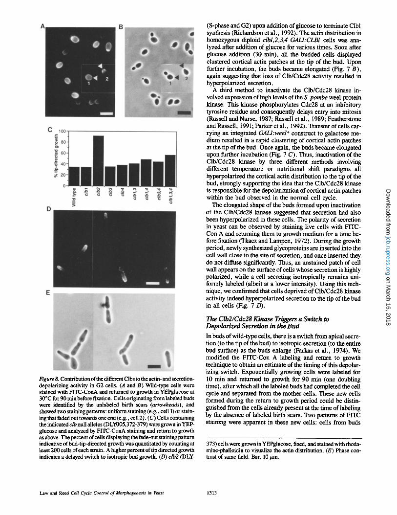
Figure 8. Contribution of the different Clbs to the actin- and secretion- depolarizing activity in G2 cells. (A and B) Wild-type cells were stained with FITC-ConA and returned to growth in YEPglucose at 30~ for 90 min before fixation. Cells originating from labeled buds were identified by the unlabeled birth scars (arrowheads), and showed two staining patterns: uniform staining (e.g., cell 1) or stain- ing that faded out towards one end (e.g., cell 2). (C) Cells containing the indicated clb null alleles (DLY005,372-379) were grown in YEP- glucose and analyzed by FITC-ConA staining and return to growth as above. The percent of cells displaying the fade-out staining pattern indicative of bud-tip-directed growth was quantitated by counting at least 200 cells of each strain. A higher percent of tip directed growth indicates a delayed switch to isotropic bud growth. (D) clb2 (DLY-
(S-phase and G2) upon addition of glucose to terminate Clbl synthesis (Richardson et al., 1992). The actin distribution in homozygous diploid clbl,2,3,4 GALI:CLB1 cells was ana- lyzed after addition of glucose for various times. Soon after glucose addition (30 min), all the budded cells displayed clustered cortical actin patches at the tip of the bud. Upon further incubation, the buds became elongated (Fig. 7 B), again suggesting that loss of Clb/Cdc28 activity resulted in hyperpolarized secretion.
A third method to inactivate the Clb/Cdc28 kinase in- volved expression of high levels of the S. pombe weel protein kinase. This kinase phosphorylates Cdc28 at an inhibitory tyrosine residue and consequently delays entry into mitosis (Russell and Nurse, 1987; Russell et al., 1989; Featherstone and Russell, 1991; Parker et al., 1992). Transfer of cells car- rying an integrated GALl:wee1 + construct to galactose me- dium resulted in a rapid clustering of cortical actin patches at the tip of the bud. Once again, the buds became elongated upon further incubation (Fig. 7 C). Thus, inactivation of the Clb/Cdc28 kinase by three different methods involving different temperature or nutritional shift paradigms all hyperpolarized the cortical actin distribution to the tip of the bud, strongly supporting the idea that the Clb/Cdc28 kinase is responsible for the depolarization of cortical actin patches within the bud observed in the normal cell cycle.
The elongated shape of the buds formed upon inactivation of the Clb/Cdc28 kinase suggested that secretion had also been hyperpolarized in these ceils. The polarity of secretion in yeast can be observed by staining live cells with FITC- Con A and returning them to growth medium for a time be- fore fixation (Tkacz and Lampen, 1972). During the growth period, newly synthesized glycoproteins are inserted into the cell wall close to the site of secretion, and once inserted they do not diffuse significantly. Thus, an unstained patch of cell wall appears on the surface of cells whose secretion is highly polarized, while a cell secreting isotropically remains uni- formly labeled (albeit at a lower intensity). Using this tech- nique, we confirmed that cells deprived of Clb/Cdc28 kinase activity indeed hyperpolarized secretion to the tip of the bud in all ceils (Fig. 7 D).
The Clb2/Cdc28 Kinase Triggers a Switch to Depolarized Secretion in the Bud
In buds of wild-type cells, there is a switch from apical secre- tion (to the tip of the bud) to isotropic secretion (to the entire bud surface) as the buds enlarge (Farkas et al., 1974). We modified the FITC-Con A labeling and return to growth technique to obtain an estimate of the timing of this depolar- izing switch. Exponentially growing cells were labeled for 10 min and returned to growth for 90 min (one doubling time), after which all the labeled buds had completed the cell cycle and separated from the mother cells. These new cells formed during the return to growth period could be distin- guished from the cells already present at the time of labeling by the absence of labeled birth scars. Two patterns of FITC staining were apparent in these new cells: cells from buds
373) cells were grown in YEPglucose, fixed, and stained with rhoda- mine-phalloidin to visualize the actin distribution. (E) Phase con- trast of same field. Bar, 10 ttm.
Lew and Reed Cell Cycle Control of Morphogenesis in Yeast 1313
on March 16, 2018
jcb.rupress.orgD
ownloaded from
that had undergone apical growth displayed intense labeling at the pole adjacent to the unlabeled birth scar which dimin- ished towards the opposite pole, while cells from buds that grew isotropically were uniformly labeled over their surface except for the birth scar (Fig. 8, A and B). Depending on the growth medium 35-50% of the new cells from a wild-type population exhibited apical growth, suggesting that the switch to isotropic secretion occurred one third to one half of the way through the budded phase (corresponding to early G2 in this strain).
The hyperpolarized secretion observed upon inactivation of the Clb/Cdc28 forms of the kinase (Fig. 7) suggested that one or more of these were responsible for triggering the depolarizing switch in wild type cells. To determine which Clbs were involved in this switch, we repeated the FITC- Con A labeling and return to growth experiment with cells carrying individual null mutations in the four CLB genes, as well as the viable multiple clb null combinations (Fig. 8 C). It was clear from this experiment that Clb2 was the major contributor to the actin- and secretion-depolarizing activity of Clb/Cdc28 complexes, clb2 cells retained an apical secre- tion pattern through most of the budded interval, resulting in elongated buds and hence elongated cells. In addition, the actin distribution in these cells was hyperpolarized, with most ceils displaying cortical actin patches clustered at the tip of the bud (Fig. 8, D and E).
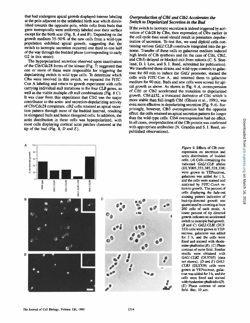
Overproduction of Clbl and Clb2 Accelerates the Switch to Depolarized Secretion in the Bud
If the switch to isotropic secretion is indeed triggered by acti- vation of Cdc28 by Clbs, then expression of Clbs earlier in the cell cycle than usual should result in premature depolar- ization of secretion. To test this, we used diploid cells con- taining various GALI:CLB constructs integrated into the ge- nome. Transfer of these cells to galactose medium induced high levels of Clb synthesis and (in the case of Clbl, Clb2, and Clb3) delayed or blocked exit from mitosis (C. S. Stue- land, D. J. Lew, and S. I. Reed, submitted for publication). We transferred these strains and wild-type controls to galac- tose for 60 min to induce the GAL/promoter, stained the cells with FITC-Con A, and returned them to galactose medium for 90 min. Buds and new cells were scored for api- cal growth as above. As shown in Fig. 9 A, overexpression of Clbl or Clb2 accelerated the transition to depolarized growth. ClblA152, a truncated derivative of Clbl which is more stable than full-length Clbl (Ghiara et al., 1991), was even more effective in depolarizing secretion (Fig. 9 A). Sur- prisingly, however, Clb3 overexpression had the opposite effect: the ceils retained an apical secretion pattern for longer than the wild-type cells. Clb4 overexpression had no effect. In all cases, overproduction of the Clb protein was confirmed with appropriate antibodies (N. Grandin and S. I. Reed, un- published observations).
Figure 9. Effects of Clb over- expression on secretion and actin distribution of budded cells. (A) Cells containing the indicated GALI:CLB alleles (DLY005,333,385,328,338) were grown in YEPsucrose, galactose was added for 1 h, and the cells were stained and analyzed by FITC-ConA re- turn to growth. The percent of cells displaying the fade-out staining pattern indicative of bud-tip-directed growth was quantitated by counting at least 200 cells of each strain. A lower percent of tip directed growth indicates an accelerated switch to isotropic bud growth. (B and C) GALI:CLB1 (DLY- 333) cells were grown in YEP- sucrose, galactose was added for 3 h, and the ceils were fixed and stained with rhoda- mine-phalloidin (B). (C) Phase contrast of same field. Similar results were obtained with GALI:CLB2 (DLY385) (data not shown). (D and E) GALl: CLB3 (DLY338) cells were grown in YEPsucrose, galac- tose was added for 3 h, and the cells were fixed and stained with rhodamine-phaUoidin (D). (E) Phase contrast of same field. Bar, 10/zm.
The Journal of Cell Biology, Volume 120, 1993 1314
on March 16, 2018
jcb.rupress.orgD
ownloaded from
The actin distribution in these ceils was monitored 3 h af- ter transfer of the cells to galactose to induce Clb expression (Fig. 9, B and C). A high proportion of the cells overproduc- ing Clbl or Clb2 displayed cortical actin patches spread evenly over the entire bud surface, and some cells also had patches in the mother portion of the cell. Cells with cortical actin patches at the tip of the bud were virtually absent from these populations. The buds formed by these cells were spherical (Fig. 9 C), rather than the normal oval shape for diploids (inset), consistent with a premature depolarization of secretion. Cells overexpressing ClblA152 had a similar morphology, and almost all the budded cells had cortical ac- tin patches delocalized over the entire cell surface (mother and bud: see Fig. 11 A). These cells were arrested in mitosis. Cells overexpressing Clb3 were also arrested in mitosis, but many of these cells had elongated buds and cortical actin patches clustered at the tip of the bud (Fig. 9, D an E). Clb4 overexpression had no effect (data not shown).
These results suggest that depolarization of the actin cytoskeleton and secretory apparatus in G2 is triggered by activation of the Clbl/Cdc28 and Clb2/Cdc28 kinase. The strikingly different results obtained with Clb3 suggest that the Clb3/Cdc28 kinase may have different targets than the Clbl/Cdc28 and Clb2/Cdc28 forms of the kinase.
Overexpression of ClblAl52 in G1 Accelerates Cell Cycle Progression but Prevents Actin Polarization and Budding
Previous experiments showed that ectopic expression of Clns in G2 resulted in hyperpolarization of the actin cytoskeleton and secretion (Fig. 6), counteracting the depolarizing effects of Clbl and Clb2. We tested whether ectopic Clb expression in G1 would have any effect on the cytoskeleton or secretion.
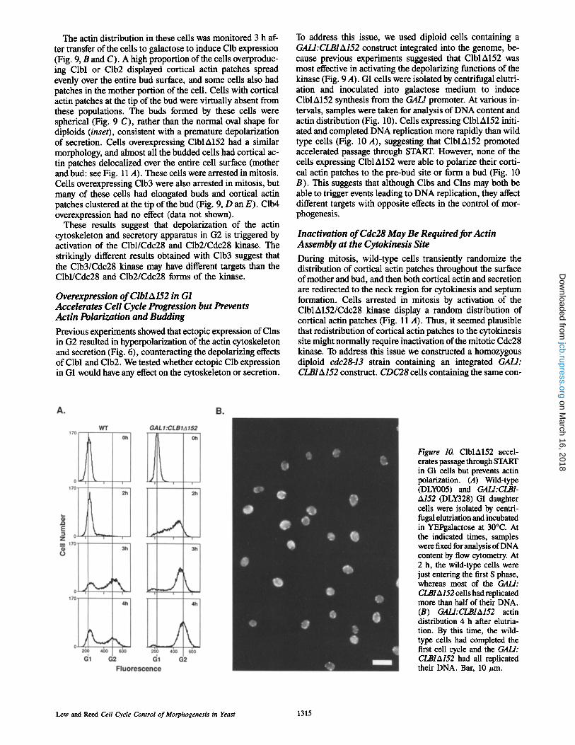
To address this issue, we used diploid cells containing a GALI:CLBIA152 construct integrated into the genome, be- cause previous experiments suggested that ClblA152 was most effective in activating the depolarizing functions of the kinase (Fig. 9 A). G1 cells were isolated by centrifugal elutri- ation and inoculated into galactose medium to induce ClblA152 synthesis from the GAL/promoter. At various in- tervals, samples were taken for analysis of DNA content and actin distribution (Fig. 10). Cells expressing ClblA152 initi- ated and completed DNA replication more rapidly than wild type ceils (Fig. 10 A), suggesting that ClblA152 promoted accelerated passage through START. However, none of the cells expressing ClblA152 were able to polarize their corti- cal actin patches to the pre-bud site or form a bud (Fig. 10 B). This suggests that although Clbs and Clns may both be able to trigger events leading to DNA replication, they affect different targets with opposite effects in the control of mor- phogenesis.
Inactivation of Cdc28 May Be Required for Actin Assembly at the Cytokinesis Site
During mitosis, wild-type cells transiently randomize the distribution of cortical actin patches throughout the surface of mother and bud, and then both cortical actin and secretion are redirected to the neck region for cytokinesis and septum formation. Cells arrested in mitosis by activation of the ClblA152/Cdc28 kinase display a random distribution of cortical actin patches (Fig. 11 A). Thus, it seemed plausible that redistribution of cortical actin patches to the cytokinesis site might normally require inactivation of the mitotic Cdc28 kinase. To address this issue we constructed a homozygous diploid cdc28-13 strain containing an integrated GALl: CLBIA152 construct. CDC28 cells containing the same con-
Figure 10. ClblA152 accel- erates passage through START in GI ceils but prevents actin polarization. (A) Wild-type (DLY005) and GALl:CLB1- A152 (DLY328) G1 daughter cells were isolated by centri- fugal elutriation and incubated in YEPgalactose at 30~ At the indicated times, samples were fixed for analysis of DNA content by flow cytometry. At 2 h, the wild-type cells were just entering the first S phase, whereas most of the GALl: CLBIA152 cells had replicated more than half of their DNA. (B) GALl:CLBIA152 actin distribution 4 h after elutria- tion. By this time, the wild- type cells had completed the first cell cycle and the GALl: CI.,BIA152 had all replicated their DNA. Bar, 10 ttm.
Lew and Reed Cell Cycle Control of Morphogenesis in Yeast 1315
on March 16, 2018
jcb.rupress.orgD
ownloaded from
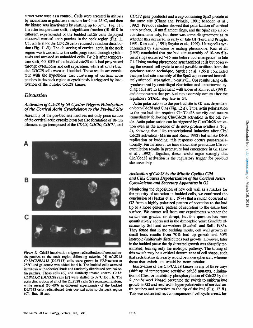
struct were used as a control. Ceils were arrested in mitosis by incubation in galactose medium for 4 h at 25~ and then the kinase was inactivated by shifting up to 37~ (Fig. 11). 1 h after temperature shift, a significant fraction (10-40% in different experiments) of the budded cdc28 cells displayed clustered cortical actin patches in the neck region (Fig. 11 C), while all of the CDC28 cells retained a random distribu- tion (Fig. 11 B). The clustering of cortical actin in the neck region was transient, as the cells progressed through cytoki- nesis and arrested as unbudded cells. By 2 h after tempera- ture shift, 60-80% of the budded cdc28 ceils had progressed through cytokinesis and cell separation, while all of the bud- ded CDC28 cells were still budded. These results are consis- tent with the hypothesis that clustering of cortical actin patches in the neck region at cytokinesis is triggered by inac- tivation of the mitotic Cdc28 kinase.
Discussion
Activation of Cdc28 by G1 Cyclins Triggers Polarization of the Cortical Actin Cytoskeleton to the Pre-bud Site
Assembly of the pre-bud site involves not only polarization of the cortical actin cytoskeleton but also formation of 10-nm filament rings (composed of the CDC3, CDCIO, CDC11, and
Figure 11. Cdc28 inactivation triggers redistribution of cortical ac- tin patches to the neck region following mitosis. (A) cdc28-13 GALl:CLBIA152 (DLYll3) cells were grown in YEPsucrose at 25~ and galactose was added for 4 h. The budded cells arrested in mitosis with spherical buds and randomly distributed cortical ac- tin patches. These cells (C) and similarly treated control GAL/: CLBIA152 (DLY328) cells (B) were shifted to 37"C for 1 h. The actin distribution of all of the DLY328 cells (B) remained random, while several (10-40% in different experiments) of the budded DLYII3 cells redistributed their cortical actin to the neck region (C). Bar, 10/~m.
CDC12 gene products) and a cap containing Spa2 protein at the same site (Chant and Pringle, 1991; Madden et al., 1992). Previous studies showed that polarization of cortical actin patches, 10 nm filament rings, and the Spa2 cap all oc- cur simultaneously, but there was some disagreement as to whether this occurred in early or late G1 (Ford and Pringle, 1991; Kim et al., 1991; Snyder et al., 1991). Using ceils syn- chronized by starvation or mating pheromone, Kim et al. (1991) concluded that pre-bud site assembly of 10-nm fila- ment rings occurred •15 min before bud emergence, in late G1. Using mating pheromone synchronized cells but observ- ing the second cell cycle to avoid possible artifacts from the synchronization technique, Snyder et al. (1991) concluded that pre-bud site assembly of the Spa2 cap occurred immedi- ately after cell separation, in early G1. Our results using cells synchronized by centrifugal elutriation and unperturbed cy- cling cells are in agreement with those of Kim et al. (1991), and demonstrate that pre-bud site assembly occurs after the regulatory START step late in G1.
Actin polarization to the pre-bud site in G1 was dependent on both Cdc28 and Clns (Fig. 12 A). Thus, actin polarization to the pre-bud site requires Cln/Cdc28 activity and occurs immediately following Cln/Cdc28 activation in the cell cy- cle. Actin polarization can be triggered by Cln/Cdc28 activa- tion even in the absence of de novo protein synthesis (Fig. 4), showing that, like transcriptional induction after Cln/ Cdc28 activation (Marini and Reed, 1992) but unlike DNA replication or budding, this response occurs post-transla- tionaUy. Furthermore, we have shown that premature Cln ac- cumulation results in premature bud emergence in G1 (Lew et al., 1992). Together, these results argue strongly that Cln/Cdc28 activation is the regulatory trigger for pre-bud site assembly.
Activation of Cdc28 by the Mitotic Cyclins Clbl and Clb2 Causes Depolarization of the Cortical Actin Cytoskeleton and Secretory Apparatus in G2
Monitoring the deposition of new cell wall as a marker for the polarity of secretion in budded cells, we confirmed the conclusion of (Farkas et al., 1974) that a switch occurred in G2 from a highly polarized pattern of secretion to the bud tip to a more general pattern of secretion to the entire bud surface. We cannot tell from our experiments whether the switch was gradual or abrupt, but this question has been quantitatively addressed in the dimorphic yeast Candida al- bicans by Soil and co-workers (Staebell and Soil, 1985). They found that in the budding mode, cell wall growth in small buds results from 70% bud tip growth and 30% isotropic (uniformly distributed) bud growth. However, later in the budded phase the tip-directed growth was abruptly ter- minated, leaving only the isotropic pathway. The timing of this switch may be a critical determinant of cell shape, such that cells that switch early would be more spherical, whereas those that switch late would be more tubular.
Inactivation of the Clb/Cdc28 kinase in any of three ways (shift-up of temperature sensitive cdc28 mutants, elimina- tion of Clbs, or inhibitory phosphorylation of Cdc28 by the S. pombe weel kinase) prevented the switch to uniform bud growth in G2 and resulted in hyperpolarization of cortical ac- tin patches and secretion to the tip of the bud (Fig. 12 B). This was not an indirect consequence of cell cycle arrest, be-
The Journal of Cell Biology, Volume 120, 1993 1316
on March 16, 2018
jcb.rupress.orgD
ownloaded from
A ( • Hyperactive ~ CllVCdc28
,.~ -.-..1~ (~ Wild type
B ~ Hyperactive Clb1,2/Cdc28
NN~ ~ Inactive Q ~ CIb1,2/Cdc28
~ ~ a (~._~ ~ ._~1~ ~_.~ ICortical actin distribution
Polarity of secretion
Cdc28 J Inactivation
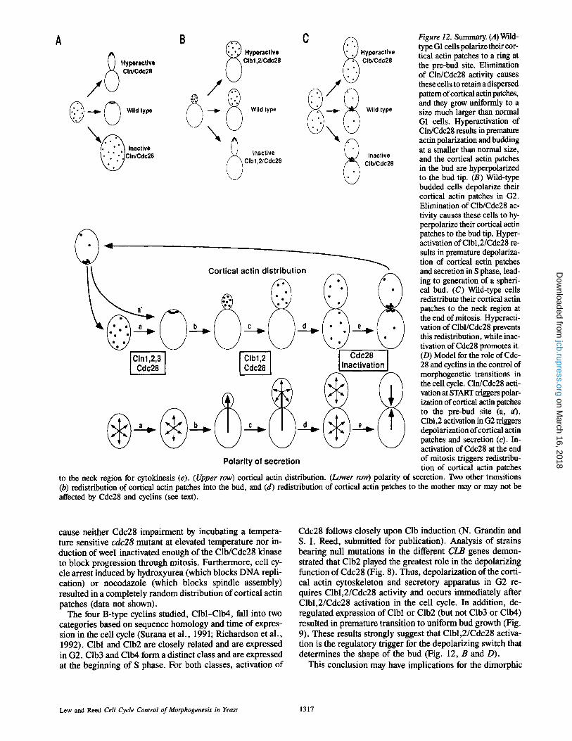
Figure 12. Summary. (A)Wild- type G1 cells polarize their cor- tical actin patches to a ring at the pre-bud site. Elimination of Cln/Cdc28 activity causes these cells to retain a dispersed pattern of cortical actin patches, and they grow uniformly to a size much larger than normal G1 cells. Hyperactivation of Cln/Cdc28 results in premature actin polarization and budding at a smaller than normal size, and the cortical actin patches in the bud are hyperpolarized to the bud tip. (B) Wild-type budded cells depolarize their cortical actin patches in G2. Elimination of Clb/Cdc28 ac- tivity causes these cells to hy- perpolarize their cortical actin patches to the bud tip. Hyper- activation of Clbl,2/Cdc28 re- suits in premature depolariza- tion of cortical actin patches and secretion in S phase, lead- ing to generation of a spheri- cal bud. (C) Wild-type cells redistribute their cortical actin patches to the neck region at the end of mitosis. Hyperacti- vation of Clbl/Cdc28 prevents this redistribution, while inac- tivation of Cdc28 promotes it. (D) Model for the role of Cdc- 28 and cyclins in the control of morphogenetic transitions in the cell cycle. Cln/Cdc28 acti- vation at START triggers polar- ization of cortical actin patches to the pre-bud site (a, a). Clbl,2 activation in G2 triggers depolarization of cortical actin patches and secretion (c). In- activation of Cdc28 at the end of mitosis triggers redistribu- tion of cortical actin patches
to the neck region for cytokinesis (e). (Upper row) cortical actin distribution. (Lower row) polarity of secretion. Two other transitions (b) redistribution of cortical actin patches into the bud, and (d) redistribution of cortical actin patches to the mother may or may not be affected by Cdc28 and cyclins (see text).
cause neither Cdc28 impairment by incubating a tempera- ture sensitive cdc28 mutant at elevated temperature nor in- duction of weel inactivated enough of the Clb/Cdc28 kinase to block progression through mitosis. Furthermore, cell cy- cle arrest induced by hydroxyurea (which blocks DNA repli- cation) or nocodazole (which blocks spindle assembly) resulted in a completely random distribution of cortical actin patches (data not shown).
The four B-type cyclins studied, Clbl-Clb4, fall into two categories based on sequence homology and time of expres- sion in the cell cycle (Surana et al., 1991; Richardson et al., 1992). Clbl and Clb2 are closely related and are expressed in G2. Clb3 and Clb4 form a distinct class and are expressed at the beginning of S phase. For both classes, activation of
Cdc28 follows closely upon Clb induction (N. Grandin and S. I. Reed, submitted for publication). Analysis of strains bearing null mutations in the different CLB genes demon- strated that Clb2 played the greatest role in the depolarizing function of Cdc28 (Fig. 8). Thus, depolarization of the corti- cal actin cytoskeleton and secretory apparatus in G2 re- quires Clbl,2/Cdc28 activity and occurs immediately after Clbl,2/Cdc28 activation in the cell cycle. In addition, de- regulated expression of Clbl or Clb2 (but not Clb3 or Clb4) resulted in premature transition to uniform bud growth (Fig. 9). These results strongly suggest that Clbl,2/Cdc28 activa- tion is the regulatory trigger for the depolarizing switch that determines the shape of the bud (Fig. 12, B and D).
This conclusion may have implications for the dimorphic
Lew and Reed Cell Cycle Control of Morphogenesis in Yeast 1317
on March 16, 2018
jcb.rupress.orgD
ownloaded from
switch in C albicans. These cells can be induced to switch from the yeast mode to a mycelial mode of growth by incuba- tion in medium of appropriate pH. Soil and co-workers found that budded yeast cells could form mycelia upon pH change, but only if they had not yet made the switch to isotropic bud growth (Soil et al., 1985). If this transition had passed, the cells would complete the cell cycle in the yeast mode and only switch to the mycelial mode in the next cell cycle. Thus, a developmental option is closed after switching to general bud growth. This situation is analogous to the de- velopmental options (budding, mating, or entrance to sta- tionary phase) available to cells in G1. Activation of Cln/ Cdc28 to pass START constitutes a decision in favor of bud- ding, and closes off the other options until the next cell cycle. Perhaps activation of the Candida equivalents of Clbl,2/ Cdc28 to switch to isotropic bud growth is similarly a deci- sion in favor of budding, and closes off the mycelial option until the next cell cycle.
Inactivation of Cdc28 after Mitosis May Trigger Actin Redistribution to the CytoMnesis Site
At the end of mitosis, cortical actin patches cluster on both sides of the neck region. Recently, it has been shown that capping protein, an actin-binding protein that colocalizes with cortical actin patches during most of the cell cycle, is absent from the patches that cluster in the neck region (Amatruda and Cooper, 1992). Thus, this redistribution also involves a change in the composition of the actin-rich patches. We found that sustained Cdc28 activation using a nondestructible derivative of Clbl (ClblA152) prevented ac- tin redistribution to the cytokinesis site (Fig. 12, C). This was not a nonspecific consequence of ClblA152 expression, because the redistribution could be triggered in the arrested cells by Cdc28 inactivation (achieved by shift-up of a temper- ature sensitive cdc28 mutant). Thus, actin redistribution to the cytokinesis site requires Clb/Cdc28 inactivation and oc- curs immediately after Clb destruction in the cell cycle, sug- gesting that inactivation of Cdc28 may be the regulatory trig- ger for this rearrangement (Fig. 12, C and D).
Cdc28 Substrates Involved in the Control of Morphogenesis
Our results indicate that changes in Cdc28 activity at three different stages of the cell cycle regulate distinct rearrange- ments of the cortical actin cytoskeleton and secretory ap- paratus. Two other rearrangements, the redistribution of cor- tical actin patches into the small bud in S phase and the redistribution of some cortical actin patches to the mother in mitosis remain to be explained. We have been unable to de- termine whether Cdc28 activity plays a role in these events.
In the case of pre-bud site assembly after START, we have shown that the actin rearrangement can be triggered by Cln/Cdc28 activation in the absence of de novo protein syn- thesis. This suggests that the kinase may directly phosphor- ylate substrates (such as actin-binding proteins) that regulate actin distribution in cells. Alternatively, the target substrates might be "polarity establishment" genes, such as CDC24, CDC42, BEM1, and BEM2 (Sloat et al., 1981; Adams et al., 1990; Johnson and Pringle, 1990; Bender and Pringle, 1991; Chenevert et al., 1992). Mutations in these genes prevent polarization of cortical actin patches, 10-nm filaments, and
Spa2 protein to the pre-bud site. Cdc42 is a ras-like GTP binding protein (Johnson and Pringle, 1990), while Cdc24 is related to the oncogene dbl, which catalyzes dissociation of GDP from the human homolog of Cdc42 (Hart et al., 1991). These proteins could potentially participate in a sig- nal transduction pathway whereby Cln/Cdc28 activation trig- gers assembly of the pre-bud site.
Different Cdc28/Cyclin Kinases Can Have Opposing Effects on Both the Actin Cytoskeleton and the Secretory Apparatus
In principle, the different effects of Cln/Cdc28 activation in G1 and Clb/Cdc28 activation in G2 could arise without the different forms of the kinase displaying any differences in substrate specificity. In this scenario, distinct substrates would be available to the kinase at different times in the cell cycle, so that activation of essentially the same kinase at different points in the cell cycle would have different effects. However, the results obtained with ectopic activation of specific forms of the kinase at inappropriate times in the cell cycle argue strongly against this model. Expression of Clnl or Cln2 in G2 resulted in hyperpolarization of cortical actin and secretion to the bud, while expression of ClblA152 in G1 prevented polarization of cortical actin to the pre-bud site. These results suggest that similar substrates may be available throughout the cell cycle, and that the different roles of the kinase are enacted by targeting the Cln/Cdc28 and Clb/Cdc28 complexes to different substrates.
Differences were also observed upon overproduction of different Clbs in G2. Specifically, Clbl and Clb2 overpro- duction had a depolarizing effect, while Clb3 overproduction had a hyperpolarizing effect. This suggests that there may be differences in the substrates of the Clbl,2/Cdc28 and Clb3/ Cdc28 complexes. In all cyclin overexpression experiments, there is a possibility that the effects of overexpression are not a direct consequence of activation of the targeted cyclin/ Cdc28 complex, but rather an indirect "dominant negative" consequence of inhibition of other cyclin/Cdc28 complexes. In the simplest case, overexpression of one cyclin might re- sult in its binding to all of the cellular Cdc28, leaving none for other cyclins. This does not appear to be the case in our experiments because co-overexpression of Cdc28 together with a cyclin had the same effect as overexpression of the cy- clin alone (data not shown). However, we cannot rule out the possibility that some of the cyclin overexpression results arose through dominant negative competition between cy- clins for other potentially limiting factors. This does not af- fect our conclusion that different forms of the Cdc28 kinase are targeted to different substrates because such differences are a prerequisite for any dominant negative model.
Understanding the molecular basis of the regulation of cytoskeletal function is the key not only to morphogenesis in yeast but also to regulation of cell shape and cell motility in nonwalled cells. The structural homology between various yeast genes involved in morphogenesis and their mammalian counterparts (e.g., between CDC42 and G25K, or CDC24 and dbl and bcr [Chant and Pringle, 1991]) suggests that in- sights gained in the genetically tractable yeast system will have far-reaching implications for other cells.
We thank Sally Kornbluth, Nick Marini, and anonymous reviewers for crit-
The Journal of Cell Biology, Volume 120, 1993 1318
on March 16, 2018
jcb.rupress.orgD
ownloaded from
ical comments on the manuscript, and John Pringle for stimulating discus-
sions. Thanks also to John McKinney and Fred Cross for yeast strains. D. J. Lew is supported by postdoctoral grant DRG1078 from the Damon
Runyon/Walter Winchell Cancer Research Fund. This work was supported by Public Health Service grant GM38328 to S. I. Reed.
Received for publication 30 September 1992 and in revised form 15 De- cember 1992.
References
Adams, A. E. M., D. Botstein, and D. G. Drubin. 1991. Requirement of yeast fimbrin for actin organization and morphogenesis in vivo. Nature (Lond.). 354:404-408.
Adams, A. E. M., D. I. Johnson, R. M. Longnecker, B. F. Sloat, and J. R. Pringle. 1990. CDC42 and CDC43, two additional genes involved in bud- ding and the establishment of cell polarity in the yeast Saccharomyces cerevisiae. J. Cell Biol. 111:131-142.
Adams, A. E. M., and J. R. Pringle. 1984. Relationship of actin and tubulin distribution to bud growth in wild-type and morphogenetic-mutant Sac- charomyces cerevisiae. J. Cell Biol. 98:934-945.
Adams, A. E. M., and J. R. Pringle. 1991. Staining of actin with fluorochrome- conjugated phalloidin. Methods Enzymol. 194:729-731.
Amatruda, J. F., J. F. Cannon, K. Tatchell, C. Hug, and J. A. Cooper. 1990. Disruption of the actin cytoskeleton in yeast capping protein mutants. Nature (Lond.). 344:352-354.
Amatruda, J. F., and J. A. Cooper. 1992. Purification, characterization, and immunofluorescence localization of Saccharomyces cerevisiae capping pro- tein. J. Cell Biol. 117:1067-107fi.
Arion, D., L. Meijer, L. Brizuela, and D. Beach. 1988. cdc2 is a component of the M phase-specific histone HI kinase: evidence for identity with MPF. Cell. 55:371-378.
Ballou, C. E. 1981. The yeast cell wall and cell surface. In The Molecular Biol- ogy of the Yeast Saccharomyces: Metabolism and Gene Expression. J. N. Strathern, E. W. Jones, and J. R. Broach, editors. Cold Spring Harbor Labo- ratory, Cold Spring Harbor, New York. 335-360.
Barton, A. A. 1950. Some aspects of cell division in Saccharomyces cerevisiae. J. Gen. Microbiol. 4:84-87.
Bender, A., and J. R. Pringle. 1991. Use of a screen for synthetic lethal and multicopy suppressed mutants to identify two new genes involved in morpho- genesis in Sacchoromyces cerevisiae. Mol. Cell. BioL 11 : 1295-1305.
Beran, K., 1968. Budding of yeast cells, their scars and ageing. Adv. Microbial Physiol. 2:143-171.
Brewer, B. J., E. Chlebowicz-Sledziewska, and W. L. Fangman. 1984. Cell cycle phases in the unequal mother/daughter cell cycles of Saccharomyces cerevisiae. Mol. Cell BioL 4:2529-2531.
Cabib, E., B. Bowers, A. Sburlati, and S. J. Silverman. 1982. Synthesis of the yeast cell wall and its regulation. Annu. Rev. Biochem. 51:763-793.
Chant, J., and J. R. Pringle. 1991. Budding and cell polarity in Sacchoromyces cerevisiae. Curr. Opin. Genet. Dee. 1:342-350.
Chenevert, J., K. Corrado, A. Bender, J. R. Pringle, and I. Herskowitz. 1992. A yeast gene (BEM1) necessary for cell polarization whose product contains two SH3 domains. Nature (Lond.). 356:77-79.
Cooper, J. A. 1987. Effects of cytochalasin and phalloidin on actin. J. Cell Biol. 105:1473-1478.
Cross, F. 1988. DAF1, a mutant gene affecting size control, pheromone arrest and cell cycle kinetics of S. cerevisiae. Mol. Cell. BioL 8:4675--4684.
Cross, F. R., and A. H. Tinkelenberg. 1991. A potential positive feedback loop controlling CLN1 and CLN2 gene expression at the start of the yeast cell cy- cle. Cell. 65:875-883.
Dirick, L., and K. Nasmyth. 1991. Positive feedback in the activation of G1 cyelins in yeast. Nature (Lond.). 351:754-757.
Draetta, G., F. Luca, J. Westendorf, J. Ruderman, and D. Beach. 1989. cdc2 protein kinase is complexed with cyclin A and B: Evidence for proteolytic inactivation of MPF. Cell. 56:829-838.
Dunphy, W. G., L. Brizuela, D. Beach, and J. Newport. 1988. The Xenopus cdc2 protein is a component of MPF, a cytoplasmic regulator of mitosis. Cell. 54:423-431.
Evans, T., E. T. Rosenthal, J. Youngbloom, D. Distel, and T. Hunt. 1983. Cy- clin: a protein specified by maternal mRNA in sea urchin eggs that is de- stroyed at each cleavage division. Cell. 33:389-396.
Farkas, V., J. Kovarik, A. Kosinova, and S. Bauer. 1974. Autoradiographic study of mannan incorporation into the growing walls of Sacchoromyces cerevisiae. J. Bacteriol. 117:265-269.
Featherstone, C., and P. Russell. 1991. Fission yeast p107 "~ mitotic inhibi- tor is a tyrosine/serine kinase. Nature (Lond.). 349:808-811.
Ford, S. K., and J. R. Pringle. 1991. Cellular morphogenesis in the Sacchoro- myces cerevisiae cell cycle: localization of the CDCll gene product and the timing of events at the budding site. Dee. Genet. 12:281-292.
Ghiara, J. B., H. E. Richardson, K. Sugimoto, M. Heaze, D. J. Lew, C. Wit- tenberg, and S. I. Reed. 1991. A cyclin B homolog in S. cerevisiae: chronic activation of the Cdc28 protein kinase by cyclin prevents exit from mitosis. Cell. 65:163-174.
Haarer, B. K., S. H. Lillie, A. E. M. Adams, V. Magdolen, W. Bandlow, and S. S. Brown. 1990. Purification of profilin from Saccharomyces cerevisiae and analysis of profilin-deficient cells. J. Cell Biol. 110:105-114.
Hadwiger, J. A., C. Wittenberg, W. A. de Barros Lopes, H. E. Richardson, and S. I. Reed. 1989. A family of cyclin homologs that control G1 phase in yeast. Proc. Natl. Acad. Sci. USA. 86:6255-6259.
Harold, F. M. 1990. To shape a cell: an inquiry into the causes of morpbo- genesis of microorganisms. Microbiol. Rev. 54:381-431.
Hart, M. J., A. Eva, T. Evans, S. A. Aaronson, and R. A. Cerione. 1991. Catalysis of guanine nucleotide exchange on the CDC42Hs protein by the dbl oncogene product. Nature (Lond.). 354:311-314.
Hartwell, L. H., J. Culotti, J. R. Pringle, and B. J. Reid. 1984. Genetic control of the cell division cycle in yeast. Science (Wash. DC). 183:46-51.
Hunt, T. 1989. Maturation promoting factor, cyclin and the control of M-phase. Curr. Opin. Cell Biol. 1:268-274.
Johnson, D. I., and J. R. Pringle. 1990. Molecular characterization of CDC42, a Saccharomyces cerevisiae gene involved in the development of cell polar- ity. J. Cell Biol. 111:143-152.
Johnston, G. C., J. A. Prendergast, and R. A. Singer. 1991. The Saccharo- myces cerevisiae MYO2 gene encodes an essential myosin for vectorial transport of vesicles. J. Cell Biol. 113:539-551.
Kilmartin, J. V., and A. E. M. Adams. 1984. Structural rearrangements oftu- bulin and actin during the cell cycle of the yeast Saccharomyces. J. Cell Biol. 98:922-933.
Kim, H. B., B. K. Haarer, and J. R. Pringle. 1991. Cellular morphogenesis in the Sacchoromyces cerevisiae cell cycle: localization of the CDC3 gene product and the timing of events at the budding site. J. Cell Biol. 112:535- 544.
Labbe, J. C., J.-P. Capony, D. Caput, J. C. Cavadore, J. Derancourt, M, Kag- had, J.-M. Lelias, A. Picard, and M. Doree. 1989. MPF from starfish oocytes at first meiotic metaphase is a heterodimer containing one molecule of cdc2 and one molecule of cyclin B. EMBO (Eur. Mol. Biol. Organ.) J. 8:3053-3058.
Lew, D. J., V. Dulic, and S. I. Reed. 1991. Isolation of three novel human cyclins by rescue of GI cyclin (Cln) function in yeast. Cell. 66:1197-1206.
Lew, D. J., N. J. Marini, and S. I. Reed. 1992. Different G1 cyclins control the timing of cell cycle commitment in mother and daughter cells in the bud- ding yeast Saccharomyces cerevisiae. Cell. 69:317-327.
Lew, D. J., and S. I. Reed. 1992. A proliferation of cyclins. Trends CellBiol. 2:77-81.
Liu, H., and A. Bretscher. 1992. Characterization of TPM1 disrupted yeast cells indicates an involvement of tropomyosin in directed vesicular transport. J. Cell Biol. 118:285-299.
L6rincz, A. T., and S. I. Reed. 1984. Primary structure homology between the product of yeast cell division control gene CDC28 and vertebrate oncogenes. Nature (Lond.). 307:183-185.
Madden, K., C. Costigan, and M. Snyder. 1992. Cell polarity and morphogene- sis in Saccharomyces cerevisiae. Trends Cell Biol. 2:22-29.
Marini, N. J., and S. I. Reed. 1992. Direct induction of Gl-specific transcripts following re-activation of the Cdc28 kinase in the absence of de novo protein synthesis. Genes Dee. 6:557-567.
Meijer, L., D. Arion, R. Golsteyn, J. Pines, L. Brizuela, T. Hunt, and D. Beach. 1989. Cyclin is a component of the sea urchin M-phase specific his- tone H1 kinase. EMBO (Fur. Mol. Biol. Organ.) J. 8:2275-2282.
Nash, R., G. Tokiwa, S. Anand, K. Erickson, and A. B. Futcher. 1988. The WHII + gene of S. cerevisiae tethers cell division to cell size and is a cyclin homolog. EMBO (Eur. Mol. Biol. Organ.) J. 7:4335-4346.
Novick, P., and D. Botstein. 1985. Phenotypic analysis of temperature- sensitive yeast actin mutants. Cell. 40:405-416.
Nurse, P. 1990. Universal control mechanism regulating onset of M-phase. Na- ture (Lond.). 344:503-508.
Nurse, P., and Y. Bissett. 1981. Gene required in G1 for commitment to the cell cycle and in G2 for control of mitosis in fission yeast. Nature (Lond.). 292:558-560.
Parker, L. L., S. Atherton-Fessler, and H. Piwnica-Worms. 1992. p107 "~'/is a dual specificity kinase that phosphorylates p34 '~c2 on tyrosine 15. Proc. Natl. Acad. Sci. USA. 89:2917-2921.
Pringle, J. R. 1991. Staining of bud scars and other cell wall chitin with calcofluor. Methods Enzymol. 194:732-735.
Pringle, J. R., A. E. M. Adams, D. G. Drubin, and B. K. Haarer. 1991. Im- munofluorescence methods for yeast. Methods Enzymol. 194:565-602.
Pringle, J. R., and L. H. Hartwell. 1981. The Saccharomyces cerevisiae cell cycle. In The Molecular Biology of the Yeast Saccharomyces. J. D. Strathem, E. W. Jones, and J. R. Broach, editors. Cold Spring Harbor Labo- ratory, Cold Spring Harbor, NY. 97-142.
Reed, S. I. 1980. The selection of S. cerevisiae mutants defective in the start event of cell division. Genetics. 95:561-577.
Reed, S. I., J. A. Hadwiger, and A. T. Lorincz. 1985. Protein kinase activity associated with the product of the yeast cell division cycle gene CDC28. Proc. Natl. Acad. Sci. USA. 82:4055-4059.
Reed, S. I., and C. Wittenberg. 1990. A mitotic function for the Cdc28 protein kinase of S. cerevisiae. Proc. Natl. Acad. Sci. USA. 87:5697-5701.
Richardson, H. E., D. J. Lew, M. Henze, K. Sugimoto, and S. I. Reed. 1992. Cyclin-B homologs in Saccharomyces cerevisiae function in S phase and in G2. Genes Dee. 6:2021-2034.
Lew and Reed Cell Cycle Control of Morphogenesis in Yeast 1319
on March 16, 2018
jcb.rupress.orgD
ownloaded from

Richardson, H. E., C. Wittenberg, F. R. Cross, and S. I. Reed. 1989. An essen- tial G1 function for cyclin-like proteins in yeast. Cell. 59:1127-1133.
Russell, P., S. Moreno, and S. I. Reed. 1989. Conservation of mitotic controls in fission and budding yeast. Cell. 57:295-303.
Russell, P., and P. Nurse. 1987. Negative regulation of mitosis by wee1 +, a gene encoding a protein kinase homolog. Cell. 45:145-153.
Sherman, F., G. Fink, and J. B. Hicks. 1982. Methods in Yeast Genetics. Cold Spring Harbor Laboratory, Cold Spring Harbor, New York.
Sloat, B. F., A. E. M. Adams, andJ. R. Pringle. 1981. Roles of the CDC24 gene product in cellular morphogenesis during the Saccharomyces cerevisiae cell cycle. J. Cell Biol. 89:395--405.
Snyder, M., S. Gehrung, and B. D. Page. 1991. Studies concerning the tem- poral and genetic control of cell polarity in Saccharomyces cerevisiae. J. Cell Biol. 114:515-532.
Soll, D. R., M. A. Herman, and M. A. Staebell. 1985. The involvement of cell wall expansion in the two modes of mycelium formation of Candida albi- cans. J. Gen. Microbiol. 131:2367-2375.
Staebell, M., and D. R. SoU. 1985. Temporal and spatial differences in cell wall expansion during bud and mycelium formation in Candida albicans. J. Gen. Microbiol. 131:1467-1480.
Sudbery, P. E., A. R. Goodey, and B. L. A. Carter. 1980. Genes which control cell proliferation in the yeast Saccharomyces cerevisiae. Nature (Lond.). 288:401-404.
Surana, U., H. Robitsch, C. Price, T. Shuster, I. Fitch, A. B. Futcher, and K. Nasmyth. 1991. The role of CDC28 and cyclins during mitosis in the bud- ding yeast S. cerevisiae. Cell. 65:145-161.
Tkacz, J. S., and J. O. Lampen. 1972. Wall replication in Saccharomyces spe- cies: use of fluorescein-conjugated concanavalin A to reveal the site of man- nan insertion. J. Gen. Microbiol. 72:243-247.
Tyers, M., G. Tokiwa, R. Nash, and B. Futcher. 1992. The Cln3-Cdc28 kinase complex of S. cerevisiae is regulated by proteolysis and phosphorylation. EMBO (Eur. MoL Biol. Organ.). J. 11:1773-1784.
Wittenberg, C., and S. I. Reed. 1988. Control of the yeast cell cycle is as- sociated with assembly/disassembly of the Cdc28 protein kinase complex. Cell. 54:1061-1072.
Wittenberg, C., K. Sugimoto, and S. I. Reed. 1990. Gl-specific cyclins of Sac- charomyces cerevisiae: cell cycle periodicity, regulation by mating phero- mone and association with the p34 ca~ protein kinase. Cell. 62:225-237.
Xiong, Y., and D. Beach. 1991. Population explosion in the cyclin family. Curr. Biol. 1:362-364.
The Journal of Cell Biology, Volume 120, 1993 1320
on March 16, 2018
jcb.rupress.orgD
ownloaded from