monitech headgrear
TRANSCRIPT

Design Studio IISpring Project
2015
Contents
Tutors 4
Partners 5
Team 6
I phase, Research 9
Discovering the brain 10
Discovering potential technologies 12
Discovering potential focus areas 13
Analysis of initial research / Defining a focus area 14
II phase, Defining 15
Identifying a field of application 16
Journey map 17
Identifying users and design requirements 21
Contents
Design Brief 22
III phase, Development 23
Brainstorming 24
Early concepts 25
Prototyping & testing 26
Evaluation and focus 28
IV pahse, Delivery 29
MoniTech 31
Visual identity 34
Analysis & conclusion 35
Opportunity (vision for the future) 36

Tutors
Sven Sõrmus DesignerSupervisor, Estonian Academy of Arts
Janno NõuDesignerSupervisor, Estonian Academy of Arts
Martin Pärn DesignerDead of D&E, Estonian Academy of Arts

Partners
ELIKO
Rauno Gordon Senior research scientist
Eiko PriidelEngineering Manager, PhD student
Annuka MikolaClinical Engineer
Technology Competence Centre in Electronics-, Info- and Communication Technologies
The mission of ELIKO is to improve the compet-itiveness of Estonian ICT industry through the collaboration of top research institutions and businesses in the fields of electronics-, info- and communication technologies.
PERHPõhja-Eesti Regionaalhaigla / The North Estonia Medical Centre
The North Estonia Medical Centre is one of the top health care providers in the country. A pa-tient-centred institution committed to profes-sionalism, innovation and teamwork, the Medical Centre has more than 3,700 people – doctors, nurses, caregivers and specialists – working for the good of patients.
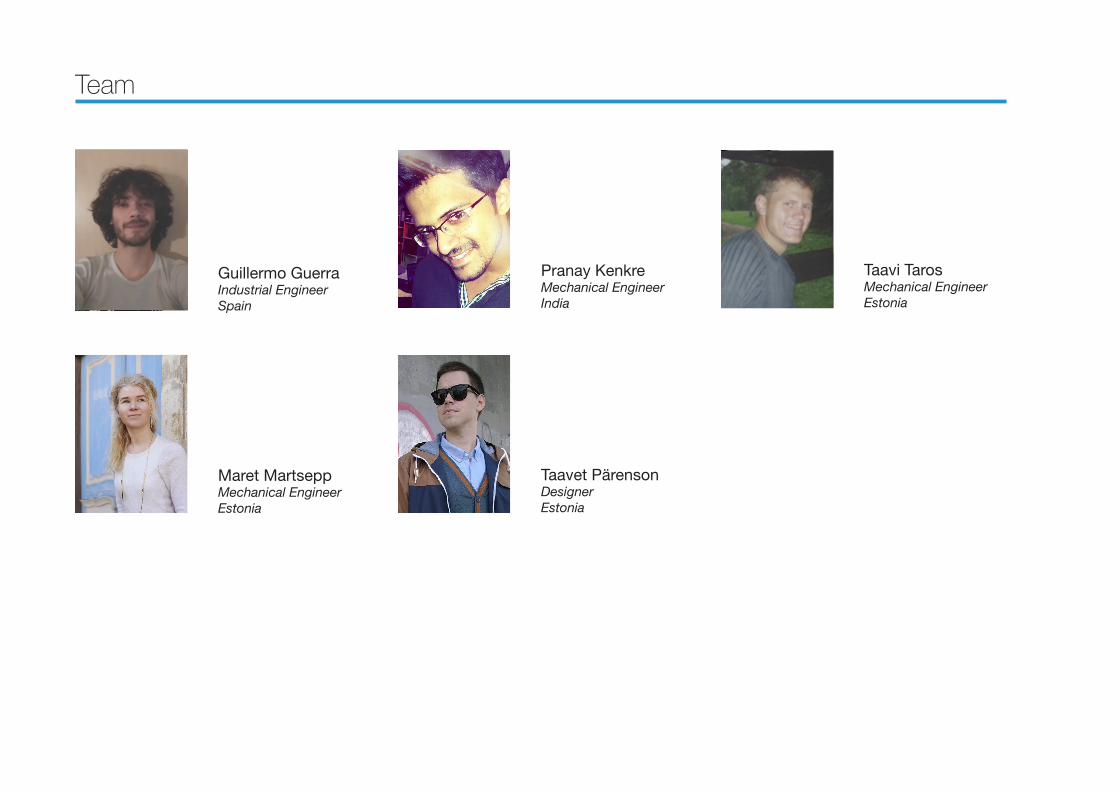
Team
Guillermo GuerraIndustrial Engineer Spain
Maret Martsepp Mechanical EngineerEstonia
Pranay KenkreMechanical EngineerIndia
Taavet Pärenson DesignerEstonia
Taavi Taros Mechanical EngineerEstonia

The goal of the project
Eliko deals with research and devel-opment of technologies in the medi-cal field. They have been involved in the development of a Portable Brain scanner for nearly two years.
The task and goal for our team was to identify an appropriate area of use,understand the environment, the people involved and create a con-cept of form and operating procedure for such a device.


I phase
ResearchThe goal of conducting the inital re-search phase is to identifie potential
areas of use.
Discovering the brain
Through desk research, going through PERHs research materials and interviewing hospital personnel we aimed to understand the anatomy of the human head and learn why we would even want to look inside it.
In the development process we were facing a product for emergency situa-tions so the research around the brain anatomy was oriented at the problems that appear after a head injury.
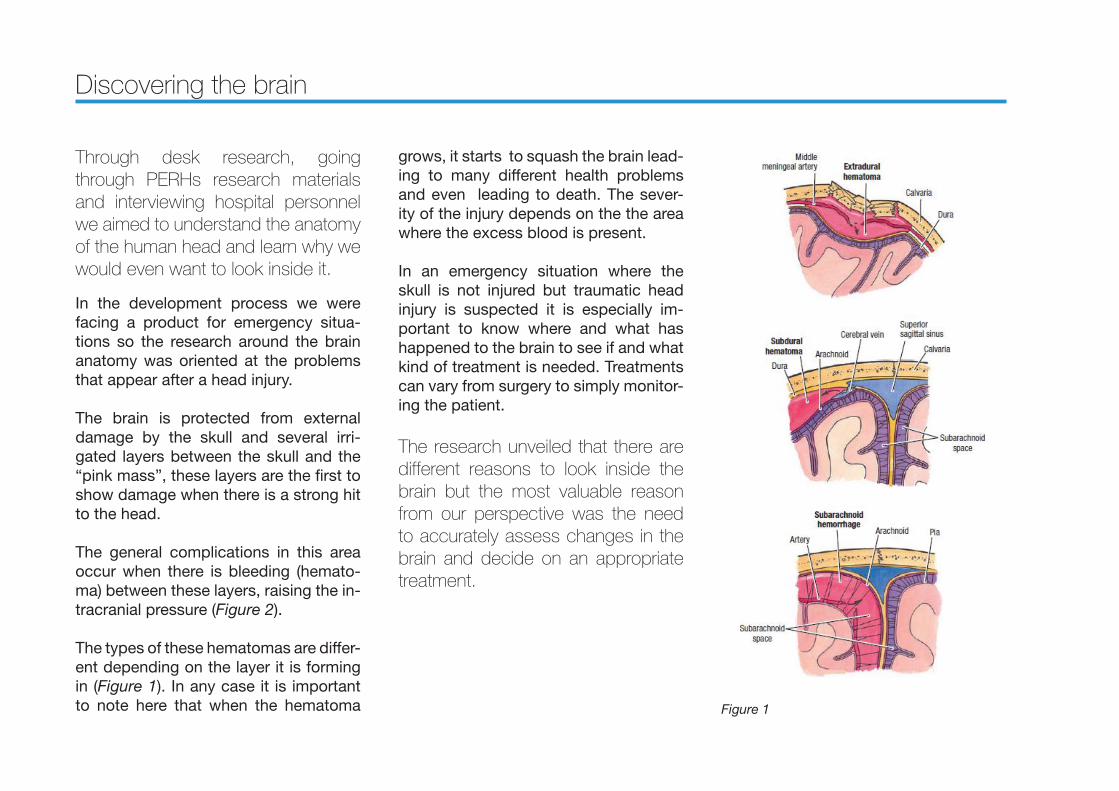
The brain is protected from external damage by the skull and several irri-gated layers between the skull and the “pink mass”, these layers are the first to show damage when there is a strong hit to the head.
The general complications in this area occur when there is bleeding (hemato-ma) between these layers, raising the in-tracranial pressure (Figure 2).
The types of these hematomas are differ-ent depending on the layer it is forming in (Figure 1). In any case it is important to note here that when the hematoma
grows, it starts to squash the brain lead-ing to many different health problems and even leading to death. The sever-ity of the injury depends on the the area where the excess blood is present.
In an emergency situation where the skull is not injured but traumatic head injury is suspected it is especially im-portant to know where and what has happened to the brain to see if and what kind of treatment is needed. Treatments can vary from surgery to simply monitor-ing the patient.
The research unveiled that there are different reasons to look inside the brain but the most valuable reason from our perspective was the need to accurately assess changes in the brain and decide on an appropriate treatment.
Figure 1

Figure 2

Discovering potential technologies
Through desk research, studying ELIKOs research materials and inter-viewing hospital personnel we aimed to understand what are the current non invasive technologies used to see inside the head and wich of those could be conidered for portable use.
Nowadays when there is a need for a non invasive way to detect injuries in the patients brain for any reason there are mainly two stationary options, MRI and CT scans.
CT (computer tomography) is a tech-nology that uses computer-processed X-rays in order to produce images (slic-es) of different parts of an object allow-ing the user to see inside without cutting, this technology is fully developed and is a perfect tool in order to diagnosticate many different problems in the head.
MRI (magnetic resonance image) is an-other technology used in radiology to study the state of the body by images. MRI scanners use radio waves and mag-netic fields to form images of the body.
Both technologies show the same defi-ciency which is that both are stationary, big machines. Because how thtese tech-nologies work, they cant not be used for portable solutions. Also both machines are rather expensive and need contin-uous maintenance. But this is under-standable as it takes alot of power to generate images of that quality.
Some portable technologies also exist that can be used to examine the brain.
NIR (Near infrared Technology) can be used for early detection of brain injury. A device is used to compare the left and right side of different areas of the brain to find irregularities that might indicate brain injury.
The research team at ELIKO are devel-oping technologies that show promise in this field.
MIT (Magnetic induction tomography) uses multible small coils around the hea compile an image of the brain. an electromagnetic field gives us informa-tion depending on the differences in the
frequency of the wave back from the head. The changes in the wave patterns are made by the bouncing of the wave on the molecular links inside the head.
UWB radar (Ultra-wideband radar) uses an antenna to sends short pulse of elec-tromagnetic waves inside the brain and compiles and image based on the fre-quency signals that bounce back. This can be used as a single antenna moved around the head.
Researching technologies unveiled that depending on the injury and the results needed different technologies can to be used for examining the brain. Most importantly current wide-ly used technologies are not suitable for use in portable devices. Thankful-ly ELIKO is developing technologies suitable for such use.

Discovering potential focus areas
Through studying research materi-als, desk research scenario building we aimed to find potential application opportunities and find who would po-tentially benfit from a portable brain scanning system.
As we are dealing with brain trauma we focused on areas of life where such inju-ries are a part of everyday life.
From civilian life we identified sports events as potential application areas. Repeated head injuries like concussions are a part of everyday life in sports like american football and extreme sports to name a few. There a documented cas-es where such injuries go unnoticed in sports events and lead to future com-plication in athletes. A portable device could help with early detection on inju-ries.
Military use proved to be a potentally huge opportunity for such devices as access to hightech medical machinery in war zones is limited and traumatic head injuries occur often. A portable device in
such situations could help in streamlin-ing the process of getting the appropri-ate treatment for the patient in time.
One of the most interesting potential opportunities are first response teams/paramedics who are first on the scene and could be in a position to be the first to identify brain injury.
Hospitals of cource are another obvious potential application area as not all hos-pitals have access to sophisticated brain imaging technologies. It can also be the case that different procedures could be streamlined by having access to porta-ble imaging devices.
In any case the hospital is a big area of interest as there we have the profession-als who can provide treatment and de-tailed diagnosis fot the patient. This is an important feature that other potential application areas (especially civilian cas-es) are missing.
From the research we concluded that whatever happens outside the hos-pital the patient still ends up in the
hospital in the hands of professionals as the non professionals do not have the knowhow, authority or machinery to make decisions concerning diag-nosis and treatment.

Analysis of initial research / Defining a focus area
In our research into brain anatomy, brain scanning technologies and po-tential focus areas we found that it’s not about who wants to find what in-side the head but about what can be done with that information or more importanly about who can do some-thing with that information.
This lead to us shifting our focus onto hospitals and the the patients who experience a traumatic brain injury (TBI) and are sent to the hospital for diagnosis and treatment.

II phase
DefiningThe goal of defining phase is to de-fine the field of application, users and
design requirements.

Identifying a field of application
By creating a patient journey map and conducting interviews with PERH personnel and pitching hypothetical cases to nurses we aim to find the approprate field of application fot a portable brain scanning device.
As we are ultimately interested in improv-ing the patients quality of life we chose to create a patient journey map (Figure 3) to unterstand and find opportunities where portable brain scanning device can help streamline patient recovery.
This method helped us create three hy-pothetical cases for testing. These were pitched and presented to medical per-sonnel to indentify users and require-ments and the need for a portable brain scanner in those situation.
The following the analysis of these three cases.
Bike crash, Paul sustains a head injury
Paul in unconsious
Pauls friend calls for help
Pauls friend Pauls friend
shows the crash site to the
paramedics
Paul getting his clinical status
checked
Paul getting his clinical status
checkedPATIENT PAUL
PARAMEDICS
PHYSICIAN
RADIOLOGIST
NURSES
Paramedics arrive to the crash site
Paramdics assess patients
clinical status
Paramedics stabilize the patient
Paul is being stabilized
Paramedics take the patient to the
hospital
Paramedics keep patient stable
Paul is put in to the ambulance
car and taken to the hospital
Paul recives an IV drip and
some medication
Paul is taken of the strecher
and lifted on to a hospital bed
Handing over the patient
Nurses recive the patient
Physician assesses
patients clinical
status
Physician orders a CT
scan
Doctor ordering
another CT scan
Paramedics infroming the hospital about condition of the arriving patient
Nurses start peparing for the patient
Radiologist prepares the CT
scanner
Radiologist recives a scan
request
Radiologist prepares the CT
scanner
Radiologist recives a scan
request
Nurses transport the patient to the CT scnanner
Nurses transport the patient to the CT scnanner
Nurses transport the patient to the
CU
Nurses prepare the patient for scanning
Nurses prepare the patient for scanning
Nurses prepare the patient for transport
Nurses prepare the patient for transport
Paul is lifted into the CT
scanner
Paul is lifted into the CT
scanner
Paul gets scanned
Paul gets scanned
Radiologist operates the
scanner
Radiologist operates the
scanner
Paul is taken to the
emergancy room (ER)
Paul is taken to a care unit
(CU)
Radiologists
delivers assessment,
diagnosis
Nurses transport the patient
Nurses monitor the patient
Nurses monitor the patient
Nurses monitor the patient
Paul is taken to a CT scanner
Physician reciving
information about the
arriving patient
Physician decides on treatment
Physician recives the scan results
Nurses prepare the patient for transport
Nurses prepare the patient for transport
Nurses prepare the patient for transport
Paul is lifted out of the CT
scanner
Paul is lifted out of the CT
scanner
Paul is taken back to care
unit
Radiologists
delivers assessment,
diagnosis
Transporting the patient
Palus is taken to the CT scanner
Doctor decides on further
treatment
Doctor reciving scan
results
Paul recives treatment at
the ER
Treating patient
Treating patient
Paul is at the CU
Paul is at the CU
Paul is at the CU
N
u
r
ses
transport t
h
e
p
atient to the
C
T scnanner
N
u
r
ses
prepare t
h
e
p
atient for scannin
g
N
urses prepare the patient
f
or
t
ranspor
t
3rd case
2nd case
1st case
Figure 3

Journey map
Bike crash, Paul sustains a head injury
Paul in unconsious
Pauls friend calls for help
Pauls friend gives first aid
Pauls friend shows the crash
site to the paramedics
Paul getting his clinical status
checked
Paul getting his clinical status
checkedPATIENT PAUL
PARAMEDICS
PHYSICIAN
RADIOLOGIST
NURSES
Paramedics arrive to the
crash site
Paramdics assess patients
clinical status
Paramedics stabilize the
patient
Paul is being stabilized
Paramedics take the
patient to the hospital
Paramedics keep patient
stable
Paul is put in to the ambulance
car and taken to the hospital
Paul recives an IV drip and
some medication
Paul is taken of the strecher
and lifted on to a hospital bed
Handing over the patient
Nurses recive the patient
Physician assesses
patients clinical status
Physician orders a CT
scan
Doctor ordering
another CT scan
Paramedics infroming the hospital about
condition of the arriving patient
Nurses start peparing for the patient
Radiologist prepares the CT
scanner
Radiologist recives a scan
request
Radiologist prepares the CT
scanner
Radiologist recives a scan
request
Nurses transport the patient to the CT scnanner
Nurses transport the patient to the CT scnanner
Nurses transport the patient to the
CU
Nurses prepare the patient for scanning
Nurses prepare the patient for scanning
Nurses prepare the patient for
transport
Nurses prepare the patient for transport
Paul is lifted into the CT
scanner
Paul is lifted into the CT
scanner
Paul gets scanned
Paul gets scanned
Radiologist operates the
scanner
Radiologist operates the
scanner
Paul is taken to the
emergancy room (ER)
Paul is taken to a care unit
(CU)
Radiologists delivers
assessment, diagnosis
Nurses transport the
patient
Nurses monitor the
patient
Nurses monitor the
patient
Nurses monitor the
patient
Paul is taken to a CT scanner
Physician reciving
information about the
arriving patient
Physician decides on treatment
Physician recives the scan results
Nurses prepare the patient for
transport
Nurses prepare the patient for transport
Nurses prepare the patient for
transport
Paul is lifted out of the CT
scanner
Paul is lifted out of the CT
scanner
Paul is taken back to care
unit
Radiologists delivers
assessment, diagnosis
Transporting the patient
Palus is taken to the CT scanner
Doctor decides on further treatment
Doctor reciving scan
results
Paul recives treatment at
the ER
Treating patient
Treating patient
Paul is at the CU
Paul is at the CU
Paul is at the CU
3rd case
2nd case
1st case

A portable device part of paramadics equipment for “in the field” scanning.
This was seen as one of the most poten-tial application fields. A paramdics are the first medical professionals to come in contact with the patient they have the potentil to initiate the early detection of possible brain injury and the severity of it.
When presenting this application case to the head of emergancy medicine in PERH it became apparent that there are a lot of complications. As any unexpected movement can in-terfier with the scanning procedure the conditions in a moving ambulace car are far from perfect. As Estonia is lucky to have so many hos-pitals the potential time window from the accident site to the hospital is to short to give any meaningful benefits.
In addition paramedics are not qualified to decide on a diagnosis and treatment based on the scanner readings. They would not take the responibility.
1. hypothetical case

A portable device in the hospital for early detection of brain injury.
As some hospitals limited brain scan-ning capabilities a portable, low cost detection device was presented as a fit for these hospitals also to work as back-up system in case CT/MRI machines are overloaded with patients.
Talking with PERHs medical personnel it became evident that right now there is no other technology besides CT/MRI the doctors trust to base their diagnosis on.
Making decision on diagnosis and treat-ment is a lot of resposibility and it’s not a risk the doctors are willing to take.
As the brain is a delicate organ there is no room for misinformation. A machine that makes 1 mistake for every 1000 scans in not accurate enough to base life or deth decison on.
No portable technologie can provide accurate enough readings for detecting brain injury as of now.
2. hypothetical case
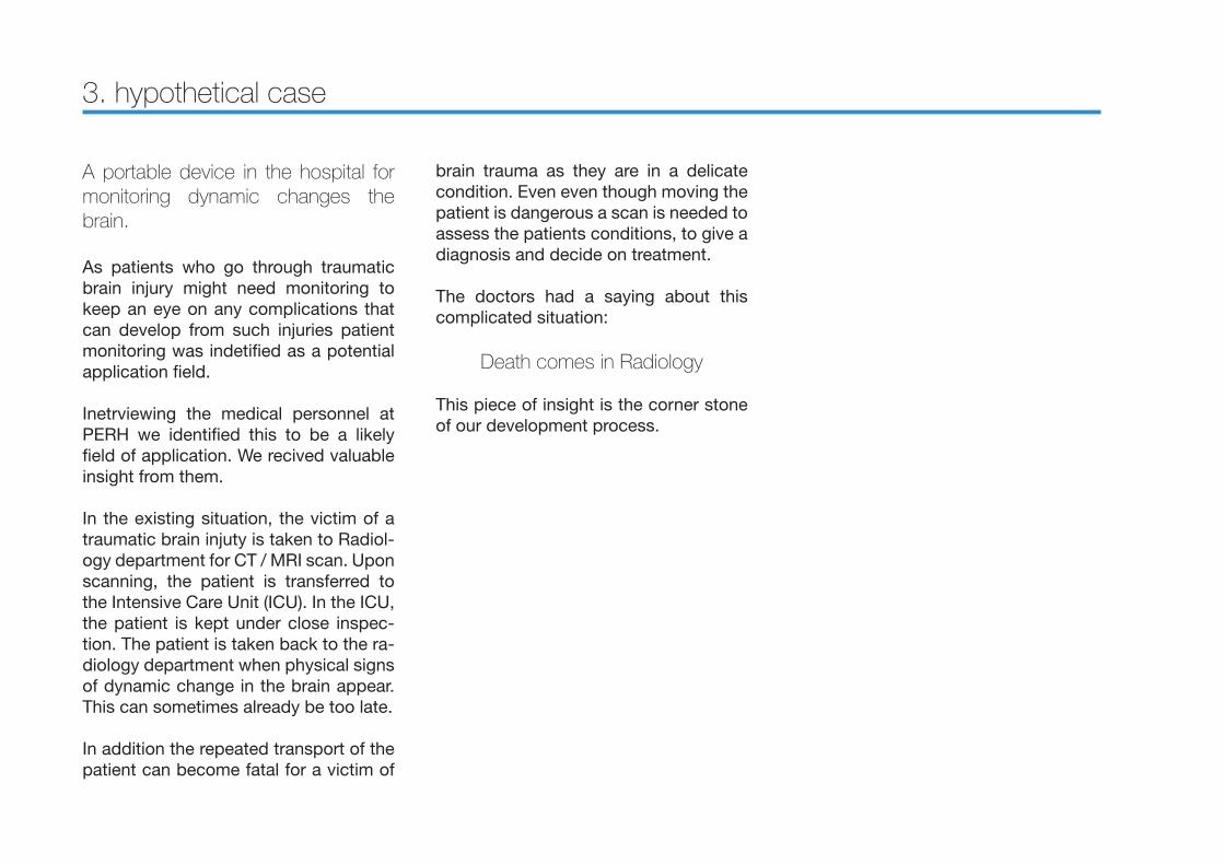
A portable device in the hospital for monitoring dynamic changes the brain.
As patients who go through traumatic brain injury might need monitoring to keep an eye on any complications that can develop from such injuries patient monitoring was indetified as a potential application field.
Inetrviewing the medical personnel at PERH we identified this to be a likely field of application. We recived valuable insight from them.
In the existing situation, the victim of a traumatic brain injuty is taken to Radiol-ogy department for CT / MRI scan. Upon scanning, the patient is transferred to the Intensive Care Unit (ICU). In the ICU, the patient is kept under close inspec-tion. The patient is taken back to the ra-diology department when physical signs of dynamic change in the brain appear. This can sometimes already be too late.
In addition the repeated transport of the patient can become fatal for a victim of
3. hypothetical case
brain trauma as they are in a delicate condition. Even even though moving the patient is dangerous a scan is needed to assess the patients conditions, to give a diagnosis and decide on treatment.
The doctors had a saying about this complicated situation:
Death comes in Radiology
This piece of insight is the corner stone of our development process.

Identifying users and design requirements
By conducting phenomenological observations in PERHs ICU and con-ductiong interviews with medical per-sonnel we aimed to idetify the users and possible requrements for a por-table brain scanning system in ICU.
We identified the users as following:Primary user - Nurse. Nurses will be the users who are hands on with this device.Secondary user - Physician. Physicians are not directly using the system but in-teract with the readings from the deviceTertiary user - Hospital board. They are influence the other users as they decide if this device is up to the task and worth purchasing for the secondary and pri-mary users to use.
As we identified nurses as our primary users we focused on observing them. By being able to observe the nurses working in the ICU we were able to un-dersand their workflow. By interviewing and observing them in action we got valuable infights on how nurses interact with medical equipment and what kind of requirements that creates.
Even though the patient is not in this case a user they affect the requirements the most.

Design Brief
There is a need or a non invasive brain scanning device that detects the need for a CT scan by monitoring dynamic changes in the brain.
Design guidelines:
• Primary users are Nurses.
• Simplifie use for the Nurses.
• Easy mounting and detaching of the device.
• The device may not interfere with the patients recovery.
• Easily maintainable
• Able to accommodate future technologies.
III phase
DevelopmentThe goal of development phase is to create concepts of form and oper-ating procedure for a portable brain
scanning system in the ICU.

Brainstorming
Multiple Brainstorming sessions were conducted (Figure..)
Throught these sessions we narrowed the possibilities for our focus to few con-crete ideas: flowers, pillows and head-phones.
Flower: A device which will wrap around the head of the patient and will monitor his/her brain activity without constraining his/her movement.
Pillow: A smart surface that will be able to get information from the patient who lay down on it, making a comfort situation during the scanner.
Head-phones: A concept to implement Eliko’s long de-veloped technology possibilities, based on a moving single antenna around the surface of the head with the help of two electrical motors and a conductive chain.
Figure

Early concepts
In the beginning of the project many ideas were rized upon. Some of them are following:
Helmet with 10 to 20 antennas inside. Antennas are sending signals to the main device, pulse generator is the one collecting the information. Not all of them are working together, switching them is complicated and also having wires around.
Kinect device usage.
MEG - small combs, so you don’t need to shave the hair, easy to contact with the head (head massage circular comb).
Smart pillow - patient is lying on the bed, the pillow/towel comes around the head and start to scan the head, so we don’t need to move the head later and it could be a little softer surface.
Smart towel - will be wrapped around patient head in a certain order.
Smart mattress - like a pillow concept, except it is a part of the mattress.
Bed attachment device, such as a spe-cial lamp.
Robot spiders, are moving in that area, that needs to be scanned.
Smart arm - the smart hand is combing the head meanwhile scanning the brain.
Fabric hat with antenna.flower pillow- the blossoms are moving automatically around the head for mon-itoring.
Fabric headband, which is fastened on the forehead part.
Shaver/ hand held cylinder - nurse will need to move the “shaver” alike device itself, where on top of it there is an an-tenna working and scanning.
Smart fluid hat, where there are coils built inside

Prototyping & testing
After realizing that we can not fix the technology we are going to use yet, we decided to start to work with dif-ferent concepts to find out the best solution.
Three ideas were chosen for developing to a mature stage.
Radar Headphones
Smart Pillow
Modular Helmet


Evaluation and focus
Thanks to good co-operation with Eliko we managed to take into consideration all the technical issues while building those concepts. After receiving the confirmation from Eliko that all those concepts are doable in real-life we went to PERH to join a morning meeting of the nurses to introduce them the ideas and ask feedback. The feedback we got from them is following:
Radar headphones - NO GO!
• The head support is not good- patientcan get bed sores
• Neck lengths are different, not suitablefor everyone
• Patient will sometimes lay on the side(then you can not use it)
• Pressure on the neck
• You can not move the patient
Smart pillow - NEEDS A LOT OF IMPROVEMENT
• Needs to be cleanable (BIG PROBLEM)
• Liquids will run also inside of the coils holes (foam can be filled with liquid such as blood)
• Coils and pillow should be changeable
• Will fit for most head sizes
• Perhaps the best option to move the patient
• The pillow will take the shape of the head, it will keep the head stable
• The angle of the pillow must be pointed up (so the head is at the upper part- it will keep away the lung infection)
• There should be signs which coilsshould be connected with which placesthey might change the pillow cover many times a day so it will be uncomfortable to unconnect and connect the coils in between
Modular helmet - WE GOT THIS!
• we need to change the material (can not be porous, because neet to be cleanable)
• Cleanability
• Breathability
• wires should come out from top of the head
• We need to take care there is not too much pressure on the head.Perhaps add some gel pads next to the head to make it softer
• The blood flow is not as good on the head trauma patient as a normal personit is adapting different head sizes
• If some bolt or some injury pieces coming out from the head, one piece of the module could be removed (have to think about how to connect the wires in this case)

IV phase
DeliveryThe goal of deliveryphase is to illus-trate the concept of form and oper-ating procedure for a portable brain
scanning system in the ICU.

MoniTechHeadgear

MoniTech
Introduction to our product
With Monitech Headgear the require-ments of users have been met with with constraints of technology.
Technology
Monitech Headgear is designed to fit MIT technology. Magnetic Induction To-mography: The theoretical base of this technology is close to the base on MRT. Electromagnetic field gives us informa-tion depending on the difference in the frequency on the wave back from the inspection, the changes on the wave are made by the bounce of the wave on the molecular links inside the head. The main difference is that instead of create a close electromagnetic field inside a jail, we use microwaves that go from one coil attached to the head to another one crossing it. Using a lot of coils we get to cover the whole surface of the head, creating a different jail and achieving all the possibilities through the head to get all the information that is need.
Design
The use of MIT technology enables to design the device in most suitable way for users. Placing it to patient’s head is intuitional and requires very little knowl-edge. It is easy to clean to meet the strict hygiene requirements of ICU envi-ronment.


Visual identity
Since the project is focused on cre-ating a product which helps the med-ical people to monitor a human brain and discover the changes in the brain dynamics we decided to keep the design of the product as simple and clear as possible.
Picking a project name was not an easy task. We decided that the name should give a straight hint what the device is doing, even when the person is hearing the name for the first time. Our project name MoniTech comes from the words Monitoring Technology combination which is pretty much the simplest way to say what is it doing.The goal is to show the product clear, visible and understandable. We are us-ing grey and blue colour, since they car-ry the right feeling.

Analysis & conclusion
Our concept was trying to design a new brain scanner which would be cheaper, “portable” and open using the technol-ogy that eliko was developing, the three different concepts that we get during the creative process fulfill these needs, but not all of them were suitable to use on real patients due to the physical con-straints around the products so we final-ly choose the “Modular Helmet” which was the concept nurses were more in-terested in.This concept has many improvements compared to the actuals techniques of brain monitoring, As it is a small device the patient will not get stressed if s/he has claustrophobic fear,it will improve the treatment of the patient, there will not be a need to displace the patient so there will no be complication attached to these movement to the radiology area, it will be possible to make a continuous monitor of the brain while the patient is in the ICU so the doctors will be able of taking decisions on the critical points in the development of the patient’s state the price of this device compare to the price of a CT or a MRI would be much lower so it will be possible to monitor the
the brain activities in rural areas which were not able to afford one of these ma-chines on their local hospitals or health centers.

Opportunity (vision for the future)
With the develop of Eliko’s technology the future for medicine scanner would get revolutionized if they succeed on their purpose.Their work on Magnetic Induction To-mography could get to release a func-tional portable scanner that could have applications not only on the ICU also in emergency situations or even satisfying the needs of the public (monitoring the brain activity from the residence of the small users who can afford to buy it as example). And even more interesting the potential of their works on Eddy currents which could finish on the developing of an ef-ficiency device to monitor bones, mus-cles or internal organs on any scenario (emergencies, residences, hospital pro-tocols etc) that also would not be too expensive.
