monash biomedical imaging and linked laboratories · pdf fileaustralia china india malaysia...
TRANSCRIPT
AUSTRALIA CHINA INDIA MALAYSIA SOUTH AFRICA
Monash Biomedical Imagingand Linked Laboratories Annual Report 2014

Monash Biomedical Imaging
Member
Collaborators
Supporters
Partners
National Node of Victoria, Australia
2

3
Vice Provost’s Report
Director’s Report
Overview
FacilitiesResearch HighlightsStrategic PlanGovernance
Personnel
StaffStudents
Research Activities
Monash Biomedical ImagingMonash Clinical and Imaging NeuroscienceMonash Neuroscience of Consciousness
Operations
Collaborations
Partnerships
Research Outputs
PublicationsGrantsSelected Presentations and OutreachAwards
Abbreviations
45
6-7
8
9-17
18-1920-21
2122-27
27
Table Of Contents
Vice Provost’s Report
4
Throughout 2014 Monash Biomedical Imaging (MBI) experienced continued growth in the provision of research services, with expanded and increased research utilisation and collaborative links with the broader Monash University research community. The research programs of Monash Clinical and Imaging Neuroscience and the Monash Neuroscience of Consciousness linked laboratories were further expanded during the year, and the NHMRC funded ASPREE (ASPirin Reducing Events in the Elderly)healthy ageing brain imaging study was commenced.
Cooperation between the Australian Synchrotron at the Imaging and Medical Beam Line and MBI scientists in preclinical models of cardiovascular dysfunction has continued. Furthermore, the increased focus from primarily brain imaging to lung, kidney and cardiovascular imaging is of strategic importance. The focus on cardiovascular research and therapeutic interventions has given impetus to the future establishment of a specialist cardiovascular University hospital on the Clayton campus, opposite MBI and the Australian Synchrotron.
Significantly greater scientific outputs were achieved in 2014 via high impact publications and increased grant success, including the Australian Research Council Centre of Excellence for Integrative Brain Function (CIBF). Internationally, the role that MBI is playing in leading the CIBF is providing a critical springboard for furthering our international reputation in biomedical imaging and brain research. Within the CIBF, MBI is playing a major role in the International Neuroinformatics Coordinating Facility (INCF), an international network of interest related to international neuroscience informatics and image sharing standards.
Across the suite of Monash University platform technologies, a systematic improvement has been undertaken to the research platform operations to ensure the systems and processes are operating to world’s best practice. In 2014 the MBI platform was incorporated and integrated into the Australian Research Infrastructure Network (ARIN) systems. This now provides researchers with streamlined access to the
outstanding biomedical imaging equipment and expertise at MBI. Participation in the Victorian Platform Technology Network (VPTN) and ARIN has increased the knowledge about MBI amongst the research community, with additional MBI research infrastructure to be added to ARIN in 2015.
MBI’s involvement in communities of interest including the Victorian Biomedical Imaging Capability (VBIC) and the National Imaging Facility (NIF) continues to enable the platform to have a major impact amongst researchers across Melbourne and Australia. There was continued cooperation with the Australian Nuclear Science and Technology Organisation (ANSTO) and Monash Health, including a number of joint appointments and projects.
The success of the Science and Industry Endowment Fund (SIEF) grant application late in 2014, in partnership with the Commonwealth Scientific Industrial Research Organisation (CSIRO), will enable establishment of the Biomedical Materials Translational Facility. The procurement and installation of an Magnetic Resonance Imaging - Positron Emission Tomography (MRI-PET) scanner within the facility will make MBI one of the most comprehensive biomedical imaging facilities in the world. The success of the application was underpinned by Monash University’s long-term collaborative approach with its partners, particularly CSIRO.
In the coming year MBI is poised for significant further development based on the growth of industry links. I’m delighted that 2014 was another successful year for MBI and I look forward to the forthcoming commissioning of the MRI-PET facility in 2015, and the expanded translational science discoveries and industry transformational outcomes that will follow.
Monash Biomedical Imaging Highlights of 2014
• Continued growth in the provision of research services to the Monash University research community
• Integration of Monash Clinical and Imaging Neuroscience and the Monash Neuroscience of Consciousness Linked Laboratories
• Expanded cooperation with Australian Synchrotron
• Significant grant success including the Australian Research Council Centre of Excellence for Integrative Brain Function
• Establishment of the ASPREE healthy ageing brain imaging study
• Incorporation and integration into Victorian Platform Technology Network and the Australian Research Infrastructure Network
• Leadership of the Victorian Biomedical Imaging Capability and contributions to the National Imaging Facility
• Expanded cooperation with the Australian Nuclear Science and Technology Organisation and Monash Health
• Science and Industry Endowment Fund funding success in partnership with the Commonwealth Scientific Industrial Research Organisation
• Poised for significant development and growth of industry links in 2015
Ian SmithVice Provost (Research and Research Infrastructure)
5
Director’s Report
I am delighted to report that during 2014 MBI continued to grow in the breadth and depth of biomedical imaging research services provided to the Monash University and Victorian research communities. In less than three years since the establishment of MBI in 2012, the University has emerged as an international leader in biomedical imaging research and development. Throughout 2014 MBI has continued to build strong collaborative links with the School of Psychological Sciences in the Faculty of Medicine, Nursing and Health Sciences via the MBI Linked Laboratories program. The Monash Clinical and Imaging Neurosciences (MCIN) Laboratory led by Professor Murat Yucel and A/Professor Alex Fornito, have continued to expand the breadth and collaborative nature of their research activities based at MBI. The activities of the laboratory will continue from 2015 as the Monash Brain and Mental Health Laboratory (BMH) that identifies the increasing importance of their research focus on mental health research.
During 2014 the Monash Consciousness Research Laboratory (MCRL), an MBI Linked Laboratory jointly led by Associate Professor Tsuchiya and Associate Professor Jeroen van Boxtel from the School of Psychological Sciences, continued research into understanding the neural basis of consciousness. The expanded group recently established the Monash Neuroscience of Consciousness (MoNoC) Laboratory to promote the group’s expanded research vision investigating the scope and limits of non-conscious processing, and the influence of attention on conscious and unconscious perception. The collective research productivity from MBI, MCIN and MCRL in 2014 were substantial with over 80 peer-reviewed articles, books and book chapters published. MBI and researchers and staff from the MCIN and MCRL Linked Laboratories were involved in over 25 outreach activities, and collectively they currently hold more than 20 competitive grants.
A major competitive grant success was achieved in 2014 with the award of the Australian Research Council (ARC) Centre of Excellence for Integrative Brain Function
(CIBF) grant for the period 2014-20. The establishment phase of the Centre was undertaken in early 2014 with the research collaboration arrangements put in place for the commencement of the Centre’s research projects in the second half of the year. The CIBF research management and administration staff were integrated with the MBI research management staff to help facilitate the smooth operation of the Centre. The involvement of MBI research support staff in CIBF is now providing significant new research collaboration opportunities for the Centre and MBI. The establishment of the ASPREE NEURO brain imaging study was a major achievement following the award of a NHMRC project grant in late 2014. Over the next four years more than 550 healthy aged volunteers will be scanned multiple times to determine whether daily low dose aspirin has an effect on brain health. The study outcomes will be of major importance for determining the overall benefits of the use of aspirin in the elderly.
The Victorian Biomedical Imaging Capability is a strategically important Victorian based biomedical imaging research community that is led from Monash University and MBI. MBI is a node of the NIF and continues to provide state-of-the-art high quality biomedical imaging research infrastructure to NIF users and researchers from across Melbourne and Australia. The success of the Science and Industry Endowment Fund grant application late in 2014, in partnership with the CSIRO, will enable establishment of the Biomedical Materials Translational Facility. The procurement and installation of an MRI-PET scanner within the facility will result in one of the most comprehensive biomedical imaging facilities in the world. The success of the application was underpinned by Monash University’s long-term collaborative approach with its partners, particularly CSIRO, and has poised MBI for significant development and growth of industry linkages in 2015-16.
I would like to thank Professor Ian Smith (Vice Provost, Research and Research Infrastructure) for his continued support of MBI during 2014. I would also like to particularly thank Ms Sue Renkin, Chair of the MBI Advisory Board, and the
MBI Advisory Board members for their governance and advice concerning the operations and further development of the MBI research platform. During 2014 Dr Lisa Hutton‘s role as Research and Operations Manager expanded to include Centre Manager for the ARC Centre of Excellence. My thanks to Lisa for her continued outstanding efforts during the establishment phase of CIBF and her oversight of the day-to-day operations of the MBI facilities throughout the first part of 2014. I would like to thank Dr Richard Huysmans for his work in continuing the establishment of CIBF during the second half of 2014 whilst Lisa was on maternity leave. Thanks also to Ms Catherine Harold and Dr Charles Hardy who took over responsibility for the MBI Research and Operations activities throughout 2014.
The MBI team leaders, Dr James Pearson, Associate Professor Nicholas Ferris, Dr David Barnes, Dr Zhaolin Chen, and Associate Professor Jeroen van Boxtel, have successfully led their teams’ research and imaging research support activities throughout the year. Their work has ensured the successful provision of outstanding imaging research services across the multidisciplinary and multimodal imaging environment at MBI and I sincerely thank them and their teams for their invaluable contributions. On a personal and family note, 2014 was a highly productive year with six staff members and researchers across MBI and the Linked Laboratories celebrating new arrivals. Congratulations to Sharna Jamadar, Lisa Hutton, Ruth Vreys, Chao Suo, Beth Johnson, and Alex Fornito and their partners on the safe arrival of their babies Rufus, Lincoln, Jasper, Felix, Jemima, and Nicholas.
Gary EganDirector MBI and CIBF; Distinguished Professorial Fellow, School of Psychology; Computational Imaging Theme Leader, LSCC, VLSCI
Strategic Plan
The MBI strategic plan was developed in 2012 in close alignment with the key objectives of Monash University’s 2025 vision: to be known internationally for a commitment to quality, and differentiated by a research intensive, international focus that will enable the University to address the important challenges of our times. The MBI strategy is also closely aligned with the Monash University Research Strategy (2011-15), which sets out to achieve impact through research excellence and relevance. Underpinning this pursuit of research excellence and ability to deliver impact from our research, is a commitment by the University to six key enabling “pillars” including: talent enhancement, world class research infrastructure, encouraging interdisciplinary research, superior research training, professional research management, and a focus on research translation.
MBI is a core University Technology Research Platform under the world class research infrastructure pillar. Through provision of biomedical imaging facilities together with close integration with other Monash University Technology Research Platforms, MBI is now ideally positioned to support the University’s strategic research objectives. The MBI strategic plan is contributing to the Monash University Research Strategy by:
• providing and maintaining world-class instrumentation and expertise
in biomedical imaging, that provides Monash University researchers and collaborating partners with access to a capability that enables innovative research of the highest quality possible;
• operating at world’s best practice to maximise the efficiencies and effectiveness of the MBI capability and capacity;
• providing strong leadership and connections within key national and international biomedical imaging and research networks;
• enabling academic and industry collaboration and partnership; and
• working towards a sustainable funding model to maintain and expand the biomedical imaging technology research platform.
The MBI research platform also importantly contributes towards other key University strategies, including enhancing the attraction and retention of high performing researchers, enabling multidisciplinary research, providing opportunities for
6
OverviewOverview
Facilities
MBI is one of the worlds pre-eminent sites for biomedical imaging and clinical neurosciences research. MBI facilities are located primarily at the Clayton Campus, and include high-resolution clinical and pre-clinical magnetic resonance imaging (MRI) scanners, and pre-clinical X-Ray computed tomography (CT), Positron Emission Tomography (PET), Single Photon Emission CT (SPECT). We manage access to a pre-clinical PET-CT scanner (Mediso) at the Alfred Medical Research and Education Precinct (AMREP, Prahran) and a FLECT-CT scanner (TriFoil) at the Monash Institute of Pharmceutical Sciences (MIPS, Parkville). We also manage access to a suite of clinical and neurocognitive testing facilities at Clayton, including Electroencephalogram
(EEG), Transcranial Magnetic Stimulation (TMS), and ocularmotor and gait testing equipment.
The expertise and technological capabilities at MBI support a wide range of pre-clinical and clinical research projects undertaken by researchers and clinicians from Monash University and collaborating organisations throughout Victoria. These facilities are co-located with the Australian Synchrotron Imaging and Medical Beam Line, providing capability for ultra-high resolution imaging of soft tissues in living animals. MBI scientists collaborate with global experts in medicine, science and engineering, as well as industry and government to create innovative solutions to clinical health challenges.

Governance
MBI has an Advisory Board with an independent chairperson that meets quarterly. The functions of the board are:
• to assist the Director with strategic planning including advice in alignment with government policy on research infrastructure and industry trends;
• to monitor the performance of MBI;
• to help define appropriate metrics (key performance indicators) for the platform;
• to provide representation for stakeholders; and
• to make recommendations on strategies for the further development of MBI.
Ms Sue Renkin Director, Intuitively Focused; Distinguished Alumnus, Monash University
Professor Ian Smith Vice Provost, Research and Research Infrastructure, Monash University
Professor Paul Bonnington Director, Monash e-Research Centre
Professor Ross Coppel Deputy Dean (Research), Faculty of Medicine, Nursing and Health Sciences, Monash University
Professor Gary Egan Director, Monash Biomedical Imaging, Monash University
A/Professor Nicholas Ferris Clinical Head, Monash Biomedical Imaging, Monash University
Dr Michael JamesHead of Science, Australian Synchrotron
Professor Nellie Georgiou-Karistianis Associate Dean, Graduate Research, Faculty of Medicine, Nursing and Health Sciences
Dr Gareth MoorheadResearch Program Leader, Materials Science and Engineering, CSIRO
Professor Andrew Peele Director, Australian Synchrotron
Professor Stephen Stuckey Director Diagnostic Imaging, Monash Health (formerly Southern Health)
Chair
Deputy Chair
Members
MBI Advisory Board
7
superior research training, and strengthening and contributing to the translation of biomedical research with the potential to improve healthcare outcomes. MBI is now providing the University’s interdisciplinary research community with an advanced biomedical imaging research environment and partnerships to invent, develop and exploit imaging based solutions that have international impact. The research projects now being undertaken at MBI are being led by some of the best researchers in the world, in collaboration with their national and international peers, and supported by one of Australia’s leading biomedical imaging research platforms.

Dr Bryan Paton Research Fellow, School of Psychology and MBI
Students
Phil Ward PhD Candidate
Shenjun Zhong PhD Candidate
Saman Kashuk PhD Candidate
Sulaiman Al Hasani PhD Candidate (co-supervision)
Steffen Krieger PhD Candidate
Imaging Analysis
Clinical Research ImagingCognitive Neuroimaging Research
A/Prof Jeroen van Boxtel Head; Senior Research Fellow, School of Psychology and MBI
Dr Sharna Jamadar Research Fellow, School of Psychology and MBI
Dr Zhaolin Chen Head, November onwards
Dr David Barnes Head, January – March; Senior Research Fellow, MBI and Monash e-Research Centre, LSCC, VLSC Senior Research Fellow (Adjunct) Faculty of IT
Dr Michael EagerSoftware programmer, MBI and Monash e-Research Centre
Ms Amanda Ng January – June Computational Biomedical Imaging Scientist, MBI and Monash e-Research Centre; LSCC, VLSCI
Dr Toan Nguyen Imaging Informatics Officer, MBI and Monash e-Research Centre
Dr Parnesh Raniga Medical Imaging Scientist; MBI Research Scientist, ICT Centre CSIRO
A/Prof Nicholas Ferris Head; Clinical MRI, MBI and Clinical Radiologist, Monash Health
Mr Richard McIntyre Radiographer, Monash Health & MBI
MBI Personnel
Director
Professor Gary EganDirector, MBI and CIBF; Distinguished Professorial Fellow, School of Psychology; Computational Imaging Theme Leader, LSCC, VLSCI
Professor Michael FarrellAssociate Director (Academic)
Management & Administration
Preclinical Imaging
Associate Director
Dr Lisa Hutton Senior Research and Operations Manager, January – June; General Manager December onwards
Ms Cathryn HaroldResearch and Operations Manager, June – August
Dr Charles Hardy Research and Operations Manager, September onwards
Ms Nichola Thompson Resources Coordinator
Ms Samantha Goode Executive Assistant, June onwards
Ms Janelle Giling Administrative Assisstant
Ms Louise Mitchell Clinical Facilities Administrator
Dr James Pearson Head; Senior Research Fellow, MBI Staff Scientist, Imaging and Medical Beamline, Australian Synchrotron
Dr Ruth Vreys MRI Imaging Scientist
Dr Qi-Zhu Wu Research Fellow, MR Physicist, MBI Research Scientist, CMSE, CSIRO
Mr Aldo Besmer Pre-Clinical Imaging Scientist
Katharina Voigt Technical Assistant
Patricia HeidmannRadiographer, Monash Health & MBI
Jeff ChenRadiographer, Monash Health & MBI
Arlene HobsonRadiographer, Monash Health & MBI
Fiona GouldRadiographer, Monash Health & MBI
8
MBI Research Activities
uncomfortable procedure, which in itself carries a small risk to the transplant.
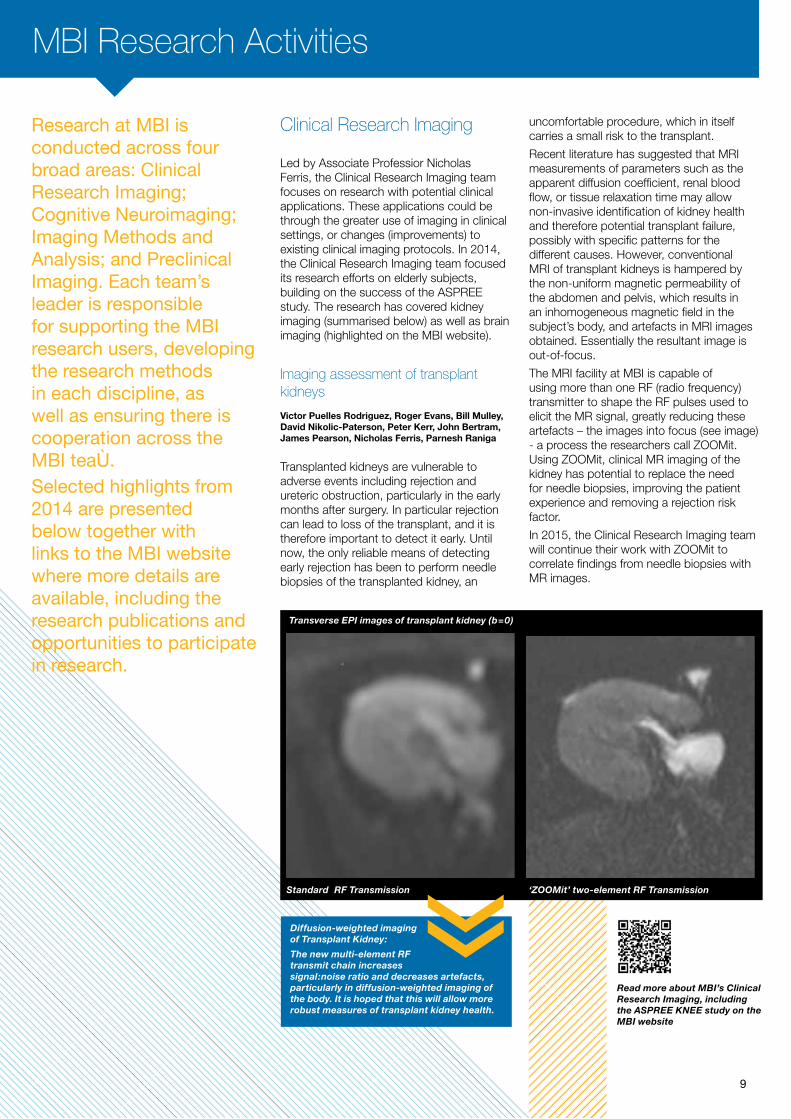
Recent literature has suggested that MRI measurements of parameters such as the apparent diffusion coefficient, renal blood flow, or tissue relaxation time may allow non-invasive identification of kidney health and therefore potential transplant failure, possibly with specific patterns for the different causes. However, conventional MRI of transplant kidneys is hampered by the non-uniform magnetic permeability of the abdomen and pelvis, which results in an inhomogeneous magnetic field in the subject’s body, and artefacts in MRI images obtained. Essentially the resultant image is out-of-focus.
The MRI facility at MBI is capable of using more than one RF (radio frequency) transmitter to shape the RF pulses used to elicit the MR signal, greatly reducing these artefacts – the images into focus (see image) - a process the researchers call ZOOMit. Using ZOOMit, clinical MR imaging of the kidney has potential to replace the need for needle biopsies, improving the patient experience and removing a rejection risk factor.
In 2015, the Clinical Research Imaging team will continue their work with ZOOMit to correlate findings from needle biopsies with MR images.
Research at MBI is conducted across four broad areas: Clinical Research Imaging; Cognitive Neuroimaging; Imaging Methods and Analysis; and Preclinical Imaging. Each team’s leader is responsible for supporting the MBI research users, developing the research methods in each discipline, as well as ensuring there is cooperation across the MBI teams.Selected highlights from 2014 are presented below together with links to the MBI website where more details are available, including the research publications and opportunities to participate in research.
Clinical Research Imaging
Led by Associate Professior Nicholas Ferris, the Clinical Research Imaging team focuses on research with potential clinical applications. These applications could be through the greater use of imaging in clinical settings, or changes (improvements) to existing clinical imaging protocols. In 2014, the Clinical Research Imaging team focused its research efforts on elderly subjects, building on the success of the ASPREE study. The research has covered kidney imaging (summarised below) as well as brain imaging (highlighted on the MBI website).
Imaging assessment of transplant kidneys
Victor Puelles Rodriguez, Roger Evans, Bill Mulley, David Nikolic-Paterson, Peter Kerr, John Bertram, James Pearson, Nicholas Ferris, Parnesh Raniga
Transplanted kidneys are vulnerable to adverse events including rejection and ureteric obstruction, particularly in the early months after surgery. In particular rejection can lead to loss of the transplant, and it is therefore important to detect it early. Until now, the only reliable means of detecting early rejection has been to perform needle biopsies of the transplanted kidney, an
Standard RF Transmission ‘ZOOMit’ two-element RF Transmission
Diffusion-weighted imaging of Transplant Kidney:
The new multi-element RF transmit chain increases signal:noise ratio and decreases artefacts, particularly in diffusion-weighted imaging of the body. It is hoped that this will allow more robust measures of transplant kidney health.
Transverse EPI images of transplant kidney (b=0)
9
Read more about MBI’s Clinical Research Imaging, including the ASPREE KNEE study on the MBI website
MBI Research Activities
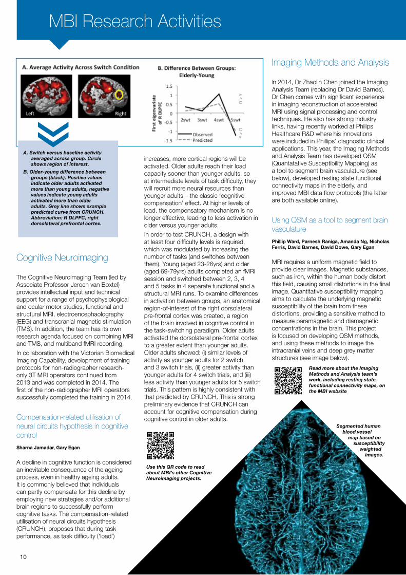
A. Switch versus baseline activity averaged across group. Circle shows region of interest.
B. Older-young difference between groups (black). Positive values indicate older adults activated more than young adults, negative values indicate young adults activated more than older adults. Grey line shows example predicted curve from CRUNCH. Abbreviation: R DLPFC, right dorsolateral prefrontal cortex.
Cognitive Neuroimaging
The Cognitive Neuroimaging Team (led by Associate Professor Jeroen van Boxtel) provides intellectual input and technical support for a range of psychophysiological and ocular motor studies, functional and structural MRI, electroencephaolography (EEG) and transcranial magnetic stimulation (TMS). In addition, the team has its own research agenda focused on combining MRI and TMS, and multiband fMRI recording.
In collaboration with the Victorian Biomedical Imaging Capability, development of training protocols for non-radiographer research-only 3T MRI operators continued from 2013 and was completed in 2014. The first of the non-radiographer MRI operators successfully completed the training in 2014.
Compensation-related utilisation of neural circuits hypothesis in cognitive control
Sharna Jamadar, Gary Egan
A decline in cognitive function is considered an inevitable consequence of the ageing process, even in healthy ageing adults. It is commonly believed that individuals can partly compensate for this decline by employing new strategies and/or additional brain regions to successfully perform cognitive tasks. The compensation-related utilisation of neural circuits hypothesis (CRUNCH), proposes that during task performance, as task difficulty (‘load’)
increases, more cortical regions will be activated. Older adults reach their load capacity sooner than younger adults, so at intermediate levels of task difficulty, they will recruit more neural resources than younger adults – the classic ‘cognitive compensation’ effect. At higher levels of load, the compensatory mechanism is no longer effective, leading to less activation in older versus younger adults.
In order to test CRUNCH, a design with at least four difficulty levels is required, which was modulated by increasing the number of tasks (and switches between them). Young (aged 23-26yrs) and older (aged 69-79yrs) adults completed an fMRI session and switched between 2, 3, 4 and 5 tasks in 4 separate functional and a structural MRI runs. To examine differences in activation between groups, an anatomical region-of-interest of the right dorsolateral pre-frontal cortex was created, a region of the brain involved in cognitive control in the task-switching paradigm. Older adults activated the dorsolateral pre-frontal cortex to a greater extent than younger adults. Older adults showed: (i) similar levels of activity as younger adults for 2 switch and 3 switch trials, (ii) greater activity than younger adults for 4 switch trials, and (iii) less activity than younger adults for 5 switch trials. This pattern is highly consistent with that predicted by CRUNCH. This is strong preliminary evidence that CRUNCH can account for cognitive compensation during cognitive control in older adults.
Imaging Methods and Analysis
In 2014, Dr Zhaolin Chen joined the Imaging Analysis Team (replacing Dr David Barnes). Dr Chen comes with significant experience in imaging reconstruction of accelerated MRI using signal processing and control techniques. He also has strong industry links, having recently worked at Philips Healthcare R&D where his innovations were included in Phillips’ diagnostic clinical applications. This year, the Imaging Methods and Analysis Team has developed QSM (Quantatative Susceptibility Mapping) as a tool to segment brain vasculature (see below), developed resting state functional connectivity maps in the elderly, and improved MBI data flow protocols (the latter are both available online).
Using QSM as a tool to segment brain vasculature
Phillip Ward, Parnesh Raniga, Amanda Ng, Nicholas Ferris, David Barnes, David Dowe, Gary Egan
MRI requires a uniform magnetic field to provide clear images. Magnetic substances, such as iron, within the human body distort this field, causing small distortions in the final image. Quantitative susceptibility mapping aims to calculate the underlying magnetic susceptibility of the brain from these distortions, providing a sensitive method to measure paramagnetic and diamagnetic concentrations in the brain. This project is focused on developing QSM methods, and using these methods to image the intracranial veins and deep grey matter structures (see image below).
Segmented human blood vessel
map based on susceptibility
weighted images.
10
Use this QR code to read about MBI’s other Cognitive Neuroimaging projects.
Read more about the Imaging Methods and Analysis team’s work, including resting state functional connectivity maps, on the MBI website

Developing novel molecular FLECT-CT tracers (Monash Institute of Pharmaceutical Sciences)
Karlheinz Peter (Baker IDI), Bock Lim, Joy Yao, May
Lin, Ben Chen
Dr Bock Lim and team under the supervision of Professor Karlheinz Peter (Baker IDI) have been employing the world-first 360-degree fluorescence CT (FLECT-CT Scanner) located at the Monash Institute of Pharmaceutical Sciences, Parkville to generate new methods of disease diagnosis. Their work investigates novel fluorescence molecular tracers that can detect important changes of disease inflammation and thereby allow various stages of pathological processes to be tracked. Besides detailed disease-diagnosis, assessment of drug-candidate efficacy can also be monitored using this method. These studies will have great implications for the design of future preclinical research and subsequent pharmaceutical development.
Multimodal image of a mouse using X-ray Computed Tomography (CT, in grayscale) and Positron Emission Tomography (PET, in colour, resolution 0.8 mm) with the PET-tracer Fluorodeoxyglucose (FDG) to monitor glucose metabolism over time.
FLECT-CT scan image from MIPS, showing the liver (coloured section) of a rat, using one of the newly developed novel fluorescence molecular tracers.
Small-animal PET-CT imaging of an in vivo model of mouse carotid artery thrombosis with the emerging radioisotope 64Cu and the next generation Cu ligand MeCOSar. The results demonstrate specific binding of a single-chain antibody against activated platelets on the left and no binding of a mutated control antibody on the right.
Pre-clinical Imaging
In 2014 new projects commenced in the fields of cardiovascular science, neuroscience, metabolic control and nanoparticle contrast agents. Collaborations spanned multiple departments across Monash University, the Australian Regenerative Medicine Institute, the Ritchie Centre Centre for Baby Health Research (Monash Health) and RMIT University. Using diffusion tensor and T1/T2-weighted imaging sequences the Preclinical Imaging Team are concentrating on the development of protocols for repeated imaging of the same animals over the course of time to investigate pathophysiological changes in disease states such as stroke and heart failure and also fundamental studies of fetal brain vasculature development (highlighted on the MBI website). CT imaging continues to be used frequently for materials studies and in 2014 we started MBI’s first in vivo PET-CT study of metabolic regulation in mice (highlighted below).
Hypothalamic neuron involvement in food intake regulation
Michael Cowley, Stephanie Simonds, Aldo Besmer, Ruth Vreys
The work of Professor Michael Cowley (Department of Physiology, Monash University) and his team focuses on receptor pathways and various nuclei within the hypothalamus in mice that produce hormones to regulate food intake. Taking advantage of MBI’s small animal PET-CT imaging expertise, Professor Cowley has shown that activation of specific receptor pathways of the brain increases neural activity in the hypothalamic neurons and glucose utilisation in multiple organ systems, thereby influencing whole body metabolism.
Developing PET-CT tracers for blood-based research (AMREP)
Christoph Hagemeyer, Karen Alt (Baker IDI), Karlheinz Peter (Baker IDI), Paul Donnelly (Bio 21), Frank Caruso (University of Melbourne).
MBI equipment also extends to AMREP (Alfred-Monash Research and Education Precinct) at The Alfred Hospital in Prahran, where a microPET-CT is located. Co-operated by MBI, AMREP Animal Services and the Baker IDI, much of the research has a strong focus on cardiovascular and metabolic disease and is led by Professor Hagemeryer. There is also a focus on nanotechnology, particularly as it relates to the development of PET-CT tracers.
In 2014, a research collaboration including AMREP and Bio21 (A/Prof Donnelly) demonstrated that site-specific bioconjugation using the bacterial enzyme Sortase A is more efficient than chemical conjugation for tracking and tracing platelets. Platelets play an essential role in thrombosis as well as inflammation, making them attractive markers for molecular imaging. The newly developed tracer is now under clinical development by the Australian Biotech company Clarity Pharmaceuticals for human use.
In a separate piece of work, collaboratively conducted with the University of Melbourne (Prof Caruso), researchers quantitatively evaluated the blood circulation times and clearance rate of novel tannic acid nanoparticles in vivo. This research was featured on the cover page of the leading journal Angewandte Chemie and received international press coverage.
11
The Preclinical Imaging team has undertaken studies into fetal brain vasculature development - you can read about this and other Preclinical Imaging projects on the MBI website

Cannabis, cognition, brain and mental health
Australians are among the highest per capita users of cannabis in the world. Over a ten-year period, our research team have documented the neurobiological, cognitive and mental health harms associated with regular cannabis use. We are currently undertaking studies that extend these findings, including investigations that examine whether brain alterations in cannabis users can be restored following cessation or substantive reduction of use. With approximately 200 million cannabis users worldwide, the findings (positive or negative) will be of relevance to researchers, clinicians, policy-makers, and those affected by cannabis. Recently, we reviewed the entire literature on the relationship between cannabis use and structural and functional alterations in the brain. We found that some of the brain alterations are similar in cannabis users without psychosis and people with psychosis who don’t use cannabis. Additionally, these brain alterations are associated with the degree of cannabis use and severity of psychotic symptoms.
Linked Laboratories
The Monash Clinical and Imaging Neuroscience Laboratory aims to understand the principles and mechanisms of human brain function in order to uncover causes and treatments of mental illness.
MCIN research activities primarily use brain imaging and other tools from cognitive neuroscience to understand human brain structure and function in health and disease. The MCIN team has a diverse range of expertise, with particular strengths in structural, functional, diffusion and spectroscopic MRI, EEG, TMS, PET, graph theory and network science, clinical neuropsychology, and translational research. Here, we present some of the highlights of our work across a range of areas. You can see other aspects of our work, including recent research outcomes on our website (use the QR Code below). Note MCIN underwent a name change in early 2015 and is now called the Brain and Mental Health Laboratory (BMH).
Substance and behavioural addictions
Substance dependence, problem gambling, excessive eating and Obsessive-Compulsive Disorder (OCD) are common examples of addictions and compulsive behaviours with high personal impact and societal costs. These behaviours involve changes in the brain of those affected, making it hard for the person to stop. We seek to understand how individual differences in reward/punishment learning, cognitive control and decision-making and their neural underpinnings contribute to vulnerability to develop and shape different forms of addictive and compulsive behaviour, and we aim to manipulate these systems to facilitate recovery. Ultimately, we seek to pave the way from vulnerability to expression of disease and eventual recovery. We have recently reviewed the latest discoveries in neuroscience of gambling behaviour, and the links between adolescent substance use and associated alterations in the brain’s connection.
Review the MCIN home page and latest research findings (note MCIN underwent a name change in early 2015 and is now called the Brain and Mental Health Laboratory (BMH).
Use this QR code to access the review: Goudriaan AE, Yücel M, van Holst RJ (2014) Getting a grip on problem gambling - What can neuroscience tell us? Frontiers in Behavioral Neuroscience. 8:141.
Access A/Prof Fornito’s article on Brain network structure and function in schizophrenia.
Read the article on cannabis and brain structure and function via this QR code.
The Monash Clinical and Imaging Neuroscience Laboratory (MCIN)
12

Schizophrenia and other psychoses
Psychosis is a debilitating neuropsychiatric syndrome characterised by delusions, hallucinations, disorganised thinking and cognitive and emotional disturbances. We have recently identified an MRI-based biological marker of risk for psychosis based on changes in brain activity. We are currently developing new therapies to remediate these changes, and hopefully minimize the risk of disease in young people. We are also conducting research into the genetic and molecular basis of schizophrenia.
Neuroethics and policy
Neuroscience research utilising emerging diagnostic technologies (e.g. fMRI, EEG, genetics) and neural interventions (e.g. brain stimulation, targeted pharmacologies and implants) including the research conducted at MCIN, has the potential to transform our ability to treat and prevent mental illness and addiction. These technologies may influence a person’s sense of self, agency and identity that are hard to predict and identify. Neural explanations of mental illness can exert powerful influences on social attitudes towards mental illness (e.g. stigma, discrimination) and the way that society responds to those with a mental illness (e.g. increased medical treatment versus compulsory treatment orders). The research performed at MCIN will help ensure that we realise the benefits of neuroscience research while minimising any unintentional harm, and assuring that these developments are provided in a manner that is equitable, economical, efficient and fair.
Network science and connectomics
The human brain is an extraordinarily complex network, comprising billions of neurons connected by trillions of fibres. All our thoughts, emotions and behaviour arise from interactions unfolding on this intricate web of connectivity. Accordingly, generating a comprehensive map of the brain’s myriad connections — a so-called human connectome —has become a central goal of neuroscience. Our team has led the way in this field, developing new methods for mapping the connectome with MRI and using these techniques to understand brain network function in health and disease. The images below show various ways of visualising the connectome. These particular images map connections in the brain that increase in strength during late adolescence. Most of these connections were long-range projections between highly connected brain network ‘hubs’.
Above: A brain-wide map of structural connectivity deficits in patients with schizophrenia. The relatively diffuse impairment that particularly affects fronto-posterior anatomical connectivity is highlighted. In this whole-brain analysis, no increases of structural connectivity were found. Letters denote different regions.
School of Psychological Sciences
13

Staff
MCIN Personnel
Prof Murat Yücel Director
A/Prof Alex FornitoDeputy Director
Prof Leonardo Fontenelle Visiting Professor of Clinical Neuropsychiatry
Dr Chao Suo Lab Manager and Postdoctoral Research Fellow
Dr Carsten Murawski Honorary Fellow of Neuroeconomics
Dr Valentina Lorenzetti Postdoctoral Research Fellow
Dr George Youssef Postdoctoral Research Fellow
Dr Paul Klauser Postdoctoral Research Fellow
Dr Ben Fulcher Postdoctoral Research Fellow
Dr Nigel Rogasch Postdoctoral Research Fellow
Dr Adrian Carter Senior Research Fellow
Amy Allen Research Assistant
Amy Finlay Research Assistant
Orwa Dandash, PhD Candidate
Simon Baker, PhD Candidate
Leah Braganza, PhD Candidate
Ari Pinar, PhD Candidate
Fernanda Mata, PhD Candidate
Linden Parkes, PhD Candidate
Michelle Lamblin, PhD Candidate
Marni Kras, PhD Candidate
Rothanthi Daglas, PhD Candidate
Sungwook Chung, PhD Candidate
Aron Hill, PhD Candidate
Melanie Emonson, PhD Candidate
Judy Luigjes, International Trainee (Netherlands)
Laura Koenders, International Trainee (Netherlands)
Lianne Schmaal, International Trainee (Netherlands)
Sanne Oostermiejer, International Trainee (Netherlands)
Yvonne van Dalen, International Trainee (Netherlands)
Silvia Alonso, International Trainee (Spain)
Juan Verdejo, International Trainee (Spain)
Antoine Klauser, International Trainee (Switzerland)
Gareth Ball, International Trainee (UK)
Chris Greenwood, Honours Student
Karen Guo, Honours Student
Erin Oldenhof, Honours Student
James Marrow, Honours Student
Maria Soloveva, Honours Student
Lauren Kate den Ouden, Volunteer Student
Maya Shepherd, Volunteer Student
Ellen Stavrinos, Volunteer Student
Stuart Oldham, Volunteer Student
Callum Robert Dark, Volunteer Student
William Ryan McMahon, Volunteer Student
Students
14
Spectroscopic imaging
Magnetic Resonance spectroscopy and Chemical Shift Imaging are powerful magnetic resonance tools that can quantify the concentration of various metabolites within one or multiple brain regions. With the support from MBI and Siemens, our team have developed reliable methods to quantify neurotransmitters gamma-Aminobutyric acid (GABA), Glutamate (Glu), Glutamine (Gln) and Glutathione (GSH) using the 3T MRI scanner. In addition, we have developed a toolbox to measure the tissue fraction within the Voxel of Interest (the spatial unit of measure in an MRI), which has been used to correct for partial volume effect (errors inadvertently introduced when measuring voxel volume) in more than 10 national/international projects. This tool can help to estimate the concentration of metabolites within brain tissue more precisely.
Computational modelling
Recently, large-scale, complex datasets have become common across neuroscience, driving the need for new, more sophisticated methods of analysis. Within MCIN, we have developed analysis and modelling techniques for understanding how brain signals change in brain disorders that could uncover new biomarkers of psychological diseases. We have also implemented models for simulating how brain connections form during brain development, and introduced a methodology for understanding the relationship between gene expression and brain wiring. Below right is a visualisation of the expression levels of over 17000 genes (columns) across 213 regions of the mouse brain (rows).
Brain stimulation
Transcranial magnetic stimulation (TMS) passes a brief magnetic pulse through the skull to alter the activity of neurons in underlying regions of the brain. Using fMRI and EEG, we are trying to understand how stimulation of different points on the scalp surface results in changes to the activity of connected surface regions and regions deep in the brain. Developing individual-tailored methods for stimulating wider brain networks including deep brain structures is critical for improving the efficacy of TMS in both understanding and treating a broad range of disorders, including addiction, depression and psychosis.
Link to developmental TMS work undertaken by MCIN, investigating the link between TMS and long-range neural stimulation in people with schizophrenia.
A visualisation of the expression levels of over 17000 genes (columns) across 213 regions of the mouse brain (rows)
School of Psychological Sciences

Monash Neuroscience of Consciousness(MoNoC)
Linked Laboratories
15
The Monash Neuroscience of Consciousness (MoNoC) Research Laboratory aims to understand the neural basis of consciousness. Our approaches focus on three areas:1. Consciousness itself -
Developing the theory of consciousness and empirically testing it, revealing the boundary condition of conscious and non-conscious processing.
2. Attention - The relationship between consciousness and attention.
3. Biological motion processing - How does the perception of social motion stimuli depend on attention, and individual differences.
To learn more about their research, visit the MoNoC Research Lab website
On the relationship between consciousness and attention
Naotsugu Tsuchiya, Christof Koch (the Allen Institute, USA)
Over the last 20 years, our understanding of the neuronal basis of perceptual consciousness and selective attention has greatly progressed. This advancement was facilitated by research using visual illusions and task designs that keep sensory input constant yet vary internal factors such as top-down attention and subjective visibility.
To isolate the neuronal mechanisms of consciousness and attention, however, it has become increasingly clear that keeping the sensory input constant is not enough. Unless manipulated independently, consciousness and attention usually covary. Recent studies that independently vary both consciousness
This theoretical research is further detailed in The Cognitive Neurosciences, where Naotsugu Tsuchiya and Christof Koch (the Allen Institute, USA) co-authored a chapter.
and attention have found that the behavioral and neuronal effects of consciousness and attention can be dissociated, implying that their neuronal mechanisms may be largely independent. Yet, even if independent neuronal mechanisms underlie consciousness and attention, there remains a conceptual dispute over the exact relationship between these processes.
It is now generally accepted that subjects can selectively attend to attributes of events or objects without becoming aware of them. Whether the converse is also true is much more contentious. That is, is attentional amplification of the neural representation of an event or object always necessary to experience it (i.e., is consciousness without attention possible)? We argue that attentional amplification is necessary to experience an object only when it needs to be “selected” among other objects that compete with it
in space and time. In a situation without any competition (e.g., an isolated object or a uniform texture), selective attention may not play any significant role. Accordingly, we argue that the neuronal mechanisms that gives rise to consciousness need to be carefully disentangled from the neuronal mechanisms that resolve competition.
Using an isolated-object paradigm, future studies can test the possibility of consciousness with no top-down attentional amplification in mice or monkeys by inactivating synaptic inputs from frontoparietal attentional areas back to visual areas using the rapidly advancing technology of optogenetics.
Linked Laboratories
Measuring integrated information from the decoding perspective
Masafumi Oizumi, Shun-ichi Amari (RIKEN, Japan), Toru Yanagawa (RIKEN, Japan), Naotaka Fujii (RIKEN, Japan), Naotsugu Tsuchiya
Accumulating evidence indicates that the capacity to integrate information in the brain is a prerequisite for consciousness. Integrated Information Theory (IIT) of consciousness provides a mathematical approach to quantifying the information integrated in a system, called integrated information, Φ. Integrated information is defined theoretically as the amount of information a system generates as a whole, above and beyond the sum of the amount of information its parts independently generate. IIT predicts that the amount of integrated information in the brain should reflect levels of consciousness. Empirical evaluation of this theory requires computing integrated information from neural data acquired from experiments, although difficulties with using the original measure Φ precludes such computations.
Although some practical measures have been previously proposed, we found that these measures fail to satisfy the theoretical requirements as a measure of integrated information. Measures of integrated information should satisfy the lower and
You can read the findings of this research in a paper due to be published in May 2015.
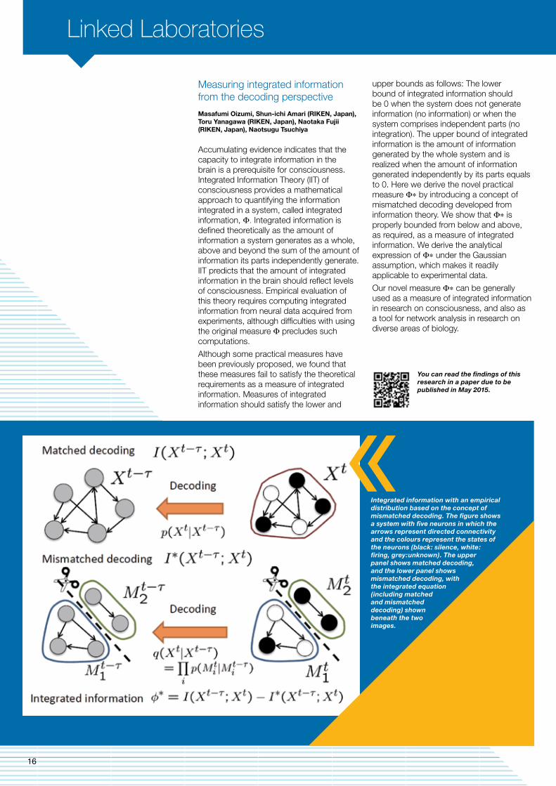
upper bounds as follows: The lower bound of integrated information should be 0 when the system does not generate information (no information) or when the system comprises independent parts (no integration). The upper bound of integrated information is the amount of information generated by the whole system and is realized when the amount of information generated independently by its parts equals to 0. Here we derive the novel practical measure Φ∗ by introducing a concept of mismatched decoding developed from information theory. We show that Φ∗ is properly bounded from below and above, as required, as a measure of integrated information. We derive the analytical expression of Φ∗ under the Gaussian assumption, which makes it readily applicable to experimental data.
Our novel measure Φ∗ can be generally used as a measure of integrated information in research on consciousness, and also as a tool for network analysis in research on diverse areas of biology.
Integrated information with an empirical distribution based on the concept of mismatched decoding. The figure shows a system with five neurons in which the arrows represent directed connectivity and the colours represent the states of the neurons (black: silence, white: firing, grey:unknown). The upper panel shows matched decoding, and the lower panel shows mismatched decoding, with the integrated equation (including matched and mismatched decoding) shown beneath the two images.
16

The importance to humans to correctly perceive and interpret social stimuli is hard to overstate. However, is social information so important that it actually determines which stimuli we will perceive and which are rendered invisible?
Using binocular rivalry, we investigated whether visually presented interactions between human actors receive preferential access to visual awareness over equally visible but non-interacting actors. When stimuli are presented using a binocular rivalry paradigm, perception fluctuates between the two eyes’ views. Our specific question was whether we could bias perception to the eye that receives the visual input containing interactive actions. We found indeed that interactive information was granted preferential access to visual awareness. Moreover, in conditions that required mentalising (i.e., the ability to understand the mental state of oneself and others), we found a strong preferential access for social actions, especially in those participants who were strong mentalisers. Our findings show that high-level perceptual mechanisms specialised for processing socially significant cues directly influence the availability of stimuli to visual awareness.
Clearly, to perceive and communicate within the social world, the human visual system allows social actions to bias selection processes very early in the visual hierarchy, thereby structuring perception almost from the first moment these cues enter the visual system.
Researchers
MoNoC Personnel
Students
Dror Cohen, PhD Candidate
Julian Mathews, PhD Candidate
William Wong, PhD Candidate
Matt Davidson, PhD Candidate
Noam Gordon, PhD Candidate (co-supervision)
Ben Chen, PhD Candidate (co-supervision)
Catherine Ding, PhD Candidate (co-supervision)
Gerick Lee, visiting Masters Student
Jochem van Kempen, visiting Masters Student
Pia Schroeder, visiting Masters Student
Miao Cao, Intern
Mana Fujiwara, Intern
Kristel Krella, Honours Student
Elise Rowe, Honours Student
Bianca Chen, Honours Student
On Zhi Xiang, Undergraduate Student
Trisha D’Lima, Undergraduate Student
Drisika Acharya, Undergraduate Student
Dinesh Giritharan, Undergraduate Student
Rannee Li, Undergraduate Student
Brandon Lam, Undergraduate Student
A/Prof Naotsugu TsuchiyaHead
A/Prof Jeroen van BoxtelHead
Dr Fabiano Baroni Postdoctoral Fellow
Dr Lisandro Kaunitz Postdoctoral Fellow
Dr Roger Koenig-Robert Postdoctoral Fellow
Dr Andrew Haun Postdoctoral Fellow
Dr Masafumi Oizumi Postdoctoral Fellow
Social interactions receive priority access to conscious perception
Jeroen van Boxtel, Junzhu Su (UCLA, USA), Hongjing Lu (UCLA, USA)
We previously showed that when adapting to a biological motion stimulus, a space invariant (global) action after effect can be observed in people with few autistic traits (Autism Spectrum Quotient (AQ);), while healthy people with many such traits do not show this global after effect (van Boxtel & Lu, 2013). Here we wanted to investigate both high and low level adaptation. The adaptation stimulus was an array of point-light walkers or runners, of which we only showed the leftward or rightward moving dots during the 6s adaptation period. This allowed for action adaptation, while the imbalance in motion energy allowed for a motion after effect to build up. Only the test stimulus differed between action and motion adaptation trials, being a morphed action (50% walker-50% runner), or a static random dot array, respectively. People with a high AQ showed less action adaptation (consistent with our previous finding), but more motion adaptation. This inverse relationship between high- and low-level adaptation is consistent with a predictive coding account. Interestingly, the finding that people with an increased number of autistic traits show increased motion adaptation is inconsistent with the idea that people with Autism Spectrum Disorder are generally less sensitive to visual illusions.
High-level action adaptation and low-level motion adaptation correlate with Autistic traits
Jeroen van Boxtel, Kristel Krella, Hongjing Lu (UCLA, USA)
(top) Schematic display of the stimuli used. (bottom) The results show that there was no difference between the partnered an un-partnered conditions when there was no mentalising task (determining whether there was an interaction between the actors). When there was a mentalising task, the interactive dance stimuli were propelled into consciousness.
School of Psychological Sciences
17

Interactions between MBI and the School of Psychological Sciences
Interactions between MBI and the co-located School of Psychological Sciences (SPS) Linked Laboratories are a natural synergy and strong benefit to both MBI and SPS researchers. This proximity promotes ready exchange of information, both informally and formally through the numerous courses and workshops held at MBI throughout the year, fostering an energetic research environment. As was the case in 2013, SPS once again promoted interactions with MBI in 2014 by jointly awarding MBI-SPS Small Grants. These grants are aimed primarily at early and mid-career researchers, to encourage imaging related research and help generate preliminary data to provide a competitive advantage in external grant funding applications. In 2014 three of these grants were awarded to projects undertaking studies to compare data obtained from the recently installed 7T MRI scanner at the University of Melbourne with 3T MRI data (awardees Drs Bryan Paton and Parnesh Raniga, both of MBI and Dr Paul Klauser of MCIN). A fourth grant was awarded to Dr Sophie Andrews (SPS) for the study of brain function in Huntington’s disease using functional magnetoencephalography (MEG) and MRI.
Training courses, tours and visits
MBI held a variety of courses and seminars throughout the year. Together with the CIBF, MBI hosted two courses for researchers in the use of MATLAB, a numerical computing environment and programming language. We hosted a number of research workshops including:
• Information and Communication Technology (ICT) for Life Sciences Forums: “Brain Structure, Brain Dynamics and Brain Initiatives: Cross currents in Neuroscience” presented by Dr Partha Mitra, Cold Spring Harbor Laboratory, and “The Human Connectome Project: Progress & Prospects”, presented by Prof David C. Van Essen, Washington University.
• A Neuroanatomy Workshop: “The organisation of the fore brain: new concepts based on gene expression during development” presented by Professor Charles Watson, Curtin University.
MBI Operations
In 2014, the major focus of the operations team was streamlining the user experience and the facilitation of improved user interactions with MBI staff and facilities. A new website was launched and a quarterly MBI Users Newsletter was distributed.
Staffing changesThere were several changes to staffing, with Dr Lisa Hutton (Senior Research and Operations Manager) taking maternity leave from June, and returning in the new role of Centre Manager of CIBF, in December, 2014. Lisa retains a fractional role, as the General Manager for MBI. Dr Charles Hardy, previously from the Monash University Department of Immunology, commenced as Research and Operations Manager in September. The Management and Administration team was strengthened by the arrival of Ms Nichola Thompson in the position of Resources Coordinator. Nikki’s previous experience within the University has been invaluable, particularly with respect to the management of building, facilities and occupational health and safety issues.
Facility utilisation and equipment
There was continued strong use of MBI facilities throughout the year. There were 33 new projects, evenly distributed between pre-clinical and clinical projects, representing 70% of all current projects. Users of our facilities came predominantly from within Monash University, with 13% from external research organisations. There was sustained high usage of the 3T MRI scanner, regularly over 70% of available hours were utilised, and this approached 90% utilisation during peak months, spread across approximately 25 separate projects. Usage of facilities for human studies, including electroencephalogy (EEG), transcranial magnetic stimulation (TMS) and oculomotor remained consistent throughout the year. The 9.4T MRI was the most heavily used of the pre-clinical facilities, frequently reaching 50% of available scan hours. We recorded dramatically increased utilisation of the Mediso PET-CT instrument located at the Alfred Hospital Campus, with billable hours approximately 5-fold greater than 2013.
18
communications. The MBI Imaging Team held a ‘Wrap-Up’ in November which provided a concise review of the projects undertaken by the team in 2014, highlighted specific and general outcomes, and provided an opportunity to meet the team and identify opportunities for collaboration.
• An Imaging Informatics Workshop (in conjunction with MASSIVE), to highlight projects that had been undertaken in data acquisition, collection and management, computing infrastructure, analysis, and visualisation of imaging data.
• Jointly with the CIBF, “Interactive Brain Function Workshop: Multi-modal approaches to understand brain function” with keynote speaker Dr David Leopold from NIH/NIMH, USA.
MBI provided a presentation and tour of the facilities for Year 10 students from John Monash Science School studying Imaging Science. We also hosted delegations from the USA National Science Foundation (NSF) to assess international research on the computational aspects of biomedical imaging, and tours for visiting academics including Professor Jon Shah, Director Institute of Neuroscience and Medicine, Forschungszentrum Jülich, Germany; Professor Warwick Anderson, CEO of the National Health and Medical Research Council; Professor Herbert Herzog, Division Head, Neuroscience, Garvan Institute, and Professor John Davey, Director of Research Technology Platforms at The University of Warwick, UK.
MBI held numerous talks throughout the year from national and international visitors, speaking on a range of topics covering imaging techniques such as magnetic resonance elastography, magnetic resonance spectroscopy, cognitive neurosciences and social
19
MBI Collaborations
MBI is focused on collaborative research efforts for both the development of biomedical imaging research techniques as well as their use in research projects. Throughout 2014 we sought to develop and maintain relationships with key research organisations or collaborations across
In 2014 the Victorian Biomedical Imaging Capability continued to be largely managed from MBI at Monash University. The on-going cooperation is a positive result for VBIC towards achieving the vision of Victoria “becoming a world leading capability dedicated to advancing the biomedical imaging research community in Victoria”, and the mission “to deliver the next generation of discoveries and enhance excellence in biomedical imaging to benefit translational research and to increase Victoria’s global competitiveness in Australia’s priority health areas.”
In 2014, the VBIC partners (including MBI) have jointly developed guidelines and policies for:
• Managing incidental findings identified as part of imaging (particularly brain MRI) studies.
• Processes for quality control and quality improvement within 3T MRI facilities.
The guidelines are planned for released in early 2015.
The Multi-modal Australian ScienceS Imaging and Visualisation Environment (MASSIVE) is a national imaging and visualisation facility operated by Monash University on behalf of the founding partners (the Australian Synchrotron, the CSIRO and Monash University). In 2014 MASSIVE completed its stage 2 upgrade and substantially expanded under the next stage (MASSIVE 3) of works. This included finalising the Characterisation Virtual Laboratory Project, which supported 450 researchers. M1 and M2 – the MASSIVE supercomputers – were upgraded to ensure future research requirements can be met.
MASSIVE has integrated multiple neuroimaging analysis software components with MBI equipment, enabling cross-platform and cross-modality integration of neuroinformatics tools, and neuroimaging databases and analysis workflows.
The Computational Bioimaging Hub of the Life Sciences Computation Centre (LSCC), based at MBI, provides access to support and training in biomedical imaging, assisting researchers to take advantage of informatics and imaging solutions, and enhancing their science output through the use of high performance computing facilities. The computational imaging scientists based in the hub also undertake research into advanced image analysis algorithms, informatics for data management, and visualisation tools. The outputs from these developments are accelerating analyses of large cohort studies, improving the characteristics of biomedical images, and providing tools for visualisation-led scientific discovery.
In mid 2014, Dr Michael Eager took over the role of LSCC Imaging Scientist following the departure of Dr Amanda Ng. Dr Eager collaborated with the Monash Immersive Visualisation Platform to generate ultra-high definition tractography images in the Monash University CAVE2; a large-scale virtual reality environment that enables interactive exploration of digital data.
The collaboration formed with CSIRO in 2011 (as part of VBIC), has further developed throughout 2014 with the announcement of funding a simultaneous MRI-PET scanner to be located at MBI. The equipment procurement and installation will take place throughout 2015 with the facility expected to be fully operational in early 2016.
In addition, CSIRO previously provided support for research fellows who have played a significant role in MRI protocol optimisation for the acquisition of MR images for the ASPREE NEURO and KNEE studies in 2014. ASPREE is a clinical trial investigating the benefits of low dose aspirin in the elderly, and MBI will be involved in two sub-studies; one focused the brain (ASPREE NEURO) and one on the knee (ASPREE KNEE).
20

CIBF is investigating integrative brain functions including attention, prediction and decision across four scales (Research Themes) – Brain Systems; Neural Circuits; Cells and Synapses; and Models and Technologies. Through understanding these complex functions, CIBF is gaining unique insights into how the brain interacts with the world. CIBF researchers (including several based at MBI) are actively pursuing multidisciplinary approaches to investigate some of the most complex problems in neuroscience.
CIBF 2014 Annual Report
MBI continued its contribution of the provision of national imaging research infrastructure through participation in the National Imaging Facility. During 2014 MBI
The International Neuroinformatics Coordinating Facility (INCF) was established in 2006 by the Organisation for Economic Co-operation and Development. Seventeen countries have INCF nodes including the USA, UK, Germany, Sweden, and Japan. Officially launched and headquartered at MBI, the INCF Victorian Node was established in 2012 through a collaboration between Monash University and The University of Melbourne. The INCF develops and maintains databases and computational infrastructure for neuroscientists. Software tools and standards for the international neuroinformatics community are being developed through the INCF Programs, which address infrastructure issues of high importance to the neuroscience community. The INCF also collects and makes available neuroinformatics tools in the INCF Software Center, where researchers can upload documentation, executables and related files; track use of their software; create a wiki; and establish development teams.
Monash University and The University of Melbourne, through their computational
The ARC Centre of Excellence for Integrative Brain Function formally began its operations in 2014. Led by Monash University (at MBI), the CIBF aims to understand how the brain interacts with the world. The centre is a collaboration between researchers at Monash University, The University of Queensland, The University of Melbourne, The University of Sydney, Australian National University and The University of New South Wales. CIBF investigators are also based at Queensland Institute of Medical Research and 11 other partnering institutions in Europe, Japan and the USA.
MBI recognises the importance of forming strategic alliances with key partners for the development of imaging infrastructure and research capabilities.
MBI and Siemens Australia continue to work closely on a number of key research projects, particularly in the MRI modality. The Monash-Siemens Collaboration Management Committee have continued to ensure the direction of the collaboration aligns with the strategies of both organisations. Indeed, in 2014 the agreement was extended for an additional year.
Five key projects were undertaken in 2014 as part of the Cooperative Agreement. Three of these focussed on the development, optimisation and validation of new or existing MR scanner sequences. One study uses a ZOOMit technique to produce extremely high-resolution images, which are being used at MBI to investigate rejection of kidneys in transplant patients, and to investigate mechanism to correct visual impairment.
Agilent has identified Monash University as a key research partner to work with in the Australian biomedical imaging research sector. In 2013, Agilent made a decision to cease production of MRI preclinical imaging systems, but continue to work closely with MBI regarding machine maintenance and servicing.
Agilent also provided funding for three key projects. Two of these were undertaken in the area of stem cell tracking. These projects will allow the development of techniques to assess how stem cells travel through the body, and whether they reach the organs where they are required (intended). One study specifically assesses the role for stem cells in the repair of brain injury, whilst the other focuses on the type of stem cell, and uptake throughout the body. The third study aims to develop a new series of techniques, which will allow optimal cardiac imaging in rodent models.
Partnerships
facilities, MASSIVE and the Victorian Life Sciences Computation Initiative (VLSCI) respectively, established the Victorian node of the INCF in 2012 for an initial three year period. The aims of the Victorian node are to support the neuroscience research communities at Monash University and The University of Melbourne using the MASSIVE and the VLSCI supercomputers; to develop neuroimaging data management and analytic tools; and to develop a program in clinical neuroinformatics research in cooperation with clinical neuroscientists and informaticians. A particular area of interest that has developed is the initiation of a new taskforce in the area of multi-scale imaging connectomics.
The MASSIVE and the VLSCI super-computing infrastructure are now contributing to the development of scalable, portable, and extensible applications that are being used by neuroscience research laboratories across Victoria for storing, analysing and simulating brain mechanisms and processes. This is furthering our knowledge of the human brain and aiding our understanding of the causes of brain diseases. In particular, MASSIVE and VLSCI are being used for management, analysis and visualisation of bioimaging datasets from VBIC nodes, and from the Imaging and Medical Beam Line at the Australian Synchrotron.
CIBF website
received staff support from NIF including part funding for the head of the Preclinical Imaging team, Dr James Pearson and the preclinical MR scientist Dr Qi-zhu Wu. Support was also received for the Imaging Informatics Officer, Dr Toan Dinh Nguyen, who is a software engineer and expert in imaging processing, computer vision, video coding, and 3D graphics.
National Node of Victoria, Australia
21

MBI Cognitive Neuroimaging
18. Akhlaghi, H., J. Yu, L. Corben, N. Georgiou-Karistianis, J. L. Bradshaw, E. Storey, M. B. Delatycki and G. F. Egan (2014). “Cognitive deficits in Friedreich ataxia correlate with micro-structural changes in dentatorubral tract.” Cerebellum 13(2): 187-198.
19. Davis, S. R., S. L. Davison, M. Gavrilescu, K. Searle, A. Gogos, S. L. Rossell, G. F. Egan and R. J. Bell (2014). “Effects of testosterone on visuospatial function and verbal fluency in postmenopausal women: results from a functional magnetic resonance imaging pilot study.” Menopause 21(4): 410-414.
20. Georgiou-Karistianis, N., J. C. Stout, D. J. Dominguez, S. P. Carron, A. Ando, A. Churchyard, P. Chua, I. Bohanna, A. R. Dymowski, G. Poudel and G. F. Egan (2014). “Functional magnetic resonance imaging of working memory in Huntington’s disease: cross-sectional data from the IMAGE-HD study.” Hum Brain Mapp 35(5): 1847-1864.
21. Karayanidis F., S. Jamadar (2014). “ Event-related potentials reveal multiple components of proactive and reactive control in task-switching’ J.A. Grange & G. Houghton, Task Switching and Cognitive Control, Oxford University Press, New York.
22. Poudel, G. R., J. C. Stout, D. J. Dominguez, L. Salmon, A. Churchyard, P. Chua, N. Georgiou-Karistianis and G. F. Egan (2014). “White matter connectivity reflects clinical and cognitive status in Huntington’s disease.” Neurobiol Dis 65: 180-187.
23. Saker, P., M. J. Farrell, F. R. Adib, G. F. Egan, M. J. McKinley and D. A. Denton (2014). “Regional brain responses associated with drinking water during thirst and after its satiation.” Proc Natl Acad Sci U S A 111(14): 5379-5384.
24. Smith, J. L., R. P. Mattick, S. D. Jamadar, J. M. Iredale (2014). “Deficits in behavioural inhibition in substance abuse and addiction: a meta-analysis.” Drug and Alcohol Dependence, 145, 1-33.
25. Thompson, D. K., C. Omizzolo, C. Adamson, K. J. Lee, R. Stargatt, G. F. Egan, L. W. Doyle, T. E. Inder and P. J. Anderson (2014). “Longitudinal growth and morphology of the hippocampus through childhood: Impact of prematurity and implications for memory and learning.” Hum Brain Mapp 35(8): 4129-4139.
26. Verghese, A., S. C. Kolbe, A. J. Anderson, G. F. Egan and T. R. Vidyasagar (2014). “Functional size of human visual area V1: a neural correlate of top-down attention.” Neuroimage 93 Pt 1: 47-52.
MBI Imaging Methods and Analysis
27. Duche, Q., P. Raniga, G. F. Egan, O. Acosta, G. Gambarota, O. Salvado and H. Saint-Jalmes (2014). “New partial volume estimation methods for MRI MP2RAGE.” Med Image Comput Comput Assist Interv 17(Pt 3): 129-136.
28. Goscinski, W. J., P. McIntosh, U. Felzmann, A. Maksimenko, C. J. Hall, T. Gureyev, D. Thompson, A. Janke, G. Galloway, N. E. Killeen, P. Raniga, O. Kaluza, A. Ng, G. Poudel, D. G. Barnes, T. Nguyen, P. Bonnington and G. F. Egan (2014). “The multi-modal Australian ScienceS Imaging and Visualization Environment (MASSIVE) high performance computing infrastructure: applications in neuroscience and neuroinformatics research.” Front Neuroinform 8: 30.
29. Krieger, S. N., C. J. Gauthier, D. Ivanov, L. Huber, E. Roggenhofer, B. Sehm, R. Turner and G. F. Egan (2014). “Regional reproducibility of calibrated BOLD functional MRI: implications for the study of cognition and plasticity.” Neuroimage 101: 8-20.
30. Krieger, S. N., D. Ivanov, L. Huber, E. Roggenhofer, B. Sehm, R. Turner, G. F. Egan and C. J. Gauthier (2014). “Using carbogen for calibrated fMRI at 7Tesla: comparison of direct and modelled estimation of the M parameter.” Neuroimage 84: 605-614.
31. Krieger, S. N., L. Huber, B. A. Poser, R. Turner and G. F. Egan (2014). “Simultaneous acquisition of cerebral blood volume-, blood flow-, and blood oxygenation-weighted MRI signals at ultra-high magnetic field.” Magn Reson Med.
MBI, MCIN and MoNoC Research Outputs
PublicationsMBI Clinical Research Imaging
1. Adlard, P. A., B. A. Tran, D. I. Finkelstein, P. M. Desmond, L. A. Johnston, A. I. Bush and G. F. Egan (2014). “A review of beta-amyloid neuroimaging in Alzheimer’s disease.” Front Neurosci 8: 327.
2. Ando, A., M. J. Farrell and S. B. Mazzone (2014). “Cough-related neural processing in the brain: a roadmap for cough dysfunction?” Neurosci Biobehav Rev 47: 457-468.
3. Beeman, S. C., L. A. Cullen-McEwen, V. G. Puelles, M. Zhang, T. Wu, E. J. Baldelomar, J. Dowling, J. R. Charlton, M. S. Forbes, A. Ng, Q. Z. Wu, J. A. Armitage, G. F. Egan, J. F. Bertram and K. M. Bennett (2014). “MRI-based glomerular morphology and pathology in whole human kidneys.” Am J Physiol Renal Physiol 306(11): F1381-1390.
4. Corben, L. A., S. R. Kashuk, H. Akhlaghi, S. Jamadar, M. B. Delatycki, J. Fielding, B. Johnson, N. Georgiou-Karistianis and G. F. Egan (2014). “Myelin paucity of the superior cerebellar peduncle in individuals with Friedreich ataxia: an MRI magnetization transfer imaging study.” J Neurol Sci 343(1-2): 138-143.
5. Farrell, M. J. and S. B. Mazzone (2014). “Sensations and regional brain responses evoked by tussive stimulation of the airways.” Respir Physiol Neurobiol 204: 58-63.
6. Farrell, M. J., D. Trevaks and R. M. McAllen (2014). “Preoptic activation and connectivity during thermal sweating in humans.” Temperature 1(2): 135-141.
7. Farrell, M. J., S. Koch, A. Ando, L. J. Cole, G. F. Egan and S. B. Mazzone (2014). “Functionally connected brain regions in the network activated during capsaicin inhalation.” Hum Brain Mapp 35(11): 5341-5355.
8. Gong, Q., L. Li, S. Tognin, Q. Wu, W. Pettersson-Yeo, S. Lui, X. Huang, A. F. Marquand and A. Mechelli (2014). “Using structural neuroanatomy to identify trauma survivors with and without post-traumatic stress disorder at the individual level.” Psychological medicine 44(1): 195-203.
9. Guo, J., N. Chen, R. Li, Q. Wu, H. Chen, Q. Gong and L. He (2014). “Regional homogeneity abnormalities in patients with transient ischaemic attack: a resting-state fMRI study.” Clinical neurophysiology : official journal of the International Federation of Clinical Neurophysiology 125(3): 520-525.
10. Jia, Z., Y. Wang, X. Huang, W. Kuang, Q. Wu, S. Lui, J. A. Sweeney and Q. Gong (2014). “Impaired frontothalamic circuitry in suicidal patients with depression revealed by diffusion tensor imaging at 3.0 T.” J Psychiatry Neurosci 39(3): 170-177.
11. Kolbe, S. C., T. J. Kilpatrick, P. J. Mitchell, O. White, G. F. Egan and J. Fielding (2014). “Inhibitory saccadic dysfunction is associated with cerebellar injury in multiple sclerosis.” Hum Brain Mapp 35(5): 2310-2319.
12. Morice, A. H., A. D. Jakes, S. Faruqi, S. S. Birring, L. McGarvey, B. Canning, J. A. Smith, S. M. Parker, K. F. Chung, K. Lai, I. D. Pavord, J. van den Berg, W. J. Song, E. Millqvist, M. J. Farrell, S. B. Mazzone, P. Dicpinigaitis and R. Chronic Cough (2014). “A worldwide survey of chronic cough: a manifestation of enhanced somatosensory response.” Eur Respir J 44(5): 1149-1155.
13. Narula, M., A. E. McGovern, S. K. Yang, M. J. Farrell and S. B. Mazzone (2014). “Afferent neural pathways mediating cough in animals and humans.” J Thorac Dis 6(Suppl 7): S712-719.
14. Poudel, G. R., G. F. Egan, A. Churchyard, P. Chua, J. C. Stout and N. Georgiou-Karistianis (2014). “Abnormal synchrony of resting state networks in premanifest and symptomatic Huntington disease: the IMAGE-HD study.” J Psychiatry Neurosci 39(2): 87-96.
15. Shanahan, C. J., T. V. Wrigley, M. J. Farrell, K. L. Bennell and P. W. Hodges (2014). “Postural response to vibration of triceps surae, but not quadriceps muscles, differs between people with and without knee osteoarthritis.” J Orthop Res 32(8): 989-996.
16. Thompson, D. K., K. J. Lee, G. F. Egan, S. K. Warfield, L. W. Doyle, P. J. Anderson and T. E. Inder (2014). “Regional white matter microstructure in very preterm infants: predictors and 7 year outcomes.” Cortex 52: 60-74.
17. Zalesky, A., H. Akhlaghi, L. A. Corben, J. L. Bradshaw, M. B. Delatycki, E. Storey, N. Georgiou-Karistianis and G. F. Egan (2014). “Cerebello-cerebral connectivity deficits in Friedreich ataxia.” Brain Struct Funct 219(3): 969-981.
22

MBI Preclinical Imaging
32. Bertram, J. F., L. A. Cullen-McEwen, G. F. Egan, N. Gretz, E. Baldelomar, S. C. Beeman and K. M. Bennett (2014). “Why and how we determine nephron number.” Pediatr Nephrol 29(4): 575-580.
33. Furtado, M. B., M. W. Costa, E. A. Pranoto, E. Salimova, A. R. Pinto, N. T. Lam, A. Park, P. Snider, A. Chandran, R. P. Harvey, R. Boyd, S. J. Conway, J. Pearson, D. M. Kaye and N. A. Rosenthal (2014). “Cardiogenic Genes Expressed in Cardiac Fibroblasts Contribute to Heart Development and Repair.” Circulation research 114(9): 1422-1434.
34. Lang, J. A., J. T. Pearson, A. B. te Pas, M. J. Wallace, M. L. Siew, M. J. Kitchen, A. Fouras, R. A. Lewis, K. I. Wheeler, G. R. Polglase, M. Shirai, T. Sonobe and S. B. Hooper (2014). “Ventilation/perfusion mismatch during lung aeration at birth.” J Appl Physiol (1985) 117(5): 535-543.
35. Nagai, H., I. Kuwahira, D. O. Schwenke, H. Tsuchimochi, A. Nara, T. Inagaki, S. Ogura, Y. Fujii, K. Umetani, T. Shimosawa, K. Yoshida, J. T. Pearson, K. Uemura and M. Shirai (2014). “β2-Adrenergic receptor-dependent attenuation of hypoxic pulmonary vasoconstriction prevents progression of pulmonary arterial hypertension in intermittent hypoxic rats.” PLoS One 9(10): e110693.
36. Ngo, J. P., S. Kar, M. M. Kett, B. S. Gardiner, J. T. Pearson, D. W. Smith, J. Ludbrook, J. F. Bertram and R. G. Evans (2014). “Vascular geometry and oxygen diffusion in the vicinity of artery-vein pairs in the kidney.” American journal of physiology. Renal physiology 307(10): F1111-1122.
37. Shirai, M., H. Tsuchimochi, H. Nagai, E. Gray, J. T. Pearson, T. Sonobe, M. Yoshimoto, T. Inagaki, Y. Fujii, K. Umetani, I. Kuwahira and D. O. Schwenke (2014). “Pulmonary vascular tone is dependent on the central modulation of sympathetic nerve activity following chronic intermittent hypoxia.” Basic Res Cardiol 109(5): 432.
38. Skiold, B., Q. Wu, S. B. Hooper, P. G. Davis, R. McIntyre, M. Tolcos, J. Pearson, R. Vreys, G. F. Egan, S. K. Barton, J. L. Cheong and G. R. Polglase (2014). “Early detection of ventilation-induced brain injury using magnetic resonance spectroscopy and diffusion tensor imaging: an in vivo study in preterm lambs.” PLoS One 9(4): e95804.
39. Zhou, L., O. Salvado, V. Dore, P. Bourgeat, P. Raniga, S. L. Macaulay, D. Ames, C. L. Masters, K. A. Ellis, V. L. Villemagne, C. C. Rowe and J. Fripp (2014). “MR-less surface-based amyloid assessment based on 11C PiB PET.” PLoS One 9(1): e84777.
40. Zohdi, V., J. T. Pearson, M. M. Kett, P. Lombardo, M. Schneider and M. J. Black (2014). “When early life growth restriction in rats is followed by attenuated postnatal growth: effects on cardiac function in adulthood.” Eur J Nutr
MCIN
41. Bell, S., A. Carter, R. Mathews, C. Gartner, J. Lucke and W. Hall (2014). “Views of Addiction Neuroscientists and Clinicians on the Clinical Impact of a ‘Brain Disease Model of Addiction’.” Neuroethics 7(1): 19-27.
42. Carter, A., R. Mathews, S. Bell, J. Lucke and W. Hall (2014). “Control and Responsibility in Addicted Individuals: What do Addiction Neuroscientists and Clinicians Think?” Neuroethics 7(2): 205-214.
43. Carter, A. (2014). “Demons: Our Changing Attitudes to Alcohol, Tobacco and Drugs by Virginia Berridge Oxford, UK, Oxford University Press, 2013, 304 pp. ISBN‐13: 978‐0‐19‐960498‐2 (hardback). Addiction 109(8): 1387-1387.
44. Cheetham, A., N. B. Allen, S. Whittle, M. Dennison, J. Simmons, M. Yücel and D. I. Lubman (2014). “Psychiatric symptoms, affective temperament, and neuroanatomy prospectively predict alcohol-related problems in adolescence.” Psychopharmacology 231(8): 1731-1742.
45. Cocchi, L., I. H. Harding, A. Lord, C. Pantelis, M. Yücel and A. Zalesky (2014). “Disruption of structure-function coupling in the schizophrenia connectome.” Neuroimage Clin 4: 779-787.
46. Dandash, O., A. Fornito, J. Lee, R. S. Keefe, M. W. Chee, R. A. Adcock, C. Pantelis, S. J. Wood and B. J. Harrison (2014). “Altered striatal functional connectivity in subjects with an at-risk mental state for psychosis.” Schizophr Bull 40(4): 904-913.
47. Dwyer, D. B., B. J. Harrison, M. Yücel, S. Whittle, A. Zalesky, C. Pantelis, N. B. Allen and A. Fornito (2014). “Large-scale brain network dynamics supporting adolescent cognitive control.” J Neurosci 34(42): 14096-14107.
48. Fiatarone Singh, M. A., N. Gates, N. Saigal, G. C. Wilson, J. Meiklejohn, H. Brodaty, W. Wen, N. Singh, B. T. Baune, C. Suo, M. K. Baker, N. Foroughi, Y. Wang, P. S. Sachdev and M. Valenzuela (2014). “The Study of Mental and Resistance Training (SMART) study-resistance training and/or cognitive training in mild cognitive impairment: a randomized, double-blind, double-sham controlled trial.” J Am Med Dir Assoc 15(12): 873-880.
49. Fontenelle, L. F., N. M. Lins-Martins, I. A. Melca, A. L. Lima, G. B. de Menezes, A. R. Torres, M. Yücel, E. C. Miguel and M. V. Mendlowicz (2014). “Exaggerating, mislabeling or simulating obsessive-compulsive symptoms: case reports of patients claiming to have obsessive-compulsive disorder.” Compr Psychiatry 55(5): 1188-1194.
23

MBI, MCIN and MoNoC Research Outputs
50. Fulcher, B. and N. Jones (2014). “Highly comparative feature-based time-series classification.” IEEE Trans. Knowl. Data Eng. 26: 3026–3037.
51. Fulcher, B. D., A. J. K. Phillips, S. Postnova and P. A. Robinson (2014). “A Physiologically Based Model of Orexinergic Stabilization of Sleep and Wake.” PLoS ONE 9(3): e91982.
52. Garfield, J. B., D. I. Lubman and M. Yücel (2014). “Anhedonia in substance use disorders: A systematic review of its nature, course and clinical correlates.” Aust N Z J Psychiatry 48(1): 36-51.
53. Goudriaan, A. E., M. Yücel and R. J. van Holst (2014). “Getting a grip on problem gambling: what can neuroscience tell us?” Front Behav Neurosci 8: 141.
54. Hall, W. D., A. Carter and M. Yücel (2014). “Ethical issues in the neuroprediction of addiction risk and treatment response.” Handbook of Neuroethics. N. Levy and J. Clausen. Heidelberg, Springer
55. Hall, W., M. Farrell and A. Carter (2014). “Compulsory treatment of addiction in the patient’s best interests: More rigorous evaluations are essential.” Drug and Alcohol Review 33(3): 268-271.
56. Harding, I. H., B. J. Harrison, M. Breakspear, C. Pantelis and M. Yücel (2014). “Cortical Representations of Cognitive Control and Working Memory Are Dependent Yet Non-Interacting.” Cereb Cortex.
57. Hong, S. B., A. Zalesky, A. Fornito, S. Park, Y. H. Yang, M. H. Park, I. C. Song, C. H. Sohn, M. S. Shin, B. N. Kim, S. C. Cho, D. H. Han, J. H. Cheong and J. W. Kim (2014). “Connectomic disturbances in attention-deficit/hyperactivity disorder: a whole-brain tractography analysis.” Biol Psychiatry 76(8): 656-663.
58. Hong, S. B., B. J. Harrison, O. Dandash, E. J. Choi, S. C. Kim, H. H. Kim, D. H. Shim, C. D. Kim, J. W. Kim and S. H. Yi (2014). “A selective involvement of putamen functional connectivity in youth with internet gaming disorder.” Brain Res.
59. Hong, S. B., E. J. Choi, H. H. Kim, J. E. Suh, M. J. Takagi, D. I. Lubman, J. W. Kim, C. D. Kim, S. H. Yi and M. Yücel (2014). “Decreased thalamic volumes in adolescent inhalant users from Korea and Australia.” World J Biol Psychiatry 15(8): 636-640.
60. Korgaonkar, M. S., A. Fornito, L. M. Williams and S. M. Grieve (2014). “Abnormal structural networks characterize major depressive disorder: a connectome analysis.” Biol Psychiatry 76(7): 567-574.
61. Lee, N. M., W. D. Hall, J. Lucke, C. Forlini and A. Carter (2014). “Food addiction and its impact on weight-based stigma and the treatment of obese individuals in the U.S. and Australia.” Nutrients 6(11): 5312-5326.
62. Liberg, B., P. Klauser, I. H. Harding, M. Adler, C. Rahm, J. Lundberg, T. Masterman, C. Wachtler, T. Jonsson, M. Kristoffersen-Wiberg, C. Pantelis and B. Wahlund (2014). “Functional and structural alterations in the cingulate motor area relate to decreased fronto-striatal coupling in major depressive disorder with psychomotor disturbances.” Front Psychiatry 5: 176.
63. Little, K., C. A. Olsson, S. Whittle, G. J. Youssef, M. L. Byrne, J. G. Simmons, M. Yücel, D. L. Foley and N. B. Allen (2014). “Association between serotonin transporter genotype, brain structure and adolescent-onset major depressive disorder: a longitudinal prospective study.” Transl Psychiatry 4: e445.
64. Lopez-Sola, C., L. F. Fontenelle, P. Alonso, D. Cuadras, D. L. Foley, C. Pantelis, J. Pujol, M. Yücel, N. Cardoner, C. Soriano-Mas, J. M. Menchon and B. J. Harrison (2014). “Prevalence and heritability of obsessive-compulsive spectrum and anxiety disorder symptoms: A survey of the Australian Twin Registry.” Am J Med Genet B Neuropsychiatr Genet 165b(4): 314-325.
65. Lorenzetti, V. and J. Cousjin (2014). “Cannabis use disorders and brain morphology.” Neuropathology of drug addictions and substance misuse Volume 1: Common substances of abuse - tobacco, alcohol, cannabinoids and opioids. V. R. Preedy. London, UK., Elsevier Inc
66. Lorenzetti, V., N. Solowij, A. Fornito, D. I. Lubman and M. Yücel (2014). “The association between regular cannabis exposure and alterations of human brain morphology: an updated review of the literature.” Curr Pharm Des 20(13): 2138-2167.
67. Meurk, C., A. Carter, B. Partridge, J. Lucke and W. Hall (2014). “How is acceptance of the brain disease model of addiction related to Australians’ attitudes towards addicted individuals and treatments for addiction?” BMC Psychiatry 14(1): 9.
68. Meurk, C., A. Carter, W. Hall and J. Lucke (2014). “Public Understandings of Addiction: Where do Neurobiological Explanations Fit?” Neuroethics 7(1): 51-62.
69. Meurk, C., B. Partridge, A. Carter, W. Hall, K. Morphett and J. Lucke (2014). “Public attitudes in Australia towards the claim that addiction is a (brain) disease.” Drug and Alcohol Review 33(3): 272-279.
70. Meurk, C., W. Hall, A. Carter and H. Chenery (2014). “Collecting real-time data from substance users raises unique legal and ethical issues: reply to Kuntsche & Labhart.” Addiction 109(10): 1760.
71. Mosley, P. E., W. Hall, C. Forlini and A. Carter (2014). “Alienation and Authenticity in Parkinson’s Disease and Its Treatment.” AJOB Neuroscience 5(4): 54-56.
72. Pizarro, M., L. F. Fontenelle, D. C. Paravidino, M. Yücel, E. C. Miguel and G. B. de Menezes (2014). “An updated review of antidepressants with marked serotonergic effects in obsessive-compulsive disorder.” Expert Opin Pharmacother 15(10): 1391-1401.
73. Rogasch, N. C., R. H. Thomson, F. Farzan, B. M. Fitzgibbon, N. W. Bailey, J. C. Hernandez-Pavon, Z. J. Daskalakis and P. B. Fitzgerald (2014). “Removing artefacts from TMS-EEG recordings using independent component analysis: importance for assessing prefrontal and motor cortex network properties.” Neuroimage 101: 425-439.
74. Rogasch, N. C., Z. J. Daskalakis and P. B. Fitzgerald (2014). “Cortical inhibition, excitation, and connectivity in schizophrenia: a review of insights from transcranial magnetic stimulation.” Schizophr Bull 40(3): 685-696.
75. Sjoerds, Z., J. Luigjes, W. van den Brink, D. Denys and M. Yücel (2014). “The role of habits and motivation in human drug addiction: a reflection.” Front Psychiatry 5: 8.
76. Takagi, M. J., D. I. Lubman, S. M. Cotton, A. Verdejo-Garcia, R. Vilar-Lopez and M. Yücel (2014). “A signal detection analysis of executive control performance among adolescent inhalant and cannabis users.” Subst Use Misuse 49(14): 1920-1927.
77. Takahashi, T., S. J. Wood, A. R. Yung, B. Nelson, A. Lin, M. Yücel, L. J. Phillips, Y. Nakamura, M. Suzuki, W. J. Brewer, T. M. Proffitt, P. D. McGorry, D. Velakoulis and C. Pantelis (2014). “Altered depth of the olfactory sulcus in ultra high-risk individuals and patients with psychotic disorders.” Schizophr Res 153(1-3): 18-24.
78. van den Heuvel, M. P. and A. Fornito (2014). “Brain Networks in Schizophrenia.” Neuropsychol Rev.
79. Via, E., A. Zalesky, I. Sanchez, L. Forcano, B. J. Harrison, J. Pujol, F. Fernandez-Aranda, J. M. Menchon, C. Soriano-Mas, N. Cardoner and A. Fornito (2014). “Disruption of brain white matter microstructure in women with anorexia nervosa.” J Psychiatry Neurosci 39(6): 367-375.
80. Vijayakumar, N., S. Whittle, M. Dennison, M. Yücel, J. Simmons and N. B. Allen (2014). “Development of temperamental effortful control mediates the relationship between maturation of the prefrontal cortex and psychopathology during adolescence: a 4-year longitudinal study.” Dev Cogn Neurosci 9: 30-43.
81. Vijayakumar, N., S. Whittle, M. Yücel, M. Dennison, J. Simmons and N. B. Allen (2014). “Thinning of the lateral prefrontal cortex during adolescence predicts emotion regulation in females.” Soc Cogn Affect Neurosci 9(11): 1845-1854.
82. Whittle, S., R. Lichter, M. Dennison, N. Vijayakumar, O. Schwartz, M. L. Byrne, J. G. Simmons, M. Yücel, C. Pantelis, P. McGorry and N. B. Allen (2014). “Structural brain development and depression onset during adolescence: a prospective longitudinal study.” Am J Psychiatry 171(5): 564-571.
83. Zalesky, A., A. Fornito, L. Cocchi, L. L. Gollo and M. Breakspear (2014). “Time-resolved resting-state brain networks.” Proc Natl Acad Sci U S A 111(28): 10341-10346.
MoNoC
84. Baroni, F., A. N. Burkitt and D. B. Grayden (2014). “Interplay of intrinsic and synaptic conductances in the generation of high-frequency oscillations in interneuronal networks with irregular spiking.” PLoS computational biology 10(5): e1003574.
85. Burt, J. S., T. J. Kipps and J. R. Matthews (2014). “Repetition in visual word identification: benefits and costs.” Q J Exp Psychol (Hove) 67(10): 1986-2009.
86. Capurro, A., F. Baroni, L. S. Kuebler, Z. Kárpáti, T. Dekker, H. Lei, B. S. Hansson, T. C. Pearce and S. B. Olsson (2014). “Temporal features of spike trains in the moth antennal lobe revealed by a comparative time-frequency analysis.” PloS one 9(1): e84037.
87. Kaunitz, L. N., J. E. Kamienkowski, A. Varatharajah, M. Sigman, R. Q. Quiroga and M. J. Ison (2014). “Looking for a face in the crowd: Fixation-related potentials in an eye-movement visual search task.” NeuroImage 89(1): 297-305.
88. Tsuchiya, N. and C. Koch (2014). “On the relationship between consciousness and attention.” The Cognitive Neurosciences. M. S. Gazzaniga. Cambridge, MA, MIT Press: 839-853
89. Wong, W. and N. S. Chiang Price (2014). “Testing neuronal accounts of anisotropic motion perception with computational modelling.” PLoS One 9(11): e113061.
24
Selected Presentations/OutreachMBI
1. Egan G.F. Australian Society for Molecular Imaging Symposium, Brisbane (June, 2014) – “Challenges and opportunities in preclinical imaging”
2. Egan G.F. CAI Ultra High Field MRI Symposium, Brisbane (June, 2014) – “Investigating brain network dysfunction in neurodegenerative diseases”
3. Egan G.F. Institute of Neuroscience, Faculty of Medicine, Rsezsow University, Poland (September, 2014) – “Advanced brain imaging in the large scale international brain research initiatives - collaborative research opportunities at Monash University, Australia.
4. Egan G.F. Inter. Symp. New Horizons in Human Brain Imaging: A focus on brain development (Hawaii, March 2014) - “Development of cortical folding in the neonatal brain”
5. Egan G.F. Siemens Australia; Australia & New Zealand Magnetic Resonance R&D Users Group Meeting, Brisbane (June, 2014) – “MR research at Monash Biomedical Imaging”
6. Egan G.F. Society for Biomedical Engineering, Melbourne (July, 2014), “Advanced brain imaging in the large scale international brain research initiatives”.
7. Pearson J.T. Bench to Bedside and Back: New Advances in Cardiovascular Research, HeartOtago Opening, University of Otago, New Zealand (Nov 2014). “Beta-blockade Prevents Coronary Dysfunction Induced by a High Salt Diet and Insulin Resistance in Rats”.
8. Pearson J.T. Gordon Research Conference on Angiotensin, Barga, Italy (Feb 2014). “Exploring RAS roles in Sex Differences in Coronary Dysfunction in Insulin Resistance with Synchrotron Imaging”.
9. Pearson J.T. Melbourne Convention Centre, World Congress of Cardiology, World Heart Federation (2014). “Functional Molecular Defects in the Diabetic Myocardium”.
MCIN
10. Lead role in professional workshops and public lectures involving international experts in the domains of problem gambling, addiction neuroscience, and neuroethics. We were also involved in professional activities linked to policy such as the Festival of Ideas, Theo Murphy High Flyers Think Tank.
11. Our research and staff were featured on ABC/SBS current affairs programs such as ‘Catalyst, and ‘Insight’, as well as documentary ‘Living with the Enemy’ and children’s science program ‘Scope’ on the ten network.
12. Public event at the Melbourne Convention and Exhibition Centre on the ethics of “Manipulating Morality” broadcast on ABC RN’s Big Ideas.
13. Multiple staff involved in ‘Scientists in Schools’, a national program that creates and supports long-term partnerships between primary or secondary school teachers and scientists or mathematicians.
MoNoC
14. 10th Conference of the Australasian Society for Cognitive Science (2014), Caulfield, Monash University “Recent computational and experimental insights into the mechanisms underlying conscious percepts” (organized by Jeroen van Voxtel. Speakers: van Boxtel, Seymour, Tsuchiya)
15. Attention Workshop (2014), University of Queensland, Australia (organised by Naotsugu Tsuchiya)
16. Baroni F., H. Kawasaki, K. Kovach K, H. Oya, M.A. Howard, R. Adolphs, and N. Tsuchiya. “Intracranial markers of conscious face perception in humans” (2014). NeuroEng2014, Adelaide (talk by NT)
17. Consciousness – here, there, everywhere? The prospects for panpsychism (2014), Byron Bay, Australia (organised by Naotsugu Tsuchiya, Bruno van Swinderen, Olivia Carter)
18. Consciousness, integrated information, and predictive coding (2014), Brisbane, ICON 12, Australia (Organised by Jakob Hohwy. Speakers: Hohwy, Tononi, Seth, Tsuchiya)
19. Fujiwara M., P. Riccarda, R. Koenig-Robert, and N. Tsuchiya “Predicting stimulus visibility based on pre-stimulus EEG responses” (2014). ANS Vision Satellite Meeting, Adelaide, Australia (poster by NT)
20. Haun A.M., B. Fabiano, J. van Kempen, H. Kawasaki, C.K. Kovach, O. Hiroyuki, M.A. Howard, R. Adolphs, N. Tsuchiya, “Informational Structure of Perceptual Experiences” (2014). ASSC18, Brisbane, Australia (poster by AMH)
21. Kaunitz L.N., E.G. Rowe, N. Tsuchiya. “How much do we see and remember outside of fixations in natural vision?” (2014). Australasian Neuroscience Society (ANS), Adelaide (poster by NT)
Current GrantsMBI
• ARC Centre of Excellence CE140100007 (2014-20). “ARC Centre of Excellence for Integrative Brain Function.” G. F. Egan, M. Rosa, J. Mattingley, P. Robinson, P. Sah, G. Stuart, M. Ibbotson, A. Lowery, E. Arabzadeh, G. Paxinos, P. Martin, S. Petrou, U. Grunert, E. Skafidis, M. Garrido; PIs - M. Breakspear, P. Mitra, J. Victor, T. Margrie, M. Diamond, G. Johnson, D. Leopold, J. Movshon, H. Markram, S. Hill.
• ARC Discovery Project DP140103045 (2014-2016). “The comparative physiology of oxygen delivery to the kidney.” D.W. Smith, R. G. Evans, B. S. Gardiner, J. T. Pearson, M. M. Kett.
• International Synchrotron Access Program Grant IA143 (2014). “Effects of intermittent hypoxia on the aging diabetic heart’s circulation.” J.T. Pearson, Y.C. Chen
• NHMRC (Monash University) Equipment Grant (2014). “Stimulating brain connections: an atlas of effective connections in the human brain.” G. F. Egan, M. Rosa, A. Lowery, J. Hohwy, A. Fornito
• NHMRC (Monash University) Equipment Grant (2014). “MRI compatible TMS coil, cooled TMS cortical coil, neuro-navigation systems and TMS coil cooling system.” G. F. Egan, P. Fitzgerald, J. Hohwy, A. Fornito
• NHMRC Principal Research Fellowship Grant #1003993 (2011-15). “Development of ultrahigh resolution brain imaging for investigating neurological and neurodegenerative diseases”, G. F. Egan
• NHMRC Project APP1050672 (2013-15). “The pathophysiology of septic acute kidney injury.” C. May, R. G. Evans, J. T. Pearson
• NHMRC Project APP1064973 (2014-16). “Facilitating the pulmonary increase in blood flow at birth.” S. B. Hooper, M. J. Kitchen, M. Siew, J. T. Pearson.
• NHMRC Project Grant APP1042528 (2013-15). “Activity in central cough networks in patients with cough hypersensitivity.” S. Mazzone, M. Farrell, G. F. Egan
• NHMRC Project Grant APP1042893 (2013-15). “A role for the pulvinar nucleus in visual cortical development and plasticity.” J. Bourne, D. Leopold, G. F. Egan
• NHMRC Project Grant APP1046037 (2013-16). “A longitudinal neuroimaging study investigating reorganisation of cerebellar-cerebral networks in Friedreich ataxia.” N. Georgiou-Karistianis, G. F. Egan, M. Delatycki, A. Churchyard, L. Corben
• NHMRC Project Grant APP1061291 (2014-16). “Development of folding in the fetal cerebral cortex.” D. W. Walker, J. Britto, L. Johnston, M. Tolcos, G. F. Egan
• NHMRC Project Grant APP1086188 (2015-18). “ASPREE NEURO Study: Cerebral micro-haemorrhages and aspirin in the elderly: cognitive and clinical consequences. A prospective randomised controlled trial.” J. McNeil, G. F. Egan, A. Brodtmann, S. Ward, N. Ferris, M. Bailey
MCIN
• Central Research Grants Scheme (2014-15). “An investigation of decision-making and self-regulation during pregnancy and its association with gestational weight gain”. Hayden, Fuller-Tyszkiewicz, Kremer, Yücel, Verdejo-Garcia, Boyle, Nagle, Skouteris.
• Faculty Strategic Grants (Monash University, Faculty of Medicine, Nursing and Health Sciences) (2014-15). “The neurobiology of overeating: ethical, clinical and policy implications of “food addiction.” Carter, Yücel, Verdejo-Garcia, Loxton and Hall.
• Faculty Strategic Grants (Monash University, Faculty of Medicine, Nursing and Health Sciences) (2014-15). “Brain mapping of nutrient sensing during food choice in obesity.” Verdejo-Garcia, Andrews, Yücel, Truby, Stice.
• NHMRC Early Career Fellowship (2015-18). “From brain maps to mechanisms: modelling the pathophysiology of schizophrenia. Fulcher
• NHMRC Equipment Grant ID (GNT9000248) (2014-15). “Brain Stimulation Equipment Suite.” Hoy, Fitzgerald, Segrave, Fitzgibbon, Maller, Rogasch, Fornito, Yücel.
• NHMRC Equipment Grant ID (GNT9000248) (2014-15). “Portable Sleep and Respiratory Phenotyping System.” Rajaratnam, Lubman, Ogeil, Cain, Edwards, Anderson, Yücel, Wang, Cornish, Hamilton, Ponsford.
MoNoC
• ARC Discovery Project (2013-15) “The neuronal basis of visual consciousness: how brain rhythms control the doors of perception.” Tsuchiya, Maller, Foster, Takaura
25

22. Koenig-Robert R.O., R. VanRullen, N. Tsuchiya. “Localizing category-selective BOLD signals in fMRI using SWIFT” (2014). Australasian Neuroscience Society (ANS), Adelaide (poster by NT)
23. Lee G., T. Noda, H. Takahashi, N. Tsuchiya, “Coding of auditory stimulus parameters between conscious states in the rat” (2014). Integrative Brain Function Workshop: Multi-modal approaches to understand brain function, Melbourne, Australia (poster by GL)
24. NeuroCog (2014), Coffs Harbour, Australia (organised by Naotsugu Tsuchiya)
25. Pathways of Consciousness - the future of Cognitive Neuroscience (2014). Latrobe University (presentation by Naotsugu Tsuchiya)
26. Paton B., J. Hohwy and N. Tsuchiya “Predicting stochastic perceptual fluctuation in continuous flash suppression in EEG” (2014). ANS Vision Satellite Meeting, Adelaide, Australia (poster by NT)
27. Su, J., J. van Boxtel and H. Lu. Meaningful actions and interactions receive priority in conscious perception (2014). VSS, St Petersburg, FL, USA (poster by JS)
28. Takaura K., N. Tsuchiya, N. Fujii, “Frequency-dependent spatiotemporal profiles of visual responses recorded with subdural electrodes in awake monkeys” (2014). SFN, USA (poster by KT).
29. Tononi, Koch, Oizumi, Albantakis, Tsuchiya (2014). “Integrated information theory” Association for the Scientific Study of Consciousness, 18th annual meeting. Brisbane, Australia. (3-hour tutorial)
30. Van Boxtel J. Attention and consciousness may operate through different gain functions, (2014). ASSC18, Brisbane, Australia (talk by JvB)
31. Van Boxtel J., and H. Lu. High-level action adaptation and low-level motion adaptation correlate with autism traits (2014). ECVP, Belgrade, Serbia. (poster by JvB)
32. Van Kempen J., H. Kawasaki, C.K. Kovach, H. Oya, M.A. Howard, R. Adolphs, N. Tsuchiya “Spectral phase and power predict stimulus category, but only power predicts visual awareness in intracranial EEG in humans” (2014). ASSC18, Brisbane, Australia (talk by JvK)
Professional Contributions
MBIGary EganExpert of National Standing
• Member, NHMRC Boosting Dementia Research Initiative Stakeholder Forum
Editorial Appointments
• Deputy Editor in Chief, Human Brain Mapping [IF 6.92] (2014 - )
Reviewer
• National Health Medical Research Council
• National Heart Foundation
• Australian Research Council
• Australian and New Zealand College of Anaesthetists
• NSW Department of Health
• Medical Research Council, London UK
• Health Research Council of New Zealand
• Qatar National Research Fund
• International Neuroinformatics Co-ordinating Facility, Stockholm
Board Memberships
• Member, Editorial Board, International Journal of Imaging Systems and Technology, Wiley, USA
• Member, Editorial Board, Frontiers in Neuroscience, Frontiers Research Foundation, Switzerland
MBI, MCIN and MoNoC Research Outputs
• Member, Editorial Board, Human Brain Mapping, Wiley Publishers, San Diego
• Member, Editorial Board, BrainMap Database, Research Imaging Centre, San Antonio, Texas, USA & Wiley Publishers, San Diego
• Governing Board Member, International Neuroinformatics Co-ordinating Facility Stockholm
International Committee Representation
• Member, Local Organizing Committee, ISMRM Annual Meeting & Exhibition, Singapore 7-13 May 2016.
• Member, International Expert Review Panel, Human Brain Project
• Member, International Program Committee of NEUROTECHNIX 2014
• Member, International Neuroinformatics Co-ordinating Facility Training Committee
• Member, Australian Synchrotron International Synchrotron Access Program Program Advisory Committee
National Committee Representation
• Scientific Advisory Board Member, Herston Imaging Research Facility, Brisbane, Australia
• Member, Australian Academy of Science Brain Implementation Committee
• Member, Scientific Program Committee, NeuroEng 2014: The 7th Australian Workshop on Computational Neuroscience, Adelaide
• Member, NHMRC Research Translation Faculty
• Member, NHMRC Assigners Academy
• Chairperson, Imaging and Visualisation Scientific Advisory Committee, Multi-modal Australian Sciences
• Member & Ambassador, Club Melbourne, State Government of Victoria
Leadership Positions
• Director, Centre of Excellence for Integrative Brain Function, Monash University
James Pearson• International Society of Heart Research
• High Blood Pressure Research Council of Australia
• American Physiological Society
• Member, Editorial Board, Frontiers in Integrative Physiology
Nicholas FerrisMemberships
• International Society for Magnetic Resonance in Medicine
• Radiological Society of North America (corresponding)
• Australian Medical Association
• Royal Australian and New Zealand College of Radiologists
• Member of Standards of Practice and Accreditation Committee
• Chair of MRI reference group
• Chair of e-Radiology Reference group
• Representative to Standards Australia committee IT-014
• Australian and New Zealand Society for Neuroradiology
• Society for Imaging Informatics in Medicine
Peer Review
• Assessor, National Health & Medical Research Council Project Grants
• Reviewer, Journal of Medical Imaging and Radiation Oncology
Jeroen van Boxtel• Associate Editor, Frontiers in Psychology (Consciousness Research)
Sharna Jamadar• Review Editor, Frontiers in Psychology (Cognition).
MCINMurat YücelProfessional Registrations
• APS College of Clinical Neuropsychologists
• APHRA endorsement in the clinical practise area of Clinical Neuropsychology
26
• Australian Health Practitioner Registration Agency
• Australian Psychological Society
Professional Affiliations
• Member, International Drug Abuse Research Society
• Member, Asia-Pacific Society for Alcohol and Addiction Research
• Member, Biological Psychiatry Australia
• Member, Australian Neuroscience Society
• Member, International College of Problems of Drug Dependence
• Member, International Society for Magnetic Resonance in Medicine
• Member, New York Academy of Sciences
• Member, Australasian Society for Psychiatric Research
Leadership Positions
• Program Leader, ‘Addiction Studies’, School of Psychological Sciences, Monash University
• Director, Brain and Mental Health Laboratory, Monash University
• Ambassador, Victorian Tall Poppy Campaign, Australian Institute of Policy and Science
• Editorial Board Member, Neurosci Biobeh Rev2011-, Psychiatry Res Neuroimag 2008-, Revista Brasileira Psiquiatria 2014-, The Open NeuroImag J 2009-,
Peer Review - Journals
• Proceedings of the National Academy of Sciences
• JAMA Psychiatry
• American Journal of Psychiatry
• British Journal of Psychiatry
• Biological Psychiatry
• ANZ Journal of Psychiatry
• Hum Brain Mapping
• Neuropsychopharm
• Neuroimage
• Psychological Medicine
• Biological Psychology
• Brain
• Addiction
• Addiction Biology
• Schiz Bull
• Schizophrenia Research
• Current Biology
• PlosOne
• Translation Psychiatry
Peer Review - Grant Bodies
• National Health & Medical Research Council of Australia, NHMRC
• Australian Research Council, ARC
• National Institute of Health, NIH (USA)
• Medical Research Council, MRC (UK)
• National Medical Research Council, NMRC (SING)
• ZonMW (NETH)
• Health Review Board, HRB (IRE)
• Health Research & Development, Ontario Mental Health Foundation (CAN)
Alex Fornito• Editorial Board, Biological Psychiatry
• Editorial Board, Journal of Neuroscience
• Executive Council Member, Biological Psychiatry Australia
• Local Organising Committee, Annual Meeting of Biological
• Psychiatry Australia
• Local Organising Committee, Annual Meeting of the Australasian Society for Cognitive Neuroscience
MoNoC• Organised symposium at the meeting of the Australasian Society for Cognitive
Science 2014 (J van Boxtel, K. Seymour, and N. Tsuchiya): Recent computational and experimental insights into the mechanisms underlying conscious percepts
Abbreviations3T / 9.4 T 3 Tesla / 9.4 Tesla
AMREP Alfred Medical Research and Education Precinct
ANSTO Australian Nuclear Science and Technology Organisation
AQ Autism Spectrum Quotient
ARC Australian Research Council
ARIN Australian Research Infrastructure Network
ASPREE Aspirin Reducing Events in the Elderly
BMH Brain and Mental Health (research laboratory)
CIBF Australian Research Council Centre of Excellence for Integrative Brain Function
CRUNCH Compensation-related utilisation of neural circuits hypothesis
CSIRO Commonwealth Scientific Industrial Research Organisation
CT Computed Tomography
EEG Electroencephalogram
FDG Fluorodeoxyglucose
FLECT Fluorescence Computed Tomography
fMRI Functional Magnetic Resonance Imaging
GABA Gamma-aminobutryic acid
Gln Glutamine
Glu Glutamate
ICT Information Communication Technology
IIT Integrated Information Theory
INCF International Neuroinformatics Coordinating Facility
LSCC Life Sciences Computation Centre
MASSIVE Multimodal Australian Sciences Visualisation Environment
MBI Monash Biomedical Imaging
MEG Magnetoencephalography MCIN Monash Clinical and Imaging Neurosciences (research
laboratory)
MCRL Monash Consciousness Research Laboratory
MIPS Monash Institute of Pharmaceutical Sciences
MoNoC Monash Neuroscience of Consciousness (research laboratory)
MRI Magnetic Resonance Imaging
NHMRC National Health and Medical Research Council
NIF National Imaging Facility
NIH National Institutes of Health
NSF National Science Foundation
OCD Obsessive Compulsive Disorder
PET Positron Emission Tomography
QSM Quantitative Susceptibility Mapping
RF Radio Frequency
SIEF Science and Industry Endowment Fund
SPECT Single Photon Emission Computed Tomography
SPS School of Psychological Sciences
TMS Transcranial Magnetic Stimulation
VBIC Victorian Biomedical Imaging Capability
VLSCI Victorian Life Sciences Computational Initiative
VPTN Victorian Platform Technology Network
27

Biomedical Imaging
Monash Biomedical Imaging
Monash University
770 Blackburn Road
Clayton VIC 3800
Telephone: 9905 0100
Email: [email protected]
https://platforms.monash.edu/mbi/
All information contained in this document is current at the time of publication. Monash University reserves the right to alter this information at any time (should the need arise). Please check the Monash University website for updates: www.monash.edu
Published August 2015.
CRICOS provider: Monash University 00008C