molecular physiology of water balance
DESCRIPTION
Fisiologia molecular del balance del agua, revisiónTRANSCRIPT
T h e n e w e ngl a nd j o u r na l o f m e dic i n e
n engl j med 372;14 nejm.org April 2, 2015 1349
Review Article
From the Epithelial Systems Biology Lab-oratory, National Heart, Lung, and Blood Institute, National Institutes of Health, Bethesda, MD (M.A.K.); the Department of Biochemistry and Cell Biology, School of Medicine, Kyungpook National Uni-versity, Taegu, South Korea (T.-H.K.); and the Institute of Medicine and Health Technology, Aalborg University, Aalborg, Denmark (S.N.). Address reprint requests to Dr. Knepper at the National Institutes of Health, 10 Center Dr., Bldg. 10, Rm. 6N307, Bethesda, MD 20892-1603, or at knep@ helix . nih . gov.
N Engl J Med 2015;372:1349-58.DOI: 10.1056/NEJMra1404726Copyright © 2015 Massachusetts Medical Society.
The hypothalamic–neurohypophyseal–renal axis normally main-tains water balance during variations in water intake and nonrenal losses of water. Failure of this mechanism is common in hospitalized patients, and
it results in a variety of water-balance disorders. In this article, we begin by review-ing the classic, integrative principles of water balance in mammals and then use this classic model as a framework to discuss the genes and gene products (pro-teins) involved in water balance. In so doing, our goal is to provide clinicians with a mechanistic basis for decisions regarding the diagnosis and treatment of water-balance disorders.
The regulation of water balance is governed by a high-gain feedback mecha-nism involving the hypothalamus, the neurohypophysis, and the kidneys (Fig. 1). Osmoreceptors in the hypothalamus, which originally were described by Verney,1 sense plasma osmolality. The molecular mechanism of “osmosensing” has re-cently been described by Danziger and Zeidel.2 It is, in part, dependent on activa-tion of nonselective calcium-permeable cation channels in osmosensing neurons that can serve as stretch receptors.
When plasma osmolality increases to levels above a physiologic threshold (290 to 295 mOsm per kilogram of water in most persons), there is increased secretion of the peptide hormone vasopressin from vasopressinergic nerve endings in the neu-rohypophysis. High osmolality also triggers thirst. Vasopressin binds to receptors in the kidney that decrease excretion of water (Fig. 2), and a greater fraction of filtered water is returned to the blood. The rate of water excretion can vary over a broad range in response to changes in plasma vasopressin levels without substan-tial changes in net solute excretion (osmolar clearance). This independent control of water and solute excretion is the result of specialized urinary concentrating and diluting mechanisms; these mechanisms are reviewed elsewhere.3
Increased renal reabsorption of water in response to vasopressin lowers plasma osmolality, thereby reducing the stimulus for vasopressin secretion and thirst and completing the feedback loop (Fig. 1). Table 1 provides a list of the major proteins that are responsible for components of the integrative model shown in Figure 1. These proteins are the focus of this review.
A rginine Va sopr essin
The gene coding for arginine vasopressin (AVP) is expressed in neurons of the supraoptic and paraventricular nuclei of the hypothalamus. Arginine vasopressin is a typical neuropeptide, since its gene codes for a prohormone that must un-dergo specific proteolytic processing to produce the active hormone. Thus, AVP codes for three peptides — the 9–amino acid peptide arginine vasopressin, a car-
Disorders of Fluids and ElectrolytesJulie R. Ingelfinger, M.D., Editor
Molecular Physiology of Water BalanceMark A. Knepper, M.D., Ph.D., Tae-Hwan Kwon, M.D., Ph.D.,
and Soren Nielsen, M.D., Ph.D.
The New England Journal of Medicine Downloaded from nejm.org by Edson Catacora on October 1, 2015. For personal use only. No other uses without permission.
Copyright © 2015 Massachusetts Medical Society. All rights reserved.

n engl j med 372;14 nejm.org April 2, 20151350
T h e n e w e ngl a nd j o u r na l o f m e dic i n e
rier protein called neurophysin-2, and a small glycoprotein called copeptin. Because vasopres-sin itself is difficult to measure in plasma sam-ples, some investigators are using measurements of copeptin in plasma as a surrogate for arginine vasopressin.4 Mutations in the arginine vaso-pressin gene that interfere with the processing and release of arginine vasopressin are associ-ated with central diabetes insipidus.
The oxytocin gene has a structure that is very similar to that of the arginine vasopressin gene. It is expressed in distinct oxytocinergic cells in the supraoptic and paraventricular nuclei of the hypothalamus and, like vasopressin, its secre-tion is increased by osmotic stimuli.5 It binds to
vasopressin receptors in the kidney and produc-es similar, although weaker, responses than ar-ginine vasopressin.6 Consequently, oxytocin is sometimes considered to be a second “antidi-uretic hormone.” Rarely, in the third trimester of pregnancy, a syndrome called transient vaso-pressin-resistant diabetes insipidus of pregnancy occurs as a result of placental secretion of vaso-pressinase (also called oxytocinase), which hy-drolyzes circulating vasopressin and oxytocin.7
Affected patients have a response to desmopres-sin acetate, which is resistant to this enzyme.
Va sopr essin R ecep t or s
After secretion into the general circulation from the posterior pituitary gland (neurohypophysis) (Fig. 1), arginine vasopressin is delivered to the kidney, where it exerts regulatory actions through the V2 receptor (gene symbol, AVPR2). The V2 va-sopressin receptor is a G protein–coupled recep-tor with physiologic functions that are mediated largely by the heterotrimeric G-protein Gs, result-ing in activation of adenylyl cyclases to increase the intracellular level of cyclic AMP (cAMP).3
Mutations in AVPR2 are responsible for X-linked nephrogenic diabetes insipidus.8
The kidney also expresses the V1a vasopressin receptor, largely in the vasculature of the renal medulla9; this receptor mediates the effects of va-sopressin on renal blood flow.10 The V1a vasopres-sin receptor signals chiefly through the hetero-trimeric G-protein Gq/11; this G protein activates phospholipase C and stimulates calcium mobili-zation. The V1a receptor is widely expressed throughout the body, whereas the V2 receptor is located chiefly in renal epithelia. A variety of localization studies and corresponding func-tional studies have shown that the V2 receptor acts chiefly in the principal cells of the renal collecting duct, the connecting tubule cells, the distal convoluted tubule cells, and the cells of the thick ascending limb of Henle (Fig. 3).
Bume ta nide-Sensi ti v e Sodium –Po ta ssium – Chl or ide
Co tr a nsporter
Vasopressin increases the rate of active absorp-tion of sodium chloride in the medullary thick ascending limbs of Henle,11,12 enhancing counter-current multiplication, which is the process re-
Figure 1. Feedback Loop Governing Regulation of Plas-ma Osmolality through Control of Arginine Vasopres-sin Secretion and Thirst.
An increase in plasma osmolality activates hypotha-lamic osmoreceptors to stimulate vasopressin secre-tion by the posterior pituitary gland. The resulting in-crease in the level of plasma vasopressin leads to an increase in renal water reabsorption and a decrease in water excretion. Increased water reabsorption reduces plasma osmolality. Osmosensing in the hypothalamus also stimulates thirst and drinking to help restore plas-ma osmolality. AVP denotes arginine vasopressin, PVN paraventricular nucleus, and SON supraoptic nucleus.
+
+
+
+
+
+
–
–
–
Waterreabsorption
Plasmaosmolality
Osmoreceptoractivation
PITUITARYGLAND
AVP secretion
Plasmavasopressin
Vasopressin-receptor activation
Water excretion
Thirst
Drinking
SONPVN
HYPOTHALAMUS
KIDNEY
The New England Journal of Medicine Downloaded from nejm.org by Edson Catacora on October 1, 2015. For personal use only. No other uses without permission.
Copyright © 2015 Massachusetts Medical Society. All rights reserved.

n engl j med 372;14 nejm.org April 2, 2015 1351
Molecular Physiology of Water Balance
sponsible for the medullary accumulation of solutes.3 The high concentration of solutes in the medullary interstitium furnishes the osmotic gra-dient needed to drive reabsorption of water from the renal collecting ducts. Consequently, the up-regulation of medullary interstitial accumulation of solutes by vasopressin contributes to the reg-ulation of water excretion.
In the thick ascending limbs of Henle, the transport of sodium and chloride from the lumen is mediated by the bumetanide-sensitive sodium–potassium–chloride cotransporter.13 Vasopressin up-regulates this cotransporter in at least two ways: short-term regulation that is a consequence of vesicular trafficking14 and long-term regula-tion that is a consequence of an increase in the expression of SLC12A1, which codes for the co-transporter protein.15 Up-regulation of the sodi-um–potassium–chloride cotransporter generally does not affect salt excretion, primarily because the thick ascending limb is upstream from the macula densa (Fig. 3), which compensates for changes in salt delivery by adjusting the glomeru-lar filtration rate. This compensatory process is called glomerulotubular feedback.
The diuretic bumetanide and other loop di-uretics increase salt excretion because they in-hibit the sodium–potassium–chloride cotrans-porter in the macula densa, thereby blocking the feedback to the glomerulus.16 Similarly, type I Bartter’s syndrome (loss-of-function mutations in SLC12A1) is manifested by a salt-losing syn-drome rather than by a simple defect in water balance.
Thi a zide-Sensi ti v e Sodium –Chl or ide Co tr a nsporter
Vasopressin also regulates salt transport in the distal convoluted tubule, which is present in each nephron a short distance downstream from the macula densa. It transports salt at a high rate but is impermeable to water and thereby contributes to dilution of the tubular fluid. The classic studies of Gottschalk and Mylle showed that the luminal fluid is dilute relative to blood plasma in this segment (independently of wheth-er vasopressin levels are high or low),17 thereby establishing that vasopressin increases water re-absorption downstream from this site in the col-lecting ducts. Vasopressin-induced increases in
salt transport out of the distal convoluted tubule can, in principle, increase the extent of luminal dilution, thereby increasing the driving force for reabsorption of water downstream.
Vasopressin exerts its effects on salt transport in the distal convoluted tubule by up-regulating the apical thiazide-sensitive sodium–chloride co-transporter, in part through effects on protein phosphorylation.18,19 This cotransporter is also regulated by aldosterone, which increases the abundance of the cotransporter protein in the distal convoluted tubule cells.20 As a result, the thiazide-sensitive sodium–chloride cotransport-er also plays a critical role in the regulation of sodium and chloride balance.
Inactivating mutations in the thiazide-sensitive sodium–chloride cotransporter cause Gitelman’s syndrome, which is manifested by hypotension, hypokalemia, hypomagnesemia, hypocalciuria, and metabolic alkalosis. Polyuria, which also oc-curs in patients with this syndrome, is primarily the result of hypokalemia21 rather than a direct effect of the loss of active sodium–chloride co-transport in the distal convoluted tubule.
Figure 2. Relationships among Plasma Vasopressin Concentration, Rate of Water Excretion, and Solute Excretion (Osmolar Clearance).
Water excretion decreases with increased levels of plasma vasopressin, whereas solute excretion remains relatively constant. This results in con-centrated urine at a high vasopressin concentration and dilute urine at a low vasopressin concentration.
Rat
e (µ
l/m
in)
100
80
60
40
20
0
90
110
70
50
30
10
1 10 100
Plasma Vasopressin Concentration (pmol/liter)
Dilute Urine Concentrated Urine
Osmolar clearance
Water excretion
The New England Journal of Medicine Downloaded from nejm.org by Edson Catacora on October 1, 2015. For personal use only. No other uses without permission.
Copyright © 2015 Massachusetts Medical Society. All rights reserved.
n engl j med 372;14 nejm.org April 2, 20151352
T h e n e w e ngl a nd j o u r na l o f m e dic i n e
Aqua por ins
Aquaporin-1
In the early 1990s, Agre and colleagues identified the first molecular water channel, aquaporin-1, which was found to be ubiquitously expressed.22 In the kidney, this water channel is expressed in the proximal tubule and thin descending limb of the loop of Henle.23 In the thin descending limb, it plays an important role in the countercurrent multiplier mechanism, allowing a rapid osmoti-cally driven exit of water from the lumen, thereby concentrating the luminal fluid.
In mice24 and humans,25 a lack of aquaporin-1 results in a near inability to concentrate urine.
Aquaporin-1 appears to be constitutively expressed in the kidney and is not regulated by vasopressin.
Aquaporin-2
Another aquaporin, aquaporin-2, is expressed throughout the collecting-duct system26 (i.e., in the region of the renal tubule where vasopressin regulates osmotic transport of water). Aquapo-rin-2 mediates the apical component of transepi-thelial water transport, and its regulation by va-sopressin controls the overall rate of water permeation across the collecting-duct epithelium. Most patients with non–X-linked nephrogenic di-abetes insipidus have mutations in AQP2.8
Extensive physiological studies involving rats
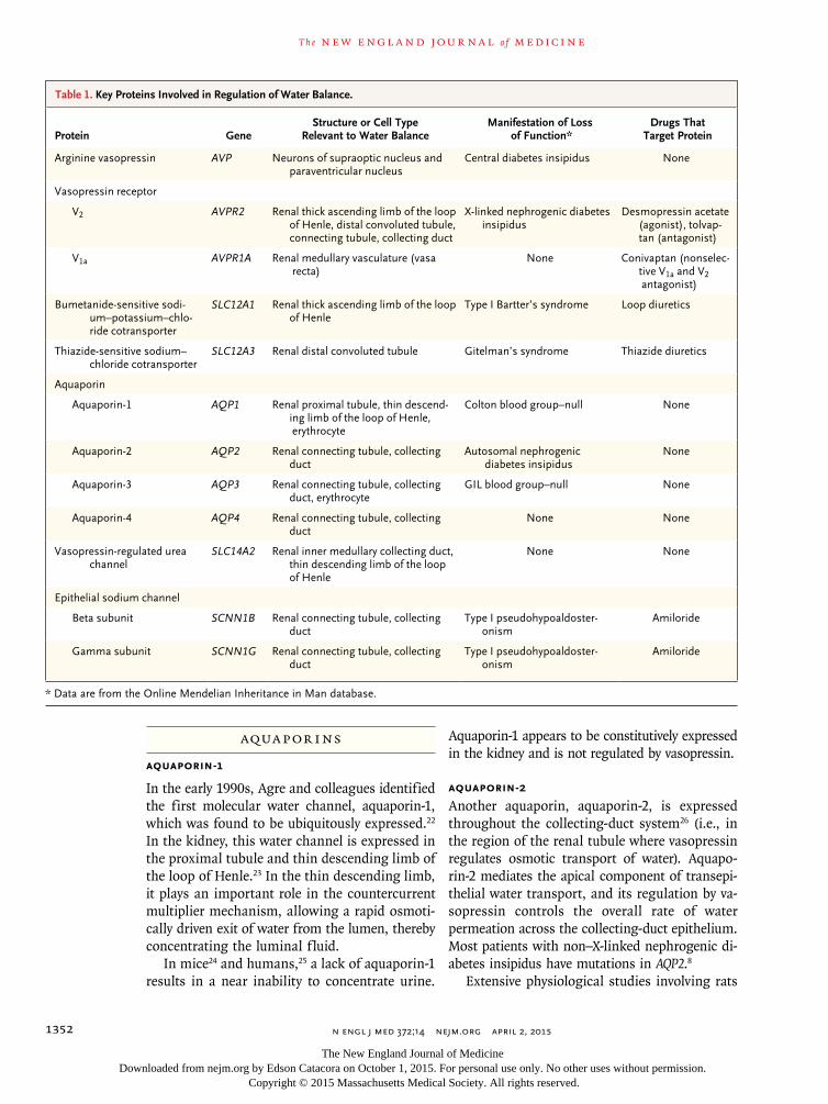
Protein GeneStructure or Cell Type
Relevant to Water BalanceManifestation of Loss
of Function*Drugs That
Target Protein
Arginine vasopressin AVP Neurons of supraoptic nucleus and paraventricular nucleus
Central diabetes insipidus None
Vasopressin receptor
V2 AVPR2 Renal thick ascending limb of the loop of Henle, distal convoluted tubule, connecting tubule, collecting duct
X-linked nephrogenic diabetes insipidus
Desmopressin acetate (agonist), tolvap-tan (antagonist)
V1a AVPR1A Renal medullary vasculature (vasa recta)
None Conivaptan (nonselec-tive V1a and V2 antagonist)
Bumetanide-sensitive sodi-um–potassium–chlo-ride cotransporter
SLC12A1 Renal thick ascending limb of the loop of Henle
Type I Bartter’s syndrome Loop diuretics
Thiazide-sensitive sodium–chloride cotransporter
SLC12A3 Renal distal convoluted tubule Gitelman’s syndrome Thiazide diuretics
Aquaporin
Aquaporin-1 AQP1 Renal proximal tubule, thin descend-ing limb of the loop of Henle, erythrocyte
Colton blood group–null None
Aquaporin-2 AQP2 Renal connecting tubule, collecting duct
Autosomal nephrogenic diabetes insipidus
None
Aquaporin-3 AQP3 Renal connecting tubule, collecting duct, erythrocyte
GIL blood group–null None
Aquaporin-4 AQP4 Renal connecting tubule, collecting duct
None None
Vasopressin-regulated urea channel
SLC14A2 Renal inner medullary collecting duct, thin descending limb of the loop of Henle
None None
Epithelial sodium channel
Beta subunit SCNN1B Renal connecting tubule, collecting duct
Type I pseudohypoaldoster- onism
Amiloride
Gamma subunit SCNN1G Renal connecting tubule, collecting duct
Type I pseudohypoaldoster- onism
Amiloride
* Data are from the Online Mendelian Inheritance in Man database.
Table 1. Key Proteins Involved in Regulation of Water Balance.
The New England Journal of Medicine Downloaded from nejm.org by Edson Catacora on October 1, 2015. For personal use only. No other uses without permission.
Copyright © 2015 Massachusetts Medical Society. All rights reserved.

n engl j med 372;14 nejm.org April 2, 2015 1353
Molecular Physiology of Water Balance
and mice have revealed two basic forms of vaso-pressin-mediated regulation of the aquaporin-2 water channel. Short-term regulation occurs over a period of a few minutes, and long-term regula-tion occurs over a period of hours to days.27
The short-term regulation of aquaporin-2 oc-curs as a result of membrane trafficking.28 Im-munoelectron microscopy showed that in the absence of vasopressin, aquaporin-2 water chan-nels were located predominantly in intracellular vesicles (endosomes), but when vasopressin was added to isolated collecting ducts, water chan-nels were seen predominantly in the luminal plasma membrane. Other studies showed that the translocation of aquaporin-2 occurs as a re-sult of both stimulated exocytosis and inhibited endocytosis29,30 (Fig. 4).
These actions of vasopressin are associated with changes in phosphorylation of the aquapo-rin-2 protein at four sites near the carboxyl ter-minus.31-33 Exocytosis of aquaporin-2 appears to require phosphorylation at serine 256.34-36 Vaso-pressin also markedly increases aquaporin-2 phosphorylation at serine 269.33 This phosphory-lation event inhibits aquaporin-2 endocytosis.33,37,38
Vasopressin decreases phosphorylation of serine 261 by reducing the activity of one or more MAP kinases.39,40 Phosphorylation at this site appears to decrease the stability of the aquaporin-2 pro-tein,40 but it was not found to affect aquaporin-2 trafficking.41 In addition to its requirement for phosphorylation, vasopressin-induced redistribu-tion to the apical plasma membrane has been shown to be dependent on actin depolymeriza-tion in the apical region of collecting-duct cells, secondary to inhibition of the small guanosine triphosphate–binding protein RhoA42 and bind-ing of aquaporin-2 to tropomyosin.43
The long-term regulation of aquaporin-2 occurs as a result of a vasopressin-induced increase in the total abundance of the aquaporin-2 protein in col-lecting-duct cells.44 At least two independent pro-cesses are involved. First, the half-life of the aqua-porin-2 protein is increased by vasopressin.40,45 One study showed that in cultured mpkCCD cells, the half-life increased from 9 to 14 hours.45 The pro-cess of endocytosis and degradation of aquaporin-2 is regulated in part through ubiquitylation of the C-terminal tail of the aquaporin-2 protein.46 Sec-ond, transcription of the aquaporin-2 gene is mark-edly increased by vasopressin,47 resulting in in-creased aquaporin-2 translation rates.45
The identification of the transcription factors involved in this long-term regulation of aquapo-rin-2 transcription is under investigation.48 Typi-cally, transcriptional regulation is combinatorial and involves two or more transcription factors acting simultaneously.49 In the 5′-flanking region of AQP2, there are two conserved clusters of pu-tative transcriptional-regulator binding elements centered at −513 bp from the transcription start site (corresponding to the SF1, NFAT, and FKHD transcription factor families) and at −224 bp
Figure 3. Renal Tubule.
The segments shown in orange are targets for vasopressin regulation through the V2 receptor. Loop of Henle segments generate a corticomedul-lary osmolality gradient through the process of countercurrent multiplica-tion. Connecting-tubule and collecting-duct segments are those that mani-fest regulated osmotic water transport through the action of vasopressin to regulate the water channel aquaporin-2. The macula densa is the point along the nephron where contact is made with the glomerulus of the same nephron. It provides a feedback signal (luminal sodium chloride concentra-tion) that regulates the glomerular filtration rate to stabilize the sodium chloride concentration in the luminal fluid delivered to the distal convolut-ed tubule.
Thin descending limb
Thick ascending limb
Proximalconvolutedtubule
Distalconvolutedtubule
Connectingtubule arcade
INNERMEDULLA
OUTERMEDULLA
KIDNEY
CALYX
CORTEX
V2-receptor distributionArea of detail
Thin ascending limb
Proximal straight tubule
GlomerulusCorticalcollecting duct
Maculadensa
Outer medullarycollecting duct
Inner medullarycollecting duct
Loo
p o
f H
enle
The New England Journal of Medicine Downloaded from nejm.org by Edson Catacora on October 1, 2015. For personal use only. No other uses without permission.
Copyright © 2015 Massachusetts Medical Society. All rights reserved.
n engl j med 372;14 nejm.org April 2, 20151354
T h e n e w e ngl a nd j o u r na l o f m e dic i n e
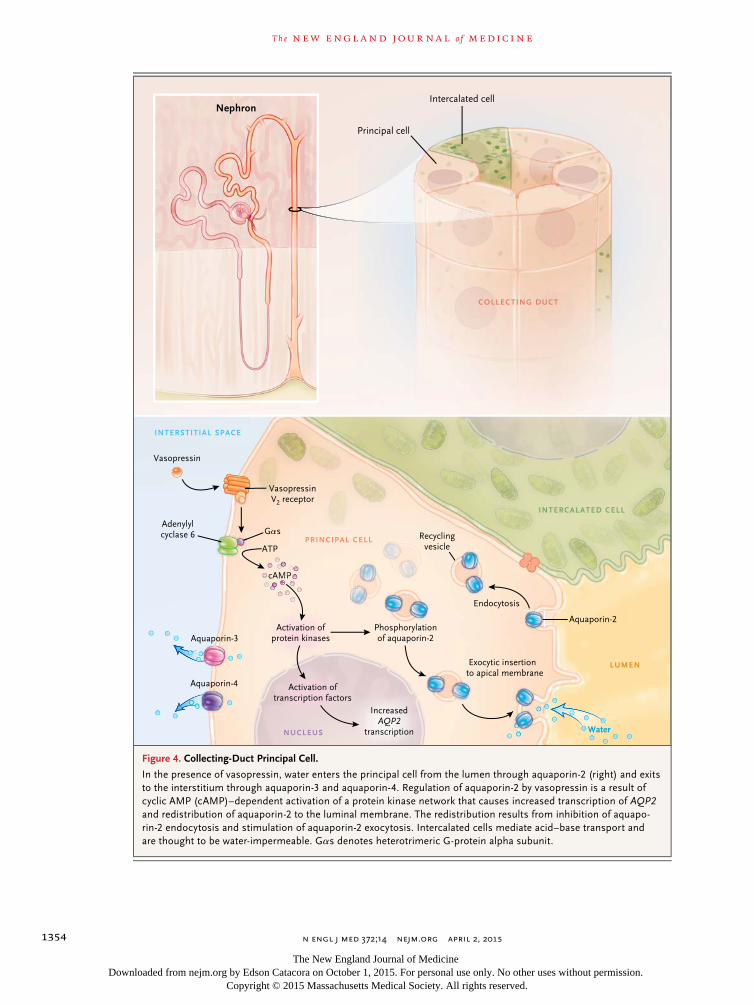
Figure 4. Collecting-Duct Principal Cell.
In the presence of vasopressin, water enters the principal cell from the lumen through aquaporin-2 (right) and exits to the interstitium through aquaporin-3 and aquaporin-4. Regulation of aquaporin-2 by vasopressin is a result of cyclic AMP (cAMP)–dependent activation of a protein kinase network that causes increased transcription of AQP2 and redistribution of aquaporin-2 to the luminal membrane. The redistribution results from inhibition of aquapo-rin-2 endocytosis and stimulation of aquaporin-2 exocytosis. Intercalated cells mediate acid–base transport and are thought to be water-impermeable. Gαs denotes heterotrimeric G-protein alpha subunit.
Intercalated cell
NUCLEUS
PRINCIPAL CELL
INTERSTITIAL SPACE
COLLECTING DUCT
INTERCALATED CELL
LUMEN
Water
Aquaporin-2
Endocytosis
Recyclingvesicle
Exocytic insertion to apical membrane
Aquaporin-3
Vasopressin
Aquaporin-4
GαsAdenylylcyclase 6
VasopressinV2 receptor
ATP
cAMP
Activation ofprotein kinases
Phosphorylationof aquaporin-2
Activation oftranscription factors
IncreasedAQP2
transcription
Principal cell
Nephron
The New England Journal of Medicine Downloaded from nejm.org by Edson Catacora on October 1, 2015. For personal use only. No other uses without permission.
Copyright © 2015 Massachusetts Medical Society. All rights reserved.

n engl j med 372;14 nejm.org April 2, 2015 1355
Molecular Physiology of Water Balance
(corresponding to the AP2, SRF, CREB, GATA, and HOX transcription factor families).50
Studies of animal models of disease have shown that dysregulation of aquaporin-2 plays a central role in both polyuric disorders and disor-ders associated with dilutional hyponatremia.27 Polyuric disorders due to abnormalities in the regulation of water transport that are intrinsic to the kidney are referred to as nephrogenic diabe-tes insipidus syndromes. Heritable nephrogenic diabetes insipidus syndromes have been reviewed by Fujiwara and Bichet.8 Acquired nephrogenic diabetes insipidus syndromes are much more common in clinical practice and can occur in patients who have hypokalemia, hypercalcemia, or partial urinary tract obstruction, as well as in patients who receive certain drugs such as lithi-um carbonate27 (see the case reports in the Sup-plementary Appendix, available with the full text of this article at NEJM.org). Animal models of each of these syndromes have shown a marked reduction in the abundance of the aquaporin-2 protein, presumably because of abnormalities in the normal long-term regulatory mechanisms de-scribed above.
Aquaporin-2 dysregulation also occurs in a number of syndromes associated with renal wa-ter retention and dilutional hyponatremia, chiefly severe congestive heart failure, hepatic cirrhosis, and the syndrome of inappropriate antidiuretic hormone secretion (SIADH)27 (see the case re-ports in the Supplementary Appendix). In these states, increased levels of circulating vasopressin occur “inappropriately” (i.e., independently of reg-ulation through the hypothalamic osmoreceptors) (Fig. 1). The pathophysiological mechanisms that are involved have been reviewed by Schrier.51 SIADH is the most common cause of hypona-tremia in hospitalized patients.52
Animal models of SIADH have shown marked increases in aquaporin-2 protein abundance.53,54 However, these increases are attenuated by a coun-terregulatory process called “vasopressin escape.”54 This escape phenomenon is associated with re-sistance to vasopressin in the collecting duct owing to decoupling of the liganded V2 receptor from cAMP production.55 Thus, despite high lev-els of circulating vasopressin, the renal collecting ducts become relatively impermeable to water,55 thereby limiting the decrease in the serum sodium to a concentration typically in the range of 120 to
129 mmol per liter. Although data from studies are lacking, it seems likely that similar mecha-nisms limit the hyponatremia seen in severe congestive heart failure.
The development of a class of orally available drugs that block the V2 vasopressin receptor — the vaptans — offers a new type of therapy for the treatment of chronic, symptomatic dilutional hyponatremia. However, the use of these drugs is limited by high cost.56
Aquaporin-3
A third aquaporin, aquaporin-3, which is consti-tutively localized to the basolateral plasma mem-brane (Fig. 4) of collecting-duct principal cells, connecting-tubule cells, and inner medullary collecting-duct cells,57 provides an exit pathway for the water that enters across the apical plasma membrane through aquaporin-2. Unlike aquapo-rin-1, aquaporin-2, and aquaporin-4, aquapo-rin-3 conducts glycerol in addition to water and may have a role in the regulation of metabolism. Like aquaporin-2, its abundance is regulated over a period of hours to days by vasopressin57 through changes in its messenger RNA (mRNA) levels.54
Aquaporin-3 is widely expressed throughout the body. In erythrocytes, it is responsible for the GIL blood-group antigen. AQP3-null persons and GIL-negative persons have no obvious clinical manifestations.58 In contrast, AQP3-null mice have severe polyuria.59
Aquaporin-4
The water channel aquaporin-4 is localized to the basolateral plasma membrane in the collect-ing-duct system (i.e., the same cells that express aquaporin-2 and aquaporin-3).27 In contrast to aquaporin-3, its abundance is not regulated by vasopressin.27
The genetic deletion of aquaporin-4 in mice results in a modest concentrating defect.60 This result is in contrast to the much more severe phenotype seen with the genetic deletion of aquaporin-3.
Va sopr essin-R egul ated Ur e a Ch a nnel
In the inner medullary collecting duct, vasopressin rapidly and reversibly increases transepithelial
The New England Journal of Medicine Downloaded from nejm.org by Edson Catacora on October 1, 2015. For personal use only. No other uses without permission.
Copyright © 2015 Massachusetts Medical Society. All rights reserved.

n engl j med 372;14 nejm.org April 2, 20151356
T h e n e w e ngl a nd j o u r na l o f m e dic i n e
urea permeability, allowing urea to exit and be-come trapped within the countercurrent ex-change system.3 Accumulation of urea in the medullary interstitium by this mechanism con-tributes to the high osmolality in the inner me-dulla. The high urea permeability of the inner medullary collecting duct is attributable to two urea-channel proteins (UT-A1 and UT-A3) that are produced from the same gene, SLC14A2.3
As seen with aquaporin-2, the regulation of SLC14A2 proteins by vasopressin is dependent on phosphorylation at multiple sites.61,62 These phosphorylation events are associated with in-creases in the number of the urea channels that are present in the apical plasma membrane.61
Genetic deletion of the UT-A1 and UT-A3 urea channels in mice markedly reduces the urea permeability of the inner medullary collecting duct63 and eliminates the corticomedullary urea gradient.63,64 Despite elimination of urea accu-mulation in the inner medulla, accumulation of sodium and chloride is unaffected. This finding seemingly ruled out concentrating models that predict that the accumulation of sodium chlo-ride in the inner medulla is dependent on urea efflux from the inner medullary collecting duct.65
A third isoform of SLC14A2, called UT-A2, is produced by transcription from a different pro-moter and is expressed in the descending limbs of the loop of Henle. Urea transport in this seg-ment is thought to be important for the recy-cling of the urea that is reabsorbed from the collecting duct back into the renal tubule.3
Epi theli a l Sodium Ch a nnel
Transport of sodium ions out of the lumen of the cortical collecting duct is strongly and rap-idly up-regulated by vasopressin.66,67 Transepi-thelial sodium transport in this segment is me-diated both by an electroneutral mechanism68,69 and by an electrogenic mechanism that is regu-lated by vasopressin. The electrogenic component depends on apical entry of sodium ions through the epithelial sodium channel. The epithelial so-dium channel is a heterotrimeric complex con-sisting of alpha, beta, and gamma subunits.
Studies by Snyder70 suggest that the rapid regu-lation by vasopressin is due to membrane traf-ficking of the epithelial sodium channel, which results from regulation of the ubiquitin ligase Nedd4-2. In addition, there appears to be long-
term regulation of the epithelial sodium channel in the collecting duct in response to vasopressin. Specifically, the abundances of the beta and gamma subunits of the epithelial sodium chan-nel are increased by vasopressin in a period of hours to days.71 The increases in protein abun-dance are associated with increases in beta and gamma subunit mRNA levels; this association points to pre-translational mechanisms of regu-lation.72
In contrast to vasopressin, aldosterone selec-tively increases the abundance of the alpha sub-unit protein without affecting levels of the beta and gamma subunits.73 Thus, the overall regula-tion of electrogenic transport in the cortical collecting duct appears to be synergistically de-pendent on both vasopressin and aldosterone. As shown in Figure 2, however, increases in va-sopressin concentrations do not generally result in reduced excretion of total solute because of compensatory effects of the renin–angiotensin–aldosterone system.
In mice, selective deletion of the epithelial sodium channel in the connecting tubule and collecting duct is associated predominantly with abnormalities in the regulation of extracellular fluid volume (i.e., salt balance), rather than with abnormalities of water balance.74 Likewise, in humans, type I pseudohypoaldosteronism is due to loss-of-function mutations in the subunit genes of the epithelial sodium channel.75
Conclusions
In this brief review, we have described recent progress in understanding the roles of selected gene products in the regulation of water balance, with an emphasis on aspects relevant to the wa-ter-balance disorders that are most common in clinical practice. In addition, we have described a compendium of protein targets for pharmaco-logic agents that are useful in the treatment of disorders of salt and water balance (Table 1).
Agents that block water channels (aquapo-rins) or urea channels are not currently avail-able. However, the important roles of these channels in normal water balance suggest that such agents (which are currently under develop-ment) may be useful in the treatment of water-balance disorders.
Disclosure forms provided by the authors are available with the full text of this article at NEJM.org.
The New England Journal of Medicine Downloaded from nejm.org by Edson Catacora on October 1, 2015. For personal use only. No other uses without permission.
Copyright © 2015 Massachusetts Medical Society. All rights reserved.

n engl j med 372;14 nejm.org April 2, 2015 1357
Molecular Physiology of Water Balance
References1. Verney EB. The antidiuretic hormone and the factors which determine its re-lease. Proc R Soc Lond B Biol Sci 1947; 135: 25-106.2. Danziger J, Zeidel ML. Osmotic ho-meostasis. Clin J Am Soc Nephrol 2014 July 30 (Epub ahead of print).3. Fenton RA, Knepper MA. Mouse models and the urinary concentrating mechanism in the new millennium. Physiol Rev 2007; 87: 1083-112.4. Fenske WK, Christ-Crain M, Hörning A, et al. A copeptin-based classification of the osmoregulatory defects in the syn-drome of inappropriate antidiuresis. J Am Soc Nephrol 2014; 25: 2376-83.5. Windle RJ, Forsling ML, Smith CP, Balment RJ. Patterns of neurohypophysial hormone release during dehydration in the rat. J Endocrinol 1993; 137: 311-9.6. Chou CL, DiGiovanni SR, Mejia R, Nielsen S, Knepper MA. Oxytocin as an antidiuretic hormone. I. Concentration dependence of action. Am J Physiol 1995; 269: F70-77.7. Brewster UC, Hayslett JP. Diabetes in-sipidus in the third trimester of pregnan-cy. Obstet Gynecol 2005; 105: 1173-6.8. Fujiwara TM, Bichet DG. Molecular biology of hereditary diabetes insipidus. J Am Soc Nephrol 2005; 16: 2836-46.9. Ostrowski NL, Young WS III, Knepper MA, Lolait SJ. Expression of vasopressin V1a and V2 receptor messenger ribonucle-ic acid in the liver and kidney of embry-onic, developing, and adult rats. Endocri-nology 1993; 133: 1849-59.10. Nakanishi K, Mattson DL, Gross V, Roman RJ, Cowley AWJ Jr. Control of re-nal medullary blood flow by vasopressin V1 and V2 receptors. Am J Physiol 1995; 269: R193-R200.11. Hall DA, Varney DM. Effect of vaso-pressin on electrical potential difference and chloride transport in mouse medul-lary thick ascending limb of Henle’s loop. J Clin Invest 1980; 66: 792-802.12. Sasaki S, Imai M. Effects of vasopres-sin on water and NaCl transport across the in vitro perfused medullary thick as-cending limb of Henle’s loop of mouse, rat, and rabbit kidneys. Pflugers Arch 1980; 383: 215-21.13. Greger R, Schlatter E, Lang F. Evi-dence for electroneutral sodium chloride cotransport in the cortical thick ascend-ing limb of Henle’s loop of rabbit kidney. Pflugers Arch 1983; 396: 308-14.14. Ortiz PA. cAMP increases surface ex-pression of NKCC2 in rat thick ascending limbs: role of VAMP. Am J Physiol Renal Physiol 2006; 290: F608-F616.15. Kim GH, Ecelbarger CA, Mitchell C, Packer RK, Wade JB, Knepper MA. Vaso-pressin increases Na-K-2Cl cotransporter expression in thick ascending limb of Henle’s loop. Am J Physiol 1999; 276: F96-F103.
16. Schnermann J, Briggs JP. Tubuloglo-merular feedback: mechanistic insights from gene-manipulated mice. Kidney Int 2008; 74: 418-26.17. Gottschalk CW, Mylle M. Micropunc-ture study of the mammalian urinary concentrating mechanism: evidence for the countercurrent hypothesis. Am J Physiol 1959; 196: 927-36.18. Mutig K, Saritas T, Uchida S, et al. Short-term stimulation of the thiazide-sensitive Na+-Cl− cotransporter by vaso-pressin involves phosphorylation and membrane translocation. Am J Physiol Renal Physiol 2010; 298: F502-F509.19. Pedersen NB, Hofmeister MV, Rosen-baek LL, Nielsen J, Fenton RA. Vasopressin induces phosphorylation of the thiazide-sensitive sodium chloride cotransporter in the distal convoluted tubule. Kidney Int 2010; 78: 160-9.20. Kim GH, Masilamani S, Turner R, Mitchell C, Wade JB, Knepper MA. The thiazide-sensitive Na-Cl cotransporter is an aldosterone-induced protein. Proc Natl Acad Sci U S A 1998; 95: 14552-7.21. Morris RG, Hoorn EJ, Knepper MA. Hypokalemia in a mouse model of Gitel-man’s syndrome. Am J Physiol Renal Physiol 2006; 290: F1416-F1420.22. Agre P, Preston GM, Smith BL, et al. Aquaporin CHIP: the archetypal molecu-lar water channel. Am J Physiol 1993; 265: F463-F76.23. Nielsen S, Smith BL, Christensen EI, Knepper MA, Agre P. CHIP28 water chan-nels are localized in constitutively water-permeable segments of the nephron. J Cell Biol 1993; 120: 371-83.24. Ma T, Yang B, Gillespie A, Carlson EJ, Epstein CJ, Verkman AS. Severely im-paired urinary concentrating ability in transgenic mice lacking aquaporin-1 wa-ter channels. J Biol Chem 1998; 273: 4296-9.25. King LS, Choi M, Fernandez PC, Car-tron JP, Agre P. Defective urinary-concen-trating ability due to a complete deficien-cy of aquaporin-1. N Engl J Med 2001; 345: 175-9.26. Fushimi K, Uchida S, Hara Y, Hirata Y, Marumo F, Sasaki S. Cloning and ex-pression of apical membrane water chan-nel of rat kidney collecting tubule. Nature 1993; 361: 549-52.27. Nielsen S, Frøkiaer J, Marples D, Kwon TH, Agre P, Knepper MA. Aquapo-rins in the kidney: from molecules to medicine. Physiol Rev 2002; 82: 205-44.28. Nielsen S, Chou CL, Marples D, Chris-tensen EI, Kishore BK, Knepper MA. Va-sopressin increases water permeability of kidney collecting duct by inducing trans-location of aquaporin-CD water channels to plasma membrane. Proc Natl Acad Sci U S A 1995; 92: 1013-7.29. Nielsen S, Knepper MA. Vasopressin activates collecting duct urea transporters
and water channels by distinct physical processes. Am J Physiol 1993; 265: F204-F213.30. Brown D. The ins and outs of aquapo-rin-2 trafficking. Am J Physiol Renal Physiol 2003; 284: F893-F901.31. Nishimoto G, Zelenina M, Li D, et al. Arginine vasopressin stimulates phos-phorylation of aquaporin-2 in rat renal tissue. Am J Physiol 1999; 276: F254-F259.32. Hoffert JD, Pisitkun T, Wang G, Shen RF, Knepper MA. Quantitative phospho-proteomics of vasopressin-sensitive renal cells: regulation of aquaporin-2 phos-phorylation at two sites. Proc Natl Acad Sci U S A 2006; 103: 7159-64.33. Hoffert JD, Fenton RA, Moeller HB, et al. Vasopressin-stimulated increase in phosphorylation at Ser269 potentiates plasma membrane retention of aquapo-rin-2. J Biol Chem 2008; 283: 24617-27.34. Katsura T, Gustafson CE, Ausiello DA, Brown D. Protein kinase A phosphor-ylation is involved in regulated exocytosis of aquaporin-2 in transfected LLC-PK1 cells. Am J Physiol 1997; 272: F817-F822.35. Fushimi K, Sasaki S, Marumo F. Phos-phorylation of serine 256 is required for cAMP-dependent regulatory exocytosis of the aquaporin-2 water channel. J Biol Chem 1997; 272: 14800-4.36. van Balkom BW, Savelkoul PJ, Mar-kovich D, et al. The role of putative phos-phorylation sites in the targeting and shuttling of the aquaporin-2 water chan-nel. J Biol Chem 2002; 277: 41473-9.37. Moeller HB, Knepper MA, Fenton RA. Serine 269 phosphorylated aquaporin-2 is targeted to the apical membrane of col-lecting duct principal cells. Kidney Int 2009; 75: 295-303.38. Moeller HB, Praetorius J, Rützler MR, Fenton RA. Phosphorylation of aquapo-rin-2 regulates its endocytosis and pro-tein-protein interactions. Proc Natl Acad Sci U S A 2010; 107: 424-9.39. Rinschen MM, Yu MJ, Wang G, et al. Quantitative phosphoproteomic analysis reveals vasopressin V2-receptor-depen-dent signaling pathways in renal collect-ing duct cells. Proc Natl Acad Sci U S A 2010; 107: 3882-7.40. Nedvetsky PI, Tabor V, Tamma G, et al. Reciprocal regulation of aquaporin-2 abundance and degradation by protein kinase A and p38-MAP kinase. J Am Soc Nephrol 2010; 21: 1645-56.41. Lu HJ, Matsuzaki T, Bouley R, Hasler U, Qin QH, Brown D. The phosphoryla-tion state of serine 256 is dominant over that of serine 261 in the regulation of AQP2 trafficking in renal epithelial cells. Am J Physiol Renal Physiol 2008; 295: F290-F294.42. Valenti G, Procino G, Tamma G, Car-mosino M, Svelto M. Minireview: aquapo-rin 2 trafficking. Endocrinology 2005; 146: 5063-70.
The New England Journal of Medicine Downloaded from nejm.org by Edson Catacora on October 1, 2015. For personal use only. No other uses without permission.
Copyright © 2015 Massachusetts Medical Society. All rights reserved.
n engl j med 372;14 nejm.org April 2, 20151358
Molecular Physiology of Water Balance
43. Noda Y, Horikawa S, Kanda E, et al. Reciprocal interaction with G-actin and tropomyosin is essential for aquaporin-2 trafficking. J Cell Biol 2008; 182: 587-601.44. DiGiovanni SR, Nielsen S, Chris-tensen EI, Knepper MA. Regulation of collecting duct water channel expression by vasopressin in Brattleboro rat. Proc Natl Acad Sci U S A 1994; 91: 8984-8.45. Sandoval PC, Slentz DH, Pisitkun T, et al. Proteome-wide measurement of pro-tein half-lives and translation rates in va-sopressin-sensitive collecting duct cells. J Am Soc Nephrol 2013; 24: 1793-805.46. Kamsteeg EJ, Hendriks G, Boone M, et al. Short-chain ubiquitination mediates the regulated endocytosis of the aquapo-rin-2 water channel. Proc Natl Acad Sci U S A 2006; 103: 18344-9.47. Matsumura Y, Uchida S, Rai T, Sasaki S, Marumo F. Transcriptional regulation of aquaporin-2 water channel gene by cAMP. J Am Soc Nephrol 1997; 8: 861-7.48. Wilson JL, Miranda CA, Knepper MA. Vasopressin and the regulation of aqua-porin-2. Clin Exp Nephrol 2013; 17: 751-64.49. Weingarten-Gabbay S, Segal E. The grammar of transcriptional regulation. Hum Genet 2014; 133: 701-11.50. Yu MJ, Miller RL, Uawithya P, et al. Systems-level analysis of cell-specific AQP2 gene expression in renal collecting duct. Proc Natl Acad Sci U S A 2009; 106: 2441-6.51. Schrier RW. Vasopressin and aquapo-rin 2 in clinical disorders of water homeo-stasis. Semin Nephrol 2008; 28: 289-96.52. Thompson C, Hoorn EJ. Hyponatrae-mia: an overview of frequency, clinical pre-sentation and complications. Best Pract Res Clin Endocrinol Metab 2012; 26: Suppl 1: S1-S6.53. Fujita N, Ishikawa SE, Sasaki S, et al. Role of water channel AQP-CD in water retention in SIADH and cirrhotic rats. Am J Physiol 1995; 269: F926-F931.54. Ecelbarger CA, Nielsen S, Olson BR, et al. Role of renal aquaporins in escape from vasopressin-induced antidiuresis in rat. J Clin Invest 1997; 99: 1852-63.55. Ecelbarger CA, Chou CL, Lee AJ, Di-
Giovanni SR, Verbalis JG, Knepper MA. Escape from vasopressin-induced antidi-uresis: role of vasopressin resistance of the collecting duct. Am J Physiol 1998; 274: F1161-F1166.56. Jovanovich AJ, Berl T. Where vaptans do and do not fit in the treatment of hypo-natremia. Kidney Int 2013; 83: 563-7.57. Ecelbarger CA, Terris J, Frindt G, et al. Aquaporin-3 water channel localization and regulation in rat kidney. Am J Physiol 1995; 269: F663-F672.58. Roudier N, Ripoche P, Gane P, et al. AQP3 deficiency in humans and the mo-lecular basis of a novel blood group sys-tem, GIL. J Biol Chem 2002; 277: 45854-9.59. Ma T, Song Y, Yang B, et al. Nephro-genic diabetes insipidus in mice lacking aquaporin-3 water channels. Proc Natl Acad Sci U S A 2000; 97: 4386-91.60. Chou CL, Ma T, Yang B, Knepper MA, Verkman AS. Fourfold reduction of water permeability in inner medullary collect-ing duct of aquaporin-4 knockout mice. Am J Physiol 1998; 274: C549-C554.61. Blount MA, Mistry AC, Fröhlich O, et al. Phosphorylation of UT-A1 urea trans-porter at serines 486 and 499 is important for vasopressin-regulated activity and mem-brane accumulation. Am J Physiol Renal Physiol 2008; 295: F295-F299.62. Bansal AD, Hoffert JD, Pisitkun T, et al. Phosphoproteomic profiling reveals vaso-pressin-regulated phosphorylation sites in collecting duct. J Am Soc Nephrol 2010; 21: 303-15.63. Fenton RA, Chou CL, Stewart GS, Smith CP, Knepper MA. Urinary concen-trating defect in mice with selective dele-tion of phloretin-sensitive urea transport-ers in the renal collecting duct. Proc Natl Acad Sci U S A 2004; 101: 7469-74.64. Fenton RA, Flynn A, Shodeinde A, Smith CP, Schnermann J, Knepper MA. Renal phenotype of UT-A urea transporter knockout mice. J Am Soc Nephrol 2005; 16: 1583-92.65. Kokko JP, Rector FC Jr. Countercur-rent multiplication system without active transport in inner medulla. Kidney Int 1972; 2: 214-23.
66. Tomita K, Pisano JJ, Knepper MA. Control of sodium and potassium trans-port in the cortical collecting duct of the rat: effects of bradykinin, vasopressin, and deoxycorticosterone. J Clin Invest 1985; 76: 132-6.67. Reif MC, Troutman SL, Schafer JA. Sodium transport by rat cortical collect-ing tubule: effects of vasopressin and des-oxycorticosterone. J Clin Invest 1986; 77: 1291-8.68. Tomita K, Pisano JJ, Burg MB, Knep-per MA. Effects of vasopressin and brady-kinin on anion transport by the rat corti-cal collecting duct: evidence for an electroneutral sodium chloride transport pathway. J Clin Invest 1986; 77: 136-41.69. Leviel F, Hübner CA, Houillier P, et al. The Na+-dependent chloride-bicarbonate exchanger SLC4A8 mediates an electro-neutral Na+ reabsorption process in the renal cortical collecting ducts of mice. J Clin Invest 2010; 120: 1627-35.70. Snyder PM. Minireview: regulation of epithelial Na+ channel trafficking. Endo-crinology 2005; 146: 5079-85.71. Ecelbarger CA, Kim GH, Terris J, et al. Vasopressin-mediated regulation of epi-thelial sodium channel abundance in rat kidney. Am J Physiol Renal Physiol 2000; 279: F46-F53.72. Nicco C, Wittner M, DiStefano A, Jounier S, Bankir L, Bouby N. Chronic ex-posure to vasopressin upregulates ENaC and sodium transport in the rat renal col-lecting duct and lung. Hypertension 2001; 38: 1143-9.73. Masilamani S, Kim GH, Mitchell C, Wade JB, Knepper MA. Aldosterone-medi-ated regulation of ENaC alpha, beta, and gamma subunit proteins in rat kidney. J Clin Invest 1999; 104: R19-R23.74. Christensen BM, Perrier R, Wang Q, et al. Sodium and potassium balance de-pends on αENaC expression in connect-ing tubule. J Am Soc Nephrol 2010; 21: 1942-51.75. Furgeson SB, Linas S. Mechanisms of type I and type II pseudohypoaldosteron-ism. J Am Soc Nephrol 2010; 21: 1842-5.Copyright © 2015 Massachusetts Medical Society.
The New England Journal of Medicine Downloaded from nejm.org by Edson Catacora on October 1, 2015. For personal use only. No other uses without permission.
Copyright © 2015 Massachusetts Medical Society. All rights reserved.