molecular phylogeny of the ceratocystis moniliformis complex and
TRANSCRIPT

Fungal Diversity
Molecular phylogeny of theCeratocystis moniliformis complexand description of C.tribiliformis sp. novo
Marelize van Wykl', Jolanda Rouxl, Irene Barnes2, Brenda D. Wingfield2and Michael J. Wingfieldl.
IDepartment of Microbiology and Plant Pathology, Forestry and Agricultural BiotechnologyInstitute (FABI), University of Pretoria, Pretoria, South Africa, 00022Department of Genetics, Forestry and Agricultural Biotechnology Institute (FABI), Universityof Preloria, Pretoria, South Africa, 0002
Van Wyk, M., Roux, 1., Barnes,!., Wingfield, B.D. and Wingfield, M.J.(2006). Molecularphylogeny of theCeratocystis moniliformiscomplex and description of C.tribiliformis sp. novoFungal Diversity 21: 181-201.
Ceratocystis moniliformis is a colonist of fresh wounds on trees, mainly in the tropics. Thefungus is not known to be a pathogen and has thusnot been widely studied.Ceratoeystismoniliformis has been reported on many taxonomically different plants and in many differentclimatic zones. It is thought to represent a complex of morphologically similar butphysiologically and phylogenetically different species, some of which have recently beendescribed. The aim of this study was to consider the phylogenetic relationship of C.moniliformis isolates from various hosts and origins, based on comparisons of DNA sequencesof the Internal Transcrihed Spacer (ITS) regions including the 5.8S gene of rDNA, P-tubulinand EFl-a genes. Four distinct clades were discemable in the phylogenetic tree. Two recentlydescribed species, C.bhutanensis and C. moniliformopsis, were confinned to bephylogenetically distinct.Ceratocystis moniliformis sensu strictoisolated from Bhutan, CostaRica, Ecuador and South Africa represented a well-resolved monophyletic group. A collectionof isolates initially identified as C.moniliformis, from Pinus merkusii in Sumatra, Indonesiawas found to reside in a distinct clade.Ceratocystis tribiliformis sp. nov. is described here toaccommodate this group of isolates. Morphologically, C.tribiliformis differs from C.moniliformis in having obpyrifonn to globose ascomata and both smooth and granular hyphae.Ceratocystis tribiliformis also has distinct optimal growth at 20-25"C on malt extract agar.
Key words: DNA sequences, insect/fungus interactions, phylogenetics, phylogeography,taxonomy, Thielaviopsis
Introduction
Ceratocystis moniliformisHedgcock is a cosmopolitan fungus that hasbeen reported from many hosts and continents (Davidson, 1935; Bakshi, 1951;Hunt, 1956; Upadhyay, 1981) (Table I). The fungus was first isolated from
.Corresponding author: Marelize van Wyk; e-mail: [email protected]
181
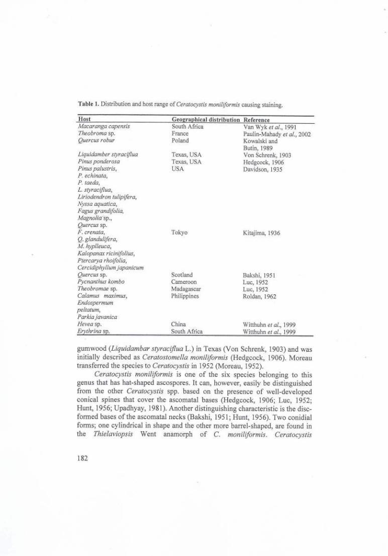
Table J. Distribution and host range of Ceratocystis moniliformis causing staining.
HostMacaranga capensisTheobroma sp.Quercus robur
Liquidamber styracifluaPinus ponderosaPinus pa/ustris,P. echinata,P. taeda,L. styraciflua,Liriodendron tu/ipifera,Nyssa aquatica,Fagus grandifo/ia,Magnolia-sp.,Quercus sp.F. crenata,Q. glandulifera,M. hyplleuca,Kalopanax ricinifolius,Ptercarya rhoifolia,Cercidiphyl/um japanicumQuercus sp.Pycnanthus komboTheobromae sp.Calamus maximus,Endospermumpeltatum,ParkiajavanicaHevea sp.Er thrina s .
Ceo ra hical distributionSouth AfricaFrancePoland
ReferenceVan Wyk el al., 1991Paulin-Mahady el aI., 2002Kowalski andButin, 1989Von Schrenk, 1903Hedgcock, 1906Davidson, 1935
Texas, USATexas, USAUSA
Tokyo Kitajlma, 1936
ScotlandCameroonMadagascarPhilippines
Bakshi,1951Luc, 1952Luc, 1952Roldan, 1962
ChinaSouth Africa
Witthuhn el al., 1999Witthuhn el al., 1999
gumwood (Liquidambarstyraciflua L.) in Texas (Von Schrenk, 1903) and wasinitially described asCera/os/omella moniliformis(Hedgcock, 1906). Moreautransferred the species toCera/ocys/isin 1952 (Moreau, 1952).
Ceralocys/is moniliformis is one of the six species belonging to thisgenus that has hat-shaped ascospores. It can, however, easily be distinguishedfrom the other Cera/ocys/is spp. based on the presence of well-developedconical spines that cover the ascomatal bases (Hedgcock, 1906; Luc, 1952;Hunt, 1956; Upadhyay, 1981). Another distinguishing characteristic is the disc-formed bases of the ascomatal necks (Bakshi, 195]; Hunt, 1956). Two conidialforms; one cylindrical in shape and the other more barrel-shaped, are found inthe Thielaviopsis Went anamorph of C. moniliformis. Cera/ocys/is
182

Fungal Diversity
moniliformis is also one of the fewCeralocyslis spp. not known to producechlamydospores (Davidson, 1935; Paulin-Mahady elal., 2002).
Descriptions for C.moniliformis have tended to be somewhat disparate.Some authors for example, failed to note the conical spines on the ascomatalbases (Kitajima, 1936; Luc, 1952; Roldan, 1962) but rather referred to thestructures as hyphal ornamentation. The ascomatal bases have been describedvariably as globose (Hedgcock, 1906), elongate to pear-shaped (Davidson,1935) and spherical or elongated (Luc, 1952). Dimensions for neck length andascomatal base widths have also varied (Hedgcock, 1906; Kitajima, 1936;Bakshi, 1951; Hunt, 1956; Upadhyay, 1981) (Table 2). Likewise, there hasbeen little agreement regarding the morphology of the conidiophores in variousdescriptions (Davidson, 1935; Kitajima, 1936; Bakshi, 1951; Hunt, 1956;Upadhyay, 1981) (Table 3).
,reralocyslis moniliformis commonly infects wounds on woody plantsandespecially trees (Grylls and Seifert, 1993; Kile, 1993). It is not consideredto be a pathogen as it causes only sap stain. There is also no evidence of C.moniliformis imparting structural changes to infected wood, but discolourationcan lead to a reduction in timber value (Davidson, 1935).
The wide host range and geographic distribution of C.moniliformis hasled us to question whether this fungus might represent a species complex. Thiswould be similar to the view that has emerged for the important tree pathogen,C.fimbriala Ell. & Haist. (Barnes, 2001a; Baker elal., 2003; Van Wyk elal.,2004b). The recent description of C.moniliformopsis Yuan & Mohammed(Yuan and Mohammed, 2002), a species morphologically very similar to C.moniliformis, provides the first support for the view that various species mighthave been aggregated with C.moniliformis. Recent phylogenetic studies havethus led us to describe a new species, C.bhulanensisM. van Wyk, M.J. Wingf.& T. Kirisits (Van Wyk el al., 2004a) that is morphologically very similar to C.moniliformis.
The aim of this study was to compare a collection of C.moniliformissensu laloisolates based on phylogenetic analysis of multiple gene sequences.Morphology and physiological growth at different temperatures of theseCeralocyslis isolates were also studied.
Materials and methods
Isolales
Isolates collected for this study were identified as C.moniliformis basedon morphological characteristics such as the presence of spines on the
183

Table 2. Morphology of the telcomorph of Cera/ocys/is moniliformis as described by various authorsa,
References! lIedgcock Davidson Kitajima Bakshi Luc Lue \ lIunt Roldan UpadhyayCharacter 1906 1935 1936 1951 1952b 1952b \ 1956 1962 1981Ascomatal baseColour Drown! Black Transparent Brown! Dark brown Black Dlack Brown!
black green tinge H!ack blackDiameter 90-180 160-235 104 -244 190-245 210-270 135-190 120-160 150-250 90-210
Ornamentation Conical Brown bristles, Brown bristles 2 types: Dark brown Elongated, Bro\\l1, Dark, Brown conicalspmes 18-60 x (numerous), (1) 65 x lfUll conical spines, Setae, straight, short,conical hyaline tip, spines,(sparse), 3-4 85-1241ong (2) 11-36 x 7- 7-20 x 12-57 x spines, 10-65 x 12-45 x12-16 x 6 15 (Base) 6-12 (Base) 6-11 (Base), 30 x 8 2-3 3-8 (Base)(base) 2-3 (Tip) 2-4 (Tip)
Shape Globose Elongatel Flask-shaped! Round! Sub-spherical/ Sphericall Globose! Globose! Globose/pear- spherical elongate Oval Elongated pear shaped Pear shaped Pearshaped shaped
Ascomatal neckColour Black Black Black Black Black BlackLength 550-1000 305-609 731-896 600-900 500-700 900 920 550-1000Width: base 30-36 39-52 29-42 21-30 20-30 20-45 20-35Width:ti 14-15 14 16-19 10-13 10-15 10-14 11-15

Table 2 continued. Morphology of the tclc<?ffiorphofCeratocystis moniliformisas described by various authors3.
References!CharacterOstiolar hyphae
Ascus
ColourAscospores
Colour
Hcdgcock1906
Brown-black,short, thick,12-18 x 2
Hyaline
Hyaline
Davidson Kitajima1935 19368-12 hyphae, 7-12 hyphae,hyaline, hyaline,filaments filaments15-25 11-63
Hyaline
LengthWidthTexture
Shape: side view Oval, one sideflat4 -53-4Long,slimy,
e mass
Broad ovoid Kidney(hat) Shaped4-5 2-42-3 4-5Gelatinous Mucilaginoussheath substance
Bakshi1951
2-14 hyphae,hyaline,34-41 x2-3 (Base)
Hyaline
Oval, brim(hat)6-83-4Oval globule,mucilaginous
Luc1952b8-15 hyphae,hyaline setae,19-46 x 2
Luc i
1952b \7-12hyphae,12-21
Hunt1956
8-10 hyphae,hyaline,25 x 2
Barely visible Poorly visible Not seen
Roldan Upadhyay1962 198110-16 hyphae, 1-25 hyphae,hyaline, bent, hyaline,10-30 x 2 divergent,
2-3
Evanescent
Hat-shaped4-63-4Gelatinoussheath
Not seen
Hyaline
Oblongrenifonn
Gelatinoussheath
Hyaline! Hyaline!yellowish yellowishOval, Oval, Hat-Flattened Flattened shaped4-6 4-5 3-63-5 3-5 2 - 3Pinkish yellow Pinkish yellow Gelatinousin mass in mass sheath
3AlI measurements are presented in microns (/-1-).bLuc described two fOnTISof C.moniliformis; C. pycnanthi (left column) and C.theobromae (right column).
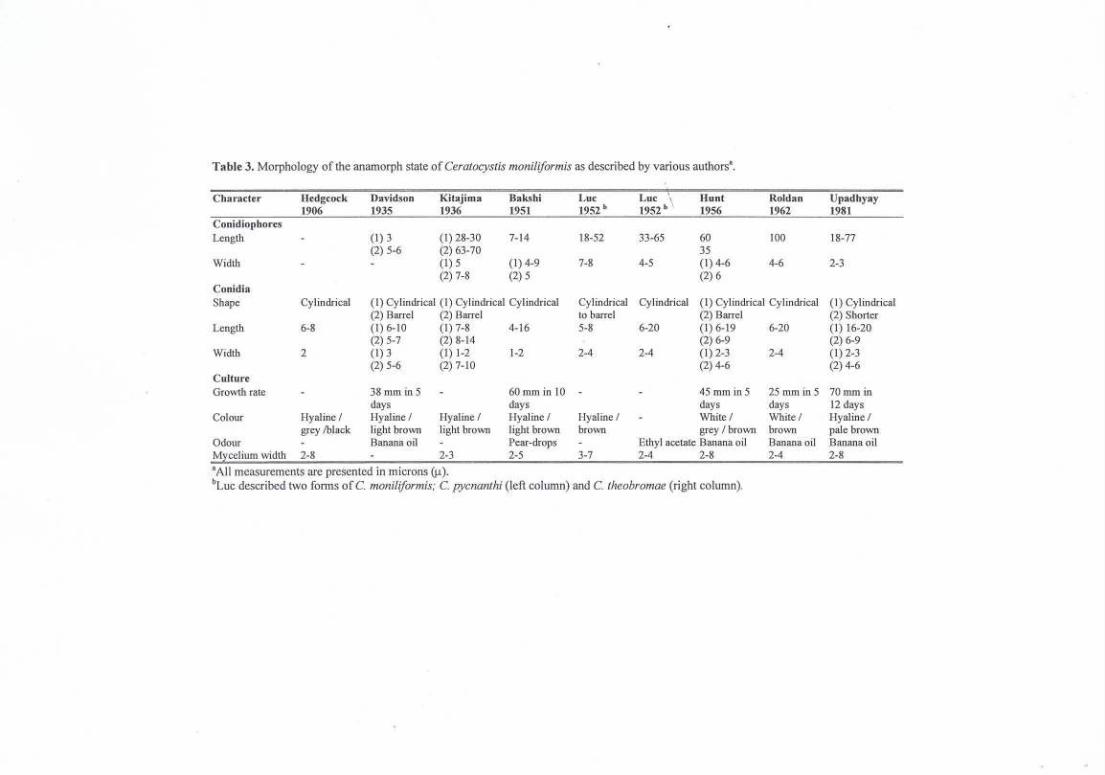
Table 3. Morphology of the anamorph state of Ceratocystis moniliJormis as described by various authorsa.
Character IIedgcock Davidson Kitajima Bak..<;hi Luc Lur"
Hunt Roldan Upadhyay1906 1935 1936 1951 1952b 1952b I. 1956 1962 1981
Conidiophores
Length (1)3 (1) 28-30 7-14 18-52 33-65 60 100 18-77(2) 5-6 (2) 63-70 35
Width (1) 5 (1) 4-9 7-8 4-5 (1) 4-6 4-6 2-3(2) 7-8 (2) 5 (2) 6
ConidiaShape Cylindrical (1) Cylindrical (1) Cylindrical Cylindrical Cylindrical Cylindrical (I) Cylindrical Cylindrical (1) Cylindri<al
(2) BaITel (2) BaITei to barTel (2)BaITei (2) ShorterLength 6-8 (I) 6-10 (1) 7-8 4-16 5-8 6-20 (1) 6-19 6-20 (1) 16-20
(2) 5-7 (2) 8-14 (2) 6-9 (2) 6-9Width 2 (1) 3 (1) 1-2 1-2 2-4 2-4 (I) 2-3 2-4 (I) 2-3
(2) 5-6 (2) 7-10 (2) 4-6 (2) 4-6CultureGrO\vth rate 38 mm in 5 60mminiO 45 mm in 5 25mmin5 70 mm in
days days days days 12 daysColour Hyaline / Hyaline / Hyaline/ Hyaline / Hyaline I White/ White/ Hyaline/
grey !black light brown light brown light brown brown grey / brown brown pale brownOdour Banana oil Pear-drops Ethyl acetate Banana oil Banana oil Banana oilM celium width 2-8 2-3 2-5 3-7 2-4 2-8 2-4 2-8aAli measurements are presented in microns (J.t).
bLue described two fonns ofe. moniliJormis; e. pycnanthi (left column) and C. theobromae (right column).

Fungal Diversity
ascomatal bases. Isolates were collected over a seven-year period from varioustree species in South Africa, Bhutan, Ecuador, Costa Rica and Sumatra(Indonesia) (Table 4). The closely related species, C.moniliformopsis and C.bhutanensis, originally thought to represent C.moniliformis due to theirmorphological similarities were also included (Table 4). All isolates used inthis study are maintained in the culture collection (CMW) of the Forestry andAgricultural Biotechnology Institute (FABl), University of Pretoria, SouthAfrica. Representative isolates have also been deposited with theCentraalbureau voor Schimmelcutures (CBS), Utrecht, The Netherlands.Holotype material of theCeratocystis sp. from Sumatra, including driedcultures on malt extract agar (MEA) has been deposited in the National FungalHerbarium, Pretoria, South Africa (PREM) (Table 4).
DNAf!XIraction
Cultures for DNA extraction were grown on 2% MEA, (Biolab, Midrand,South Africa) at 25 "C for two weeks. Masses of fungal mycelium andascomata were scraped directly from the actively growing cultures, transferredto Eppendorf tubes and lyophilised for two days. The lyophilised myceliumwas placed in liquid nitrogen, ground to a fine powder and DNA was extractedusing the method described by Barneset al. (200Ib).
PCR amplificmion
Three gene regions were amplified using the Polymerase Chain Reaction(PCR). The Internal Transcribed Spacer regions (ITS 1 and ITS2) and the 5.8Sgene of the ribosomal DNA (rDNA) operon were amplified using the primersITS 1 and ITS4 (Whiteet al., 1990). A region of the p-tubulin gene wasamplified using the primers ptla and ptlb (Glass and Donaldson, 1995) and aportion of the EFI-a gene was amplified using the primers EFI-n8F and EFI-986R (Carbone and Kohn, 1999).
PCR reaction mixtures, for all three gene regions, consisted of 200 11Mofthe forward and reverse primers, 200 flM of each dNTP, Expand High FidelityPCR System enzyme mix (1.75 U) (Roche Diagnostics, Mannheim, Germany),1 x Expand HF Buffer containing 1.5 mM MgCh (supplied with the enzyme)and 2-10 ng DNA. Reaction volumes were adjusted to 25 f1Lwith sterile water.The PCR programme was set at 96"C for 2 min for DNA denaturation,followed by 10 cycles at 94"C for 20 s, 55"C for 40 s for annealing andn"Cfor 45 s for elongation. A further 30 cycles at 94"C for 20 s, 55"C for 40 s
187

Table 4. Ceratocystis isolates used in this studya.
Fun~us Alternativenumhers
Collector(s)
AY528991AY529001AY529012AY528992AY529002AY529013AY528993AY529003AY529014AY528994AY529004AY529015AY528985AY528996AY529006N/AAY528986AY528997AY529007
a Isolates marked with # were sequenced in this study, those followed by... were used for morphological descriptions and those marked with+ were included in the growth studies.b CMW refers to the culture collection of the Forestry and Agricultural I3iotcchnology Institute (FABI), University of Pretoria, South Africa.CBS to the Ccntraalbureau voor Schimmelcultures, Utrecht, The Netherlands, and I'REM to the National Fungal Herbarium (I'REM),Pretoria, South Africa.
Isolate no.
C. moniliformis CMW 13011 PREM 57825
CMW 13012" PREM 57826
CMW 13013'" PREM 57827
PREM 57828
CMW 9590N+
CMW 10134"CMW 4114#
GenBank accession
nr.Date ofisolation
\ Geographical Associated\ ori in insect
Pinus merkusii Sumatra, NoneIndonesia
MJ. Wingfield
lIost
1996
2002 Eucalyptusgrandis
Mpumalanga,
South Africa
J.Roux
1997M. van Wyk
M. J. WingfieldSchizolobium Ecuador, Southparahybum America

Table 4 continued. Ceratocystis isolates used in this study.
Fungus Isolate no. Alternative GenBank Date of Host \ Geographical Associated Collector(s)
numbers accession nr. isolation \ .. insecton mC. moniliformis CMW 9990 CBS 155.62 NIA 1962 Theobroma Costa Rica A. 1. Hansen
cacaoCMW 8379' AY528995 2001 Cassia fistula Punaka, Bhutan M.J. Wingfield,
AY529005 T.Kirisits&AY529016 D.B. Clilictri
CMW 8240' AY528989 Wangdi, Bhutan "AY529000AY529010
CMW 8238' N/AC. bhutanensis CMW 8242 CBS 112907 i\ Y528956 Ips
PREM 57809 AY528961 schmutzenhoferiAY528951
CMW 8217 CBS 114289 AY528957PREM 57807 AY528962
AY528952CMW 8244 CBS 114287 N/A
PREM 57811CMW 8241 CBS 115773 N/A
C. moniliformopsis CMW 9986,> CBS 109441 AY528987 1999 Eucalyptus Tasmania, None Z.Q. YuanAY528998 obliqua AustraliaAY529008

Alternative GenBank Date ofnumbers accession nr. isolationCBS 115793 N/A 1999
A Y 528984 1963AY528990AY529011
Table 4 continued. Ceratocystis isolates used in this study.
Fungus Isolate no. Collector(s)
C. moniliformopsis CMW 10215'
C. virescens CMW 3276'
lIost '",Geographical origin AssociatedinsectNone Z.Q. Yuan
T.Hinds
Eucalyptus ohliquaTasmania, Australia
Quercus Warrenber, U.S.A.

Fungal Diversity
with a 5 s extension step, after each cycle was included, followed by 72°C for45 s. A final step of 10 min at 72 °C completed the programme. Amplificationof the respective genes was confirmed under UV illumination using 2%agarose (Roche diagnostics, Mannheim, Germany) gel electrophoresis in thepresence of ethidium bromide. After amplification, amplicons were purifiedusing Sephadex G-50 columns (I g in 15 ml H20, SIGMA, Steinheim,Germany).
Sequencing and analysis
PCR amplicons were sequenced in both directions using the ABIPRISMTM Big DYE Terminator Cycle Sequencing Ready Reaction Kit(Applied BioSystems, Foster City, California). The same primers as those usedin the,PCR reactions were used for sequencing. Sequencing reactions were runon an ABI PRISMTM 3100 Autosequencer (Applied BioSystems, Foster City,California, USA) and sequences were analysed using Sequence Navigatorversion 1.0.1 (Applied BioSystems, Foster City, California).
Sequences were manually aligned and analysed using PAUP version4.0b 10* (Swofford, 2002). A partition homogeneity test (Swofford, 2002) wasused to determine whether the sequence data sets for the three different generegions could be combined. Gaps were treated as "newstate" and trees wereobtained via stepwise addition of 1,000 replicates with the Mulpar optioneffective. The heuristic search option based on parsimony with tree bisectionreconnection (TBR) was used to obtain the phylogram. Confidence intervalsusing 1000 bootstrap replicates were calculated.Ceratocystis virescens(Davidson) Moreau was designated as the out-group taxon. All sequencesderived from this study have been deposited in GenBank (Table 4).
The Markov Chain Monte Carlo (MCMC) method (Larget and Simon,1999), with a Bayesian framework was used to estimate the posteriorprobability of nodes in the phylogenetic tree. One hundred thousand randomtrees were generated using the MCMC procedure, sampling every 100th treeand saving every 10th tree. To avoid including trees sampled beforeconvergence of the Markov chain, the first 4,700 trees were discarded. For thecombined dataset of the three gene sequences, gamma rate heterogeneity wasset, and no codon specific sites were included for the ITS gene. For ~-tubulinand EFt-a sequences, codon specific sites were specified with a site-specificsubstitution rate and the site partition was treated as a by-codon.
191
Cultural characteristics
Growth of three selected isolates for each species was compared on MEAat different temperatures (Table 5). Inocula for the growth study were preparedby growing the fungi on 2% MEA. After a two-week incubation period at25°C, mycelial plugs were taken from the margins of the actively growingcultures using a 5 mm diameter cork borer, and transferred to the centres of 90mm Petri dishes containing 2% MEA. Five plates of each isolate wereincubated at 4°C, lOoC, 15°C, 20°C, 25°C, 30°C and 35°C respectively. Growthwas assessed by taking two measurements of the colony diameter at rightangles to each other, every day for 3 days. Averages and standard deviationswere computed for all growth measurements. The entire experiment wasrepeated once. Colony colour of the isolates was determined using the colourtables_of Rayner (1970) and colony textures were noted.
Morphology
Morphological characteristics of the isolates from Sumatra (Indonesia)were described from 10-day-old cultures on 2% MEA. Sexual and asexualstates of the fungus were mounted in lactophenol on glass slides and examinedunder a Zeiss light microscope. Fifty measurements were taken for eachtaxonomically relevant structure of isolate CMW 13013, which was chosen torepresent the group of isolates from Sumatra. In addition, ten measurementswere made forCeratocystis moniliformis sensu stricto,C. bhutanensisand C.moniliformopsis.
Results
Phylogenetic analysis
PCR amplification of the ITS regions (including the 5.8S gene) and~-tubulin gene resulted in fragments of -500 base pairs (bp) while theamplification of the EF I-a gene resulted in fragments of -300 bp. All three-gene regions were successfully amplified for all isolates.
The partition homogeneity test showed that the sequence data sets for~-tubulin, ITS and EFI-a could be combined (P= 0.05). The combined data setresulted in 1 396 characters, of which 873 characters were constant, 353 wereparsimony-uninformative and 170 were parsimony informative. Analysis of thedata set resulted in one most parsimonious tree (Fig. 1), with a consistency
192

Fungal Diversity
index (CI) of 0.941, homoplasy index (HI) of 0.059, retention index (Rr) of0.889 and a rescaled consistency index (Re) of 0.837.
Four distinct clades, representing four different phylogenetic species,were observed in the combined analysis, each with a strong bootstrap support(77-100%) (Fig. I). The C.moniliformis group was separated into two distinctclades. One clade included only isolates from Sumatra, which were from cutpine timber, while the second clade represented all the other C.moniliformisisolates from Bhutan, South Africa, Ecuador and Costa Rica. The other twoclades represented the two closely related but distinct species, C.moniliformopis and C. bhutanensis.The posterior probability of the branchnodes of the combined tree, generated using Bayesian inference, supported thebootstrap values (Fig. I).
Cultu!al characteristics
There were distinct differences in the growth and culture morphology forthe C. moniliformis isolates studied. No growth was observed for the isolatesfrom Sumatra at 4°C, JOoC,15°C, 30°C or at 35°C and they grew only at 20°Cand 25°C. The isolates from Sumatra grew more slowly than the C.moniliformis isolates from South Africa, Bhutan, Ecuador and Costa Rica. TheC. moniliformis isolates grew at 15°C and above but not at 35°C and they grewrapidly, reaching the edges of the Petri dishes (90 mm) in 72 hours at 30°C(Table 5). Isolates of C.moniliformopsis grew slowly compared to those of C.moniliformis. They displayed optimal growth between 15°C and 20°C and didnot grow at temperatures above 20°C (Table 5). Isolates of C.bhutanensisgrewwell at most temperatures and had an optimum growth between 20°C to 25°C.
Ceratocystis moniliformopsiscultures had buff-yellow (19d) aerialmycelium and Isabella colour (l9"i) submerged mycelium, older cultures havefawn (13"') aerial and submerged mycelium, with few observed ascomata. Thesubmerged mycelium of C.bhutanensiswas umber (15 m) with the well-developed aerial mycelium being ecru-drab (l3''''''d). The C.moniliformisisolates could be separated into two groups that were consistent with thoseresulting from the phylogenetic comparisons. Isolates from Sumatra differedfrom others in having virtually no aerial mycelium and they had masses ofascomata covering the plates. The remaining C.moniliformis isolates hadabundant buff-yellow (19d) aerial mycelium with ascomata tending to occurbelow the mycelium, consistent with the description of this species (Hedgcock,1906; Upadhyay, 1981).
193
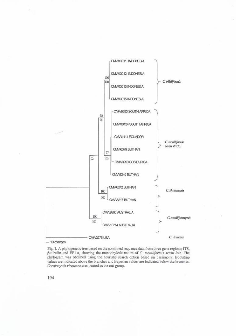
Fig. 1. A phylogenetic tree based on the combined sequence data from three gene regions; ITS,p-tubulin and EF I-a, showing the monophyletic nature of C.moniliformis sensu lato.Thephylogram was obtained using the heuristic search option based on parsimony. Bootstrapvalues are indicated above the branches and Bayesian values are indicated below the branches.Ceratocystis virescenswas treated as the out-group.
194

Table 5. Comparison of growth of Ceratocystis moniliformis sensu stricto, C. bhutanensis, C. moniliformopsis and isolates from SumatraBO.
S ecies Isolate number 4Growth mm at different tem eratures °C
10 15 20 25 ' 30 35
C. moniliformis CMW 9590 0,0 I L3(3.2) I L3(3,0) 64,0(1,9) 72.3(0.8) 90.0(0.0) 0.0sensu stricto CMW 8238 0.0 0.0 0.0 5L3(2.4) 66.9(1,4) 62.6(2.9) 0.0
CMW 10134 0.0 0.0 0.0 19.0(4.0) 17.9(5.0) 14.0(1,4) 0.0CMW 13014 0.0 0.0 0.0 52.8(3.0) 37.9(2.1) 0.0 0.0
Isolates from CMW 13013 0.0 0.0 0.0 45.1(1,9) 37.7(2.7) 0.0 0.0Sumatra CMW 13015 0.0 0.0 0.0 45.0(1.7) 37.5(2.4) 0.0 0.0
CMW 8217 16.8(L3) 17.6(0.5) 30.1(0.5) 60.0(3.9) 60.0(3.7) 11.8(2.0) 0.0CMW 8244 18.6(1.1) 16.8(0.6) 31,6(0.6) 64.6(7.3) 51,0(3.2) 0.0 0.0
C. bhutanensis CMW 8241 16.3(0.6) 13.9(0.8) 28.3(0.8) 70.0(5.0) 44.1(4.1) 0.0 0.0CMW 9986 0.0 0.0 18.5(1,8) 36.6(1.1) 0.0 0.0 0.0CMW 10214 0.0 0.0 16.2(6.6) 50.1(0.7) 0.0 0.0 0.0
C. monili ormo sis CMW 10215 0.0 0.0 12.6 1,2 27.75.8 0.0 0.0 0.0B. Growth assessed as averagecolony diameter for five cultures per isolate on 2% MEA after 3 d.o/Standard deviations are presented in parentheses.

Morphology
The ascomatal bases for the Sumatra isolates were black and obpyriform(Fig. 2.1) and the ostiolar hyphae were divergent (Fig. 2.2). The bases of thenecks at the points of attachment resembled those of C.moniliformis (Fig. 2.3).The ascomatal bases also resembled those of typical C.moniliformis, whichwere covered in short conical spines (Fig. 2.4). Masses of hat-shaped
Fig. 3. Cera/oeys/is /ribiliformis (from holotype) (CMW 13015). 1. Obpyriform aseoma. 2.Divergent ostiolar hyphae. 3. Ascornatal neckdisc-shaped at base. 4. Ascomatalbase withshort, conical spines and hyphal ornamentation. 5. Hat-shaped ascospore in side view,oval inface view. 6. Phialidic conidiogenous cell. 7. Hyphaewith rough edges. 8.Barrel~shapedconidia. 9. Cylindrical conidia. Bars:1 ~ 100 ~m; 2 ~ 10 ~m; 3 ~ 10 ~m; 4 ~ 10 ~m; 5 ~ 10~m; 6 ~ 5 ~m; 7 ~ 5 ~m; 8~ 1a ~m; 9~ 1a ~m.
196

Fungal Diversity
ascospores were observed in both groups of isolates (Fig. 2.5). The anamorphwas typical of Thielaviopsis, with phialidic conidiogenous cells (Fig. 2.6).Both smooth-walled hyphae and those having a granular appearance wereobserved (Fig. 2.7) and both barrel-shaped (Fig. 2.8) and cylindrical conidiawere present (Fig. 2.9).
Taxonomy
DNA sequence analyses, growth comparisons, cultural characteristicsand fruit-body morphology provided support for the view that the isolatesthought to be C. moniliformis from Sumatra, represent a previouslyundescribed species ofCeratocystis. The fungus is therefore described asfollows:
"Ceratocystis tribiliformis M. van Wyk & M.J. Wingf., sp. novo (Fig. 2)Etymology: Shapeof the ascomata similar to the fruit of the plant Tribu/us terrestris L.,
known in the Afrikaans language as the "dubbeltjie".Coloniae albae. ,Uycelium rarum, praecipue in medio immersurn. Temperatura
faustissima 20-25°C, supra 30°C. Hyphae laeves vel granulatae, in septis non-constrictae, 2-4).lID latae. Bases ascomatum atrohrunneae vel nigrae, obpyriformes, spinis hyphibusqueornatae, spinis atrobrunneisvel nigris, 4-12 ~m(X ~ 6-10 ~m), bases 196-264~m(x ~ 203-249 ~m) diametro.Colla oscoma/umatrobrunneavel nigra, 741-1047 x 43-53 ~m(X ~ 782-986 x 44-50 ~m basi discoidea) 741-1047 x 13-20(X ~ 782-986 x 14-18 ~m apice).Hyphaeostia/ares divergentes, hyalinae, 22-32 /-UTI(f = 25-31 ).lm)longae.Asci non visi. Ascosporaelateraliter visae cucullatae, aseptatae, hyalinae, in vagina investitae, cum vagina 5-6 x 2-3!lm,sine ilia 4-5 x 2-3 !lm. Ascosporae in massis bubalino-luteis mucilaginis in apicibus collorumascomatum convenientes. Anamorpha Thielaviopsis: conidiophora singula in myceliocrescentia, apicem versus angustata, 21-46 x 3-4 ~m(X ~ 22-40 ~m x 3-4 ~m) (longa, basi),21-46 x 1-3 ~m (X = 22-40 ~m x 1-3 ~m) (apice). Evo1utio conidii phialidici per parietes
annulares faciendas,conidia biforrniia: conidia primaria hyalina, aseptata, cylindrica 7-9 x 2!lITI,conidia secondaria hyalina, aseptata, doliiforrnia, 7-9 x 3-4 !lIn.
Colony white on malt extract agar.Mycelium sparse mostly submerged inmedium. Optimal temperaturerange for growth 20-25DC, no growth above30DC.Hyphae smooth or granulated, not constricted at septa, 2-4 I'mwide.Ascomatal basesdark brown to black, globose to obpyriforrn, ornamented withspines and hyphae, spines dark brown to black, 4-12 I'm(x = 6-10 I'm) long,bases 196-264 I'm(x = 203-249 I'm) in diam. Ascomatal necksdark brown toblack, 741-1047 x 43-53 I'm(x = 782-986 x 44-50 I'mwide at base) 741-1047x 13-20(x = 782-986x 14-18 I'm wide at the apex), with a disc-like base.Ostio/ar hyphaedivergent, hyaline, 22-32 I'm(x = 25-31 I'm) long. Asci notobserved.Ascosporeshat-shaped in side view, aseptate, hyaline, invested insheath, 5-6x 2-3 I'm with sheath, 4-5 x 2-3 I'm without sheath. Ascosporesaccumulating in buff-yellow (19d) mucilaginous masses on the apices of
197

ascomatal necks.Thielaviopsis anamorph: conidiophores occurring singly onmycelium, hyaline, swollen at the base, tapering towards the apex, 21-46 x 3-4J1m(x = 22-40 J1m x 3-4 J1m) (at base), 21-46 x 1-3 J1m(x = 22-40 J1m x 1-3J1m) (at the apices).Conidium development through ring wall building, conidiaof two types: primary conidia hyaline, aseptate, cylindrical 7-9 x 2 J1m,secondary conidia hyaline, aseptate, barrel-shaped 7-9 x 3-4 J1m.
Habitat: Wood of Pinus merkusii.Known distribution: Indonesia, Sumatra.Material examined:Indonesia:Sumatraisolated fromwood ofPinus merkusii, 1996,
M.J. Wingfield(PREM 57827-holotypus, living culture:CMW 13013).Additional specimens examined (paratypes): Indonesia: Sumatra, isolated from wood of
Pinus rnerkusH,1996, M.J. Wingfield (PREM 57825, living culture CMW 13011); samecollection data (PREM 57826, living culture CMW 13012/CBS 118242);same collection data(PREM 57828, living culture CMW 13015/CBS115949).
Discussion
In this study, we considered the taxonomy and relationships of acollection ofCeratocystis moniliformis sensu latoisolates from a wide range ofhosts and geographic areas. These included isolates from Sumatra tentativelyidentified as C. moniliformis and the two closely related species, C.bhutanensis and C. moniliformopsis. Comparisons were based on DNAsequences, morphology and growth characteristics in culture. Our resultsshowed that the isolates identified as C.moniliformis represent two discretephylogenetic lineages. One of the clades, consisting of isolates from a widegeographic and host range represents C.moniliformis sensu stricto.The secondgroup of isolates from Sumatra represents an undescribed species, describedhere as C.tribiliformis. Data emerging from this study also provide additionalsupport for the separation of C.bhutanensisand C.moniliformopsis, the twospecies most closely related to C.moniliformis. We consider these species toform part of the larger C.moniliformis sensu latocomplex, characterised bythe formation of hat-shaped ascospores, disc-shaped bases to the ascomatalnecks and short conical spines on their ascomatal bases.
Ceratocystis tribiliformis is morphologically very similar to C.moniliformis. Small but distinct differences in the morphology of the fruitingstructures can be used to distinguish between them. The ascomatal bases of C.tribiliformis are characteristically obpyriform to globose while C.moniliformis,C. bhutanensis and C. moniliformopsis have distinctly globose bases.Ceratocystis tribiliformis and C. bhutanensis both have smooth hyphae asobserved in C.moniliformis and C.moniliformopsis, and hyphae with granularsurfaces. These fungi can also easily be distinguished from each other based on
198

Fungal Diversity
growth characteristics in culture. In culture, C.tribiliformis, has very little, ifany, aerial mycelium, while all the other species in the C.moniliformis sensulato complex produce prolific aerial mycelium (Hedgcock, 1906; Davidson,1935; Yuan and Mohammed, 2002; Van Wyket al., 2004a). Ascomata coverthe agar in cultures of C.tribiliformis, while isolates of C.moniliformissporulate less prolifically and generally require the addition of thiamine toenhance the production of ascomata (Robbins and Ma, 1942; Hawker, 1966;Upadhyay, 1981).
Analysis of sequence data for the ITS regions alone did not provideconvincing separation between isolates of C.bhutanensis,C. moniliformis, C.moniliformopsis, and C. tribiliformis. However, addition of sequences for the~-tubulin and EF1-a gene areas provided clear resolution of these four speciesinto distinct clades with robust bootstrap and Bayesian support. This supportsthe l1}orphological and cultural differences observed in these species andemphasises the importance of considering multiple gene regions in bothtaxonomic and phylogenetic studies.
Results of this study provide the first phylogenetic comparison for acollection of isolates, many of which have previously been referred to as C.moniliformis. Clearly, C. moniliformis sensu strictohas a wider geographicdistribution than previously thought. Other apparently cryptic species that haveemerged ITomthis and other recent studies appear, however, to be restricted tospecific geographical areas.Ceratocystis moniliformopsisis known only fromAustralia (Yuan and Mohammed, 2002); C.bhutanensisis associated with thescolytine bark beetle,Ips schmutzenhoferiHolzschuh and found only in Bhutan(Van Wyk et al., 2004a); and C.tribiliformis described in this study has, todate, been found only on the island of Sumatra in Indonesia. The knowndistribution for some of these species is likely to expand if additionalcollections are undertaken. However, some species such as C.bhutanensis,appear to be ecologically adapted to their areas of origin and hosts.
Very little is known regarding the ecology of the species in the C.moniliformis sensu latocomplex. In pathogenicity trials (Davidson, 1935), ithas been concluded that the fungus is a wound inhabiting saprophyte. With therecognition of more species, additional pathogenicity tests should be carriedout, on their hosts of origin and in the areas from which they have originated.These tests will expandour understanding of the C.moniliformis speciescomplex and thereby possibly, lead to the discovery of new pathogens.
Acknowledgements
We thank the National Research Foundation (NRF), members of the Tree ProtectionCo-operative Programme (TPCP) and the THRJP initiative of the Department of Trade and
199

Industry for funding. We also thaok Dr. Hugh Glen for providing the Latin description aod forsuggesting a name for the new species.
References
Baker, c.J., Harrington, T.c., Krauss, U. and Alfenas, A.C. (2003). Genetic variability andhost specialization in the Latin American clade of Ceratocystis fimbriata.Phytopathology 93: 1274-1284.
Bakshi, B.K. (1951). Studies on four species ofCeratocystis, with a discussion of fungicausing sap-stain in Britain. Mycological Paper 35: 1-16.
Barces, 1., Guar, A., Burgess, T., Raux, J., Wingfield, B.D. aod Wingfield, M.J. (200Ia).Microsatellite markers reflect intra-specific relationships between isolates of thevascular wilt pathogen,Ceratocystis jimbriata. Molecular Plant Pathology 2: 319-325.
Barces, 1., Raux, J., Coetzee M.P.A. aod Wingfield, M.J. (2001b), Characterization ofSeiridium spp. associated with cypress cankerbased on p-tubulin and histonesequences. Plaot Disease 85: 317-321.
Carborie:I. and Kahn, L.M. (1999). A method for designing primer sets for speciation studiesin filamentous ascomycetes. Mycologia 91: 553-556.
Davidson, R.W. (1935). Fungi causing stain in logs and lumber in the Southern states,including five new species. Journalof Agricultural Research 50: 789-807.
Glass, N.L. aod Donaldson, G.c. (1995). Development of primer sets designed for use with thePCR to amplify conserved genes from filamentous Ascomycetes. Applied andEnvironmental Microbiology 61: 1323-1330.
Grylls, B.T. aod Seifert, K.A. (1993). A synoptic key to species ofOphios/oma, Cera/ocys/isand Ceratocystiopsis. In: Ceratocystis and Ophiostoma: Taxonomy, Ecology, andPathogenicity (eds. M.J. Wingfield, K.A. Seifert aod J.F. Webber). APS Press, SI. Paul,Minnesota: 261-268.
Hedgcock, G.G. (1906). Studies upon some chromogenic fungi which discolor wood. MissouriBotaoica! Garden 17: 59-124.
Hawker, E.L. (1966). Environmental influences on reproduction. In:The Fungi: An AdvancedTreatise. Volume II, The Fungal Organism (eds. G.C. Ainsworth and A.S. Sussman)Academic Press. New York. London: 435-469.
Hunt, J. (1956). Taxonomy of the genusCera/ocys/is.Lloydia 19: I-58.Kite,G.A. (1993). Plantdiseasescausedby speciesof Cerato cyst is sensu stricto andChalara.
In: Ceratocystis and Ophiostoma: Taxonomy, Ecology, and Pathogenicity (eds. M.J.Wingfield, K.A. Seifert and J.F. Webber). APS Press, SI. Paul, Minnesota: 173-183.
Kitajirna,K. (1936). Researches on the discolourations oflogs ofFagus crenata Blume causedby Endoconidiophora Bunae, n. sp. and on its preventive method. Bulletin of ImperialForest Experiments station, Meguro, Tokyo 35: 1-134.
Kowalski, T. and Butin, H. (1989). Taxonomie bekannter und neuerCeratocystis-Arten anEiche (Quercus roburL.). Phytopathology 124: 236-248.
Larget, B. aod Simon, D.L. (1999). Markov Chain Monte Carlo algorithms for the Bayesiananalysis of phylogenetic trees. Molecular Biology aod Evolution 16:750-759.
Luc, M. (1952). Ophios/oma moniliforme (Hedgc.) H. et P. Syd. and its various forms.Reviews in Mycology 17: 10-16.
Moreau, C. (1952). Coexistence des fonnesThielaviopsis et Graphium chez une souche deCeratocystis major (van Beyma) novo comb.Revue de Mycologie. (Paris) SupplementColonial 17: 17-22.
200

Fungal Diversity
Paulin-Mahady, A.E., Harrington, T.e. and McNew, D.L. (2002). Phylogenetic and taxonomicevaluation of Chalara, Chalaropsis, and Thielaviopsis anamorphs associated withCeratocystis. Mycologia 94: 62-72.
Rayner, R.W. (1970). A mycological colour chart. Commonwealth Mycological Institute andBritish Mycological Society, Kew, SUITey.
Robbins, W.J. and Ma, R. (1942). Vitamin deficiencies ofCeralastomella and related fungi.American Journal of Botany 29: 835-843.
Roldan, E.P. (1962). Species ofCeratocystis (Ceralas/omella) causing stain in rattan. ThePhilippine Journal of Science 91: 4 I 5-423.
Swofford, D.L. (2002). PAUP'. Phylogenetic Analysis Using Parsimony ('and othermethods). Version 4.0blO. Sunderland,Massachusetts: Sinauer Associates.
Upadhyay, H.P. (1981). A monograph of Ceratocystis and Ceratocystiopsis. University ofGeorgia Press. Athens.
Van Wyk, M., Raux, 1., Bames, I., Wingfield, B.D., Chhetri, D.B., Kirisits, T. and Wingfield,M.J. (2004a). Ceratocystis bhutanensis sp. nov., associated with the bark beetleIpsschmutzenhoferi on Picea spinulosa in Bhutan. Studies in Mycology 50: 365-379.
Van Wyk, M., Roux, 1., Barnes, L, Wingfield, B.D., Liew, E.C.Y., Assa, B., SummereIl, B.A.and Wingfield, M.J. (2004b). Ceratocystis polychroma sp. nov., a new species fromSyzygium aromaticum in Sulawesi. Studies in Mycology 50: 272-282.
Van Wyk, P.W.J., Wingfield, M.J. and Van Wyk, P.S. (1991). Ascospore development inCeratocystis moniliformis. Mycological Research 95: 96-103.
Von Schrenk, H. (1903). The "bluing" and the "red-hot" of the western yellow pine, withspecial reference to the Black Hills Forest Reserve. U,S. Department of Agriculture.Bureau of Plant IndustryBulletin36: 1-46.
White, T.J., Bruns, T., Lee, S. and Taylor, J. (1990). Amplification and direct sequencing offungal ribosomal RNA genes for phylogenetics. In:PCR Protocols: A sequencing guideto methods and applications.(eds. M.A. Innis, D.H. Gelfand, J.J. Sninsky and T.J.White). Academic Press, San Diego: 315-322.
Witthuhn, R.e., Wingfield, B.D., Wingfield, M.J. and Harrington, T.C. (1999). PCR-basedidentification and phylogeny of species ofCeratocystis sensu stricto. MycologicalResearch 103: 743-749.
Yuan, Z.Q. and Mohammed, C. (2002). Ceratocystis moniliformopsis sp. nov., an earlycolonizer of Eucalyptus obliqua logs in Tasmania, Australia. Australian SystematicBotany 15: 125-133.
(Received 12 March 2005; accepted16 November 2005)
201