molecular mechanisms of bmaa stress-response and ... · figure 5 - (a) gsts activity and (b) ache...
TRANSCRIPT

Molecular mechanisms of BMAA stress-response and detoxification in Mytilus galloprovincialis Marta Isabel Póvoas Monteiro Masters in Biologic Aquatic Resources Biology 2015
Supervisor Mafalda Baptista, Post Doctoral Researcher, CIIMAR Coorientador Co Supervisor Vitor Vasconcelos, Professor, Faculty of Sciences of University of Porto
Todas as correções determinadas
pelo júri, e só essas, foram efetuadas.
O Presidente do Júri,
Porto, ______/______/_________

FCUP Molecular mechanisms of BMAA stress-response and detoxification in Mytilus galloprovincialis
i
Acknowledgments
This research was partially funded by the FCT project UID/Multi/04423/2013.
I want to thank my supervisors, Mafalda Baptista and Vitor Vasconcelos, for the
opportunity to be included this project and for their guidance through the
development of this work. To my laboratory colleagues in CIIMAR, thank you for the
support with laboratory work.
Moreover, I would like to thank to my supervisor in Padova University, Paola
Venier and laboratory colleagues, Umberto Rosani, Stefania Domeneghetti, and Laura
Varotto for kindly accepting and welcoming me into their team.
To all my friends, especially to Sara Morgado, Maria João Xavier, and Ana Isabel
Tavares, thanks for brightening up these past couple of years. I would also like to thank
Fábio Rangel for the patience and companionship during this all process.
Finally, I am very grateful for my parents and brother, for their constant support and
motivation to always smile towards the future.
FCUP Molecular mechanisms of BMAA stress-response and detoxification in Mytilus galloprovincialis
ii
Abstract
BMAA is a putative neurotoxin that, in marine environments, has been shown to
find its way from the phytoplankton first producers (e.g. cyanobacteria) to higher trophic
levels. In vitro, BMAA has been shown to act as an ionotropic glutamate receptor
(iGluR) agonist and to induce excitotoxic effects. Despite the fact that mussels have
been shown to accumulate BMAA, toxicity effects have not been yet described. This
work aimed at testing the hypothesis that exposure to BMAA results in changes in the
expression of iGluR, and in the activity of the enzymes Acetylcholinesterase (AChE)
and Glutathione S-transferases (GSTs), in the Mediterranean mussel Mytilus
galloprovincialis, and evaluate their potential as biomarkers of BMAA-induced toxicity.
M. galloprovincialis were exposed to 10, 100 and 1000 µg L-1 of BMAA standard
in seawater up to 48h, and afterwards depurated until 96h. In another experiment, M.
galloprovincialis were fed with BMAA producing cyanobacteria Nostoc sp, BMAA non-
producing cyanobacteria Microcoleus sp., and the green alga Chlorella sp., up to 48h.
Gills and digestive gland of exposed and unexposed animals were separated for
enzymatic analysis and total RNA extraction. Two transcripts, termed GLU4 and GLU5,
were selected from Mytibase, a catalogue of M. galloprovincialis expressed sequence
tags (ESTs). Relative expression of the transcripts was assessed by qPCR, using the
elongation factor alpha-1 (EF-1 α) as internal reference.
In mussels exposed to BMAA standard gills showed increased GSTs activity
during exposure and depuration. Digestive gland also showed increased GSTs activity
during exposure to BMAA. AChE activity decreased its activity during exposure to
BMAA in gills, and no effects could be seen for the digestive gland. In mussels fed with
cyanobacteria, both AChE and GSTs displayed an increase in activity, in digestive
gland, while in gills GSTs activity decreased, and AChE activity increased. Regarding
iGluR expression in mussels exposed to BMAA standard, in gills, both transcripts
displayed a clear downregulation during the exposure period that was reversed after
depuration, while in digestive gland results were not conclusive.
The results suggest that GSTs could be considered a potentially useful
biomarker of BMAA exposure, when it is known that M. galloprovincialis has been
exposed this amino acid, while AChE was considered a poor biomarker. The
transcription of iGluR genes can potentially be used as tool to assess BMAA induced
toxicity in biomonitoring studies using M. galloprovincialis. Nevertheless, further work is
needed to better understand the regulatory mechanisms of iGLuR genes as well as

FCUP Molecular mechanisms of BMAA stress-response and detoxification in Mytilus galloprovincialis
iii
their functional role in mussels.
FCUP Molecular mechanisms of BMAA stress-response and detoxification in Mytilus galloprovincialis
iv
Resumo
BMAA é uma neurotoxina putativa que, em ambientes marinhos, foi encontrada
desde entre os produtores primários fitoplanctónicos (por exemplo, cianobactérias) até
aos níveis tróficos superiores. In vitro, foi provado que o BMAA atua como um agonista
de receptores de glutamato ionotrópicos (iGluR) e induz efeitos de excitotoxicidade.
Estes efeitos sobre os iGluR não foram ainda descritos em organismos marinhos,
apesar do facto destes organismos terem sido reconhecidos como capazes de
acumular BMAA.
Este trabalho teve como objetivo testar a hipótese de que a exposição ao
BMAA resulta em alterações na expressão dos iGluR, bem como na atividade das
enzimas Acetilcolinesterase (AChE) e Glutationa-S-transferases (GSTs) e avaliar o seu
potencial como biomarcadores de toxicidade induzida pelo BMAA, no mexilhão
mediterrânico, Mytilus galloprovincialis.
M. galloprovincialis foram expostos a 10, 100 e 1000 µg L-1 de padrão de BMAA
em água do mar, durante 48h e depurados até às 96h. Noutra experiência, M.
galloprovincialis foram alimentados com cianobactérias produtoras de BMAA, Nostoc
sp., cianobactérias não produtoras de BMAA, Microcoleus sp. e com a alga verde
Chlorella sp durante 48h. Brânquias e glândulas digestivas dos animais expostos e
não expostos foram separados para análise enzimática e extracção de RNA total. Dois
transcritos, denominados GLU4 e GLU5 foram selecionados a partir de um catálogo de
M. galloprovincialis "Expressed Sequence Tag" (ESTs). A expressão relativa dos
transcritos foi avaliada por qPCR, utilizando o gene do factor de alongamento alfa-1
(EF-1 α) como referência interna.
Em mexilhões expostos ao padrão BMAA, nas brânquias, observou-se uma
atividade aumentada da GSTs durante a exposição e depuração. Nas glândulas
digestivas também se verificou um aumento da atividade das GSTs durante a
exposição ao BMAA. A atividade da AChE diminuiu durante a exposição ao BMAA nas
brânquias, mas não ocorreram efeitos observáveis nas glândulas digestivas. Em
mexilhões alimentados com cianobactérias, ambas AChE e GSTs apresentaram um
aumento na atividade nas glândulas digestivas, enquanto nas brânquias a actividade
das GSTs diminuíu, e a atividade da AChE aumentou. Em relação aos iGluR,
expressos em mexilhões expostos a padrão de BMAA, nas brânquias ambos os
transcritos exibiram uma regulação negativa clara durante o período de exposição, que

FCUP Molecular mechanisms of BMAA stress-response and detoxification in Mytilus galloprovincialis
v
foi revertida após depuração, enquanto na glândula digestiva os resultados não foram
conclusivos.
Os resultados sugerem que as GSTs podem ser consideradas um marcador
potencialmente útil de exposição ao BMAA, quando se sabe que M. galloprovincialis
foi exposto a este aminoácido, enquanto que a AChE foi considerado um fraco
biomarcador. A expressão de genes de iGluR poderá ser utilizada como ferramenta
para avaliar a toxicidade induzida por BMAA em estudos de biomonitoração de M.
galloprovincialis. No entanto, mais estudos são necessários para entender melhor os
mecanismos de regulação de genes de iGluR, bem como o seu papel funcional no
mexilhão.
FCUP Molecular mechanisms of BMAA stress-response and detoxification in Mytilus galloprovincialis
vi
Table of Contents
Acknowledgments .......................................................................................................... i
Abstract ........................................................................................................................ ii
Resumo ....................................................................................................................... iv
Figures Index .............................................................................................................. viii
Tables Index ................................................................................................................. x
List of Abbreviations ..................................................................................................... xi
1. Introduction ............................................................................................................... 1
1.1. Cyanobacteria and cyanotoxins ......................................................................... 1
1.2. The cyanotoxin BMAA ........................................................................................ 2
1.2.1. History and relevance .................................................................................. 2
1.2.2. Presence in cyanobacteria .......................................................................... 3
1.2.3. Biosynthesis ................................................................................................ 5
1.2.4. Mechanisms of toxicity ................................................................................ 6
1.3. Health and environmental risks of exposure to BMAA ........................................ 8
1.3.1. Biomagnification hypothesis and human exposure ...................................... 8
1.3.2. Biomarkers of toxic exposure ...................................................................... 9
1.4. Objectives ........................................................................................................ 11
2. Material & Methods ................................................................................................. 12
2.1. Mussel collection .............................................................................................. 12
2.2. Bioaccumulation experiment ............................................................................ 12
2.2.1. Mussels exposed to BMAA standard ......................................................... 12
2.2.2. Mussels fed with cyanobacteria ................................................................. 13
2.3. Enzymatic analysis ........................................................................................... 13
2.4. iGluR expression .............................................................................................. 15
2.4.1. Sequence selection and preliminary analysis ............................................ 15
2.4.2. Primer design ............................................................................................ 16
2.4.3. RNA extraction and purification ................................................................. 17
2.4.4. Quantitative PCR for gene expression analysis ......................................... 19
2.5. BMAA quantification ......................................................................................... 20
2.5.1. Microwave-assisted digestion .................................................................... 20
2.5.2. Liquid chromatography with mass detection (LC-MS/MS) analysis. ........... 20
2.6. Data treatment ................................................................................................. 21
3. Results .................................................................................................................... 22
3.1. BMAA quantification in mussels and cyanobacteria .......................................... 22

FCUP Molecular mechanisms of BMAA stress-response and detoxification in Mytilus galloprovincialis
vii
3.2. AChE and GSTs analysis ................................................................................. 23
3.2.1. Mussels exposed to BMAA standard ......................................................... 23
3.2.1.1. Digestive gland ....................................................................................... 23
3.3.1.2. Gills ........................................................................................................ 24
3.3.2. Mussels fed with cyanobacteria ................................................................. 26
3.3.2.1. Digestive gland ....................................................................................... 26
3.3.2.2. Gills ........................................................................................................ 27
3.3. iGluR expression in M. galloprovincialis ........................................................... 28
3.3.1. Amplification of GLU4 and GLU5 ............................................................... 28
3.3.2. Mussels exposed to BMAA standard ......................................................... 29
4. Discussion .............................................................................................................. 32
4.1. Effects of BMAA on AChE and GSTs activity .................................................... 33
4.2. Effects of Exposure to BMAA on iGluR Expression .......................................... 35
5. Conclusions ............................................................................................................ 37
6. References ............................................................................................................. 38
7. Annexes .................................................................................................................. 61
FCUP Molecular mechanisms of BMAA stress-response and detoxification in Mytilus galloprovincialis
viii
Figures Index
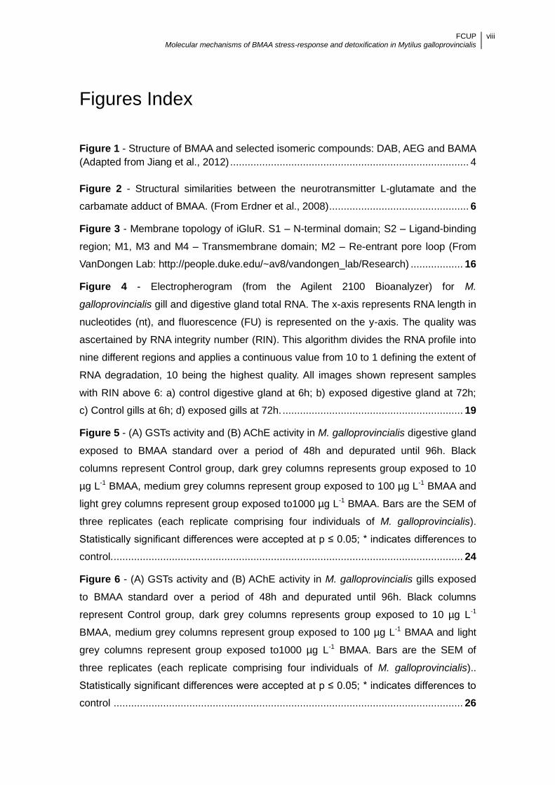
Figure 1 - Structure of BMAA and selected isomeric compounds: DAB, AEG and BAMA
(Adapted from Jiang et al., 2012) .................................................................................. 4
Figure 2 - Structural similarities between the neurotransmitter L-glutamate and the
carbamate adduct of BMAA. (From Erdner et al., 2008) ................................................ 6
Figure 3 - Membrane topology of iGluR. S1 – N-terminal domain; S2 – Ligand-binding
region; M1, M3 and M4 – Transmembrane domain; M2 – Re-entrant pore loop (From
VanDongen Lab: http://people.duke.edu/~av8/vandongen_lab/Research) .................. 16
Figure 4 - Electropherogram (from the Agilent 2100 Bioanalyzer) for M.
galloprovincialis gill and digestive gland total RNA. The x-axis represents RNA length in
nucleotides (nt), and fluorescence (FU) is represented on the y-axis. The quality was
ascertained by RNA integrity number (RIN). This algorithm divides the RNA profile into
nine different regions and applies a continuous value from 10 to 1 defining the extent of
RNA degradation, 10 being the highest quality. All images shown represent samples
with RIN above 6: a) control digestive gland at 6h; b) exposed digestive gland at 72h;
c) Control gills at 6h; d) exposed gills at 72h. .............................................................. 19
Figure 5 - (A) GSTs activity and (B) AChE activity in M. galloprovincialis digestive gland
exposed to BMAA standard over a period of 48h and depurated until 96h. Black
columns represent Control group, dark grey columns represents group exposed to 10
µg L-1 BMAA, medium grey columns represent group exposed to 100 µg L-1 BMAA and
light grey columns represent group exposed to1000 µg L-1 BMAA. Bars are the SEM of
three replicates (each replicate comprising four individuals of M. galloprovincialis).
Statistically significant differences were accepted at p ≤ 0.05; * indicates differences to
control. ........................................................................................................................ 24
Figure 6 - (A) GSTs activity and (B) AChE activity in M. galloprovincialis gills exposed
to BMAA standard over a period of 48h and depurated until 96h. Black columns
represent Control group, dark grey columns represents group exposed to 10 µg L-1
BMAA, medium grey columns represent group exposed to 100 µg L-1 BMAA and light
grey columns represent group exposed to1000 µg L-1 BMAA. Bars are the SEM of
three replicates (each replicate comprising four individuals of M. galloprovincialis)..
Statistically significant differences were accepted at p ≤ 0.05; * indicates differences to
control ........................................................................................................................ 26

FCUP Molecular mechanisms of BMAA stress-response and detoxification in Mytilus galloprovincialis
ix
Figure 7 - (A) GSTs activity and (B) AChE activity in digestive gland of M.
galloprovincialis fed with Nostoc sp., Microcoleus sp or Chlorella sp. Black columns
represent group fed with Nostoc sp., medium grey columns represent group fed with
Chlorella sp. and light grey columns represent group fed with Microcoleus sp. Bars are
the SEM of three replicates (each replicate comprising four individuals of M.
galloprovincialis). Statistically significant differences were accepted at p ≤ 0.05.
Asterisk bracket indicates significant differences to Nostoc sp. ................................... 27
Figure 8 - (A) GSTs activity and (B) AChE activity in gills of M. galloprovincialis fed with
Nostoc sp., Microcoleus sp or Chlorella sp. Black columns represent group fed with
Nostoc sp., medium grey columns represent group fed with Chlorella sp. and light grey
columns represent group fed with Microcoleus sp. Bars are the SEM of three replicates
(each replicate comprising four individuals of M. galloprovincialis). Statistically
significant differences were accepted at p ≤ 0.05. Asterisk bracket indicates significant
differences to Nostoc sp.............................................................................................. 28
Figure 9 - Gill changes of GLU4 and GLU5 transcripts expression during (A) exposure
to BMAA and (B) depuration period compared with controls. Black columns represent
GLU4 transcript and grey columns represent GLU5 transcript (Results are expressed
as mean ± SEM. Statistically significant differences were accepted at p ≤ 0.05; *
indicates differences to control). EF-1 α as internal reference .................................... 30

FCUP Molecular mechanisms of BMAA stress-response and detoxification in Mytilus galloprovincialis
x
Tables Index
Table 1 – Biomarker Classification according to NRC (1987) and WHO (1993).……. ... 9
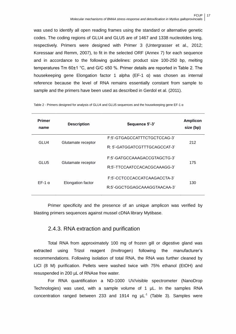
Table 2 - Primers designed for analysis of GLU4 and GLU5 sequences and the
housekeeping gene EF-1 α ........................................................................................ 17
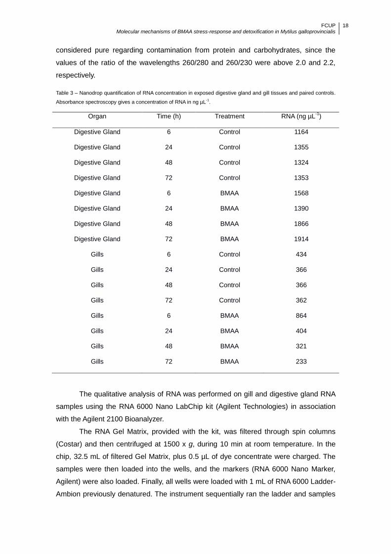
Table 3 - Nanodrop quantification of RNA concentration in exposed digestive gland and
gill tissues and paired controls. Absorbance spectroscopy gives a concentration of RNA
in ng µL-1 ..................................................................................................................... 18
Table 4 - Concentration of BMAA (µg g-1) on mussels exposed to 1000 µg L-1 for 24h
and 48h Mean ± SEM for the concentration of BMAA ................................................ 22
Table 5 - BMAA quantification (µg g-1) in the phytoplankton used to feed M.
galloprovincialis and the culture media in which the phytoplankton was grown. .......... 23
Table 6 – Mean Ct ± SEM of GLU4, GLU5 and EF-1 α in untreated gills collected from
(A) Spring Mussels and (B) Winter mussels ............................................................... 29
Table 7 - Mean Ct ± SEM values of control and treated mussels digestive gland of
GLU4, GLU5 and EF-1 α. Different exposure times have been averaged. ................. 31
FCUP Molecular mechanisms of BMAA stress-response and detoxification in Mytilus galloprovincialis
xi
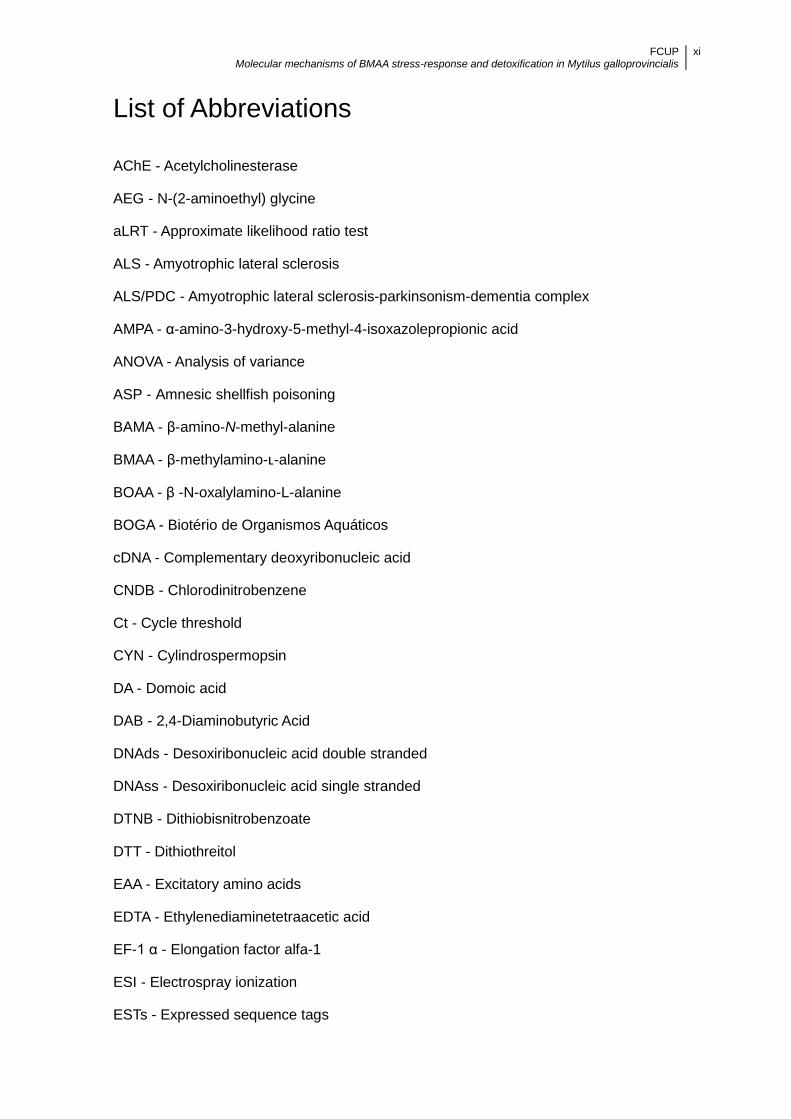
List of Abbreviations
AChE - Acetylcholinesterase
AEG - N-(2-aminoethyl) glycine
aLRT - Approximate likelihood ratio test
ALS - Amyotrophic lateral sclerosis
ALS/PDC - Amyotrophic lateral sclerosis-parkinsonism-dementia complex
AMPA - α-amino-3-hydroxy-5-methyl-4-isoxazolepropionic acid
ANOVA - Analysis of variance
ASP - Amnesic shellfish poisoning
BAMA - β-amino-N-methyl-alanine
BMAA - β-methylamino-ʟ-alanine
BOAA - β -N-oxalylamino-L-alanine
BOGA - Biotério de Organismos Aquáticos
cDNA - Complementary deoxyribonucleic acid
CNDB - Chlorodinitrobenzene
Ct - Cycle threshold
CYN - Cylindrospermopsin
DA - Domoic acid
DAB - 2,4-Diaminobutyric Acid
DNAds - Desoxiribonucleic acid double stranded
DNAss - Desoxiribonucleic acid single stranded
DTNB - Dithiobisnitrobenzoate
DTT - Dithiothreitol
EAA - Excitatory amino acids
EDTA - Ethylenediaminetetraacetic acid
EF-1 α - Elongation factor alfa-1
ESI - Electrospray ionization
ESTs - Expressed sequence tags
FCUP Molecular mechanisms of BMAA stress-response and detoxification in Mytilus galloprovincialis
xii
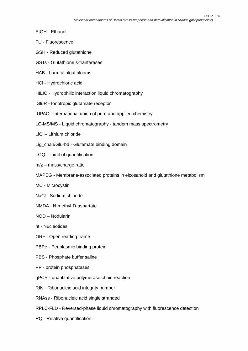
EtOH - Ethanol
FU - Fluorescence
GSH - Reduced glutathione
GSTs - Glutathione s-tranferases
HAB - harmful algal blooms
HCl - Hydrochloric acid
HILIC - Hydrophilic interaction liquid chromatography
iGluR - Ionotropic glutamate receptor
IUPAC - International union of pure and applied chemistry
LC-MS/MS - Liquid chromatography - tandem mass spectrometry
LiCl – Lithium chloride
Lig_chan/Glu-bd - Glutamate binding domain
LOQ – Limit of quantification
m/z – mass/charge ratio
MAPEG - Membrane-associated proteins in eicosanoid and glutathione metabolism
MC - Microcystin
NaCl - Sodium chloride
NMDA - N-methyl-D-aspartate
NOD – Nodularin
nt - Nucleotides
ORF - Open reading frame
PBPe - Periplasmic binding protein
PBS - Phosphate buffer saline
PP - protein phosphatases
qPCR - quantitative polymerase chain reaction
RIN - Ribonucleic acid integrity number
RNAss - Ribonucleic acid single stranded
RPLC-FLD - Reversed-phase liquid chromatography with fluorescence detection
RQ - Relative quantification

FCUP Molecular mechanisms of BMAA stress-response and detoxification in Mytilus galloprovincialis
xiii
SH - Shimodaira-Hasegawa
TMRs - Transmembranar
ZIC-HILIC - Zwitterionic hydrophilic interaction liquid chromatography

FCUP Molecular mechanisms of BMAA stress-response and detoxification in Mytilus galloprovincialis
1
1. Introduction
1.1. Cyanobacteria and cyanotoxins
Cyanobacteria are photosynthetic ubiquitous micro-organisms (Sompong et al.,
2005; Taton et al., 2006) as well as very important nitrogen fixing organisms that played
a key role in the oxygenation of Earth’s atmosphere (Paul, 2008; Bláha et al., 2009).
They provide an extraordinary wide-ranging contribution to human affairs in everyday
life, and are of economic importance (Bartram and Chorus, 2002). In fact, they are a
source of a series of biologically active compounds with applications in areas such as
medicine (Burja et al., 2001), cosmetics (Sasaki et al., 2004), and industrial production
of fuel (Parmar et al., 2011)
Sometimes they produce massive growth, or blooms, mainly as a consequence
of nutrient enrichment of natural waters from agricultural fields by run off, or from
domestic, industrial and sewage effluents, resulting in eutrophication of the water
(Codd et al. 2005, Singh et al. 2008).
Often these organisms produce various types of toxic secondary metabolites,
commonly known as cyanotoxins (Sivonen and Jones, 1999). These toxins exert
harmful effects on aquatic communities, and may be fatal to animals and human
beings, when ingested or taken intraperitoneally (Jochimsen et al., 1998; Carmichael,
2001). Moreover, recent studies appoint global climate change as a catalyst of
hazardous cyanobacterial species proliferation, persistence, dominance and activity
(Bláha et al., 2009; Paerl and Huisman, 2009). Depending on their concentration in the
aquatic environment, cyanotoxins can cause severe poisoning, induce chronic effects
and ultimately lead to death.
There are many types of toxins, but those produced by cyanobacteria mainly fall
into three categories including hepatotoxins, neurotoxins and dermatoxins (Carmichael,
1997; Sivonen and Jones, 1999; Briand et al., 2003). Predominantly, hepatotoxins
affect the liver, neurotoxins affect the nervous system, and the dermatotoxins affect the
skin and mucous membranes. Hepatotoxin-producing cyanobacteria are the most
common (Vasconcelos et al., 1996; Chorus et al., 2000; Soares et al., 2013), although
neurotoxin-producing harmful algal blooms (HAB) have been reported (Esteves et al.,
1992; Batorèu et al., 2005; Bargu et al, 2012). Hepatotoxins comprise highly diverse
compounds such as microcystins (MC), nodularin (NOD), and cylindrospermopsin
(CYN) (Pearson, et al., 2010). On the other hand, cyanobacterial neurotoxins include
alkaloid compounds such as anatoxin-a, homoanatoxin-a, anatoxin-a(s) and saxitoxins
FCUP Molecular mechanisms of BMAA stress-response and detoxification in Mytilus galloprovincialis
2
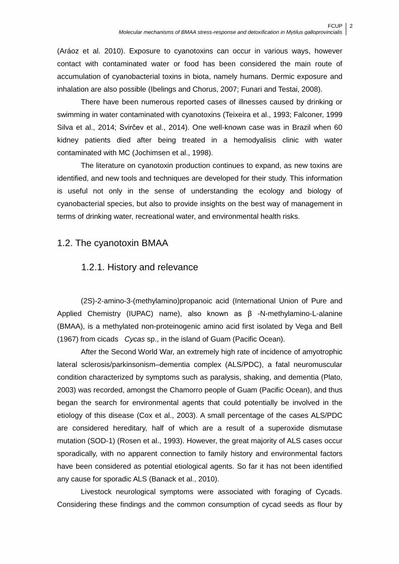
(Aráoz et al. 2010). Exposure to cyanotoxins can occur in various ways, however
contact with contaminated water or food has been considered the main route of
accumulation of cyanobacterial toxins in biota, namely humans. Dermic exposure and
inhalation are also possible (Ibelings and Chorus, 2007; Funari and Testai, 2008).
There have been numerous reported cases of illnesses caused by drinking or
swimming in water contaminated with cyanotoxins (Teixeira et al., 1993; Falconer, 1999
Silva et al., 2014; Svirčev et al., 2014). One well-known case was in Brazil when 60
kidney patients died after being treated in a hemodyalisis clinic with water
contaminated with MC (Jochimsen et al., 1998).
The literature on cyanotoxin production continues to expand, as new toxins are
identified, and new tools and techniques are developed for their study. This information
is useful not only in the sense of understanding the ecology and biology of
cyanobacterial species, but also to provide insights on the best way of management in
terms of drinking water, recreational water, and environmental health risks.
1.2. The cyanotoxin BMAA
1.2.1. History and relevance
(2S)-2-amino-3-(methylamino)propanoic acid (International Union of Pure and
Applied Chemistry (IUPAC) name), also known as β -N-methylamino-L-alanine
(BMAA), is a methylated non-proteinogenic amino acid first isolated by Vega and Bell
(1967) from cicads Cycas sp., in the island of Guam (Pacific Ocean).
After the Second World War, an extremely high rate of incidence of amyotrophic
lateral sclerosis/parkinsonism–dementia complex (ALS/PDC), a fatal neuromuscular
condition characterized by symptoms such as paralysis, shaking, and dementia (Plato,
2003) was recorded, amongst the Chamorro people of Guam (Pacific Ocean), and thus
began the search for environmental agents that could potentially be involved in the
etiology of this disease (Cox et al., 2003). A small percentage of the cases ALS/PDC
are considered hereditary, half of which are a result of a superoxide dismutase
mutation (SOD-1) (Rosen et al., 1993). However, the great majority of ALS cases occur
sporadically, with no apparent connection to family history and environmental factors
have been considered as potential etiological agents. So far it has not been identified
any cause for sporadic ALS (Banack et al., 2010).
Livestock neurological symptoms were associated with foraging of Cycads.
Considering these findings and the common consumption of cycad seeds as flour by

FCUP Molecular mechanisms of BMAA stress-response and detoxification in Mytilus galloprovincialis
3
the indigenous Chamorro people, a link with ALS/PDC was suggested (Whiting et al.,
1966). After isolation in cycad tissues BMAA was put forward as being a possible cause
of ALS/PDC in the Chamorro people of Guam (Vega and Bell, 1967).
Parallel research showed that injection of BMAA into chicks and rats caused
convulsions, supporting the hypothesis of neurotoxic behavior of this compound (Vega
and Bell, 1967; Vega and Bell, 1968). However, dietary exposure to the toxin was not
sufficient to induce long term effects in macaques (Spencer et al., 1987) whereas
ALS/PDC took years or even decades after exposure to become symptomatic.
Moreover, cycad seeds processing was suggested to remove over 80% of BMAA,
which implied extremely high doses of the toxin would have to be consumed to induce
neurotoxic effects. Consequently, BMAA was questioned as causative agent of ALS
(Duncan et al., 1990).
Only later, when the amino acid was found to be biomagnified in animals, which
forage on cycad seeds, and are subsequently consumed by the Chamorro people did
the hypothesis of BMAA neurotoxicity re-emerge (Charlton et al., 1992). BMAA
biomagnification was reported when an increased concentration of the toxicant along
the Guam food chain was observed. Samples of Mariana flying fox (Pteropus
mariannus mariannus) skin presented BMAA levels three times more elevated than the
cycad seed coat eaten by them (Murch et al. 2004). Flying foxes were part of the
traditional diet of the Chamorro people, which suggested that BMAA was consumed by
humans at much higher concentrations than previously thought. This toxin was then
identified in the brains of deceased ALS/PDC patients from Guam (Cox et al., 2003).
Later, BMAA was also identified in the brain of patients in Canada and USA,
meaning that the biomagnification hypothesis of BMAA may be more widespread than
initially thought and not exclusive to Guam (Cox et al., 2003; Banack et al., 2006).
However, these results were subjected to some controversy as they could not be
replicated by other research group (Montine et al., 2005; Snyder et al., 2009).
1.2.2. Presence in cyanobacteria
BMAA presence in symbiotic Nostoc sp. that colonized the coralloid roots of the
Cycas sp. was the first report in cyanobacteria (Cox et al., 2003).
This discovery prompted the screening of cyanobacteria from all over the world
for BMAA. Since then, BMAA has been found in several other strains of cyanobacteria,
as symbiont or as free-living species, in terrestrial, marine, brackish or freshwater
environments (Cox et al., 2005; Esterhuizen and Downing, 2008; Metcalf et al., 2008,
Baptista et al., 2011, Cianca et al., 2012). However, many works reported BMAA not to
FCUP Molecular mechanisms of BMAA stress-response and detoxification in Mytilus galloprovincialis
4
be detected (Krüger et al., 2010; Rosén and Hellenäs, 2008; Fan et al., 2015), detected
in only a few samples (Faassen et al., 2009) or detected at residual concentrations
(Jonasson et al., 2010; Li et al., 2010).
This suggested that, since cyanobacteria have a global distribution the
occurrence of BMAA may be globally widespread, especially since blooms of
cyanobacteria are becoming increasingly common.
BMAA presence in biological samples has been reported using different
extraction and analytical methods (Faassen 2014). The existence of numerous isomers
of BMAA constitutes a challenge when detecting this amino acid in biological samples.
The risk of quantifying these isomers instead of BMAA has been addressed (Jiang et
al. 2012).
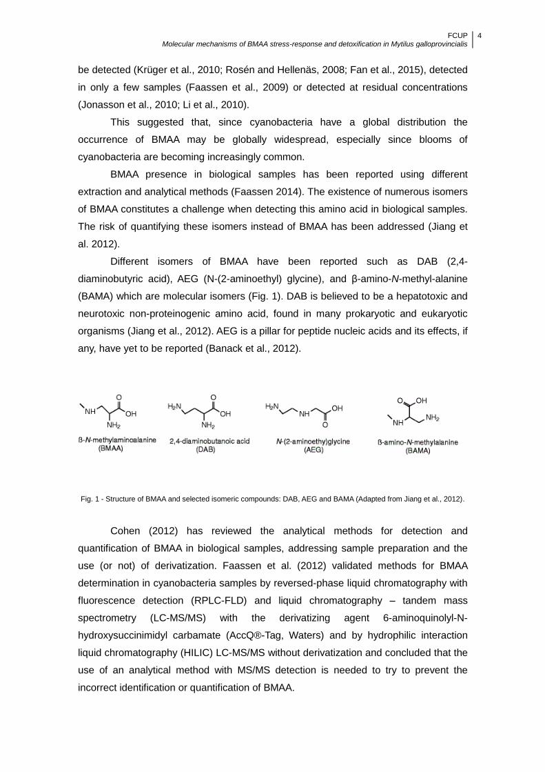
Different isomers of BMAA have been reported such as DAB (2,4-
diaminobutyric acid), AEG (N-(2-aminoethyl) glycine), and β-amino-N-methyl-alanine
(BAMA) which are molecular isomers (Fig. 1). DAB is believed to be a hepatotoxic and
neurotoxic non-proteinogenic amino acid, found in many prokaryotic and eukaryotic
organisms (Jiang et al., 2012). AEG is a pillar for peptide nucleic acids and its effects, if
any, have yet to be reported (Banack et al., 2012).
Fig. 1 - Structure of BMAA and selected isomeric compounds: DAB, AEG and BAMA (Adapted from Jiang et al., 2012).
Cohen (2012) has reviewed the analytical methods for detection and
quantification of BMAA in biological samples, addressing sample preparation and the
use (or not) of derivatization. Faassen et al. (2012) validated methods for BMAA
determination in cyanobacteria samples by reversed-phase liquid chromatography with
fluorescence detection (RPLC-FLD) and liquid chromatography – tandem mass
spectrometry (LC-MS/MS) with the derivatizing agent 6-aminoquinolyl-N-
hydroxysuccinimidyl carbamate (AccQ®-Tag, Waters) and by hydrophilic interaction
liquid chromatography (HILIC) LC-MS/MS without derivatization and concluded that the
use of an analytical method with MS/MS detection is needed to try to prevent the
incorrect identification or quantification of BMAA.

FCUP Molecular mechanisms of BMAA stress-response and detoxification in Mytilus galloprovincialis
5
Faassen et al. (2012) suggested that the variation in results may to be related to
the analytical method used. It has been also suggested that matrix effects may be
complicating factors in BMAA analysis (Glover et al., 2012). BMAA is a highly reactive
molecule and it may interact with other molecules during the analysis, which could
prevent accurate quantification (Li et al., 2010; Glover et al., 2012). If the matrix is of
higher complexity, other chemical interactions could interfere with the analysis (Li et al.,
2010; Glover et al., 2012). Finally, the lack of a standard procedure to determine BMAA
in various samples (e.g. cyanobacteria, marine invertebrates and vertebrates, etc.)
constitutes a problem in the accurate determination of BMAA: Therefore, failure to
detect BMAA cannot be taken as a total absence of the compound.
1.2.3. Biosynthesis
Variation of cellular toxin levels under different growth conditions have been
studied for a number of toxin-producing cyanobacteria (Rapala and Sivonen, 1998,
Sivonen and Jones, 1999). Some studies have established a relation between
environmental conditions and cyanotoxin production (Rapala and Sivonen, 1998; Vézie
et al., 2002; Jähnichen et al., 2007). Efforts rely on unveiling not only the mechanism
that underlie toxin biosynthesis but also its role in cyanobacteria, which remains a
mystery. Nonetheless, it remains a difficulty to identify all the factors involved in
cyanotoxin biosynthesis, how these are regulated at a molecular level and how this
translates to actual responses in the environment (Neilan et al., 2013). Some
cyanotoxins are constitutively produced and potentially less dependent on the
environmental conditions.
Some cyanotoxins, such as MC, NOD and CYN have a complete gene cluster
for its biosynthesis characterized in different strains of cyanobacteria (Moffit and Neilan,
2004; Mbedi et al., 2005; Micallef et al., 2015). However, for the vast majority of the
toxic secondary metabolites produced by cyanobacteria, a clearly identified
biosynthesis pathway has not been put forward. Such is the case of BMAA.
The production of BMAA by cyanobacteria in culture media has been reported
not to be consistent (Downing et al. 2011; Li et al. 2010), and since no pathway of
BMAA synthesis has been described, the conditions that favour its production by
cyanobacteria remain to be ascertained. Downing et al. (2011) suggested that BMAA
would be synthesized as a response to nitrogen depletion in culture media. However, in
other studies, this was not verified (Baptista et al., 2015). In fact, different strains of
cyanobacteria appeared to respond better, to the presence of nitrogen in the medium
regarding BMAA productivity.
FCUP Molecular mechanisms of BMAA stress-response and detoxification in Mytilus galloprovincialis
6
1.2.4. Mechanisms of toxicity
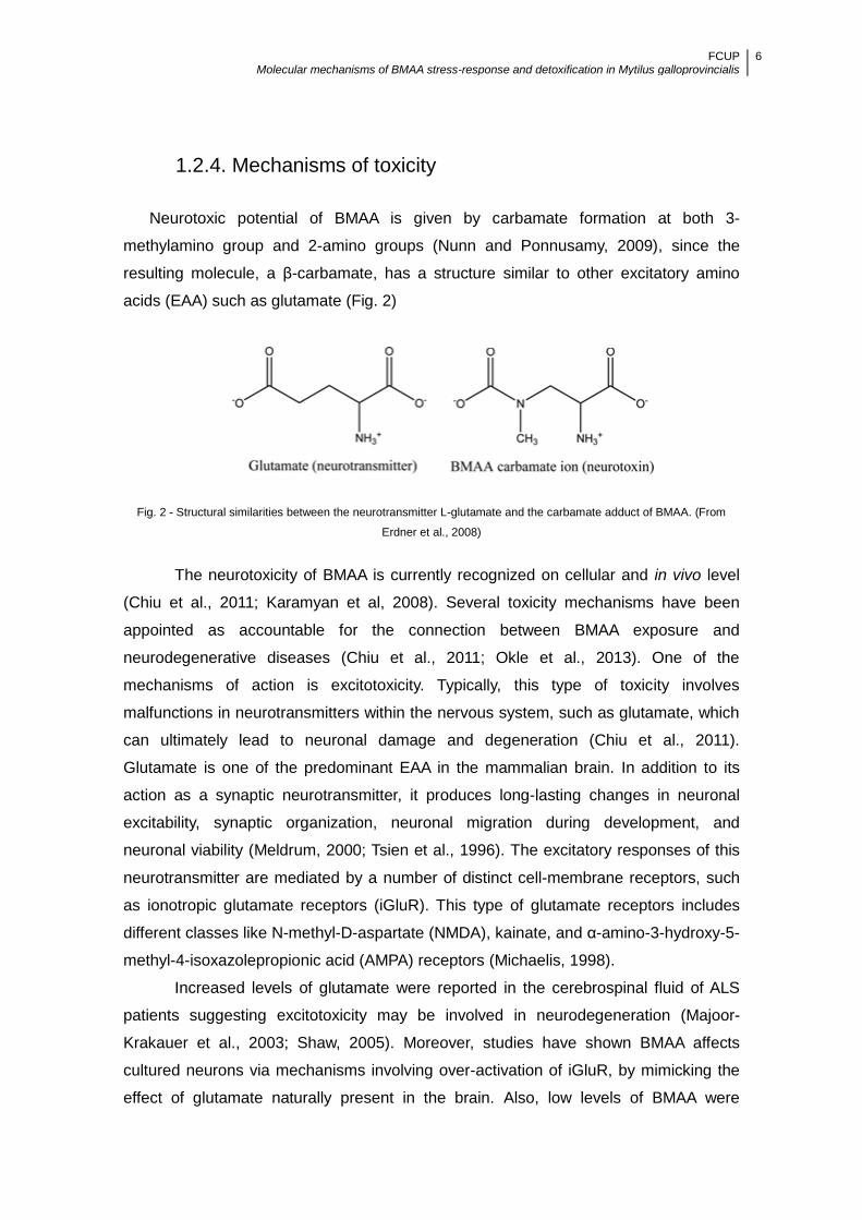
Neurotoxic potential of BMAA is given by carbamate formation at both 3-
methylamino group and 2-amino groups (Nunn and Ponnusamy, 2009), since the
resulting molecule, a β-carbamate, has a structure similar to other excitatory amino
acids (EAA) such as glutamate (Fig. 2)
Fig. 2 - Structural similarities between the neurotransmitter L-glutamate and the carbamate adduct of BMAA. (From
Erdner et al., 2008)
The neurotoxicity of BMAA is currently recognized on cellular and in vivo level
(Chiu et al., 2011; Karamyan et al, 2008). Several toxicity mechanisms have been
appointed as accountable for the connection between BMAA exposure and
neurodegenerative diseases (Chiu et al., 2011; Okle et al., 2013). One of the
mechanisms of action is excitotoxicity. Typically, this type of toxicity involves
malfunctions in neurotransmitters within the nervous system, such as glutamate, which
can ultimately lead to neuronal damage and degeneration (Chiu et al., 2011).
Glutamate is one of the predominant EAA in the mammalian brain. In addition to its
action as a synaptic neurotransmitter, it produces long-lasting changes in neuronal
excitability, synaptic organization, neuronal migration during development, and
neuronal viability (Meldrum, 2000; Tsien et al., 1996). The excitatory responses of this
neurotransmitter are mediated by a number of distinct cell-membrane receptors, such
as ionotropic glutamate receptors (iGluR). This type of glutamate receptors includes
different classes like N-methyl-D-aspartate (NMDA), kainate, and α-amino-3-hydroxy-5-
methyl-4-isoxazolepropionic acid (AMPA) receptors (Michaelis, 1998).
Increased levels of glutamate were reported in the cerebrospinal fluid of ALS
patients suggesting excitotoxicity may be involved in neurodegeneration (Majoor-
Krakauer et al., 2003; Shaw, 2005). Moreover, studies have shown BMAA affects
cultured neurons via mechanisms involving over-activation of iGluR, by mimicking the
effect of glutamate naturally present in the brain. Also, low levels of BMAA were

FCUP Molecular mechanisms of BMAA stress-response and detoxification in Mytilus galloprovincialis
7
associated with selective damage of sub-populations of neurons, such as motor
neurons, via activation of AMPA/kainate receptors (Rao et al., 2006).
After consumption, BMAA passes from the gut into the blood stream and
crosses the blood-brain barrier via large neutral amino acid carriers (Chiu et al., 2011).
At this point, the formation of a carbamate adduct of the side-chain amino group
produces structures capable of activating glutamate receptors by mimicking the effect
of glutamate naturally present in the brain (Weiss et al, 1989; Rakonczay et al., 1991).
The excess of BMAA leads to an overflow of calcium ions into the cells, and
subsequent cell damage (Choi, 1988). The increased Ca2+ concentration in the cell
causes the activation of lysozymes that inactivate the mitochondria by preventing the
ATP synthesis associated with respiratory oxidation. Less ATP leads to less ATPases
and Ca²+ enzymes, forming a vicious cycle with increased intracellular calcium,
ultimately ending in cell death (Stout et al., 1998; Lobner et al., 2007; Chiu et al., 2011).
Excitotoxicity has been described for other amino acids such as β-N-oxalyl-
amino-L-alanine (BOAA) and Domoic Acid (DA). BOAA can be found in the legume
Lathyrus sativus and is responsible for the occurrence of Human Lathyrism (Spencer et
al., 1987; Weiss et al.1989), by acting as a selective AMPA agonist. DA is a kainate
agonist and its presence in algal blooms is associated with the occurrence of amnesic
shellfish poisoning (ASP) (Wright et al., 1989; Todd, 1993). Activation of iGluR is also
related to free radical production by calcium-dependent activation of the arachidonic
acid cascade, nitric oxide synthase and calpain (Szydlowska and Tymianski, 2010).
With time, reactive oxygen species can lead to irreversible cellular injury and death, by
damaging proteins, lipids and DNA, and consequently leading to function impairment of
vital macromolecules and organelles. BMAA is also responsible for the inhibition of the
cysteine/glutamate antiporter system Xc-. Thus, the uptake of cysteine is blocked,
which leads to glutathione depletion, and ultimately results in oxidative stress increase.
Simultaneously, the system Xc- potentiates the release of glutamate from the cell. By
binding to iGluR, excess glutamate induces further neurological damage by
excitotoxicity (Liu et al., 2009). Despite the information regarding BMAA effects in
neuronal cells the, effects upon iGluR have not been yet tested in marine organisms.
Recent studies appoint BMAA as capable of being incorporated into proteins
and subsequently lead to protein misfolding (Banack et al., 2010). The tRNA
synthetase enzyme for the amino acid serine mistakenly picks up BMAA and
incorporates it into proteins in vitro. Amino acid misincorporation leads to mistakes in
translation, exposing the hydrophobic parts of the protein. The resulting misfolded
proteins start to ―aggregate‖ forming larger clusters, until the cells are no longer able to
function effectively (Dunlop et al., 2013).

FCUP Molecular mechanisms of BMAA stress-response and detoxification in Mytilus galloprovincialis
8
1.3. Health and environmental risks of exposure to BMAA
1.3.1. Biomagnification hypothesis and human exposure
The bioaccumulation of cyanotoxins by aquatic organisms is widely described in
literature. Several studies have shown that mollusks are among the most harmed
organisms, being able to accumulate several cyanotoxins, such as MC (Amorim and
Vasconcelos, 1999, Pires et al., 2004; Paldavičienė, et al., 2015), NOD (Sipiä, et al.,
2002; Karlsson et al., 2003), paralytic shellfish poisoning toxins (Pereira et al., 2004;
Setälä et al., 2014), CYN (Saker et al., 2004), anatoxin-a (Osswald et al., 2008) and
okadaic acid (Silva et al., 2013; Garcia et al., 2015). More recently, the presence of
BMAA in food webs of the North Atlantic was verified, suggesting that certain diets and
locations may put people at particular risk (Jonasson et al., 2010; Brand et al., 2010).
In the Baltic Sea, BMAA was found in organisms of higher trophic levels such as
various vertebrates and invertebrates such as mussels and oysters (Jonasson et al.,
2010). Also, in a recent long term monitoring of BMAA in cyanobacteria, mollusks,
crustaceans and various fish species at different trophic levels in Gonghu Bay,
bioaccumulation of the toxin was observed among the aquatic animals, and within the
food web (Jiao et al., 2014). Over 7 months of study, BMAA content in cyanobacteria,
mollusks, crustaceans and various fish species averaged between 4.12 – 6.05 µg
BMAA g-1 dry weight (dw). The transfer and bioaccumulation of BMAA within these and
other food webs illuminate the possible pathways of human exposure.
Mytilus galloprovincialis is a mollusk native to the Mediterranean coast and the
Black and Adriatic Seas. It has succeeded in establishing itself at widely distributed
points around the globe, occurring essentially in temperate regions, including the
Atlantic Ocean (Branch and Stephanni, 2004). It is a smooth-shelled mussel with
coloring usually ranging from blue-violet to black. The two shells are of quadrangular
shape and equal dimensions. Generally, on one side, the edge of the shell ends with a
pointed and slightly bent umbo while the other side is rounder. This animal can grow up
to 140 mm in length and it can inhabit from exposed rocky outer coasts to sandy
bottoms (Ceccherelli and Rossi 1984). M. galloprovincialis constitutes an important
component of estuarine and marine food webs and because of its sessile filter feeder
nature, it may be exposed to high density of cyanobacteria and their toxins (Osswald et
al., 2008). This species is eaten by other animals (including humans), providing a way
FCUP Molecular mechanisms of BMAA stress-response and detoxification in Mytilus galloprovincialis
9
of exposure to cyanotoxins, namely BMAA, through food chain. In fact, it was recently
shown that BMAA is accumulated in this organism (Baptista et al., 2015).
Thus human exposure to BMAA may come from direct contact with
cyanobacterial blooms or consumption of aquatic organisms exposed to such blooms
(Jonasson et al. 2010; Cox et al., 2005). This suggests that BMAA, like cyanobacteria,
may have a global distribution and therefore the occurrence of BMAA may be a
worldwide concern.
Due to this fact, and to the discrepancies documented on BMAA measurement
in environmental samples (Banack et al., 2010; Faassen, 2014; Glover et al., 2012) the
assessment of this amino acid in the aquatic environment, should take into account not
only its quantification, but also its effects towards aquatic organisms.
1.3.2. Biomarkers of toxic exposure
Biomarkers are helpful tools in detecting and evaluating the harmful
consequences of chemical contamination on organisms. They can be subdivided into
three classes (NRC, 1987; WHO, 1993). However, one biomarker can be classified
within several groups
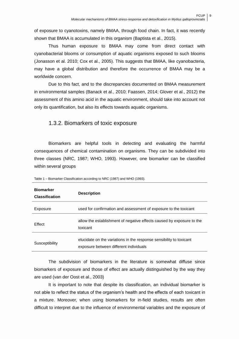
Table 1 – Biomarker Classification according to NRC (1987) and WHO (1993).
Biomarker
Classification Description
Exposure used for confirmation and assessment of exposure to the toxicant
Effect allow the establishment of negative effects caused by exposure to the
toxicant
Susceptibility elucidate on the variations in the response sensibility to toxicant
exposure between different individuals
The subdivision of biomarkers in the literature is somewhat diffuse since
biomarkers of exposure and those of effect are actually distinguished by the way they
are used (van der Oost et al., 2003)
It is important to note that despite its classification, an individual biomarker is
not able to reflect the status of the organism’s health and the effects of each toxicant in
a mixture. Moreover, when using biomarkers for in-field studies, results are often
difficult to interpret due to the influence of environmental variables and the exposure of

FCUP Molecular mechanisms of BMAA stress-response and detoxification in Mytilus galloprovincialis
10
the studied organisms to a mixture of several chemicals with influence on the
measured molecular and/or cellular responses (Frasco et al., 2005). For these reasons,
the use of a biomarker battery is recommended when assessing the biological impact
of chemical pollutants (Regoli et al., 2004).
Biomarkers often provide faster biological answers and information about
toxicant bioavailability in comparison with other toxicological methods or assays
(Shugart et al. 1992). An accurate prediction of effects at higher levels of organization
in the ecosystem makes biochemical changes useful biomarkers. These variations
allow the assessment of whether toxicants are present, as well as the natural response
of a particular organism to a specific environmental stressor. Regarding cyanobacterial
exposure, parameters of biotransformation or oxidative stress are usually addressed
(Bláha et al. 2004; Ziková et al., 2013; Guzmán-Guillén et al., 2014).
Biomarkers are common in the biomonitoring of contaminated marine
environments. Typically, they rely on the study of potential stimulation or inhibition of
specific enzymes, such as Glutathione s-tranferases (GSTs) and Acetylcholinesterase
(AChE) (Moreira and Guilhermino, 2005). GSTs are a family of phase II dimeric
enzymes involved in the detoxification of xenobiotics with an electrophilic center by
conjugating them with reduced glutathione (GSH) (Hayes et al., 2005). The activity of
these detoxification enzymes has been successfully used as a biomarker of exposure
to toxic compounds in bivalves (Sheehan and Power, 1999). Increased GSTs activity
has been reported, not only in mussels but also in various other aquatic organisms
(Pflugmacher et al., 1998) in connection to exposure to cyanobacterial toxins, such as
MC and CYN (Pflugmacher et al., 1998; Wiegand et al.,1999; Nogueira et al., 2004;
Sipiä et al., 2002, Vasconcelos et al., 2007). AChE is an indicator of neurotoxic effects
(Kirby et al., 2000) and has traditionally been used as a biomarker of exposure to
organophosphate and carbamate pesticides (Bocquené and Galgani, 1998). It is
usually a general indicator of pollution (Schiedek et al., 2006), and recently was also
proven to be affected by exposure to NOD (Lehtonen et al., 2003), as well as other
cyanotoxins (Ferrão-Filho and Kozlowsky-Suzuki, 2011).
More recently, research efforts have been converging on particular genes
sequencing and transcription to be used as potential biomarkers. In fact, this molecular
biology approach (transcriptomics) has been already used in the biomonitoring of
contaminated sites (Hoarau et al., 2006; Sarkar et al, 2006). Gene expression
biomarkers can enable rapid assessment of physiological conditions in situ, providing a
valuable tool for linking organism physiology with cyanobacterial toxic exposure. In
most studies, there is a focus on the genes which are either involved in the metabolic
activation and detoxification of xenobiotics, or in oxidative stress regulation (Kurelec et

FCUP Molecular mechanisms of BMAA stress-response and detoxification in Mytilus galloprovincialis
11
al., 1996; Blanchette et al., 2007; Izagirre et al, 2014). However, it has become
increasingly clear with time that single biomarkers are not able to describe the
physiological status of the mussel. Instead, the use of a battery of biomarkers is
becoming a standard (Narbonne et al., 2005; Cotou et al., 2013; Izagirre et al, 2014).
1.4. Objectives
In marine environments BMAA has been shown to find its way from the
phytoplankton first producers (e.g. cyanobacteria) to marine invertebrates first
consumers such as M. galloprovincialis, which accumulate this amino acid. Therefore,
the aim of this work was to assess the use of the activity of the enzymes GSTs and
AChE, and the expression of iGluR genes, as biomarkers of BMAA exposure in M.
galloprovincialis.
To this aim, genetic expression of two iGluR genes was evaluated by a
quantitative polymerase chain reaction (qPCR), and GSTs and AChE activity was
measured spectrophotometrically in gills and digestive gland of M. galloprovincialis
exposed to BMAA standard, or fed with BMAA-producing cyanobacterium.
BMAA effects upon enzyme activity, in M. galloprovincialis, have not been
tested yet, despite the fact that these are commonly assessed biomarkers for exposure
to cyanotoxins.
Effects upon glutamate receptors have also not been yet tested in M.
galloprovincialis. In fact, the presence of iGluR in M. galloprovincialis has only been
putatively ascertained, through comparison with iGluR sequences from other
organisms. Therefore, the first part of this analysis consisted in determining iGluR
sequences of interest and characterizing them with the support of bioinformatics tools.
FCUP Molecular mechanisms of BMAA stress-response and detoxification in Mytilus galloprovincialis
12
2. Material & Methods
2.1. Mussel collection
Collection of M. galloprovincialis took place in a beach at Matosinhos, Portugal,
in October 2014. The mussels were placed in aquariums at BOGA (Biotério de
organismos aquáticos) at CIIMAR (Centro Interdisciplinar de Investigação Marinha e
Ambiental), for acclimation, during 3 days. The aquariums used in this experiment had
capacity of 5 L, were filled with 3 L of filtered sea water, aerated and maintained at
18ºC.
Collection of mussels was performed in Italy. M. galloprovincialis were brought
from a lagoon outside of Venice, in February 2015. They were maintained in aquariums
with 14 L of capacity, filled with 10 L aerated sea water at 14°C for 5 days for
acclimation.
In both cases the mussels were not fed. Water was renewed every two days.
2.2. Bioaccumulation experiment
2.2.1. Mussels exposed to BMAA standard
For analysis of AChE and GSTs activity M. galloprovincialis, collected at
Portugal, at densities of 24 individuals per tank, were exposed to BMAA standard
(Sigma–Aldrich) with concentrations of 10 µg L-1, 100 µg L-1, and 1000 µg L-1 dissolved
in filtered seawater for a period of 48h. All experiments were conducted in triplicate.
Control mussels (not exposed to BMAA) were kept likewise in filtered seawater. Four
individuals were collected from the BMAA treatment and control aquariums at 12h, 24h
and 48h. At this point the water was changed and mussels depurated for 2 days.
During this period, at 72h and 96h, 4 individuals were also collected from the BMAA
treatment and control aquariums. The mussels were not fed, and no deaths were
observed during this experiment. Upon collection, mussels were dissected and
separated in gills and digestive gland. Tissues were then frozen at -80ºC.
For analysis of iGluR expression, mussels collected at Italy, at densities of 24
individuals per tank, were exposed to 1000 ug L-1 standard BMAA dissolved in
seawater for a period of 48h. Four individuals were collected from the BMAA treatment
and control aquariums at 6h, 24h, 48h. At this point the water was changed and

FCUP Molecular mechanisms of BMAA stress-response and detoxification in Mytilus galloprovincialis
13
mussels depurated for 1 day. At 72h 4 individuals were also collected from the BMAA
treatment and control aquariums. The mussels were not fed. Five deaths were
observed in the control aquarium. Five deaths were also observed in the aquarium
treated with BMAA during this experiment.
Upon collection, mussel sex and gonad status was registered. A mix of females
and males in similar conditions were used, to avoid variability induced by discrepancies
in reproductive stage. Mussels were dissected and separated in gills and digestive
gland. Tissues were then frozen at -80ºC.
2.2.2. Mussels fed with cyanobacteria
Animals were kept in 0.5 L aquariums of filtered seawater, with aeration and
natural light, at 18 °C. After an acclimation period of 3 days, mussels in each aquarium
were fed either with cyanobacteria Nostoc sp. (LEGE 06077), or Microcoleus sp.
(LEGE 07092), from the LEGE culture collection, or with the green algae Chlorella sp.
(LEGE Z-001). Cyanobacteria sequences associated with this study are available in
GenBank under the accession numbers HM217071 and HM217062, respectively.
Cyanobacteria species were grown in BG11 culture medium (Rippka et al.,
1979), with a light intensity of 25 µE m− 2s− 1 and a light:dark period of 14h:10h, at 25ºC.
Chlorella sp. was grown in Z8 culture medium (Kotai, 1972) in the same conditions
described above.
Mussels were fed once each day with approximately 105 cells mL-1 of either
Nostoc sp., Microcoleus sp or Chlorella sp. From each aquarium, 4 mussels were
retrieved, at 24h and 48h. Upon collection, mussels were dissected and gills and
digestive gland were separated for posterior analysis of enzymatic activity. Tissues
were immediately frozen at -80ºC awaiting further procedures.
2.3. Enzymatic analysis
2.3.1. Tissue homogenization and protein quantification
Approximately 250 mg of tissue from digestive gland or from gills were
homogenized with liquid nitrogen and a protein solubilization buffer (SB). SB was
prepared as a solution of phosphate buffer (100 mM, pH 7.0) containing 20% glycerol
(v/v), 1.4 mM dithiothreitol (DTT) and 1 mM Ethylenediaminetetraacetic acid (EDTA) to
aid in the extraction and stabilization of proteins present in the mussel tissues.

FCUP Molecular mechanisms of BMAA stress-response and detoxification in Mytilus galloprovincialis
14
Homogenization was made maintaining a constant ratio of 5 mL of buffer for each gram
of tissue. Cell debris were ultra-centrifuged (Beckman Coulter centrifuge, model Alegra
25R, Beckman Coulter, CA, USA) at 100 000 ×g for 60 min, at 4ºC. The supernatants
were stored at −80 °C for posterior analysis.
Total protein quantification was conducted using a microplate-adapted protocol
of the Bradford method (Bradford, 1976). The assay is based on the observation that
the absorbance maximum for an acidic solution of Coomassie Brilliant Blue G-250
shifts from 465 nm to 595 nm when binding to protein occurs. Both hydrophobic and
ionic interactions stabilize the anionic form of the dye, causing a visible color change.
A standard curve was prepared using standards containing a range of 0.25 mg
mL-1 to 1.25 mg mL-1 bovine serum albumin. In the microplate, 5 µL of standard or
sample were added to the wells. After addition of 250 µL dye reagent to the wells and
an incubation period of 15 min absorbance measurements were performed at 595 nm.
Protein quantification was obtained by comparison with standard curve previously
obtained. Sample protein content was then adjusted to 0.3 mg mL-1.
2.3.2. GSTs activity quantification
GSTs activity was measured using 1-chloro-2,4-dinitrobenzene (CDNB), which
is suitable for the broadest range of GST isoenzymes. The reaction is based on the
principle that upon conjugation of the thiol group of glutathione to the CDNB substrate,
there is an increase in the absorbance at 340 nm. Procedures were as described by
Habig, (1974) adapted to microplate, with alterations from Frasco and Guilhermino,
(2002).
To obtain the reaction mixture, CDNB (60 mM) was dissolved in ethanol, with
the final reaction concentration less than 0.01%, and GSH (10 mM) was dissolved in
phosphate buffer (100 mM, pH 7.0).
In 96-well microplates, 0.2 mL of the reaction mixture was added to 0.1 mL of
the sample and the GSTs activity was measured immediately every 20 seconds, at 340
nm, during 5 minutes, at 25ºC, in triplicate on Bioteck microplate reader (Synergy HT,
2009).
2.3.3. AChE activity quantification
AChE activity was measured using DTNB (acid dithiobisnitobenzoate) to
quantify the thiocholine produced from the hydrolysis of acetylthiocholine by AChE. The

FCUP Molecular mechanisms of BMAA stress-response and detoxification in Mytilus galloprovincialis
15
absorption intensity of DTNB adduct is used to measure the amount of thiocholine
formed, which is proportional to the AChE activity. The method of Ellman et al. (1961),
adapted to microplate, was followed.
In the assay, 0.250 mL of the reaction solution (30 mL of phosphate buffer, 1 mL
of 10 mM DTNB (acid dithiobisnitobenzoate mixed with sodium hydrogen carbonate in
phosphate buffer) and 0.200 mL of acetylthiocholine iodide 0.075 M) were added to
0.05 mL of homogenized sample tissue. The measurements were performed after an
incubation period of 5 min, at 412 nm, during 10 minutes, every 15 seconds, at 25ºC, in
triplicate on Bioteck microplate reader (Synergy HT, 2009).
2.4. iGluR expression
2.4.1. Sequence selection and preliminary analysis
A search in National Center for Biotechnology Information (NCBI) databases
showed that there were no available sequences for glutamate receptors in M.
galloprovincialis, or in Mytilus sp. in general. Therefore, the first part of this work was
devoted to finding sequences that could putatively be considered as glutamate
receptors, in M. galloprovincialis.
For that purpose, a catalogue of ESTs from M. galloprovincialis named Mytibase
was used (Venier et al., 2009), at the Department of Biology of University of Padova.
Seven putative glutamate receptor sequences were initially retrieved. Nucleotide and
deduced amino acid sequences were obtained, after similarity to orthologue sequences
was determined.
Digital gene expression was performed with CLC Genomics Workbench to
ensure sequences with higher expression values in M. galloprovincialis tissues were
selected (Annex 1). Based on this preliminary analysis, two sequences, termed GLU4
and GLU5 were chosen for further work.
iGluR all share a common membrane topology (Fig. 3) characterized by a large
extracellular N-terminus that includes the N-terminal domain (S1), a membrane region
comprising three transmembrane segments (M1, M3 and M4,) plus a re-entrant pore
loop (M2,), an extracellular loop between M3 and M4 where the ligand-binding domain
(S2) is located, and a cytoplasmic C-terminus, which varies in size and provides
multiple sites of interaction with numerous intracellular proteins (Wood et al., 1995;
Dingledine et al., 1999).

FCUP Molecular mechanisms of BMAA stress-response and detoxification in Mytilus galloprovincialis
16
Fig. 3 - Membrane topology of iGluR. S1 – N-terminal domain; S2 – Ligand-binding region; M1, M3 and M4 –
Transmembrane domain; M2 – Re-entrant pore loop (From VanDongen Lab:
http://people.duke.edu/~av8/vandongen_lab/Research).
After selection of GLU4 and GLU5, further work was carried out to characterize
the sequences. In order to have an idea of GLU4 and GLU5 domain architectures
SMART was used (Schultz et al., 1998; Letunic et al., 2015). The presence of signature
domains consistent with iGluR receptors, such as L-glutamate binding region was
observed (Annex 2). This online tool also allowed the retrieval of several sequences
from different taxa with domain organization similar to GLU4 and GLU5. Afterwards, all
sequences were aligned using the Clustal Omega (Sievers et al., 2011). After
alignment (Annex 3), a phylogenetic tree was generated with Phylogeny.fr platform
(Dereeper et al., 2008) to analyze relationships between mussel and other taxa iGluR
sequences (Annex 4).
Hydropathic analysis with Phobius (Käll et al., 2004) of the predicted mature
polypeptides of GLU4 and GLU5 showed the presence of three strongly hydrophobic
regions (Annex 5 and 6; images obtained with Protter (Omasits et al. 2013)). The N
termini of GLU4 and GLU5 contained signal peptides of 20 and 23 amino acids,
respectively. The distribution of hydrophobic regions and potential sites for N-
glycosylation suggests that the topology of GLU4 and GLU5 is similar to that predicted
for the mammalian iGluR subunits. The present sequences, however, appear to lack N-
terminal domain present typically in iGluR (Annex 2).
2.4.2. Primer design
Open reading frame (ORF) finder (http://www.ncbi.nlm.nih.gov/projects/gorf/)
FCUP Molecular mechanisms of BMAA stress-response and detoxification in Mytilus galloprovincialis
17
was used to identify all open reading frames using the standard or alternative genetic
codes. The coding regions of GLU4 and GLU5 are of 1467 and 1338 nucleotides long,
respectively. Primers were designed with Primer 3 (Untergrasser et al., 2012;
Koressaar and Remm, 2007), to fit in the selected ORF (Annex 7) for each sequence
and in accordance to the following guidelines: product size 100-250 bp, melting
temperatures Tm 60±1 °C, and G/C ≤50 %. Primer details are reported in Table 2. The
housekeeping gene Elongation factor 1 alpha (EF-1 α) was chosen as internal
reference because the level of RNA remains essentially constant from sample to
sample and the primers have been used as described in Gerdol et al. (2011).
Table 2 - Primers designed for analysis of GLU4 and GLU5 sequences and the housekeeping gene EF-1 α
Primer
name Description Sequence 5’-3’
Amplicon
size (bp)
GLU4 Glutamate receptor F:5’-GTGAGCCATTTCTGCTCCAG-3’
R: 5’-GATGGATCGTTTGCAGCCAT-3’ 212
GLU5 Glutamate receptor F:5’-GATGCCAAAGACCGTAGCTG-3’
R:5’-TTCCAATCCACACGCAAAGG-3’ 175
EF-1 α Elongation factor F:5’-CCTCCCACCATCAAGACCTA-3’
R:5’-GGCTGGAGCAAAGGTAACAA-3’ 130
Primer specificity and the presence of an unique amplicon was verified by
blasting primers sequences against mussel cDNA library Mytibase.
2.4.3. RNA extraction and purification
Total RNA from approximately 100 mg of frozen gill or digestive gland was
extracted using Trizol reagent (Invitrogen) following the manufacturer’s
recommendations. Following isolation of total RNA, the RNA was further cleaned by
LiCl (8 M) purification. Pellets were washed twice with 75% ethanol (EtOH) and
resuspended in 200 µL of RNAse free water.
For RNA quantification a ND-1000 UV/visible spectrometer (NanoDrop
Technologies) was used, with a sample volume of 1 µL. In the samples RNA
concentration ranged between 233 and 1914 ng µL-1 (Table 3). Samples were
FCUP Molecular mechanisms of BMAA stress-response and detoxification in Mytilus galloprovincialis
18
considered pure regarding contamination from protein and carbohydrates, since the
values of the ratio of the wavelengths 260/280 and 260/230 were above 2.0 and 2.2,
respectively.
Table 3 – Nanodrop quantification of RNA concentration in exposed digestive gland and gill tissues and paired controls.
Absorbance spectroscopy gives a concentration of RNA in ng µL-1.
Organ Time (h) Treatment RNA (ng µL-1
)
Digestive Gland 6 Control 1164
Digestive Gland 24 Control 1355
Digestive Gland 48 Control 1324
Digestive Gland 72 Control 1353
Digestive Gland 6 BMAA 1568
Digestive Gland 24 BMAA 1390
Digestive Gland 48 BMAA 1866
Digestive Gland 72 BMAA 1914
Gills 6 Control 434
Gills 24 Control 366
Gills 48 Control 366
Gills 72 Control 362
Gills 6 BMAA 864
Gills 24 BMAA 404
Gills 48 BMAA 321
Gills 72 BMAA 233
The qualitative analysis of RNA was performed on gill and digestive gland RNA
samples using the RNA 6000 Nano LabChip kit (Agilent Technologies) in association
with the Agilent 2100 Bioanalyzer.
The RNA Gel Matrix, provided with the kit, was filtered through spin columns
(Costar) and then centrifuged at 1500 x g, during 10 min at room temperature. In the
chip, 32.5 mL of filtered Gel Matrix, plus 0.5 µL of dye concentrate were charged. The
samples were then loaded into the wells, and the markers (RNA 6000 Nano Marker,
Agilent) were also loaded. Finally, all wells were loaded with 1 mL of RNA 6000 Ladder-
Ambion previously denatured. The instrument sequentially ran the ladder and samples

FCUP Molecular mechanisms of BMAA stress-response and detoxification in Mytilus galloprovincialis
19
quantitating the fluorescence emitted by a red fluorescent intercalating dye as the RNA
passed a fixed point within the capillary.
Figure 4 shows some of the electrophoregrams obtained showing that the
quality of the extracted RNA was suitable for the analysis of GLU4 and GLU5.
Electropherograms were obtained with Agilent 2100 Bioanalyzer, for M.
galloprovincialis gill and digestive gland total RNA.
Fig. 4 - Electropherogram (from the Agilent 2100 Bioanalyzer) for M. galloprovincialis gill and digestive gland total RNA.
The x-axis represents RNA length in nucleotides (nt), and fluorescence (FU) is represented on the y-axis. The quality
was ascertained by RNA integrity number (RIN). This algorithm divides the RNA profile into nine different regions and
applies a continuous value from 10 to 1 defining the extent of RNA degradation, 10 being the highest quality. All images
shown represent samples with RIN above 6: a) control digestive gland at 6h; b) exposed digestive gland at 72h; c)
Control gills at 6h; d) exposed gills at 72h.
2.4.4. Quantitative PCR for gene expression analysis
The expression levels of GLU4 and GLU5, were assessed in samples of the
digestive gland and gills, of four adult mussels, collected at each time point, from the
control (0 µg L-1) and the BMAA treated (1000 µg L-1) aquariums.
RNA pools were prepared with equal amounts of RNA from each individual
mussel (N = 4). The cDNA for qPCR was obtained using a Superscript II Reverse
Transcriptase 1st Strand cDNA Kit (Invitrogen) from 1 µg total RNA. PCR reactions
were performed in a 7500 Real-Time PCR System (Applied Biosystems, Foster City,
CA) using DyNAmo HS SYBR Green qPCR kit (Thermo Scientific) to amplify 1 µL of
FCUP Molecular mechanisms of BMAA stress-response and detoxification in Mytilus galloprovincialis
20
purified first-strand cDNA in a 10 µL of final reaction mixture.
Thermal cycling conditions were: 15 min denaturation at 95ºC; followed by 40
cycles of 30 s denaturation step at 95ºC, annealing and elongation steps for 1 min
each at 60ºC. A dissociation curve analysis was performed at the end of the reaction to
ensure the specificity of the primers. Three replicates were amplified of the complete
sample set (BMAA exposed mussels and paired controls, 4 time points) for each primer
pair (target and endogenous genes). The cycle threshold (Ct) is defined as the number
of cycles required for the fluorescent signal to cross the threshold in qPCR. To
calculate the relative expression ratio, the 2−ΔΔCt (RQ, relative quantification) method
(Livak and Schmittgen, 2001) implemented in the 7500 Real-Time PCR System
software was used.
Additionally, the primers were subjected to a preliminary test with spring mussel
gill samples, in order to confirm the presence of unique amplicons and exclude the
possibility of primer dimers. The gill samples for this evaluation originated from mussels
collected from an outlet of the Venice lagoon (Italy) in May 2014, and acclimated at
23ºC and 32 ‰ salinity. After 24h and 48h the mussels were retrieved and processed
as described before for qPCR. Dissociation curves of the qPCR products for both
GLU4 and GLU5 transcripts using these mussels showed single peaks.
2.5. BMAA quantification
2.5.1. Microwave-assisted digestion
Soft tissues from mussels exposed to 10 µg L-1, 100 µg L-1 and 1000 µg L-1 of
BMAA standard, or fed with cyanobacteria or microalgae, were lyophilized before
digestion (FTS System freeze dryer EZ550). Cyanobacteria and Chlorella sp. cultures
used for feeding were harvested by centrifugation and also lyophilized.
In both cases, approximately 10 mg (dw) were acid-digested with 2 mL of 6M of
HCl, at 120ºC, for 20 minutes using a high-pressure microwave system (Milestone-
Ethos 1). The samples were evaporated in a low flux of nitrogen and then reconstituted
in 0.5 mL 20 mmol L-1 HCl, prior to analysis by LC-MS/MS.
2.5.2. Liquid chromatography with mass detection (LC-
MS/MS) analysis.
The analyses were performed in a Thermo LCQ Fleet Ion Trap LC/MSn system

FCUP Molecular mechanisms of BMAA stress-response and detoxification in Mytilus galloprovincialis
21
(Thermo Scientific), using a 2.1 x 100 mm, 5 µm diameter ZIC-HILIC column
(SeQuant), and a 14 x 1 mm, 5 µm guard column (SeQuant). The mobile phase
consisted in eluent A, acetonitrile (0.1% formic acid) and eluent B, deionized water
(0.1% formic acid). In the first 20 min a 90–60% linear gradient of acetonitrile was
achieved, and afterwards 60% acetonitrile was maintained for 15 min. The system was
then equilibrated to the initial conditions during 5 min (Kubo et al. 2008). The flow rate
was 0.5 mL min–1, the injection volume was 10 µL, and the column temperature was 40
ºC. The electrospray ionization (ESI) was operated in the positive mode. Nitrogen was
used as sheath gas, at a rate of 45 (unitless), and auxiliary gas at a rate of 20
(unitless). The capillary temperature was held at 250 ºC.
Mass-to-charge ratio (m/z) scan was performed from 50 to 150, and the ion m/z
119 was monitored. At collision energy of 14 V the presence of more abundant product
ions m/z 102, 88 and 76 was verified, in this order of abundance, as reported before
(Rosén and Hellenäs, 2008) and selected-reaction monitoring (SRM) chromatograms
were retrieved. m/z 102 was used to quantitatively assess BMAA and m/z 88 and m/z
76 used to qualitatively assess BMAA. SoftwareXcalibur® was used to analyze the
data.
To account for matrix effects a calibration curve was prepared as described in
Baptista et al. (2015). Mussel and cyanobacteria (10 mg dw) were spiked with BMAA
standard in the 10 to 1000 µg L-1 range, and digested as described above. The limit of
quantification (LOQ) was obtained from the calibration curve, calculated as 10 α /S,
where α is the standard deviation of the y-intercept and S is the slope of regression
line.
2.6. Data treatment
GSTs and AChE activity results are presented as mean values ± standard error
of the mean (SEM) and were analyzed by one-way ANOVA. Post-hoc comparisons
were made using Tukey's test considering p≤0.05 as statistically significant. Individual
gene expression levels, of mussels exposed to BMAA and controls, were compared by
using an unpaired Student's t test. GraphPad Prism 6 was used for the calculations.

FCUP Molecular mechanisms of BMAA stress-response and detoxification in Mytilus galloprovincialis
22
3. Results
3.1. BMAA quantification in mussels and cyanobacteria
Mussels exposed to 1000 µg L-1 for 24h and 48h, presented detectable
amounts of BMAA (Table 4). At this concentration, accumulation of BMAA by mussels
was shown to be similar to previous exposures of M. galloprovincialis to the same
range of concentrations (Baptista et al., 2015). No mortality was registered during the
experiment, implying that acute lethal toxicity does not occur at the tested
concentrations. In mussels not exposed (control), the amino acid was not detected. For
10 µg L-1 and 100 µg L-1 quantification was not possible due to lack of availability of the
equipment.
Table 4 - Concentration of BMAA (µg g-1
) on mussels exposed to 1000 µg L-1
for 24 h and 48 h.
Mean ± SEM for the concentration of BMAA.
Concentration tested Exposure Time (h) BMAA (µg g-1
)
1000 µg L-1
24 41.59 ± 4,27
48 52.19 ± 4,43
0 µg L-1
24 < LOQ
48 < LOQ
Limit of quantification: LOQ = 0.8 µg g-1
BMAA was analysed in the cyanobacteria and Chlorella sp. used for feeding the
mussels. In Nostoc sp. BMAA could be quantified but in Microcoleus sp, no BMAA was
detected (Table 5). For the microalgae Chlorella sp. no BMAA could be detected either.
Quantification of BMAA in the mussel fed with cyanobacteria and green alga
was not possible. However, in a previous study, mussels fed with cyanobacteria
Synechocystis salina showed accumulation of 32 µg g-1 of BMAA after 4 days of
feeding (Baptista et al., 2015). In this study, one of the strains used, Nostoc sp.,
presented measurable levels of BMAA (Table 5). Given the levels of biomass used to
feed the mussels in the accumulation experiment, these results suggest that M.
galloprovincialis fed with Nostoc sp. were exposed to approximately 2 µg L-1 BMAA.
FCUP Molecular mechanisms of BMAA stress-response and detoxification in Mytilus galloprovincialis
23
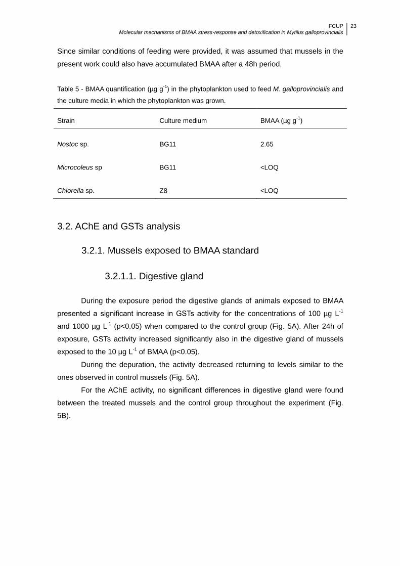
Since similar conditions of feeding were provided, it was assumed that mussels in the
present work could also have accumulated BMAA after a 48h period.
Table 5 - BMAA quantification (µg g-1
) in the phytoplankton used to feed M. galloprovincialis and
the culture media in which the phytoplankton was grown.
Strain Culture medium BMAA (µg g-1
)
Nostoc sp.
BG11
2.65
Microcoleus sp BG11 <LOQ
Chlorella sp.
Z8
<LOQ
3.2. AChE and GSTs analysis
3.2.1. Mussels exposed to BMAA standard
3.2.1.1. Digestive gland
During the exposure period the digestive glands of animals exposed to BMAA
presented a significant increase in GSTs activity for the concentrations of 100 µg L-1
and 1000 µg L-1 (p<0.05) when compared to the control group (Fig. 5A). After 24h of
exposure, GSTs activity increased significantly also in the digestive gland of mussels
exposed to the 10 µg L-1 of BMAA (p<0.05).
During the depuration, the activity decreased returning to levels similar to the
ones observed in control mussels (Fig. 5A).
For the AChE activity, no significant differences in digestive gland were found
between the treated mussels and the control group throughout the experiment (Fig.
5B).
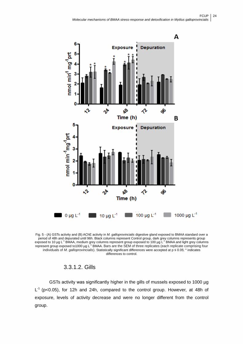
FCUP Molecular mechanisms of BMAA stress-response and detoxification in Mytilus galloprovincialis
24
Fig. 5 - (A) GSTs activity and (B) AChE activity in M. galloprovincialis digestive gland exposed to BMAA standard over a period of 48h and depurated until 96h. Black columns represent Control group, dark grey columns represents group
exposed to 10 µg L-1 BMAA, medium grey columns represent group exposed to 100 µg L
-1 BMAA and light grey columns
represent group exposed to1000 µg L-1 BMAA. Bars are the SEM of three replicates (each replicate comprising four
individuals of M. galloprovincialis). Statistically significant differences were accepted at p ≤ 0.05; * indicates differences to control.
3.3.1.2. Gills
GSTs activity was significantly higher in the gills of mussels exposed to 1000 µg
L-1 (p<0.05), for 12h and 24h, compared to the control group. However, at 48h of
exposure, levels of activity decrease and were no longer different from the control
group.

FCUP Molecular mechanisms of BMAA stress-response and detoxification in Mytilus galloprovincialis
25
During depuration, at 72h and 96h, the activity was significantly higher in the
mussels exposed to 100 µg L-1 and 1000 µg L-1 (p<0.05) in comparison to the control
group. For the lowest concentration, BMAA did not produce any significant effect in
GSTs activity, as there were no differences between exposed and control mussels (Fig.
6A).
A significant decrease in gill AChE activity was verified between the treated and
the control groups at 48h of exposure to BMAA, for 100 µg L-1 and 1000 µg L-1
concentrations (p<0.05) (Fig. 6B). During depuration, activity levels raised for these
concentrations, returning to levels similar to the control group. At a concentration of 10
µg L-1, no significant alterations were observed in gill AChE activity of M.
galloprovincilalis during the whole experiment.
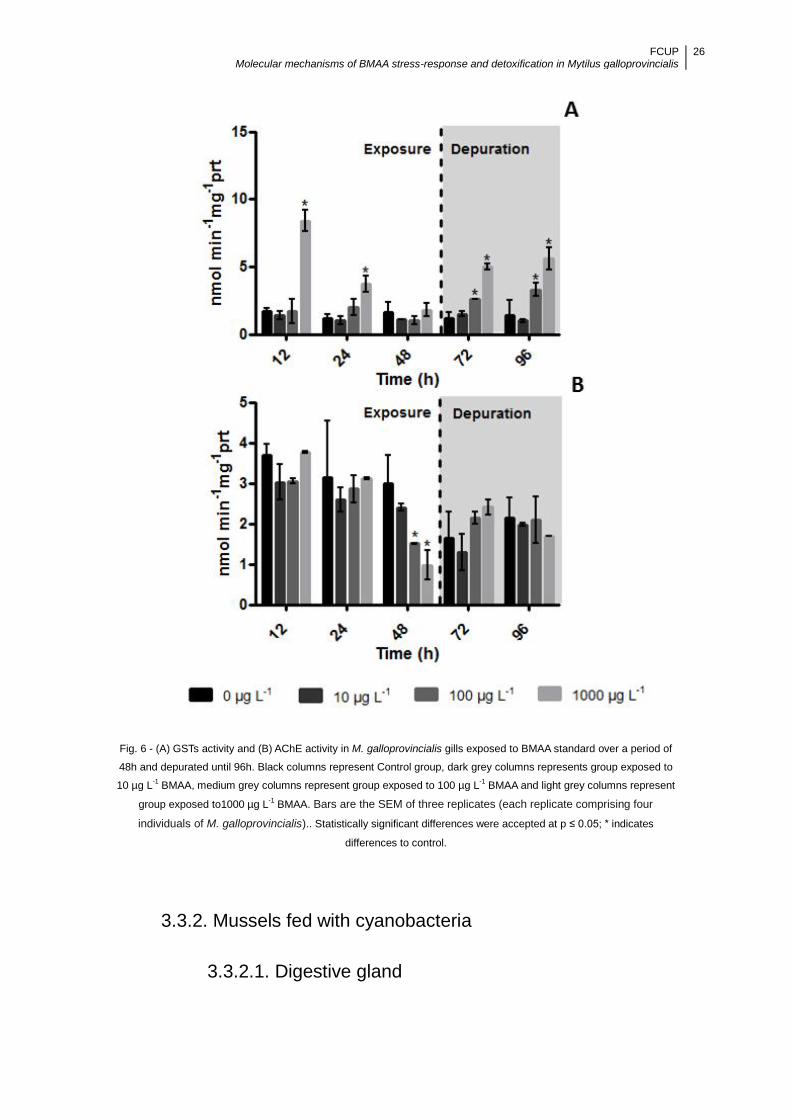
FCUP Molecular mechanisms of BMAA stress-response and detoxification in Mytilus galloprovincialis
26
Fig. 6 - (A) GSTs activity and (B) AChE activity in M. galloprovincialis gills exposed to BMAA standard over a period of
48h and depurated until 96h. Black columns represent Control group, dark grey columns represents group exposed to
10 µg L-1 BMAA, medium grey columns represent group exposed to 100 µg L
-1 BMAA and light grey columns represent
group exposed to1000 µg L-1 BMAA. Bars are the SEM of three replicates (each replicate comprising four
individuals of M. galloprovincialis).. Statistically significant differences were accepted at p ≤ 0.05; * indicates
differences to control.
3.3.2. Mussels fed with cyanobacteria
3.3.2.1. Digestive gland
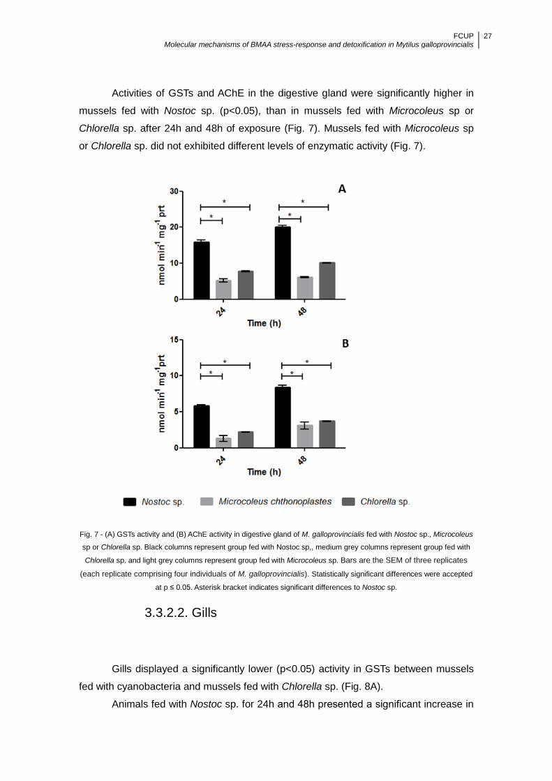
FCUP Molecular mechanisms of BMAA stress-response and detoxification in Mytilus galloprovincialis
27
Activities of GSTs and AChE in the digestive gland were significantly higher in
mussels fed with Nostoc sp. (p<0.05), than in mussels fed with Microcoleus sp or
Chlorella sp. after 24h and 48h of exposure (Fig. 7). Mussels fed with Microcoleus sp
or Chlorella sp. did not exhibited different levels of enzymatic activity (Fig. 7).
Fig. 7 - (A) GSTs activity and (B) AChE activity in digestive gland of M. galloprovincialis fed with Nostoc sp., Microcoleus
sp or Chlorella sp. Black columns represent group fed with Nostoc sp,, medium grey columns represent group fed with
Chlorella sp. and light grey columns represent group fed with Microcoleus sp. Bars are the SEM of three replicates
(each replicate comprising four individuals of M. galloprovincialis). Statistically significant differences were accepted
at p ≤ 0.05. Asterisk bracket indicates significant differences to Nostoc sp.
3.3.2.2. Gills
Gills displayed a significantly lower (p<0.05) activity in GSTs between mussels
fed with cyanobacteria and mussels fed with Chlorella sp. (Fig. 8A).
Animals fed with Nostoc sp. for 24h and 48h presented a significant increase in

FCUP Molecular mechanisms of BMAA stress-response and detoxification in Mytilus galloprovincialis
28
AChE activity (p<0.05) in gills when compared to the mussels fed with Microcoleus sp
or Chlorella sp. (Fig. 8B).
Fig. 8 - (A) GSTs activity and (B) AChE activity in gills of M. galloprovincialis fed with Nostoc sp., Microcoleus sp or
Chlorella sp. Black columns represent group fed with Nostoc sp., medium grey columns represent group fed with
Chlorella sp. and light grey columns represent group fed with Microcoleus sp. Bars are the SEM of three replicates
(each replicate comprising four individuals of M. galloprovincialis). Statistically significant differences were accepted
at p ≤ 0.05. Asterisk bracket indicates significant differences to Nostoc sp.
Moreover, a significant difference in mean activity of AChE and GSTs was
observed between the mussels used in both bioaccumulation experiments. While unfed
mussels exposed to BMAA standard exhbited activities ranging between 1 and 10 nmol
min-1 mg-1 of protein, mussels fed displayed higher enzimatic activities ranging between
2 and 20 nmol min-1 mg-1 of protein.
3.3. iGluR expression in M. galloprovincialis
3.3.1. Amplification of GLU4 and GLU5
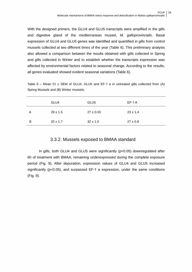
FCUP Molecular mechanisms of BMAA stress-response and detoxification in Mytilus galloprovincialis
29
With the designed primers, the GLU4 and GLU5 transcripts were amplified in the gills
and digestive gland of the mediterranean mussel, M. galloprovincialis. Basal
expression of GLU4 and GLU5 genes was identified and quantified in gills from control
mussels collected at two different times of the year (Table 6). This preliminary analysis
also allowed a comparison between the results obtained with gills collected in Spring
and gills collected in Winter and to establish whether the transcripts expression was
affected by environmental factors related to seasonal change. According to the results,
all genes evaluated showed evident seasonal variations (Table 6).
Table 6 – Mean Ct ± SEM of GLU4, GLU5 and EF-1 α in untreated gills collected from (A)
Spring Mussels and (B) Winter mussels.
GLU4 GLU5 EF-1 Α
A 29 ± 1.5 27 ± 0.03 23 ± 1.4
B 32 ± 1.7 32 ± 1.0 27 ± 0.8
3.3.2. Mussels exposed to BMAA standard
In gills, both GLU4 and GLU5 were significantly (p<0.05) downregulated after
6h of treatment with BMAA, remaining underexpressed during the complete exposure
period (Fig. 9). After depuration, expression values of GLU4 and GLU5 increased
significantly (p<0.05), and surpassed EF-1 α expression, under the same conditions
(Fig. 9).

FCUP Molecular mechanisms of BMAA stress-response and detoxification in Mytilus galloprovincialis
30
Fig. 9 - Gill changes of GLU4 and GLU5 transcripts expression during (A) exposure to BMAA and (B) depuration period
compared with controls. Black columns represent GLU4 transcript and grey columns represent GLU5 transcript (Results
are expressed as mean ± SEM. Statistically significant differences were accepted at p ≤ 0.05; * indicates differences to
control). EF-1 α as internal reference.
Digestive gland did not allow a relative quantification analysis, as EF-1 α expression
varied between exposed and not exposed mussels in this tissue. Thus, it was not
considered a reliable internal reference. After treatment with BMAA, EF-1 α expression
was highly depressed (Table 7). GLU4 and GLU5 expression was also affected, but to
a lesser extent.

FCUP Molecular mechanisms of BMAA stress-response and detoxification in Mytilus galloprovincialis
31
Table 7 - Mean Ct ± SEM values of control and treated mussels digestive gland of GLU4, GLU5
and EF-1 α. Different exposure times have been averaged.
GLU4 GLU5 EF-1 α
Control 31 ± 2.3 33 ± 1.0 21 ± 0.7
BMAA 36 ± 1.7 36 ± 1.7 36 ± 2.5

FCUP Molecular mechanisms of BMAA stress-response and detoxification in Mytilus galloprovincialis
32
4. Discussion
In the last two decades, marine biotoxins have received increasing attention
from scientific community, from the development of new analytical methods for toxins
detection, to get a better understanding on accumulation and trophic transfer of these
molecules in marine organisms, to their mechanisms of toxicity and the generation of
more accurate risk evaluations and recommendations (Munday, 2011; Gorbi et al.,
2012) . In this study an integrated approach with bioaccumulation of BMAA on mussel
and a set of associated subcellular responses was used.
Many discrepancies in published results on BMAA concentration have been
reported (Banack et al., 2010; Faassen et al., 2012; Glover et al., 2012). The lack of a
standard procedure to determine BMAA in different samples (such as cyanobacteria or
marine invertebrates) has been considered an issue. In fact, it was suggested by
Faassen et al. (2012) that the inconsistensies observed could be due to the analytical
method used. LC-MS/MS has been appointed as the more selective and appropriate
method for BMAA detection (Banack et al., 2010; Faassen et al., 2012).
Additionally, matrix effects may be complicating factors in BMAA analysis
(Glover et al., 2012). BMAA reactivity and potential to interact with other molecules
during the analysis may interfere with the accurate quantification (Li et al., 2010; Glover
et al., 2012). In the presence of a complex matrix even more chemical interactions are
likely to occur and interfere with the analysis (Li et al., 2010; Glover et al., 2012). Low
recovery rates have also been reported when attempting to remove BMAA from saline
waters (Glover et al. 2012) and so far, no suitable protocol for BMAA recovery from
samples with high concentration of salts has been described. Hence, failure to detect
BMAA cannot be considered an absence of the compound.
Concentrations reported in different organisms (phytoplankton, plants, animals)
(Faassen 2014) allow the assumption that concentrations ranging 10 µg L-1 can be
considered environmentally relevant, whereas concentrations above this value are less
frequent to come by. Hence, the assessment of the effects of BMAA exposure on
enzyme activity and glutamate receptors, such as the one in this study, allows the
assessement of toxicity in mussels tissues even when detection of BMAA in the mussel
tissues is not possible or reliable.
FCUP Molecular mechanisms of BMAA stress-response and detoxification in Mytilus galloprovincialis
33
4.1. Effects of BMAA on AChE and GSTs activity
In this study, levels of enzyme activity in M. galloprovincialis were in general,
significantly affected by the concentration of BMAA standard and the duration of the
exposure.
In the digestive gland, GSTs activity increased during exposure to BMAA and
was lowered during depuration. The increase of GST activity was obtained during the
accumulation period, for all concentrations, suggesting that this organ is very sensitive,
even to low concentrations of BMAA. During depuration, the drop in activity can be
explained by the removal of the toxicant from the water. This implies a significant role
of GSTs in BMAA detoxification in M. galloprovincialis. Although cellular detoxifying
pathways involved in BMAA degradation are not clearly understood, phase II
detoxification, mainly GSTs-formed conjugates, has been shown to be involved in the
degradation process of cyanobacterial compounds (Davies et al., 2005). Formation of
the cyanotoxin-GSH conjugate via GSTs that enhances the water solubility of the toxin
was observed in different aquatic organisms (Pflugmacher et al., 1998, Adamovský et
al., 2007).
In gills, GSTs activity increased during depuration and was lowered during
exposure to BMAA. This organ is the major site of uptake of waterborne toxicants, and
the presence of BMAA, that initially provoked conjugation action from the enzyme,
could have interfered with the mechanisms responsible for regulation of GSTs gene
expression, as described for other xenobiotics (Chen and Ramos, 1999; Contardo-Jara
et al., 2008). In fact, BMAA, could act in a similar way as MC and okadaic acid, which
are known to inhibit protein phosphatases (PP). These PP are involved in cell signaling
processes (Rivas et al., 2000), and an inhibition could cause major effects on signal
transduction pathways, and consequentially, a disturbance of essential cellular
functions (Svensson et al., 2003). During depuration, the GSTs activity increase could
be explained by the fact that a rise of the toxin levels could have occurred due to the
release of previously bound toxins, from the renewal of PP. This response has been
observed for MC (Fernandes et al., 2009). The decrease in activity registered during
exposure points to a high vulnerability of the gills to the toxin. Šetlíková and Wiegand
(2009) showed that gills of two different fish species had lower GSTs activities and
consequently a lower ability to biotransform MC than the respective livers.
AChE activity is commonly used as a bioindicator and there are a number of
studies in the literature concerning the effects of environmental organic pollutants,
heavy metals and other chemical toxicants on AChE activity in animals (Rao et al.,
2005, Richetti et al., 2011; Pereira et al.,2012).

FCUP Molecular mechanisms of BMAA stress-response and detoxification in Mytilus galloprovincialis
34
In gills, no apparent effect of BMAA exposure was observed in AChE activity
after 48 hours. However, in the digestive gland the results show that, in the tested
conditions, the decreased activity of AChE after 48h of exposure to BMAA was
reversible after transfer to toxin-free media. However, prolonged exposure to this toxin
may impair actions such as feeding activity of the mussels and therefore compromise
bioenergetics and general physiological condition (Kankaanpää et al., 2007).
In M.galloprovincialis fed with cyanobacterial cells, tissue-specific changes in
the enzymatic activity of GSTs and AChE were also observed.
In the digestive gland, the increase of GSTs activity was relevant at 24h and
48h of feeding with Nostoc sp. This increase in activity points to a potential stimulation
of the detoxification process of cyanobacterial secondary metabolites. Similar results
were obtained for liver GSTs activity in several fish species during Microcystis blooms
in Lake Taihu in China (Qiu et al., 2007). In gills, in contrast to the results observed in
the digestive gland, a decrease in activity was registered in response to feeding with
Nostoc sp. As discussed above, exposure to BMAA, either directly filtered from the
water or supplied from toxic cyanobacteria, may inhibit GSTs in gill tissues. However,
no differences were observed between mussels fed with Nostoc sp. or other strains,
that are not producing BMAA, which suggests this effect is not exclusive to BMAA
exposure.
M. galloprovincialis is known to present multiple GSTs isoforms divided in three
superfamilies: cytosolic, membrane-associated proteins in eicosanoid and glutathione
metabolism (MAPEG) and mytochondrial (Fitzpatrick and Sheehan, 1993). Present
data refers to total cytosolic GSTs activity, using CDNB as substrate. Thus, it is not
possible to exclude pattern variations from specific isoforms (Regoli and Principato,
1995). Different GSTs isoforms expression seems to be differentially regulated, as
different inducers can be related to different isoforms (Hoarau et al., 2006).
The increase in AChE activity was verified in both tissues of mussels fed with
BMAA producing cyanobacteria. An increase in AChE activity has been demonstrated
as a consequence of exposure to neurotoxic compounds such as aluminum (Kaizer et
al., 2010) and ethanol (Rico et al., 2007) as well as cyanotoxin MC (Kist et al., 2012).
Recently AChE has been connected to apoptosis as an important regulator (Zhang et
al., 2002). In fact, an over-expression of AChE is able to inhibit cell proliferation (Pérez-
Aguilar et al., 2015) and promote apoptosis (Zhang and Greenberg, 2012). As it is
known, apoptosis often underlies the neurotoxic effects of various compounds such as
β-amyloid and prion protein fragments (Forloni et al., 1996). However, the same effect
was not observed in mussels exposed to BMAA standard. This study cannot account
for the opposite trends verified between mussels exposed to BMAA standard or BMAA

FCUP Molecular mechanisms of BMAA stress-response and detoxification in Mytilus galloprovincialis
35
producing cyanobacteria, which compromises the uses of this enzyme as a reliable
biomarker of BMAA exposure. It is likely that the rapid and extensive accumulation of
secondary compounds in the tissues of the mussels exposed to Nostoc sp., caused
multiple cellular and physiological responses, including changes in AChE activity.
However, the actual mechanism behind the observed fluctuations in AChE activity in M.
galloprovincialis fed with Nostoc sp. is not known. Further studies on potential activity
modulating factors are recommended.
Nonetheless, analysis of AChE and GSTs activity as putative markers of
exposure to BMAA revealed differences in these parameters in gills and digestive gland
from M. galloprovincialis mussels exposed to BMAA. In fact, GSTs together with other
parameters could be indicators of BMAA exposure. AChE lack of response at lower
concentrations in digestive gland, and absence of response in gills in mussels exposed
to BMAA, excludes the use of this enzyme as a biomarker.
4.2. Effects of Exposure to BMAA on iGluR Expression
Recent studies in Mytilus spp. correlated changes in gene transcription with
responses to a variety of environmental stressors: mixtures of crude oil and mercury
(Dondero et al., 2006a), copper (Dondero et al., 2006b), benzo(a)pyrene (Brown et
al.,2006), okadaic acid (Manfrin et al., 2010) and hypoxia (Hines et al., 2007). Similarly,
in this study we identified differentially expressed transcripts in response to BMAA
exposure in M. galloprovincialis through qPCR analysis.
Given the excitotoxic potential of BMAA it was thought that iGluR gene
expression could potentially be used as a biomarker of exposure to BMAA. It was
hypothesized that exposure to the toxin would increase the activity of iGluR with
consequent increase in their transcription in a concentration-dependent manner.
However, the opposite was observed. In gills, the results obtained supported the
hypothesis of feedback loop that regulates iGluR expression. When exposed to BMAA
standard, a iGluR agonist, it was downregulated. In accordance, when the toxin was
removed, this process was reversed. This would mean that despite the lack N-terminal
domain, channel activity was not compromised. Nonetheless, full molecular
consequences are not yet understood. Thus, discrimination of pharmacologic
properties of GLU4 and GLU5 is required to better understand their role as M.
galloprovincialis glutamate receptors.
Long-term synaptic activity (or its inhibition) has been proved to have an effect
on iGluR accumulation at excitatory synapses supporting the idea that feedback

FCUP Molecular mechanisms of BMAA stress-response and detoxification in Mytilus galloprovincialis
36
mechanisms are responsible for adjusting neuronal output (Bear, 1995; Miller, 1996). In
fact, other studies show that both acute and chronic increases in synaptic activity result
in a reduction in postsynaptic iGluR clusters (O’Brien et al., 1998; Lee et al, 2002).
In the environment, the seasonal cycle is a strong determinate of invertebrate
physiology (Petrovic et al., 2004; Farcy et al., 2009). Changes in environmental factors
resulting from seasonal changes may, therefore, powerfully affect the normal metabolic
activities of mussels (Place et al., 2008; Gracey et al., 2008). Therefore, it was
expected to find seasonal variation between gene expression of spring mussels and
winter mussels, as was observed.
In the digestive gland, it was not possible to estimate the relative expression of
GLU4 and GLU5. The development of gonads and gametes that occurs during winter
requires a lot of energy, and thus genes non-related to reproduction may be less
expressed, during this period (Banni et al., 2011). The reproductive period is an
especially energy draining process that induces major changes in the digestive gland
(Wilhelm-Filho et al., 2001) In fact, studies have described differences in metabolic
activity and genetic expression in this organ , during the reproductive stage of mussels
(Seed and Suchanek, 1992; Banni et al., 2011). However, after treatment with BMAA,
severe changes in Ct level were observed. The increased Ct levels point to a similar
response of downregulation of GLU4 and GLU5, as in gills. However differences in
expression EF-1 α cannot be accounted for.
Degeneration of digestive gland tissues and several other histopathological
changes could have been induced by exposure to the cyanotoxin, as verified by other
studies (Klobuĉar et al., 1997; Auriemma and Battistella, 2004). Moreover, the
damaging effects of cyanotoxins have been shown to be enhanced when exposed
mussels are spawning (Galimany et al., 2008). Thus, a new experiment with mussels
outside of spawning season is necessary to further validate the results obtained. Also,
it would be interesting to test the effects of BMAA producing cyanobacteria to confirm
the relevance of the results in the environment. Nonetheless, preliminary analysis of
iGluR expression in gills of mussels exposed to BMAA standard, as putative
biomarkers of BMAA, suggested these genes could be useful markers of BMAA
exposure.
FCUP Molecular mechanisms of BMAA stress-response and detoxification in Mytilus galloprovincialis
37
5. Conclusions
This study provided a fundamental step in uncovering the molecular responses
to BMAA exposure in M. galloprovincialis, an organism important in the aquatic
ecosystem and present in the diet of many organisms, including humans.
This study showed that an exposure to sublethal concentrations of BMAA in a
short period of time had consequences at a molecular and biochemical level,
elucidating putative target and/or detoxification mechanisms.
The transcription of iGluR genes can potentially be used as tools to assess
BMAA induced toxicity in biomonitoring studies using M. galloprovincialis.
Nevertheless, further work is needed to better understand the regulatory mechanisms
of iGLuR genes as well as their pharmacologic properties and functional role in
mussels. Enzymes GSTs were considered a potentially useful marker of BMAA
exposure when it is known that M. galloprovincialis was exposed to BMAA but the
levels are too low for an unequivocal quantification. The lower capacity of response
from AChE suggests that this enzyme is a poor marker, though.
Further research will be needed to characterize both natural variability and
seasonal changes of M. galloprovincialis in responses to BMAA. It is also important to
account for possible sinergies of multiple stressors, that may occur in the environment,
and affect biomarker modulation. Furthermore, other antioxidant enzymes could also
be tested for changes regarding BMAA exposure, thus providing a more complete
enzymatic profile for detecting BMAA toxic effects. On a last note concerning BMAA
analysis, more investigation should be done to standardize detection and quantification
procedures in order to obtain more reliable results.
FCUP Molecular mechanisms of BMAA stress-response and detoxification in Mytilus galloprovincialis
38
6. References
Adamovský, O., Kopp, R., Hilscherová, K., Babica, P., Palíková, M., Pašková, V.,
Navrátil, S. Maršálek, B., & Bláha, L. (2007). Microcystin kinetics (bioaccumulation and
elimination) and biochemical responses in common carp (Cyprinus carpio) and silver
carp (Hypophthalmichthys molitrix) exposed to toxic cyanobacterial blooms.
Environmental Toxicology and Chemistry, 26(12), 2687-2693.
Amorim, A., Vasconcelos, V. M., 1999. Dynamics of microcystins in the mussel Mytilus
galloprovincialis. Toxicon 37: 1041-1052
Aráoz, R., Molgó, J., & De Marsac, N. T. (2010). Neurotoxic cyanobacterial toxins.
Toxicon, 56(5), 813-828.
Auriemma, R., & Battistella, S. (2004). Biochemical and histological alterations of
Mytilus galloprovincialis digestive gland after exposure to okadaic acid and derivatives.
ISJ, 1(6).
Banack, S. A., Caller, T. A., & Stommel, E. W. (2010). The cyanobacteria derived toxin
beta-N-methylamino-L-alanine and amyotrophic lateral sclerosis. Toxins, 2(12), 2837-
2850.
Banack, S. A., Metcalf, J. S., Jiang, L., Craighead, D., Ilag, L. L., & Cox, P. A. (2012).
Cyanobacteria produce N-(2-aminoethyl) glycine, a backbone for peptide nucleic acids
which may have been the first genetic molecules for life on earth. PLoS ONE
Banack, S. A., Murch, S. J., & Cox, P. A. (2006). Neurotoxic flying foxes as dietary
items for the Chamorro people, Marianas Islands. Journal of ethnopharmacology,
106(1), 97-104.
Banni, M., Negri, A., Mignone, F., Boussetta, H., Viarengo, A., & Dondero, F. (2011).
Gene expression rhythms in the mussel Mytilus galloprovincialis (Lam.) across an
annual cycle. PLoS One, 6(5), e18904.
FCUP Molecular mechanisms of BMAA stress-response and detoxification in Mytilus galloprovincialis
39
Baptista, M. S., Cianca, R. C. C., Almeida, M. R., Vasconcelos, V. M. 2011.
Determination of the non protein amino acid β-N-methylamino-L-alanine in estuarine
cyanobacteria by capillary electrophoresis. Toxicon 58: 410-414
Baptista, M. S., Vasconcelos, R. G., Ferreira, P. C., Almeida, C. M. R., & Vasconcelos,
V. M. (2015). Assessment of the non-protein amino acid BMAA in Mediterranean
mussel Mytilus galloprovincialis after feeding with estuarine cyanobacteria.
Environmental Science and Pollution Research, 1-10.
Bargu, S., Goldstein, T., Roberts, K., Li, C., & Gulland, F. (2012). Pseudo-nitzschia
blooms, domoic acid, and related California sea lion strandings in Monterey Bay,
California. Marine Mammal Science, 28(2), 237-253.
Bartram, J., & Chorus, I. (Eds.). (2002). Toxic Cyanobacteria in Water: A guide to their
public health consequences, monitoring and management. CRC Press.
Batorèu, M. C. C., Dias, E., Pereira, P., & Franca, S. (2005). Risk of human exposure
to paralytic toxins of algal origin. Environmental toxicology and pharmacology, 19(3),
401-406.
Bear, M. F. (1995). Mechanism for a sliding synaptic modification threshold. Neuron,
15(1), 1-4.
Bláha, L., Babica, P., & Maršálek, B. (2009). Toxins produced in cyanobacterial water
blooms-toxicity and risks. Interdisciplinary toxicology, 2(2), 36-41.
Bláha, L., Kopp, R., Šimková, K., & Mareš, J. (2004). Oxidative stress biomarkers are
modulated in silver carp (Hypophthalmichthys molitrix Val.) exposed to microcystin-
producing cyanobacterial water bloom. Acta Veterinaria Brno, 73(4), 477-482.
Blanchette, B., Feng, X., & Singh, B. R. (2007). Marine glutathione S-transferases.
Marine biotechnology, 9(5), 513-542.
Bocquené, G., & Galgani, F. (1998).Biological effects of contaminants: cholinesterase
inhibition by organophosphate and carbamate compounds (No. 22). Copenhagen,,
Denmark: International Council for the Exploration of the Sea.

FCUP Molecular mechanisms of BMAA stress-response and detoxification in Mytilus galloprovincialis
40
Bradford, M. M. (1976). A rapid and sensitive method for the quantitation of microgram
quantities of protein utilizing the principle of protein-dye binding. .Analytical
biochemistry, 72(1), 248-254.
Branch, G. M., & Steffani, C. N. (2004). Can we predict the effects of alien species? A
case-history of the invasion of South Africa by Mytilus galloprovincialis (Lamarck).
Journal of Experimental Marine Biology and Ecology, 300(1), 189-215.
Brand, L. E., Pablo, J., Compton, A., Hammerschlag, N., & Mash, D. C. (2010).
Cyanobacterial blooms and the occurrence of the neurotoxin, beta-N-methylamino-l-
alanine (BMAA), in South Florida aquatic food webs. Harmful algae, 9(6), 620-635.
Briand, J. F., Jacquet, S., Bernard, C., & Humbert, J. F. (2003). Health hazards for
terrestrial vertebrates from toxic cyanobacteria in surface water ecosystems. Veterinary
Research, 34(4), 361-377.
Brown, M., Davies, I. M., Moffat, C. F., & Craft, J. A. (2006). Application of SSH and a
macroarray to investigate altered gene expression in Mytilus edulis in response to
exposure to benzo [a] pyrene. Marine environmental research, 62, S128-S135.
Burja, A. M., Banaigs, B., Abou-Mansour, E., Burgess, J. G., & Wright, P. C. (2001).
Marine cyanobacteria—a prolific source of natural products. Tetrahedron, 57(46), 9347-
9377.
Carmichael, W. W. (1997). The cyanotoxins. Advances in botanical research, 27, 211-
257.
Carmichael, W. W. (2001). Health effects of toxin-producing cyanobacteria:―The
CyanoHABs‖. Human and ecological risk assessment: An International Journal, 7(5),
1393-1407.
Ceccherelli, V. U., & Rossi, R. (1984). Settlement, growth and production of the mussel
Mytilus galloprovincialis. Marine ecology. Progress series, 16(1-2), 173-184.
Charlton, T. S., Marini, A. M., Markey, S. P., Norstog, K., & Duncan, M. W. (1992).
Quantification of the neurotoxin 2-amino-3-(methylamino)-propanoic acid (BMAA) in
Cycadales. Phytochemistry, 31(10), 3429-3432.
FCUP Molecular mechanisms of BMAA stress-response and detoxification in Mytilus galloprovincialis
41
Chen, Y. H., & Ramos, K. S. (1999). Negative regulation of rat GSTS-Ya gene via
antioxidant/electrophile response element is directed by a C/EBP-like site.Biochemical
and biophysical research communications, 265(1), 18-23.
Chiu, A. S., Gehringer, M. M., Welch, J. H., & Neilan, B. A. (2011). Does α-amino-β-
methylaminopropionic acid (BMAA) play a role in neurodegeneration? International
journal of environmental research and public health, 8(9), 3728-3746.
Choi, D. W. (1988). Glutamate neurotoxicity and diseases of the nervous system.
Neuron, 1(8), 623-634.
Cianca, R. C. C., Baptista, M. S., Lopes, V. R., Vasconcelos, V. M.. 2012. b-N-
methylamino-L-alanine in novel Portuguese cyanobacterial isolates from Minho, Douro
and Vouga Rivers. Amino Acids 42:2473-2479
Chorus, I., Falconer, I. R., Salas, H. J., & Bartram, J. (2000). Health risks caused by
freshwater cyanobacteria in recreational waters. Journal of Toxicology and
Environmental Health Part B: Critical Reviews, 3(4), 323-347.
Codd, G. A., Morrison, L. F., & Metcalf, J. S. (2005). Cyanobacterial toxins: risk
management for health protection. Toxicology and applied pharmacology, 203(3), 264-
272.
Cohen, S. A. (2012). Analytical techniques for the detection of a-amino-ß-
methylaminopropionic acid. Analyst, 137(9), 1991-2005.
Contardo-Jara, V., Pflugmacher, S., & Wiegand, C. (2008). Multi-xenobiotic-resistance
a possible explanation for the insensitivity of bivalves towards cyanobacterial toxins.
Toxicon, 52(8), 936-943.
Cotou, E., Tsangaris, C., & Henry, M. (2013). Comparative study of biochemical and
immunological biomarkers in three marine bivalves exposed at a polluted site.
Environmental Science and Pollution Research, 20(3), 1812-1822.
Cox, P. A., Banack, S. A., Murch, S. J., Rasmussen, U., Tien, G., Bidigare, R. R.,
Metcalf. J.S Morrinson L. F, Codd, G. A. & Bergman, B. (2005). Diverse taxa of
FCUP Molecular mechanisms of BMAA stress-response and detoxification in Mytilus galloprovincialis
42
cyanobacteria produce β-N-methylamino-L-alanine, a neurotoxic amino acid.
Proceedings of the National Academy of Sciences of the United States of America,
102(14), 5074-5078.
Cox, P. A., Banack, S. A., & Murch, S. J. (2003). Biomagnification of cyanobacterial
neurotoxins and neurodegenerative disease among the Chamorro people of Guam.
Proceedings of the National Academy of Sciences, 100(23), 13380-13383.
Davies, W. R., Siu, W. H., Jack, R. W., Wu, R. S., Lam, P. K., & Nugegoda, D. (2005).
Comparative effects of the blue green algae Nodularia spumigena and a lysed extract
on detoxification and antioxidant enzymes in the green lipped mussel (Perna viridis).
Marine pollution bulletin, 51(8), 1026-1033.
Dereeper, A., Guignon, V., Blanc, G., Audic, S., Buffet, S., Chevenet, F., Dufayard, J. F.,
Guindon, S., Lefort, V., Lescot, M., Claverie, J. M., & Gascuel, O. (2008). Phylogeny. fr:
robust phylogenetic analysis for the non-specialist. Nucleic acids research, 36(suppl 2),
W465-W469.
Dingledine, R., Borges, K., Bowie, D., & Traynelis, S. F. (1999). The glutamate receptor
ion channels. Pharmacological reviews, 51(1), 7-62.
Dondero, F., Piacentini, L., Marsano, F., Rebelo, M., Vergani, L., Venier, P., & Viarengo,
A. (2006a). Gene transcription profiling in pollutant exposed mussels (Mytilus spp.)
using a new low-density oligonucleotide microarray. Gene, 376(1), 24-36.
Dondero, F., Dagnino, A., Jonsson, H., Caprì, F., Gastaldi, L., & Viarengo, A. (2006b).
Assessing the occurrence of a stress syndrome in mussels (Mytilus edulis) using a
combined biomarker/gene expression approach. Aquatic toxicology, 78, S13-S24.
Downing, S., Banack, S. A., Metcalf, J. S., Cox, P. A., & Downing, T. G. (2011). Nitrogen
starvation of cyanobacteria results in the production of β- N-methylamino-L-alanine.
Toxicon, 58(2), 187-194.
Duncan, M. W., Steele, J. C., Kopin, I. J., & Markey, S. P. (1990). 2-Amino-3-
(methylamino)-propanoic acid (BMAA) in cycad flour An unlikely cause of amyotrophic
lateral sclerosis and parkinsonism-dementia of Guam. Neurology,40(5), 767-767.

FCUP Molecular mechanisms of BMAA stress-response and detoxification in Mytilus galloprovincialis
43
Dunlop, R. A., Cox, P. A., Banack, S. A., & Rodgers, K. J. (2013). The non-protein
amino acid BMAA is misincorporated into human proteins in place of L-serine causing
protein misfolding and aggregation. PloS one, 8(9), e75376.
Ellman, G. L., Courtney, K. D., Andres, V., & Featherstone, R. M. (1961). A new and
rapid colorimetric determination of acetylcholinesterase activity. Biochemical
pharmacology, 7(2), 88-95.
Erdner, D., Dyble, J., Parsons, M., Stevens, R., Hubbard, K., Wrabel, M., Moore, S. K.,
Lefebvre, K. A., Anderson, D. M., Bienfang, P., Bidigare, R. R., Parker, M. S., Moeller,
P., Brand, L. E., Trainer, V. (2008). Centers for Oceans and Human Health: A unified
approach to the challenge of harmful algal blooms. Environmental Health Environ
Health, 7(Suppl 2), s2.
Esterhuizen, M., & Downing, T. G. (2008). β-N-methylamino-l-alanine (BMAA) in novel
South African cyanobacterial isolates. Ecotoxicology and environmental safety, 71(2),
309-313
Esteves, J. L., Santinelli, N., Sastre, V., Díaz, R., & Rivas, O. (1992). A toxic
dinoflagellate bloom and PSP production associated with upwelling in Golfo Nuevo,
Patagonia, Argentina. Hydrobiologia, 242(2), 115-122.
Faassen, E. J. (2014). Presence of the neurotoxin BMAA in aquatic ecosystems: what
do we really know? Toxins, 6(3), 1109-1138.
Faassen, E. J., Gillissen, F., Zweers, H. A., & Lürling, M. (2009). Determination of the
neurotoxins BMAA (ß-N-methylamino-L-alanine) and DAB (diaminobutyric acid) by LC-
MSMS in Dutch urban waters with cyanobacterial blooms. Amyotrophic Lateral
Sclerosis, 10(sup2), 79-84.
Faassen, E. J., Gillissen, F., & Lürling, M. (2012). A comparative study on three
analytical methods for the determination of the neurotoxin BMAA in cyanobacteria.
PLoS One, 7(5).
Falconer, I. R. (1999). An overview of problems caused by toxic blue–green algae
(cyanobacteria) in drinking and recreational water. Environmental Toxicology, 14(1), 5-
12.

FCUP Molecular mechanisms of BMAA stress-response and detoxification in Mytilus galloprovincialis
44
Fan, H., Qiu, J., Fan, L., & Li, A. (2015). Effects of growth conditions on the production
of neurotoxin 2, 4-diaminobutyric acid (DAB) in Microcystis aeruginosa and its universal
presence in diverse cyanobacteria isolated from freshwater in China. Environmental
Science and Pollution Research, 22(8), 5943-5951.
Farcy, É., Voiseux, C., Lebel, J. M., & Fiévet, B. (2009). Transcriptional expression
levels of cell stress marker genes in the Pacific oyster Crassostrea gigas exposed to
acute thermal stress. Cell Stress and Chaperones, 14(4), 371-380.
Fernandes, S., Welker, M., & Vasconcelos, V. M. (2009). Changes in the GSTs activity
of the mussel Mytilus galloprovincialis during exposure and depuration of microcystins.
Journal of Experimental Zoology Part A: Ecological Genetics and Physiology, 311(3),
226-230.
Ferrão-Filho, A. D. S., & Kozlowsky-Suzuki, B. (2011). Cyanotoxins: bioaccumulation
and effects on aquatic animals. Marine drugs, 9(12), 2729-2772.
Fitzpatrick, P. J., & Sheehan, D. (1993). Separation of multiple forms of glutathione S-
transferase from the blue mussel, Mytilus edulis. Xenobiotica, 23(8), 851-861.
Forloni, G., Bugiani, O., Tagliavini, F., & Salmona, M. (1996). Apoptosis-mediated
neurotoxicity induced by β-amyloid and PrP fragments. Molecular and chemical
neuropathology, 28(1-3), 163-171.
Frasco, M. F., & Guilhermino, L. (2002). Effects of dimethoate and beta-naphthoflavone
on selected biomarkers of Poecilia reticulata. Fish Physiology and Biochemistry, 26(2),
149-156.
Frasco, M. F., Fournier, D., Carvalho, F., & Guilhermino, L. (2005). Do metals inhibit
acetylcholinesterase (AChE)? Implementation of assay conditions for the use of AChE
activity as a biomarker of metal toxicity. Biomarkers, 10(5), 360-375.
Funari, E., & Testai, E. (2008). Human health risk assessment related to cyanotoxins
exposure. Critical reviews in toxicology, 38(2), 97-125.
Galimany, E., Sunila, I., Hégaret, H., Ramón, M., & Wikfors, G. H. (2008). Experimental

FCUP Molecular mechanisms of BMAA stress-response and detoxification in Mytilus galloprovincialis
45
exposure of the blue mussel (Mytilus edulis, L.) to the toxic dinoflagellate Alexandrium
fundyense: histopathology, immune responses, and recovery. Harmful Algae, 7(5), 702-
711.
García, C., Pérez, F., Contreras, C., Figueroa, D., Barriga, A., López-Rivera, A.,
Araneda, O. F. & Contreras, H. R. (2015). Saxitoxins and okadaic acid group:
accumulation and distribution in invertebrate marine vectors from Southern Chile. Food
Additives & Contaminants: Part A, (ahead-of-print), 1-19.
Gerdol, M., Manfrin, C., De Moro, G., Figueras, A., Novoa, B., Venier, P., & Pallavicini,
A. (2011). The C1q domain containing proteins of the Mediterranean mussel Mytilus
galloprovincialis: a widespread and diverse family of immune-related molecules.
Developmental & Comparative Immunology, 35(6), 635-643.
Glover, W. B., Liberto, C. M., McNeil, W. S., Banack, S. A., Shipley, P. R., & Murch, S.
J. (2012). Reactivity of ß-methylamino-L-alanine in complex sample matrixes
complicating detection and quantification by mass spectrometry. Analytical chemistry,
84(18), 7946-7953.
Gorbi, S., Bocchetti, R., Binelli, A., Bacchiocchi, S., Orletti, R., Nanetti, L., Raffaelli, F.,
Vigninic, A., Accoronia, S., Totti, C., & Regoli, F. (2012). Biological effects of palytoxin-
like compounds from Ostreopsis cf. ovata: A multibiomarkers approach with mussels
Mytilus galloprovincialis. Chemosphere, 89(5), 623-632.
Gracey, A. Y., Chaney, M. L., Boomhower, J. P., Tyburczy, W. R., Connor, K., &
Somero, G. N. (2008). Rhythms of gene expression in a fluctuating intertidal
environment. Current Biology, 18(19), 1501-1507.
Guzmán-Guillén, R., Prieto, A. I., Moreno, I., Ríos, V., Vasconcelos, V. M., & Cameán,
A. M. (2014). Effects of depuration on oxidative biomarkers in tilapia (Oreochromis
niloticus) after subchronic exposure to cyanobacterium producing cylindrospermopsin.
Aquatic Toxicology, 149, 40-49.
Habig, W. H., Pabst, M. J., & Jakoby, W. B. (1974). Glutathione S-transferases the first
enzymatic step in mercapturic acid formation. Journal of biological Chemistry, 249(22),
7130-7139.
FCUP Molecular mechanisms of BMAA stress-response and detoxification in Mytilus galloprovincialis
46
Hayes, J. D., Flanagan, J. U., & Jowsey, I. R. (2005). Glutathione transferases. Annual
Review of Pharmacology and Toxicology, 45, 51-88.
Hines, A., Oladiran, G. S., Bignell, J. P., Stentiford, G. D., & Viant, M. R. (2007). Direct
sampling of organisms from the field and knowledge of their phenotype: key
recommendations for environmental metabolomics. Environmental science &
technology, 41(9), 3375-3381.
Hoarau, P., Damiens, G., Roméo, M., Gnassia-Barelli, M., & Bebianno, M. J. (2006).
Cloning and expression of a GST-pi gene in Mytilus galloprovincialis. Attempt to use
the GST-pi transcript as a biomarker of pollution. Comparative Biochemistry and
Physiology Part C: Toxicology & Pharmacology, 143(2), 196-203.
Ibelings, B. W., & Chorus, I. (2007). Accumulation of cyanobacterial toxins in freshwater
―seafood‖ and its consequences for public health: a review. Environmental pollution,
150(1), 177-192.
Izagirre, U., Errasti, A., Bilbao, E., Múgica, M., & Marigómez, I. (2014). Combined
effects of thermal stress and Cd on lysosomal biomarkers and transcription of genes
encoding lysosomal enzymes and HSP70 in mussels, Mytilus galloprovincialis. Aquatic
Toxicology, 149, 145-156.
Jähnichen, S., Ihle, T., Petzoldt, T., & Benndorf, J. (2007). Impact of inorganic carbon
availability on microcystin production by Microcystis aeruginosa PCC 7806. Applied
and environmental microbiology, 73(21), 6994-7002.
Jiang, L., Aigret, B., De Borggraeve, W. M., Spacil, Z., & Ilag, L. L. (2012). Selective
LC-MS/MS method for the identification of BMAA from its isomers in biological
samples. Analytical and bioanalytical chemistry, 403(6), 1719-1730.
Jiao, Y., Chen, Q., Chen, X., Wang, X., Liao, X., Jiang, L., Wu., J., & Yang, L. (2014).
Occurrence and transfer of a cyanobacterial neurotoxin β-methylamino-L-alanine within
the aquatic food webs of Gonghu Bay (Lake Taihu, China) to evaluate the potential
human health risk. Science of the Total Environment, 468, 457-463.
Jochimsen, E. M., Carmichael, W. W., An, J., Cardo, D. M., Cookson, S. T., Holmes, C.
E., Antunes, M. B., Melo Filho, D. A., Lyra, T. M., Barreto, V. S. T., Azevedo, S. M. F. O.

FCUP Molecular mechanisms of BMAA stress-response and detoxification in Mytilus galloprovincialis
47
& Jarvis, W. R. (1998). Liver failure and death after exposure to microcystins at a
hemodialysis center in Brazil. New England Journal of Medicine, 338(13), 873-878.
Jonasson, S., Eriksson, J., Berntzon, L., Spáčil, Z., Ilag, L. L., Ronnevi, L. O.
Rasmussen, U. & Bergman, B. (2010). Transfer of a cyanobacterial neurotoxin within a
temperate aquatic ecosystem suggests pathways for human exposure. Proceedings of
the National Academy of Sciences, 107(20), 9252-9257.
Käll, L., Krogh, A., & Sonnhammer, E. L. (2004). A combined transmembrane topology
and signal peptide prediction method. Journal of molecular biology, 338(5), 1027-1036.
Kankaanpää, H., Leiniö, S., Olin, M., Sjövall, O., Meriluoto, J., & Lehtonen, K. K.
(2007). Accumulation and depuration of cyanobacterial toxin nodularin and biomarker
responses in the mussel Mytilus edulis. Chemosphere, 68(7), 1210-1217.
Karamyan, V. T., & Speth, R. C. (2008). Animal models of BMAA neurotoxicity: a critical
review. Life sciences, 82(5), 233-246.
Karlsson K., Sipia V., Kankaanpaa H. and Meriluoto J. (2003). Mass spectrometric
detection of nodularin and desmethylnodularin in mussels and flounders. Journal of
Chromatography B, 784: 243-253.
Kaizer, R. R., Gutierres, J. M., Schmatz, R., Spanevello, R. M., Morsch, V. M.,
Schetinger, M. R., & Rocha, J. B. (2010). In vitro and in vivo interactions of aluminum
on NTPDase and AChE activities in lymphocytes of rats. Cellular immunology, 265(2),
133-138.
Kirby, M. F., Morris, S., Hurst, M., Kirby, S. J., Neall, P., Tylor, T., & Fagg, A. (2000). The
use of cholinesterase activity in flounder (Platichthys flesus) muscle tissue as a
biomarker of neurotoxic contamination in UK estuaries. Marine Pollution Bulletin, 40(9),
780-791.
Kist, L. W., Rosemberg, D. B., Pereira, T. C. B., De Azevedo, M. B., Richetti, S. K., de
Castro Leão, J., Yunesf, J. S., Bonand, C. D. & Bogo, M. R. (2012). Microcystin-LR
acute exposure increases AChE activity via transcriptional ache activation in zebrafish
(Danio rerio) brain. Comparative Biochemistry and Physiology Part C: Toxicology &
Pharmacology, 155(2), 247-252.

FCUP Molecular mechanisms of BMAA stress-response and detoxification in Mytilus galloprovincialis
48
Klobučar, G. I. V., Lajtner, J., & Erben, R. (1997). Lipid peroxidation and
histopathological changes in the digestive gland of a freshwater snail Planorbarius
corneus L. (Gastropoda, Pulmonata) exposed to chronic and sub-chronic
concentrations of PCP. Bulletin of environmental contamination and toxicology, 58(1),
128-134.
Koressaar T., & Remm M. (2007) Enhancements and modifications of primer design
program Primer3. Bioinformatics 23(10):1289-91
Kotai, J. (1972). Instructions for preparation of modified nutrient solution Z8 for algae.
Norwegian Institute for Water Research, Oslo, 11(69), 5.
Krüger, T., Mönch, B., Oppenhäuser, S., & Luckas, B. (2010). LC–MS/MS
determination of the isomeric neurotoxins BMAA (ß-N-methylamino-l-alanine) and DAB
(2, 4-diaminobutyric acid) in cyanobacteria and seeds of Cycas revoluta and Lathyrus
latifolius. Toxicon, 55(2), 547-557.
Kubo, T., Kato, N., Hosoya, K., & Kaya, K. (2008). Effective determination method for a
cyanobacterial neurotoxin, β-N-methylamino-L-alanine. Toxicon, 51(7), 1264-1268.
Kurelec, B., Krča, S., & Lucić, D. (1996). Expression of multixenobiotic resistance
mechanism in a marine mussel Mytilus galloprovincialis as a biomarker of exposure to
polluted environments. Comparative Biochemistry and Physiology Part C:
Pharmacology, Toxicology and Endocrinology, 113(2), 283-289.
Lee, C. J., Bardoni, R., Tong, C. K., Engelman, H. S., Joseph, D. J., Magherini, P. C., &
MacDermott, A. B. (2002). Functional expression of AMPA receptors on central
terminals of rat dorsal root ganglion neurons and presynaptic inhibition of glutamate
release. Neuron, 35(1), 135-146.
Lehtonen, K. K., Kankaanpää, H., Leiniö, S., Sipiä, V. O., Pflugmacher, S., & Sandberg-
Kilpi, E. (2003). Accumulation of nodularin-like compounds from the cyanobacterium
Nodularia spumigena and changes in acetylcholinesterase activity in the clam Macoma
balthica during short-term laboratory exposure. Aquatic toxicology, 64(4), 461-476.
Letunic, I., Doerks, T., & Bork, P. (2015). SMART: recent updates, new developments
FCUP Molecular mechanisms of BMAA stress-response and detoxification in Mytilus galloprovincialis
49
and status in 2015. Nucleic acids research, 43(D1), D257-D260.
Li, A., Tian, Z., Li, J., Yu, R., Banack, S. A., & Wang, Z. (2010). Detection of the
neurotoxin BMAA within cyanobacteria isolated from freshwater in China. Toxicon,
55(5), 947-953.
Liu, X., Rush, T., Zapata, J., & Lobner, D. (2009). β-N-methylamino-l-alanine induces
oxidative stress and glutamate release through action on system Xc-. Experimental
neurology, 217(2), 429-433.
Lobner, D., Piana, P. M. T., Salous, A. K., & Peoples, R. W. (2007). β-N-methylamino-l-
alanine enhances neurotoxicity through multiple mechanisms. Neurobiology of disease,
25(2), 360-366.
Majoor-Krakauer, D., Willems, P. J., & Hofman, A. (2003). Genetic epidemiology of
amyotrophic lateral sclerosis. Clinical genetics, 63(2), 83-101.
Manfrin, C., Dreos, R., Battistella, S., Beran, A., Gerdol, M., Varotto, L., Lanfranchi, G.,
Venier, P., & Pallavicini, A. (2010). Mediterranean mussel gene expression profile
induced by okadaic acid exposure. Environmental science & technology, 44(21), 8276-
8283.
Mbedi, S., Welker, M., Fastner, J., & Wiedner, C. (2005). Variability of the microcystin
synthetase gene cluster in the genus Planktothrix (Oscillatoriales, Cyanobacteria).
FEMS microbiology letters, 245(2), 299-306.
Meldrum, B. S. (2000). Glutamate as a neurotransmitter in the brain: review of
physiology and pathology. The Journal of nutrition, 130(4), 1007S-1015S.
Metcalf, J. S., Banack, S. A., Lindsay, J., Morrison, L. F., Cox, P. A., & Codd, G. A.
(2008). Co‐occurrence of β‐N‐methylamino‐l‐alanine, a neurotoxic amino acid with
other cyanobacterial toxins in British waterbodies, 1990–2004. Environmental
Microbiology, 10(3), 702-708.
Micallef, M. L., D'Agostino, P. M., Al-Sinawi, B., Neilan, B. A., & Moffitt, M. C. (2015).
Exploring cyanobacterial genomes for natural product biosynthesis pathways. Marine
genomics, 21, 1-12.

FCUP Molecular mechanisms of BMAA stress-response and detoxification in Mytilus galloprovincialis
50
Michaelis, E. K. (1998). Molecular biology of glutamate receptors in the central nervous
system and their role in excitotoxicity, oxidative stress and aging. Progress in
neurobiology, 54(4), 369-415.
Miller, K. D. (1996). Synaptic economics: competition and cooperation in synaptic
plasticity. Neuron, 17(3), 371-374.
Moffitt, M. C., & Neilan, B. A. (2004). Characterization of the nodularin synthetase gene
cluster and proposed theory of the evolution of cyanobacterial hepatotoxins. Applied
and Environmental Microbiology, 70(11), 6353-6362.
Montine, T.J.; Li, K.; Perl, D.P. & Galasko, D. (2005) Lack of β-methylamino-L-alanine
in brain from controls, AD, or Chamorros with PDC. Neurology, 65, 768–769.
Moreira S.M. & Guilhermino L. (2005) The Use of Mytilus Galloprovincialis
Acetylcholinesterase and Glutathione S-Transferases Activities as Biomarkers of
Environmental Contamination Along the Northwest Portuguese Coast. Environmental
Monitoring and Assessment; 105 (1-3): 309-25
Munday, R., Quilliam, M. A., LeBlanc, P., Lewis, N., Gallant, P., Sperker, S. A., Ewart,
H. S., & MacKinnon, S. L. (2011). Investigations into the toxicology of spirolides, a
group of marine phycotoxins. Toxins, 4(1), 1-14.
Murch, S. J., Cox, P. A., Banack, S. A., Steele, J. C., & Sacks, O. W. (2004).
Occurrence of ß-methylamino-l-alanine (BMAA) in ALS/PDC patients from Guam. Acta
Neurologica Scandinavica, 110(4), 267-269.
Narbonne, J. F., Aarab, N., Clérandeau, C., Daubeze, M., Narbonne, J., Champeau, O.,
& Garrigues, P. (2005). Scale of classification based on biochemical markers in
mussels: application to pollution monitoring in Mediterranean coasts and temporal
trends. Biomarkers, 10(1), 58-71.
Neilan, B. A., Pearson, L. A., Muenchhoff, J., Moffitt, M. C., & Dittmann, E. (2013).
Environmental conditions that influence toxin biosynthesis in cyanobacteria.
Environmental microbiology, 15(5), 1239-1253.
FCUP Molecular mechanisms of BMAA stress-response and detoxification in Mytilus galloprovincialis
51
Nogueira, I. C., Saker, M. L., Pflugmacher, S., Wiegand, C., & Vasconcelos, V. M.
(2004). Toxicity of the cyanobacterium Cylindrospermopsis raciborskii to Daphnia
magna. Environmental toxicology, 19(5), 453-459.
NRC (National Research Council) (1987) Biological markers in environmental health
research. Environmental Health Perspectives, 74, 3-9.
Nunn, P. B., & Ponnusamy, M. (2009). ß-N-methylaminoalanine (BMAA): Metabolism
and metabolic effects in model systems and in neural and other tissues of the rat in
vitro. Toxicon, 54(2), 85-94.
O’Brien, R. J., Kamboj, S., Ehlers, M. D., Rosen, K. R., Fischbach, G. D., & Huganir, R.
L. (1998). Activity-dependent modulation of synaptic AMPA receptor accumulation.
Neuron, 21(5), 1067-1078.
Okle, O., Stemmer, K., Deschl, U., & Dietrich, D. R. (2013). L-BMAA induced ER stress
and enhanced caspase 12 cleavage in human neuroblastoma SH-SY5Y cells at low
nonexcitotoxic concentrations. Toxicological sciences, 131(1), 217-224.
Omasits, U., Ahrens, C. H., Müller, S., & Wollscheid, B. (2013). Protter: interactive
protein feature visualization and integration with experimental proteomic data.
Bioinformatics, btt607.
Osswald J., Rellán S., Gago A. and Vasconcelos V. (2008). Uptake and depuration of
anatoxin-a by the mussel Mytilus galloprovincialis (Lamarck, 1819) under laboratory
conditions. Chemosphere, 72: 1235-1241.
Paerl, H. W., & Huisman, J. (2009). Climate change: a catalyst for global expansion of
harmful cyanobacterial blooms. Environmental Microbiology Reports, 1(1), 27-37.
Paldaviciene, A., Zaiko, A., Mazur-Marzec, H., & Razinkovas-Baziukas, A. (2015).
Bioaccumulation of microcystins in invasive bivalves: A case study from the boreal
lagoon ecosystem. Oceanologia, 57(1), 93-101.
Parmar, A., Singh, N. K., Pandey, A., Gnansounou, E., & Madamwar, D. (2011).
Cyanobacteria and microalgae: a positive prospect for biofuels. Bioresource
technology, 102(22), 10163-10172.

FCUP Molecular mechanisms of BMAA stress-response and detoxification in Mytilus galloprovincialis
52
Paul, V. J. (2008). Global warming and cyanobacterial harmful algal blooms. In
Cyanobacterial Harmful Algal Blooms: State of the Science and Research Needs.
Springer New York, 239-257.
Pearson, L., Mihali, T., Moffitt, M., Kellmann, R., & Neilan, B. (2010). On the chemistry,
toxicology and genetics of the cyanobacterial toxins, microcystin, nodularin, saxitoxin
and cylindrospermopsin. Marine Drugs, 8(5), 1650-1680.
Pereira P., Dias E., Franca S., Pereira E., Carolino M. & Vasconcelos V. (2004).
Accumulation and depuration of cyanobacterial paralytic shellfish toxins by the
freshwater mussel Anodonta cygnea. Aquatic Toxicology, 68, 339-350.
Pereira, V. M., Bortolotto, J. W., Kist, L. W., de Azevedo, M. B., Fritsch, R. S., da Luz
Oliveira, R., Pereira, T. C. B., Bonan, C. D, Vianna, M. R. & Bogo, M. R. (2012).
Endosulfan exposure inhibits brain AChE activity and impairs swimming performance in
adult zebrafish (Danio rerio). Neurotoxicology, 33(3), 469-475.
Pérez-Aguilar, B., Vidal, C. J., Palomec, G., García-Dolores, F., Gutiérrez-Ruiz, M. C.,
Bucio, L., Gómez-Olivares, J. L., & Gómez-Quiroz, L. E. (2015). Acetylcholinesterase is
associated with a decrease in cell proliferation of hepatocellular carcinoma cells.
Biochimica et Biophysica Acta (BBA)-Molecular Basis of Disease, 1852(7), 1380-1387.
Petrović, S., Semenčić, L., Ozretić, B., & Ozretić, M. (2004). Seasonal variations of
physiological and cellular biomarkers and their use in the biomonitoring of north Adriatic
coastal waters (Croatia). Marine Pollution Bulletin, 49(9), 713-720.
Pflugmacher, S., Wiegand, C., Oberemm, A., Beattie, K. A., Krause, E., Codd, G. A., &
Steinberg, C. E. (1998). Identification of an enzymatically formed glutathione conjugate
of the cyanobacterial hepatotoxin microcystin-LR: the first step of detoxication.
Biochimica et Biophysica Acta (BBA)-General Subjects, 1425(3), 527-533.
Pires L.M.D., Karlsson K.M., Meriluoto J.A.O., Kardinaal E., Visser P.M., Siewertsen K.,
Donk E.V. & Ibelings B.W. (2004). Assimilation and depuration of microcystin-LR by
zebra mussel, Dreissena polymorpha. Aquatic Toxicology, 69, 385–396.
Place, S. P., ODonnell, M. J., & Hofmann, G. E. (2008). Gene expression in the

FCUP Molecular mechanisms of BMAA stress-response and detoxification in Mytilus galloprovincialis
53
intertidal mussel Mytilus californianus: physiological response to environmental factors
on a biogeographic scale. Marine Ecology Progress Series, 356, 1.
Plato, C. C., Garruto, R. M., Galasko, D., Craig, U. K., Plato, M., Gamst, A., Torres,
J.M. & Wiederholt, W. (2003). Amyotrophic lateral sclerosis and parkinsonism-dementia
complex of Guam: changing incidence rates during the past 60 years. American journal
of epidemiology, 157(2), 149-157.
Qiu, T., Xie, P., Ke, Z., Li, L., & Guo, L. (2007). In situ studies on physiological and
biochemical responses of four fishes with different trophic levels to toxic cyanobacterial
blooms in a large Chinese lake. Toxicon, 50(3), 365-376.
Rakonczay, Z., Matsuoka, Y., & Giacobini, E. (1991). Effects of L-β-N-methylamino-L-
alanine (L-BMAA) on the cortical cholinergic and glutamatergic systems of the rat.
Journal of neuroscience research, 29(1), 121-126.
Rao, J. V., Begum, G., Sridhar, V., & Reddy, N. C. (2005). Sublethal effects of
monocrotophos on locomotor behavior and gill architecture of the mosquito fish,
Gambusia affinis. Journal of Environmental Science and Health Part B, 40(6), 813-825.
Rao, S. D., Banack, S. A., Cox, P. A., & Weiss, J. H. (2006). BMAA selectively injures
motor neurons via AMPA/kainate receptor activation. Experimental neurology, 201(1),
244-252..
Rapala, J., & Sivonen, K. (1998). Assessment of environmental conditions that favor
hepatotoxic and neurotoxic Anabaena spp. strains cultured under light limitation at
different temperatures. Microbial Ecology, 36(2), 181-192.
Regoli, F., Frenzilli, G., Bocchetti, R., Annarumma, F., Scarcelli, V., Fattorini, D., &
Nigro, M. (2004). Time-course variations of oxyradical metabolism, DNA integrity and
lysosomal stability in mussels, Mytilus galloprovincialis, during a field translocation
experiment. Aquatic toxicology, 68(2), 167-178.
Regoli, F., & Principato, G. (1995). Glutathione, glutathione-dependent and antioxidant
enzymes in mussel, Mytilus galloprovincialis, exposed to metals under field and
laboratory conditions: implications for the use of biochemical biomarkers. Aquatic
Toxicology, 31(2), 143-164.

FCUP Molecular mechanisms of BMAA stress-response and detoxification in Mytilus galloprovincialis
54
Richetti, S. K., Rosemberg, D. B., Ventura-Lima, J., Monserrat, J. M., Bogo, M. R., &
Bonan, C. D. (2011). Acetylcholinesterase activity and antioxidant capacity of zebrafish
brain is altered by heavy metal exposure. Neurotoxicology, 32(1), 116-122.
Rico, E. P., Rosemberg, D. B., Dias, R. D., Bogo, M. R., & Bonan, C. D. (2007).
Ethanol alters acetylcholinesterase activity and gene expression in zebrafish brain.
Toxicology letters, 174(1), 25-30.
Rippka, R., Deruelles, J., Waterbury, J. B., Herdman, M., & Stanier, R. Y. (1979).
Generic assignments, strain histories and properties of pure cultures of cyanobacteria.
Journal of General microbiology, 111(1), 1-61.
Rivas, M., García, C., Liberona, J. L., & Lagos, N. (2000). Biochemical characterization
and inhibitory effects of dinophysistoxin-1, okadaic acid and microcystine lr on protein
phosphatase 2a purified from the mussel Mytilus chilensis. Biological research, 33(3-4),
197-206.
Rosén, J., & Hellenäs, K. E. (2008). Determination of the neurotoxin BMAA (ß-N-
methylamino-L-alanine) in cycad seed and cyanobacteria by LC-MS/MS (liquid
chromatography tandem mass spectrometry). Analyst, 133(12), 1785-1789.
Rosen, D. R., Siddique, T., Patterson, D., Figlewicz, D. A., Sapp, P., Hentati, A.,
Donaldson, D., Goto, J., O'regan, J., Deng, H., Rahmani, Z., Krizus, A. Mckenna-yasek,
D., Cayabyab, A., Gaston, S., Berger, R., Tanzi, R., Halperin, J., Herzfeldt, B., Van den
bergh, R., Hung†, W., Bird, T., Deng, G., Mulder, D., Smyth, C., Laing, N., Soriano, E.,
Pericak–vance, M., Haines, J., Rouleau, H., Gusella, J., Horvitz, R., & Brown, R. H.
(1993). Mutations in Cu/Zn superoxide dismutase gene are associated with familial
amyotrophic lateral sclerosis. Nature, 362(6415), 59-62.
Saker M.L., Metcalf J.S., Codd G.A. and Vasconcelos V.M. (2004). Accumulation and
depuration of the cyanobacterial toxin cylindrospermopsin in the freshwater mussel
Anodonta cygnea. Toxicon, 43, 185-194.
Sarkar A., Ray D., Shrivastava A., Sarker S. (2006). Molecular Biomarkers: Their
significance and application in marine pollution monitoring. Ecotoxicology, 15, 333-40.

FCUP Molecular mechanisms of BMAA stress-response and detoxification in Mytilus galloprovincialis
55
Sasaki, S., Kobayashi, T., Kumagai, S., & Hiyama, T. (2004). Enhancement of human
fibroblast growth and other dermatological effects induced by cell extract from
heat‐shocked blue green alga (Cyanobacteria). International Journal of Cosmetic
Science, 26(4), 216-216.
Schiedek, D., Broeg, K., Baršienė, J., Lehtonen, K. K., Gercken, J., Pfeifer, S.,
Vuontisjarvi, H., Vuorinen, P.J., Dedonyte, V., Koehler, A., Balk, L. & Schneider, R.
(2006). Biomarker responses as indication of contaminant effects in blue mussel
(Mytilus edulis) and female eelpout (Zoarces viviparus) from the southwestern Baltic
Sea. Marine pollution bulletin, 53(8), 387-405.
Schultz, J., Milpetz, F., Bork, P., & Ponting, C. P. (1998). SMART, a simple modular
architecture research tool: identification of signaling domains. Proceedings of the
National Academy of Sciences, 95(11), 5857-5864.
Seed, R., & Suchanek, T. H. (1992). Population and community ecology of Mytilus.
Developments in aquaculture and fisheries science, 25, 87-169.
Setälä, O., Lehtinen, S., Kremp, A., Hakanen, P., Kankaanpää, H., Erler, K., &
Suikkanen, S. (2014). Bioaccumulation of PSTs produced by Alexandrium ostenfeldii in
the northern Baltic Sea. Hydrobiologia, 726(1), 143-154.
Šetlíková, I., & Wiegand, C. (2009). Hepatic and branchial glutathione S-transferases
of two fish species: Substrate specificity and biotransformation of microcystin-LR.
Comparative Biochemistry and Physiology Part C: Toxicology & Pharmacology, 149(4),
515-523.
Shaw, P. J. (2005). Molecular and cellular pathways of neurodegeneration in motor
neurone disease. Journal of Neurology, Neurosurgery & Psychiatry, 76(8), 1046-1057.
Sheehan, D., & Power, A. (1999). Effects of seasonality on xenobiotic and antioxidant
defence mechanisms of bivalve molluscs. Comparative Biochemistry and Physiology
Part C: Pharmacology, Toxicology and Endocrinology, 123(3), 193-199.
Shugart, L. R., McCarthy, J. F., & Halbrook, R. S. (1992). Biological markers of
environmental and ecological contamination: an overview. Risk Analysis, 12(3), 353-
360.
FCUP Molecular mechanisms of BMAA stress-response and detoxification in Mytilus galloprovincialis
56
Silva, A. C., Souza, A. M., & Dutra, I. S. (2014). Occurrence of blue-green algae in the
drinking water of extensively raised cattle. Pesquisa Veterinária Brasileira, 34(5), 415-
420.
Silva, M., Barreiro, A., Rodriguez, P., Otero, P., Azevedo, J., Alfonso, A., Botana, L. M.,
& Vasconcelos, V. (2013). New invertebrate vectors for PST, spirolides and okadaic
acid in the North Atlantic. Marine drugs, 11(6), 1936-1960.
Singh, A. K., Elvitigala, T., Bhattacharyya-Pakrasi, M., Aurora, R., Ghosh, B., & Pakrasi,
H. B. (2008). Integration of carbon and nitrogen metabolism with energy production is
crucial to light acclimation in the cyanobacterium Synechocystis. Plant physiology,
148(1), 467-478.
Sipiä, V. O., Kankaanpää, H. T., Pflugmacher, S., Flinkman, J., Furey, A., & James, K.
J. (2002). Bioaccumulation and Detoxication of Nodularin in Tissues of Flounder
(Platichthys flesus, Mussels (Mytilus edulis, Dreissena polymorpha), and Clams
(Macoma balthica) from the Northern Baltic Sea. Ecotoxicology and environmental
safety, 53(2), 305-311.
Sievers, F., Wilm, A., Dineen, D., Gibson, T. J., Karplus, K., Li, W., Lopez, R.,
McWilliam, H., Remmert, M., Söding, J., Thompson, J. D. & Higgins, D. G. (2011). Fast,
scalable generation of high‐quality protein multiple sequence alignments using Clustal
Omega. Molecular systems biology, 7(1), 539.
Sivonen K., & Jones G. (1999). Cyanobacterial toxins. In: Toxic cyanobacteria in water:
A guide to public health significance, monitoring and management (Chorus I. and
Bartram J., eds.), Monitoring and Management, 55-124
Snyder, L. R., Cruz-Aguado, R., Sadilek, M., Galasko, D., Shaw, C. A., & Montine, T. J.
(2009). Lack of cerebral BMAA in human cerebral cortex. Neurology, 72(15), 1360-
1361.
Soares, M. C. S., Huszar, V. L., Miranda, M. N., Mello, M. M., Roland, F., & Lürling, M.
(2013). Cyanobacterial dominance in Brazil: distribution and environmental
preferences. Hydrobiologia, 717(1), 1-12.

FCUP Molecular mechanisms of BMAA stress-response and detoxification in Mytilus galloprovincialis
57
Sompong, U., Hawkins, P. R., Besley, C., & Peerapornpisal, Y. (2005). The distribution
of cyanobacteria across physical and chemical gradients in hot springs in northern
Thailand. FEMS microbiology ecology, 52(3), 365-376.
Spáčil, Z., Eriksson, J., Jonasson, S., Rasmussen, U., Ilag, L. L., & Bergman, B.
(2010). Analytical protocol for identification of BMAA and DAB in biological samples.
Analyst, 135(1), 127-132.
Stout, A. K., Raphael, H. M., Kanterewicz, B. I., Klann, E., & Reynolds, I. J. (1998).
Glutamate-induced neuron death requires mitochondrial calcium uptake. Nature
neuroscience, 1(5), 366-373.
Svensson, S., Särngren, A., & Förlin, L. (2003). Mussel blood cells, resistant to the
cytotoxic effects of okadaic acid, do not express cell membrane p-glycoprotein activity
(multixenobiotic resistance). Aquatic toxicology, 65(1), 27-37.
Svirčev, Z. B., Tokodi, N., Drobac, D., & Codd, G. A. (2014). Cyanobacteria in aquatic
ecosystems in Serbia: effects on water quality, human health and biodiversity.
Systematics and Biodiversity, 12(3), 261-270.
Szydlowska, K., & Tymianski, M. (2010). Calcium, ischemia and excitotoxicity. Cell
calcium, 47(2), 122-129.
Taton, A., Grubisic, S., Balthasart, P., Hodgson, D. A., Laybourn-Parry, J., & Wilmotte,
A. (2006). Biogeographical distribution and ecological ranges of benthic cyanobacteria
in East Antarctic lakes. FEMS microbiology ecology, 57(2), 272-289.
Teixeira, M. G. L., Costa, M. C. N., Carvalho, V. L. P., Pereira, M. S., & Hage, E. (1993).
Gastroenteritis epidemic in the area of the Itaparica Dam, Bahia, Brazil. Bulletin of
PAHO, 27(3), 244-253.
Todd, E. C. (1993). Domoic acid and amnesic shellfish poisoning-a review. Journal of
Food Protection®, 56(1), 69-83.
Tsien, J. Z., Huerta, P. T., & Tonegawa, S. (1996). The essential role of hippocampal
CA1 NMDA receptor–dependent synaptic plasticity in spatial memory. Cell, 87(7),
1327-1338.
FCUP Molecular mechanisms of BMAA stress-response and detoxification in Mytilus galloprovincialis
58
Untergasser, A., Cutcutache, I., Koressaar, T., Ye, J., Faircloth, B. C., Remm, M., &
Rozen, S. G. (2012). Primer3—new capabilities and interfaces. Nucleic acids research,
40(15), e115-e115.
Van der Oost, R., Beyer, J., & Vermeulen, N. P. (2003). Fish bioaccumulation and
biomarkers in environmental risk assessment: a review. Environmental toxicology and
pharmacology, 13(2), 57-149.
Varotto, L., Domeneghetti, S., Rosani, U., Manfrin, C., Cajaraville, M. P., Raccanelli, S.,
Pallavicini, A., & Venier, P. (2013). DNA damage and transcriptional changes in the gills
of Mytilus galloprovincialis exposed to nanomolar doses of combined metal salts (Cd,
Cu, Hg). PLoS One, 8(1), e54602.
Vasconcelos, V. M., Sivonen, K., Evans, W. R., Carmichael, W. W., & Namikoshi, M.
(1996). Hepatotoxic microcystin diversity in cyanobacterial blooms collected in
Portuguese freshwaters. Water Research, 30(10), 2377-2384.
Vasconcelos V.M., Wiegand, C., Pflugmacher, S. 2007. Dynamics of glutathione-s-
transferases in Mytilus galloprovincialis exposed to toxic Microcystis aeruginosa cells,
extracts and pure toxins. Toxicon 50:740-745
Vega, A., & Bell, E. A. (1967). α-Amino-β-methylaminopropionic acid, a new amino acid
from seeds of Cycas circinalis. Phytochemistry, 6(5), 759-762.
Vega, A., Bell, E. A., & Nunn, P. B. (1968). The preparation of l-and d-α-amino-β-
methylaminopropionic acids and the identification of the compound isolated from Cycas
circinalis as the l-isomer. Phytochemistry, 7(10), 1885-1887
Venier, P., De Pittà, C., Bernante, F., Varotto, L., De Nardi, B., Bovo, G., Roch, P.,
Novoa, B., Figueras, A., Pallavicini, A., & Lanfranchi, G. (2009). MytiBase: a
knowledgebase of mussel (M. galloprovincialis) transcribed sequences. BMC
genomics, 10(1), 72.
Vézie, C., Rapala, J., Vaitomaa, J., Seitsonen, J., & Sivonen, K. (2002). Effect of
nitrogen and phosphorus on growth of toxic and nontoxic Microcystis strains and on
intracellular microcystin concentrations. Microbial ecology, 43(4), 443-454.

FCUP Molecular mechanisms of BMAA stress-response and detoxification in Mytilus galloprovincialis
59
Weiss, J. H., Christine, C. W., & Choi, D. W. (1989). Bicarbonate dependence of
glutamate receptor activation by β-N-methylamino-L-alanine: channel recording and
study with related compounds. Neuron, 3(3), 321-326.
Whiting, M., Spatz, M., & Matsumoto, H. (1966). Research progress on cycads.
Economic Botany, 20(1), 98-102.
WHO (World Health Organization) (1993). Biomarkers and Risk Assessment: Concepts
and Principles.
Wood, M. W., VanDongen, H. M., & VanDongen, A. M. (1995). Structural conservation
of ion conduction pathways in K channels and glutamate receptors. Proceedings of the
National Academy of Sciences, 92(11), 4882-4886.
Wright, J. L. C., Boyd, R. K., Freitas, A. D., Falk, M., Foxall, R. A., Jamieson, W. D.,
Laycock, M. V., McCulloch, A. W., McInnes, A. G., Odense, P., Pathak, V. P., Quilliam,
M. A., Ragan, M. A., Sim, P. G., Thibault, P., Walter, J. A.,Gilgan, M., Richard, D. J. A.,
& Dewar, D. (1989). Identification of domoic acid, a neuroexcitatory amino acid, in toxic
mussels from eastern Prince Edward Island. Canadian Journal of Chemistry, 67(3),
481-490.
Wiegand, C., Pflugmacher, S., Oberemm, A., Meems, N., Beattie, K. A., Steinberg, C.
E., & Codd, G. A. (1999). Uptake and effects of microcystin‐LR on detoxication
enzymes of early life stages of the zebra fish (Danio rerio). Environmental Toxicology,
14(1), 89-95.
Wilhelm Filho, D., Tribess, T., Gaspari, C., Claudio, F. D., Torres, M. A., & Magalhaes,
A. R. M. (2001). Seasonal changes in antioxidant defenses of the digestive gland of the
brown mussel (Perna perna). Aquaculture, 203(1), 149-158.
Zhang, X. J., Yang, L., Zhao, Q., Caen, J. P., He, H. Y., Jin, Q. H., Guo L. H., Alemany
M., Zhang L. Y., & Shi, Y. F. (2002). Induction of acetylcholinesterase expression during
apoptosis in various cell types. Cell death and differentiation, 9(8), 790-800.
Zhang, X. J., & Greenberg, D. S. (2012). Acetylcholinesterase involvement in
apoptosis. Frontiers in molecular neuroscience, 5.

FCUP Molecular mechanisms of BMAA stress-response and detoxification in Mytilus galloprovincialis
60
Ziková, A., Lorenz, C., Lutz, I., Pflugmacher, S., & Kloas, W. (2013). Physiological
responses of Xenopus laevis tadpoles exposed to cyanobacterial biomass containing
microcystin-LR. Aquatic toxicology, 128, 25-33.

FCUP Molecular mechanisms of BMAA stress-response and detoxification in Mytilus galloprovincialis
61
7. Annexes
Annex 1 – Expression levels of glutamate receptor sequences (1-6) lifted from Mytibase in M. galloprovincialis tissues:
HAE – hemolymph, DG – digestive gland, GILL – gills, MAN – mantle, MUS – muscle.
Data shown was previously normalized with EF-1 α expression = 1.
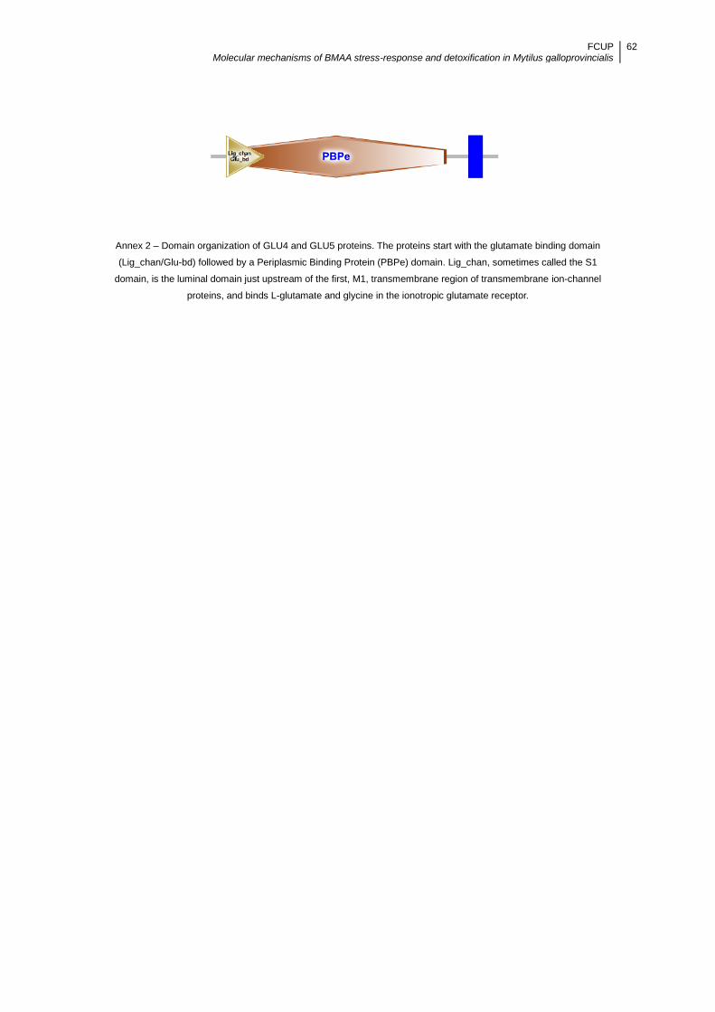
FCUP Molecular mechanisms of BMAA stress-response and detoxification in Mytilus galloprovincialis
62
Annex 2 – Domain organization of GLU4 and GLU5 proteins. The proteins start with the glutamate binding domain
(Lig_chan/Glu-bd) followed by a Periplasmic Binding Protein (PBPe) domain. Lig_chan, sometimes called the S1
domain, is the luminal domain just upstream of the first, M1, transmembrane region of transmembrane ion-channel
proteins, and binds L-glutamate and glycine in the ionotropic glutamate receptor.

FCUP Molecular mechanisms of BMAA stress-response and detoxification in Mytilus galloprovincialis
63
Annex 3 – Sequence alignment produced by Clustal Omega of iGluR from Homo sapiens, Rattus norvegicus, Xenopus
laevis, Carassius auratus, Carassius carassius, Aplysia californica, Lymnaea stagnalis, Crassostrea gigas and Mytilus
galloprovincialis (GLU4 and GLU5). Residues that are conserved across all sequences are highlighted in grey. Below
the protein sequences is a key denoting conserved sequence (*), conservative mutations (:), semi-conservative
mutations (.), and non-conservative mutations ( ).
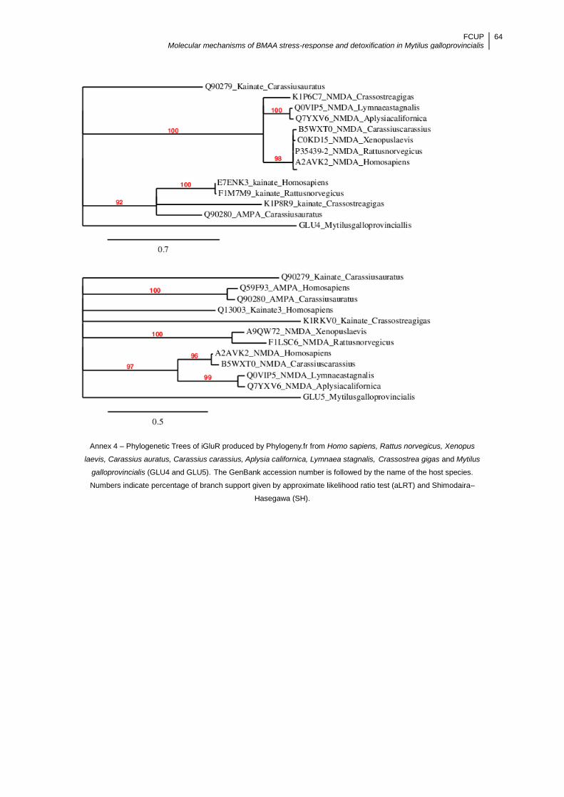
FCUP Molecular mechanisms of BMAA stress-response and detoxification in Mytilus galloprovincialis
64
Annex 4 – Phylogenetic Trees of iGluR produced by Phylogeny.fr from Homo sapiens, Rattus norvegicus, Xenopus
laevis, Carassius auratus, Carassius carassius, Aplysia californica, Lymnaea stagnalis, Crassostrea gigas and Mytilus
galloprovincialis (GLU4 and GLU5). The GenBank accession number is followed by the name of the host species.
Numbers indicate percentage of branch support given by approximate likelihood ratio test (aLRT) and Shimodaira–
Hasegawa (SH).

FCUP Molecular mechanisms of BMAA stress-response and detoxification in Mytilus galloprovincialis
65
Annex 5 - Plot of transmembranar (TMRs) domains of GLU4 obtained with Protter. TMRs domains previously predicted
with Phobius are indicated by numbers 1-3. Signal peptide (red) and glycosylation sites (green) are also shown.

FCUP Molecular mechanisms of BMAA stress-response and detoxification in Mytilus galloprovincialis
66
Annex 6 - Plot of transmembranar (TMRs) of GLU5 obtained with Protter. TMRs domains previously predicted with
Phobius are indicated by numbers 1-3. Signal peptide (red) and glycosylation sites (green) are also shown.
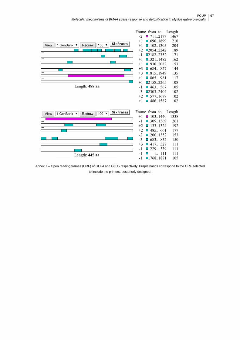
FCUP Molecular mechanisms of BMAA stress-response and detoxification in Mytilus galloprovincialis
67
Annex 7 – Open reading frames (ORF) of GLU4 and GLU5 respectively. Purple bands correspond to the ORF selected
to include the primers, posteriorly designed.