molecular interactions at the surface of extracellular ... · clude those between the ps-binding...
TRANSCRIPT
REVIEW
Molecular interactions at the surface of extracellular vesicles
Edit I. Buzás1,2 & Eszter Á. Tóth1& Barbara W. Sódar1 & Katalin É. Szabó-Taylor1
Received: 12 March 2018 /Accepted: 26 March 2018# The Author(s) 2018
AbstractExtracellular vesicles such as exosomes, microvesicles, apoptotic bodies, and large oncosomes have been shown toparticipate in a wide variety of biological processes and are currently under intense investigation in many differentfields of biomedicine. One of the key features of extracellular vesicles is that they have relatively large surfacecompared to their volume. Some extracellular vesicle surface molecules are shared with those of the plasma membraneof the releasing cell, while other molecules are characteristic for extracellular vesicular surfaces. Besides proteins,lipids, glycans, and nucleic acids are also players of extracellular vesicle surface interactions. Being secreted andpresent in high number in biological samples, collectively extracellular vesicles represent a uniquely large interactivesurface area which can establish contacts both with cells and with molecules in the extracellular microenvironment.Here, we provide a brief overview of known components of the extracellular vesicle surface interactome and highlightsome already established roles of the extracellular vesicle surface interactions in different biological processes in healthand disease.
Keywords Extracellular vesicle . Surface . Interactome . Extracellular matrix . Drug delivery
Introduction
Extracellular vesicles (EVs) are membrane-enclosed het-erogeneous structures that are secreted by all cells [1]and have many different physiological and pathophysio-logical roles [2]. They include small EVs of endosomalorigin (exosomes) as well as plasma membrane-derivedintermediate-sized (100–1000 nm) microvesicles, andlarge sized (> 1 μm) apoptotic bodies and largeoncosomes [3, 4]. In the past few years, EVs attractedrapidly growing scientific interest from various fields ofbiomedicine.
Surface molecules of EVs are of critical functional signif-icance as they (i) establish connections with the surroundingmicro milieu and with cells, (ii) determine EV mobility, (iii)mediate cellular uptake, (iv) affect immune recognition ofEVs (also via posttranslational modifications) by the innateand adaptive immune systems, and (v) may represent effectormolecules (such as FasL). On the other hand, from a re-searcher’s perspective, they enable identification, affinity iso-lation, and molecular classification of EVs and EV subtypes,and enable the use of EVs as biomarkers.
Here, we overview EV surface interactions with the sur-roundingmicroenvironment (extracellular matrix (ECM)mol-ecules or components of the blood plasma) and with cells andprovide examples for the functional relevance of the surfaceinteractions of EVs.
Evidences for exofacial localization of EVproteins as partners in EV surface interactions
When considering EV surface interactions, it is of crucial im-portance to define EV molecules with exofacial topology thatcan serve as interaction partners. EV surface molecules areidentified by immunolabeling (immunogold electron micros-copy, flow cytometry or immunochemistry using confocal or
Edit I. Buzás and Eszter Á. Tóth contributed equally to this work.
This article is a contribution to the special issue on ExtracellularVesicles - Guest Editor: Esther Nolte-’t Hoen
* Edit I. Buzá[email protected]–univ.hu
1 Department of Genetics, Cell- and Immunobiology, SemmelweisUniversity, Budapest, Hungary
2 MTA-SE Immune-Proteogenomics Research Group,Budapest, Hungary
Seminars in Immunopathologyhttps://doi.org/10.1007/s00281-018-0682-0
super resolution microscopy). These widely used approachesenabled identification of Bcanonical^ EV surface proteins in-cluding tetraspanins (CD9, CD63, and CD81), integrins(ITG), cell adhesion molecules (CAM), and growth factor re-ceptors [5]. The presence of these molecules has been con-firmed by many different laboratories.
Mass spectrometry (MS)-based proteomic characterizationhas proven to be a very efficient and widely used tool tocharacterize EVs. This approach was first used by Theryet al. [6] for the characterization of exosomes followed bymany other studies over the years. These proteomic data arealso publicly available from databases (Exocarta, EVpedia,and Vesiclepedia) (http://student4.postech.ac.kr/evpedia2_xe/xe/, http://www.exocarta.org/, http://www.microvesicles.org/). However, MS does not enable identification of theprecise topology of EV proteins. Possible membrane defectsdue to centrifuge-based EV isolation procedures or the occur-rence of inverted vesicles may enable labeled antibodies torecognize internal cargo molecules of EVs making the distinc-tion between EV surfaces and internal cargo proteins chal-lenging. This possibility cannot be completely excluded evenwhen using, e.g., antibody-coated EVarrays [7].
Recently, a combination of proteinase treatment and sub-sequent biotinylation, a strategy known from studying cellularmembrane proteins, has been suggested for the study of lumi-nal and surface-accessible EV cargo [8]. Even with this ap-proach, it cannot be determined whether the surface-accessible EV proteins were present already at the time ofEV production or they were subsequently acquired from con-ditioned media or biological fluids.
Strong evidence for EV surface localization of certain mol-ecules comes from the ability to target the putative protein (orother molecule) for affinity isolation of EVs. Anti-EpCAMand anti-A33 antibodies were used for immunocapture of co-lon cancer-derived exosomes [9]. Similarly, anti-tetraspanin(antiCD63, CD9 and CD81) antibodies can be used forimmunoisolation of EVs [3]. Immune electron microscopyrevealed that hsp70 is localized on the surface of exosomes[10], and a synthetic peptide (Vn96) with high affinity for heatshock proteins has proven useful for affinity enrichment ofcancer EVs [11–13]. Furthermore, EVs can be isolated byheparin affinity purification. Suggested heparin-binding pro-teins on EVs include histones, heat shock proteins, andannexin; however, definite interacting ligand(s) have not beendetermined yet [14]. Of note, not only proteins but also othersurface molecules are targeted for EV affinity capture. As anexample, the recently identified phosphatidyl serine (PS) re-ceptor TIM4 [15] was found efficient in capturing PS-exposing EVs [16].
For immunodetection of EV surface molecules, dot scan(antibody microarray) has been used recently. It showedmoderate/high levels of CD19, CD5, CD31, CD44, CD55,CD62Lm, CD82, HLA-A, B, and C and low levels of
CD21, CD49c, and CD63 on EVs. The authors proposedthese EV surface molecules as a diagnostic signature forchronic lymphocytic leukemia [17]. Furthermore, surfaceplasmon resonance (SPR) has been used recently for the si-multaneous detection of both EV and cancer markers onexosomes from breast cancer cells [18]. Moreover, exosomeBsurfaceome^ profiling was carried out by an initial MS test-ing EVs secreted by 13 pancreatic ductal adenocarcinoma celllines and 2 non-neoplastic cell lines. MS was followed byidentification of candidate biomarkers and validation by animmunocapture pulldown assay. In this assay, a multiplexedpanel of antibodies was used that included anti-CLDN4,EPCAM, CD151, LGALS3BP, HIST2H2BE, andHIST2H2BF antibodies for the enrichment of tumor-specificexosomes for subsequent studies [19].
Numerous pieces of evidence suggest that surface mole-cules on EVs determine the uptake and biological functionsof EVs. As one example, blockade of exosome surface SIRPα(CD47) was shown to be effective in increasing cancer cellphagocytosis [20].
Interaction of EVs with the plasma membraneof cells
Surface interactions of EVs with the plasma membrane are ofoutstanding importance since such interactions mediate bind-ing of EVs to cells resulting in signal transduction or uptake ofEVs by cells. It is now established that EV-target cell interac-tions involve tetraspanins, integrins, ECM proteins, immuno-globulin superfamily members, proteoglycans, and lectins [21,22]. Details of EV docking and entry to cells are not in thefocus of this review, as these interactions have recently beenreviewed elsewhere [21, 22]. To illustrate the outstandingfunctional significance of the interaction of EV surface mole-cules with those of the plasma membrane, here, we only referto the plethora of EV-immune cell interactions including cell-free antigen presentation by EVs [23], Fas ligand or TRAIL-mediated cell death induction by EVs [24–26], or the transferof immune checkpoint molecules (PD1, PDL-1) by EVs [27].
Here, we also point out the significance of externalization(translocation to the outer leaflet of a phospholipid bilayer) ofphosphatidyl serine (PS), a characteristic feature of manyEVs. The negatively charged, surface-exposed phospholipidPS is recognized by numerous plasma membrane receptorseither directly or indirectly, via bridging proteins. Direct PSsensing receptors include the previously mentioned TIM4[15], the receptor for advanced glycation end products,RAGE [28], brain-specific angiogenesis inhibitor 1 Bai-1[29], and stabilin-2 [30]. Indirect PS recognition and subse-quent uptake is mediated by milk fat globule-EGF factor8, MFGE8 [31] which forms a molecular bridge betweenPS and plasma membrane integrins (such as αvβ3) [31]
Semin Immunopathol
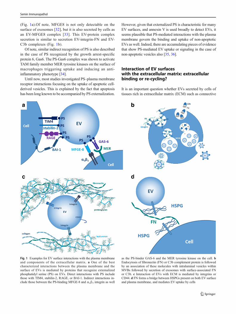
(Fig. 1a).Of note, MFGE8 is not only detectable on thesurface of exosomes [32], but it is also secreted by cells asan EV-MFGE8 complex [33]. This EV-protein complexsecretion is similar to secretion EV-integrin-FN and EV-C3b complexes (Fig. 1b).
Of note, similar indirect recognition of PS is also describedin the case of PS recognized by the growth arrest-specificprotein 6, Gas6. The PS-Gas6 complex was shown to activateTAM family member MER tyrosine kinases on the surface ofmacrophages triggering uptake and inducing an anti-inflammatory phenotype [34].
Until now, most studies investigated PS–plasma membranereceptor interactions focusing on the uptake of apoptotic cell-derived vesicles. This is explained by the fact that apoptosishas been long known to be accompanied by PS externalization.
However, given that externalized PS is characteristic for manyEV surfaces, and annexin V is used broadly to detect EVs, itseems plausible that PS-mediated interactions with the plasmamembrane govern the binding and uptake of non-apoptoticEVs as well. Indeed, there are accumulating pieces of evidencethat show PS-mediated EV uptake or signaling in the case ofnon-apoptotic vesicles also [35, 36].
Interaction of EV surfaceswith the extracellular matrix: extracellularbinding or re-cycling?
It is an important question whether EVs secreted by cells oftissues rich in extracellular matrix (ECM) such as connective
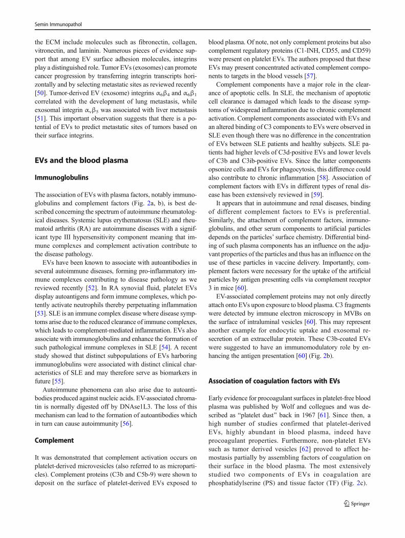
Fig. 1 Examples for EV surface interactions with the plasma membraneand components of the extracellular matrix. a One of the bestcharacterized interactions between the plasma membrane and thesurface of EVs is mediated by proteins that recognize externalizedphosphatidyl serine (PS) on EVs. Direct interactions with PS includethose with TIM4, stabilin-2, RAGE, or BAI-1. Indirect interactions in-clude those between the PS-binding MFGE-8 and αvβ3 integrin as well
as the PS-binder GAS-6 and the MER tyrosine kinase on the cell. bEndocytosis of fibronectin (FN) or C3b complement protein is followedby an association of these molecules with intraluminal vesicles withinMVBs followed by secretion of exosomes with surface-associated FNor C3b. c Interaction of EVs with ECM is mediated by integrins orCD44. d FN forms a bridge between HSPGs present on both EV surfaceand plasma membrane, and mediates EV uptake by cells
Semin Immunopathol
tissue, interact with matrix molecules. Accumulating pieces ofevidence suggest that indeed such interactions exist and theirsignificance is increasingly recognized. It may seem intuitivethat EV surfaces interact with the ECM components uponsecretion, once being surrounded by the macromolecularECM milieu. This may predict that EV membrane depositionof matrix molecules results from binding of these moleculesonto EV surfaces extracellularly. Although newly secretedEVs evidently establish interactions with ECM molecules intissues and body fluids (Fig. 1c), there seems to be anothermechanism, which may explain the presence of certain ECMmolecules on the surface of EVs. It has been proposed recentlythat cells endocytose ECM molecules and re-secrete them onthe exofacial surface of EVs (exosomes) [37] (Fig. 1b). Thiscontinuous endocytosis and re-secretion of ECM componentsguarantees an abundant source of ECM-carrying EVs, whichmay play an important role in cell migration. Such endocyto-sis and EV-associated re-secretions has been recently demon-strated in the case of fibronectin (FN)–integrin complexes. FNis endocytosed in association with integrins, it is then targetedto MVB, where it binds to the surface of intraluminal vesiclesin correct topology to interact with both the cell surface andother ECM molecules (e.g. collagen fibers) [37].
Kowal et al. used immuno-isolated EVs by CD9, CD63,and CD81-specific antibodies. The authors have demonstratedthe existence of a subtype of small EVs (sEVs) that the authorsreferred to as Bdense sEVs^ which carried FN, complement,prothrombin, and serum albumin, while another subpopula-tion of sEVs (Blight sEVs^) did not carry any of these mole-cules on its surface [3]. Whether dense sEVs acquired theirECM coat from the conditioned medium of the cells uponsecretion or were secreted with surface-bound ECM mole-cules, was not investigated in this study.
Fibronectin
One of the most extensively studied ECM molecules withrespect to surface interaction with EVs is FN. FN binds mul-tiple integrins. It has been shown that reticulocyte maturationis accompanied by release of EVs carrying α4β1 integrin(Very Late Antigen-4, VLA4) by which EVs were shown tobind to FN [38]. Myeloma-derived EVs (exosomes) werefound to carry FN on their surface [39]. This exofaciallybound FN could interact with cell surface heparan sulfate(through its Hep-II domain). The authors showed that FNcould simultaneously bind to heparan sulfate proteoglycansboth on the exosomal and the plasma membrane surfacesthereby facilitating cellular uptake of EVs [39] (Fig. 1d).
There are multiple evidences that beyond facilitating cellbinding and cellular uptake, there are other functional conse-quences of EV-association of FN. A striking function of FNon EVs is related to cellular motility. As described above, FN
bound to integrins on exosomes was shown to promote direc-tional cancer cell movement by reinforcing transient polariza-tion states and adhesion assembly [37]. Furthermore,exosomal FN was shown to induce IL-1β expression by mac-rophages [40]. We have shown recently that DNA present onthe surface of small EVs secreted by stressed cells facilitatedinteraction of EVs with FN [41]. Finally, FN on circulatingEVs in liquid biopsy samples of breast cancer patients sampleswas suggested to be a promising cancer biomarker [42].
Glycosaminoglycans (GAGs)and proteoglycans
Heparan sulfate proteoglycans (HSPGs) are abundant glyco-proteins having a core protein to which one or more heparansulfate (HS) glycosaminoglycan (GAG) chains are attachedcovalently. Membrane-bound HSPGs include syndecans andglypicans. Interestingly, syndecans and glypicans are presentboth on the plasma membrane of the EV-releasing cells andthe membrane of EVs. Cancer cell-surface HSPGs of thesyndecan and glypican types were shown to mediate internal-ization of EVs [43]. This process was readily inhibited by freeheparan sulfate. Importantly, the same study demonstratedsorting of HSPGs to EVs (exosomes) [43]. As we mentionedearlier, by forming a bridge between EV and plasma mem-brane HSPGs, FN was shown to mediate EV uptake [39].
Recently, the presence of glypican 1 associated withexosomes was demonstrated by different groups [44–46]. Itsproposed exploitation as a biomarker of pancreatic cancer iscurrently under investigation.
Among other ECM molecules, hyaluronan (HA) synthesiswas shown to be associated with the shedding of HA-coatedEVs by human mesenchymal stem cells (Fig. 1d). HA coatingon EVs was proposed to (i) contribute to HA-mediated tissueregeneration, (ii) regulate interactions of EVs with target cells,and (iii) play a role in ECM remodeling [47]. Not only HA butalso the HA receptor CD44 is associated with EV surfaces.Ovarian cancer cell invasion was shown to be supported byexosomal transfer of CD44 to peritoneal mesothelial cells[48]. CD44 was also identified as a component of the cancercell-derived circulating EV-specific diagnostic signature [17]and was recently shown to serve as one of the diagnostic andprognostic exosomal biomarkers of breast cancer [49].Interestingly, transcripts of CD44 are also carried horizontallyas internal cargo in human mesenchymal stem cell-derivedHA-coated EVs [47].
The role of integrins in EV-ECM interactions
Integrins represent a group of transmembrane receptors thatplay a role in cell-ECM adhesion. Known integrin ligands in
Semin Immunopathol
the ECM include molecules such as fibronectin, collagen,vitronectin, and laminin. Numerous pieces of evidence sup-port that among EV surface adhesion molecules, integrinsplay a distinguished role. Tumor EVs (exosomes) can promotecancer progression by transferring integrin transcripts hori-zontally and by selecting metastatic sites as reviewed recently[50]. Tumor-derived EV (exosome) integrins α6β4 and α6β1
correlated with the development of lung metastasis, whileexosomal integrin αvβ5 was associated with liver metastasis[51]. This important observation suggests that there is a po-tential of EVs to predict metastatic sites of tumors based ontheir surface integrins.
EVs and the blood plasma
Immunoglobulins
The association of EVs with plasma factors, notably immuno-globulins and complement factors (Fig. 2a, b), is best de-scribed concerning the spectrum of autoimmune rheumatolog-ical diseases. Systemic lupus erythematosus (SLE) and rheu-matoid arthritis (RA) are autoimmune diseases with a signif-icant type III hypersensitivity component meaning that im-mune complexes and complement activation contribute tothe disease pathology.
EVs have been known to associate with autoantibodies inseveral autoimmune diseases, forming pro-inflammatory im-mune complexes contributing to disease pathology as wereviewed recently [52]. In RA synovial fluid, platelet EVsdisplay autoantigens and form immune complexes, which po-tently activate neutrophils thereby perpetuating inflammation[53]. SLE is an immune complex disease where disease symp-toms arise due to the reduced clearance of immune complexes,which leads to complement-mediated inflammation. EVs alsoassociate with immunoglobulins and enhance the formation ofsuch pathological immune complexes in SLE [54]. A recentstudy showed that distinct subpopulations of EVs harboringimmunoglobulins were associated with distinct clinical char-acteristics of SLE and may therefore serve as biomarkers infuture [55].
Autoimmune phenomena can also arise due to autoanti-bodies produced against nucleic acids. EV-associated chroma-tin is normally digested off by DNAse1L3. The loss of thismechanism can lead to the formation of autoantibodies whichin turn can cause autoimmunity [56].
Complement
It was demonstrated that complement activation occurs onplatelet-derived microvesicles (also referred to as microparti-cles). Complement proteins (C3b and C5b-9) were shown todeposit on the surface of platelet-derived EVs exposed to
blood plasma. Of note, not only complement proteins but alsocomplement regulatory proteins (C1-INH, CD55, and CD59)were present on platelet EVs. The authors proposed that theseEVs may present concentrated activated complement compo-nents to targets in the blood vessels [57].
Complement components have a major role in the clear-ance of apoptotic cells. In SLE, the mechanism of apoptoticcell clearance is damaged which leads to the disease symp-toms of widespread inflammation due to chronic complementactivation. Complement components associated with EVs andan altered binding of C3 components to EVs were observed inSLE even though there was no difference in the concentrationof EVs between SLE patients and healthy subjects. SLE pa-tients had higher levels of C3d-positive EVs and lower levelsof C3b and C3ib-positive EVs. Since the latter componentsopsonize cells and EVs for phagocytosis, this difference couldalso contribute to chronic inflammation [58]. Association ofcomplement factors with EVs in different types of renal dis-ease has been extensively reviewed in [59].
It appears that in autoimmune and renal diseases, bindingof different complement factors to EVs is preferential.Similarly, the attachment of complement factors, immuno-globulins, and other serum components to artificial particlesdepends on the particles’ surface chemistry. Differential bind-ing of such plasma components has an influence on the adju-vant properties of the particles and thus has an influence on theuse of these particles in vaccine delivery. Importantly, com-plement factors were necessary for the uptake of the artificialparticles by antigen presenting cells via complement receptor3 in mice [60].
EV-associated complement proteins may not only directlyattach onto EVs upon exposure to blood plasma. C3 fragmentswere detected by immune electron microscopy in MVBs onthe surface of intraluminal vesicles [60]. This may representanother example for endocytic uptake and exosomal re-secretion of an extracellular protein. These C3b-coated EVswere suggested to have an immunomodulatory role by en-hancing the antigen presentation [60] (Fig. 2b).
Association of coagulation factors with EVs
Early evidence for procoagulant surfaces in platelet-free bloodplasma was published by Wolf and collegues and was de-scribed as Bplatelet dust^ back in 1967 [61]. Since then, ahigh number of studies confirmed that platelet-derivedEVs, highly abundant in blood plasma, indeed haveprocoagulant properties. Furthermore, non-platelet EVssuch as tumor derived vesicles [62] proved to affect he-mostasis partially by assembling factors of coagulation ontheir surface in the blood plasma. The most extensivelystudied two components of EVs in coagulation arephosphatidylserine (PS) and tissue factor (TF) (Fig. 2c).
Semin Immunopathol
As a high percentage of platelet-derived MVs bear the an-ionic phospholipid PS on their outer membrane, they facilitatethe assembly of several proteins of the coagulation cascade.These proteins contain positively charged γ-carboxyglutamicacid domains to which PS can bind with electrostatic interac-tion. These factors include VII, IX, X, and prothrombin [63].Underlining the importance of PS-positive EV formation inhemostasis, patients suffering from a rare bleeding disorder(Scott syndrome) were found to have reduced floppase activ-ity resulting in faulty PS externalization and reduced micro-particle shedding [64].
Also, TF, a transmembrane receptor of factor VII/VIIa, canbe present on MVs (vesicles often referred to as microparti-cles, MPs in coagulation studies). The elevated activity levelsof this protein have been detected in various diseases (such asin acute liver injury, cirrhosis, urinary tract infection,endotoxemia, influenza, cancer, and related thromboembo-lism [65]). The platelet origin of TF on platelet MVs and theoverall relevance of TF-bearing platelet MVs have beenquestioned by several authors [66–68]. However, the role ofTF positive EVs irrespective whether they originate formplatelets, tumor cells, endothelial cells, or leukocytes is clear
in various hemostatic diseases and diseases tipically present-ing with thrombembolic complications (as detailed in thecomprehensive review by Owens and Mackman [63]). Thepresence of TF on EVs makes the presence of its specificinhibitor molecule, tissue factor pathway inhibitor (TFPI), al-so probable [63].
In addition to assembling factors that initiate the coagula-tion cascade, platelets and their MPs also can present specificbinding sites for factors V, IX, and VIII [69–71]. Indeed, thesebinding sites can be found concentrated on MPs relative toplatelets. In the case of factor Va and VIIIa, a 10-fold, while inthe case of factor IXa, a 2-fold concentration of factor bindingsites was observed in the above mentioned studies [69–71].Another factor, von Willebrand Factor (vWF), an interactionpartner of both glycoproteins GPIb and GPIIbIIIa, was foundto be attached to platelet- and also to endothelial cell-derivedEVs [72, 73]. Together, the accumulation of PS and of othercoagulation factor binding sites enables the surface of plateletMPs to enhance coagulation approximately 50–100-fold ascompared to platelets [74].
Interestingly, depending on what stimuli the parent cellrecieved, MPs may bear different surface molecules resulting
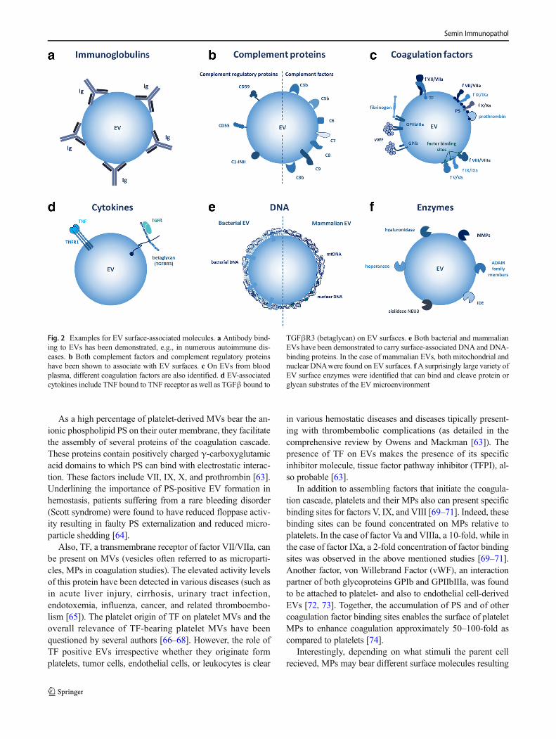
Fig. 2 Examples for EV surface-associated molecules. a Antibody bind-ing to EVs has been demonstrated, e.g., in numerous autoimmune dis-eases. b Both complement factors and complement regulatory proteinshave been shown to associate with EV surfaces. c On EVs from bloodplasma, different coagulation factors are also identified. d EV-associatedcytokines include TNF bound to TNF receptor as well as TGFβ bound to
TGFβR3 (betaglycan) on EV surfaces. e Both bacterial and mammalianEVs have been demonstrated to carry surface-associated DNA and DNA-binding proteins. In the case of mammalian EVs, both mitochondrial andnuclear DNAwere found on EV surfaces. fA surprisingly large variety ofEV surface enzymes were identified that can bind and cleave protein orglycan substrates of the EV microenvironment
Semin Immunopathol
in different binding features. For instance, platelets activatedwith thrombin or collagen were found to shed MPs exposingGPIIbIIIa complexes binding fibrinogen, while those activat-ed with C5b-9 shed non-GPIIbIIIa-exposing MPs [70].
Although the effect of platelet-derived microvesicles hasbeen studied most widely, it is important to note that activatedplatelets also secrete exosomes [75]. However, their associa-tion with coagulation factors in plasma is questionable, asthey, if at all, bear very low levels of PS [76]. Also, it iscontroversial whether plasma exosomes of different cellularorigin bear TF [77].
Association of EVs with lipoproteins
Isolation of EVs from human blood plasma or serum is oftenconfounded by the co-isolated lipoproteins [78–81].Moreover, antibody-mediated depletion of lipoproteins [82]and lipoprotein apheresis [83] both resulted in loss of EVcontent as well. On the other hand, MS analysis of VLDLand LDL particles purified from human blood plasma re-vealed the presence of EV proteins (CD14, LDL-receptor,HLA class I molecules, and protein S100-A8) in these isolates[84]. Taken together, these data suggest that beyond the sharedphysiological parameters, there might be an association be-tween lipoproteins and EVs as well. In vitro associationhas already been demonstrated by transmission electronmicroscopy [80]. However, experimental data are notavailable yet in support of an in vivo association of EVsand lipoproteins. Exchange of the protein and lipid con-tent between lipoproteins is an established phenomenon[85–88]. Exchange of ApoE between lipoproteins andhepatitis C virus lipoviral particles has been also de-scribed [89]. Moreover, in vitro SR-B1-dependent transferof a fluorescent phospholipid from engineered HDL nano-particles to exosomes was also reported [90]. Finally,ApoE has been implicated in amyloid formation of pig-ment cells [91], and it has been shown that in these cells,ApoE associates with intraluminal vesicles and is secretedon the surface of exosomes [92].
Further blood plasma proteins associatedwith the surface of circulating EVs
Beside the known role of EVs as carriers of luminal cargo,EVs may also carry a significant surface cargo. Technicallychallenging to investigate, so far, very little is known about theexternally adsorbed proteins. It is likely that the external cargoof EVs is at least partly acquired in body liquids after the EVshave been shed. As an example, blood plasma-derived EVscommonly carry substantial amounts of albumin [93]. In linewith this, proteomics data in EV databases (http://student4.postech.ac.kr/evpedia2_xe/xe/, http://www.exocarta.org/,http://www.microvesicles.org/), [94–96] show that blood
plasma-derived EVs co-isolate with numerous blood plasmaproteins. Given the known presence of integrins and HSPGson EVs, integrin ligands and heparin binding proteins areevident potential partners to establish interactions on the sur-face of circulating EVs. Furthermore, phosphatidyl serinebinding proteins (such as MFGE8) and glycan bindinggalectins are obvious interaction partners of EVs in the circu-lation. Systemic analysis of EVs surface interactions withblood plasma proteins is still lacking.
Association of EVs with cytokines/chemokines
An increasing number of data support that EVs are capable ofcarrying various cytokines [2]. In most instances, these cyto-kines are carried in EVs as part of the internal cargo. However,it was shown that EVs carry a full-length 55-kDa tumor ne-crosis factor receptor 1 (TNFR1). Importantly, it was demon-strated by the authors that HUVEC-derived exosomes carriedbound TNF [97] (Fig. 2d).
In addition, TGF beta was shown to be associatedwith the cell-surface chondroitin sulfate/heparan sulfateproteoglycan betaglycan (also referred to as transforminggrowth factor beta receptor III, TGFBR3) on the surfaceof cancer cell-derived exosomes. Although the authorsfound that the kinetics and magnitude of biological re-sponse were similar irrespective if they used soluble orEV-associated TGF beta, there were some qualitative dif-ferences in the elicited cellular responses [98] (Fig. 2d).Although it is tempting to hypothesize that additionalcytokines (including chemokines) may be carried on thesurface of EVs in association with EV surface proteogly-cans, a systemic analysis of this question has not beenperformed yet.
DNA associated with the surfaces of EVs
In bacteria, outer membrane vesicle (OMV)-associated DNAhas been shown to mediate inter- and intra-species horizontalgene transfer by carrying antibiotic resistance genes and viru-lence factor [99–101], and participating in the establishmentof bacterial biofilms [102, 103]. Recently, it was also reportedthat OMV-associated DNA was found predominantly on theouter surface of OMVs [104].
In mammalian systems, most studies so far focused onDNA encapsulated in EVs as an internal cargo, and only veryfew reports investigated the EV surface-associated DNase-sensitive DNA. Of note, these studies drew attention towardsthe potential of EV surface-associatedDNA in horizontal genetransfer [105], induction of autoimmunity [56], and cellularuptake [106]. Recently, we have shown that antibiotic-exposed cells undergoing genotoxic shock secreted smallEVs (exosomes) with surface-associated DNAwhich was pre-dominantly mitochondrial DNA [41]. The amount of this
Semin Immunopathol
DNAwas not enhanced by induction of apoptosis of the EV-releasing cells. As mentioned earlier, exosome surface-associated DNAwas capable of mediating EV binding to FN[41] (Fig. 2e).
Enzymes associated with EV surfaces
Several pieces of evidence support the presence and activity ofEV-associated enzymes as reviewed recently [107]. Thus, en-zymes do not only represent components of the EV internalcargo but are also characterized by active exofacial enzymes.These include both proteases MT1-MMP (MMP-14) [108],ADAM17 [109], insulin-degrading enzyme (IDE) (insulin-like), and EV surface-associated glycosidases such assialidase NEU3 [110, 111] and heparanase [112].Importantly, EV surface-associated proteases and glycosi-dases may exert their function in concert with one another inmatrix degradation. In addition, flow cytometry of isolatedEVs bound to latex beads demonstrated the presence of mul-tiple other enzymes (MMPs-2, -3, -9, -13, -14, ADAM-10,ADAM-17, ADAMTS-5, ADAMTS-8, uPAR, and hyaluron-idase) [113]. EV-associated enzymes may (i) facilitate cell andEV mobility by degrading ECM macromolecules as sub-strates, (ii) release bound growth factors or chemokines, and(iii) destruct amyloid β plaques [114] (Fig. 2f).
EV surface-associated thiols
Thiol interactions are relevant both in the release and uptakeof EVs, and it is highly likely that the content and compositionof exofacial thiols has a vast influence on the interactions ofEVs with their environment including macromolecules. Thetotal surface thiol content of EVs can also be utilized for la-beling purposes [115]. Plasma-derived and tissue culture-derived EVs can equally be labeled by thiol-reactive fluores-cent reagents. However, it is important to be aware that con-taminating plasma proteins interfere with such labeling andtherefore, a dual labeling protocol of EV thiols is preferablefrom plasma samples [115]. Certain plasma proteins, like al-bumin, have a reactive thiol moiety [116], and interactionswith EVs may take place via thiol interactions. In particular,in the case of albumin, it seems feasible that EVs form part ofthe Balbuminome^ potentially extending their half-life in thecirculation via interacting with albumin. Albumin certainlyappears among the molecules associated with EVs asdiscussed above. Redox regulation of cellular surface mole-cules is an emerging factor affecting cellular functions.Importantly, adhesion molecules such as integrins underlieredox regulation. Reducing the α4-integrin by N-acetyl-cys-teine leads to increased FN adhesion and cellular aggregationof Jurkat cells [117]. Similar redox regulation may be relevantin EV biology. EV-associated integrin regulation is of partic-ular interest, since distinct expression pattern of integrins on
EVs is responsible for organotropism in cancer metastasis[51]. Therefore, redox regulation of exofacial molecules onEVs is likely to affect their functions.
Several thiol-reactive antioxidants are present in the plas-ma, notably certain members of the thioredoxin family such asperoxiredoxins 1, 2, and 4 and different forms of thioredoxin[118]. These thiol-reactive antioxidants are also known to ap-pear on the surface of cells [118, 119]. Therefore, it is to beexpected that these molecules also appear on the surface ofEVs either as membrane proteins or plasma proteins associat-ing with the EVs. Indeed, peroxiredoxin 2 was present on thesurface of EVs [120, 121], and peroxiredoxin1 (Prdx1)-posi-tive EVs were elevated in rheumatoid arthritis (RA) patientplasma compared with healthy controls which may be a mark-er of inflammation [115]. Since Prdx 1 is also present as a freeprotein in plasma, it may be released together with EVs or itassociates with EVs after release, in the plasma. Protein disul-fide isomerase is thought to be responsible for regulating cellsurface thiols [122] and associated with EV surface, and it alsoseems to activate platelets [123].
Conclusions
The relatively large surface to volume ratio of EVs enableshighly efficient surface interactions of these structures withcells and extracellular molecules. Such surface interactionshave outstanding importance since they determine the fateof EVs by targeting them to the plasma membrane of cellsor to certain tissues. One of the most exciting aspects ofEV surface interactions is that they can be tailored by en-gineering the EV-releasing cells [124, 125]. This way, byhaving the designed EV-producing cells, one may achieveto produce EVs with specific targeting molecules on theirsurface and thus may be able to alter the biodistribution ofEVs used as drug delivery systems. Feasibility of this ap-proach was first demonstrated when siRNA was success-fully delivered to murine brain upon systemic administra-tion of exosomes carrying a brain-targeting peptide [126].This initial proof-of-concept study has been followed byseveral subsequent works in which designer EVs were spe-cifically targeted to tissues or cells [125]. The potential ofdesigning targeted EVs for drug delivery, and the knownability of exosomes to cross blood tissue barriers such asblood brain barrier, underlies the significance in emergingEV-based therapies including gene therapy.
Another aspect of EV surface interactions is that due totechnical limitations, it is not feasible to perform comprehen-sive analysis of EVs in situ in living tissues. Considering theinteractions of EVs both with cells and with molecules of themicroenvironment, there is an urgent need for much morecomplex systems to model EV surface interactions.
Semin Immunopathol
Recognizing the complexity of EV surface interactions, weshould change our way of thinking about EVs as Bpure^mem-brane vesicles. We should rather consider EV surface interac-tome in our experimental design since EV surface-associatedmolecules can hinder those already present on the EV surface,resulting in unexpected outcomes of both analysis and isola-tion of EVs.
Grant support This work was supported by the NationalResearch, Development and Innovation Office NKFIH,Hungary, OTKA11958, OTKA120237, NVKP_16-1-2016-0017, Ministry for National Economy of Hungary VEKOP-2.3.2-16-2016-00002, and VEKOP-2.3.315201600016.
Open Access This article is distributed under the terms of the CreativeCommons At t r ibut ion 4 .0 In te rna t ional License (h t tp : / /creativecommons.org/licenses/by/4.0/), which permits unrestricted use,distribution, and reproduction in any medium, provided you give appro-priate credit to the original author(s) and the source, provide a link to theCreative Commons license, and indicate if changes were made.
References
1. Gyorgy B, Szabo TG, Pasztoi M, Pal Z, Misjak P, Aradi B, LaszloV, Pallinger E, Pap E, Kittel A, Nagy G, Falus A, Buzas EI (2011)Membrane vesicles, current state-of-the-art: emerging role of ex-tracellular vesicles. Cell Mol Life Sci 68:2667–2688
2. Yanez-Mo M, Siljander PR, Andreu Z, Zavec AB, Borras FE,Buzas EI, Buzas K, Casal E, Cappello F, Carvalho J, Colas E,Cordeiro-da Silva A, Fais S, Falcon-Perez JM, Ghobrial IM,Giebel B, Gimona M, Graner M, Gursel I, Gursel M, HeegaardNH, Hendrix A, Kierulf P, Kokubun K, Kosanovic M, Kralj-IglicV, Kramer-Albers EM, Laitinen S, Lasser C, Lener T, Ligeti E,Line A, Lipps G, Llorente A, Lotvall J, Mancek-Keber M,Marcilla A, Mittelbrunn M, Nazarenko I, Nolte-'t Hoen EN,Nyman TA, O'Driscoll L, Olivan M, Oliveira C, Pallinger E, DelPortillo HA, Reventos J, RigauM, Rohde E, SammarM, Sanchez-Madrid F, Santarem N, Schallmoser K, Ostenfeld MS, StoorvogelW, Stukelj R, Van der Grein SG, Vasconcelos MH, Wauben MH,De Wever O (2015) Biological properties of extracellular vesiclesand their physiological functions. J Extracell Vesicles 4:27066
3. Kowal J, Arras G, Colombo M, Jouve M, Morath JP, Primdal-Bengtson B, Dingli F, Loew D, Tkach M, Thery C (2016)Proteomic comparison defines novel markers to characterize het-erogeneous populations of extracellular vesicle subtypes. ProcNatl Acad Sci U S A 113:E968–E977
4. Di Vizio D,MorelloM, Dudley AC, Schow PW, AdamRM,MorleyS, Mulholland D, Rotinen M, Hager MH, Insabato L, Moses MA,Demichelis F, Lisanti MP, Wu H, Klagsbrun M, Bhowmick NA,Rubin MA, D'Souza-Schorey C, Freeman MR (2012) Largeoncosomes in human prostate cancer tissues and in the circulationof mice with metastatic disease. Am J Pathol 181:1573–1584
5. Lotvall J, Hill AF, Hochberg F, Buzas EI, Di Vizio D, Gardiner C,Gho YS, Kurochkin IV, Mathivanan S, Quesenberry P, Sahoo S,Tahara H, Wauben MH, Witwer KW, Thery C (2014) Minimalexperimental requirements for definition of extracellular vesiclesand their functions: a position statement from the InternationalSociety for Extracellular Vesicles. J Extracell Vesicles 3:26913
6. Théry C, Regnault A, Garin J, Wolfers J, Zitvogel L, Ricciardi-Castagnoli P, Raposo G, Amigorena S (1999) Molecular charac-terization of dendritic cell-derived exosomes: selective accumula-tion of the heat shock protein hsc73. J Cell Biol 147:599–610
7. Jørgensen MM, Bæk R, Varming K (2015) Potentials and capabil-ities of the extracellular vesicle (EV) array. J Ext Vesicles 4:26048
8. Cvjetkovic A, Jang SC, Konečná B, Höög JL, Sihlbom C, LässerC, Lötvall J (2016) Detailed analysis of protein topology of extra-cellular vesicles—evidence of unconventional membrane proteinorientation. Sci Rep 6:36338
9. Tauro BJ, Greening DW, Mathias RA, Mathivanan S, Ji H,Simpson RJ (2013) Two distinct populations of exosomes arereleased from LIM1863 colon carcinoma cell-derived organoids.Mol Cell Proteomics 12:587–598
10. Radons J, Multhoff G (2005) Immunostimulatory functions ofmembrane-bound and exported heat shock protein 70. ExercImmunol Rev 11:17–33
11. Ghosh A, Davey M, Chute IC, Griffiths SG, Lewis S, Chacko S,Barnett D, Crapoulet N, Fournier S, Joy A (2014) Rapid isolationof extracellular vesicles from cell culture and biological fluidsusing a synthetic peptide with specific affinity for heat shock pro-teins. PLoS One 9:e110443
12. Griffiths SG, Lewis SE (2015) Polypeptides with affinity for heatshock proteins (HSPs) and HSP associated complexes (HACS)and their use in diagnosis and therapy
13. Griffiths SG, Cormier MT, Clayton A, Doucette AA (2017)Differential proteome analysis of extracellular vesicles from breastcancer cell lines by chaperone affinity enrichment. Proteomes 5:25
14. Balaj L, Atai NA, Chen W, Mu D, Tannous BA, Breakefield XO,Skog J, Maguire CA (2015) Heparin affinity purification of extra-cellular vesicles. Sci Rep 5:10266
15. Tietjen GT, Gong Z, Chen CH, Vargas E, Crooks JE, Cao KD,Heffern CT, Henderson JM, Meron M, Lin B, Roux B,Schlossman ML, Steck TL, Lee KY, Adams EJ (2014)Molecular mechanism for differential recognition of membranephosphatidylserine by the immune regulatory receptor Tim4.Proc Natl Acad Sci USA 111(15):E1463–E1E72
16. Nakai W, Yoshida T, Diez D, Miyatake Y, Nishibu T, Imawaka N,Naruse K, Sadamura Y, Hanayama R (2016) A novel affinity-based method for the isolation of highly purified extracellularvesicles. Sci Rep 6:33935
17. Belov L, Matic KJ, Hallal S, Best OG, Mulligan SP,Christopherson RI (2016) Extensive surface protein profiles ofextracellular vesicles from cancer cells may provide diagnosticsignatures from blood samples. J Ext Vesicles 5:25355
18. Grasso L, Wyss R, Weidenauer L, Thampi A, Demurtas D,Prudent M, Lion N, Vogel H (2015) Molecular screening ofcancer-derived exosomes by surface plasmon resonance spectros-copy. Anal Bioanal Chem 407:5425–5432
19. Castillo J, Bernard V, San Lucas F, Allenson K, Capello M, KimD, Gascoyne P, Mulu F, Stephens B, Huang J (2017) Surfaceomeprofiling enables isolation of cancer-specific exosomal cargo inliquid biopsies from pancreatic cancer patients. Ann Oncol 29:223–229
20. Koh E, Lee EJ, Nam G-H, Hong Y, Cho E, Yang Y, Kim I-S(2017) Exosome-SIRPα, a CD47 blockade increases cancer cellphagocytosis. Biomaterials 121:121–129
21. Mulcahy LA, Pink RC, Carter DRF (2014) Routes and mecha-nisms of extracellular vesicle uptake. J Ext Vesicles 3:24641
22. French KC, Antonyak MA, Cerione RA (2017) Extracellular ves-icle docking at the cellular port: extracellular vesicle binding anduptake. Presented at Seminars in cell & developmental biology
23. Raposo G, Nijman HW, Stoorvogel W, Liejendekker R, HardingCV, Melief CJ, Geuze HJ (1996) B lymphocytes secrete antigen-presenting vesicles. J Exp Med 183:1161–1172
Semin Immunopathol
24. Martínez-Lorenzo MJ, Anel A, Gamen S, Monleón I, Lasierra P,Larrad L, Piñeiro A, Alava MA, Naval J (1999) Activated humanT cells release bioactive Fas ligand and APO2 ligand inmicrovesicles. J Immunol 163:1274–1281
25. Monleón I, Martínez-Lorenzo MJ, Monteagudo L, Lasierra P,Taulés M, Iturralde M, Piñeiro A, Larrad L, Alava MA,Naval J, Anel A (2001) Differential secretion of Fas ligand-or APO2 ligand/TNF-related apoptosis-inducing ligand-car-rying microvesicles during activation-induced death of hu-man T cells. J Immunol 167(12):6736–6744
26. Rivoltini L, Chiodoni C, Squarcina P, Tortoreto M, Villa A, VerganiB, BürdekM,Botti L, Arioli I, CovaA (2016) TNF-related apoptosis-inducing ligand (TRAIL)—armed exosomes deliver proapoptotic sig-nals to tumor site. Clin Cancer Res 22:3499–3512
27. Theodoraki M-N, Yerneni S, Hoffmann TK, Gooding WE,Whiteside TL (2017) Clinical significance of PD-L1+ exosomesin plasma of head and neck cancer patients. Clin Cancer Res pp.clincanres 2664.017
28. He M, Kubo H, Morimoto K, Fujino N, Suzuki T, Takahasi T,Yamada M, Yamaya M, Maekawa T, Yamamoto Y (2011)Receptor for advanced glycation end products binds tophosphatidylserine and assists in the clearance of apoptotic cells.EMBO Rep 12:358–364
29. Park D, Tosello-Trampont A-C, Elliott MR, LuM, Haney LB, MaZ, Klibanov AL, Mandell JW, Ravichandran KS (2007) BAI1 isan engulfment receptor for apoptotic cells upstream of theELMO/Dock180/Rac module. Nature 450:430–434
30. Park SY, JungMY, Kim HJ, Lee SJ, Kim SY, Lee BH, Kwon TH,Park RW, Kim IS (2008) Rapid cell corpse clearance by stabilin-2,a membrane phosphatidylserine receptor. Cell Death Differ 15(1):192–201
31. Hanayama R, Tanaka M, Miwa K, Shinohara A, Iwamatsu A,Nagata S (2002) Identification of a factor that links apoptotic cellsto phagocytes. Nature 417:182–187
32. Véron P, Segura E, Sugano G, Amigorena S, Théry C (2005)Accumulation of MFG-E8/lactadherin on exosomes from imma-ture dendritic cells. Blood Cell Mol Dis 35:81–88
33. Oshima K, Aoki N, Kato T, Kitajima K, Matsuda T (2002)Secretion of a peripheral membrane protein, MFG-E8, as a com-plex with membrane vesicles. FEBS J 269:1209–1218
34. Graham DK, DeRyckere D, Davies KD, Earp HS (2014) TheTAM family: phosphatidylserine-sensing receptor tyrosine ki-nases gone awry in cancer. Nat Rev Cancer 14:769–785
35. Zakharova L, Svetlova M, Fomina AF (2007) T cell exosomesinduce cholesterol accumulation in human monocytes viaphosphatidylserine receptor. J Cell Physiol 212:174–181
36. Wei X, Liu C, Wang H, Wang L, Xiao F, Guo Z, Zhang H (2016)Surface phosphatidylserine is responsible for the internalization onmicrovesicles derived from hypoxia-induced human bone marrowmesenchymal stem cells into human endothelial cells. PLoS One11:e0147360
37. Sung BH, Ketova T, Hoshino D, Zijlstra A, Weaver AM (2015)Directional cell movement through tissues is controlled byexosome secretion. Nat Commun 6:7164
38. Rieu S, Géminard C, Rabesandratana H, Sainte-Marie J, Vidal M(2000) Exosomes released during reticulocyte maturation bind tofibronectin via integrin α4β1. FEBS J 267:583–590
39. Purushothaman A, Bandari SK, Liu J, Mobley JA, Brown EE,Sanderson RD (2016) Fibronectin on the surface of myelomacell-derived exosomes mediates exosome-cell interactions. J BiolChem 291:1652–1663
40. Atay S, Gercel-Taylor C, Taylor DD (2011) Human trophoblast-derived Exosomal fibronectin induces pro-inflammatory Il-1βproduction by macrophages. Am J Reprod Immunol 66:259–269
41. Németh A, Orgovan N, Sódar BW, Osteikoetxea X, Pálóczi K,Szabó-Taylor KÉ, Vukman KV, Kittel Á, Turiák L, Wiener Z
(2017) Antibiotic-induced release of small extracellular vesicles(exosomes) with surface-associated DNA. Sci Rep 7:8202
42. Moon P-G, Lee J-E, Cho Y-E, Lee SJ, Chae YS, Jung JH, Kim I-S,Park HY, Baek M-C (2016) Fibronectin on circulating extracellularvesicles as a liquid biopsy to detect breast cancer. Oncotarget 7:40189
43. Christianson HC, Svensson KJ, van Kuppevelt TH, Li J-P, BeltingM (2013) Cancer cell exosomes depend on cell-surface heparansulfate proteoglycans for their internalization and functional activ-ity. Proc Natl Acad Sci 110:17380–17385
44. Lai X, Wang M, McElyea SD, Sherman S, House M, Korc M(2017) A microRNA signature in circulating exosomes is superiorto exosomal glypican-1 levels for diagnosing pancreatic cancer.Cancer Lett 393:86–93
45. Melo SA, Luecke LB, Kahlert C, Fernandez AF, Gammon ST,Kaye J, LeBleu VS, Mittendorf EA, Weitz J, Rahbari N (2015)Glypican-1 identifies cancer exosomes and detects early pancre-atic cancer. Nature 523:177–182
46. Liang K, Liu F, Fan J, Sun D, Liu C, Lyon CJ, Bernard DW, Li Y,Yokoi K, Katz MH (2017) Nanoplasmonic quantification oftumour-derived extracellular vesicles in plasma microsamples fordiagnosis and treatment monitoring. Nat Biomed Eng 1:0021
47. Arasu UT, Kärnä R, Härkönen K, Oikari S, Koistinen A, KrögerH, Qu C, Lammi MJ, Rilla K (2017) Human mesenchymal stemcells secrete hyaluronan-coated extracellular vesicles. Matrix Biol64:54–68
48. Nakamura K, Sawada K, Kinose Y, Yoshimura A, Toda A,Nakatsuka E, Hashimoto K, Mabuchi S, Morishige K-I, KurachiH (2016) Exosomes promote ovarian cancer cell invasion throughtransfer of CD44 to peritoneal mesothelial cells. Mol Cancer Respp. molcanres 0191.2016
49. Wang M, Ji S, Shao G, Zhang J, Zhao K, Wang Z, Wu A (2017)Effect of exosome biomarkers for diagnosis and prognosis ofbreast cancer patients. Clin Transl Oncol 1–6
50. Paolillo M, Schinelli S (2017) Integrins and exosomes, a danger-ous liaison in cancer progression. Cancers 9:95
51. Hoshino A, Costa-Silva B, Shen TL, Rodrigues G, Hashimoto A,Tesic Mark M, Molina H, Kohsaka S, Di Giannatale A, Ceder S,Singh S, Williams C, Soplop N, Uryu K, Pharmer L, King T,Bojmar L, Davies AE, Ararso Y, Zhang T, Zhang H, HernandezJ,Weiss JM, Dumont-Cole VD, Kramer K,Wexler LH,NarendranA, Schwartz GK, Healey JH, Sandstrom P, Labori KJ, Kure EH,Grandgenett PM, Hollingsworth MA, de Sousa M, Kaur S, JainM, Mallya K, Batra SK, Jarnagin WR, Brady MS, Fodstad O,Muller V, Pantel K, Minn AJ, Bissell MJ, Garcia BA, Kang Y,Rajasekhar VK, Ghajar CM, Matei I, Peinado H, Bromberg J,Lyden D (2015) Tumour exosome integrins determineorganotropic metastasis. Nature 527(7578):329–335
52. Buzas EI, György B, Nagy G, Falus A, Gay S (2014) Emergingrole of extracellular vesicles in inflammatory diseases. Nat RevRheumatol 10(6):356–364
53. Cloutier N, Tan S, Boudreau LH, Cramb C, Subbaiah R, Lahey L,Albert A, Shnayder R, Gobezie R, Nigrovic PA (2013) The expo-sure of autoantigens by microparticles underlies the formation ofpotent inflammatory components: the microparticle-associatedimmune complexes. EMBO Mol Med 5:235–249
54. Perez-Hernandez J, Redon J, Cortes R (2017) Extracellular vesi-cles as therapeutic agents in systemic lupus erythematosus. Int JMol Sci 18:717
55. Fortin PR, Cloutier N, Bissonnette V, Aghdassi E, Eder L, SimonyanD, Laflamme N, Boilard E (2016) Distinct subtypes of microparticle-containing immune complexes are associated with disease activity,damage, and carotid intima-media thickness in systemic lupus erythe-matosus. J Rheumatol pp. jrheum. 160050
56. Sisirak V, Sally B, D’Agati V, Martinez-Ortiz W, Özçakar ZB,David J, Rashidfarrokhi A, Yeste A, Panea C, Chida AS (2016)
Semin Immunopathol
Digestion of chromatin in apoptotic cell microparticles preventsautoimmunity. Cell 166:88–101
57. Yin W, Ghebrehiwet B, Peerschke EI (2008) Expression of com-plement components and inhibitors on platelet microparticles.Platelets 19:225–233
58. Winberg LK, Nielsen CH, Jacobsen S (2017) Surface complementC3 fragments and cellular binding of microparticles in patientswith SLE. Lupus Sci Med 4(1):e000193
59. Karpman D, Ståhl A-L, Arvidsson I (2017) Extracellular vesiclesin renal disease. Nat Rev Nephrol 13:545–562
60. Carrillo-Conde BR, Ramer-Tait AE, Wannemuehler MJ,Narasimhan B (2012) Chemistry-dependent adsorption of serumproteins onto polyanhydride microparticles differentially influ-ences dendritic cell uptake and activation. Acta Biomater 8:3618–3628
61. Wolf P (1967) The nature and significance of platelet products inhuman plasma. Br J Haematol 13:269–288
62. Zwicker JI, Liebman HA, Neuberg D, Lacroix R, Bauer KA, FurieBC, Furie B (2009) Tumor-derived tissue factor–bearing micro-particles are associated with venous thromboembolic events inmalignancy. Clin Cancer Res 15:6830–6840
63. Owens AP, Mackman N (2011) Microparticles in hemostasis andthrombosis. Circ Res 108:1284–1297
64. Toti F, Satta N, Fressinaud E, Meyer D, Freyssinet J-M (1996)Scott syndrome, characterized by impaired transmembrane migra-tion of procoagulant phosphatidylserine and hemorrhagic compli-cations, is an inherited disorder. Blood 87:1409–1415
65. Hisada Y, Auriemma AC, Alexander W, Ay C, Mackman N(2017) Detection of tissue factor-positive extracellular vesiclesby laser scanning confocal microscopy. Thromb Res 150:65–72
66. Cointe S, Lacroix R, Dignat-George F (2017) Platelet-derivedmicroparticles. In: Platelets in thrombotic and non-thrombotic dis-orders. Springer, p 379–92
67. Lacroix R, Dubois C, Leroyer A, Sabatier F, Dignat-George F(2013) Revisited role of microparticles in arterial and venousthrombosis. J Thromb Haemost 11:24–35
68. Van Der Meijden P, Van Schilfgaarde M, Van Oerle R, Renne T,Ten Cate H, Spronk H (2012) Platelet-and erythrocyte-derivedmicroparticles trigger thrombin generation via factor XIIa. JThromb Haemost 10:1355–1362
69. Gilbert GE, Sims PJ, Wiedmer T, Furie B, Furie BC, Shattil SJ(1991) Platelet-derived microparticles express high affinity recep-tors for factor VIII. J Biol Chem 266(26):17261–17268
70. Sims PJ, Wiedmer T, Esmon CT, Weiss HJ, Shattil S (1989)Assembly of the platelet prothrombinase complex is linked tovesiculation of the platelet plasma membrane. Studies in Scottsyndrome: an isolated defect in platelet procoagulant activity. JBiol Chem 264:17049–17057
71. Hoffman M, Monroe DM, Roberts HR (1992) Coagulation factorIXa binding to activated platelets and platelet-derived microparti-cles: a flow cytometric study. Thromb Haemost 68:74–78
72. Thom SR, Bennett M, Banham ND, ChinW, Blake DF, Rosen A,Pollock NW, Madden D, Barak O, Marroni A (2015) Associationof microparticles and neutrophil activation with decompressionsickness. J Appl Physiol 119:427–434
73. JyW, Jimenez J, Mauro L, Horstman L, Cheng P, Ahn E, Bidot C,Ahn Y (2005) Endothelial microparticles induce formation ofplatelet aggregates via a von Willebrand factor/ristocetin depen-dent pathway, rendering them resistant to dissociation. J ThrombHaemost 3:1301–1308
74. Sinauridze EI, Kireev DA, Popenko NY, Pichugin AV, PanteleevMA, Krymskaya OV, Ataullakhanov FI (2007) Platelet micropar-ticle membranes have 50-to 100-fold higher specific procoagulantactivity than activated platelets. Thromb Haemost 98:425–434
75. Théry C, Zitvogel L, Amigorena S (2002) Exosomes: composi-tion, biogenesis and function. Nat Rev Immunol 2:569–579
76. Heijnen HF, Schiel AE, Fijnheer R, Geuze HJ, Sixma JJ (1999)Activated platelets release two types of membrane vesicles:microvesicles by surface shedding and exosomes derived fromexocytosis of multivesicular bodies and-granules. Blood 94:3791–3799
77. van der Pol E, Böing AN, Harrison P, Sturk A, Nieuwland R(2012) Classification, functions, and clinical relevance of extracel-lular vesicles. Pharmacol Rev 64:676–705
78. Yuana Y, Levels J, Grootemaat A, Sturk A, Nieuwland R (2014)Co-isolation of extracellular vesicles and high-density lipoproteinsusing density gradient ultracentrifugation. J Ext Vesicles 3:23262
79. Deregibus MC, Figliolini F, D'antico S, Manzini PM,Pasquino C, De Lena M, Tetta C, Brizzi MF, Camussi G(2016) Charge-based precipitation of extracellular vesicles.Int J Mol Med 38:1359–1366
80. Sódar BW, Kittel Á, Pálóczi K, Vukman KV, Osteikoetxea X, Szabó-Taylor K, Németh A, Sperlágh B, Baranyai T, Giricz Z (2016) Low-density lipoprotein mimics blood plasma-derived exosomes andmicrovesicles during isolation and detection. Sci Rep 6:24316
81. Grigor’eva A, Dyrkheeva N, Bryzgunova O, Tamkovich S,Chelobanov B, Ryabchikova E (2017) Contamination of exosomepreparations, isolated from biological fluids. Biochem (Moscow),Suppl Ser B: Biomed Chem 11:265–271
82. Mørk M, Handberg A, Pedersen S, Jørgensen MM, Bæk R,Nielsen MK, Kristensen SR (2017) Prospects and limitations ofantibody-mediated clearing of lipoproteins from blood plasmaprior to nanoparticle tracking analysis of extracellular vesicles. JExt Vesicles 6:1308779
83. Connolly KD, Willis GR, Datta DB, Ellins EA, Ladell K, PriceDA, Guschina IA, Rees DA, James PE (2014) Lipoprotein-apheresis reduces circulating microparticles in individuals withfamilial hypercholesterolemia. J Lipid Res 55:2064–2072
84. Dashty M, Motazacker MM, Levels J, de Vries M, Mahmoudi M,PeppelenboschMP, Rezaee F (2014) Proteome of human plasma verylow-density lipoprotein and low-density lipoprotein exhibits a linkwithcoagulation and lipid metabolism. Thromb Haemost 112:518–530
85. Illingworth DR, Portman OW (1972) Independence of phospho-lipid and protein exchange between plasma lipoproteins in vivoand in vitro. Biochim Biophys Acta (BBA)-Lipids Lipid Metab280:281–289
86. Barr SI, Kottke B, Mao S (1981) Postprandial exchange of apoli-poprotein C-III between plasma lipoproteins. Am J Clin Nutr 34:191–198
87. Liu X, Bagdade J (1995) Neutral lipid mass transfer among lipo-proteins in plasma from normolipidemic subjects is not an equi-molar heteroexchange. J Lipid Res 36:2574–2579
88. Ooi EM, Barrett PHR, Chan DC,Watts GF (2008) ApolipoproteinC-III: understanding an emerging cardiovascular risk factor. ClinSci (Lond) 114:611–624
89. Yang Z,Wang X, Chi X, Zhao F, Guo J, Ma P, Zhong J, Niu J, PanX, Long G (2016) Neglected but important role of apolipoproteinE exchange in hepatitis C virus infection. J Virol 90:9632–9643
90. Angeloni NL,McMahonKM, Swaminathan S, PlebanekMP,OsmanI, Volpert OV, Thaxton CS (2016) Pathways for modulating exosomelipids identified by high-density lipoprotein-like nanoparticle bindingto scavenger receptor type B-1. Sci Rep 6:22915
91. Van Niel G, Bergam P, Di Cicco A, Hurbain I, Cicero AL, DingliF, Palmulli R, Fort C, Potier MC, Schurgers LJ (2015)Apolipoprotein E regulates amyloid formation within endosomesof pigment cells. Cell Rep 13:43–51
92. van Niel G (2016) Study of exosomes shed new light on physiol-ogy of amyloidogenesis. Cell Mol Neurobiol 36:327–342
93. Baranyai T, Herczeg K, Onódi Z, Voszka I, Módos K, Marton N,Nagy G, Mäger I, Wood MJ, El Andaloussi S (2015) Isolation ofexosomes from blood plasma: qualitative and quantitative
Semin Immunopathol
comparison of ultracentrifugation and size exclusion chromatog-raphy methods. PLoS One 10:e0145686
94. Keerthikumar S, Chisanga D, Ariyaratne D, Al Saffar H, Anand S,Zhao K, Samuel M, Pathan M, Jois M, Chilamkurti N (2016)ExoCarta: a web-based compendium of exosomal cargo. J MolBiol 428:688–692
95. Mathivanan S, Simpson RJ (2009) ExoCarta: a compendium ofexosomal proteins and RNA. Proteomics 9:4997–5000
96. Kim D-K, Kang B, Kim OY, Choi D-S, Lee J, Kim SR, Go G,Yoon YJ, Kim JH, Jang SC (2013) EVpedia: an integrated data-base of high-throughput data for systemic analyses of extracellularvesicles. J Ext Vesicles 2:20384
97. Hawari FI, Rouhani FN, Cui X, Yu Z-X, Buckley C, Kaler M,Levine SJ (2004) Release of full-length 55-kDa TNF receptor 1 inexosome-like vesicles: a mechanism for generation of soluble cy-tokine receptors. Proc Natl Acad Sci U S A 101:1297–1302
98. Webber J, Steadman R, Mason MD, Tabi Z, Clayton A (2010)Cancer exosomes trigger fibroblast to myofibroblast differentia-tion. Cancer Res 70:9621–9630
99. Rumbo C, Fernández-Moreira E, Merino M, PozaM,Mendez JA,Soares NC, Mosquera A, Chaves F, Bou G (2011) Horizontaltransfer of the OXA-24 carbapenemase gene via outer membranevesicles: a new mechanism of dissemination of carbapenem resis-tance genes in Acinetobacter baumannii. Antimicrob AgentsChemother 55:3084–3090
100. Fulsundar S, Harms K, Flaten GE, Johnsen PJ, Chopade BA,Nielsen KM (2014) Gene transfer potential of outer membranevesicles of Acinetobacter baylyi and effects of stress on vesicula-tion. Appl Environ Microbiol 80:3469–3483
101. Yaron S, Kolling GL, Simon L, Matthews KR (2000) Vesicle-mediated transfer of virulence genes from Escherichia coliO157: H7 to other enteric bacteria. Appl Environ Microbiol 66:4414–4420
102. Liao S, KleinMI, HeimKP, Fan Y, Bitoun JP, Ahn S-J, Burne RA,Koo H, Brady LJ, Wen ZT (2014) Streptococcus mutans extracel-lular DNA is upregulated during growth in biofilms, actively re-leased via membrane vesicles, and influenced by components ofthe protein secretion machinery. J Bacteriol 196:2355–2366
103. Gloag ES, Turnbull L, Huang A, Vallotton P, Wang H, Nolan LM,Mililli L, Hunt C, Lu J, Osvath SR (2013) Self-organization ofbacterial biofilms is facilitated by extracellular DNA. Proc NatlAcad Sci 110:11541–11546
104. Bitto NJ, Chapman R, Pidot S, Costin A, Lo C, Choi J, D’cruze T,Reynolds EC, Dashper SG, Turnbull L (2017) Bacterial membranevesicles transport their DNA cargo into host cells. Sci Rep 7:7072
105. Fischer S, Cornils K, Speiseder T, Badbaran A, Reimer R,Indenbirken D, Grundhoff A, Brunswig-Spickenheier B, AlawiM, Lange C (2016) Indication of horizontal DNA gene transferby extracellular vesicles. PLoS One 11:e0163665
106. Shelke GV, Jang SC, Yin Y, Lässer C, Lötvall J (2016) Humanmast cells release extracellular vesicle-associated DNA.Matters 2:e201602000034
107. Sanderson RD, Bandari SK, Vlodavsky I (2017) Proteases andglycosidases on the surface of exosomes: newly discovered mech-anisms for extracellular remodeling. Matrix Biol
108. Hakulinen J, Sankkila L, Sugiyama N, Lehti K, Keski-Oja J(2008) Secretion of active membrane type 1 matrix metallopro-teinase (MMP-14) into extracellular space in microvesicularexosomes. J Cell Biochem 105:1211–1218
109. Groth E, Pruessmeyer J, Babendreyer A, Schumacher J,Pasqualon T, Dreymueller D, Higashiyama S, Lorenzen I,Grötzinger J, Cataldo D (2016) Stimulated release and functionalactivity of surface expressed metalloproteinase ADAM17 inexosomes. Biochim Biophys Acta (BBA)-Mol Cell Res 1863:2795–2808
110. Sumida M, Hane M, Yabe U, Shimoda Y, Pearce OM, Kiso M,Miyagi T, Sawada M, Varki A, Kitajima K, Sato C (2015) Rapidtrimming of cell surface polysialic acid (PolySia) by exovesicularsialidase triggers release of preexisting surface neurotrophin. JBiol Chem 290(21):13202–13214
111. Paolini L, Orizio F, Busatto S, Radeghieri A, Bresciani R, BergeseP, Monti E (2017) Exosomes secreted by HeLa cells shuttle ontheir surface the plasma membrane-associated sialidase NEU3.Biochemistry 56:6401–6408
112. Bandari SK, Purushothaman A, Ramani VC, Brinkley GJ,Chandrashekar DS, Varambally S, Mobley JA, Zhang Y, BrownEE, Vlodavsky I (2017) Chemotherapy induces secretion ofexosomes loaded with heparanase that degrades extracellular ma-trix and impacts tumor and host cell behavior. Mat Biol
113. Mu W, Rana S, Zöller M (2013) Host matrix modulationby tumor exosomes promotes motility and invasiveness.Neoplasia 15:875–IN4
114. ShimodaM, Khokha R (2017)Metalloproteinases in extracellular ves-icles. Biochim Biophys Acta (BBA)-Mol Cell Res 1864:1989–2000
115. Szabó-Taylor KÉ, Tóth EÁ, Balogh AM, Sódar BW, Kádár L,Pálóczi K, Fekete N, Németh A, Osteikoetxea X, Vukman KV,Holub M, Pállinger É, Nagy G, Winyard PG, Buzás EI (2017)Monocyte activation drives preservation of membrane thiols bypromoting release of oxidisedmembrane moieties via extracellularvesicles. Free Radic Biol Med 108:56–65
116. Turell L, Radi R, Alvarez B (2013) The thiol pool in human plas-ma: the central contribution of albumin to redox processes. FreeRadic Biol Med 65:244–253
117. Laragione T, Bonetto V, Casoni F, Massignan T, Bianchi G,Gianazza E, Ghezzi P (2003) Redox regulation of surface proteinthiols: identification of integrin α-4 as a molecular target by usingredox proteomics. Proc Natl Acad Sci 100:14737–14741
118. Szabó KÉ, Line K, Eggleton P, Littlechild JA, Winyard PG (2009)Structure and function of the human peroxiredoxin-based antiox-idant system: the interplay between peroxiredoxins, thioredoxins,thioredoxin reductases, sulfiredoxins and sestrins. Redox SignRegul Biol Med 143–79
119. Szabó-Taylor KÉ, Eggleton P, Turner CA, Faro MLL, Tarr JM,Tóth S, Whiteman M, Haigh RC, Littlechild JA, Winyard PG(2012) Lymphocytes from rheumatoid arthritis patients have ele-vated levels of intracellular peroxiredoxin 2, and a greater frequen-cy of cells with exofacial peroxiredoxin 2, compared with healthyhuman lymphocytes. Int J Biochem Cell Biol 44:1223–1231
120. Théry C, Boussac M, Véron P, Ricciardi-Castagnoli P, Raposo G,Garin J, Amigorena S (2001) Proteomic analysis of dendritic cell-derived exosomes: a secreted subcellular compartment distinctfrom apoptotic vesicles. J Immunol 166:7309–7318
121. Żmigrodzka M, Guzera M, Miśkiewicz A, Jagielski D, WinnickaA (2016) The biology of extracellular vesicles with focus on plate-let microparticles and their role in cancer development and pro-gression. Tumour Biol 37(11):14391–14401
122. Jiang X-M, Fitzgerald M, Grant CM, Hogg PJ (1999) Redox con-trol of exofacial protein thiols/disulfides by protein disulfide isom-erase. J Biol Chem 274:2416–2423
123. Fan G-Q, Qin R-R, Li Y-H, Song D-J, Chen T-S, ZhangW, ZhongM, Zhang Y, Xing Y-Q, Wang Z-H (2016) Endothelial cellsmicroparticle-associated protein disulfide isomerase promotesplatelet activation in metabolic syndrome. Oncotarget 7:83231
124. György B, Hung ME, Breakefield XO, Leonard JN (2015)Therapeutic applications of extracellular vesicles: clinical promiseand open questions. Annu Rev Pharmacol Toxicol 55:439–464
125. Kim S-M, Kim H-S (2017) Engineering of extracellular vesiclesas drug delivery vehicles. Stem Cell Investig 4:74
126. Alvarez-Erviti L, Seow Y, Yin H, Betts C, Lakhal S, Wood MJ(2011) Delivery of siRNA to the mouse brain by systemic injec-tion of targeted exosomes. Nat Biotechnol 29:341–345
Semin Immunopathol