molecular evolution of urea amidolyase and urea carboxylase in fungi
TRANSCRIPT

RESEARCH ARTICLE Open Access
Molecular evolution of urea amidolyase and ureacarboxylase in fungiPooja K Strope1, Kenneth W Nickerson1, Steven D Harris2,3 and Etsuko N Moriyama1,3*
Abstract
Background: Urea amidolyase breaks down urea into ammonia and carbon dioxide in a two-step process, whileanother enzyme, urease, does this in a one step-process. Urea amidolyase has been found only in some fungalspecies among eukaryotes. It contains two major domains: the amidase and urea carboxylase domains. A shorterform of urea amidolyase is known as urea carboxylase and has no amidase domain. Eukaryotic urea carboxylasehas been found only in several fungal species and green algae. In order to elucidate the evolutionary origin ofurea amidolyase and urea carboxylase, we studied the distribution of urea amidolyase, urea carboxylase, as well asother proteins including urease, across kingdoms.
Results: Among the 64 fungal species we examined, only those in two Ascomycota classes (Sordariomycetes andSaccharomycetes) had the urea amidolyase sequences. Urea carboxylase was found in many but not all of thespecies in the phylum Basidiomycota and in the subphylum Pezizomycotina (phylum Ascomycota). It wascompletely absent from the class Saccharomycetes (phylum Ascomycota; subphylum Saccharomycotina). FourSordariomycetes species we examined had both the urea carboxylase and the urea amidolyase sequences.Phylogenetic analysis showed that these two enzymes appeared to have gone through independent evolutionsince their bacterial origin. The amidase domain and the urea carboxylase domain sequences from fungal ureaamidolyases clustered strongly together with the amidase and urea carboxylase sequences, respectively, from asmall number of beta- and gammaproteobacteria. On the other hand, fungal urea carboxylase proteins clusteredtogether with another copy of urea carboxylases distributed broadly among bacteria. The urease proteins werefound in all the fungal species examined except for those of the subphylum Saccharomycotina.
Conclusions: We conclude that the urea amidolyase genes currently found only in fungi are the results of ahorizontal gene transfer event from beta-, gamma-, or related species of proteobacteria. The event took placebefore the divergence of the subphyla Pezizomycotina and Saccharomycotina but after the divergence of thesubphylum Taphrinomycotina. Urea carboxylase genes currently found in fungi and other limited organisms werealso likely derived from another ancestral gene in bacteria. Our study presented another important exampleshowing plastic and opportunistic genome evolution in bacteria and fungi and their evolutionary interplay.
BackgroundFungi exhibit great metabolic flexibility in the diversity ofcarbon and nitrogen sources they can use. We have beenespecially interested in their nitrogen sources, mostrecently urea [1,2]. In a previous study [1], a dichotomywas observed with regard to urea utilization in fungi.Hemiascomycetes (yeasts and yeast-like fungi; the majoritybelongs to the class Saccharomycetes of the phylum
Ascomycota) possess the urea amidolyase (DUR1,2; Degra-dation of URea) genes whereas all other fungi examinedpossess the nickel-containing urease sequences. Urea ami-dolyase is an energy dependent biotin-containing enzyme.It is encoded by the DUR1,2 gene and was first character-ized in the yeast Candida utilis, now known as Pichiajadinii [3]. The activity of this enzyme was also detectedin green algae such as Asterococcus superbus and Chlamy-domonas reinhardii. Urease and urea amidolyase activitieswere not observed together in the same green algalspecies; it was either one or the other [4,5]. This cytoplas-mic, biotin-dependent enzyme [6] consists of a single
* Correspondence: [email protected] of Biological Sciences, University of Nebraska, Lincoln, NE 68588,USAFull list of author information is available at the end of the article
Strope et al. BMC Evolutionary Biology 2011, 11:80http://www.biomedcentral.com/1471-2148/11/80
© 2011 Strope et al; licensee BioMed Central Ltd. This is an Open Access article distributed under the terms of the Creative CommonsAttribution License (http://creativecommons.org/licenses/by/2.0), which permits unrestricted use, distribution, and reproduction inany medium, provided the original work is properly cited.

polypeptide chain with regions for urea carboxylase (EC6.3.4.6) and allophanate hydrolase (also known as amidase;EC 3.5.1.54) activity. Two adjacent genes (DUR1 andDUR2) were originally considered to encode the twoenzymes; but later they were renamed as a single gene,DUR1,2 [7].Urea amidolyase breaks down urea into ammonia and
carbon dioxide in a two-step process, while urease (EC3.5.1.5) does this in a one-step process [1] as shown inthe following equations:
[Urea carboxylase] urea + ATP + HCO3− → allophanate + ADP + Pi (1)
[Allophanate hydrolase (amidase)] allophanate → 2NH3 + 2CO2 (2)
[Urease] urea → 2NH3 + CO2 (3)
There are two forms of urea amidolyase proteins.Figure 1 shows the domain structure of urea amidolyaseand related proteins. A shorter form of urea amidolyaseis known as urea carboxylase, and has no amidasedomain attached to it. This protein is found in several
fungal species [1], green algae [8], and has been alsocharacterized in bacteria [9].The urea carboxylase protein (as well as the domain) is
further divided into sub-domains: the biotin-carboxylationdomain, allophanate hydrolase subunit 1 (AHS1) domain,allophanate hydrolase subunit 2 (AHS2) domain, andthe biotin-lipoyl domain (Figure 1). The function ofthe AHS1 and AHS2 domains is still unknown. The bio-tin-carboxylation domain and the biotin-lipoyl domain ofurea carboxylase are commonly found in variousother carboxylases including pyruvate carboxylase (Pyc),methylcrotonoyl-CoA carboxylase (MccA), acetyl-CoAcarboxylase (Acc1), and propionyl-CoA carboxylase(PccA) [10].In Navarathna et al. [1], we suggested that urea ami-
dolyase likely arose before the divergence of the hemias-comycetes and the euascomycetes (filamentous fungi;the subphylum Pezizomycotina of the phylum Ascomy-cota), c. 350 - 400 million years ago, by insertion of agene encoding allophanate hydrolase into a methylcroto-nyl CoA carboxylase (mccA) gene, thus creating DUR1,2and inactivating mccA. This suggestion was made
Urea Amidolyase (UA),~1800 aa
Amidase/Allophanate hydrolase Urea carboxylase
Urea carboxylase (UC),~1200aa
Amidase (A), ~600aa
Biotin carboxylationAllophanate hydrolasesubunit 2 (AHS2)
Allophanate hydrolasesubunit 1 (AHS1)
Biotinlipoyl
Pyruvate carboxyltransferase
Carboxylase, conserved domainPyruvate carboxylase
(Pyc), ~1200aa
Acetyl-CoA carboxylase, central region Carboxyl transferase
Biotin carboxylase (BC),~450aa
Methylcrotonoyl-CoAcarboxylase alpha chain (MccA), ~700aa
Propionyl-CoA carboxylase alpha chain (PccA), ~500aa
Acetyl-CoAcarboxylase (Acc1), ~2300aa
Figure 1 Domain structures of urea amidolyase and related proteins. Proteins that share the amidase (allophanate hydrolase) or the biotin-carboxylation domain are listed. The domains colored in grey are those that are not shared among these proteins. The domain structures arebased on the InterPro protein domain database [38]. The abbreviations and approximate amino-acid lengths are given with the protein names.Amidase and urea carboxylase sequences exist as domains within the urea amidolyase protein or as single proteins by themselves. Similarly, thebiotin-carboxylation sequence exists as a domain in various proteins as well as by itself as in the biotin-carboxylase protein.
Strope et al. BMC Evolutionary Biology 2011, 11:80http://www.biomedcentral.com/1471-2148/11/80
Page 2 of 15

because of the corresponding dichotomies: the hemias-comycetes have DUR1,2 but do not have mccA whereasthe rest of the fungi have both urease and mccA [1].The present paper investigates the evolutionary origin ofDUR1,2, the urea amidolyase gene, more thoroughly.We studied the distribution of urea amidolyase, ureacarboxylase, and urease proteins in various speciesacross all kingdoms, and biotin-carboxylation domaincontaining proteins, i.e., Acc1, Pyc, PccA, and MccA, invarious fungal species. Contrary to our previous specula-tion, an ancestral urea amidolyase gene likely arose inbacteria and then appeared in the fungal lineage beforethe divergence of the subphyla Pezizomycotina and Sac-charomycotina by prokaryote-to-eukaryote horizontalgene transfer. There have been studies indicating suchbacteria-to-fungi horizontal transfers [e.g., [11-15]]. Ourstudy adds yet another important example showing evo-lutionary interplays between bacteria and fungi and howplastic and opportunistic the fungal genome evolutioncan be.
Results and DiscussionUrea amidolyase is unique to the kingdom fungi amongeukaryotesWe have previously shown that long and short forms ofurea amidolyase are present in fungi [1]. The urea ami-dolyase protein of the yeast Saccharomyces cerevisiae(phylum Ascomycota; subphylum Saccharomycotina) is1,835 amino acids (aa) long. As shown in Figure 1, thefirst 632-aa region in the N-terminus of the protein con-sists of the amidase domain. The remainder of thesequence is the urea carboxylase domain, which consistsof four smaller sub-domains. As mentioned before, theshorter form of urea amidolyase lacks the amidasedomain and the urea carboxylase domain exists as awhole protein. This urea carboxylase sequence (1,241aa) has been identified from a filamentous fungus Asper-gillus nidulans (phylum Ascomycota; subphylum Pezizo-mycotina). Using these protein and domain sequences,we first examined if these two forms of urea amidolyaseexist in eukaryotes outside of the fungal kingdom.As shown in Table 1 (see also Additional file 1), urea
amidolyase is absent in non-fungal eukaryotic genomeswe examined. Blastp similarity search against the NCBInon-redundant (nr) database also showed no sequencesimilar to urea amidolyase from any other eukaryoticspecies. However, urea carboxylase and amidase genesare present in all four green algae we examined. Inthree of the four green algae, the amidase genes arelocated near the urea carboxylase genes but not adjacentto them. The distance between these two genes rangedfrom 588 to 6,236 bp in these green algae (see Addi-tional file 2). The absence of urea amidolyase gene butthe presence of urea carboxylase and amidase genes in
C. reinhardtii suggests that the activity of urea amido-lyase seen previously in this species [3-5] is not due tothe urea amidolyase protein but the combined activityof urea carboxylase and amidase proteins. Although wedid not find sequences similar to urea carboxylase fromany of the metazoan genomes we examined, similaritysearch against NCBI nr database turned up twosequences from Hydra (Hydra magnipapillata). One ofthem, however, was found actually to be a sequence of aputative bacterial symbiont. These Hydra sequences arediscussed further later. No amidase sequence was foundfrom Hydra or any other eukaryotes other than fungiand green algae.Urease was found in both plant genomes we exam-
ined: Arabidopsis thaliana (a dicot) and Oryza sativa (amonocot). Similarity search against NCBI nr databasealso showed a wide distribution of urease in higherplants. While none of the green algal genomes we exam-ined had urease (Table 1), it was identified in distantlyrelated and more ancestral types of green algae (Ostreo-coccus and Micromonas) by searching against NCBI nrdatabase. On the other hand, in metazoa, urease wasfound only in a limited number of genomes. In additionto Nematostella vectensis (a sea anemone, Table 1), onlythree metazoan urease sequences were found in the
Table 1 Distribution of urea amidolyase and relatedproteins in eukaryotic species other than fungia
Enzymesb
Kingdom Species UA UC Ac Urease
Plantae(green algae)
Chlamydomonas reinhardtii - 1 1+ -
Volvox carteri - 1 1+ -
Chlorella sp. NC64A - 1 1+ -
Coccomyxa sp. C-169 - 1 1 -
Plantae (land plants)
Arabidopsis thaliana - - - 1
Oryza sativa - - - 1d
Amoebozoa
Dictyostelium discoideum - - - -
Animalia
Nematostella vectensis - - - 1
Drosophila melanogaster - - - -
Homo sapiens - - - -aSee Additional file 1 for the sequence sources.bSee Figure 1 for the enzyme name abbreviations. The number of sequencesfound from each genome is shown. ‘-’ indicates that no similar sequence wasfound.cThe amidase gene located close to the urea carboxylase gene (less than6,250 bp) is indicated with +. See Additional file 2 for the distance betweenthe genes.dBlastp similarity search against the downloaded rice genome showed nosequence similar to urease. However, similarity search against NCBI nrdatabase found a sequence highly similar to urease from Oryza sativa.
Strope et al. BMC Evolutionary Biology 2011, 11:80http://www.biomedcentral.com/1471-2148/11/80
Page 3 of 15
NCBI nr database (from Strongylocentrotus purpuratus,Branchiostoma floridae, and Ixodes scapularis). Theseobservations are not consistent with what we observedearlier in fungi, where all fungi that lack urea amido-lyase seemed to possess urease ([1]; also described next).
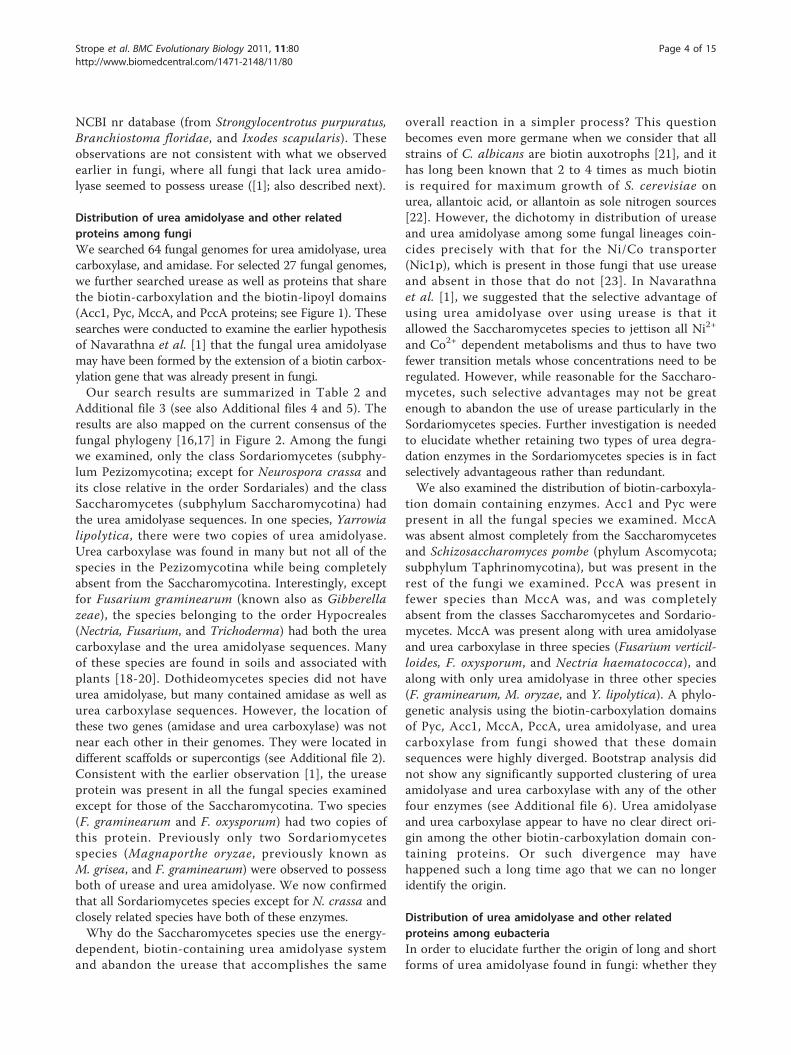
Distribution of urea amidolyase and other relatedproteins among fungiWe searched 64 fungal genomes for urea amidolyase, ureacarboxylase, and amidase. For selected 27 fungal genomes,we further searched urease as well as proteins that sharethe biotin-carboxylation and the biotin-lipoyl domains(Acc1, Pyc, MccA, and PccA proteins; see Figure 1). Thesesearches were conducted to examine the earlier hypothesisof Navarathna et al. [1] that the fungal urea amidolyasemay have been formed by the extension of a biotin carbox-ylation gene that was already present in fungi.Our search results are summarized in Table 2 and
Additional file 3 (see also Additional files 4 and 5). Theresults are also mapped on the current consensus of thefungal phylogeny [16,17] in Figure 2. Among the fungiwe examined, only the class Sordariomycetes (subphy-lum Pezizomycotina; except for Neurospora crassa andits close relative in the order Sordariales) and the classSaccharomycetes (subphylum Saccharomycotina) hadthe urea amidolyase sequences. In one species, Yarrowialipolytica, there were two copies of urea amidolyase.Urea carboxylase was found in many but not all of thespecies in the Pezizomycotina while being completelyabsent from the Saccharomycotina. Interestingly, exceptfor Fusarium graminearum (known also as Gibberellazeae), the species belonging to the order Hypocreales(Nectria, Fusarium, and Trichoderma) had both the ureacarboxylase and the urea amidolyase sequences. Manyof these species are found in soils and associated withplants [18-20]. Dothideomycetes species did not haveurea amidolyase, but many contained amidase as well asurea carboxylase sequences. However, the location ofthese two genes (amidase and urea carboxylase) was notnear each other in their genomes. They were located indifferent scaffolds or supercontigs (see Additional file 2).Consistent with the earlier observation [1], the ureaseprotein was present in all the fungal species examinedexcept for those of the Saccharomycotina. Two species(F. graminearum and F. oxysporum) had two copies ofthis protein. Previously only two Sordariomycetesspecies (Magnaporthe oryzae, previously known asM. grisea, and F. graminearum) were observed to possessboth of urease and urea amidolyase. We now confirmedthat all Sordariomycetes species except for N. crassa andclosely related species have both of these enzymes.Why do the Saccharomycetes species use the energy-
dependent, biotin-containing urea amidolyase systemand abandon the urease that accomplishes the same
overall reaction in a simpler process? This questionbecomes even more germane when we consider that allstrains of C. albicans are biotin auxotrophs [21], and ithas long been known that 2 to 4 times as much biotinis required for maximum growth of S. cerevisiae onurea, allantoic acid, or allantoin as sole nitrogen sources[22]. However, the dichotomy in distribution of ureaseand urea amidolyase among some fungal lineages coin-cides precisely with that for the Ni/Co transporter(Nic1p), which is present in those fungi that use ureaseand absent in those that do not [23]. In Navarathnaet al. [1], we suggested that the selective advantage ofusing urea amidolyase over using urease is that itallowed the Saccharomycetes species to jettison all Ni2+
and Co2+ dependent metabolisms and thus to have twofewer transition metals whose concentrations need to beregulated. However, while reasonable for the Saccharo-mycetes, such selective advantages may not be greatenough to abandon the use of urease particularly in theSordariomycetes species. Further investigation is neededto elucidate whether retaining two types of urea degra-dation enzymes in the Sordariomycetes species is in factselectively advantageous rather than redundant.We also examined the distribution of biotin-carboxyla-
tion domain containing enzymes. Acc1 and Pyc werepresent in all the fungal species we examined. MccAwas absent almost completely from the Saccharomycetesand Schizosaccharomyces pombe (phylum Ascomycota;subphylum Taphrinomycotina), but was present in therest of the fungi we examined. PccA was present infewer species than MccA was, and was completelyabsent from the classes Saccharomycetes and Sordario-mycetes. MccA was present along with urea amidolyaseand urea carboxylase in three species (Fusarium verticil-loides, F. oxysporum, and Nectria haematococca), andalong with only urea amidolyase in three other species(F. graminearum, M. oryzae, and Y. lipolytica). A phylo-genetic analysis using the biotin-carboxylation domainsof Pyc, Acc1, MccA, PccA, urea amidolyase, and ureacarboxylase from fungi showed that these domainsequences were highly diverged. Bootstrap analysis didnot show any significantly supported clustering of ureaamidolyase and urea carboxylase with any of the otherfour enzymes (see Additional file 6). Urea amidolyaseand urea carboxylase appear to have no clear direct ori-gin among the other biotin-carboxylation domain con-taining proteins. Or such divergence may havehappened such a long time ago that we can no longeridentify the origin.
Distribution of urea amidolyase and other relatedproteins among eubacteriaIn order to elucidate further the origin of long and shortforms of urea amidolyase found in fungi: whether they
Strope et al. BMC Evolutionary Biology 2011, 11:80http://www.biomedcentral.com/1471-2148/11/80
Page 4 of 15

share a common evolutionary origin or arose indepen-dently, we performed extensive similarity searches usingthese protein and domain sequences among 56 bacterialgenomes. As shown in Table 3 (see also Additionalfile 7), the longer form of urea amidolyase (~1,800 aa)was found only in one bacterium, Pantoea ananatis(class Gammaproteobacteria). This bacterium, which pre-viously belonged to the genus Erwinia but was recentlyreclassified into the genus Pantoea, is a well-known plantpathogen with a reported case of it also being a human-pathogen [24,25]. This bacterium and its related speciesare usually isolated from soil, fruits, and vegetables [24].Urea carboxylase (~1,200 aa), the shorter form of ureaamidolyase, was found in bacterial species scatteredamong a wide range of groups. Almost all bacteria with
urea carboxylase also had amidase. These two enzymes areencoded in two different genes in bacteria, but are locatednext to each other in most of the bacterial genomes weexamined (see Additional file 8). In two species (Wolinellasuccinogenes, class Epsilonproteobacteria; and Gloeobacterviolaceus, phylum Cyanobacteria), the two genes were notadjacent to each other but only 943 bp and 1,701 bp apart,respectively, while in another Cyanobacteria species (Cya-nothece sp.), the two genes were located far apart (979,743bp). Sorangium cellulosum (class Deltaproteobacteria) andNitrosomonas europaea (class Alphaproteobacteria) hadurea carboxylase but lacked amidase. Three Gammapro-teobacteria species have two urea carboxylase genes, onlyone of which lies next to the amidase gene. P. ananatis, agammaproteobacteria, which has urea amidolyase (the
Table 2 Distribution of urea amidolyase and related proteins in fungal speciesa
Enzymesc
Taxonomical groupb Species UA UC Ad Urease MccA PccA
[Zygomycota] Rhizopus oryzae - - - 1 1 1
[Basidiomycota] Ustilago maydis - - - 1 1 -
Cryptococcus neoformans - 1 - 1 1 -
Coprinus cinereus - - - 1 1 1
[Ascomycota/Taphrinomycotina]
Schizosaccharomycetes Schizosaccharomyces pombe - - - 1 - -
[Ascomycota/Pezizomycotina]
Eurotiomycetes Coccidioides immitis - - - 1 1 1
Aspergillus nidulans - 1 - 1 1 1
Aspergillus fumigatus - 1 - 1 1 1
Aspergillus terreus - 1 - 1 1 1
Aspergillus oryzae - - - 1 1 -
Dothideomycetes Mycosphaerella graminicola - - 1 1 1 1
Stagonospora nodorum - 1 1 1 1 1
Cochliobolus heterostrophus - 1 1 1 1 1
Leotiomycetes Botritis cinerea - - - 1 1 1
Sordariomycetes Neurospora crassa - - - 1 1 -
Magnaporthe oryzae 1 - (1) 1 1 -
Nectria haematococca 1 1 (1) 1 1 -
Fusarium graminearum 1 - (1) 2 1 -
Fusarium oxysporum 1 1 (1) 2 1 -
Fusarium verticillioides 1 1 (1) 1 1 -
[Ascomycota/Saccharomycotina]
Saccharomycetes Yarrowia lipolytica 2 - (2) - 1 -
Candida albicans 1 - (1) - - -
Candida lusitaniae 1 - (1) - - -
Debaryomyces hansenii 1 - (1) - - -
Ashbya gossypii 1 - (1) - - -
Candida glabrata 1 - (1) - - -
Saccharomyces cerevisiae 1 - (1) - - -aSee Additional files 4 and 5 for the sequence sources.bThe phylum/subphylum (in square brackets) and class are given.cSee Figure 1 for the enzyme name abbreviations. The number of sequences found from each genome is shown. ‘-’ indicates that no similar sequence wasfound.dThe amidase sequences that are a part of the urea amidolyase sequences are shown in parentheses.
Strope et al. BMC Evolutionary Biology 2011, 11:80http://www.biomedcentral.com/1471-2148/11/80
Page 5 of 15
long form), also has urea carboxylase (the short form).Furthermore, P. ananatis has no independent amidasegene. The only amidase sequence present in this bacter-ium is the domain of the urea amidolyase gene. It seemsreasonably likely that fusion of the amidase and urea car-boxylase genes occurred in P. ananatis to generate thelong form of the urea amidolyase gene similar to thosefound in fungi.
The urease protein in bacteria occurs as a trimer ofalpha, beta, and gamma subunits encoded by separategenes forming a gene cluster, whereas in eukaryotes asingle gene encodes the urease protein, a fused proteinrepresenting the three bacterial subunits [26]. In somebacteria, beta and gamma subunits are fused andencoded by one gene (denoted with ß/g in Table 3)while in others either beta- or gamma-subunit gene was
Asp. terreusAsp. oryzaeAsp. fumigatusAsp. nidulans
Cocc. immitusCoch. heterotrophusSta. nodorum
F. verticillioidesMyc. graminicola
Mag. oryzaeNec. haematococcaF. graminearum
F. oxysporum
Neu. crassa
B. cinerea
Can. glabrataSac. cerevisiaeAsh. gossypiiD. hanseniiCan. lusitaniae
Can. albicansY. lipolyticaSch. pombe
Cry. neoformans
Cop. cinereusU. maydisR. oryzaeZygomycota
Leotiomycetes
Basidiomycota
Ascomycota
Saccharomycotina
Pezizomycotina
Eurotiomycetes
Dothideomycetes
Sordariomycetes
Taphrinomycotina
270mya
290mya
500mya
630mya
400mya
UAUCMccAPccAUrease
320mya
Figure 2 Distribution of urea amidolyase and related proteins in fungi. Existence of urea amidolyase and four other proteins are mappedalong the current consensus of the fungal phylogeny (summarized from [16,17]). The estimated divergence times (million years ago or mya) aretaken from [39]. Refer to Figure 1 for protein name abbreviations. See Table 2 and Additional file 3 for the complete search results.
Strope et al. BMC Evolutionary Biology 2011, 11:80http://www.biomedcentral.com/1471-2148/11/80
Page 6 of 15

Table 3 Distribution of urea amidolyase and related proteins in eubacterial speciesa
Enzymesb
Phylum or Class Species UA UC Ac Ureased
Alphaproteobacteria Caulobacter crescentus NA1000 - 1 1* -
Asticcacaulis excentricus CB 48 - 1 1* -
Sinorhizobium medicae WSM419 - - - a,b,gBetaproteobacteria Achromobacter piechaudii ATCC 43553 - 1 1* -
Bordetella pertussis Tohama I - - - a,b,gNitrosomonas europaea ATCC 19718 - 1 - -
Neisseria meningitidis FAM18 - - - -
Burkholderia sp. CCGE1001 - 1 1* a,b,gGammaproteobacteria Escherichia coli O111:H- str. 11128 - - - a,b,g
Yersinia pestis Angola - - - a,bHaemophilus influenzae 86-028NP - - - a,b,gPantoea ananatis LMG 20103 1 1 (1) -
Pantoea sp. At-9b - 2 1* -
Shewanella oneidensis MR-1 - - - -
Pseudomonas aeruginosa LESB58 - - - a,b,gCoxiella burnetii Dugway 5J108-111 - - - -
Pectobacterium carotovorum subsp. carotovorum PC1 - 2 1* -
Xanthomonas campestris pv. campestris str. B100 - - - -
Cellvibrio japonicus Ueda107 - 2 1* -
Teredinibacter turnerae T7901 - 1 1* a,b,gMarinomonas sp. MED121 - 1 1* a,gKlebsiella pneumoniae 342 - 1 1* a,b,gPseudomonas fluorescens SBW25 - - - a,b,g
Deltaproteobacteria Geobacter sp. M21 - - - -
Sorangium cellulosum ‘So ce 56’ - 1 - a,b/gEpsilonproteobacteria Helicobacter pylori B38 - - - a,b/g
Wolinella succinogenes DSM 1740 - 1 1+ -
Acidobacteria Acidobacterium capsulatum ATCC 51196 - - - -
Solibacter usitatus Ellin6076 - 1 1* -
Cyanobacteria Synechococcus sp. PCC 7002 - - - a,b,gGloeobacter violaceus PCC 7421 - 1 1+ -
Cyanothece sp. PCC 7425 - 1 1 a,b,gDeinococcus-Thermus Thermus thermophilus HB8 - - - -
Deinococcus deserti VCD115 - - - -
Chloroflexi Dehalococcoides ethenogenes 195 - - - -
Aquificae Aquifex aeolicus VF5 - - - -
Thermotogae Thermotoga maritima MSB8 - - - -
Fusobacteria Fusobacterium nucleatum subsp. Nucleatum ATCC 25586 - - - -
Verrucomicrobia Verrucomicrobium spinosum DSM 4136 - 1 1* a,b,gChlamydiae Chlamydophila pneumoniae CWL029 - - - -
Chlamydia trachomatis B/TZ1A828/OT - - - -
Bacterioidetes Porphyromonas gingivalis W83 - - - -
Chlorobi Chlorobium limicola DSM 245 - - - -
Fibrobacteres Fibrobacter succinogenes subsp. succinogenes S85 - - - -
Actinobacteria Mycobacterium tuberculosis F11 - - - a,b,gCorynebacterium aurimucosum ATCC 700975 - - - -
Streptomyces avermitilis MA-4680 - 1 1* a,b,g; a,b/gBifidobacterium longum subsp. infantis ATCC 15697 - - - a,b/g
Spirochaetes Borrelia burgdorferi ZS7 - - - -
Treponema denticola ATCC 35405 - - - -
Strope et al. BMC Evolutionary Biology 2011, 11:80http://www.biomedcentral.com/1471-2148/11/80
Page 7 of 15

missing. As shown in Table 3, existence of these urease-subunit genes was scattered throughout the bacterialgroups. Of 56 bacterial genomes we examined, 31 hadeither or both of urease and amidase/urea carboxylase(or urea amidolyase). Only seven of 31 bacterial specieshad all three genes. Consistent with what we observedin fungi, there appears to be a certain degree of dichot-omy in possession of urease genes or amidase/urea car-boxylase (or urea amidolyase) genes among bacterialgenomes.
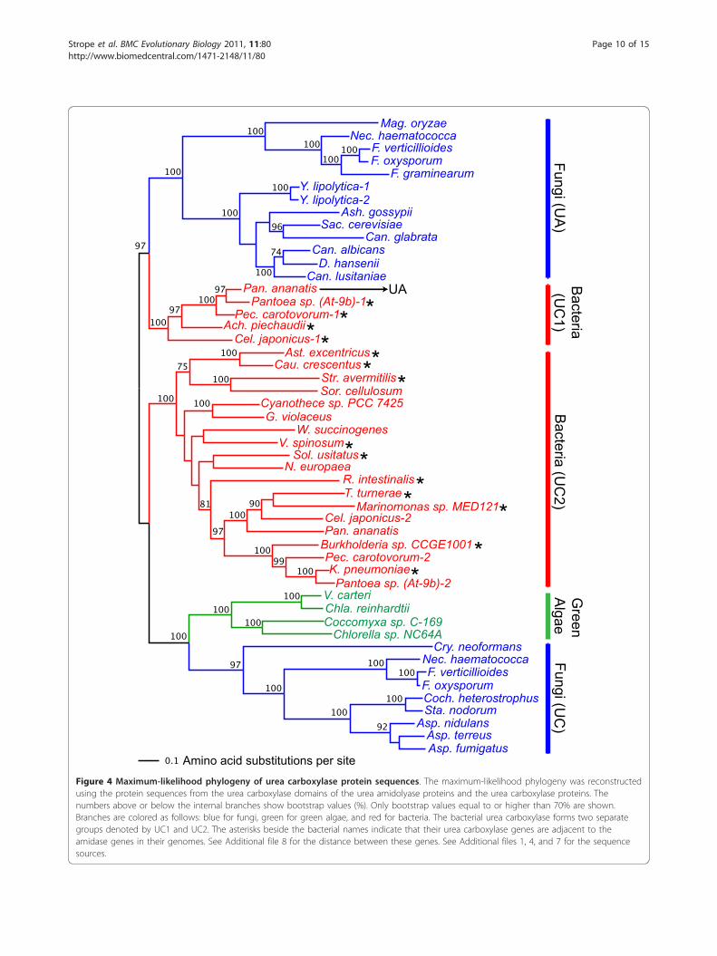
Phylogenetic analysis of amidase domain sequencesIn order to elucidate the evolutionary origin of eukaryo-tic urea amidolyase proteins, we performed phylogeneticanalysis among amidase, urea amidolyase, and urea car-boxylase identified across kingdoms. Phylogenies werereconstructed using amidase and urea carboxylasesequences separately.Figure 3 is the maximum-likelihood phylogenetic tree
reconstructed from the amidase domain sequences fromurea amidolyase and the amidase protein sequencesfrom fungi, green algae, and bacteria (the minimum-evolution tree is shown in Additional file 9). It showsthat the fungal amidase domain from urea amidolyase(shown in blue and denoted by UA in Figure 3), and thestand-alone fungal amidase protein that exists on itsown (shown in blue and denoted by A in Figure 3) clus-ter separately, implying that they have evolved indepen-dently. The amidase sequences from green algae (shownin green in Figure 3) cluster with the stand-alone ami-dase protein from fungi, however, with not very strongbootstrap support (76%).Bacterial amidase sequences also cluster into two
groups (shown in red in Figure 3). Amidases from fourgammaproteobacteria species (P. ananatis, Pantoea sp.At-9b, Pectobacterium carotovorum, and Cellvibrio japo-nicus) and one betaproteobacteria species (Achromobacterpiechaudii) form a cluster (denoted by A1 in Figure 3).Notably, the amidase sequence of P. ananatis is part of
the urea amidolyase, and the amidase genes of the otherthree gammaproteobacteria species lie immediately adja-cent to their urea carboxylase genes (see Additional file8). These bacterial amidases cluster with fungal amidasesfrom urea amidolyase with a strong bootstrap support(100%). Compared to the fungal stand-alone amidases(Fungi A), the fungal amidase-domain sequences fromurea amidolyase (Fungi UA) are clearly more closelyrelated to the bacterial amidases, especially to those fromP. ananatis and a small number of gamma- and betapro-teobacteria species (Bacteria A1).
Phylogenetic analysis of urea carboxylase domainsequencesFigure 4 shows the result of maximum-likelihood phylo-genetic analysis using the urea carboxylase protein andurea carboxylase domain sequences from urea amido-lyase (the minimum-evolution tree is shown in Addi-tional file 10). The urea carboxylase sequence (~1,200aa) is twice longer than the amidase sequence (~600 aa),which resulted in a better resolution in the recon-structed phylogeny. Bacterial urea carboxylase sequenceswere clearly divided into two clusters (denoted by UC1and UC2 in Figure 4) where both were supported by100% bootstrap values. The UC1 group, which consistsof the five species of gamma- and betaproteobacteria(P. ananatis, Pantoea At-9b, P. carotovorum, C. japoni-cus, and A piechaudii), clustered closely with the fungalurea amidolyase (UA) with a high bootstrap value (97%).These five bacterial species are the same five speciesfound in Figure 3 (A1) whose amidases clustered withthe amidase-domain sequences of the fungal ureaamidolyase. Four of these five bacterial species have asecond urea carboxylase gene. Thus, the duplicationevent that created these two sets of urea carboxylasegenes must have happened before the divergence ofthe five proteobacteria. Based on the deep divergencebetween the paralogous groups (UC1 and UC2) and thesomewhat slower evolution observed in UC1 (the urea
Table 3 Distribution of urea amidolyase and related proteins in eubacterial speciesa (Continued)
Planctomycetes Rhodopirellula baltica SH 1 - - - -
Firmicutes Clostridium botulinum A2 str. Kyoto - - - -
Mycoplasma hyopneumoniae 7448 - - - -
Streptococcus pneumoniae 70585 - - - -
Bacillus anthracis str. CDC 684 - - - -
Roseburia intestinalis L1-82 - 1 1* -aSee Additional file 7 for the sequence sources.bSee Figure 1 for the enzyme name abbreviations. The number of sequences found from each genome is shown. ‘-’ indicates that no similar sequence wasfound.cThe amidase gene located next to (within 200 bp) the urea carboxylase gene is indicated with *. The amidase gene located close to (within 6,500 bp) but notnext to the urea carboxylase gene is indicated with +. See Additional file 8 for the distance between the genes. The amidase sequences that are a part of theurea amidolyase sequences are shown in parentheses.dFor urease, the search results for three subunits (a, b, or g) are shown. b/g indicates that the b and g subunits are fused into one gene.
Strope et al. BMC Evolutionary Biology 2011, 11:80http://www.biomedcentral.com/1471-2148/11/80
Page 8 of 15

carboxylase genes found only in five gamma/betaproteo-bacteria species), we speculate that the close functionalassociation with amidase likely arose in the UC1group to create a fused single gene, urea amidolyase, inP. ananatis, and thus changed the evolutionary rate andpattern in this copy of urea carboxylase.
We also see two separate and strongly supported clus-ters of urea carboxylase sequences in fungi. One clusteris of the urea carboxylase domain from urea amidolyase(UA, 100% bootstrap support) whereas the other clusteris of the urea carboxylase protein sequence (UC, 97%bootstrap support). It shows that the urea carboxylase
Y. lipolytica-2Y. lipolytica-1
D. hanseniiCan. albicans
Can. lusitaniaeSac. cerevisiaeCan. glabrata
Ash. gossypiiNec. haematococca
F. graminearumF. oxysporum
F. verticillioidesMag. oryzae
Fungi (UA
)
Pan. ananatis UAPantoea sp. (At-9b)
Pec. carotovorumAch. piechaudii
Cel. japonicus
Bacteria(A
1)
Amino acid substitutions per site
T. turneraeBurkholderia sp. CCGE1001
K. pneumoniaeMarinomonas sp. MED121
R. intestinalisW. succinogenesSol. usitatus
G. violaceusCyanothece sp. PCC 7425
Ast. excentricusCau. crescentus
V. spinosumStr. avermitilis
Bacteria (A
2)
Chlorella sp. NC64ACoccomyxa sp. C-169
Chla. reinhardtiiV. carteri
Green
Algae
Coch. heterostrophusSta. nodorum
Myc. graminicola
Fungi(A
)
Figure 3 Maximum-likelihood phylogeny of amidase protein sequences. The maximum-likelihood phylogeny was reconstructed using theprotein sequences from the amidase domains of the urea amidolyase proteins and the amidase proteins. The numbers above or below theinternal branches show bootstrap values (%). Only bootstrap values equal to or higher than 70% are shown. Branches are colored as follows:blue for fungi, green for green algae, and red for bacteria. The bacterial urea carboxylase forms two separate groups denoted by A1 and A2. SeeAdditional files 1, 4, and 7 for the sequence sources.
Strope et al. BMC Evolutionary Biology 2011, 11:80http://www.biomedcentral.com/1471-2148/11/80
Page 9 of 15
Mag. oryzaeNec. haematococca
F. verticillioidesF. oxysporum
F. graminearumY. lipolytica-1Y. lipolytica-2
Ash. gossypiiSac. cerevisiae
Can. glabrataCan. albicans
D. hanseniiCan. lusitaniae
Pan. ananatis UAPantoea sp. (At-9b)-1*Pec. carotovorum-1*Ach. piechaudii
Cel. japonicus-1* *Ast. excentricus*Cau. crescentus*Str. avermitilis*Sor. cellulosum
N. europaeaSol. usitatus*
R. intestinalis*
V. spinosumW. succinogenes
Cyanothece sp. PCC 7425G. violaceus
T. turneraeMarinomonas sp. MED121
Cel. japonicus-2Pan. ananatis
Burkholderia sp. CCGE1001Pec. carotovorum-2K. pneumoniaePantoea sp. (At-9b)-2
*
**
* *
Chlorella sp. NC64ACoccomyxa sp. C-169Chla. reinhardtiiV. carteri
Cry. neoformans
Coch. heterostrophusSta. nodorum
Asp. nidulansAsp. terreusAsp. fumigatus
F. oxysporumF. verticillioides
Nec. haematococca
Amino acid substitutions per site
Fungi (UA
)B
acteria(U
C1)
Bacteria (U
C2)
Green
Algae
Fungi (UC
)
Figure 4 Maximum-likelihood phylogeny of urea carboxylase protein sequences. The maximum-likelihood phylogeny was reconstructedusing the protein sequences from the urea carboxylase domains of the urea amidolyase proteins and the urea carboxylase proteins. Thenumbers above or below the internal branches show bootstrap values (%). Only bootstrap values equal to or higher than 70% are shown.Branches are colored as follows: blue for fungi, green for green algae, and red for bacteria. The bacterial urea carboxylase forms two separategroups denoted by UC1 and UC2. The asterisks beside the bacterial names indicate that their urea carboxylase genes are adjacent to theamidase genes in their genomes. See Additional file 8 for the distance between these genes. See Additional files 1, 4, and 7 for the sequencesources.
Strope et al. BMC Evolutionary Biology 2011, 11:80http://www.biomedcentral.com/1471-2148/11/80
Page 10 of 15

sequences in the two groups have independently evolvedover a long period of time. Since urea carboxylase wasfound in the phylum Basidiomycota (represented byCryptococcus neoformans in Figure 4) and it clusteredwith other urea carboxylase proteins, the divergencebetween urea carboxylase and urea amidolyase in fungimust have preceded the Ascomycota-Basidiomycotadivergence. As we discuss in the next section, the for-mation of urea amidolyase with acquisition of the ami-dase domain seems to have happened most likely in abacterial lineage. Note that the urea carboxylases fromgreen algae clustered with the fungal urea carboxylases(with 100% bootstrap support) rather than with the fun-gal urea amidolyases. This clustering pattern is consis-tent with what we observed in the amidase phylogeny(Figure 3) where green algal genes clustered with thestand-alone version of the fungal amidase genes ratherthan with the amidase-domain sequence of urea amido-lyase. Although in some green algae, amidase and ureacarboxylase genes are located relatively closely (within588 to 6,236 bp; Additional file 2), their evolution iscompletely independent from urea amidolyase genesfound in fungi.As mentioned before, two Hydra urea carboxylase
sequences were found from the NCBI nr databasesearch. One of them was actually found to be asequence of a putative bacterial symbiont, Curvibacter(betaproteobacteria) (described in NCBI gi|260221606entry). Phylogenetic analysis clearly showed that thissequence belongs to the bacterial urea carboxylase(UC2) group (see Additional file 11). The other Hydrasequence clustered with urea carboxylase sequencesfrom green algae and fungi (93% bootstrap support).
Bacterial origins of the fungal urea amidolyase and ureacarboxylaseOur phylogenetic analysis did not support the previoushypothesis that the fungal urea amidolyase and urea car-boxylase sequences are formed from fungal biotin-carboxylation domain containing proteins such as MccAor PccA. Instead, our conclusion is that the urea amido-lyase and urea carboxylase genes currently found in fungiand green algae, as well as in Hydra, are the results of hor-izontal gene transfer events from bacteria. This is basedon observations such as the abundant distribution of theshorter form of urea amidolyase, i.e., urea carboxylase, aswell as the single occurrence so far of urea amidolyase(the long form) in bacteria, coupled with the rarity of bothforms of urea amidolyase in eukaryotes except in the fun-gal kingdom, in some green algae, and in Hydra.Phylogenetic analysis of amidase and urea carboxylase
sequences across kingdoms showed that the urea car-boxylase domain in urea amidolyase and the urea
carboxylase protein itself have undergone extensiveindependent evolution. Fungal urea amidolyase proteinsare more closely related to one of the two groups ofbacterial urea carboxylase. Furthermore, one of thesebacteria (P. ananatis) has a unique urea amidolyasegene, a product of amidase/urea carboxylase genefusion. The direction of the horizontal gene transferseems to be from a bacterial lineage to a fungal lineage,since in bacteria other than P. ananatis, urea carboxy-lase and amidase exist as two independent genesalthough they are located next to each other. Inspectionof introns in fungal urea amidolyase genes corroboratesthis hypothesis further. Fungal urea amidolyases areeither single or double-exon genes (see Additionalfile 2). All Saccharomycetes species except for Y. lipoly-tica have single-exon urea amidolyase genes. While inthe three Sordariomyetes species (M. oryzae, N. haema-tococca, and F. graminearum) the single intron wasinserted towards the end of the urea carboxylasedomain, in the duplicated Y. lipolytica genes the singleintron was inserted at the beginning of the amidasedomain. These observations indicate that the introns inthese fungal urea amidolyase genes must have beenacquired independently during their evolution as fungalgenes. Therefore, fusion of the two genes appears tohave happened in the ancestral bacterial species close toP. ananatis, and this fused gene must have been trans-ferred to a fungal lineage.Since so far we found the urea amidolyase protein
only in one bacterial species, it is probable that thefusion of urea carboxylase and amidase to form bacterialurea amidolyase is a recent event specific to this bacter-ial lineage. If this is the case, the fusion event in P. ana-natis could be also independent from those thatproduced fungal urea amidolyases. However, we did notfind any unfused fungal urea carboxylase sequencesclustered with urea amidolyase in our phylogeneticanalysis (Figure 4), nor did we find any unfused fungalamidase sequences clustered with urea amidolyase(Figure 3). Therefore, if the fusion happened in fungallineage, it must have happened soon after the two bac-terial genes (amidase and urea carboxylase) wereacquired by an ancestral fungal species. Regardless ofthe timing of the fusion event, association between theamidase and urea carboxylase sequences for the ureaamidolyase function and subsequent divergence of thesesequences from the other paralogous set must havestarted in bacterial lineage.Compared to urea amidolyase, urea carboxylase genes
in fungi have a wider range in the number of exons, 1-16exons, implying again their independent evolution as wellas a greater number of accumulated changes. Note thatthe single introns found in the urea carboxylase genes of
Strope et al. BMC Evolutionary Biology 2011, 11:80http://www.biomedcentral.com/1471-2148/11/80
Page 11 of 15

N. haematococca and F. oxysporum are both at the begin-ning of the genes and of similar lengths (55-56 bp; seeAdditional file 2). It indicates that the common ancestorof these species acquired a single intron in the urea car-boxylase gene and it happened independently from theintron acquisition in N. haematococca urea amidolyase.Interestingly, the number of introns in urea carboxylaseand amidase genes in green algae is much higher thanthe number of introns in the fungal orthologues. This isin agreement with the observation that the Chlamydomo-nas reinhardtii genome has much higher percentage ofgenes with introns and a much greater number of exonsper gene (88% and 7.4) as compared to S. cerevisiae (5%and 1) and S. pombe (43% and 2) [27].There have been studies presenting cases of bacteria-to-
fungi horizontal gene transfers. For example, Hall et al.[11] found ten potential such cases in S. cerevisiae and onein Ashbya gossypii. Fitzpatrick et al. [12] reported twoCandida parapsilosis genes as bacterial origin. Garcia-Vallvé et al. [13] showed that many glycosyl hydrolasegenes in the rumen fungus Orpinomyces joyonii wereacquired from bacteria. Schmitt and Lumbsch [14] showedthat the polyketide synthase in lichen-forming fungi wereresults of ancient horizontal gene transfer from Actinobac-teria. A recent study, the largest of its kind, by Marcet-Houben and Gabaldón [15] detected 713 transferred genesin 60 fungal genomes. Therefore, horizontal gene transfersfrom bacteria to fungi do not appear to be rare events. Weidentified yet another such example.
Proposed model for the urea carboxylase and ureaamidolyase evolutionFigure 5 illustrates our proposed model for the evolu-tion of urea carboxylase and urea amidolyase genes infungi. As presented in Figure 5A, an ancestral urea car-boxylase sequence in bacteria duplicated in the beta/gammaproteobacteria lineage and evolved into twogenes (UC1 and UC2). Since in many bacterial genomes,urea carboxylase and amidase genes are located adjacentto each other (see Additional file 8), it is plausible thatbefore the duplication, the ancestral urea carboxylasegene already had an associated function with the ami-dase gene. However, the creation of duplicated redun-dant copies of the urea carboxylase gene in beta/gammaproteobacteria species appears to have reinforcedthe association between the two genes and changedtheir evolutionary pattern and rate in these bacteria.This amidase-associated copy of bacterial urea carboxy-lase gene (UC1) was subsequently fused with the ami-dase gene to form a single urea amidolyase gene. Thefused gene was later transferred to an ancestral ascomy-cete lineage before the divergence of the Pezizomycotinaand Saccharomycotina. Alternatively, the gene fusioncould have happened in an ancestral fungal species soon
after the region containing amidase and urea carboxy-lase genes was transferred from bacteria.The other bacterial urea carboxylase gene (UC2) may
have also been acquired by fungi, green algae, as well asHydra. Since our phylogenetic analysis did not showindependent origins for these urea carboxylase genes, theacquisition of this enzyme into fungi, green algae, andHydra must have happened around the time of diver-gence among these groups of organisms. It may havebeen by a single event, likely before the divergence ofthese organisms. Then we cannot eliminate the possibi-lity that what we observed in the urea carboxylase genesis the result of simple vertical evolution from bacteria toeukaryotes. Either way, however, many eukaryotesincluding the entire metazoa and land plants must havelost these genes. As we mentioned before (and shownalso in Figure 5B), even within fungi, the urea carboxylasegene is not retained in many species. Considering thateither scenario requires such a high number of lossevents, there would be other possible scenarios. Onegroup of organisms (either green algae, Hydra, or fungi)may have acquired a urea carboxylase gene from bacteriafirst. Later this gene may have been transferred to otherorganisms. Although this scenario requires fewer lossevents, the main question is how such horizontal genetransfers can happen between green algae, Hydra, andfungi, or among any of their ancestral organisms.In fungi, the introduction of the urea carboxylase gene
happened earlier than that of the urea amidolyase gene asshown in Figure 5B. The urea carboxylase gene (red cir-cle) was acquired in fungi before the divergence of thephyla Ascomycota and Basidiomycota. The acquisitioncould have been after the divergence of the phylumZygomycota or alternatively the gene was lost from theZygomycota lineage. Some Basidiomycota species subse-quently lost the gene (the lost events are indicated withgrey symbols in Figure 5B). In the phylum Ascomycota,this gene was again lost in the subphyla Taphrinomyco-tina (it includes S. pombe) and Saccharomycotina.Further losses of this gene happened in some species ofthe subphylum Pezizomycotina. The urea carboxylasegene appears to become easily dispensable in many spe-cies, which may be related to the genomic and metabolicenvironment of the organisms. The same seems to be thecase with MccA and PccA. The introduction of the ureaamidolyase gene (black square) in fungi took place beforethe divergence of the subphyla Pezizomycotina and Sac-charomycotina but probably after the divergence of thesubphylum Taphrinomycotina (at least after the phylumAscomycota diverged from the ancestral lineage). Withinthe subphylum Pezizomycotina, the urea amidolyase genewas lost in many groups but retained in almost all speciesin the class Sordariomycetes (absent in the order Sordar-iales species). The urea amidolyase gene was retained in
Strope et al. BMC Evolutionary Biology 2011, 11:80http://www.biomedcentral.com/1471-2148/11/80
Page 12 of 15

Asp. terreusAsp. oryzaeAsp. fumigatusAsp. nidulans
Cocc. immitusCoch. heterotrophusSta. nodorum
F. verticillioidesMyc. graminicola
Mag. oryzaeNec. haematococcaF. graminearum
F. oxysporum
Neu. crassa
B. cinerea
Can. glabrataSac. cerevisiaeAsh. gossypiiD. hanseniiCan. lusitaniae
Can. albicansY. lipolyticaSch. pombe
Cry. neoformans
Cop. cinereusU. maydisR. oryzaeZygomycota
Leotiomycetes
Basidiomycota
Ascomycota
Saccharomycotina
Pezizomycotina
Eurotiomycetes
Dothideomycetes
Sordariomycetes
Taphrinomycotina
270mya
290mya
500mya
630mya
400mya
UAUCMccAPccAUrease
AncestralUC Fungal UC
Fungal UA(A)
(B)
Green Algal UCBacterial UC2
320mya
+UC1 AmidaseBeta/gammaproteobacteria
Figure 5 Evolutionary model of urea carboxylase and urea amidolyase in fungi. (A) The evolution of the two types of bacterial ureacarboxylases, UC1 and UC2, and the subsequent transfer of those genes to fungi and green algae. The arrows represent possible horizontalgene-transfer events. Dashed arrows indicate that both horizontal transfer and vertical transmission are possible. (B) Acquisition and loss eventsof the urea amidolyase and related proteins inferred within fungal evolution. The fungal consensus phylogeny and the presence/absence tablefor five proteins are the same as Figure 2. Within the tree, the colored symbols indicate gene-acquisition events while the grey symbols indicatethe deletion of that gene.
Strope et al. BMC Evolutionary Biology 2011, 11:80http://www.biomedcentral.com/1471-2148/11/80
Page 13 of 15

all Saccharomycotina species, and even recently dupli-cated in Y. lipolytica.
ConclusionsWe have presented a possible scenario of horizontalgene transfer of the urea amidolyase and urea carboxy-lase genes from bacteria to fungi. Plastic and opportu-nistic genome evolution in bacteria and fungi and theirevolutionary interplay must have allowed the Saccharo-mycetes fungi to abandon the use of nickel-containingurease. It contributed to optimizing these organismstoward Ni2+ (and Co2+)-independent cellular metabo-lisms. Further detailed studies of a wider range of genefamilies would reveal the importance of acquisition ofbacterial genes in fungal evolution.
MethodsSimilarity searchesSimilarity searches for protein sequences were per-formed using blastp (version 2.2.17 [28]). For urea ami-dolyase search, the S. cerevisiae sequence (P32528) wasused as a query. Search was performed using both thefull sequence as well as only the amidase domain of thissequence. To search for urea carboxylase sequences,A. nidulans sequence (P38095) was used as a query. Tosearch for other urea carboxylase domain containingproteins, the S. cerevisiae Acc1 (Q00955) and Pyc(P11154), A. nidulans MccA (Q6T5L7), and Aspergillusrelated Neosartorya fischeri PccA (A1DF70) were usedas query sequences. The urease sequence from A. fumi-gatus (Q6A3P9) was used as a query sequence to searchfor urease.We performed these searches against 56 bacterial gen-
omes, 64 fungal genomes, and 10 non-fungal eukaryoticgenomes (including 4 green algae, 2 land plants, 1amoebozoa, and 3 animals). The species names, taxono-mical groups, and the sources of the sequences are listedin Additional files 1, 4, 5, and 7. The species were cho-sen such that all major bacterial, fungal, and othereukaryotic groups were represented from a tree of life[e.g., [29]]. For fungi, preliminary search for urea amido-lyase, urea carboxylase, and amidase was done in 64genomes and further analysis was done using 27selected fungal genomes (noted with * in Additional file3). The non-redundant (nr) database at National Centerfor Biotechnology Information (NCBI) was also searchedfor urea amidolyase, urea carboxylase, and urease pro-tein sequences using blastp.All protein sequences were highly conserved, and
similar sequences were clearly identifiable in the resultsobtained by blastp similarity search. The E-value thresh-old for each protein hit was as follows: 1 × 10-49 foramidase, 0 for urea amidolyase and urea carboxylase, 1× 10-12 for urease, 1 × 10-111 for MccA, 1 × 10-115 for
PccA, and 0 for Pyc and Acc1. The default parameterswere used with blastp program (version 2.2.17), whichinclude BLOSUM62 scoring matrix, low-complexity fil-tering, gap-open and gap-extend penalties of 11 and 1,respectively. In order to obtain the E-values comparableamong different genome sizes, the “effective length ofdatabase” was set to 500,000,000 (using -z option). Thisalso makes the E-values obtained from each genomesearch equivalent to those obtained against NCBI nrdatabase.
Multiple alignment and phylogenetic analysisMultiple alignments of protein sequences were generatedusing MAFFT (version 6.240 [30]) with default parameters(FFT-NS-2, a progressive FFT alignment with two tree-building cycles). The maximum-likelihood phylogeny [31]was reconstructed as implemented in raxmlHPC-MPI (ver-sion 7.0.4 [32]) using the following options: ‘-m PROTMIX-WAG’ to use WAG amino-acid substitution model [33]with a fixed number approximation followed by a refinedgamma-model of rate heterogeneity, ‘-f a’ for a rapid boot-strap analysis, ‘-x 1234’ to set the random seed, and ‘-# 1000’for 1000 pseudoreplicates for bootstrap analysis. To gatherthe bootstrap values, the ‘consense’ program of the Phylippackage (v. 3.68 [34]) was used. The minimum-evolution phylogeny [35] was reconstructed as implementedin MEGA4 [36] using the JTT amino-acid substitutionmodel [37] with 1000 pseudoreplicates for bootstrapanalysis.
Additional material
Additional file 1: Sequence sources for the non-fungal eukaryoticsequences used in this study.
Additional file 2: Number of exons in urea amidolyase and relatedgenes and their distance in eukaryotic genomes.
Additional file 3: Distribution of urea amidolyase, urea carboxylase,and amidase proteins in 64 fungal species.
Additional file 4: Sequence sources of the urea amidolyase, ureacarboxylase, and amidase from 64 fungal species.
Additional file 5: Sequence sources of the urease, methylcrotonoyl-CoA carboxylase, and propionyl-CoA carboxylase from the selected27 fungal species.
Additional file 6: Maximum-likelihood phylogeny of thecarboxylation-domain sequences from urea carboxylase, ureaamidolyase, methylcrotonoyl-CoA carboxylase, propionyl-CoAcarboxylase, pyruvate carboxylase, and acetyl-CoA carboxylase.
Additional file 7: Sequence sources of urea amidolyase, ureacarboxylase, and amidase in eubacterial genomes.
Additional file 8: Distance between amidase and urea carboxylasegenes in eubacterial genomes.
Additional file 9: Minimum-evolution phylogeny of amidase proteinsequences.
Additional file 10: Minimum evolution phylogeny of ureacarboxylase protein sequences.
Additional file 11: Maximum-likelihood phylogeny of ureacarboxylase protein sequences including the two Hydra sequences.
Strope et al. BMC Evolutionary Biology 2011, 11:80http://www.biomedcentral.com/1471-2148/11/80
Page 14 of 15
AcknowledgementsWe thank Catherine Anderson (UNL) for her help on the use of the localfungal genome database. This work was in part funded by the NationalLibrary of Medicine grant (R01LM009219) to E.N.M. and by the NationalScience Foundation grant (BDI-0743783) to E.N.M. and S.D.H.
Author details1School of Biological Sciences, University of Nebraska, Lincoln, NE 68588,USA. 2Department of Plant Pathology, University of Nebraska, Lincoln, NE68588, USA. 3Center for Plant Science Innovation, University of Nebraska,Lincoln, NE 68588, USA.
Authors’ contributionsPKS collected data, carried out all the analyses, and drafted the manuscript.KWN and SDH conceived of the study, contributed the discussion, andrevised the manuscript. ENM supervised the entire process of the study,contributed the discussion, and revised the manuscript. All authors read andapproved the final manuscript.
Received: 20 September 2010 Accepted: 29 March 2011Published: 29 March 2011
References1. Navarathna DH, Harris SD, Roberts DD, Nickerson KW: Evolutionary aspects
of urea utilization by fungi. FEMS Yeast Res 2010, 10(2):209-213.2. Ghosh S, Navarathna DH, Roberts DD, Cooper JT, Atkin AL, Petro TM,
Nickerson KW: Arginine-induced germ tube formation in Candidaalbicans is essential for escape from murine macrophage line RAW264.7. Infect Immun 2009, 77(4):1596-1605.
3. Roon RJ, Levenberg B: Urea amidolyase. I. Properties of the enzyme fromCandida utilis. J Biol Chem 1972, 247(13):4107-4113.
4. Leftley JW, Syrett PJ: Urease and ATP:urea amidolyase activity inunicellular algae. J Gen Microbio 1973, 77.
5. Al-Houty FAA, Syrett PJ: The occurrence of urease/urea amidolyase andglycollate oxidase/dehydrogenase in Klebsormidium spp. and membersof the Ulotrichales. European J of Phycology 1984, 19:1-10.
6. Roon RJ, Hampshire J, Levenberg B: Urea amidolyase. The involvement ofbiotin in urea cleavage. J Biol Chem 1972, 247(23):7539-7545.
7. Cooper TG, Lam C, Turoscy V: Structural analysis of the dur loci in S.cerevisiae: two domains of a single multifunctional gene. Genetics 1980,94(3):555-580.
8. Hodson RC, Williams SK, Davidson WR Jr: Metabolic control of ureacatabolism in Chlamydomonas reinhardi and Chlorella pyrenoidosa.J Bacteriol 1975, 121(3):1022-1035.
9. Kanamori T, Kanou N, Atomi H, Imanaka T: Enzymatic characterization of aprokaryotic urea carboxylase. J Bacteriol 2004, 186(9):2532-2539.
10. Jitrapakdee S, Wallace JC: The biotin enzyme family: conserved structuralmotifs and domain rearrangements. Curr Protein Pept Sci 2003,4(3):217-229.
11. Hall C, Brachat S, Dietrich FS: Contribution of horizontal gene transfer tothe evolution of Saccharomyces cerevisiae. Eukaryot Cell 2005,4(6):1102-1115.
12. Fitzpatrick DA, Logue ME, Butler G: Evidence of recent interkingdomhorizontal gene transfer between bacteria and Candida parapsilosis.BMC Evol Biol 2008, 8:181.
13. Garcia-Vallve S, Romeu A, Palau J: Horizontal gene transfer of glycosylhydrolases of the rumen fungi. Mol Biol Evol 2000, 17(3):352-361.
14. Schmitt I, Lumbsch HT: Ancient horizontal gene transfer from bacteriaenhances biosynthetic capabilities of fungi. PLoS One 2009, 4(2):e4437.
15. Marcet-Houben M, Gabaldon T: Acquisition of prokaryotic genes byfungal genomes. Trends Genet 2010, 26(1):5-8.
16. Marcet-Houben M, Gabaldon T: The tree versus the forest: the fungal treeof life and the topological diversity within the yeast phylome. PLoS One2009, 4(2):e4357.
17. Wang H, Xu Z, Gao L, Hao B: A fungal phylogeny based on 82 completegenomes using the composition vector method. BMC Evol Biol 2009,9:195.
18. Oren L, Ezrati S, Cohen D, Sharon A: Early events in the Fusariumverticillioides-maize interaction characterized by using a greenfluorescent protein-expressing transgenic isolate. Appl Environ Microbiol2003, 69(3):1695-1701.
19. Enya J, Togawa M, Takeuchi T, Yoshida S, Tsushima S, Arie T, Sakai T:Biological and phylogenetic characterization of Fusarium oxysporumcomplex, which causes yellows on Brassica spp., and proposal of F.oxysporum f. sp. rapae, a novel forma specialis pathogenic on B. rapa inJapan. Phytopathology 2008, 98(4):475-483.
20. Gunawardena U, Rodriguez M, Straney D, Romeo JT, VanEtten HD,Hawes MC: Tissue-specific localization of pea root infection by Nectriahaematococca. Mechanisms and consequences. Plant Physiol 2005,137(4):1363-1374.
21. Odds FC: Candida and candidiasis. Bailliere: Tindall; 1988.22. DiCarlo FJ, Schultz AS, Kent AM: The mechanism of allantoin catabolism
by yeast. Arch Biochem Biophys 1953, 44:468-474.23. Zhang Y, Rodionov DA, Gelfand MS, Gladyshev VN: Comparative genomic
analyses of nickel, cobalt and vitamin B12 utilization. BMC Genomics2009, 10:78.
24. De Baere T, Verhelst R, Labit C, Verschraegen G, Wauters G, Claeys G,Vaneechoutte M: Bacteremic infection with Pantoea ananatis. J ClinMicrobiol 2004, 42(9):4393-4395.
25. De Maayer P, Chan WY, Venter SN, Toth IK, Birch PR, Joubert F,Coutinho TA: Genome sequence of Pantoea ananatis LMG20103, thecausative agent of Eucalyptus blight and dieback. J Bacteriol 2010,192(11):2936-2937.
26. Carter EL, Flugga N, Boer JL, Mulrooney SB, Hausinger RP: Interplay ofmetal ions and urease. Metallomics 2009, 1(3):207-221.
27. Labadorf A, Link A, Rogers MF, Thomas J, Reddy AS, Ben-Hur A: Genome-wide analysis of alternative splicing in Chlamydomonas reinhardtii. BMCGenomics 2010, 11:114.
28. Altschul SF, Madden TL, Schaffer AA, Zhang J, Zhang Z, Miller W,Lipman DJ: Gapped BLAST and PSI-BLAST: a new generation of proteindatabase search programs. Nucleic Acids Res 1997, 25(17):3389-3402.
29. Ciccarelli FD, Doerks T, von Mering C, Creevey CJ, Snel B, Bork P: Towardautomatic reconstruction of a highly resolved tree of life. Science 2006,311(5765):1283-1287.
30. Katoh K, Misawa K, Kuma K, Miyata T: MAFFT: a novel method for rapidmultiple sequence alignment based on fast Fourier transform. NucleicAcids Res 2002, 30(14):3059-3066.
31. Felsenstein J: Evolutionary trees from DNA sequences: a maximumlikelihood approach. J Mol Evol 1981, 17(6):368-376.
32. Stamatakis A: RAxML-VI-HPC: maximum likelihood-based phylogeneticanalyses with thousands of taxa and mixed models. Bioinformatics 2006,22(21):2688-2690.
33. Whelan S, Goldman N: A general empirical model of protein evolutionderived from multiple protein families using a maximum-likelihoodapproach. Mol Biol Evol 2001, 18(5):691-699.
34. Felsenstein J: Phylogeny Inference Package (Version 3.2). Cladistics 1989,5:164-166.
35. Rzhetsky A, Nei M: Statistical properties of the ordinary least-squares,generalized least-squares, and minimum-evolution methods ofphylogenetic inference. J Mol Evol 1992, 35(4):367-375.
36. Tamura K, Dudley J, Nei M, Kumar S: MEGA4: Molecular EvolutionaryGenetics Analysis (MEGA) software version 4.0. Mol Biol Evol 2007,24(8):1596-1599.
37. Jones DT, Taylor WR, Thornton JM: The rapid generation of mutation datamatrices from protein sequences. Comput Appl Biosci 1992, 8(3):275-282.
38. Hunter S, Apweiler R, Attwood TK, Bairoch A, Bateman A, Binns D, Bork P,Das U, Daugherty L, Duquenne L, et al: InterPro: the integrative proteinsignature database. Nucleic Acids Res 2009, , 37 Database: D211-215.
39. Lucking R, Huhndorf S, Pfister DH, Plata ER, Lumbsch HT: Fungi evolvedright on track. Mycologia 2009, 101(6):810-822.
doi:10.1186/1471-2148-11-80Cite this article as: Strope et al.: Molecular evolution of urea amidolyaseand urea carboxylase in fungi. BMC Evolutionary Biology 2011 11:80.
Strope et al. BMC Evolutionary Biology 2011, 11:80http://www.biomedcentral.com/1471-2148/11/80
Page 15 of 15