molecular drug imaging: 89zr-bevacizumab pet in children with
TRANSCRIPT
Molecular Drug Imaging:
89Zr-bevacizumab PET in Children with Diffuse Intrinsic Pontine Glioma
- 89Zr-bevacizumab PET in DIPG –
Marc H Jansen1,i and Sophie EM Veldhuijzen van Zanten1,i, Dannis G van Vuurden1, Marc C
Huisman2, Danielle J Vugts2, Otto S Hoekstra2, Guus A van Dongen2, Gert-Jan L Kaspers1
iEqual contribution
1 Department of Pediatrics, Pediatric Oncology/Hematology, VU University Medical Center,
Amsterdam, The Netherlands.
2 Department of Radiology & Nuclear Medicine, VU University Medical Center, Amsterdam, The
Netherlands.
First authors:
M.H.A. Jansen, MD, PhD
Pediatric Oncology/Hematology, VU University Medical Center
De Boelelaan 1117, Room KTC4.027, Amsterdam, NL 1081 HV
Phone: +31 20 4446201 Fax: +31 20 44 42422 E-mail: [email protected]
S.E.M. Veldhuijzen van Zanten, MD, PhD candidate
Pediatric Oncology/Hematology, VU University Medical Center
De Boelelaan 1117, Room KTC4.027, Amsterdam, NL 1081 HV
Phone: +31 20 4446201 Fax: +31 20 44 42422 E-mail: [email protected]
Journal of Nuclear Medicine, published on October 20, 2016 as doi:10.2967/jnumed.116.180216by on April 2, 2018. For personal use only. jnm.snmjournals.org Downloaded from
2
Corresponding author:
Prof. Dr. G.A.M.S. van Dongen, MD, PhD
Department of Radiology & Nuclear Medicine, VU University Medical Center
De Boelelaan 1118, Amsterdam, NL 1081 HV
Phone: +31 20 44 42869 Fax: +31 20 44 43688 Email: [email protected]
Abstract:
292 words
Manuscript:
4516 words
Role of the founding source:
This DIPG study is funded by the Semmy Foundation (Stichting Semmy) and the Egbers
Foundation (Egbers Stichting).
by on April 2, 2018. For personal use only. jnm.snmjournals.org Downloaded from
3
ABSTRACT
Predictive tools to guide therapy in children with brain tumors are urgently needed. We
introduced molecular imaging to facilitate this. We investigated whether bevacizumab can reach
the tumor in children with diffuse intrinsic pontine glioma (DIPG) by measuring the tumor uptake
of zirconium-89(89Zr)-labeled bevacizumab by PET. In addition we evaluated the safety of the
procedure in children and determined the optimal timing of imaging.
Methods
Patients received 0.1 mg/kg (0.9 MBq/kg) 89Zr-bevacizumab, ≥ two weeks after completing
radiotherapy. Whole body PET-CT scans were performed 1, 72 and 144 hours post-injection
(p.i.). All patients underwent contrast (gadolinium)-enhanced MRI. Biodistribution of 89Zr-
bevacizumab was quantified as Standardized Uptake Values (SUVs).
Results
We included seven DIPG patients (4 males, age range 6-17 years) who were scanned without
anesthesia. No adverse events occurred. Five out of seven primary tumors showed focal 89Zr-
bevacizumab uptake (SUVs range 1.0-6.7 at 144 hours p.i.), while there was no significant
uptake in healthy brain. One patient had multiple metastases, which were all PET positive. There
was inter- and intra-tumoral heterogeneity of uptake and 89Zr-bevacizumab was predominantly
(in 4 out of 5 patients) present within the MRI contrast-enhancing areas, although 89Zr-
bevacizumab uptake was variable among MRI contrast-enhancing areas. Tumor targeting was
quantitatively similar at 72 and 144 hours post-injection, but tumor-to-blood SUV ratios increased
over post-injection time (p=0.045). The mean effective dose per patient was 0.9±0.3 mSv/MBq.
Conclusion
by on April 2, 2018. For personal use only. jnm.snmjournals.org Downloaded from
4
89Zr-bevacizumab PET-studies are feasible in children with DIPG. The data suggest
considerable heterogeneity in drug delivery between patients and within DIPG tumors and a
positive, but not 1-to-1, correlation between MRI contrast-enhancement and 89Zr-bevacizumab
uptake. The optimal moment of scanning is 144 hours p.i.. Tumor 89Zr-bevacizumab
accumulation as assessed by PET scanning might help to select patients with highest chance of
benefit from bevacizumab treatment.
Keywords: Brainstem neoplasm, Positron Emission Tomography, 89Zr-bevacizumab,
pharmacokinetics.
by on April 2, 2018. For personal use only. jnm.snmjournals.org Downloaded from
5
INTRODUCTION
The need for predictors to guide therapy in the individual patient applies to the entire field of
pediatric oncology but in particular to malignant brain tumors. One of the most challenging brain
tumors in children is DIPG, a lethal childhood malignancy of the brainstem comprising 10% of all
pediatric central nervous system tumors (1). DIPG tumors are resistant to all kinds of systemic
therapies, including targeted agents, and hardly any patient survives beyond two years from
diagnosis (2,3). One hypothesis of therapy failure is that drugs actually do not reach the tumor,
as in most DIPGs at diagnosis MRI shows little or no gadolinium-contrast enhancement, which
suggests an intact blood-brain barrier for large molecules (4). Molecular PET imaging could help
to investigate this hypothesis, but despite a recent boost of molecular drug imaging in adults,
thus far no immuno-PET imaging studies have been performed in children (5).
Recent mRNA profiling studies revealed overexpression of the pro-angiogenic vascular
endothelial growth factor (VEGF-A) in DIPG compared to normal brain and compared to adult
high grade glioma, which makes VEGF-A a potential drug target (6,7). The biologic activity of
VEGF can be neutralized by bevacizumab, a recombinant humanized monoclonal antibody.
Several trials are ongoing in which the potential of bevacizumab is being studied in DIPG
(NCT01182350; clinicaltrials.gov, NTR2391; Trialregister.nl) and few trials have been published
(8-10). The overall survival outcome in these DIPG trials was as poor as in historical controls
(8,9); however, individual patients with significant responses and prolonged survival after
bevacizumab treatment have been reported (10,11). Hence, the challenge is to identify those
patients who will benefit from bevacizumab treatment. Zirconium-89 (89Zr) labeled bevacizumab
PET imaging might help assessing inter- and intrapatient heterogeneity of drug biodistribution in
vivo, hereby predicting (absence of) response in patients subsequently treated with
bevacizumab. Studies in both mice and adult patients confirmed that 89Zr-bevacizumab PET
by on April 2, 2018. For personal use only. jnm.snmjournals.org Downloaded from
6
imaging is feasible and able to show bevacizumab accumulation in VEGF-positive tumors (7,12-
14). In addition, in adult renal cancer tumors, 89Zr-bevacizumab uptake was correlated to
response to bevacizumab treatment (13). Results from our 89Zr-bevacizumab preclinical
research in mice suggest poor bevacizumab uptake in intracranial tumors; in three different
pontine-, striatal- and subcutaneous glioma mouse models we found no significant uptake of
89Zr-bevacizumab in the intracranial tumors at any stage of the disease, nor in the normal non-
neoplastic surrounding brain in any of the tumor models used (7). In contrast, high accumulation
of 89Zr–bevacizumab was observed in the subcutaneous glioma xenograft. Poor bevacizumab
distribution in the brain therefore seemed to play a major issue, however we also observed a
significantly lower level of VEGF expression in the intracranial tumors (and in one intracranial
tumor even absence) compared to the subcutaneous glioma xenograft.
As a first step to investigate the clinical utility of immuno-PET in this setting, we
performed a pilot study to investigate whether bevacizumab can reach the tumor in children with
DIPG by measuring the tumor uptake of 89Zr-labeled bevacizumab.In addition, we assessed the
optimal moment of PET imaging, biodistribution and radiation dosimetry of 89Zr-bevacizumab,
and ascertained safety and tolerability of the scanning procedures without anesthesia.
MATERIAL AND METHODS
Study population
The study was conducted at VU University Medical Center, Amsterdam. DIPG patients,
aged between 4 and 18 years, were eligible. All patients were included at least 2 weeks after
completing radiotherapy, the standard DIPG therapy. Patients with known hypersensitivity
against humanized monoclonal antibodies were excluded, as well as those previously treated
with bevacizumab or any other anti-VEGF agent. To reduce patient burden, PET-related blood
by on April 2, 2018. For personal use only. jnm.snmjournals.org Downloaded from
7
sampling and the use of anesthetics were not allowed. The latter determined the minimum age
of 4 years, as children under 4 were not expected to complete a PET scan without movement
artifacts without the use of anesthesia. For all subjects, both parents signed a written informed
consent form. In addition, all subjects aged 12 years and older signed a written “informed
assent” form. The study (registered as NTR3518 in The Netherlands National Trial Register -
NTR) was approved by the Dutch Central Committee on Research involving Human Subjects
and the Institutional Review Board of VU University Medical Center, and was performed
according to the declaration of Helsinki.
To determine the optimal moment of scanning (72 or 144 hours post-injection), we aimed
to include at least five patients with visible tumor uptake of 89Zr-bevacizumab. These post-
injection times were based on previous research in mice and clinical studies in adults with 89Zr-
bevacizumab (7,12,14).
89Zr-bevacizumab labeling, infusion and PET procedures
The production and purification of 89Zr and its coupling to bevacizumab was performed
according to good manufacturing practice at the VU University Medical Center. The labeling
process as well as quality controls have been described previously (15-17), and showed similar
results as described by Bahce et al. (14). The administered dosage of 89Zr-bevacizumab (0.9
MBq/kg-0.1 mg/kg) is 1% of the therapeutic bevacizumab dose in humans. Patients were
imaged using a whole body Philips Gemini TF64 PET-CT scanner (18). Each PET scan was
preceded by a low-dose CT using routinely applied settings. Following the CT, a 10-minute static
PET-CT scan was performed covering the brain, followed by a whole body PET scan (4 minutes
per bed position covering neck to upper legs and 1 min covering the legs). PET scans were
performed at 1, 72 and 144 hours post-injection (p.i.). Patients were clinically observed for 3
hours p.i. to check for allergic reactions, and they were informed to contact the hospital in case
of later adverse events. All patients underwent T1-weighted pre- and post-gadolinium and T2-
by on April 2, 2018. For personal use only. jnm.snmjournals.org Downloaded from

8
weighted MRI without anesthesia (Siemens Sonata 1.5 Tesla MRI, Erlangen, Germany) within 2
weeks before PET.
Image analysis
Visual analysis of the PET scans was first performed blinded to the MRI results,
considering focal uptake exceeding local background as positive. Manually, volumes of interest
were generated of tumors and metastases with enhanced tracer uptake, and of blood pool (1.6
mL in the aortic arch), liver, kidneys, spleen, lung and bone (vertebra). Standard Uptake Values
(SUVs) were calculated as decay-corrected activity concentration (kBq/mL) per (injected dose
(MBq)/body weight (kg). PET-images of the brain were co-registered with gadolinium (Gd)-
enhanced T1-weighted MRI to enable comparison of MR-post contrast-images with PET-
images. T2-weighted MRI was used to determine whether 89Zr-bevacizumab uptake was present
in the whole tumor or focally. Whole body dosimetry was performed using Olinda software (19).
This dosimetry model takes into account an age-dependent weight-factor.
Statistics
Statistical analyses were performed using SPSS version 19. For correlations between
SUV mean ratios and post-injection time a non-parametric correlation test (Kendall’s tau_b) was
used.
RESULTS
Population: safety and feasibility
We included 7 patients (baseline characteristics: Table 1) with primary DIPG tumors, one
of whom with metastatic disease in the myelum and subependymal space and another with
tumor extension in the facial nerve. Median age was 8 years (range 6-17) and all patients were
by on April 2, 2018. For personal use only. jnm.snmjournals.org Downloaded from
9
pretreated with radiotherapy of whom 3 in combination with gemcitabine and one with
temozolomide. There were no adverse events observed after injection of 89Zr-bevacizumab, and
all patients tolerated the scans and related procedures well. The duration of the whole body PET
scans (including brain) ranged from 40 to 50 minutes. All PET and MRI images were of good
quality without movement artifacts.
89Zr-bevacizumab uptake in DIPG
PET scans of five patients showed focally enhanced 89Zr-bevacizumab accumulation in
their primary DIPG tumors [MRI transversal tumor size range 25-37mm], while two were
negative at PET (Figs. 1C and 1E) [MRI transversal tumor size range 36-39mm]. Tumor PET
scans were all negative at 1 hour p.i. and became positive as of 72 hours p.i. (Fig. 2A). In none
of the patients 89Zr-bevacizumab uptake was present in the entire tumor as depicted at T2-MRI.
For example, in Fig. 1B, 89Zr-bevacizumab uptake concentrates on the lower right side of the
tumor, while the tumor extends into the whole pons (see T2-weighted image). There was no
visually detectable 89Zr-bevacizumab uptake in healthy brain tissue in any patient. Tracer tumor
uptake between patients was heterogeneous, with tumor SUVs ranging from 1.0 to 5.3 and 1.0
to 6.7 at 72 and 144 hours p.i., respectively (Fig. 2B). Five DIPG tumors showed focal areas of
modest to strong contrast enhancement on Gd-enhanced T1-weighted MRI (Fig. 1). Co-
registration of PET-MRI images revealed focal 89Zr-bevacizumab uptake in these contrast-
enhancing areas in four out of five DIPG tumors. One tumor with an area of Gd-contrast
enhancement (transversal enhancement size 13 mm) was PET negative (Fig. 1C). In one DIPG
tumor the area of 89Zr-bevacizumab uptake on PET was not in an area of Gd-contrast
enhancement (Fig. 1D). SUVs varied widely between gadolinium positive tumors (Table 1).
In one DIPG patient, 11 metastases were observed which where all PET positive
(Fig. 3). The two largest metastases were in the myelum at C1 level and in the subependymal
space (volumes 2.0 mL and 1.2 mL, respectively), with a SUVmean of 2.4 and 6.5 at 72 hours
by on April 2, 2018. For personal use only. jnm.snmjournals.org Downloaded from
10
p.i., and 2.5 and 7.2 at 144 hours p.i., respectively. In one patient extension of the tumor in the
facial nerve was seen at T2-weighted MRI, without visually detectable PET signal.
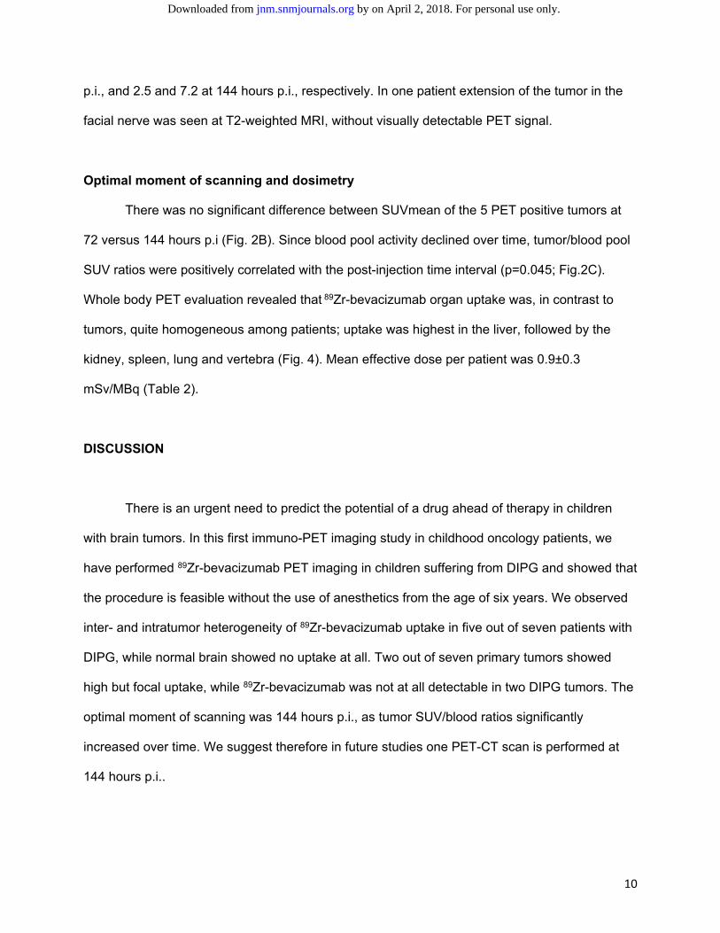
Optimal moment of scanning and dosimetry
There was no significant difference between SUVmean of the 5 PET positive tumors at
72 versus 144 hours p.i (Fig. 2B). Since blood pool activity declined over time, tumor/blood pool
SUV ratios were positively correlated with the post-injection time interval (p=0.045; Fig.2C).
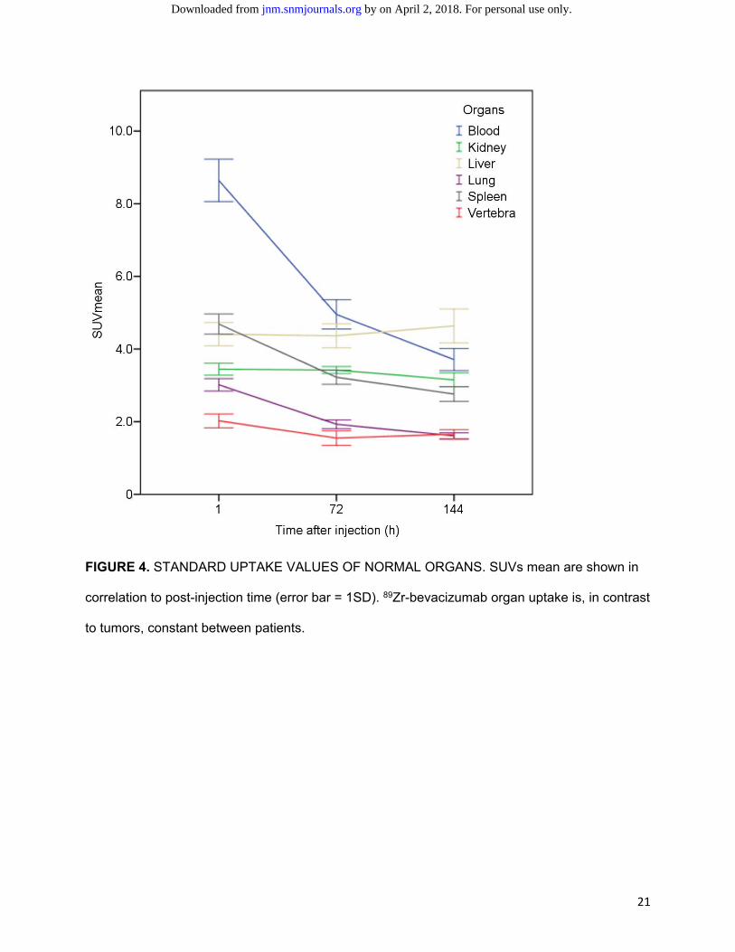
Whole body PET evaluation revealed that 89Zr-bevacizumab organ uptake was, in contrast to
tumors, quite homogeneous among patients; uptake was highest in the liver, followed by the
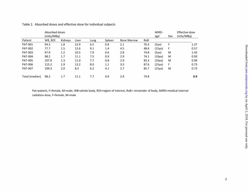
kidney, spleen, lung and vertebra (Fig. 4). Mean effective dose per patient was 0.9±0.3
mSv/MBq (Table 2).
DISCUSSION
There is an urgent need to predict the potential of a drug ahead of therapy in children
with brain tumors. In this first immuno-PET imaging study in childhood oncology patients, we
have performed 89Zr-bevacizumab PET imaging in children suffering from DIPG and showed that
the procedure is feasible without the use of anesthetics from the age of six years. We observed
inter- and intratumor heterogeneity of 89Zr-bevacizumab uptake in five out of seven patients with
DIPG, while normal brain showed no uptake at all. Two out of seven primary tumors showed
high but focal uptake, while 89Zr-bevacizumab was not at all detectable in two DIPG tumors. The
optimal moment of scanning was 144 hours p.i., as tumor SUV/blood ratios significantly
increased over time. We suggest therefore in future studies one PET-CT scan is performed at
144 hours p.i..
by on April 2, 2018. For personal use only. jnm.snmjournals.org Downloaded from

11
The 89Zr-bevacizumab uptake pattern was focal in all tumors; interestingly, four out of five
tumors only showed significant 89Zr-bevacizumab uptake within MRI contrast-enhancing areas of
the tumor. Gd-uptake in the brain is associated with BBB degradation, thus when Gd-DPTA, with
an average molecular weight of 545 kDa, is able to pass the BBB, other large molecules like
bevacizumab (149 kDa), might be able to pass as well, although BBB permeability is of course
dependent of more than molecular size only. In addition, contrast-enhancing “leaky” tumors have
been associated with higher local VEGF expression (20). We show, however, a high variability in
the level of 89Zr-bevacizumab uptake between Gd-enhancing areas (from intense to absent 89Zr-
bevacizumab uptake) which suggests large differences in local VEGF expression between
DIPG. Moreover one tumor showed 89Zr-bevacizumab uptake in an area without Gd-
enhancement. Unfortunately, we were not able to validate the VEGF expression in tissue, as
DIPG patients are not routinely biopsied. At least, the clear differences in SUVs between Gd-
enhancing tumors (reflecting differences in local drug uptake), and the presence of drug uptake
in a Gd-absent tumor area suggest that MRI alone is insufficient to predict tumor accumulation of
large molecules like bevacizumab, and that immuno-PET is of additional value.
We realize that an important aspect is the influence of radiotherapy. In our preclinical
study in non-irradiated DIPG mouse models, we observed poor uptake of 89Zr-bevacizumab in
the intracranial tumors (7). In our study, all patients were included at least 2 weeks after
completing radiotherapy, which may have induced temporary disruption of the blood-brain
barrier, as well as increased VEGF expression via the mitogen-activated protein kinase (MAPK)
pathway (21). We therefore expect DIPG patients to be more eligible for bevacizumab after
radiotherapy, and probably this effect on the blood-brain barrier integrity fades out with
increasing time after radiotherapy. Therefore, ideally, subsequent scans before, during and after
radiotherapy are performed in each patient to find out the susceptibility of the tumor for
bevacizumab over time. Future studies are needed to show the correlation of 89Zr-bevacizumab
by on April 2, 2018. For personal use only. jnm.snmjournals.org Downloaded from
12
tumor uptake and treatment response in DIPG. In adults with renal cell carcinoma, it has been
shown that high baseline 89Zr-bevacizumab uptake in the tumor prior to treatment is positively
associated with time to progression in bevacizumab-treated patients (13). A difficulty in running
such a study in DIPG patients is that current DIPG trials involve multi-agent therapy regimens.
The finding that bevacizumab can reduce the penetration of other drugs into the tumor (22,23)
underlines the need for labeled drug imaging in this multi-agent setting. On the other hand, in
contrast with single agent settings, multi-agent therapy may confound the association of the
imaging biomarker and outcome so that clinical validation of this immuno-PET method as a
predictive biomarker of therapy response requires larger datasets. However, since immuno-PET
allows for international application (long physical half-life of 89Zr, relatively simple, centrally or
locally performed, radiochemistry), we suggest that immuno-PET, using the data of the present
pilot study, is included in future international DIPG clinical trials. Furthermore, molecular imaging
data from different trials may be included in the recently established SIOPE DIPG Registry,
which enables comparison of results.
Whole body molecular drug imaging can help to predict organ-related toxicity. This is
particularly important in children since drugs developed to target over-expressed cancer-specific
signal proteins (such as VEGF), also target tissues in which these proteins are expressed during
pediatric development. We found that whole body 89Zr-bevacizumab biodistribution showed
relatively high organ-uptake in liver, followed by blood, kidneys, lungs, and bone. These results
are comparable to the results of the two 89Zr-bevacizumab clinical trials in adults and correspond
to the results of toxicity trials on bevacizumab in children reporting hypertension, bleeding,
aspartate aminotransferase elevation and proteinuria as the main side effects (13,14,24). We
observed moderate bone uptake, however, osteonecrosis is a rarely reported symptom in
bevacizumab-treated children (1%). Whether 89Zr-bevacizumab uptake also correlates with long-
by on April 2, 2018. For personal use only. jnm.snmjournals.org Downloaded from
13
term organ and/or bone toxicity needs to be addressed in long-term follow up studies of children
treated with bevacizumab.
The mean effective dose was slightly higher than doses of 89Zr-labeled compounds
published in adult studies (25), which is likely due to the age-dependent weight-factors of
dosimetry models. In our study, the 29 mSv radiation burden of immuno-PET (including 3 low
dose total body CT’s) for an average DIPG patient (25 kg) is considerable, although in future
studies the radiation dose can be reduced to 22mSv as only one PET-low dose CT of the brain
will be performed. However, the possible benefits may outweigh the risks, especially in light of
the poor prognosis (2-year survival of less than 10%) for patients with DIPG.
CONCLUSION
We introduced immuno-PET imaging in childhood cancer patients. The procedure is safe
and feasible in children without using anesthetics and the optimal moment of scanning is 144
hours p.i.. Clear differences in 89Zr-bevacizumab uptake between DIPG tumors were observed,
with 2 tumors showing no uptake at all. Interestingly, 4 out of 5 tumors showed significant 89Zr-
bevacizumab uptake on PET within the contrast-enhancing area on MRI of the tumor. However,
we show high variability in SUVs between these contrast-enhancing areas suggesting
differences in local VEGF expression, and we observed one patient with a positive PET in a non-
Gd enhancing area. MRI alone seems therefore insufficient to predict drug accumulation in the
tumor. The addition of 89Zr-bevacizumab PET imaging may therefore be of help to select
potential candidates for bevacizumab treatment in DIPG, as it assesses target availability and
drug accessibility of the tumor within the same procedure.
by on April 2, 2018. For personal use only. jnm.snmjournals.org Downloaded from

14
ACKNOWLEDGEMENTS
This DIPG study is funded by the Semmy Foundation (Stichting Semmy) and the Egbers
Foundation (Egbers Stichting).
DISCLOSURE
There is no conflict of interest to disclose. The Semmy Foundation (Stichting Semmy)
and the Egbers Foundation (Egbers Stichting) who fund DIPG research in VU University Medical
Center had no role in the study and/or preparation of this manuscript.
by on April 2, 2018. For personal use only. jnm.snmjournals.org Downloaded from

15
REFERENCES
1. Kaatsch P, Rickert CH, Kuhl J, Schuz J, Michaelis J. Population-based epidemiologic
data on brain tumors in German children. Cancer. 2001;92:3155-3164.
2. Hargrave D, Bartels U, Bouffet E. Diffuse brainstem glioma in children: critical review of
clinical trials. Lancet Oncol. 2006;7:241-248.
3. Jansen MH, Van Vuurden DG, Vandertop WP, Kaspers GJ. Diffuse intrinsic pontine
gliomas: a systematic update on clinical trials and biology. Cancer Treat Rev.
2012;38:27-35.
4. Jansen MH, Veldhuijzen van Zanten SE, Sanchez Aliaga, et al. Survival prediction
model of children with diffuse intrinsic pontine glioma based on clinical and radiological
criteria. Neuro Oncol. 2015;17:160-166.
5. Van Dongen GAMS, Huisman MC, Boellaard R, et al. 89Zr-immuno-PET for imaging of
long circulating drugs and disease targets: why, how and when to be applied? Q J Nucl
Med Mol Imaging. 2015;59:18-38.
6. Puget S, Philippe C, Bax DA, et al. Mesenchymal transition and PDGFRA
amplification/mutation are key distinct oncogenic events in pediatric diffuse intrinsic
pontine gliomas. PLoS One. 2012;7:30313.
7. Jansen MJ, Lagerweij T, Sewing AC, et al. Bevacizumab targeting diffuse intrinsic
pontine glioma: results of 89Zr-bevacizumab PET imaging in brain tumor models. Mol
Cancer Ther. 2016;15:2166-2174
8. Gururangan S, Chi SN, Young PT, et al. Lack of efficacy of bevacizumab plus irinotecan
in children with recurrent malignant glioma and diffuse brainstem glioma: a Pediatric
Brain Tumor Consortium study. J Clin Oncol. 2010;28:3069-3075.
9. Hummel TR, Salloum R, Drissi R, et al. A pilot study of bevacizumab-based therapy in
patients with newly diagnosed high-grade gliomas and diffuse intrinsic pontine gliomas.
J Neurooncol. 2016;127:53-61
by on April 2, 2018. For personal use only. jnm.snmjournals.org Downloaded from
16
10. Zaky W, Wellner M, Brown RJ, et al. Treatment of children with diffuse intrinsic pontine
gliomas with chemoradiotherapy followed by a combination of temozolomide, irinotecan,
and bevacizumab. Pediatr Hematol Oncol. 2013;30:623-632
11. Aguilera DG, Mazewski C, Hayes L, et al. Prolonged survival after treatment of diffuse
intrinsic pontine glioma with radiation, temozolamide, and bevacizumab: report of 2
cases. J Pediatr Hematol Oncol. 2013;35:42-46.
12. Van der Bilt AR, Terwisscha van Scheltinga AG, Timmer-Bosscha H, et al.
Measurement of tumor VEGF-A levels with 89Zr-bevacizumab PET as an early
biomarker for the antiangiogenic effect of everolimus treatment in an ovarian cancer
xenograft model. Clin Cancer Res. 2012;18:6306-6314
13. Oosting SF, Brouwers AH, Van Es SC, et al. 89Zr-bevacizumab PET visualizes
heterogeneous tracer accumulation in tumor lesions of renal cell carcinoma patients and
differential effects of antiangiogenic treatment. J Nucl Med. 2015;56:63-69
14. Bahce I, Huisman MC, Verwer EE, et al. Pilot study of (89)Zr-bevacizumab positron
emission tomography in patients with advanced non-small cell lung cancer. EJNMMI
Res. 2014;4:35
15. Verel I, Visser GW, Boellaard R, Stigter-van WM, Snow GB, Van Dongen GA. 89Zr
immuno-PET: comprehensive procedures for the production of 89Zr-labeled monoclonal
antibodies. J Nucl Med. 2003;44:1271-1281.
16. Cohen R, Stammes MA, de Roos IH, Stigter-van WM, Visser GW, Van Dongen GA.
Inert coupling of IRDye800CW to monoclonal antibodies for clinical optical imaging of
tumor targets. EJNMMI Res. 2011;1:31.
17. Cohen R, Vugts DJ, Stigter-van Walsum M, Visser GWM, Van Dongen GAMS. Inert
coupling of IRDye800CW and zirconium-89 to monoclonal antibodies for single- or dual-
mode fluorescence and PET imaging. Nat Protoc. 2013;8:1010–1018.
by on April 2, 2018. For personal use only. jnm.snmjournals.org Downloaded from
17
18. Surti S, Kuhn A, Werner ME, Perkins AE, Kolthammer J, Karp JS. Performance of
Philips Gemini TF PET/CT scanner with special consideration for its time-of-flight
imaging capabilities. J Nucl Med. 2007;48:471-480.
19. Stabin MG, Sparks RB, Crowe E. OLINDA/EXM: the second-generation personal
computer software for internal dose assessment in nuclear medicine. J Nucl Med.
2005;46:1023-1027.
20. Johansson M, Brannstrom T, Bergenheim AT, Henriksson R. Spatial expression of
VEGF-A in human glioma. J Neurooncol. 2002;59:1-6.
21. Park JS, Qiao L, Su ZZ, et al. Ionizing radiation modulates vascular endothelial growth
factor (VEGF) expression through multiple mitogen activated protein kinase dependent
pathways. Oncogene. 2001;20:3266-3280
22. Arjaans M, Oude Munnink TH, Oosting SF, et al. Bevacizumab-induced normalization of
blood vessels in tumors hampers antibody uptake. Cancer Res. 2013;73:3347-3355
23. Van der Veldt AA, Lubberink M, Mathijssen RH, et al. Toward prediction of efficacy of
chemotherapy: a proof of concept study in lung cancer patients using ((1)(1)C)docetaxel
and positron emission tomography. Clin Cancer Res. 2013;19:4163-4173
24. Fangusaro J, Gururangan S, Poussaint TY, et al. Bevacizumab (BVZ)-associated
toxicities in children with recurrent central nervous sys tem tumors treated with BVZ and
irinotecan (CPT-11): a Pediatric Brain Tumor Consortium Study (PBTC-022). Cancer.
2013;1;119:4180-4187.
25. Borjesson PK, Jauw YW, Boellaard R, et al. Performance of immuno-positron emission
tomography with zirconium-89-labeled chimeric monoclonal antibody U36 in the
detection of lymph node metastases in head and neck cancer patients. Clin Cancer Res.
2006;12:2133-2140
by on April 2, 2018. For personal use only. jnm.snmjournals.org Downloaded from
18
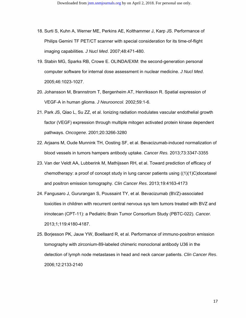
FIGURE 1. MRI AND PET-MRI FUSION IMAGES OF PATIENTS WITH DIPG. Top row: 89Zr-
bevacizumab PET (144 hrs p.i.) fused with T1-Gd weighted MRI per patient; middle row: T1-Gd
weighted MRI; lower row: T2-weighted/FLAIR MR-images. Five tumors show variable uptake of
89Zr-bevacizumab (white arrows), with both PET negative and positive areas within each tumor.
Two primary tumors are completely PET negative (Fig. 1C and 1E), while the T2 weighted
images show tumor infiltration in the whole pons of both patients. In the middle row, the red
arrows represent the areas of contrast enhancement within the tumor. In four out of five primary
tumors the PET-positive area corresponds with the contrast-enhancing area on MRI of the
tumors (Fig. 1A, 1B, 1F and 1G). In Fig. 1C, the tumor shows an MRI contrast enhancing area,
while there is no 89Zr-bevacizumab uptake. Fig. 1D shows a PET positive tumor, while no Gd-
enhancement is observed on MRI.
by on April 2, 2018. For personal use only. jnm.snmjournals.org Downloaded from
19
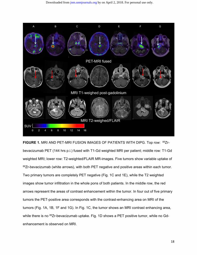
FIGURE 2. 89ZR-
BEVACIZUMAB TUMOR
UPTAKE IN CORRELATION
TO BLOOD POOL. Fig. 2A
shows the whole body PET
images of one DIPG patient
(PAT-002) at 1, 72 and 144
hours post-injection (p.i.). At
1 hour p.i., maximum uptake
is observed within the blood
pool. With increasing post-
injection time, the blood pool
activity decreases
significantly. There is no
uptake of 89Zr-bevacizumab
in the tumor at 1 hour post-
injection while there is clear
uptake both at 72 and 144 hours p.i. (red arrows). Uptake in the liver is stable and represents
vascularisation (1 hour p.i.) and metabolisation (72 and 144 hours p.i.) of 89Zr-bevacizumab. Fig.
2B shows the boxplots of the tumor SUVs at 72 and 144 hours post-injection; SUVs were not
significantly different (p=0.6). Fig. 2C shows SUV ratios of tumor/blood pool (measured in the
aortic arch) per patient as a function of post injection time (PI). SUV ratios are positively
correlated with increasing post-injection time (Kendall’s tau_b correlation coefficient 0,524
p=0.045 (2-tailed).
by on April 2, 2018. For personal use only. jnm.snmjournals.org Downloaded from
20
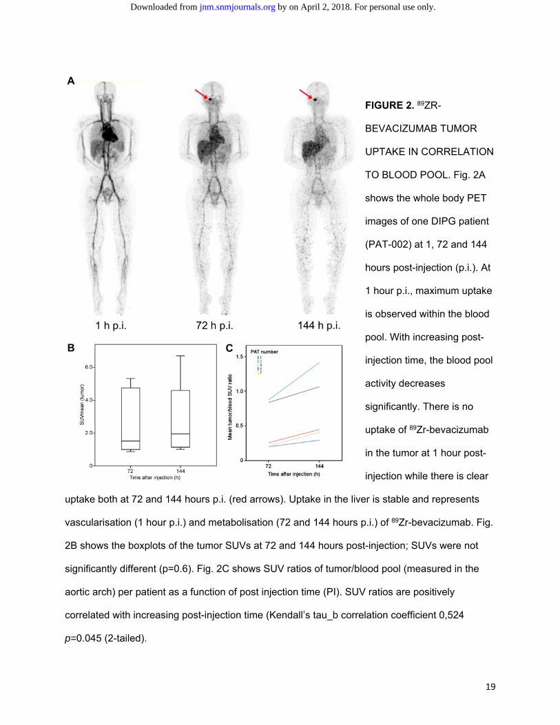
FIGURE 3. MRI AND PET-MRI FUSION IMAGES OF ONE PATIENT WITH PRIMARY DIPG
AND METASTASES IN THE MYELUM. The left 89Zr-bevacizumab PET-MRI image is a sagittal
plane of the myelum showing six 89Zr-bevacizumab PET hotspots, which were all confirmed
metastases by T1-Gd weighted MRI (right image). Some of these metastases were even clearer
on PET than MRI. All (11 in total) metastases were PET positive in this patient, whose primary
tumor also showed high but focal 89Zr-bevacizumab uptake (Fig. 1F).
by on April 2, 2018. For personal use only. jnm.snmjournals.org Downloaded from
21
FIGURE 4. STANDARD UPTAKE VALUES OF NORMAL ORGANS. SUVs mean are shown in
correlation to post-injection time (error bar = 1SD). 89Zr-bevacizumab organ uptake is, in contrast
to tumors, constant between patients.
by on April 2, 2018. For personal use only. jnm.snmjournals.org Downloaded from
Tables
Table 1. Baseline characteristics and scanning results
Patient Sex Age (years)
Weight (kg)
Metastases/ tumor extension
Treatment before PET study:
89Zr dose (Mbq)
Adverse events
Tumor size transversal diameter (mm)
Gd‐Contrast enhancement on MRI
89Zr‐Bmab tumor uptake
Tumor SUV 72 hrs p.i.
Tumor SUV 144 hrs p.i.
PAT‐001 F 6 33 None RT 30.7 None 25 Small nodular Yes 1.0 1.0
PAT‐002 F 17 55 None RT & gemcitabine 37.1 None 37 Ring Yes 5.3 6.7
PAT‐003 M 7 28 None RT 24.2 None 36 Ring No No No
PAT‐004 M 8 30 None RT 25.6 None 33 Patchy Yes† 0.9 1.2
PAT‐005 M 11 37 None RT & gemcitabine 31.2 None 39 No No No No
PAT‐006 F 13 44 LM and arachnoidal RT & gemcitabine 34.7 None 32 Patchy Yes 4.7 4.6
PAT‐007 M 15 65 Tumor extension nVII RT & temozolomide 36.7 None 37 Ring Yes 1.5 1.9
Total M=57% 11* 37* 31.5# 34.1# 2.7# 3.1#
F=female, M=male, LM= leptomeningeal metastasis, RT=radiotherapy, nVII= facial nerve, 89Zr=Zirconium‐89, Gd=Gadolinium, Bmab=bevacizumab,
SUV=standard uptake value, hrs=hours, p.i. =post‐injec on , *=median, # mean, †PET uptake was in an area without Gd‐contrast enhancement
by on April 2, 2018. For personal use only.
jnm.snm
journals.org D
ownloaded from
2
Table 2. Absorbed doses and effective dose for individual subjects
Absorbed doses (mGy/MBq)
MIRD‐age' Sex
Effective dose (mSv/MBq)
Patient WB_ROI Kidneys Liver Lung Spleen Bone Marrow RoB
PAT‐001 94.3 1.8 12.9 6.5 0.8 2.1 70.3 (5yo) F 1.37
PAT‐002 77.7 1.5 12.6 9.1 1.4 4.5 48.6 (15yo) F 0.57
PAT‐003 97.9 1.2 10.5 7.9 0.6 2.8 74.8 (5yo) M 1.43
PAT‐004 98.2 1.7 11.1 7.5 0.9 2.9 74.1 (10yo) M 0.93
PAT‐005 107.0 1.3 11.0 7.7 0.8 2.9 83.3 (10yo) M 0.99
PAT‐006 115.2 1.9 13.2 8.0 1.1 3.5 87.6 (15yo) F 0.73
PAT‐007 109.3 2.0 8.5 6.2 4.1 2.7 85.7 (15yo) M 0.73
Total (median) 98.2 1.7 11.1 7.7 0.9 2.9 74.8 0.9
Pat=patient, F=female, M=male, WB=whole body, ROI=region of interest, RoB= remainder of body, MIRD=medical internal
radiation dose, F=female, M=male by on April 2, 2018. For personal use only.
jnm.snm
journals.org D
ownloaded from
Doi: 10.2967/jnumed.116.180216Published online: October 20, 2016.J Nucl Med. Hoekstra, Guus A.M.S. van Dongen and Gert-Jan JL KaspersMarc Jansen, Sophie EM Veldhuijzen van Zanten, Dannis G Van Vuurden, Marc Huisman, Danielle J. Vugts, Otto S. Pontine Glioma
Zr-bevacizumab PET in Children with Diffuse Intrinsic89Molecular Drug Imaging:
http://jnm.snmjournals.org/content/early/2016/10/19/jnumed.116.180216This article and updated information are available at:
http://jnm.snmjournals.org/site/subscriptions/online.xhtml
Information about subscriptions to JNM can be found at:
http://jnm.snmjournals.org/site/misc/permission.xhtmlInformation about reproducing figures, tables, or other portions of this article can be found online at:
and the final, published version.proofreading, and author review. This process may lead to differences between the accepted version of the manuscript
ahead of print area, they will be prepared for print and online publication, which includes copyediting, typesetting,JNMcopyedited, nor have they appeared in a print or online issue of the journal. Once the accepted manuscripts appear in the
. They have not beenJNM ahead of print articles have been peer reviewed and accepted for publication in JNM
(Print ISSN: 0161-5505, Online ISSN: 2159-662X)1850 Samuel Morse Drive, Reston, VA 20190.SNMMI | Society of Nuclear Medicine and Molecular Imaging
is published monthly.The Journal of Nuclear Medicine
© Copyright 2016 SNMMI; all rights reserved.
by on April 2, 2018. For personal use only. jnm.snmjournals.org Downloaded from