molecular characterizations of cytolethal distending …iai.asm.org/content/80/4/1323.full.pdf ·...
TRANSCRIPT
Molecular Characterizations of Cytolethal Distending Toxin Producedby Providencia alcalifaciens Strains Isolated from Patients withDiarrhea
Ayaka Shima,a Atsushi Hinenoya,a Masahiro Asakura,a Norihiko Sugimoto,a Teizo Tsukamoto,a,b Hideaki Ito,a Akira Nagita,c
Shah M. Faruque,d and Shinji Yamasakia
Graduate School of Life and Environmental Sciences, Osaka Prefecture University, Osaka, Japana; Osaka Prefectural Institute of Public Health, Osaka, Japanb; Departmentof Pediatrics, Mizushima Central Hospital, Okayama, Japanc; and Molecular Genetics Laboratory, International Centre for Diarrhoeal Diseases Research, Dhaka,Bangladeshd
Cytolethal distending toxins (CDTs), which block eukaryotic cell proliferation by acting as inhibitory cyclomodulins, areproduced by diverse groups of Gram-negative bacteria. Active CDT is composed of three polypeptides—CdtA, CdtB, andCdtC— encoded by the genes cdtA, cdtB, and cdtC, respectively. We developed a PCR-restriction fragment length polymor-phism assay for the detection and differentiation of five alleles of cdtB (Cdt-I through Cdt-V) in Escherichia coli and usedthe assay to investigate the prevalence and characteristic of CDT-producing E. coli in children with diarrhea (A. Hinenoyaet al., Microbiol. Immunol. 53:206 –215, 2009). In these assays, two untypable cdtB genes were detected and the organismsharboring the cdtB gene were identified as Providencia alcalifaciens (strains AH-31 and AS-1). Nucleotide sequence analy-sis of the cdt gene cluster revealed that the cdtA, cdtB, and cdtC genes of P. alcalifaciens are of 750, 810, and 549 bp, respec-tively. To understand the possible horizontal transfer of the cdt genes among closely related species, the presence of cdtgenes was screened in various Providencia spp. by colony hybridization assay, and the cdt gene cluster was found in onlylimited strains of P. alcalifaciens. Genome walking revealed that the cdt gene cluster of P. alcalifaciens is located adjacentto a putative transposase gene, suggesting the locus might be horizontally transferable. Interestingly, the CDT of P. alcali-faciens (PaCDT) showed some homology with the CDT of Shigella boydii. Whereas filter-sterilized lysates of strains AH-31and AS-1 showed distention of CHO but not of HeLa cells, E. coli CDT-I exhibited distention of both cells. This activity ofPaCDT was confirmed by generating recombinant PaCDT protein, which could also be neutralized by rabbit anti-PaCdtBantibody. Furthermore, recombinant PaCDT was found to induce G2/M cell cycle arrest and phosphorylation of host his-tone H2AX, a sensitive marker of DNA double-strand breaks. To our knowledge, this is the first report showing that cer-tain clinical P. alcalifaciens strains could produce variants of the CDTs compared.
Cytolethal distending toxin (CDT) was first discovered as a newtype of toxin in Escherichia coli strains isolated from patient
with diarrhea in 1987 (29). CDT has a unique activity, which dif-fers from heat-labile enterotoxin (LT). Although LT causes onlycell elongation, CDT causes not only cell elongation but also celldistention and blocking of eukaryotic cell cycle at G2/M phase,leading to cell death. Since 1987, the presence of CDTs has beenreported in various Gram-negative bacteria, such as Aggregatibac-ter actinomycetemcomitans, Campylobacter spp., Escherichia alber-tii, Haemophilus ducreyi, Helicobacter spp., and Shigella spp. (55).CDTs are composed of three polypeptides, namely, CdtA, CdtB,and CdtC, which form a complex structure needed for the toxinactivity (55). CdtA and CdtC subunits bind the eukaryotic cellsurface, followed by entry of the CdtB subunit into the cell (55).After entering the cell, the CdtB was transferred in the nucleus(55), and ultimately it causes DNA double-strand breaks by usingits DNase I activity (15, 32). Therefore, CDT has recently beenrecognized as a new family of bacterial toxins, called genotoxin(37) or cyclomodulin (38).
In E. coli, at least five different types of EcCDTs have beenreported thus far. EcCDT-I and EcCDT-II were initially identifiedin enteropathogenic E. coli (EPEC) strains isolated from patientswith diarrhea (45, 46). EcCDT-III was discovered in an E. colistrain isolated from a calf with septicemia (43). EcCDT-IV wasdetected in pathogenic E. coli strains isolated from human or an-
imal intestinal and extraintestinal sources (50), whereas EcCDT-Vwas identified in Shiga toxin-producing E. coli (STEC) or entero-hemorrhagic E. coli (EHEC) strains (28). Among five differentEcCDTs, it has been reported that the EcCDT-III gene is carried bya conjugative plasmid, called pVir (43), whereas EcCDT-IV andEcCDT-V genes are carried by the lambdoid and P2 phages, re-spectively (28, 51). Asakura et al. (5) has recently reported thatEcCDT-I produced by certain strains of EPEC was encoded on aninducible lambdoid phage.
Although a number of studies regarding the isolation and char-acterization of CDT-producing bacteria from patients with diar-rhea have been reported (2, 4, 9, 20, 24, 34, 39, 41), the role of CDTin human diseases, including diarrhea, has not yet been estab-lished. However, it has been demonstrated that shedding of Heli-cobacter hepaticus cdt-negative mutant was much shorter, and
Received 29 August 2011 Returned for modification 25 October 2011Accepted 5 January 2012
Published ahead of print 17 January 2012
Editor: S. R. Blanke
Address correspondence to Shinji Yamasaki, [email protected].
Copyright © 2012, American Society for Microbiology. All Rights Reserved.
doi:10.1128/IAI.05831-11
0019-9567/12/$12.00 Infection and Immunity p. 1323–1332 iai.asm.org 1323
on Septem
ber 21, 2018 by guesthttp://iai.asm
.org/D
ownloaded from

there was also mild inflammation of intestine compared to that ofits isogenic wild-type strain (57). Recombinant S. dysenteriae CDTcould cause diarrhea in the suckling mouse model (40). Further-more, coadministration of H. ducreyi and purified H. ducreyi CDTcould induce more severe inflammation than H. ducreyi alone(54). It is noteworthy that high amount of CDT-producing E. coli(CTEC) strains have been isolated from patients with bloody di-arrhea in India (41). CTEC was also isolated from patients withbloody diarrhea as a sole pathogen in Japan (23). It has been re-ported that EcCDT-V genes in EHEC O157:H7 strains were sig-nificantly more frequent in isolates from patients with diarrheathan in isolates from asymptomatic carriers (18) and that cdt in eae(E. coli attaching and effacing)-negative STEC was significantlymore frequent in patients with hemolytic-uremic syndrome andin patients with diarrhea than in asymptomatic carriers (7). Thesedata suggest that CDT could possibly be a virulence factor andmay contribute to persistence of infection or it could enhancepathogenicity in host. To elucidate the relevance of CTEC in di-arrhea, we developed a PCR-restriction fragment length polymor-phism (RFLP) assay for the detection and differentiation of cdtgenes in E. coli and examined the prevalence and characteristics ofCTEC among children with diarrhea in Japan (24). UntypeablecdtB genes were detected directly from two stool samples of pa-tients with diarrhea by the PCR-RFLP assay, and the bacteria har-boring these untypeable cdtB genes were isolated and identified asProvidencia alcalifaciens (24; unpublished data).
In the present study we attempted to analyze the cdt gene clus-ter and its flanking region in the genome of P. alcalifaciens. Toexamine the possible horizontal transfer of the cdt gene clusteramong closely related species, the distribution of cdt genes in var-ious Providencia spp. was also checked. In addition, the biologicalactivity and possible mechanism of action of these CDTs pro-duced by the P. alcalifaciens strains were examined and discussed.
(This study was performed in partial fulfillment of the require-ments of a Ph.D. thesis for A.S. from Graduate School of Life andEnvironmental Sciences, Osaka Prefecture University, Osaka, Ja-pan.)
MATERIALS AND METHODSBacterial strains and growth condition. The bacterial strains used in thepresent study are listed in Table 1. Two P. alcalifaciens isolated from chil-dren with diarrhea during surveillance of cdt gene harboring E. coli andidentified by biochemical test using API 20E and adonitol and galactoseutility tests (24, unpublished) were examined. Furthermore, eight P. al-califaciens strains, six P. rettgeri strains, two P. rustigianii strains, one P.heimbachae strain, and one P. stuartii strain were also investigated for thepresence of cdt genes. Bacteria were grown aerobically in Luria-Bertani(LB) medium (Becton Dickinson, Franklin Lakes, NJ), in brain heart in-fusion (BHI) medium (Becton Dickinson) or on LB agar (Becton Dickin-son) containing 30 �g of kanamycin (Nacalai Tesque, Inc., Kyoto, Ja-pan)/ml when appropriate.
Isolation of Providencia spp. Rectal swabs collected from patientswith diarrhea were plated on polymyxin-mannitol-xylitol medium for
TABLE 1 Bacterial strains used in this study
Bacterium Strain Characteristic Source or reference
P. alcalifaciens AH-31 Clinical isolate (PaCDT) 24AS-1 Clinical isolate (PaCDT) Diarrheal children (this study)F90-2004 Clinical isolate ICDDR,B24717 Clinical isolate ICDDR,BGTC2020 Clinical isolate Purchased from Gifu University18H253 Clinical isolate Osaka Prefectural Institute of Public Health18H399 Clinical isolate Osaka Prefectural Institute of Public Health19H270 Clinical isolate Osaka Prefectural Institute of Public HealthP2556 Clinical isolate Diarrheal adult (unpublished)RME362 Human isolate Specimen from regular medical examination (unpublished)
P. rettgeri GTC1263 (ATCC 29944) Unknown Purchased from Gifu UniversityP2234 Clinical isolate Diarrheal children (unpublished)P2253 Clinical isolate Diarrheal children (unpublished)P2312 Clinical isolate Diarrheal children (unpublished)P2536 Clinical isolate Diarrheal children (unpublished)RME220 Human isolate Specimen from regular medical examination (unpublished)
P. rustigianii GTC1504 (ATCC 33673) Human isolate Purchased from Gifu UniversityRME3 Human isolate Specimen from regular medical examination (unpublished)
P. heimbachae GTC1501 (ATCC 35613) Penguin isolate Purchased from Gifu University
P. stuartii GTC1444 (ATCC 29914) Human isolate Purchased from Gifu University
E. coli GB1371 Clinical isolate (EcCDT-I) 41C600 NAa Laboratory strain (C. Sasakawa)BL21(DE3) NA Laboratory strain (Promega)BL21(DE3) With pET28a This studyBL21(DE3)/TAS-1 With pAS-1 (rPaCdtB) This studyBL21(DE3)/TAS-2 With pAS-2 (rPaCDT) This study
a NA, not applicable.
Shima et al.
1324 iai.asm.org Infection and Immunity
on Septem
ber 21, 2018 by guesthttp://iai.asm
.org/D
ownloaded from

Providencia (PMXMP) agar (56). In addition, stool specimens obtainedfrom a regular medical examination of 3- to 9-year-old children (kinder-garten and primary school) were also screened by plating on PMXMPagar. Suspected bacterial colonies were isolated and identified as Providen-cia spp. by the API 20E system, and adonitol and galactose utility tests.
Colony hybridization assay for detection of cdt genes. To examinethe presence of cdt genes in P. alcalifaciens strains and in other Providenciaspp., including P. rettgeri, P. rustigianii, P. heimbachae, and P. stuartii, thecdtB DNA fragment was generated by PCR using the Cdt-Bcomu andCdt-Bcomd primers, and the fragment was used as a probe in colonyhybridization assay (24). Strain AH-31 was always used as a positive con-trol.
PCR. PCR for the detection of cdtB gene was performed as describedpreviously (24).
Nucleotide sequence analysis. To determine the nucleotide sequenceof the cdt gene cluster, PCR products of cdtB gene and its flanking regionwere sequenced either by a standard method or by genome walking (6).Briefly, PCR product was purified by QIAquick PCR products purifica-tion kit (Qiagen, GmbH, Hilden, Germany), and the nucleotide sequenceof the PCR product was determined by using a BigDye terminator cyclesequencing kit on an ABI Prism 3100 genetic analyzer (Applied Biosys-tems, Foster City, CA) essentially as described by the manufacturer. Syn-thetic primers were designed on the basis of obtained sequence and ge-nome walking was performed as described previously (6). Nucleotide andamino acid sequences were analyzed and compared by using GenBank,DDBJ (the DNA Data Bank of Japan), and DNASIS software (HitachiSoftware Engineering Co., Ltd., Tokyo, Japan). Dendrogram analysis wasperformed by using the software CLUSTAL W of MegAlign (DNASTAR,Inc., Madison, WI).
Preparation of recombinant proteins. The P. alcalifaciens cdt andcdtB genes (Pacdt and PacdtB) were amplified from the genomic DNA ofP. alcalifaciens strain AH-31 by PCR using the primer set PacdtABC-F(5=-ATATGGATCCATGAATAATAAACGCACAT-3=) and PacdtABC-R(5=-ATATCTCGAGTTTAAATAACGGGTGACTC-3=) and the primerset PacdtB-F (5=-GAGAGGATCCGTGTTTTTATCGTTTTACGC-3=)and PacdtB-R (5=-GAGACTCGAGTTTACCTTCTGAATACGCC-3=),respectively. PCR was carried out in a 50-�l reaction mixture for each tubecontaining 2.5 �l of DNA template, 1� ExTaq PCR buffer (Takara Bio,Inc., Shiga, Japan), 0.2 mM deoxynucleoside triphosphate mixture, 0.5�M concentrations of each primer set, and 1.25 U of ExTaq polymerase(Takara Bio, Inc.). DNA template was prepared from an overnight cul-ture, which was diluted 10-fold in sterile TE buffer (10 mM Tris-HCl, 1mM EDTA [pH 8.0]) and boiled for 10 min, followed by centrifugation at12,000 � g at 4°C for 5 min. The PCR conditions were optimized as aninitial denaturation of 5 min at 94°C, followed by 30 cycles of denatur-ation for 30 s at 94°C, annealing for 30 s at 55°C, and extension for 90 s or60 s at 72°C, with a final extension step for 5 min at 72°C in GeneAmp PCRSystem 9700 (Perkin-Elmer, Waltham, MA). Each PCR product was di-gested with BamHI and XhoI and ligated into pET-28a (pAS-1 for PacdtB,pAS-2 for Pacdt), and pAS-1 and pAS-2 were transformed into E. coliBL21(DE3) (strain TAS-1 with pAS-1 and strain TAS-2 with pAS-2). E.coli strain BL21(DE3) with pET-28a was similarly prepared and used as avector control. Recombinant PaCdtB (rPaCdtB) was purified from cruderPaCdtB expressed in E. coli strain TAS-1. Briefly, E. coli strain TAS-1 wasgrown at 37°C overnight in LB broth containing kanamycin (30 �g/ml).The culture was diluted 1:100 in the fresh medium and incubated at 37°Cuntil the optical density at 600 nm of the culture reached 0.6. IPTG (iso-propyl-�-D-thiogalactopyranoside) was added to a final concentration of0.1 mM, and the culture was further incubated at 18°C for 16 h withvigorous shaking. The bacterial cells were then collected by centrifugationat 6,000 � g at 4°C for 15 min. The cells were suspended in 50 mMTris-HCl (pH 8.0) containing 150 mM NaCl and 50 mM imidazole andthen sonicated using an Astrason ultrasonic processor (Heat-System Ul-trasonics, Farmingdale, NY). The lysates were centrifuged at 15,000 � g at4°C for 15 min, and the supernatants were collected and used for further
purification. The rPaCdtB was purified by using an Ni-Sepharose column(GE Healthcare UK, Ltd., Buckinghamshire, England). The purity of therPaCdtB was confirmed by sodium dodecyl sulfate–15% polyacrylamidegel electrophoresis (SDS–15% PAGE) (31). For the preparation of cruderPaCDT, E. coli strain TAS-2 was cultured at 37°C overnight with vigorousshaking. The culture was sonicated as described above, the lysates werecentrifuged at 12,000 � g at 4°C for 5 min, and the supernatants werefiltrated using a 0.22-�m-pore-size filter (Asahi Glass Co., Ltd., Tokyo,Japan). The filter-sterilized lysate of E. coli strain BL21(DE3)/pET28a wassimilarly prepared as a vector control. The filtrate was either directly usedfor cytotoxicity assay or used for suckling mouse assay after concentrationby ammonium sulfate precipitation. For ammonium sulfate precipita-tion, solid ammonium sulfate was first added to 40% saturation, and thenthe precipitate was removed by centrifugation at 20,000 � g at 4°C for 20min. Finally, the supernatant solution was brought to 60% saturation withrespect to ammonium sulfate. After centrifugation at 20,000 � g at 4°C for20 min, the precipitate was dialyzed against phosphate-buffered saline(PBS; pH 7.4) until ammonium sulfate was completely removed. Thecrude rPaCDT was then used for suckling mouse assay. The endotoxincontent of rPaCDT was determined by using the ToxinSensor endotoxindetection system (GenScript USA, Inc., Piscataway, NJ) and confirmed tobe similar to the preparation from negative control [E. coli BL21(DE3)harboring pET28a].
Preparation of antisera against rPaCdtB. Purified rPaCdtB was im-munized against 8-week-old male New Zealand White rabbit (OrientalYeast Co., Ltd., Tokyo, Japan). Briefly, 300 �g of purified rPaCdtB wasinjected into four sites: subcutaneously into the shoulders and intramus-cularly into the thighs every 2 weeks interval with Freund complete adju-vant (Becton Dickinson) first and subsequently with Freund incompleteadjuvant (Becton Dickinson) for 8 weeks. The rabbits were anesthetizedwith ketamine (35 mg/kg [body weight]) and xylazine (5 mg/kg [bodyweight]), blood was collected, and serum was obtained by centrifugationat 6,000 � g for 10 min.
Determination of titer. The titer was determined by using an Ouch-terlony double gel diffusion test as described previously (58). Briefly, thedouble gel diffusion test was carried out with 1.2% Noble agar (Difco) in50 mM Tris-HCl buffer (pH 8.0) containing 150 mM NaCl. Each samplewas applied into a hole, and the plate was placed in a humidified chamberat room temperature for about 16 to 24 h. The plate was then washedextensively with a solution of 0.4% NaCl and 0.4% sodium borate anddried. The plate was stained with 0.5% Coomassie brilliant blue (CBB)dissolved in a solution of 50% methanol, 10% acetic acid, and 40% waterand destained with the same solution without CBB. The antibody titer wasdefined as the highest dilution of serum that yielded a visible precipitationline by CBB staining.
Western blotting. P. alcalifaciens strains AH-31 and AS-1 were cul-tured at 37°C for 16 h in BHI medium, respectively. A 1-ml portion of theculture was centrifuged at 6,000 � g for 10 min, and the cells were resus-pended and sonicated in 200 �l of PBS for 1 min on ice using a handysonicator UR-20P (Tomy Seiko Co., Ltd., Tokyo, Japan). Cell lysates wereseparated by SDS-15% PAGE as described above. The proteins were blot-ted to polyvinylidene difluoride (PVDF) membranes (Bio-Rad, Hercules,CA) using a Trans-Blot SD semidry electrophoretic transfer cell essentiallyas described by the manufacturer (Bio-Rad) and probed with anti-rPaCdtB antiserum, followed by goat anti-rabbit IgG tagged with horse-radish peroxidase (HRP) as a secondary antibody (Invitrogen, Carlsbad,CA). Color development was performed with 4CN-PLUS (Perkin-Elmer)at room temperature.
Cell culture. HeLa, Vero, Int407, HEp2, and Caco-2 cells were cul-tured in minimum essential medium (MEM; Invitrogen). CHO, Y-1, orNIH/3T3 was cultured either in MEM-� (Invitrogen), Ham F-12 (Invit-rogen), or Dulbecco’s modified Eagle medium (Invitrogen), respectively.All media contained 10% fetal bovine serum (Invitrogen) and 1% antibi-otic, including antimycotic (�100) liquid (penicillin G sodium [10,000U/ml], streptomycin sulfate [10,000 U/ml], and 25 �g of amphotericin
CDT Produced by Providencia alcalifaciens
April 2012 Volume 80 Number 4 iai.asm.org 1325
on Septem
ber 21, 2018 by guesthttp://iai.asm
.org/D
ownloaded from
B/ml as Fungizone in 0.85% saline [Invitrogen]). In addition, 1% nones-sential amino acids solution (�100; Invitrogen) was added to the MEMfor Caco-2 cells. The cells were cultured at 37°C under 5% CO2 in air.
Cytotoxicity assay. P. alcalifaciens strains (AH-31 and AS-1) harbor-ing Pacdt genes (Table 1) were cultured at 37°C for 16 h in an appropriatemedium, and the culture was sonicated as described above. The lysateswere passed through a sterile disposable filter with a 0.22-�m pore size,and filter-sterilized bacterial lysates were examined for the ability to causethe distension and death of CHO, HeLa, Vero, HEp2, Int407, Caco-2, Y-1,and NIH/3T3 cells. Filter-sterilized lysates of E. coli strain TAS-2(rPaCDT) were also included. Filter-sterilized lysates of E. coli strainsGB1371 (EcCDT-I) and C600 were used as positive and negative controls,respectively. The cells were seeded at a density of 5 � 103 cells in a 96-wellplate (Asahi Glass Co., Ltd.). After 24 h of incubation, 20 �l of 2-foldserially diluted crude PaCDT was added. For the neutralization assay, 20�l of 2-fold serially diluted rabbit anti-rPaCdtB serum, as well as preim-mune serum, was added with filter-sterilized bacterial lysates into the cellculture. Cell morphology was observed after 72 h of incubation undermicroscopy. A neutralizing titer was defined as the highest dilution ofserum that inhibits the 50% cytotoxicity caused by PaCDT.
Plasmid isolation. Plasmid DNA was extracted from 100 ml of over-night bacterial culture by the alkaline lysis method (8) and electropho-resed in 0.7% agarose.
Analysis of cell cycle inhibition. To measure cell cycle arrest inducedby PaCDT, HeLa or CHO cells were seeded at density of 2 � 105 cells in a25-cm2 flask (Corning, NY). After 24 h of incubation, 1 ml of a 5-foldserially diluted filter-sterilized lysate of P. alcalifaciens strain AH-31 orAS-1 (PaCDT) or E. coli TAS-2 strain (rPaCDT) was added to the flask.Filter-sterilized lysates of E. coli strains GB1371 (EcCDT-I) and C600 wereused as positive and negative controls, respectively. After 24 h of incuba-tion, the medium was replaced, and incubation continued for another 24h. Cells were collected and fixed for 1 h on ice with 70% ethanol. The cellswere then stained with propidium iodide (50 �g/ml) in PBS containing0.25 mg of RNase A (Sigma, St. Louis, MO)/ml at 4°C for 20 min in thedark. For each flask, 104 cells were analyzed by using FACSCalibur (Bec-ton Dickinson). Cell cycle analysis was performed using BD CellQuest Prosoftware (Becton Dickinson).
Fluorescence microscopy. CHO cells (104) were seeded on a glassslide (Nalge Nunc International, Rochester, NY) and allowed to adherefor 24 h. The cells were incubated with the filter-sterilized bacterial lysateof strain AH-31 for 16 h. Intoxicated cells were fixed in 3.7% formalde-hyde for 10 min, treated with ice-cold methanol for 20 min at �30°C, andthen treated with 0.5% Triton X-100 for 20 min. Cells were stained withAlexa Fluor 546-conjugated phalloidin (Invitrogen). For immunostain-ing, the cells were blocked in PBS containing 0.3% Triton X-100 and 1%bovine serum albumin for 1 h and then treated with Alexa Fluor 488-conjugated anti-phospho-histone H2AX (phosphor-Ser139) antibodies(Cell Signaling Technology, Danvers, MA) at 4°C overnight.
Purification of recombinant cholera toxin. Recombinant choleratoxin (rCT) used as a positive control for suckling mice assay was purifiedas described previously (52).
Suckling mouse assay and diarrhea score. The enterotoxic activity ofCDT produced by P. alcalifaciens was examined by using suckling mouseassay and diarrhea score was calculated as described previously (40). Ei-ther rPaCDT, which showed a high titer (50% cytotoxic dose [CD50] �2,560) to CHO cell cytotoxicity, or 1010 CFU of live bacteria such as P.alcalifaciens strains AH-31 and AS-1 and E. coli C600 (negative control),respectively, was given orally to each mouse.
Nucleotide sequence accession number. The nucleotide sequences ofcdt genes and its flanking region of P. alcalifaciens strains AH-31 and AS-1have been registered in DDBL under accession numbers AB583184 andAB583185, respectively.
Statistical analysis. To compare the effect of CDT-producing strains[strains AH-31, AS-1, GB1371, and BL21(DE3)/TAS-2] and nontoxigeniccontrol strains [strains C600 and BL21(DE3)] on CHO cells (G1 and G2/
M), statistical analysis was performed using the Student t test. A P value of�0.05 was considered significant (n � 3).
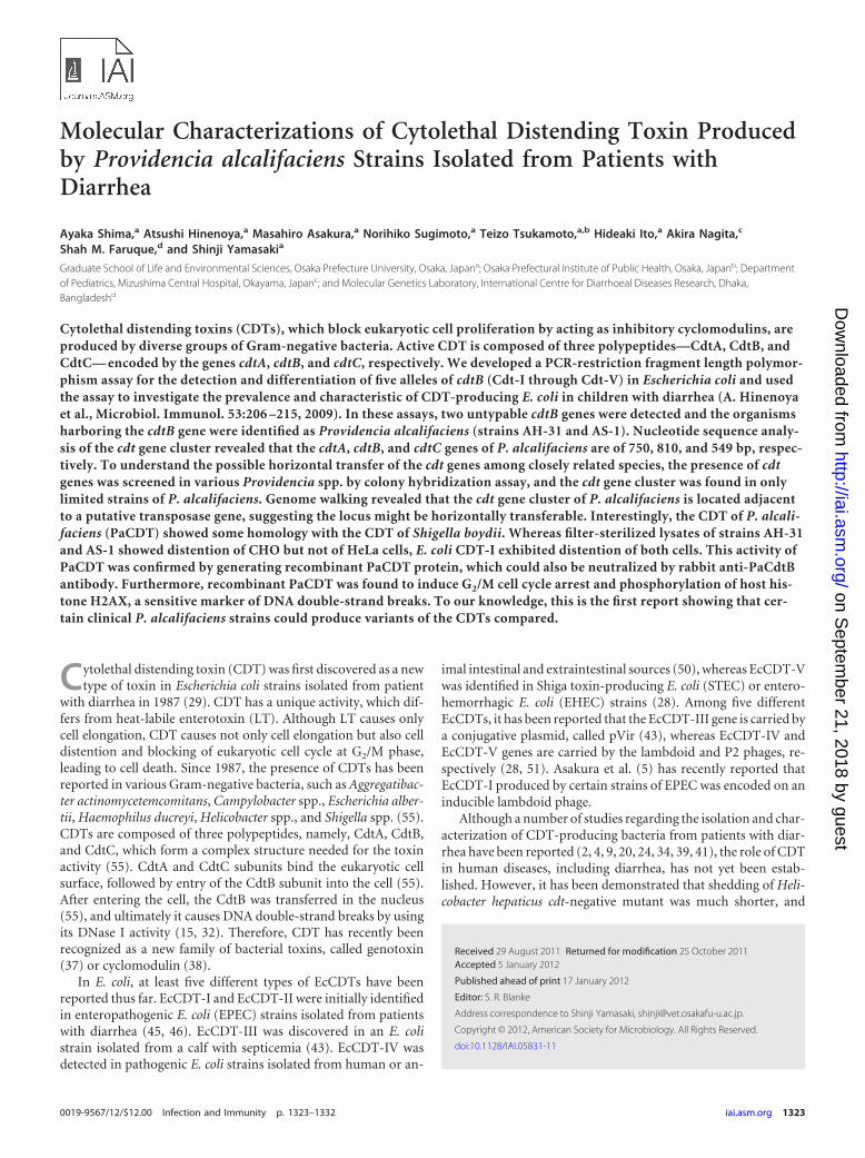
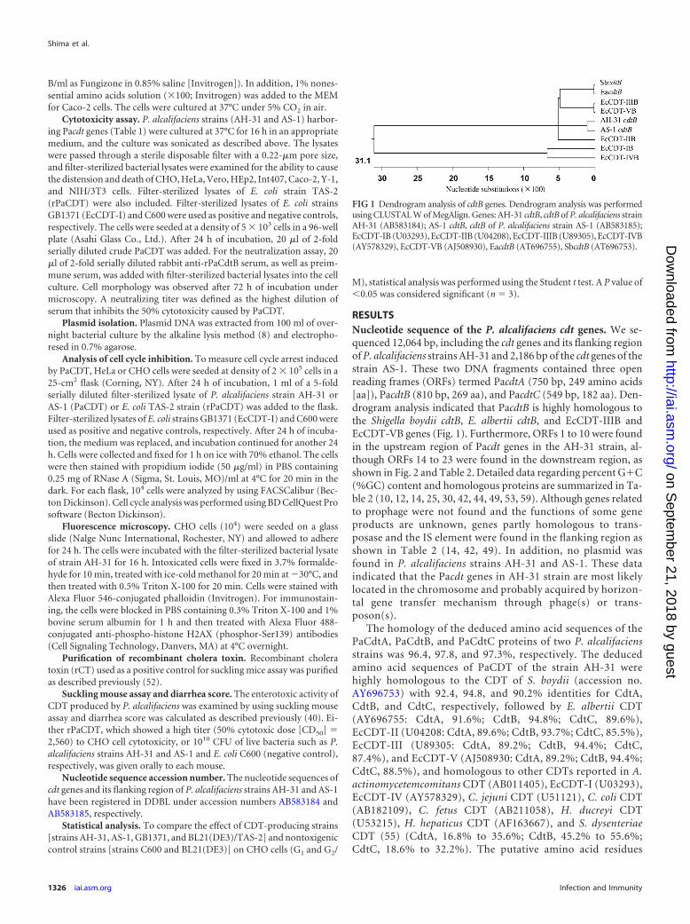
RESULTSNucleotide sequence of the P. alcalifaciens cdt genes. We se-quenced 12,064 bp, including the cdt genes and its flanking regionof P. alcalifaciens strains AH-31 and 2,186 bp of the cdt genes of thestrain AS-1. These two DNA fragments contained three openreading frames (ORFs) termed PacdtA (750 bp, 249 amino acids[aa]), PacdtB (810 bp, 269 aa), and PacdtC (549 bp, 182 aa). Den-drogram analysis indicated that PacdtB is highly homologous tothe Shigella boydii cdtB, E. albertii cdtB, and EcCDT-IIIB andEcCDT-VB genes (Fig. 1). Furthermore, ORFs 1 to 10 were foundin the upstream region of Pacdt genes in the AH-31 strain, al-though ORFs 14 to 23 were found in the downstream region, asshown in Fig. 2 and Table 2. Detailed data regarding percent G�C(%GC) content and homologous proteins are summarized in Ta-ble 2 (10, 12, 14, 25, 30, 42, 44, 49, 53, 59). Although genes relatedto prophage were not found and the functions of some geneproducts are unknown, genes partly homologous to trans-posase and the IS element were found in the flanking region asshown in Table 2 (14, 42, 49). In addition, no plasmid wasfound in P. alcalifaciens strains AH-31 and AS-1. These dataindicated that the Pacdt genes in AH-31 strain are most likelylocated in the chromosome and probably acquired by horizon-tal gene transfer mechanism through phage(s) or trans-poson(s).
The homology of the deduced amino acid sequences of thePaCdtA, PaCdtB, and PaCdtC proteins of two P. alcalifaciensstrains was 96.4, 97.8, and 97.3%, respectively. The deducedamino acid sequences of PaCDT of the strain AH-31 werehighly homologous to the CDT of S. boydii (accession no.AY696753) with 92.4, 94.8, and 90.2% identities for CdtA,CdtB, and CdtC, respectively, followed by E. albertii CDT(AY696755: CdtA, 91.6%; CdtB, 94.8%; CdtC, 89.6%),EcCDT-II (U04208: CdtA, 89.6%; CdtB, 93.7%; CdtC, 85.5%),EcCDT-III (U89305: CdtA, 89.2%; CdtB, 94.4%; CdtC,87.4%), and EcCDT-V (AJ508930: CdtA, 89.2%; CdtB, 94.4%;CdtC, 88.5%), and homologous to other CDTs reported in A.actinomycetemcomitans CDT (AB011405), EcCDT-I (U03293),EcCDT-IV (AY578329), C. jejuni CDT (U51121), C. coli CDT(AB182109), C. fetus CDT (AB211058), H. ducreyi CDT(U53215), H. hepaticus CDT (AF163667), and S. dysenteriaeCDT (55) (CdtA, 16.8% to 35.6%; CdtB, 45.2% to 55.6%;CdtC, 18.6% to 32.2%). The putative amino acid residues
FIG 1 Dendrogram analysis of cdtB genes. Dendrogram analysis was performedusing CLUSTAL W of MegAlign. Genes: AH-31 cdtB, cdtB of P. alcalifaciens strainAH-31 (AB583184); AS-1 cdtB, cdtB of P. alcalifaciens strain AS-1 (AB583185);EcCDT-IB (U03293), EcCDT-IIB (U04208), EcCDT-IIIB (U89305), EcCDT-IVB(AY578329), EcCDT-VB (AJ508930), EacdtB (AT696755), SbcdtB (AT696753).
Shima et al.
1326 iai.asm.org Infection and Immunity
on Septem
ber 21, 2018 by guesthttp://iai.asm
.org/D
ownloaded from

(H154, G191-N194, D229, and S259-V264) important forDNase I activity were perfectly conserved in PaCdtB. Thenuclear localization signals (NLS1 and NLS2) detected inEcCDT-II were also almost conserved in PaCdtB, except for two
amino acid substitutions in each of the regions (NLS1, A198D andR210N; NLS2, F262Y and S267F). The RR(X)10-20RR motif (33) inNLS2 was found to be completely conserved, suggesting that PaCDTmay enter into the nucleus for its genotoxic activity.
FIG 2 Schematic representation of the Pacdt genes and its flanking regions of the strain AH-31. Closed and open arrows indicate Pacdt genes and ORFs,respectively, located in the flanking region. The number indicated above each bar represents the %GC content of a noncoding region.
TABLE 2 Characteristics of the ORFs of the Pacdt gene and its flanking regions
ORFaGene coordinatesand direction
Geneproductsize (aa) GC%
Related bacterial proteins
Source orreferenceProduct(s) and origin
GenBankaccession no.
BLAST E-value(identity), %
1 651¢1001 117 47.86 Transposase (fragment), Xenorhabdusnematophila ATCC 19061
FN667742 4e–19 (64/110), 58% Direct submission
2 745¡885 47 49.65 Transposase (fragment), Xenorhabdusnematophila ATCC 19061
FN667742 7e–05 (22/39), 56% Direct submission
3 882¡1004 41 46.34 No hits found4 1270¡1368 33 36.36 Unknown, Comamonas testosteroni PtL5 AF076997 0.24 (15/22), 68% Direct submission5 1387¡1947 187 34.22 Unknown, Photorhabdus luminescens subsp.
laumondii TTO1BX571866 8e–65 (126/187), 67% 53
6 1812¢1898 29 42.53 Putative DNA-binding protein, Proteus mirabilisHI4320
AM942759 8e–06 (24/28), 85% 42
7 1996¢2574 193 43.01 Transposase, Salmonella enterica subsp. salamaeserovar Sofia
FJ496648 7e–86 (154/192), 80% Direct submission
8 2957¢3445 163 47.65 Putative exported protein, Citrobacter rodentiumICC168
FN543503 1e–27 (55/100), 55% 44
9 3464¢3655 64 44.27 Transposase, Proteus mirabilis HI4320 AM942759 5e–24 (54/67), 80% 4210 3604¢3801 66 41.92 Putative putative IS element transposase, Proteus
mirabilis HI4320AM942759 3e–10 (23/32), 71% 42
11* 4264¡5013 750 42.80 Cytolethal distending toxin A, Shigella boydiistrain K-1
AY696753 92% 25
12* 5034¡5843 810 42.10 Cytolethal distending toxin B, Shigella boydiistrain K-1
AY696753 95% 25
13* 5858¡6406 549 37.34 Cytolethal distending toxin C, Shigella boydiistrain K-1
AY696753 90% 25
14 6884¡7090 69 29.47 Replication initiator and transcription repressor,Pantoea stewartii subsp. stewartii
L42524 3e–16 (47/94), 50% 19
15 7390¡7542 51 41.83 Unknown, Escherichia coli strain CB853 FM210347 0.044 (22/50), 44% 1016 7539¡8168 210 45.40 Hypothetical protein, Enterobacter cloacae AY780889 4e–78 (142/205), 69% 5917 8168¡8281 38 51.75 Transposase, Escherichia coli strain BEN2908 AY857617 1e–09 (30/41), 73% 1418 8186¢8323 46 44.93 Hypothetical ORF in IS2, Klebsiella pneumoniae AY378100 0.020 (24/47), 51% 1219 8384¢8542 53 35.85 Transposase, Clostridium difficile CD196 FN538970 1.7 (15/34), 44% 4920 8629¢9567 313 37.49 EspG protein, Escherichia coli strain 71074 GQ338312 2e–44 (104/314), 33% Direct submission21 8960¡9190 77 37.66 Pyridine nucleotide-disulfide oxidoreductase,
Listeria monocytogenes HCC23CP001175 32.7 (17/50), 34% Direct submission
22 9861¢11549 563 38.90 Protein tyrosine phosphatase SptP, Salmonellaenterica subsp. enterica serovar Typhimuriumstrain SL1344
U63293 9e–82 (190/559), 33% 30
23 11579¢1195 124 31.45 Chaperone protein SicP, Salmonella entericasubsp. salamae serovar Sofia
FJ496648 2e–16 (43/93), 46% Direct submission
a *, Results obtained from the CLUSTAL W method.
CDT Produced by Providencia alcalifaciens
April 2012 Volume 80 Number 4 iai.asm.org 1327
on Septem
ber 21, 2018 by guesthttp://iai.asm
.org/D
ownloaded from

Distribution of cdt genes among Providencia spp. To exam-ine the distribution of cdt genes among Providencia spp., 18 strainsbelonging to the genus Providencia, including eight P. alcalifaciensstrains, six P. rettgeri strains, two P. rustigianii strains, one P.heimbachae strain, and one P. stuartii strain, were tested using thecdtB gene as a probe by colony hybridization assay. The probe wasable to detect only a single strain of P. alcalifaciens, and further
analysis revealed that the strain harbors a truncated cdt gene clus-ter, which was the most likely reason for the absence of CDTactivity in this strain.
Expression of recombinant PaCDT. To confirm whether P.alcalifaciens strains AH-31 and AS-1 produced PaCDTs, we at-tempted to raise an antibody against PaCdtB for Western blottingand cytotoxic assay. For this purpose, a PacdtB gene was clonedand expressed in E. coli as rPaCdtB with His tag (see the details inMaterials and Methods). Purified rPaCdtB was used to immunizerabbits and antiserum against rPaCdtB was successfully obtained.Western blotting revealed that antiserum against rPaCdtB wasspecific for PaCdtB (Fig. 3). Two bands (of about 35 and 33 kDa),which seem to be intact and degraded products of rPaCdtB, re-spectively, were obtained in lane 1 (rPaCdtB), while only one band(about 32 kDa) was obtained in lanes 2 and 3 with lysates of P.alcalifaciens strains AH-31 and AS-1, respectively. However, noreactive bands were obtained from the lysate of E. coli strainBL21(DE3) carrying the empty vector pET28a, indicating that P.alcalifaciens strains AH-31 and AS-1 produced PaCdtB and anti-body against rPaCdtB was specifically reactive to the protein.
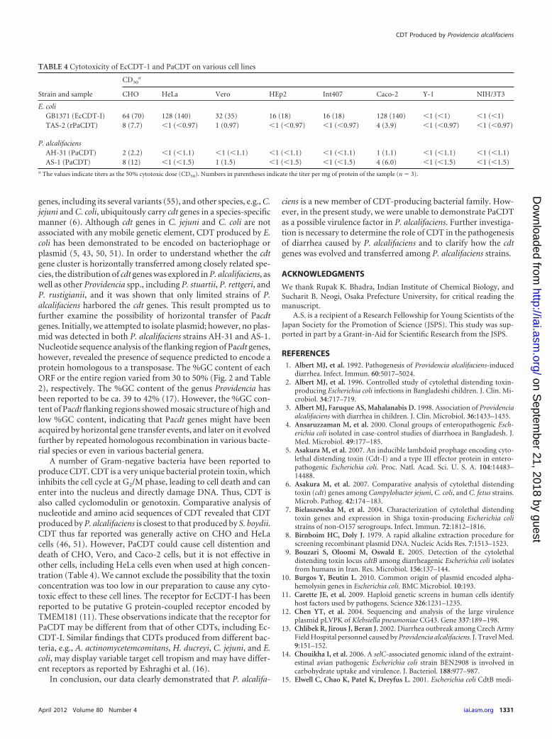
Genotoxic activity of PaCDT. Since production of PaCdtBwas confirmed in both P. alcalifaciens strains AH-31 and AS-1, wefurther examined whether biologically active CDT was producedby these strains. Filter-sterilized lysates of both P. alcalifaciensstrains AH-31 (Fig. 4C) and AS-1 (Fig. 4D) induced cell distentionon CHO cells. Although filter-sterilized lysate of E. coli strainGB1371 (EcCDT-I) used as a positive control showed cell disten-tion in both CHO (Fig. 4B) and HeLa (data not shown) cells, thefilter-sterilized lysates of P. alcalifaciens strains AH-31 and AS-1(PaCDT) did not show any morphological changes of HeLa cells(data not shown). Since the activities of PaCDT and EcCDT-I toCHO and HeLa cells were different, an additional six cell lines,namely, Vero, HEp2, Int407, Caco-2, Y-1, and NIH/3T3, were alsotested to examine any such differential activities by these two
FIG 3 Detection of PaCdtB of wild-type P. alcalifaciens by Western blotting.Whole-cell lysate of P. alcalifaciens was separated by SDS-PAGE (15%), trans-ferred into a PVDF membrane, and probed with rabbit anti-rPaCdtB antise-rum, followed by treatment with goat anti-rabbit IgG tagged with HRP as asecondary antibody. Color development was performed with 4CN-PLUS.Lanes: M, prestained SDS-PAGE broad-range marker (Bio-Rad);1, purifiedrPaCdtB; 2, bacterial lysate of AH-31 (P. alcalifaciens); 3, bacterial lysate ofAS-1 (P. alcalifaciens); 4, bacterial lysate of E. coli strain BL21(DE3) carryingempty vector pET28a. The closed and open arrowheads and the arrow indicaterPaCdtB, PaCdtB, and degraded product of rPaCdtB, respectively. The exper-iment was carried out at least thrice.
FIG 4 Cytotoxic effect of CDT produced by P. alcalifaciens on CHO cells. Whereas alteration of cellular morphology was not apparent at 72 h after exposure ofthe filter-sterilized samples from E. coli strain C600 (A) and E. coli strain BL21(DE3) carrying pET-28a as a vector control (E) to CHO cells, cytoplasmic distensionwas apparent by the filter-sterilized samples from E. coli strain GB1371 (EcCDT-I) (B), P. alcalifaciens strains AH-31 (C) and AS-1 (D), E. coli strain TSA-1(rPaCDT) (F), and P. alcalifaciens strain AH-31 with preimmunization serum (G). The CDT activity of P. alcalifaciens strain AH-31 was neutralized in thepresence of anti-rabbit rPaCdtB (H). The experiment was performed at least thrice.
Shima et al.
1328 iai.asm.org Infection and Immunity
on Septem
ber 21, 2018 by guesthttp://iai.asm
.org/D
ownloaded from

toxins. Although EcCDT-I showed cytotoxicity to Vero, HEp2,Int407, and Caco-2 cells, PaCDT was cytotoxic only to Vero andCaco-2 cells. Both toxins failed to show any cytotoxic effects onY-1 and NIH/3T3 cells. To further examine whether the cytotoxicactivity of PaCDT on CHO cells is specific, CHO cells were incu-bated with filter-sterilized lysate of P. alcalifaciens strains AH-31or AS-1 in the presence of rabbit antiserum raised against rPaCdtBor filter-sterilized lysate of E. coli strain TAS-2 (rPaCDT) alone.CHO cell distention was not observed only when filter-sterilizedlysate of P. alcalifaciens strain AH-31 or AS-1 was mixed withanti-rPaCdtB (Fig. 4H) but not with preimmunized rabbit serum(Fig. 4G). Furthermore, rPaCDT alone also caused morphologicalchanges of CHO cells (Fig. 4F), which was similar to EcCDT-I,indicating that the CHO cell cytotoxic effect was most likely due tothe CDT produced by P. alcalifaciens strain AH-31 or AS-1. Wefurther explored the DNA contents of CHO cells treated withPaCDT. The filter-sterilized lysates of both parental P. alcalifaciensstrains AH-31 (Fig. 5C) and AS-1 (Fig. 5D) caused G2/M cell cyclearrest on CHO cells (P � 0.05) compared to that of the lysate of E.coli strain C600, a CDT-negative wild-type strain used as a control(Fig. 5A). Similarly, filter-sterilized lysate of E. coli strain TAS-2producing rPaCDT (Fig. 5F) caused G2/M cell cycle arrest onCHO cells (P � 0.05) compared to that of E. coli BL21(DE3) straincarrying the empty vector pET-28a (Fig. 5E).
Since EcCDT-I has been shown to cause phosphorylation ofthe histone H2AX, a sensitive marker for double-strand DNAbreaks, we also examined whether PaCDT is involved in phos-
phorylation of the histone H2AX (�H2AX). As shown in Fig. 6,phosphorylated H2AX was visualized by direct immunofluores-cence using antibodies against �H2AX, and a strong nuclear signalwas detected in PaCDT-treated cells such as EcCDT-I-treated cellsbut not in control cells, indicating that PaCDT entered into thenucleus and induced DNA double-strand breaks of CHO cells.
Enterotoxic activity. Enterotoxicity of CDT produced by P.alcalifaciens was examined by suckling mouse assay. CruderPaCDT prepared from E. coli strain TAS-2, rCT, or PBS wasorally administered to each mouse, followed by an evaluation ofthe diarrheal score for each sample. rCT and PBS showed 100%positive and negative results, respectively. However, cruderPaCDT did not show any enterotoxicity (data not shown).
Subsequently, live bacteria were orally inoculated into sucklingmice to see whether there was any fluid accumulation. Although1010 CFU of P. alcalifaciens strain AS-1 did not cause any diarrheain 12 mice tested, the same dose of P. alcalifaciens strain AH-31could cause diarrhea in 7 of 12 mice. Further study is needed toprove the enterotoxic activity of purified PaCDT.
DISCUSSION
Despite tremendous efforts toward understanding the pathogen-esis of P. alcalifaciens, it is still unclear how P. alcalifaciens causesdiarrhea in humans. In the present study we show that P. alcalifa-ciens strains isolated from patients with diarrhea could produceCDT, and this is the first report, to our knowledge, regarding the
FIG 5 Analysis of CHO cell cycle after treatment with various preparations. The cell cycle distribution of 10,000 cells was determined by flow cytometry, andrepresentative results are shown. The average percentages and the standard deviations of cells in each cell cycle phase calculated with three independentexperiments are indicated. CHO cells were unaffected when exposed to lysates of nontoxigenic control E. coli strain such as C600 (A) or BL21(DE3) carrying theempty vector pET-28a (E) cells, but the cells were blocked in the G2/M cell cycle phase when they were exposed to EcCDT-I (B) or PaCDT (C, D, and F). DNAcontent of CHO cells was monitored by flow cytometry as described in Materials and Methods. The effects of EcCDT-I (B) and PaCDT (C and D) on the CHOcell cycle (G1 and G2/M) were compared to the lysate of E. coli strain C600 (A) used as a negative control. The effect of rPaCDT (F) on CHO cell cycle (G1 andG2/M) was compared to that of E. coli strain BL21(DE3) carrying the empty vector pET-28a (F). *, P � 0.05 (Student t test, n � 3).
CDT Produced by Providencia alcalifaciens
April 2012 Volume 80 Number 4 iai.asm.org 1329
on Septem
ber 21, 2018 by guesthttp://iai.asm
.org/D
ownloaded from

production of the toxin by the genus Providencia, including P.alcalifaciens.
Genus Providencia, belonging to the family Enterobacteriaceae,consists of five species: P. alcalifaciens, P. stuartii, P. rettgeri, P.rustigianii, and P. heimbachae (27). Among these, P. alcalifacienshas been described as a causative agent of diarrhea because a num-ber of P. alcalifaciens strains were isolated from patients with di-arrhea in developing countries (21, 22, 47, 48). Indeed, a casecontrol study conducted by Albert et al. (3) demonstrated that P.alcalifaciens was associated with diarrhea in children in Bangla-desh. Haynes and Hawkey (22) reported that P. alcalifaciens wasassociated with traveler’s diarrhea. Yoh et al. (56) showed that notonly P. alcalifaciens but also P. rettgeri in particular is an importantpathogen for traveler’s diarrhea. Furthermore, two large out-breaks of food poisoning caused by P. alcalifaciens have been re-ported from Japan and the Czech Republic (13, 36).
Several studies demonstrated that P. alcalifaciens is able to in-vade cultured epithelial cells (1, 21, 26). Invasion was also ob-served in intestinal tissues by using a removable intestinal tie adultrabbit diarrhea (RITARD) model and an adult rabbit ileal loopmodel (1, 35). Although invasion was considered as one of thevirulence mechanisms to cause diarrhea by P. alcalifaciens strains,noninvasive P. alcalifaciens were also isolated from patients withdiarrhea (21, 48). In our study, one strain, P. alcalifaciens AH-31,showed invasiveness to HeLa cells; however, another strain, P.alcalifaciens strain AS-1, did not show any invasiveness (data notshown). This observation suggests that invasiveness could be oneof several possible virulence mechanisms. Therefore, other mech-
anisms by which P. alcalifaciens is involved in diarrhea have beenconsidered but are not fully understood. Until the present study,no toxin was reported to be the virulence factor of P. alcalifaciens.CDT produced by P. alcalifaciens may be a candidate virulencefactor in these strains other than invasiveness. Colony hybridiza-tion assay revealed that cdt genes are present only in certain strainsof P. alcalifaciens. In addition to the cytotoxicity test, we also at-tempted to examine the enterotoxic activity of PaCDT in a suck-ling mouse assay. However, the suckling mouse assay did not showany enterotoxicity by crude concentrated rPaCDT, but one of theP. alcalifaciens strains (the strain AH-31) showed enterotoxicitywhen live bacteria were orally administered (Table 3). Based onthese findings, it is not clear whether PaCDT is indeed a virulencefactor for this pathogen, and further studies are needed to shedlight on this aspect.
Some bacterial species, e.g., certain strains of E. coli carry cdt
FIG 6 Genotoxic effect of PaCDT. CHO cells were treated with filter-sterilized lysates of bacteria producing EcCDT-I (GB1371) or PaCDT (AH-31). After 16 hof treatment, the cells were stained as described in Materials and Methods with Alexa Fluor 546-conjugated phalloidin (upper panels) or with fluoresceinisothiocyanate-conjugated anti-phospho-histone H2AX (H2AX) monoclonal antibody (middle panels). EcCDT-I- and PaCDT-treated cells exhibited nuclearH2AX indicating host DNA double-strand breaks, enlarged nuclei and cell bodies, and the absence of mitotic features. Filter-sterilized lysate of E. coli C600 wasused as a negative control. The experiment was repeated at least three times.
TABLE 3 Suckling mouse assay with Providencia alcalifaciens
SampleDiarrhea score (no. of positiveanimals/total no. of animals)a
E. coli C600 0 (0/12)P. alcalifaciens AH-31 58 (7/12)P. alcalifaciens AS-1 0 (0/12)rCTb 100 (9/9)a The animals excreting stained loose and/or watery feces over 24 h after sampleadministration were judged positive.b Recombinant cholera toxin.
Shima et al.
1330 iai.asm.org Infection and Immunity
on Septem
ber 21, 2018 by guesthttp://iai.asm
.org/D
ownloaded from
genes, including its several variants (55), and other species, e.g., C.jejuni and C. coli, ubiquitously carry cdt genes in a species-specificmanner (6). Although cdt genes in C. jejuni and C. coli are notassociated with any mobile genetic element, CDT produced by E.coli has been demonstrated to be encoded on bacteriophage orplasmid (5, 43, 50, 51). In order to understand whether the cdtgene cluster is horizontally transferred among closely related spe-cies, the distribution of cdt genes was explored in P. alcalifaciens, aswell as other Providencia spp., including P. stuartii, P. rettgeri, andP. rustigianii, and it was shown that only limited strains of P.alcalifaciens harbored the cdt genes. This result prompted us tofurther examine the possibility of horizontal transfer of Pacdtgenes. Initially, we attempted to isolate plasmid; however, no plas-mid was detected in both P. alcalifaciens strains AH-31 and AS-1.Nucleotide sequence analysis of the flanking region of Pacdt genes,however, revealed the presence of sequence predicted to encode aprotein homologous to a transposase. The %GC content of eachORF or the entire region varied from 30 to 50% (Fig. 2 and Table2), respectively. The %GC content of the genus Providencia hasbeen reported to be ca. 39 to 42% (17). However, the %GC con-tent of Pacdt flanking regions showed mosaic structure of high andlow %GC content, indicating that Pacdt genes might have beenacquired by horizontal gene transfer events, and later on it evolvedfurther by repeated homologous recombination in various bacte-rial species or even in various bacterial genera.
A number of Gram-negative bacteria have been reported toproduce CDT. CDT is a very unique bacterial protein toxin, whichinhibits the cell cycle at G2/M phase, leading to cell death and canenter into the nucleus and directly damage DNA. Thus, CDT isalso called cyclomodulin or genotoxin. Comparative analysis ofnucleotide and amino acid sequences of CDT revealed that CDTproduced by P. alcalifaciens is closest to that produced by S. boydii.CDT thus far reported was generally active on CHO and HeLacells (46, 51). However, PaCDT could cause cell distention anddeath of CHO, Vero, and Caco-2 cells, but it is not effective inother cells, including HeLa cells even when used at high concen-tration (Table 4). We cannot exclude the possibility that the toxinconcentration was too low in our preparation to cause any cyto-toxic effect to these cell lines. The receptor for EcCDT-I has beenreported to be putative G protein-coupled receptor encoded byTMEM181 (11). These observations indicate that the receptor forPaCDT may be different from that of other CDTs, including Ec-CDT-I. Similar findings that CDTs produced from different bac-teria, e.g., A. actinomycetemcomitans, H. ducreyi, C. jejuni, and E.coli, may display variable target cell tropism and may have differ-ent receptors as reported by Eshraghi et al. (16).
In conclusion, our data clearly demonstrated that P. alcalifa-
ciens is a new member of CDT-producing bacterial family. How-ever, in the present study, we were unable to demonstrate PaCDTas a possible virulence factor in P. alcalifaciens. Further investiga-tion is necessary to determine the role of CDT in the pathogenesisof diarrhea caused by P. alcalifaciens and to clarify how the cdtgenes was evolved and transferred among P. alcalifaciens strains.
ACKNOWLEDGMENTS
We thank Rupak K. Bhadra, Indian Institute of Chemical Biology, andSucharit B. Neogi, Osaka Prefecture University, for critical reading themanuscript.
A.S. is a recipient of a Research Fellowship for Young Scientists of theJapan Society for the Promotion of Science (JSPS). This study was sup-ported in part by a Grant-in-Aid for Scientific Research from the JSPS.
REFERENCES1. Albert MJ, et al. 1992. Pathogenesis of Providencia alcalifaciens-induced
diarrhea. Infect. Immun. 60:5017–5024.2. Albert MJ, et al. 1996. Controlled study of cytolethal distending toxin-
producing Escherichia coli infections in Bangladeshi children. J. Clin. Mi-crobiol. 34:717–719.
3. Albert MJ, Faruque AS, Mahalanabis D. 1998. Association of Providenciaalcalifaciens with diarrhea in children. J. Clin. Microbiol. 36:1433–1435.
4. Ansaruzzaman M, et al. 2000. Clonal groups of enteropathogenic Esch-erichia coli isolated in case-control studies of diarrhoea in Bangladesh. J.Med. Microbiol. 49:177–185.
5. Asakura M, et al. 2007. An inducible lambdoid prophage encoding cyto-lethal distending toxin (Cdt-I) and a type III effector protein in entero-pathogenic Escherichia coli. Proc. Natl. Acad. Sci. U. S. A. 104:14483–14488.
6. Asakura M, et al. 2007. Comparative analysis of cytolethal distendingtoxin (cdt) genes among Campylobacter jejuni, C. coli, and C. fetus strains.Microb. Pathog. 42:174 –183.
7. Bielaszewska M, et al. 2004. Characterization of cytolethal distendingtoxin genes and expression in Shiga toxin-producing Escherichia colistrains of non-O157 serogroups. Infect. Immun. 72:1812–1816.
8. Birnboim HC, Doly J. 1979. A rapid alkaline extraction procedure forscreening recombinant plasmid DNA. Nucleic Acids Res. 7:1513–1523.
9. Bouzari S, Oloomi M, Oswald E. 2005. Detection of the cytolethaldistending toxin locus cdtB among diarrheagenic Escherichia coli isolatesfrom humans in Iran. Res. Microbiol. 156:137–144.
10. Burgos Y, Beutin L. 2010. Common origin of plasmid encoded alpha-hemolysin genes in Escherichia coli. BMC Microbiol. 10:193.
11. Carette JE, et al. 2009. Haploid genetic screens in human cells identifyhost factors used by pathogens. Science 326:1231–1235.
12. Chen YT, et al. 2004. Sequencing and analysis of the large virulenceplasmid pLVPK of Klebsiella pneumoniae CG43. Gene 337:189 –198.
13. Chlibek R, Jirous J, Beran J. 2002. Diarrhea outbreak among Czech ArmyField Hospital personnel caused by Providencia alcalifaciens. J. Travel Med.9:151–152.
14. Chouikha I, et al. 2006. A selC-associated genomic island of the extraint-estinal avian pathogenic Escherichia coli strain BEN2908 is involved incarbohydrate uptake and virulence. J. Bacteriol. 188:977–987.
15. Elwell C, Chao K, Patel K, Dreyfus L. 2001. Escherichia coli CdtB medi-
TABLE 4 Cytotoxicity of EcCDT-1 and PaCDT on various cell lines
Strain and sample
CD50a
CHO HeLa Vero HEp2 Int407 Caco-2 Y-1 NIH/3T3
E. coliGB1371 (EcCDT-I) 64 (70) 128 (140) 32 (35) 16 (18) 16 (18) 128 (140) �1 (�1) �1 (�1)TAS-2 (rPaCDT) 8 (7.7) �1 (�0.97) 1 (0.97) �1 (�0.97) �1 (�0.97) 4 (3.9) �1 (�0.97) �1 (�0.97)
P. alcalifaciensAH-31 (PaCDT) 2 (2.2) �1 (�1.1) �1 (�1.1) �1 (�1.1) �1 (�1.1) 1 (1.1) �1 (�1.1) �1 (�1.1)AS-1 (PaCDT) 8 (12) �1 (�1.5) 1 (1.5) �1 (�1.5) �1 (�1.5) 4 (6.0) �1 (�1.5) �1 (�1.5)
a The values indicate titers as the 50% cytotoxic dose (CD50). Numbers in parentheses indicate the titer per mg of protein of the sample (n � 3).
CDT Produced by Providencia alcalifaciens
April 2012 Volume 80 Number 4 iai.asm.org 1331
on Septem
ber 21, 2018 by guesthttp://iai.asm
.org/D
ownloaded from

ates cytolethal distending toxin cell cycle arrest. Infect. Immun. 69:3418 –3422.
16. Eshraghi A, et al. 2010. Cytolethal distending toxin family members aredifferentially affected by alterations in host glycans and membrane cho-lesterol. J. Biol. Chem. 285:18199 –18207.
17. Falkow S, Ryman IR, Washington O. 1962. Deoxyribonucleic acid basecomposition of Proteus and Providencia organism. J. Bacteriol. 83:1318 –1321.
18. Friedrich AW, et al. 2006. Cytolethal distending toxin in Escherichia coliO157:H7: spectrum of conservation, structure, and endothelial toxicity. J.Clin. Microbiol. 44:1844 –1846.
19. Fu JF, Chang HC, Chen YM, Chang YS, Liu ST. 1995. Sequence analysisof an Erwinia stewartii plasmid, pSW100. Plasmid 34:75– 84.
20. Ghilardi AC, Gomes TA, Trabulsi LR. 2001. Production of cytolethaldistending toxin and other virulence characteristics of Escherichia colistrains of serogroup O86. Mem. Inst. Oswaldo Cruz 96:703–708.
21. Guth BEC, Perrella E. 1996. Prevalence of invasive ability and othervirulence-associated characteristics in Providencia alcalifaciens strains iso-lated in Sao Paulo, Brazil. J. Med. Microbiol. 45:459 – 462.
22. Haynes J, Hawkey PM. 1989. Providencia alcalifaciens and traveller’sdiarrhoea. BMJ 299:94 –95.
23. Hinenoya A, et al. 2007. Cytolethal distending toxin (Cdt)-producingEscherichia coli isolated from a child with bloody diarrhea in Japan. Mi-crobiol. Immunol. 51:435– 438.
24. Hinenoya A, et al. 2009. Prevalence and characteristics of cytolethaldistending toxin-producing Escherichia coli from children with diarrhea inJapan. Microbiol. Immunol. 53:206 –215.
25. Hyma KE, et al. 2005. Evolutionary genetics of a new pathogenic Esche-richia species: Escherichia albertii and related Shigella boydii strains J. Bac-teriol. 187:619 – 628.
26. Janda JM, Abbott SL, Woodward D, Khashe S. 1998. Invasion of HEp-2and other eukaryotic cell lines by Providenciae: further evidence support-ing the role of Providencia alcalifaciens in bacterial gastroenteritis. Curr.Microbiol. 37:159 –165.
27. Janda JM, Abbott LS. 2006. The enterobacteria, 2nd ed, p 279 –299. ASMPress, Washington, DC.
28. Janka A, et al. 2003. Cytolethal distending toxin gene cluster in entero-hemorrhagic Escherichia coli O157:H� and O157:H7: characterizationand evolutionary considerations. Infect. Immun. 71:3634 –3638.
29. Johnson WM, Lior H. 1987. Response of Chinese hamster ovary cells toa cytolethal distending toxin (CDT) of Escherichia coli and possible mis-interpretation as heat-labile (LT) enterotoxin. FEMS Microbiol. Lett. 43:19 –23.
30. Kaniga K, Uralil J, Bliska JB, Galán JE. 1996. A secreted protein tyrosinephosphatase with modular effector domains in the bacterial pathogenSalmonella typhimurium. Mol. Microbiol. 21:633– 641.
31. Laemmli UK. 1970. Cleavage of structural proteins during the assembly ofthe head of bacteriophage T4. Nature 227:680 – 685.
32. Lara-Tejero M, Galan JE. 2000. A bacterial toxin that controls cell cycleprogression as a deoxyribonuclease I-like protein. Science 290:354 –357.
33. Leslie A, McSweeney LA, Dreyfus LA. 2004. Nuclear localization of theEscherichia coli cytolethal distending toxin CdtB subunit. Cell. Microbiol.6:447– 458.
34. Marques LR, Tavechio AT, Abe CM, Gomes TA. 2003. Search forcytolethal distending toxin production among fecal Escherichia coli iso-lates from Brazilian children with diarrhea and without diarrhea. J. Clin.Microbiol. 41:2206 –2208.
35. Mathan MM, Mathan VI, Albert MJ. 1993. Electron microscopic studyof the attachment and penetration of rabbit intestinal epithelium by Provi-dencia alcalifaciens. J. Pathol. 171:67–71.
36. Murata T, et al. 2001. A large outbreak of food-borne infection attributedto Providencia alcalifaciens. J. Infect. Dis. 184:1050 –1055.
37. Nesic D, Hsu Y, Stebbins CE. 2004. Assembly and function of a bacterialgenotoxin. Nature 429:429 – 433.
38. Nougayrede JP, Taibe F, Rycke JD, Oswald E. 2005. Cyclomodulins:bacterial effectors that modulate the eukaryotic cell cycle. Trends Micro-biol. 13:103–110.
39. Okeke IN, Lamikanra A, Steinruck H, Kaper JB. 2000. Characterizationof Escherichia coli strains from cases of childhood diarrhea in provincialsouthwestern Nigeria. J. Clin. Microbiol. 38:7–12.
40. Okuda J, Fukumoto M, Takeda Y, Nishibuchi M. 1997. Examination ofdiarrheagenicity of cytolethal distending toxin: suckling mouse responseto the products of the cdtABC genes of Shigella dysenteriae. Infect. Immun.65:428 – 433.
41. Pandey M, et al. 2003. Association of cytolethal distending toxin locuscdtB with enteropathogenic Escherichia coli isolated from patients withacute diarrhea in Calcutta, India. J. Clin. Microbiol. 41:5277–5281.
42. Pearson MM, et al. 2008. Complete genome sequence of uropathogenicProteus mirabilis, a master of both adherence and motility. J. Bacteriol.190:4027– 4037.
43. Pérès SY, et al. 1997. A new cytolethal distending toxin (CDT) fromEscherichia coli producing CNF2 blocks HeLa cell division in G2/M phase.Mol. Microbiol. 24:1095–1107.
44. Petty NK, et al. 2010. The Citrobacter rodentium genome sequence revealsconvergent evolution with human pathogenic Escherichia coli. J. Bacteriol.192:525–538.
45. Pickett CL, Cottle DL, Pesci EC, Bikah G. 1994. Cloning, sequencing,and expression of the Escherichia coli cytolethal distending toxin genes.Infect. Immun. 62:1046 –1051.
46. Scott DA, Kaper JB. 1994. Cloning and sequencing of the genes encodingEscherichia coli cytolethal distending toxin. Infect. Immun. 62:244 –251.
47. Sen R. 1962. Isolation of strains of Providencia group from cases withdiarrhoea in Ibadan, Nigeria, West Africa. Indian J. Med. Res. 50:622– 626.
48. Sobreira M, Leal NC, Magalhães M, Guth BE, Almeida AM. 2001.Molecular analysis of clinical isolates of Providencia alcalifaciens. J. Med.Microbiol. 50:29 –34.
49. Stabler RA, et al. 2009. Comparative genome and phenotypic analysis ofClostridium difficile 027 strains provides insight into the evolution of ahypervirulent bacterium. Genome Biol. 10:R102.
50. Tóth I, Herault F, Beutin L, Oswald E. 2003. Production of cytolethaldistending toxins by pathogenic Escherichia coli strains isolated from hu-man and animal sources: establishment of the existence of a new cdt vari-ant (type IV). J. Clin. Microbiol. 41:4285– 4291.
51. Tóth I, et al. 2009. Cytolethal distending toxin type I and type IV genes areframed with lambdoid prophage genes in extraintestinal pathogenic Esch-erichia coli. Infect. Immun. 77:492–500.
52. Uesaka Y, et al. 1994. Simple method of purification of Escherichia coliheat-labile enterotoxin and cholera toxin using immobilized galactose.Microb. Pathog. 16:71–76.
53. Williamson VM, Kaya HK. 2003. Sequence of a symbiont. Nat. Biotech-nol. 21:1294 –1295.
54. Wising C, Molne L, Jonsson IM, Ahlman K, Lagergard T. 2005. Thecytolethal distending toxin of Haemophilus ducreyi aggravates dermal le-sions in a rabbit model of chancroid. Microbes Infect. 7:867– 874.
55. Yamasaki S, et al. 2006. Cytolethal distending toxin (CDT): genetic di-versity, structure and role in diarrheal disease. Toxin Rev. 25:61– 88.
56. Yoh M, et al. 2005. Importance of Providencia species as a major cause oftravellers’ diarrhoea. J. Med. Microbiol. 54:1077–1082.
57. Young VB, et al. 2004. In vitro and in vivo characterization of Helicobacterhepaticus cytolethal distending toxin mutants. Infect. Immun. 72:2521–2527.
58. Yutsudo T, Nakabayashi N, Hirayama T, Takeda Y. 1987. Purificationand some properties of a Vero toxin from Escherichia coli O157:H7 that isimmunologically unrelated to Shiga toxin. Microb. Pathog. 3:21–30.
59. Yu YS, Du XX, Zhou ZH, Chen YG, Li LJ. 2006. First isolation ofblaIMI-2 in an Enterobacter cloacae clinical isolate from China. Antimi-crob. Agents Chemother. 50:1610 –1611.
Shima et al.
1332 iai.asm.org Infection and Immunity
on Septem
ber 21, 2018 by guesthttp://iai.asm
.org/D
ownloaded from