molecular characterization of dematiaceous root endophytes
TRANSCRIPT

1397
S. K. HARNEY*, S. O. ROGERS AND C. J. K. WANG
Faculty of Environmental and Forest Biology, State University of New York, College of Environmental Science and Forestry, Syracuse, New York
13210-2788, U.S.A.
Sterile dematiaceous fungi are commonly isolated from plant roots. They are often assigned to Mycelium radicis atrovirens, a name
originally proposed for black, sterile, fast-growing, pseudomycorrhizal fungi. Dematiaceous fungi isolated from roots may be
mutualists, commensalists, or pathogens and, in the absence of sporulation, identification is not possible. Forty-six isolates of
dematiaceous fungi from the roots of different hosts and locations were characterized using restriction site mapping of polymerase
chain reaction amplified nuclear ribosomal DNA internal transcribed spacers. The restriction site maps were compared to identified
dematiaceous mycorrhizal and pseudomycorrhizal fungi. Computer generated trees (UPGMA and parsimony analysis) characterized
two unknown isolates as Phialophora finlandia, an ectendomycorrhizal fungus. The majority of the isolates were characterized as
Phialocephala fortinii-like. Phialocephala fortinii has been reported as both pathogenic and non-pathogenic in a number of hosts. There
was variation within the P. fortinii-like group suggesting intraspecific variation or a species complex.
Sterile, dematiaceous fungi are commonly isolated from roots
and often remain unidentified (e.g. Melin, 1923 ; Haselwandter
& Read, 1980 ; Currah et al., 1988 ; Stoyke & Currah, 1991 ;
O’Dell & Trappe, 1992). In a recent study examining the
mycorrhizas of outplanted white pine seedlings, a large
number of isolates were non-sporulating and dark (Wilcox,
Wang, Prabhu, LoBuglio & Harney, unpublished), of the sort
commonly assigned to Mycelium radicis atrovirens (Mra).
Culture manipulation allowed Wang & Wilcox (1985) to
induce three Mra-like isolates to sporulate : Chloridium
paucisporumC. J. K. Wang & H. E. Wilcox, Phialophora finlandia
C. J. K. Wang & H. E. Wilcox, and Phialocephala fortinii C. J. K.
Wang & H. E. Wilcox. Chloridium paucisporum (Wilcox &
Ganmore-Neumann, 1974 ; Wilcox et al., 1974) and P. finlandia
(Wilcox & Wang, 1987a, b) formed ectendomycorrhizas with
Pinus resinosa Ait. and mainly ectomycorrhizas with Picea
rubens Sarg. Phialocephala fortinii formed pseudomycorrhizal
(pathogenic) associations with Picea mariana (Mill.) B.S.P.
(Richard & Fortin, 1974), P. resinosa and P. rubens (Wilcox &
Wang, 1987a ; Wang & Wilcox, 1985). Phialophora finlandia
and C. paucisporum, morphologically similar to the cosmo-
politan ectomycorrhizal fungus Cenococcum geophilum, have
never been reported elsewhere.
Phialocephala fortinii has been isolated from orchid roots
(Currah et al., 1987), alpine plant roots (Ericaceae, Rosaceae)
(Stoyke & Currah, 1991) and lupin roots (O’Dell & Trappe,
1992). Its role in orchidaceous and ericaceous plants is
uncertain, although it does not appear to cause serious
* Current address : Biology Department, San Diego State University, SanDiego, CA 92182, U.S.A.
damage (e.g. Stoyke & Currah, 1991). In lupin (Lupinus
latifolius Agardh.) and Pinus contorta Dougl. there is evidence
that it lives within the root without penetrating the vascular
tissue or forming any mycorrhizal structures, and is neither
beneficial nor pathogenic (O’Dell et al., 1993).
Because the role of dematiaceous endophytes is variable,
accurate identification of root isolates is the first step in
eliciting their ecological role. Culture manipulation is time
consuming, and often unsuccessful. PCR (polymerase chain
reaction) amplification and restriction site mapping of the
internal transcribed spacer (ITS) region have previously been
utilized to characterize and identify ectomycorrhizal fungi
(Gardes et al., 1991 ; Gardes & Bruns, 1993). We evaluated
restriction site mapping and phenetic analysis of PCR amplified
ribosomal DNA (rDNA) ITS regions as a means to characterize
a number of Mra-like isolates.
MATERIALS AND METHODS
Forty-six isolates were used in this study (Table 1). Cultures
were maintained on 2% malt extract agar (MEA). All
unknown isolates were sterile and chosen either because they
resemble P. finlandia or P. fortinii (Mra-like), or randomly from
a selection of dematiaceous isolates.
Isolates were incubated at room temperature in 2% liquid
malt extract broth, either in static or agitating conditions on
a rotating shaker, for DNA extraction. Mycelium was
harvested by vacuum filtration over filter paper (Whatman no.
1), frozen at ®80 °C and lyophilized. Lyophilized mycelium
was stored at ®20°.Total DNA was extracted using a CTAB
(cetyltrimethylammonium bromide) micropreparation method
Mycol. Res. 101 (11), 1397–1404 (1997) Printed in the United Kingdom
Molecular characterization of dematiaceous root endophytes
Dematiaceous root endophytes 1398
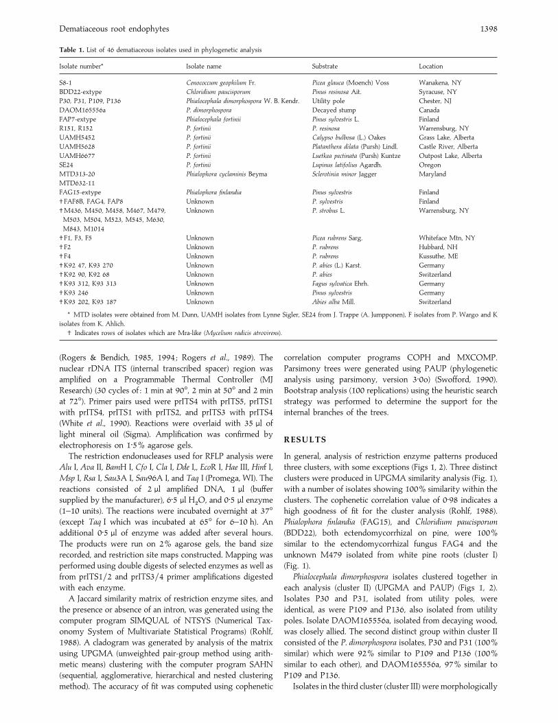
Table 1. List of 46 dematiaceous isolates used in phylogenetic analysis
Isolate number* Isolate name Substrate Location
S8-1 Cenococcum geophilum Fr. Picea glauca (Moench) Voss Wanakena, NY
BDD22-extype Chloridium paucisporum Pinus resinosa Ait. Syracuse, NY
P30, P31, P109, P136 Phialocephala dimorphospora W. B. Kendr. Utility pole Chester, NJ
DAOM165556a P. dimorphospora Decayed stump Canada
FAP7-extype Phialocephala fortinii Pinus sylvestris L. Finland
R151, R152 P. fortinii P. resinosa Warrensburg, NY
UAMH5452 P. fortinii Calypso bulbosa (L.) Oakes Grass Lake, Alberta
UAMH5628 P. fortinii Platanthera dilata (Pursh) Lindl. Castle River, Alberta
UAMH6677 P. fortinii Luetkea pectinata (Pursh) Kuntze Outpost Lake, Alberta
SE24 P. fortinii Lupinus latifolius Agardh. Oregon
MTD313-20 Phialophora cyclaminis Beyma Sclerotinia minor Jagger Maryland
MTD632-11
FAG15-extype Phialophora finlandia Pinus sylvestris Finland
† FAF8B, FAG4, FAP8 Unknown P. sylvestris Finland
†M436, M450, M458, M467, M479, Unknown P. strobus L. Warrensburg, NY
M503, M504, M523, M545, M630,
M843, M1014
† F1, F3, F5 Unknown Picea rubrens Sarg. Whiteface Mtn, NY
† F2 Unknown P. rubrens Hubbard, NH
† F4 Unknown P. rubrens Kussuthe, ME
†K92 47, K93 270 Unknown P. abies (L.) Karst. Germany
†K92 90, K92 68 Unknown P. abies Switzerland
†K93 312, K93 313 Unknown Fagus sylvatica Ehrh. Germany
†K93 246 Unknown Pinus sylvestris Germany
†K93 202, K93 187 Unknown Abies alba Mill. Switzerland
* MTD isolates were obtained from M. Dunn, UAMH isolates from Lynne Sigler, SE24 from J. Trappe (A. Jumpponen), F isolates from P. Wargo and K
isolates from K. Ahlich.
† Indicates rows of isolates which are Mra-like (Mycelium radicis atrovirens).
(Rogers & Bendich, 1985, 1994 ; Rogers et al., 1989). The
nuclear rDNA ITS (internal transcribed spacer) region was
amplified on a Programmable Thermal Controller (MJ
Research) (30 cycles of : 1 min at 90°, 2 min at 50° and 2 min
at 72°). Primer pairs used were prITS4 with prITS5, prITS1
with prITS4, prITS1 with prITS2, and prITS3 with prITS4
(White et al., 1990). Reactions were overlaid with 35 µl of
light mineral oil (Sigma). Amplification was confirmed by
electrophoresis on 1±5% agarose gels.
The restriction endonucleases used for RFLP analysis were
Alu I, Ava II, BamH I, Cfo I, Cla I, Dde I,, EcoR I, Hae III, Hinf I,
Msp I, Rsa I, Sau3A I, Sau96A I, and Taq I (Promega, WI). The
reactions consisted of 2 µl amplified DNA, 1 µl (buffer
supplied by the manufacturer), 6±5 µl H#O, and 0±5 µl enzyme
(1–10 units). The reactions were incubated overnight at 37°(except Taq I which was incubated at 65° for 6–10 h). An
additional 0±5 µl of enzyme was added after several hours.
The products were run on 2% agarose gels, the band size
recorded, and restriction site maps constructed. Mapping was
performed using double digests of selected enzymes as well as
from prITS1}2 and prITS3}4 primer amplifications digested
with each enzyme.
A Jaccard similarity matrix of restriction enzyme sites, and
the presence or absence of an intron, was generated using the
computer program SIMQUAL of NTSYS (Numerical Tax-
onomy System of Multivariate Statistical Programs) (Rohlf,
1988). A cladogram was generated by analysis of the matrix
using UPGMA (unweighted pair-group method using arith-
metic means) clustering with the computer program SAHN
(sequential, agglomerative, hierarchical and nested clustering
method). The accuracy of fit was computed using cophenetic
correlation computer programs COPH and MXCOMP.
Parsimony trees were generated using PAUP (phylogenetic
analysis using parsimony, version 3±0o) (Swofford, 1990).
Bootstrap analysis (100 replications) using the heuristic search
strategy was performed to determine the support for the
internal branches of the trees.
RESULTS
In general, analysis of restriction enzyme patterns produced
three clusters, with some exceptions (Figs 1, 2). Three distinct
clusters were produced in UPGMA similarity analysis (Fig. 1),
with a number of isolates showing 100% similarity within the
clusters. The cophenetic correlation value of 0±98 indicates a
high goodness of fit for the cluster analysis (Rohlf, 1988).
Phialophora finlandia (FAG15), and Chloridium paucisporum
(BDD22), both ectendomycorrhizal on pine, were 100%
similar to the ectendomycorrhizal fungus FAG4 and the
unknown M479 isolated from white pine roots (cluster I)
(Fig. 1).
Phialocephala dimorphospora isolates clustered together in
each analysis (cluster II) (UPGMA and PAUP) (Figs 1, 2).
Isolates P30 and P31, isolated from utility poles, were
identical, as were P109 and P136, also isolated from utility
poles. Isolate DAOM165556a, isolated from decaying wood,
was closely allied. The second distinct group within cluster II
consisted of the P. dimorphospora isolates, P30 and P31 (100%
similar) which were 92% similar to P109 and P136 (100%
similar to each other), and DAOM165556a, 97% similar to
P109 and P136.
Isolates in the third cluster (cluster III) were morphologically

S. K. Harney, S. O. Rogers and C. J. K. Wang 1399
BDD22 Chloridium paucisporum – extypeFAG4FAG15 Phialophora finlandia – extypeM479P30 Phialocephala dimorphospora
FAP7 Phialocephala fortinii – extypeDAOM165556a P. dimorphospora
P31 Phialocephala dimorphosporaP109 Phialocephala dimorphosporaP136 Phialocephala dimorphospora
R151 Phialocephala fortiniiUAMH6677 Phialocephala fortiniiK93 187K93 246K93 312M450M458M467M630SE24 Phialocephala fortiniiUAMH5452 Phialocephala fortiniiK93 202M843FAF8BK92 47F1F4K92 68M523
FAP8R152 Phialocephala fortinii
R503 Phialocephala fortiniiR504 Phialocephala fortinii
M436
M1014K93 270K92 90K93 313
F2
F3F5M545
S8-1 Cenococcum geophilum
UAMH5628 Phialocephala fortinii
MTD313-20 Phialocephala cyclaminisMTD632-11 Phialocephala cyclaminis
0 20 40 60 80 100
Percent similarity
I
II
III
Fig. 1. UPGMA cluster diagram (NTSYS) of relationships among dematiaceous isolates based on similarity coefficients of rDNA
restriction enzyme sites. The cophenetic correlations is 0±98. Unnamed isolates were sterile in culture.
Mra-like (Figs 1, 2). All were from roots and included several
P. fortinii cultures : FAP7 (extype culture), R151 and R152,
UAMH6677, SE24, UAMH5452, and M503 and M504.
Support for individual groups was high, but in general,
dropped between groups. Isolates did not cluster according to
geographic origin.
The third cluster included almost all the Mra-like isolates
and could be divided into smaller clusters. A number of
isolates clustered at 100% similarity : FAP7, R151,
UAMH6677, K93 187, K93 312, M450, M458, M467 and
M630; SE24, UAMH5452, and K93 202 ; FAF8B and K92 47 ;
F1 and F4 ; R152, M436, M503, M504 and M1014 ; K92 90
and K93 313 ; F3 and F5. The FAP7 group and SE24 group
clustered at 97% similarity, with the SE24 group showing
93% similarity to M843.
Isolates with similar colony morphology did not closely
cluster within the Mra-like group (cluster III) (Figs 1, 2).
Isolates K92 90, K92 270 (both from Picea abies), K93 313
(from Fagus sylvatica) and F1, F3 and F5 (from Picea rubrens)
loosely grouped with the Mra-like cluster but with low
percent similarity. Isolate UAMH5628 (from Platanthera dilata),
identified as P. fortinii, did not cluster with the Mra-like
isolates.
The remaining clusters could not be easily interpreted, since
they exhibited low percent similarities. Several individual
isolates (F2, M545, S8-1 and UAMH5628) did not closely
group with any other isolates. The P. cyclaminis isolates
(MTD313-20 and MTD632-11) showed a low percent
similarity to all isolates examined.
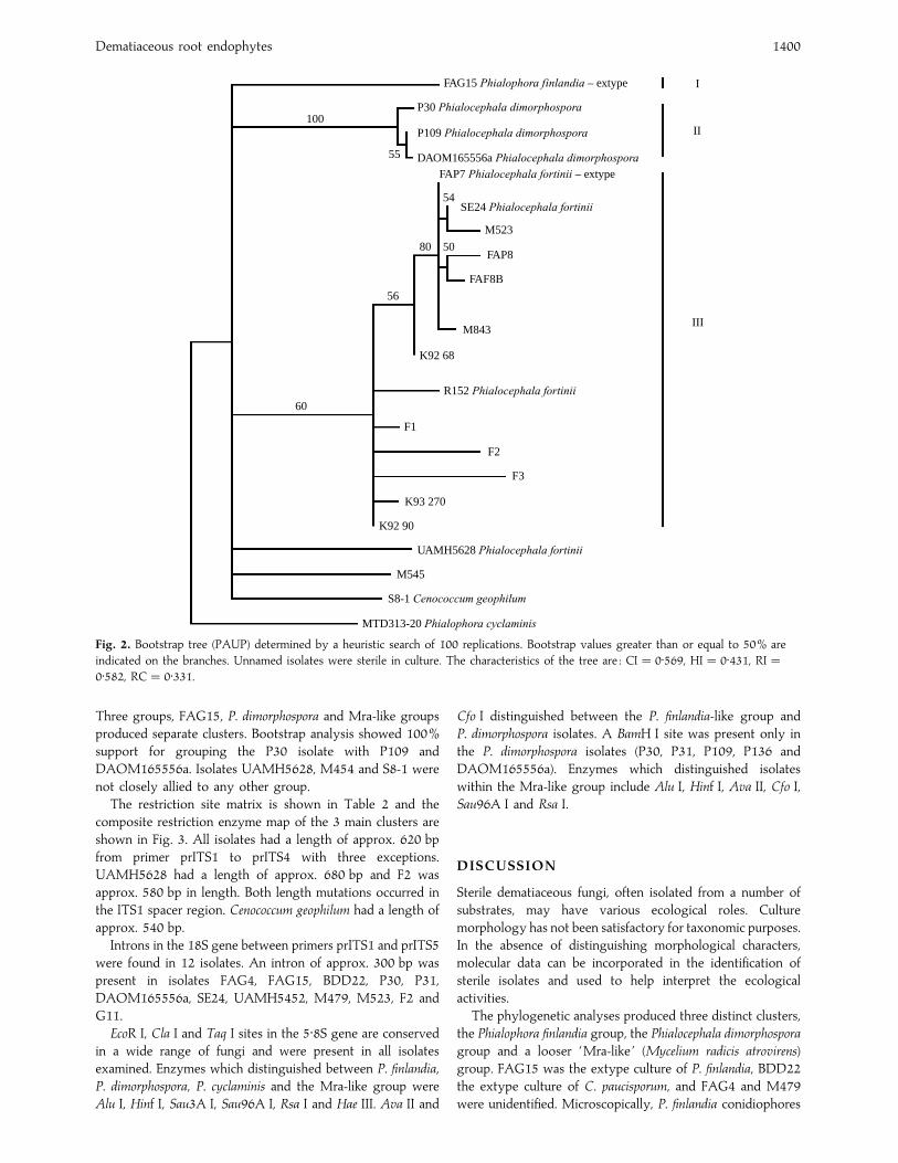
The phylogramgenerated using the heuristic search strategy
of PAUP (Fig. 2) was similar to the UPGMA tree in Fig. 1. Not
all isolates are included in the phylogram to increase clarity.
Representatives from each cluster are presumed. The P.
cyclaminis isolate (MTD313-20) was used as the outgroup.
Dematiaceous root endophytes 1400
100
55
80
56
60
FAG15 Phialophora finlandia – extype
P30 Phialocephala dimorphospora
P109 Phialocephala dimorphospora
DAOM165556a Phialocephala dimorphospora
I
FAP7 Phialocephala fortinii – extype
54SE24 Phialocephala fortinii
II
III
M523
FAP8
FAF8B
M843
K92 68
R152 Phialocephala fortinii
F1
F2
F3
K93 270
K92 90
UAMH5628 Phialocephala fortinii
M545
S8-1 Cenococcum geophilum
MTD313-20 Phialophora cyclaminis
50
Fig. 2. Bootstrap tree (PAUP) determined by a heuristic search of 100 replications. Bootstrap values greater than or equal to 50% are
indicated on the branches. Unnamed isolates were sterile in culture. The characteristics of the tree are : CI¯ 0±569, HI¯ 0±431, RI¯0±582, RC¯ 0±331.
Three groups, FAG15, P. dimorphospora and Mra-like groups
produced separate clusters. Bootstrap analysis showed 100%
support for grouping the P30 isolate with P109 and
DAOM165556a. Isolates UAMH5628, M454 and S8-1 were
not closely allied to any other group.
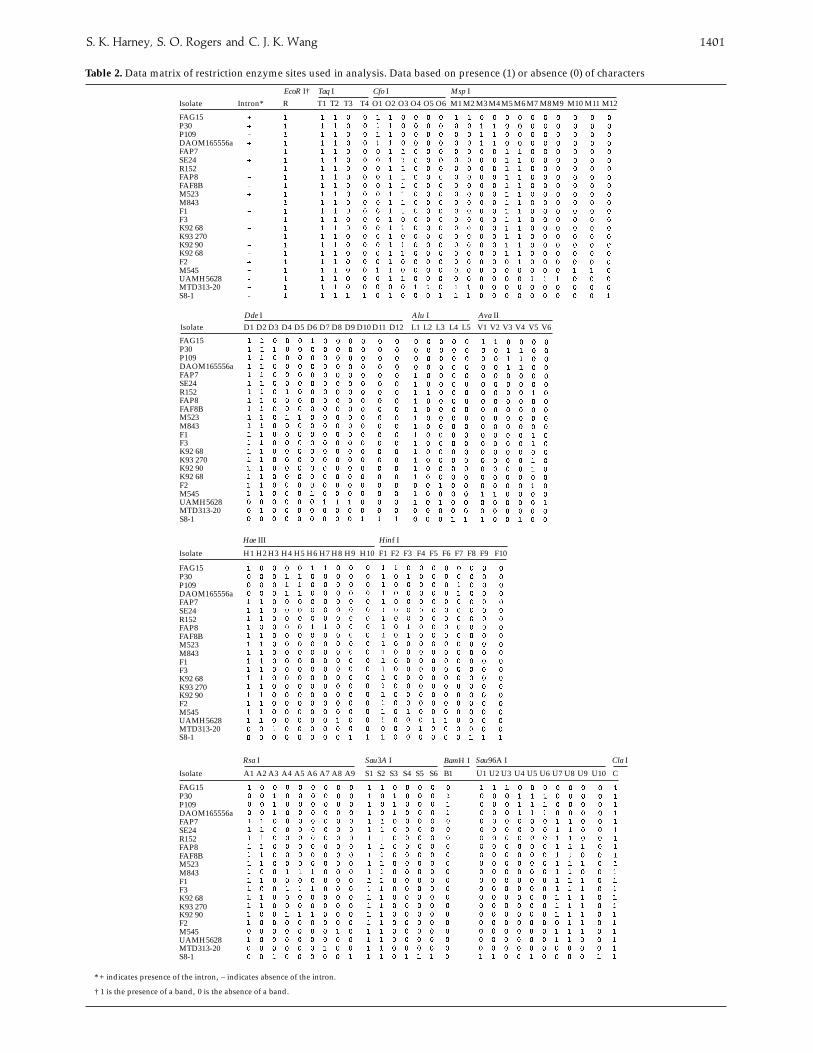
The restriction site matrix is shown in Table 2 and the
composite restriction enzyme map of the 3 main clusters are
shown in Fig. 3. All isolates had a length of approx. 620 bp
from primer prITS1 to prITS4 with three exceptions.
UAMH5628 had a length of approx. 680 bp and F2 was
approx. 580 bp in length. Both length mutations occurred in
the ITS1 spacer region. Cenococcum geophilum had a length of
approx. 540 bp.
Introns in the 18S gene between primers prITS1 and prITS5
were found in 12 isolates. An intron of approx. 300 bp was
present in isolates FAG4, FAG15, BDD22, P30, P31,
DAOM165556a, SE24, UAMH5452, M479, M523, F2 and
G11.
EcoR I, Cla I and Taq I sites in the 5±8S gene are conserved
in a wide range of fungi and were present in all isolates
examined. Enzymes which distinguished between P. finlandia,
P. dimorphospora, P. cyclaminis and the Mra-like group were
Alu I, Hinf I, Sau3A I, Sau96A I, Rsa I and Hae III. Ava II and
Cfo I distinguished between the P. finlandia-like group and
P. dimorphospora isolates. A BamH I site was present only in
the P. dimorphospora isolates (P30, P31, P109, P136 and
DAOM165556a). Enzymes which distinguished isolates
within the Mra-like group include Alu I, Hinf I, Ava II, Cfo I,
Sau96A I and Rsa I.
DISCUSSION
Sterile dematiaceous fungi, often isolated from a number of
substrates, may have various ecological roles. Culture
morphology has not been satisfactory for taxonomic purposes.
In the absence of distinguishing morphological characters,
molecular data can be incorporated in the identification of
sterile isolates and used to help interpret the ecological
activities.
The phylogenetic analyses produced three distinct clusters,
the Phialophora finlandia group, the Phialocephala dimorphospora
group and a looser ‘Mra-like ’ (Mycelium radicis atrovirens)
group. FAG15 was the extype culture of P. finlandia, BDD22
the extype culture of C. paucisporum, and FAG4 and M479
were unidentified. Microscopically, P. finlandia conidiophores
S. K. Harney, S. O. Rogers and C. J. K. Wang 1401
DAOM165556a
FAG15P30P109
FAP7SE24R152FAP8FAF8BM523M843F1F3K92 68K93 270K92 90K92 68F2M545UAMH5628MTD313-20S8-1
DAOM165556a
FAG15P30P109
FAP7SE24R152FAP8FAF8BM523M843F1F3K92 68K93 270K92 90K92 68F2M545UAMH5628MTD313-20S8-1
DAOM165556a
FAG15P30P109
FAP7SE24R152FAP8FAF8BM523M843F1F3K92 68K93 270K92 90F2M545UAMH5628MTD313-20S8-1
DAOM165556a
FAG15P30P109
FAP7SE24R152FAP8FAF8BM523M843F1F3K92 68K93 270K92 90F2M545UAMH5628MTD313-20S8-1
T1 T2 T3 T4 O1 O2 O3 O4 O5 O6 M1 M2 M3 M4 M5 M6 M7 M8M9 M10 M11 M12R
EcoR I†
Intron*Isolate
Taq I Cfo I Msp I
Table 2. Data matrix of restriction enzyme sites used in analysis. Data based on presence (1) or absence (0) of characters
Isolate D1 D2 D3 D4 D5 D6 D7 D8 D9 D10 D11 D12 L1 L2 L3 L4 L5 V1 V2 V3 V4 V5 V6Dde I Alu I Ava II
Isolate H1 H2 H3 H4 H5 H6 H7 H8 H9 H10 F1 F2 F3 F4 F5 F6 F7 F8 F9 F10
Hae III Hinf I
Isolate A1 A2 A3 A4 A5 A6 A7 A8 A9 S1 S2 S3 S4 S5 S6 B1
Rsa I Sau3A I
U1 U2 U3 U4 U5 U6 U7 U8 U9
Sau96A I
U10 C
Cla I
* + indicates presence of the intron, – indicates absence of the intron.
† 1 is the presence of a band, 0 is the absence of a band.
BamH I

Dematiaceous root endophytes 1402
prITS1F2
18S (SSU)
Cluster I
H6 O1
U2V1
D1
A1
prITS3
prITS2
S1CT1 R F1
ITS1 5.8S
O2 D2U3H1
U1V2 S2
ITS2 25S (LSU)
T2 M1 M2prITS4
Cluster II
D3B
U6H5
U4V3 O1 D1 A3 S1
CT1 R F1 O2
M5T2
U5V4 D2 S3
M6
L1U7H2
M4
D4 H6 M3
V5 D1
U9 A1
S1CT1 R F1 O2
T2
D1 D2 F3U8H1 O3 L2 A5 S2
A4 A2
100 bp
Cluster III
Fig. 3. Composite restriction enzyme site maps. Each map is from the 3« end of the 18S rRNA gene to the 5« end of the 25S gene, and
include the 5±8S gene, ITS1 and ITS2 (positions indicated at top of figure). Maps are centred with respect to the 5±8S gene. Triangles
in the 18S gene indicate intron. Each restriction site position is indicated by the appropriate letter and number (where more than one
site for that enzyme exists). Positions of primers prITS1–prITS4 are shown by half-arrows at the top of the figure. Scale is at the
bottom. Abbreviations for enzymes are : A, Rsa I, B, BamH I ; C, Cla I ; D, Dde I ; F, Hinf I ; H, Hae III ; L, Alu I ; M, Msp I ; O, Cfo I ; R,
EcoR I ; S, Sau3A I ; T, Taq I ; U, Sau96A I ; V, Ava II.
are semimacronematous and highly branched whereas
conidiophores of Chloridium paucisporum are solitary or
loosely branched. The morphological characters of phialides
and conidia of these two fungi are very different (Wilcox et al.,
1974, Wang & Wilcox, 1985). Anatomical studies show
differences in ecto- and ectendomycorrhizal development
between P. finlandia and C. paucisporum (Wilcox & Wang,
1987a). Possible explanations of the 100% similarity observed
include that they are so closely related genetically they cannot
be distinguished or that P. finlandia and C. paucisporum may be
conspecific.
The sterile isolates FAG4 and M479 are 100% identical to
P. finlandia. FAG4 and FAG15 were collected together,
formed similar mycorrhizas and were thought to be identical,
but FAG4 could never by induced to sporulate (H. E. Wilcox
& C. J. K. Wang, pers. comm.). The 100% similarity between
FAG4, FAG15 (both collected from Finland) and M479
(isolated from an outplanted white pine seedling in
Warrensburg, New York) suggests that P. finlandia is possibly
more widespread than reported. Phialophora finlandia is a
dematiaceous mycorrhizal fungus that rarely sporulates and
could be mistaken for C. geophilum, a common dematiaceous
ectomycorrhizal fungus. Morphological descriptions of C.
geophilum mycorrhizas which undergo colour change (Park,
1970 ; Giltrap, 1983) suggest that it may be a misidentified P.
finlandia (or C. paucisporum) (Wilcox & Wang, 1987a).
There is a close relationship between the P. dimorphospora
isolates but little similarity to the other groups. P30 and P31
(100% identical) were isolated from the same utility pole in
Chester, NJ (C. J. K. Wang, pers. comm.) and probably
represent the same individual. P109 and P136, also collected
from utility poles in Chester, NJ, showed a high percent
similarity with the DAOM165556a isolate from a decayed
stump in Canada, the only difference being the presence or
absence of an intron in the 18S gene. Introns are fairly
common in deuteromycetes and intraspecific variation has
been reported (Rogers et al., 1993 ; Shinohara, 1994 ; Shinohara
et al., 1996), suggesting that P109, P136 and DAOM165556a
may be identical. P30 and P31 also had introns, and only one
additional restriction site (Dde I), suggesting a close re-
lationship to the other P. dimorphospora isolates studied.
The third cluster contained the majority of Mra-like
isolates, with some variation between the groups. Isolates
which had been identified as P. fortinii based on conidiogenesis
include FAP7 (extype), R151, R152, UAMH5452,
UAMH5628, UAMH6677, SE24, M503 and M504. Several
isolates which were identified as P. fortinii had low support
using molecular analysis. This may be due to a wide variation
within the group or it may be that the isolates represent more
than one species. Phialocephala fortinii and P. dimorphospora are
fairly similar morphologically and have been confused with
each other. Molecular analysis clearly distinguished between
these two groups. This suggests that P. fortinii is a species with
a high degree of variation.
Two groups which had a high percent similarity could only
be separated based on the presence or absence of an intron
and, therefore, are probably the same. These two groups
contain five of the nine isolates which had been identified as
P. fortinii (along with sterile unknowns).
Of the isolates identified as P. fortinii (excluding
UAMH5628), FAP7 was isolated in Finland ; M503, M504,
R151 and R152 in New York ; UAMH6677 and UAMH5452
in Alberta ; and SE24 in Oregon. Phialocephala fortinii has also
been isolated from Quebec (Richard & Fortin, 1973),
Czechoslovakia (Cerny & Cudlı!n, 1989), Germany and
Switzerland (K. Ahlich, pers. comm.), suggesting that P. fortinii

S. K. Harney, S. O. Rogers and C. J. K. Wang 1403
has a wide geographic range, and probably remains
unidentified as it rarely sporulates. Stoyke et al. (1992)
characterized a number of sterile dematiaceous isolates from
the roots of subalpine and alpine plants as P. fortinii based on
RFLP analysis of PCR amplified rDNA. The P. fortinii isolates
did not cluster according to geographic region.
Phialocephala fortinii and the P. fortinii-like isolates show
little host specificity. The cultures of P. fortinii in this study
were isolated from Pinus sylvestris (FAP7), Pinus resinosa (R151,
R152, M503 and M504), Calypso bulbosa (UAMH5452),
Luetkea pectinata (UAMH6677) and Lupinus latifolius (SE24).
Phialocephala fortinii has also been isolated from Picea mariana
(Richard & Fortin, 1973), Picea abies (Cerny & Cudlı!n, 1989 ;
K. Ahlich, pers. comm.) and Abies alba (K. Ahlich, pers.
comm.). Host origin did not show any consistency in the
cluster analysis.
The variation within the large cluster of the Mra-like
isolates is not easily explained. Isolates F2 and M545, along
with the C. geophilum isolate S8-1, can be presumed to be
different, with the F2 and M545 isolates remaining unknown.
P. fortinii may have a large amount of intraspecific variation or
the variation may indicate a species complex, with the isolates
perhaps closely related to P. fortinii. Molecular analysis
suggests that the P. fortinii-like cluster includes different
species. However, the identification of P. fortinii from the
FAP7}SE24 groups and R152}M303}M504 group suggests
that P. fortinii might show a high degree of intraspecific
variation. Stoyke et al. (1992) showed intraspecific variation
with isolates of P. fortinii-like sterile dematiaceous fungi (using
six enzymes) and concluded the isolates were conspecific or
closely related to P. fortinii. LoBuglio et al. (1991) found a high
degree of variation between isolates of C. geophilum using
RFLP analysis suggesting C. geophilum is a heterogeneous
species or a fungal complex of broad taxonomic range.
Sequencing data later resolved some of the variation seen
within the C. geophilum isolates (Shinohara, 1994) suggesting
that the variation observed in the P. fortinii and P. fortinii
isolates may also be resolved by sequencing.
It appears that a number of the isolates can be tentatively
characterized as P. fortinii-like. Phialocephala fortinii exhibits a
large amount of variation, as indicated by the high degree of
variation between sporulating cultures of P. fortinii. The low
resolution may be the result of individual isolates which are
not similar to any other isolates present. Branch and bound
parsimony analysis (not shown) generated a total of five trees
with branching differences occurring among 4–5 of the
isolates examined. Three of these isolates loosely cluster with
the P. fortinii-like group using the heuristic approach and two
clustered using UPGMA in analyses. Sequencing may help in
clarifying some of the ambiguity.
Additional experiments examining pathogenicity of isolates
from different hosts and geographic origin are being done in
order to determine if virulence can be associated with the
different groups.
The authors would like to thank Mike Allen for critically
reviewing this manuscript and Robin Pietropaolo for technical
assistance. This research was supported by the McIntire
Stennis Cooperative Forestry Research Program of the United
States Department of Agriculture. This is part I of a dissertation
submitted by S. Harney in partial fulfilment of the Ph.D.
degree at the State University of New York, College of
Environmental Science and Forestry, Syracuse, New York.
REFERENCES
Cerny, M. & Cudlı!n, P. (1989). Micromycetes from the rhizosphere of
Norway spruce stands under different pollution stress. Agriculture,
Ecosystems and Environment 28, 49–54.
Currah, R., Hambleton, S. & Smreciu, A. (1988). Mycorrhizae and mycorrhizal
fungi of Calypso bulbosa. American Journal of Botany 75, 739–752.
Currah, R., Sigler, L. & Hambleton, S. (1987). New records and new taxa of
fungi from the mycorrhizae of terrestrial orchids of Alberta. Canadian
Journal of Botany 65, 2473–2482.
Gardes, M. & Bruns, T. D. (1993). ITS primers with enhanced specificity for
basidiomycetes – application to the identification of mycorrhizae and rusts.
Molecular Ecology 2, 113–118.
Gardes, M., White, E. J., Fortin, J. A., Bruns, T. D. & Taylor, J. W. (1991).
Identification of indigenous and introduced symbiotic fungi in
ectomycorrhizae by amplification of nuclear and mitochondrial ribosomal
DNA. Canadian Journal of Botany 69, 180–190.
Giltrap, N. (1983). Influence of irradiance and concentration of glucose in the
substrate on mycorrhizal development by Cenococcum geophilum and
Paxillus involutus in axenic culture. Transactions of the British Mycological
Society 81, 627–629.
Haselwandter, K. & Read, D. (1980). Fungal associations of roots of dominant
and sub-dominant plants in high-alpine vegetation systems with special
reference to mycorrhiza. Oecologia 45, 57–62.
LoBuglio, K., Rogers, S. & Wang, C. J. K. (1991). Variation in ribosomal DNA
among isolates of the mycorrhizal fungus Cenococcum geophilum. Canadian
Journal of Botany 69, 2331–2343.
Melin, E. (1923). Experimentelle Untersuchungen u$ ber die Konstitution und
O> kologie der Mykorrhizen von Pinus silvestris L. und Picea abies (L.) Karst.
In Mykologische Untersuchungen und Berichte Zweiter Band (ed. R. Fakk), pp.
222–247, Aktiengesellschaft fu$ r Druck und Verlag : Germany.
O’Dell, T., Massicotte, H. & Trappe, J. (1993). Root colonization of Lupinus
latifolius Agardh. and Pinus contorta by Phialocephala fortinii Wang and
Wilcox. New Phytologist 124, 93–100.
O’Dell, T. & Trappe, J. (1992). Root endophytes of lupin and some other
legumes in Northwestern U.S.A. New Phytologist 122, 479–485.
Park, J. (1970). A change in color of aging mycorrhizal roots of Tilia americana
formed by Cenococcum graniforme. Canadian Journal of Botany 48,
1339–1341.
Richard, C. & Fortin, J. A. (1973). The identification of Mycelium radicis
atrovirens (Phialocephala dimorphospora). Canadian Journal of Botany 51,
2247–2248.
Richard, C. & Fortin, J. A. (1974). Distribution ge! ographique, e! cologie,
physiologie, pathoge! nicite! et sporulation du Mycelium radicis atrovirens.
Phytoprotection 55, 67–88.
Rogers, S. O. & Bendich, A. (1985). Extraction of DNA from milligram
amounts of fresh, herbarium and mummified plant tissues. Plant Molecular
Biology 5, 69–76.
Rogers, S. O. & Bendich, A. (1994). Extraction of total cellular DNA from
plants, algae and fungi. In Plant Molecular Biology Manual 3rd ed. (ed. S. B.
Gelvin & R. A. Schilperoort), pp. D1 :1–8. Kluwer Academic Publishers :
Boston, MA, U.S.A.
Rogers, S. O., Rehner, S., Bledsoe, C., Mueller, G. & Ammirati, J. (1989).
Extraction of DNA from basidiomycetes for ribosomal DNA hybridizations.
Canadian Journal of Botany 67, 1235–1243.
Rogers, S. O., Yan, Z., Shinohara, M., LoBuglio, K. & Wang, C. J. K. (1993).
Messenger RNA intron in the nuclear 18S ribosomal RNA gene of
deuteromycetes. Current Genetics 23, 338–342.
Rohlf, F. (1988). NTSYS-pc : Numerical Taxonomy and Multivariate Analysis
System. Version 1.50. Exeter Publishing Ltd : New York.
Shinohara, M. (1994). Molecular evolutionary study of Cenococcum geophilum.
Ph.D. Thesis, State University of New York, College of Environmental
Science and Forestry, Syracuse, New York.

Dematiaceous root endophytes 1404
Shinohara, M., LoBuglio, K. F. & Rogers, S. O. (1996). Group-I intron family
in the nuclear ribosomal RNA small subunit genes of Cenococcum geophilum
isolates. Current Genetics 29, 377–387.
Stoyke, G. & Currah R. (1991). Endophytic fungi from the mycorrhizae of
alpine ericoid plants. Canadian Journal of Botany 69, 347–352.
Stoyke, G., Egger, K. & Currah, R. (1992). Characterization of sterile
endophytic fungi from the mycorrhizae of subalpine plants. Canadian
Journal of Botany 69, 2009–2016.
Swofford, D. (1990). PAUP: Phylogenetic Analysis Using Parsimony, version
3.1. Computer program distributed by the Illinois History Survey :
Champaign, Illinois.
Wang, C. J. K. & Wilcox, H. E. (1985). New species of ectendomycorrhizal
and pseudomycorrhizal fungi : Phialophora finlandia, Chloridium paucisporum,
and Phialocephala fortinii. Mycologia 77, 951–958.
White, T., Bruns, T., Lee, S. & Taylor, J. (1990). Amplification and direct
sequencing of fungal ribosomal RNA genes for phylogenetics. In PCR
(Accepted 26 February 1997)
Protocols : A Guide to Methods and Applications (ed. M. Innis, D. Gelfand, J.
Sninsky & T. White), pp. 315–322. Academic Press, Inc. & Harcourt Brace
Jovanovich Publishers : New York.
Wilcox, H. E. & Ganmore-Neumann, R. (1974). Ectendomycorrhizae in Pinus
resinosa seedlings. I. Characteristics of mycorrhizae produced by a black
imperfect fungus. Canadian Journal of Botany 52, 2145–2155.
Wilcox, H. E., Ganmore-Neumann, R. & Wang, C. J. K. (1974). Characteristics
of two fungi producing ectomycorrhizae in Pinus resinosa. Canadian Journal
of Botany 52, 2279–2282.
Wilcox, H. E. & Wang, C. J. K. (1987a). Mycorrhizal and pathological
associations of dematiaceous fungi in roots of 7-month-old tree seedlings.
Canadian Journal of Forest Research 17, 884–899.
Wilcox, H. E. & Wang, C. J. K. (1987b). Ectomycorrhizal and
ectendomycorrhizal associations of Phialophora finlandia with Pinus resinosa,
Picea rubens, and Betula alleghaniensis. Canadian Journal of Forest Research 17,
976–990.