molecular characterisation of salmonella enterica serovar ... · enterica serovar typhi causing...
TRANSCRIPT

Malaysian Journal of Microbiology, Vol 8(3) 2012, pp. 148-155
148 ISSN (print): 1823-8262, ISSN (online): 2231-7538
Molecular Characterisation of Salmonella enterica Serovar Typhi Isolated from Typhoidial Humans
Arunava Das
1*, Seenivasan Sree Hari
1, Umachandran Shalini
2, Arumugam Ganeshkumar
3 and Magudeshwaran
Karthikeyan4
1Department of Biotechnology, Bannari Amman Institute of Technology,
Sathyamangalam-638401, Erode District, Tamil Nadu, India. 2Centre for Biotechnology, A. C. Tech Campus, Anna University, Chennai-600025, Tamil Nadu, India.
3Department of Biological Sciences, BITS, Pilani - K. K. Birla Goa Campus, Zuarinagar, Goa- 403726, India.
dBiocon Limited, Electronic City, Bangalore-560100, India.
E-mail: [email protected]
Received 8 February 2012; received in revised form 30 March 2012; accepted 2 April 2012
ABSTRACT Aims: Salmonella enterica serovar Typhi is the major causative agent for typhoidial fever around the globe among human population reported till date. Present research work was carried out for detection and molecular characterisation of Salmonella enterica serovar Typhi isolated from humans with Typhoidial fever by biochemical, phenotypical and virulence gene based polymerase chain reaction (PCR) techniques. The isolated strains were also investigated for antibiotic susceptibility patterns as a control measure. Methodology and Results: A total of 16 clinical samples were collected from the same numbers of patients (7 males and 9 females) from Coimbatore, Erode and Salem districts of Tamil Nadu and were processed via broth enrichment methods for isolation and identification of the causative agent S. enterica serovar Typhi. Microbiological and biochemical investigations revealed the presence of S. Typhi from 16 samples. The biotyping of the isolates showed that all the isolates belonged to biotype IV. The PCR analysis confirmed the presence of invA (Invasion gene, 244bp), tyv (Tyvelose epimerase gene, 615 bp), fliC-d (Phage-1 flagellin gene for d-antigen, 750 bp) and viaB (Vi antigen gene, 439bp) in all 16 clinical samples. The antibiotic susceptibility test that was carried out among the isolates against 12 antimicrobial agents, showed 100 % resistance to only ampicillin and 100 % sensitivity to carbenicillin, chloramphenicol, clindamycin, gentamycin, kanamycin and tetracycline. Conclusion, significance and impact of study: This study confirmed the association of virulent strains of S. enterica serovar Typhi from Typhoidial fever among human population and suggested that PCR based diagnostic could be very useful for the rapid detection of S. Typhi isolates. Present study emphasized the use of antibiotic like chloramphenicol or in combination with other antibiotics for the effective control of S. Typhi. Keywords: Salmonella enterica serovar Typhi, antibiogram, PCR, Typhoidial fever
INTRODUCTION
Salmonella enterica serovar Typhi, an inevitable etiology of sporadic outbreaks of typhoidial fever, which remains as an important public health problem, causes 16 million cases of the disease and about 600,000 deaths, annually, all over the world (Ivanoff and Levine, 1995). It also results in fatal infection among adults and children, if untreated causing bacteraemia and inflammatory destruction of the intestine and other organs (Hirose et al., 2002).
There are nearly 2,000 Salmonella serovars and for those tested so far, all seem to contain invasion gene (inv), which enable the bacteria to invade host cells (Chiu and Ou, 1996). The O antigen gene (tyv) encodes CDP - tyvelose epimerase, which converts CDP - paratose to CDP - tyvelose. The tyv gene is present in both serovars Typhi and Paratyphi A, but the tyv gene of serovar
Paratyphi A does not produce active CDP tyvelose epimerase due to the 1-bp deletion which causes the frame shift mutation and converts codon 4 of tyv to a stop codon (Verma and Reeves, 1989). All virulent strains of S. enterica serovar Typhi causing typhoidial fever possess the Vi capsular antigen gene. Thus, the DNA sequence encoding the Vi antigen, pertaining to the viaB region is useful in developing DNA based diagnostic tests for S. enterica serovar Typhi (Hashimoto et al., 1995). The flagellin gene fliC encodes the major component of the flagellum which plays a key role for the Type III Secretion system, the most widely used mechanism to secrete proteins from cytoplasm of the bacterial cell (Yonekura et al., 2003) and in case of S. enterica serovar Typhi, the H antigen gene (fliC-d) ie., phage-1 flagellin gene for d-antigen [H:d] encodes for flagellin (Hirose et al., 2002). Antibiotics such as chloramphenicol has been a choice of drug for the treatment of typhoid fever for about 40 years, but alternative drugs for treatment are now required due to
*Corresponding author
Mal. J. Microbiol. Vol 8(3) 2012, pp. 148-155
149 ISSN (print): 1823-8262, ISSN (online): 2231-7538
the emergence of multi-drug resistant S. enterica serovar Typhi showed resistant to ampicillin, chloramphenicol and trimethoprimsulfamethoxazole (Hirose et al., 2001). Geographically, the emergence and spreading of multi-drug resistant S. enterica serovar Typhi have been reported from developing countries, particularly the Indian subcontinent and Southeast Asia (Chitnis et al., 1999; Rao et al., 1993). The emergences of the drug resistant S. Typhi strains possess major challenge in the treatment and prevention of typhoid fever, particularly, in rural India population (Senthilkumar and Prabakaran, 2005). Therefore, it is essential to reappraise the antibiotic sensitivity pattern of the isolates periodically. In this study, detection and molecular characterisation of S. enterica serovar Typhi isolated from typhoidial human blood samples has been carried out by biochemical, phenotypical and molecular characterisation tools. Present study also determines the antibiotic susceptibility pattern of the S. Typhi strains and their prevalence towards the multi-drug resistance for epidemiological study. MATERIALS AND METHODS Sample collection
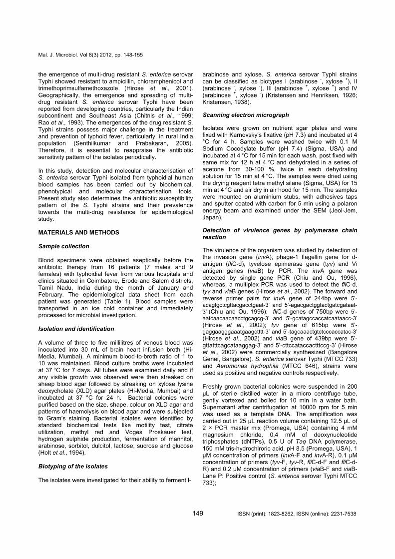
Blood specimens were obtained aseptically before the antibiotic therapy from 16 patients (7 males and 9 females) with typhoidial fever from various hospitals and clinics situated in Coimbatore, Erode and Salem districts, Tamil Nadu, India during the month of January and February. The epidemiological data sheet from each patient was generated (Table 1). Blood samples were transported in an ice cold container and immediately processed for microbial investigation. Isolation and identification
A volume of three to five millilitres of venous blood was inoculated into 30 mL of brain heart infusion broth (Hi-Media, Mumbai). A minimum blood-to-broth ratio of 1 to 10 was maintained. Blood culture broths were incubated at 37
°C for 7 days. All tubes were examined daily and if
any visible growth was observed were then streaked on sheep blood agar followed by streaking on xylose lysine deoxycholate (XLD) agar plates (Hi-Media, Mumbai) and incubated at 37 °C for 24 h. Bacterial colonies were purified based on the size, shape, colour on XLD agar and patterns of haemolysis on blood agar and were subjected to Gram’s staining. Bacterial isolates were identified by standard biochemical tests like motility test, citrate utilization, methyl red and Voges Proskauer test, hydrogen sulphide production, fermentation of mannitol, arabinose, sorbitol, dulcitol, lactose, sucrose and glucose (Holt et al., 1994). Biotyping of the isolates The isolates were investigated for their ability to ferment l-
arabinose and xylose. S. enterica serovar Typhi strains can be classified as biotypes I (arabinose
-, xylose
+), II
(arabinose -, xylose
-), III (arabinose
+, xylose
+) and IV
(arabinose +, xylose
-) (Kristensen and Henriksen, 1926;
Kristensen, 1938).
Scanning electron micrograph
Isolates were grown on nutrient agar plates and were fixed with Karnovsky’s fixative (pH 7.3) and incubated at 4
°C for 4 h. Samples were washed twice with 0.1 M Sodium Cocodylate buffer (pH 7.4) (Sigma, USA) and incubated at 4
°C for 15 min for each wash, post fixed with
same mix for 12 h at 4 °C and dehydrated in a series of
acetone from 30-100 %, twice in each dehydrating solution for 15 min at 4
°C. The samples were dried using
the drying reagent tetra methyl silane (Sigma, USA) for 15 min at 4
°C and air dry in air hood for 15 min. The samples
were mounted on aluminium stubs, with adhesives taps and sputter coated with carbon for 5 min using a polaron energy beam and examined under the SEM (Jeol-Jem, Japan).
Detection of virulence genes by polymerase chain reaction
The virulence of the organism was studied by detection of the invasion gene (invA), phage-1 flagellin gene for d-antigen (fliC-d), tyvelose epimerase gene (tyv) and Vi antigen genes (viaB) by PCR. The invA gene was detected by single gene PCR (Chiu and Ou, 1996), whereas, a multiplex PCR was used to detect the fliC-d, tyv and viaB genes (Hirose et al., 2002). The forward and reverse primer pairs for invA gene of 244bp were 5’-acagtgctcgttacgacctgaat-3’ and 5’-agacgactggtactgatcgataat-
3’ (Chiu and Ou, 1996); fliC-d genes of 750bp were 5’-
aatcaacaacaacctgcagcg-3’ and 5’-gcatagccaccatcaataacc-3’ (Hirose et al., 2002); tyv gene of 615bp were 5’-gaggaagggaaatgaagctttt-3’ and 5’-tagcaaactgtctcccaccatac-3’ (Hirose et al., 2002) and viaB gene of 439bp were 5’-gttatttcagcataaggag-3’ and 5’-cttccataccactttccg-3’ (Hirose et al., 2002) were commercially synthesized (Bangalore Genei, Bangalore). S. enterica serovar Typhi (MTCC 733) and Aeromonas hydrophila (MTCC 646), strains were used as positive and negative controls respectively. Freshly grown bacterial colonies were suspended in 200 µL of sterile distilled water in a micro centrifuge tube, gently vortexed and boiled for 10 min in a water bath. Supernatant after centrifugation at 10000 rpm for 5 min was used as a template DNA. The amplification was carried out in 25 µL reaction volume containing 12.5 µL of 2 × PCR master mix (Promega, USA) containing 4 mM magnesium chloride, 0.4 mM of deoxynucleotide triphosphates (dNTPs), 0.5 U of Taq DNA polymerase, 150 mM tris-hydrochlroric acid, pH 8.5 (Promega, USA), 1 µM concentration of primers (invA-F and invA-R), 0.1 µM concentration of primers (tyv-F, tyv-R, fliC-d-F and fliC-d-R) and 0.2 µM concentration of primers (viaB-F and viaB- Lane P: Positive control (S. enterica serovar Typhi MTCC 733);
Mal. J. Microbiol. Vol 8(3) 2012, pp. 148-155
150 ISSN (print): 1823-8262, ISSN (online): 2231-7538
Ta
ble
1:
Ep
ide
mio
log
ica
l d
ata
an
d d
eta
ils o
f to
xin
gen
es d
ete
cte
d b
y P
CR
fro
m S
. e
nte
rica
se
rova
r T
yp
hi
De
tec
tio
n o
f to
xin
ge
ne
s b
y
PC
R
via
B
+
+
+
+
+
+
+
+
+
+
+
+
+
+
+
+
fliC
-d
+
+
+
+
+
+
+
+
+
+
+
+
+
+
+
+
tyv
+
+
+
+
+
+
+
+
+
+
+
+
+
+
+
+
inv
A
+
+
+
+
+
+
+
+
+
+
+
+
+
+
+
+
Bio
typ
e
IV
Pla
ce
of
Sa
mp
le
Co
lle
cti
on
Clin
ic,
Sa
lem
Ho
sp
ita
ls,
Co
imba
tore
Clin
ic,
Ero
de
Ho
sp
ita
ls,
Co
imba
tore
Clin
ic,
Sa
lem
Clin
ic,
Co
imba
tore
Sa
mp
le
co
lle
cte
d f
rom
the
da
y o
f
on
se
t o
f
dis
ea
se
(D
ays
)
7
7
8
5
5
8
5
5
7
6
8
8
7
8
5
5
He
alt
h
Co
nd
itio
n
Typ
ho
id
feve
r
Se
x (
Ag
e
in y
ea
rs)
Ma
le (
12
)
Fe
male
(50
)
Ma
le (
23
)
Ma
le (
16
)
Fe
male
(1
8)
Ma
le (
15
)
Fe
male
(2
2)
Fe
male
(20
)
Fe
male
(16
)
Ma
le (
21
)
Ma
le (
17
)
Fe
male
(22
)
Fe
male
(50
)
Ma
le (
23
)
Fe
male
(16
)
Fe
male
(18
)
So
urc
es
Hu
ma
n
Iso
late
No
HS
T1
HS
T2
HS
T3
HS
T4
HS
T5
HS
T6
HS
T7
HS
T8
HS
T9
HS
T1
0
HS
T1
1
HS
T1
2
HS
T1
3
HS
T1
4
HS
T1
5
HS
T1
6
Sa
mp
le
No
M4
367
F5
67
3
M5
778
72
63
2B
C
74
67
5B
C
74
89
8B
C
75
03
4B
C
F7
86
1
F7
96
8
M7
988
M8
011
75
26
7B
C
F8
56
7
M8
876
75
67
8B
C
75
89
9B
C
S.
No
1.
2.
3.
4.
5.
6.
7.
8.
9.
10
.
11
.
12
.
13
.
14
.
15
.
16
.

Mal. J. Microbiol. Vol 8(3) 2012, pp. 148-155
151 ISSN (print): 1823-8262, ISSN (online): 2231-7538
Figure 1: Ultrastructure of S. enterica serovar Typhi under SEM (11,000x).
Figure 2: Detection of virulence genes from S. enterica serovar Typhi by PCR. A: Detection of invA (244 bp) gene by PCR B: Detection of tyv (615 bp), fliC-d (750bp) and viaB
(439bp) genes by mPCR Lane N: Negative control (A. hydrophila MTCC 646); Lane 1-5: Field isolates positive for virulence genes; Lane M: High range DNA rule R) and 2.5 µL of template DNA. The PCR reactions were performed in thermal Cycler (Eppendorf, USA). For the
invA gene after initial denaturation at 94 °C for 4 min, the
amplification cycle had denaturation, annealing and extension at 94
°C, 56
°C and 72
°C for 30 s, 30 s and 2
min respectively. For fliC-d, tyv and viaB genes after initial denaturation at 95
°C for 4 min, the amplification cycle had
denaturation, annealing and extension at 95 °C, 55
°C and
72 °C for 30 s, 60 s and 90 s respectively. Final extension
was done at 72 °C for 10 min. The PCR amplicons (5 µL)
were electrophoresed in 1.5 % agarose gel in TAE (Tris-acetic acid-EDTA, pH 8) buffer, stained with ethidium bromide and observed under gel doc system (Universal Hood, BIORAD, Italy). Antibiotic susceptibility test
Antibiotic susceptibility tests were performed by disc diffusion method (Bauer et al., 1996) with little modification. Overnight cultures in peptone water were spread plated on nutrient agar (Hi-media, Mumbai) plates. The antibiotics discs (Hi-media, Mumbai) were purchased and used at the following concentrations: Gentamycin (10 g), Cefuroxime (30 g), Penicillin-G (2 U/mL), Nalidixic acid (30 g), Clindamycin (10 g), Carbenicillin (100 g), Cephalothin (30 g), Kanamycin (30 g), Nitrofurantoin (100 g), Tetracyclin (30 g), Ampillicin (10 g) and Chloramphenicol (30 g). The resistance breakpoints were those defined by the National Committee for Clinical Laboratory Standards (NCCLS, 1999) for Gram negative bacteria. S. enterica serovar Typhi (MTCC 733) and A. hydrophila (MTCC 646) were used as controls. RESULTS Isolation and identification
A visible growth was observed in BHI broth on 7th
day of incubation. The isolates were found non haemolytic on sheep blood agar and showed pink colour colonies with black centre on XLD agar. Glucose, mannitol, L-arabinose and sorbitol were fermented by all isolates. In triple sugar iron slants, the butt and slant turned into yellow and red colour respectively indicating the fermentation of glucose alone and production of acid in the butt. The isolates showed production of hydrogen sulphide and no gas production in TSI. Isolates were positive for oxidase test and methyl red test and negative for indole production, urease production and citrate utilization. All the isolates were found Gram negative, flagellated and motile. Upon detailed bacteriological investigation based on the biochemical tests, 16 isolates were tentatively identified as S. enterica serovar Typhi (Table 1). Biotyping of the isolates
All the 16 isolates were able to ferment l-arabinose but not xylose. Thus S. enterica serovar Typhi strains were classified as biotype IV (Table 1) Scanning electron micrograph The ultrastructure study of S. enterica serovar Typhi in
b)
a)

Mal. J. Microbiol. Vol 8(3) 2012, pp. 148-155
152 ISSN (print): 1823-8262, ISSN (online): 2231-7538
SEM was observed to be in clusters of thick rods (Figure 1). The rods were observed to be variable in length; sometimes occured either single or in pairs and occasionally in short chains.
Detection of virulence genes by polymerase chain reaction
In PCR assay, amplification of virulence genes from all 16 isolates tested with the primers of invA, tyv, viaB and fliC-d genes resulted fragments of the predicted size at 244 bp, 615 bp, 439 bp and 750 bp respectively (Table 1, Figure 2).
Antibiotic susceptibility test
In the present study, all the 16 (100 %) isolates were found resistant to ampicillin, moderately sensitive to nalidixic acid and nitrofurantoin and sensitive to carbenicillin, chloramphenicol, clindamycin, gentamycin, kanamycin and tetracycline. However, 13 (81.25 %) isolates were also found resistance to cefuroxime, while 11 (68.75 %) isolates were found resistant to penicillin-G and cephalothin. The remaining 3 (18.75 %) were moderately sensitive to cefuroxime and 5 (31.25 %) isolates were moderately sensitive to penicillin-G and cephalothin (Figure 3, Figure 4).
DISCUSSION In the present study, blood samples were collected from 16 patients of age group 12 to 50 years from Coimbatore, Erode and Salem districts, Tamil Nadu, India. All the clinical samples were collected during the month of January and February and this end of dry season was considered to be the peak occurrence season of typhoidial fever (Lin et al., 2000). Infected and healthy carriers were the source of infection and “five Fs” (food, fingers, flies, fomites and faeces) played an important role in the spread of the disease (Old and Threlfal, 1998). All the 16 patients were diagnosed typhoid positive from the fifth to eighth days of onset of disease and the attack rate 14 (87.5 %) was significantly higher among the people below 30 years old. Very similar to the present study, higher frequency of detection of typhoid cases from the patients of less than 30 years old were previously reported from Tamil Nadu (Ganeshkumar et al., 2010). All the isolated bacteria produced pink coloured and black centred colonies on XLD plates and were positive for mannitol, l-arabinose, sorbitol, glucose fermentation, methyl red test, indole test, H2S production, citrate utilization, motility, oxidase test and urease activity. The microbiological investigation confirmed the tentative isolation of S. enterica serovar Typhi from the clinical cases of typhoid fever from patients were reported earlier (Wain et al., 1998; Ganeshkumar et al., 2010). All the 16 (100 %) isolates were classified as biotype IV for fermenting l-arabinose but not xylose. This biotyping have added data to the epidemiological based classification system according to their fermentation ability of sugars and based on other biochemical properties (Kristensen and Henriksen, 1926; Kristensen, 1938).
Figure 3: Antibiotic susceptibility of S. enterica serovar
Typhi. C: Chloramphenicol (30 µg) , Cu: Cefuroxime (30 µg), A: Ampillicin (10µg), Na: Nalidixic acid (30 µg), Ch: Cephalothin (30 µg), T: Tetracycline (30 µg), Cb: Carbenicillin (100 µg) , Cd: Clindamycin (10 µg), G: Gentamycin (10 µg), Nf: Nitrofurantoin (100 µg), P: Penicillin G (2 Units), K: Kanamycin (30 µg). In PCR, invA, tyv, fliC-d and viaB genes were targeted for the virulence based identification of S. enterica serovar Typhi which revealed the 100 % detection of all the above virulence genes from the clinical isolates originated from typhoidial human origins. Although, the pathogenesis of Salmonella has been mediated by several virulence factors, the role of invA gene was significant as this gene helped S. Typhi for adhesion and invasion to the host epithelial cells (Darwin and Miller, 1990). This study demonstrated that invA gene was predominant along with the other three genes among the isolates of S. Typhi, which could be used as specific marker gene for the rapid detection of the S. Typhi isolates from various biological samples irrespective of sample origin (Chiu and Ou, 1996). In analogy, 100 % detection frequency of inv gene among S. enterica serovars such as Typhi, Virchow, Enteritidis, Typhimurium, Senftenberg, Strasbourg and Infantis (Kumar et al., 2006) originated from poultry products, wastewater and human sources were reported in other countries (Swamy et al., 1996; Salehi et al., 2005) and also in India (Shome et al., 2006; Ganeshkumar et al., 2010). In mPCR study, the O antigen coded by tyv gene, H antigen coded by fliC-d and VI antigen coded by viaB virulence genes were used as the basis of identification of S. enterica serovar Typhi from the clinical cases of typhoid fever in humans. The mPCR result depicted in this study established that these three genes are highly conserved among the isolates of S. Typhi and could be very useful marker genes for the rapid detection of only S. Typhi isolates (Hirose et al., 2002; Kumar et al., 2006). The result of antibiotic susceptibility test revealed that isolates of S. Typhi were 100 % resistant to ampicillin, 81.25 % to cefuroxime and 68.75 % resistant to penicillin- G and cephalothin respectively. The ampicillin resistant S.

Mal. J. Microbiol. Vol 8(3) 2012, pp. 148-155
153 ISSN (print): 1823-8262, ISSN (online): 2231-7538
C (3
0µg)
Cu
(30µ
g)
P (2Uni
ts)
A (10µ
g)
Na (3
0µg)
T (3
0µg)
G (1
0µg)
Nf (
100µ
g)
K (3
0µg)
Cd
(10µ
g)
Cb
(100
µg)
Ch
(30µ
g)
0
20
40
60
80
100
Perc
en
tag
e %
Antibiotics (concentration)
Sensitive
Resistant
Moderately Sensitive
Figure 4: Antibiotic susceptibility test results of S. enterica serovar Typhi A:Ampillicin, C:Chloramphenicol, Cb:Carbenicillin, Cd:Clindamycin, Ch:Cephalothin, Cu:Cefuroxime, G:Gentamycin, K:Kanamycin, Na:Nalidixic acid, Nf:Nitrofurantoin, P:Penicillin G, T:Tetracycline
Typhi isolates from the typhoidial patients from Tamil Nadu, India were reported earlier (Ganeshkumar et al., 2010). The present result clearly indicating the tendency of the S. Typhi isolates to become resistance towards multiple drugs. In view of this, researchers from southern Vietnam reported that 90 % S. Typhi isolates were resistant to multiple antibiotics like ampicillin, chloramphenicol and co-trimoxazole (Smith et al., 1994). In India, 29.47 % and 28.42 % of S. Typhi isolates were also reported to be resistant to ampicillin and chloramphenicol respectively (Nagshetty et al., 2010). Although, chloramphenicol which has been reported many a times by the researcher as resistance to S. Typhi isolates (Agarwal, 1962; Olarte and Galindo, 1973) now found 100 % sensitive in this study along with kanamycin, clindamycin, carbenicillin, gentamycin and tetracycline. In congruence, 100 % sensitivity of S. Typhi isolates against chloramphenicol, gentamicin and tetracycline were also detected earlier (Quintaes et al., 2002). This is in full agreement with the reports of re-emergence of sensitivity of S. Typhi to chloramphenicol (Sood et al., 1999). In our study, nalidixic acid and nitrofurantoin were found 100 %
moderate. More recently, 76 % of blood culture isolates of S. Typhi were reported to be resistant to nalidixic acid (Parry et al., 1998). CONCLUSION
This study confirmed the association of virulent strains of Salmonella enterica serovar Typhi in the occurrence of the typhoidial fever in humans in Tamil Nadu. It is suggested from the present study that PCR technique could be a useful, high throughput and rapid diagnostic tool for the detection of S. enterica serovar Typhi and could be employed by the diagnostic laboratories or clinics for the clinical diagnosis of typhoidial fever from patients. Despite the use of only 12 antibiotics for susceptibility test, present findings helped to know the current status of typhoidial fever among the people in Southern part of India. Although chloramphenicol and other antibiotics showed 100 % sensitivity, still continuous evaluation of sensitivity-resistance pattern of S. Typhi isolates is necessary to make rational use of antibiotics in the management of typhoidial fever in future.

Mal. J. Microbiol. Vol 8(3) 2012, pp. 148-155
154 ISSN (print): 1823-8262, ISSN (online): 2231-7538
REFERENCES Agarwal, S. C. (1962). Chloramphenicol resistance of
Salmonella species in India, 1956-61. The International Journal of Public Health - World Health Organization 17: 331-335.
Bauer, A. W., Kirby, W. M. M, Sheris, J. C. and Turck, M. (1996). Antibiotic suspectibility test by standardized single disk method. American Journal of Clinical Pathology 36: 493-496.
Chitnis, V. D., Verma, C. S. and Hemvani, N. (1999).
Multidrug-resistant Salmonella typhi in India. Lancet 354: 514–515.
Chiu, C. H. and Ou, J. T. (1996). Rapid identification of Salmonella serovars in faeces by specific detection of virulence genes, invA and spvC, by an enrichment broth culture-multiplex PCR combination assay. Journal of Clinical Microbiology 34: 2619–2622.
Darwin, K. H. and Miller, V. L. (1990). Molecular basis of interaction of Salmonella with the intestinal mucosa. Clinical Microbiology Reviews 12: 405-4428.
Ganeshkumar, A. Shalini, U. Vaishnavi, K. Remya Ismail. Lakshmanaswamy, A. Vasanthi, N. S. and Das, A. (2010). Rapid detection of Salmonella enterica serovar Typhi from humans. Journal of Pure and Applied Microbiology 4: 837-841.
Hashimoto, Y., Itho, Y., Fujinaga, Y., Khan, A. Q., Sultana, F., Miyake, M., Hirose, K., Yamamoto, H. and Ezaki, T. (1995). Development of nested PCR based on the ViaB sequence to detect Salmonella typhi. Journal of Clinical Microbiology 33: 775–777.
Hirose, K., Itoh, K. I., Nakajima, H., Kurazono, T., Yamaguchi, M., Moriya, K., Ezaki, T., Kawamura, Y., Tamura, K. and Watanabe, H. (2002). Selective amplification of tyv (rfbE), prt (rfbS), viaB, and fliC genes by multiplex PCR for identification of Salmonella enteric Serovars Typhi and Paratyphi A. Journal of Clinical Microbiology 40: 633-636.
Hirose, K., Tamura, K., Sagara, H. and Watanabe, H. (2001). Antibiotic susceptibilities of Salmonella
enterica serovar Typhi and S. enterica serovar Paratyphi A isolated from patients in Japan. Antimicrobial Agents and Chemotheraphy 45: 956–958.
Holt, J. C., Kreig, N. R., Sneath, P. H. A. and Stanley, J. Y. (1994). Bergey's Manual of determinative Bacteriology. Springer. USA. pp 764-799.
Ivanoff, B. and Levine, M. M. (1997). Typhoid fever:
continuing challenges from a resilient foe. Bulletin de I’Institut Pasteur 95: 129–142.
Kristensen, M. (1938). Studies on the type division of the
typhoid and paratyphoid B bacilli by fermentations.The Journal of Hygiene 38: 688.
Kristensen, M. and Henriksen, H. C. D. (1926).
Reactions fermentatives du bacille typhique et leur rôle epidémiologique. Acta Pathologica Microbiologica Scandinavica 3: 551-582.
Kumar, S., Balakrishna, K. and Batra, H. V. (2006). Detection of Salmonella enterica Serovar Typhi (S.
Typhi) by selective amplification of invA, viaB, fliC-d and prt genes by polymerase chain reaction in multiplex format. Letters in Applied Microbiology 42: 149-154.
Lin, F. C., Ho V. A., Bay, P. V., Thuy N. T. T., Bryla, D., Thanh, T. C., Khiem, H. B., Trach, D. D. and Robbins, J. B. (2000). The epidemiology of typhoid
fever in the Dong Thap Province, Mekong Delta region of Vietnam. The American Society of Tropical Medicine and Hygiene 62(5): 644–648.
Nagshetty, K., Channappa, S. T. and Gaddad, S. M. (2010). Antimicrobial susceptibility of Salmonella Typhi in India. The Journal of Infection in Developing Countries 4(2):070-073.
Olarte, J. and Galindo, E. (1973). S. typhi resistant to
chloramphenicol, ampicillin and other antimicrobial agents: strains isolated in extensive typhoid fever epidemic in Mexico. Antimicrobial Agents in Chemotherapy 4: 597-601.
Old, D. C. and Threlfal, E. J. (1998). Salmonella In: Topley and Wilson’s Microbiology and Microbial Infections, 9th edition, volume 2 (Systemic Bacteriology), Arnold, London. pp. 969-97.
Parry, C., Wain, J., Chinh, N. T., Vinh, H., Farrah, J. J. (1998). Quinoloneresistant S. typhi in Vietnam. Lancet 351: 1289.
Quintaes, B. R., Leal, N. C., Reis, E. M. F., Fonseca, E. L. and Hofer, E. (2002). Conventional and molecular typing of Salmonella Typhi strains from Brazil. Journal of the Institute of Tropical Medicine of São Paulo 44: 315-319.
Rao, P. S., Rajashekar, V., Varghese, G. K. and Shivananda, P. G. (1993). Emergence of multidrug-resistant Salmonella typhi in rural southern India. The American Journal of Tropical Medicine and Hygiene 48:108–111.
Salehi, Z., Mahzounieh, M. and Saeedzadeh, A. (2005). Detection of invA gene in isolated Salmonella from broilers by PCR method. International Journal of Poultry Science 4: 557-559.
Senthilkumar, B. and Prabakaran, G. (2005). Multidrug resistant Salmonella Typhi in asymptomatic Typhoid carriers among food handlers in Namakkal District, Tamilnadu. Indian Journal of Medical Microbiology 23: 92-94.
Shome, B. R., Rahman, H., Shome, R., Murugkar, H. V., Mazumder, Y., Das, A., Kumar, A. and Bujarbaruah, K. M. (2006).
Detection of virulent
genes and genotyping of Salmonella enterica isolated from livestock and poultry. Indian Veterinary Journal 83: 934-938.
Smith, M. D., Doung, N. M., Hoa, N. T., Wain, J., Ha, H. D., Diep, T. S., Day, N. T., Hien, T. T. and White, N. J. (1994). Comparison of ofloxacin and ceftriaxone for short-course treatment of enteric fever. Antimicrobial Agents and Chemotherapy 38: 1716–1720.
Sood, S., Kapil, A., Das, B., Jain, Y. and Kabra, S. K. (1999). Reemergence of chloramphenicol sensitive Salmonella typhi. Lancet 353: 1241-1242.
Swamy, S. C., Barnhart, H., Lee, M. D. and Dreesen, D.

Mal. J. Microbiol. Vol 8(3) 2012, pp. 148-155
155 ISSN (print): 1823-8262, ISSN (online): 2231-7538
W. (1996). Virulence determinants invA and spvC in Salmonellae isolated from poultry products, wastewater and human sources. Applied and Environmental Microbiology 62: 3768-3771.
Verma, N. and Reeves, P. (1989). Identification and sequence of rfbS and rfbE, which determine antigenic specificity of group A and group D salmonellae. Journal of Bacteriology 171: 5694–5701.
Wain, J., Diep, T. S., Ho, V. A., Walsh, A. M., Hoa, N. T. T., Parry, C. M. and White, N. J. (1998).
Quantitation of Bacteria in Blood of Typhoid Fever Patients and Relationship between Counts and Clinical Features, Transmissibility and Antibiotic Resistance. Journal of Clinical Microbiology 36: 1683-1687.
Yonekura, K., Yonekura, S. M. and Namba, K. (2003).
Complete atomic model of the bacterial flagellar filament by electron cryomicroscopy. Nature 424: 643–650.