molecular cell review - university of cambridge
TRANSCRIPT

Molecular Cell
Review
Quality Control of Orphaned Proteins
Szymon Juszkiewicz1 and Ramanujan S. Hegde1,*1MRC Laboratory of Molecular Biology, Cambridge CB2 0QH, UK*Correspondence: [email protected]://doi.org/10.1016/j.molcel.2018.07.001
The billions of proteins inside a eukaryotic cell are organized among dozens of sub-cellular compartments,within which they are further organized into protein complexes. The maintenance of both levels of organiza-tion is crucial for normal cellular function. Newly made proteins that fail to be segregated to the correctcompartment or assembled into the appropriate complex are defined as orphans. In this review, we discussthe challenges faced by a cell of minimizing orphaned proteins, the quality control systems that recognizeorphans, and the consequences of excess orphans for protein homeostasis and disease.
IntroductionThe advent of methods to observe the interior of cells by electron
microscopy in the 1950s revealed far more compartmentaliza-
tion than was apparent previously using light microscopy with vi-
tal dyes and stains (Porter, 1955–1956). This led to the definition
of many membrane-bound organelles in the cell and our first
views of many non-membrane-bound structures such as the
ribosome (Palade, 1955). In parallel with direct visualization,
biochemical fractionation was able to isolate these various
morphologically identifiable structures (Claude, 1943, 1946).
The structures were soon shown to have distinct functions (De
Duve, 1965) and eventually associated with specific subsets of
cellular proteins that are often part of larger complexes with
defined composition and stoichiometry. These studies initiated
modern cell biology, a major goal of which is to understand
how intracellular organization is generated and maintained to
facilitate cellular function.
The most extensive efforts have been aimed at determining
how newly made proteins are segregated to the appropriate
organelle (Blobel, 1980; Wickner and Schekman, 2005).
Although the accuracy of intracellular protein targeting was usu-
ally assumed to be high, one could anticipate that it cannot be
perfect. As detailed below, it is increasingly clear that the recog-
nition and disposal of mistargeted copies of a protein is an
important facet of achieving effective net segregation. Similarly,
the assembly of proteins into complexes is likely to be imperfect
(Harper and Bennett, 2016), necessitating the degradation of un-
assembled components. Thus, the prompt degradation of pro-
teins that fail to be correctly localized or assembled is critical
to the maintenance of intracellular organization. We refer to pro-
teins that are terminally separated from their correct location or
partners as orphans.
In this review, we discuss our understanding of how cells
recognize orphans and selectively route them for degradation.
We begin by providing an accounting of the eukaryotic prote-
ome and the extent of its sub-cellular organization into com-
partments and complexes. After defining the challenges to
achieving a well-organized proteome, we consider successively
the mechanisms cells use to identify mislocalized and misas-
sembled proteins. We end with a consideration of how path-
ways for the quality control of orphaned proteins impact cellular
physiology and disease.
Organization of the Eukaryotic ProteomeBioinformatic tools to predict protein location based on known
trafficking signals (Emanuelsson et al., 2007), together with
increasingly thorough proteome-wide analytic tools (Itzhak
et al., 2016; Mulvey et al., 2017), provide estimates of how
many proteins are delivered to non-cytosolic destinations. In
parallel, systematic analyses of protein complexes (Babu et al.,
2012; Gavin et al., 2006; Krogan et al., 2006) and their relative
abundances (Kulak et al., 2014) inform on what proportion of
nascent proteins need additional assembly. These studies
show that the vast majority of newly made proteins are destined
for a different location, assembled with other cellular factors, or
both (Figure 1).
Of the 20,341 reference proteins of the human genome (Uni-
Prot Consortium, 2018),�7,000 are targeted to the endoplasmic
reticulum for eventual residence in the endomembrane system,
nuclear envelope, plasma membrane, outside the cell, and
peroxisomal membrane. Approximately 1,000 proteins are
destined for mitochondria, �50 for the peroxisomal lumen, and
a relative handful for more specialized structures such as lipid
droplets. Around 5,000 proteins operate primarily in the nucleus,
although the entry and exit of many of these are often dynamic
and regulated. Thus, �65% of genes encode for proteins that
must be recognized for selective trafficking to a membrane-en-
closed compartment (Figure 1A).
Analysis of protein interactions by mass spectrometry across
the yeast proteome (Gavin et al., 2006; Krogan et al., 2006) indi-
cates that over half of all proteins may be in stable multi-protein
complexes (Figure 1B).While the proportion is somewhat less for
membrane proteins (Babu et al., 2012), this may be due to the
additional challenge of retaining interactions during detergent
solubilization. Although data on the human proteome is less
complete, analysis of the relatively abundant cytosolic proteins
in HeLa and HEK293 cells indicate that a little less than half of
them are in stable complexes (Havugimana et al., 2012). This
means that of the�35% of proteins that do not traffic to another
destination (see above), fewer than half are unaccompanied by
partners. Thus, only�15% of genes encode proteins that simply
fold and function without first requiring sub-cellular localization
and assembly.
When abundance, cell type, and growth rate are taken into
account, there are many circumstances where the vast majority
Molecular Cell 71, August 2, 2018 ª 2018 Elsevier Inc. 443

A
B
Figure 1. Accounting of Protein Localization and Assembly(A) The approximate percentages of human genes whose proteins aredestined for various intracellular compartments are shown.(B) Data from three proteomic studies indicating that roughly half of all proteinsin yeast and mammals are in protein complexes.
Molecular Cell
Review
of synthesized proteins must be localized or assembled. For
example, in highly secretory cell types such as pancreatic
exocrine cells or antibody-secreting plasma cells, nearly all ribo-
somes are synthesizing proteins destined for the ER (Brewer and
Hendershot, 2005; Pfeffer et al., 2016). In reticulocytes, almost
all protein synthesis is dedicated to a- and b-globin, which
assemble into a heterotetramer (Benz and Forget, 1974). Even
in a less specialized cell type making a diverse proteome, the
cell’s most abundant proteins are localized (e.g., histones to
the nucleus) or part of complexes (e.g., tubulin, ribosomes,
and proteasomes).
These considerations indicate that even if the rate of failure for
localization or assembly is less than 0.1%, the absolute number
of polypeptides that must be recognized by quality control is
high. As argued below, the actual failure rates are likely to be a
few percent, imposing a substantial constitutive burden on qual-
ity control pathways. Based on these estimates, we propose that
orphans are the major source of substrates for most cellular
quality control pathways in normal unstressed cells. Thus, a
444 Molecular Cell 71, August 2, 2018
mechanistic framework for their selective recognition and degra-
dation is of substantial importance to understanding how protein
homeostasis is maintained in cells to avoid various disease
states.
Challenges to Accurate Protein LocalizationThe fundamental principle underlying protein segregation to or-
ganelles is the recognition of signal sequences within a nascent
protein by targeting factors that specify the appropriate destina-
tion (Blobel, 1980). With few exceptions, the targeting signal for a
particular organelle is not a specific sequence. Instead, it is
typically specified bymore general properties such as hydropho-
bicity, length, and charge (von Heijne, 1995). Thus, the set of pro-
teins that are recognized by a targeting factor have signals that
can differ substantially in sequence as long as they share the
relevant underlying biochemical feature(s).
While molecular recognition of a distinct sequence can be
extremely specific, recognition of a diverse set of sequences
with loosely shared features will necessarily be limited in speci-
ficity. The actual rate of failure has been studied only cursorily
and is best understood for secretory protein segregation to the
ER. Very early studies anecdotally noted that the signal se-
quences of some proteins are more efficient than others in
cell-free translocation assays. It was later appreciated that not
only do signals differ in their relative efficiencies (Kim et al.,
2002; Levine et al., 2005), but also in their requirements for trans-
location machinery (Fons et al., 2003; Ng et al., 1996; Voigt
et al., 1996).
Mammalian cell culture experiments comparing different ER
signal sequences showed that even the best signals fail �5%
of the time (Levine et al., 2005; Rane et al., 2004). In mouse brain,
it has been estimated that the efficient and well-characterized
signal from prolactin fails �1%–2% of the time, while the more
average signal from prion protein fails �5% of the time (Rane
et al., 2010). These in vitro, cell culture, and in vivo experiments
all indicate that failure rates of protein segregation to the ER are
on the order of 1%–10%. Although similar measurements of
mitochondrial import efficiency remain to be performed, the
comparable diversity of signal sequences (von Heijne, 1995)
and analogous mechanisms of recognition suggest that failure
rates may be comparable.
In addition to intrinsic failure of localization under normal con-
ditions, acute organelle stress has been shown to impair protein
import into the ER and mitochondria (Kang et al., 2006; Wright
et al., 2001). The step that fails appears to involve the actual
translocation reaction across the membrane, albeit by different
mechanisms. In the case of ER translocation, it is thought that
chaperones in the lumen bind partially translocated polypeptides
to prevent their back-sliding (Brodsky et al., 1995; Matlack et al.,
1999). Engagement of these chaperones with misfolded proteins
during ER stress reduces their availability for driving transloca-
tion. This is thought to be a protective mechanism to minimize
the load of misfolded proteins in the ER during stress (Kang
et al., 2006) but results in an increased number of mislocalized
proteins orphaned from the ER.
In the case of mitochondria, import of many proteins relies on
the electrochemical gradient across the inner membrane (Wie-
demann and Pfanner, 2017). Stress conditions that impair this
nascentprotein
TMDSRP
(ER membrane) BAG6 complex
RNF126(E3)
HSPs
(mito. membrane)
E3
+ ++
IPOs
+ ++
UBE2O
UBQLNs
+ + +
?
(nucleus)
(mito. matrix)
+ + +
?
E2/E3
proteasome
QC factor
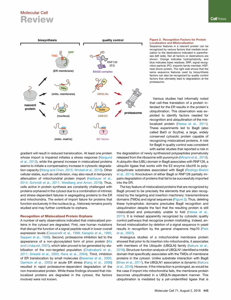
biosynthesis quality control Figure 2. Recognition Factors for ProteinLocalization and MislocalizationSequence features in a nascent protein can berecognized by various factors that mediate local-ization to the destinations indicated in parenthe-ses (left side). Not all factors or destinations areshown. Orange indicates hydrophobicity, andblue indicates basic residues. SRP, signal recog-nition particle; IPO, importin family member; HSP,heat shock protein. The right side shows that thesame sequence features used by biosynthesisfactors can also be recognized by quality controlfactors that ultimately lead to degradation at theproteasome.
Molecular Cell
Review
gradient will result in reduced translocation. At least one protein
whose import is impaired initiates a stress response (Nargund
et al., 2012), while the general increase in mislocalized proteins
seems to initiate a compensatory increase in cytosolic degrada-
tion capacity (Wang and Chen, 2015; Wrobel et al., 2015). Other
cellular states, such as cell division, may also result in temporary
attenuation of mitochondrial protein import (Harbauer et al.,
2014; Schmidt et al., 2011; Weidberg and Amon, 2018). Thus,
cells active in protein synthesis are constantly challenged with
proteins orphaned in the cytosol due to a combination of intrinsic
and stress-dependent failures in segregating proteins to the ER
and mitochondria. The extent of import failure for proteins that
function exclusively in the nucleus (e.g., histones) remains poorly
studied and may further contribute to orphans.
Recognition of Mislocalized Protein OrphansA number of early observations indicated that mislocalized pro-
teins in the cytosol are degraded. First, rare human mutations
that disrupt the function of a signal peptide result in lower overall
expression levels (Cassanelli et al., 1998; Karaplis et al., 1995;
Seppen et al., 1996). Second, proteasome inhibition led to the
appearance of a non-glycosylated form of prion protein (Ma
and Lindquist, 2001), which later proved to be generated by sta-
bilization of the non-targeted population (Chakrabarti et al.,
2011; Drisaldi et al., 2003; Rane et al., 2004). Third, inhibition
of ER translocation by small molecules (Besemer et al., 2005;
Garrison et al., 2005) or acute ER stress (Kang et al., 2006)
resulted in rapid proteasome-dependent degradation of the
non-translocated protein. While these findings showed that mis-
localized proteins are degraded in the cytosol, the factors
involved were not known.
Various studies had informally noted
that cell-free translation of a protein in-
tended for the ER results in the protein’s
ubiquitination. This observation was ex-
ploited to identify factors needed for
recognition and ubiquitination of the mis-
localized protein (Hessa et al., 2011).
These experiments led to Bag6 (also
called Bat3 or Scythe), a large, widely
conserved cytosolic protein capable of
recognizing mislocalized proteins. A role
for Bag6 in quality control was consistent
with earlier studies that reported a role in
the degradation of newly synthesized polypeptides prematurely
released from the ribosomewith puromycin (Minami et al., 2010).
A ubiquitin-like (UBL) domain in Bag6 associates with RNF126, a
ubiquitin ligase that works with the E2 enzyme UbcH5 to poly-
ubiquitinate substrates associated with Bag6 (Rodrigo-Brenni
et al., 2014). Knockdown of either Bag6 or RNF126 partially im-
pairs degradation of proteins that fail to be successfully imported
into the ER.
The key feature of mislocalized proteins that are recognized by
Bag6 proved to be precisely the elements that are also recog-
nized by the targeting and insertion machinery: transmembrane
domains (TMDs) and signal sequences (Figure 2). Thus, deleting
these hydrophobic domains precludes Bag6 recognition and
ubiquitination despite the fact that the resulting protein is still
mislocalized and presumably unable to fold (Hessa et al.,
2011). It is instead apparently recognized by cytosolic quality
control pathways that recognize protein misfolding. Indeed, en-
forced mislocalization by deletion of a signal sequence in yeast
results in recognition by the general chaperone Hsp70 (Park
et al., 2007).
Analogous studies of a mitochondrial membrane protein
showed that prior to its insertion into mitochondria, it associates
with members of the Ubiquilin (UBQLN) family (Itakura et al.,
2016). Structure-function analysis of UBQLN1 identified amiddle
domain that specifically associates with the TMDs of membrane
proteins in the cytosol. Unlike substrate interaction with Bag6
(Shao et al., 2017), the UBQLN1 association is dynamic (Itakura
et al., 2016). However, if the interaction is prolonged, aswould be
the case if import into mitochondria fails, the membrane protein
becomes ubiquitinated in a UBQLN-dependent manner. This
ubiquitination is mediated by a yet-unidentified ligase that is
Molecular Cell 71, August 2, 2018 445

Molecular Cell
Review
recruited to the ubiquitin-associating (UBA) domain in UBQLN.
Although other members of the UBQLN family (mammals have
four) have been less well studied, they have similar middle do-
mains that may also interact with TMDs of mislocalized proteins
(Itakura et al., 2016; Suzuki and Kawahara, 2016). It is attractive
to speculate that their specificities are somewhat different from
each other to collectively cover the wide range of sequences
that define TMDs. Indeed, Bag6 appears to prefer more hydro-
phobic TMDs than UBQLN1 (Itakura et al., 2016). Additional
studies are needed to investigate the substrate ranges and mo-
lecular basis of specificity of these factors.
Unlike membrane proteins whose exposed TMDs provide a
distinctive cue for recognition of mislocalization, the soluble pro-
teins of membrane-bound compartmentsmore closely resemble
cytosolic proteins. In the case of proteins destined for the ER,
signal sequences are sufficiently similar to TMDs that they
seem to be recognized by Bag6 (Hessa et al., 2011). Soluble pro-
teins destined for mitochondria or the nucleus have amphipathic
or basic signals, respectively. Whether dedicated factors recog-
nize persistent residence of these signals in the cytosol for
degradation remains largely unexplored. One candidate factor
for this role is UBE2O, a hybrid E2-E3 enzyme that was recently
shown to recognize and ubiquitinate ribosomal proteins in the
cytosol (Nguyen et al., 2017; Yanagitani et al., 2017). Under
normal circumstances, newly made ribosomal proteins are
recognized by dedicated nuclear import factors (J€akel and Gor-
lich, 1998), delivered to the nucleolus, and assembled with rRNA
into pre-ribosomal subunits (Pena et al., 2017). It appears that
failure of nuclear import leads to recognition by UBE2O, multi-
mono-ubiquitination, and proteasomal degradation.
Although the precise sequence features recognized by
UBE2O are unclear, it appears to be the juxtaposition of hydro-
phobic and basic residues (Yanagitani et al., 2017). This is note-
worthy because it is similar in some ways to both nuclear import
(J€akel and Gorlich, 1998) and mitochondrial import signals (von
Heijne, 1995), as well as a common feature of nucleic acid-bind-
ing proteins (Hentze et al., 2018; Nelson, 1995). Ribosomal pro-
tein interactions with UBE2O and nuclear import factors appear
to bemutually exclusive (Yanagitani et al., 2017), suggesting that
UBE2O may interact with the nuclear import signal. It will there-
fore be interesting to determinewhether this is a general property
of UBE2O and whether other nucleic acid binding proteins, such
as histones, subunits of the signal recognition particle (SRP), and
mitoribosomal proteins, are recognized by UBE2O when they
are orphaned in the cytosol.
Thus, the cytosol appears to be patrolled by a set of factors
that can interact with signal sequences and TMDs on the one
hand and ubiquitin ligases on the other (Figure 2). These factors
collectively recognize proteins that expose organelle targeting
domains, an indicator that targeting may have failed, and
mediate their tagging for degradation. It is noteworthy that the
substrate specificity of these factors, while likely to be rather
broad, appears to be especially well suited to the same elements
recognized by protein targeting factors such as SRP and TRC40
(also called Get3), which mediate membrane protein targeting to
the ER (Shao and Hegde, 2011), and Importins 5 and 7, which
mediate nuclear import of ribosomal proteins (J€akel and Gorlich,
1998). In order to not interfere with targeting, Bag6, UBQLNs,
446 Molecular Cell 71, August 2, 2018
and UBE2O must act after substrates have attempted targeting,
themechanisms of which are discussed later. Even though these
factors collectively recognize a broad range of orphansmislocal-
ized to the cytosol, additional quality control machinery that
recognize nuclear and mitochondrial targeting signals may
remain to be discovered.
Proteins Orphaned to the Wrong OrganelleThe TMDs of membrane proteins targeted to the ER and mito-
chondria are very similar in their biophysical properties (Guna
and Hegde, 2018), consistent with their eventual residence in a
lipid bilayer. For many of these proteins, the TMD(s) serve as
the main or sole targeting signal. Due to the similarities among
these TMDs, membrane proteins can be routed to the wrong
organelle. How cells deal with this problem is only partially un-
derstood.
The simplest and best-studied case involves tail-anchored
(TA) membrane proteins (Hegde and Keenan, 2011). These pro-
teins contain a single TMD close to the C terminus, which serves
as the sole targeting signal. TA proteins destined for the mito-
chondria tend to have slightly less hydrophobic TMDs (Watten-
berg et al., 2007) and often have downstream basic residues in
the C-terminal tail (Horie et al., 2002). Nevertheless, there is sub-
stantial overlap in their properties and a clear risk ofmistargeting.
The best-characterized TA targeting pathway to the ER is
known as the TRC (TMD recognition complex) pathway in mam-
mals and the homologous GET (guided entry of TA proteins)
pathway in yeast (Hegde and Keenan, 2011). Deletion of GET
pathway components in yeast results in mislocalization to the
cytosol (often as protein aggregates) and mitochondria (Jonikas
et al., 2009; Schuldiner et al., 2008). A membrane-embedded
AAA-ATPase Msp1 in the mitochondrial outer membrane is
thought to be involved in removing TA proteins mislocalized to
mitochondria (Chen et al., 2014; Okreglak and Walter, 2014).
A sub-population of Msp1 is also found in the peroxisomal mem-
brane, where it is thought to serve a similar role (Weir et al., 2017).
Hence, the levels of mislocalized TA proteins in mitochondria or
peroxisomes increases when Msp1 (or its human homolog,
ATAD1) is deleted. As this stabilization is seen even in wild-
type cells containing an intact TRC/GET pathway (Chen et al.,
2014), mislocalization must be a constitutive problem in cells
and tissues, consistent with the challenges of achieving precise
targeting specificity.
The mechanistic basis of Msp1 function in clearance of mislo-
calized protein substrates is not well understood. Purified Msp1
reconstituted into synthetic liposomes was shown to drive ATP-
dependent extraction of a co-reconstituted TA protein to the
cytosol (Wohlever et al., 2017). The ATPase domain of Msp1,
which faces the cytosol, was shown to be a hexamer containing
a central pore. Because mutations within the pore impair the TA
protein extraction activity, it has been posited that the substrate
is pulled through the pore into the cytosol (Wohlever et al., 2017).
Such a model would be consistent with the mechanism of action
of other AAA protein unfoldases for which translocation through
the central pore is strongly supported (Sauer and Baker, 2011).
How the substrate is kept in a soluble form after extraction,
how it is ubiquitinated, and how it is delivered to the proteasome
all remain unknown. One possibility is that cytosolic factors such

endoplasmicreticulum
peroxisome
proteasome
mitochondrion
Msp1
Msp1ERAD?
Figure 3. Recognition of ProteinMislocalization at OrganellesMembrane protein insertion into the wrongorganelle can be recognized by its lack of associ-ation with an interaction partner. In mitochondriaand peroxisomes, the hexameric ATPase Msp1 isinvolved in extracting such mislocalized proteinsto the cytosol for degradation by the proteasome.In the ER, the specific machinery for mislocalizedproteins has not been studied, but probably in-volves known ER-associated degradation (ERAD)pathways. The molecular basis of recognition isnot known, but may involve the same features thatfacilitate assembly into protein complexes, ex-plaining why successful assembly would preventrecognition by quality control factors.
Molecular Cell
Review
as Bag6 or UBQLNsmaymediate these downstream steps simi-
larly to how they recognize and degrade cytosolic orphans
generated by failed targeting. Consistent with this concept,
Bag6 has been shown to maintain the solubility of membrane
proteins extracted from the ER until their degradation at the pro-
teasome (Wang et al., 2011).
The basis of substrate selection by Msp1 is also only partially
understood. Studies of the peroxisomal TA protein Pex15 have
provided initial insights into this key issue (Weir et al., 2017).
Pex15 is normally found in complex with Pex3. It was observed
that excess Pex15 in peroxisomes is degraded in an Msp1-
dependent manner, while Pex15 in complex with Pex3 evades
Msp1 (Weir et al., 2017). As discussed in detail later, orphans
of multi-protein complexes are often targets for quality control
via recognition of the regions that are normally shielded by
interaction partner(s). It appears that Msp1 may use such a
mechanism to recognize orphaned Pex15 and perhaps other
substrates (Figure 3).
This finding suggests that when Pex15 is mistargeted to mito-
chondria, its recognition by Msp1 for degradation is due to the
absence of Pex3. Whether all Msp1 substrates are normally
part of larger complexes whose absence cues their recognition
remains to be determined. Furthermore, it is unclear whether
Msp1 directly recognizes substrates or uses adaptors. In this
context, it is noteworthy that Msp1 associates with Cis1, a pro-
tein that is upregulated by excessive mitochondrial import failure
and plays a yet-unknown role in clearance of failed import prod-
ucts (Weidberg and Amon, 2018). Whether Cis1 is involved in
identifying substrates for delivery to Msp1 remains unclear.
Thus, both the specific features of mistargeted proteins that
are recognized and the factors that mediate recognition remain
to be elucidated in molecular terms.
The factor(s) that play the analogous role toMsp1 in the ER are
unknown. It has recently been shown that mitochondrial TA
proteins can be mistargeted to the ER, particularly under
overexpression conditions (Vitali et al.,
2018). Furthermore, the ribosome-asso-
ciated chaperone-like protein NAC
appears to antagonize SRP and limit
its promiscuous recognition of nascent
mitochondrial proteins (Gamerdinger
et al., 2015; Wiedmann et al., 1994).
Thus, when NAC levels are reduced, mitochondrial proteins
can be detected at the ER. Although these examples of ER mis-
targeting are seen under perturbed conditions, it is likely that a
low level of mistargeting occurs in normal cells. How these mis-
targeted proteins are recognized and degraded is unclear. One
possibility is that known ER-associated degradation pathways
are involved (Vembar and Brodsky, 2008). This problemwarrants
investigation. Tools to induce excessive ER mistargeting should
facilitate this line of inquiry.
Challenges to Assembly of Protein ComplexesAfter segregating proteins to the correct compartment, most
proteins must additionally assemble with one or more partners
(typically other proteins, but also RNAs). The assembly of
multi-protein complexes imposes three major challenges to the
cell. First, the components of the complex must be synthesized
in the appropriate stoichiometry. Second, the unassembled sub-
units of a complex must avoid inappropriate interactions until
their assembly. Third, partners must find each other within a
crowded cell. As with protein localization, limitations to the
overall efficiency of achieving these essential steps in complex
assembly results in orphans that must be recognized and
degraded.
Direct analysis of translation rates by sequencing of ribosome-
protected footprints (ribosome profiling) indicates that cells typi-
cally express subunits of a multi-protein complex at close to the
stoichiometry found in the final complex (Li et al., 2014). A com-
bination of several mechanisms contributes to this concordance.
First, the genes for proteins that function together are frequently
organized in a single operon in prokaryotes (Ames and Martin,
1964). This allows the transcript levels to necessarily be
increased and decreased in unison. Differences in the order of
genes in an operon and their respective Shine-Dalgarno se-
quences can presumably further tune their relative levels of
translation (Bonde et al., 2016; Lim et al., 2011).
Molecular Cell 71, August 2, 2018 447

Molecular Cell
Review
In eukaryotes, gene order (Davila Lopez et al., 2010) and the
similarities of promoters engaged by the same transcription fac-
tors (Lee et al., 2009) can also coordinate transcript levels of pro-
tein subunits. Beyond this, further fine-tuning of mRNA half-lives
and translation rates via untranslated regions and codon opti-
mality probably contribute to expression at the desired stoichi-
ometry. For example, the 50 end of mRNAs encoding ribosomal
proteins and many translation factors contain terminal oligo-py-
rimidine sequences (Meyuhas and Kahan, 2015). These se-
quences allow the translation of these mRNAs to be repressed
and de-repressed in concert, thereby maintaining their approxi-
mate stoichiometry. Nevertheless, these mechanisms are all
subject to stochasticity (Munsky et al., 2012), meaning that the
inevitable imbalances in synthesis levels generate orphans. It is
reasonable to assume that achieving expression stoichiometry
to greater than 90% precision is very challenging given the
inherent noisiness of gene expression. For complexes with
many subunits, such as ribosomes and proteasomes, matching
expression levels may be particularly difficult.
The mechanisms by which proteins are assembled into com-
plexes has been studied in a number of contexts, including ribo-
some biogenesis in the nucleolus (Pena et al., 2017), hemoglobin
assembly in pre-erythrocytes (Feng et al., 2004; Kihm et al.,
2002), and histone assembly (Hammond et al., 2017). Each of
these processes is distinctive in its use of specific chaperones
and assembly factors. The main underlying principle from these
studies is that unassembled subunits of a complex are held
temporarily by an assembly factor until it is displaced by the
appropriate interacting subunit. In many cases, the precise order
of protein complex assembly is important and under selective
pressure to be maintained, presumably to minimize misas-
sembled products (Marsh et al., 2013). However, the efficiency
of these processes is unknown in most cases, especially in vivo,
so their relative contributions to the generation of orphans re-
mains to be determined.
Recent proteomic pulse-chase experiments indicate a sub-
stantial constitutive burden of unassembled orphans in cultured
cells (McShane et al., 2016). In this study, kinetic analysis of pro-
tein degradation found �10% of proteins are degraded non-
exponentially, with a proportion (ranging from 10% to 70%)
being degraded immediately after synthesis. Approximately
70% of these non-exponentially degraded proteins are subunits
of known multi-protein complexes, suggesting that the rapidly
degraded populations are those polypeptides that failed assem-
bly. As this analysis identified only the most abundant proteins, it
appears that cells must constantly identify and degrade unas-
sembled orphans.
Recognition of Unassembled Cytosolic Protein OrphansIt has been apparent for many decades that unassembled or-
phans of multi-protein complexes are selectively degraded.
The earliest appreciation of this phenomenon comes from the
study of hemoglobin, a hetero-tetramer normally formed of two
a and two b subunits (Perutz et al., 1960). Mutations in hemoglo-
bin that markedly reduce its unusually long half-life were corre-
lated with an increased tendency of tetramer dissociation into
subunits and decreased binding to heme (Jacob et al., 1968;
Rieder, 1974). Many years later, studies of the multi-subunit
448 Molecular Cell 71, August 2, 2018
T cell receptor (TCR) showed that the a subunit is rapidly
degraded when expressed without its interaction partners (Lip-
pincott-Schwartz et al., 1988). Conversely, it is well appreciated
that knockdown or knockout of one subunit of a multi-protein
complex often destabilizes the other subunits. Thus, in
numerous contexts and cellular compartments, cells have
mechanisms to selectively identify orphaned subunits and target
them for degradation.
Two general mechanisms can explain how orphans are recog-
nized. First, some proteins may not be able to achieve a stable
folded state in the absence of its interaction partner. This insta-
bility would result in its unfolding and recognition by general
quality control pathways that monitor protein folding. Second,
a subunit interface that is normally shielded in the intact complex
is recognized by a factor that ultimately leads to degradation.
Factors for both mechanisms have been identified in different
biological contexts.
The most challenging multi-subunit complex to assemble in
cells is probably the ribosome. Its biogenesis from 80 proteins
and pre-rRNA involves dozens of dedicated assembly factors,
numerous processing reactions of both proteins and rRNA, a va-
riety of post-translational modifications, and multiple transport
steps across the nuclear envelope (Pena et al., 2017). Individual
ribosomal proteins have long been known to be refractory to
overexpression, suggesting efficient mechanism(s) for degrad-
ing excess copies (Abovich et al., 1985; Warner et al., 1985).
Despite the fact that ribosomes have a very long half-life, ribo-
somal proteins are a major set of proteins that accumulate
when proteasome activity is diminished (Mayor et al., 2005,
2007). This suggests that ribosomal proteins may be a major
source of orphans in actively growing cells. The need to destroy
orphaned ribosomal proteins may be especially critical as these
factors are aggregation-prone in the absence of an RNA scaffold
or a ribosome assembly chaperone (J€akel et al., 2002).
As discussed above, a ribosomal protein orphaned at the step
of import into the nucleus is recognized in the cytosol by UBE2O
(Yanagitani et al., 2017). After nuclear import, failure at the step of
assembly with rRNA is recognized by a different mechanism. In
yeast, the large nuclear-localized E3 ligase Tom1 was shown
to interact with unassembled ribosomal proteins and mediate
their ubiquitination (Sung et al., 2016). The sites of ubiquitination
suggest that Tom1 probably interacts with basic regions that
would ordinarily interact with rRNA. These regions are inacces-
sible in intact ribosomes, which are not recognized by Tom1.
HUWE1 is the closest homolog of Tom1 in mammals. Its knock-
down partially increases the levels of overexpressed uL24/
RPL26, but the basis for this effect was not explored in sufficient
depth to conclude a direct effect (Sung et al., 2016).
In separate studies, however, it was observed that HUWE1 is
required for efficient degradation of farnesyltransferase alpha
(FTNA) and UBL4A when their respective binding partners are
absent (Xu et al., 2016). Although interaction studies were not
performed, HUWE1 presumably recognizes the exposed inter-
faces of these and other proteins to mediate their ubiquitination.
Proteomic analysis identified 72 candidate HUWE1 substrates
(although, curiously, not ribosomal proteins), almost all of which
are known subunits of multi-protein complexes. Most of these
candidate substrates operate in the nucleus, yet HUWE1
A
B
C
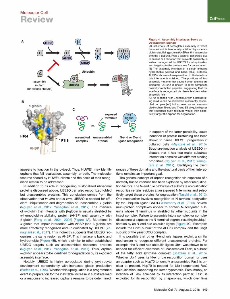
Figure 4. Assembly Interfaces Serve asDegradation Signals(A) Schematic of hemoglobin assembly in whichthe a subunit is temporarily shielded by a-hemo-globin-stabilizing protein (AHSP) until it assembleswith the b subunit. Free a subunit, generated dueto excess or a mutation that prevents assembly, isinstead recognized by UBE2O for ubiquitinationand targeting to the proteasome for degradation.(B) The assembly interface of a-globin showinghydrophobic (yellow) and basic (blue) surfaces.AHSP is shown in transparent tan to illustrate howthis interface is shielded. The positions of twoassembly mutants that cause human anemia areindicated. UBE2O is known to bind compositebasic/hydrophobic peptides, suggesting that theinterface is recognized via these features whenassembly fails.(C) An exposed N or C terminus with a destabiliz-ing residue can be shielded in a correctly assem-bled complex (left) but exposed as an unassem-bled orphan. N-end andC-end E3 ubiquitin ligasesthat recognize such residues would then selec-tively target the orphan for degradation.
Molecular Cell
Review
appears to function in the cytosol. Thus, HUWE1 may identify
orphans that fail localization, assembly, or both. The molecular
features shared by HUWE1 clients and the basis of their recog-
nition remain to be addressed.
In addition to its role in recognizing mislocalized ribosomal
proteins discussed above, UBE2O can also recognized folded
but unassembled proteins. This conclusion comes from the
observation that in vitro and in vivo, UBE2O is needed for effi-
cient ubiquitination and degradation of unassembled a-globin
(Nguyen et al., 2017; Yanagitani et al., 2017). The interface
of a-globin that interacts with b-globin is usually shielded by
a-hemoglobin-stabilizing protein (AHSP) until assembly with
b-globin (Feng et al., 2004, 2005) (Figure 4A). Mutations in
a-globin that impair interaction with AHSP (and b-globin) are
more effectively recognized and ubiquitinated by UBE2O (Ya-
nagitani et al., 2017). This indirectly suggests that UBE2O rec-
ognizes the same region as AHSP. This interface is basic and
hydrophobic (Figure 4B), which is similar to other established
UBE2O targets such as unassembled ribosomal proteins
(Nguyen et al., 2017; Yanagitani et al., 2017). Thus, orphan
a-globin appears to be identified for degradation by its exposed
assembly interface.
Notably, UBE2O is highly upregulated during erythrocyte
development concordantly with increased globin production
(Wefes et al., 1995). Whether this upregulation is a programmed
event in preparation for the inevitable increase in substrate load
or a response to increased orphans remains to be determined.
In support of the latter possibility, acute
induction of protein misfolding has been
shown to cause UBE2O upregulation in
cultured cells (Miyazaki et al., 2015).
Structure-function analysis of UBE2O in-
dicates that it has two major substrate
interaction domains with different binding
properties (Nguyen et al., 2017; Yanagi-
tani et al., 2017). Identifying the client
ranges of these domains and the structural basis of their interac-
tions remains an important goal.
The general concept of orphan recognition via exposure of a
normally buried interface has been exploited by other ubiquitina-
tion factors. The N-end rule pathways of substrate ubiquitination
recognize certain residues at an exposed N terminus and selec-
tively target these proteins for degradation (Tasaki et al., 2012).
One mechanism involves recognition of N-terminal acetylation
by the ubiquitin ligase CNOT4 (Shemorry et al., 2013). Several
multi-protein complexes appear to contain N-acetylated sub-
units whose N terminus is shielded by other subunits in the
intact complex. Failure to assemble into a complex (or complex
disassembly) exposes the N-terminal degron, resulting in ubiqui-
tination by an N-end rule ubiquitin ligase (Figure 4C). Examples
include the Hcn1 subunit of the APC/C complex and the Cog1
subunit of the yeast COG complex.
It is possible that other N-end rule ligases exploit a similar
mechanism to recognize different unassembled proteins. For
example, the N-end rule ubiquitin ligase Ubr1 was shown to be
needed for efficient clearance of unassembled Fas2, a subunit
of the fatty acid synthase complex (Scazzari et al., 2015).
Whether Ubr1 uses its N-end rule recognition domain or uses
an adaptor such as Hsp70 to identify unassembled Fas2 is un-
clear at present. Hsp70 is needed for Ubr1-dependent Fas2
ubiquitination, supporting the latter hypothesis. Presumably, an
interface of Fas2 shielded by its interaction partner, Fas1, is
exploited for its recognition by chaperones, which over time
Molecular Cell 71, August 2, 2018 449
Molecular Cell
Review
recruits a ubiquitin ligase to initiate degradation. The recent
discovery of a ‘‘C-end rule’’ (Koren et al., 2018; Lin et al., 2018)
raises the possibility that exposure of a normally shielded C ter-
minus might also be exploited by cells to recognize orphans
(Figure 4C). This idea remains to be investigated.
Recognition of Unassembled Protein Orphans at the EREach of the examples of unassembled protein degradation dis-
cussed thus far occur in the cytosol. Analogous processes oper-
ate in the ER membrane and lumen and probably also different
mitochondrial compartments. In the ER, the most extensively
studied multi-protein assemblies have been the immunoglobu-
lins and TCR. TCRa and CD3d, two subunits of the TCR, have
been widely used as models for ERAD (Bonifacino and Lippin-
cott-Schwartz, 1991; Bonifacino et al., 1989; Huppa and Ploegh,
1997; Lippincott-Schwartz et al., 1988). Both subunits are single-
spanning transmembrane proteins that are recognized for
degradation via their TMDs (Bonifacino et al., 1991; Cosson
et al., 1991; Manolios et al., 1990). The prevailing model is that
the orphaned TMDs of TCRa and CD3d are recognized by the
membrane-embedded regions of a ER-resident ubiquitin ligase
complex centered around Hrd1 (Kikkert et al., 2004). Although
the structural basis of their recognition remains unknown, it de-
pends critically on charged residues within their TMDs that are
shielded by other subunits in the assembled TCR. A similar
mode of recognition by Hrd1 was also shown for other integral
membrane substrates such as Hmg2 (Sato et al., 2009).
Based on these examples, one might speculate that orphans
of multi-protein complexes assembled via their TMDs are recog-
nized by the exposure of polar residues within the lipid bilayer.
This would provide a relatively universal cue for unassembled
proteins without relying on sequence-specific recognition. In
this light, it is intriguing that the recently determined cryo-EM
structure of Hrd1 shows a hydrophilic pocket that might serve
as the site of recognition of such orphaned TMDs (Schoebel
et al., 2017). Even though the functional role of this putative bind-
ing region of Hrd1 remains to be investigated experimentally, the
model is consistent with previous observations that mutations
within this hydrophilic region of Hrd1 affect recognition of
some integral membrane ERAD substrates (Sato et al., 2009).
Although recognition of orphaned TMDs within the membrane
has long been presumed, experimental evidence for this idea is
generally lacking. An alternative idea is that orphaned TMDs, due
to their exposed polar or charged residues, are unstable in the
lipid bilayer and translocate into the ER lumen (Feige and Hen-
dershot, 2013). This results in their recognition by lumenal chap-
erones, particularly BiP, which are proposed to deliver them for
degradation. Successful assembly would prevent this transloca-
tion, providing an explanation for why orphans are selectively
degraded. Although both models ultimately involve Hrd1 (or
another ubiquitin ligase complex) mediating retrotranslocation
into the cytosol for degradation, one posits recognition in the
ER lumen while the other involves recognition in the membrane.
Although the two views have yet to be reconciled, one attrac-
tion of the lumenal recognition model is its similarity to how mis-
assembled lumenal protein complexes might be monitored. One
example of protein complex assembly in the ER lumen is immu-
noglobulins typically built from two heavy and two light chains
450 Molecular Cell 71, August 2, 2018
(Huber et al., 1976). After translocation into the ER lumen, the
first heavy chain immunoglobulin domain (CH1) binds to the
chaperone BiP in an unfolded and reduced state (Bole et al.,
1986; Haas and Wabl, 1983; Hendershot et al., 1987; Vanhove
et al., 2001). CH1 can only fold upon interaction with light chain
(Feige et al., 2009). Hence, in the absence of light chains, heavy
chains are retained in the ER and are eventually dislocated to the
cytosol to be degraded by the proteasome (Mancini et al., 2000).
Even though the assembly process varies between different
immunoglobulin isotypes (Baumal et al., 1971), BiP-mediated
retention of heavy chains in the ER appears to be universal (Hen-
dershot et al., 1987). Prolonged association with BiP, whether
of orphan heavy chains or TCR subunits, may result in delivery
to ERAD machinery via adaptors such as specific J-proteins
(Shen and Hendershot, 2005).
Distinguishing Intermediates from OrphansProteins should be recognized as orphans to be degraded only
after reasonable attempt(s) at localization and assembly have
failed. Thus, the biosynthesis machinery should have higher pri-
ority for access to nascent chains than the quality control ma-
chinery, but this priority should be time limited. The mechanistic
basis of how priority and timing are determined is important to
understand because this critical decision balances promiscuous
degradation of normal biosynthetic intermediates versus exces-
sive persistence of potentially toxic and aggregation-prone
orphans.
One mechanism by which priority can be conferred is by
spatial segregation of the biosynthetic and quality control fac-
tors. The best illustration of this principle is SRP interaction
with the ribosome. The majority of eukaryotic proteins
destined for the ER are recognized co-translationally by SRP
(Chartron et al., 2016; Costa et al., 2018; Keenan et al.,
2001). This interaction is facilitated by the ability of SRP to
interact with the ribosome such that the signal sequence bind-
ing domain is positioned precisely at the ribosomal exit tunnel
(Halic et al., 2004; Voorhees and Hegde, 2015). Not only is
SRP thought to sample ribosomes for the presence of an
emerging signal, but it may be stabilized there while the signal
is still inside the ribosomal tunnel (Berndt et al., 2009; Voo-
rhees and Hegde, 2015).
In this manner, nascent secretory and membrane proteins
would not have an opportunity to engage quality control factors
such as Bag6 or UBQLNs unless SRP recognition has failed.
Thus, despite its �5- to 10-fold lower abundance (Kulak et al.,
2014), SRP nevertheless has priority due to its precise localiza-
tion at the ribosome. Once a protein has been released from
the ribosome, SRP’s relatively low abundance and poor capacity
to bind proteins in solution precludes its interference with other
processes.
At a later step in biogenesis, secretory and membrane proteins
are translocated into the ER through the ribosome-associated
Sec61 translocation channel (Rapoport et al., 2017). Any proteins
that selectively associate, even dynamically, with Sec61 will get
priority for interaction with the nascent chain over those that do
not. Indeed, processing enzymes such as oligosaccharyl trans-
ferase and signal peptidase enjoy such priority (Braunger et al.,
2018), as might the chaperone BiP (via its recruitment by the

Molecular Cell
Review
translocon component Sec63 [Brodsky et al., 1995]) and Calnexin
(Lakkaraju et al., 2012). In this manner, biosynthetic factors could
be given priority before degradation factors such as Hrd1.
A second mechanism of conferring priority is to use a combi-
nation of abundance and fast binding. This appears to be how
newly made TA proteins are prioritized for ER targeting ahead
of proteasomal degradation. Recent studies show that among
the three TMD binding factors involved in TA protein targeting,
SGTA is the fastest and first interactor (Shao et al., 2017). Its
competitive advantage may be further increased by its interac-
tion with the Bag6 complex or Hsp70, both of which can asso-
ciate with the ribosome. TA proteins can transfer from SGTA to
TRC40 (Mock et al., 2015; Shao et al., 2017) by a mechanism
that does not require release of TA protein into the bulk cytosol
(Shao et al., 2017). Loading on TRC40 is a commitment to target-
ing because TA protein dissociation is very slow relative to the
rate of targeting. TA proteins have a limited time to complete
these events as dictated by their off-rate from SGTA. Dissocia-
tion from SGTA permits an opportunity to be captured by
Bag6, which recruits an E3 ligase for substrate ubiquitination
(Rodrigo-Brenni et al., 2014). Very slow dissociation from Bag6
means TA binding to this factor is effectively a commitment for
degradation. Thus, triage by multiple factors of seemingly iden-
tical specificity can be accomplished simply by their differential
on- and off-rates combined with committed downstream events
such as membrane insertion or ubiquitination (Shao et al., 2017).
The final mechanism is based on an intrinsically slow reaction
followed by a specific commitment step. This is best exemplified
by glycoprotein quality control in which a specific irreversible
trimming event of an N-linked glycan generates a product that
is recognized by a lectin coupled to quality control pathways
(Caramelo and Parodi, 2015; Tannous et al., 2015). The trimming
event is carried out by intrinsically slow mannosidases, effec-
tively placing a time limit on protein maturation attempts. As
predicted by this model, overexpression of the mannosidase
accelerates the rate of degradation (Hosokawa et al., 2001).
An analogous mechanism of a slow enzymatic reaction linked
to a commitment step seems to be used for some orphans in the
cytosol. Membrane proteins bound to UBQLNs are not initially
committed for degradation (Itakura et al., 2016). Instead, their
dynamic release and re-binding provides opportunities at suc-
cessful insertion. However, a relatively slow ubiquitination step
seems to be the key commitment step. When ubiquitin is added
to the substrate, a UBA domain in UBQLNs binds to ubiquitin,
preventing substrate release and thereby ending insertion at-
tempts (Itakura et al., 2016). Presumably, the time allowed for
insertion is dependent on the affinity of the ligase for UBQLNs,
its abundance, and speed of ubiquitination. Identifying the ligase
should allow these aspects of the model to be tested.
Can Failures Be Detected Co-translationally?The systems for detection and degradation of orphans dis-
cussed so far all operate post-translationally. This is consistent
with the idea that biosynthesis must necessarily have an oppor-
tunity to succeed before a polypeptide is deemed an orphan.
However, many biosynthetic reactions are initiated or occur
co-translationally: recognition by SRP, the initial stages of poly-
peptide folding, various modifications, and even multi-subunit
assembly. It is therefore possible that cells have evolved
mechanisms to detect orphans co-translationally via failure of
a decisive co-translational biosynthetic step. Early detection is
advantageous because minimizing the time an aberrant poly-
peptide resides in a cell reduces the risk of inappropriate interac-
tions, aggregation, and other adverse consequences. Although
co-translational orphan detection has not been established
unambiguously, two examples are noteworthy.
The first example concerns an autoregulatory mechanism to
control b-tubulin expression. As noted already, a- and b-tubulin
form a constitutive complex (Feit et al., 1971). In the presence
of excess unpolymerized tubulin subunits, synthesis of both
a-tubulin and b-tubulin polypeptides is reduced (Cleveland
et al., 1981).One regulatorymechanism involves selective degra-
dation of b-tubulin mRNA (Pittenger and Cleveland, 1985). This
degradation is strictly dependent on translation andmore specif-
ically on the first four amino acids in the nascent polypeptide
(Bachurski et al., 1994; Gay et al., 1989). Remarkably, a mono-
clonal antibody that binds these four residues abolished transla-
tion-dependentmRNAdegradation (Theodorakis andCleveland,
1992). These findings led to the idea that co-translational associ-
ation of nascent b-tubulin with some yet-unidentified factor
(which is not a-tubulin) is needed to escape mRNA degradation.
In the intervening time since these findings, various pathways
of mRNA decay have been characterized, including ones depen-
dent on ribosome stalling (Shoemaker and Green, 2012). Thus,
one attractive model is that a limiting assembly factor binds
nascent b-tubulin, the absence of which leads to translation
arrest and mRNA degradation. Assuming that this factor is
displaced upon assembly with a-tubulin (or tubulin incorporation
into microtubules), b-tubulin orphans would sequester the
assembly factor and lead to mRNA decay. In this way, the
production of orphan b-tubulin would be tightly restricted via a
co-translational detection mechanism.
The second example concerns the consequences of failed co-
translational recognition by SRP. It has been observed that
knockdown of SRP or mutating a signal peptide such that it
cannot be recognized by SRP causes a reduction in the corre-
sponding mRNA and protein (Karamyshev et al., 2014). This
effect was dependent on Argonaute2, which was also observed
to interact with the SRP-deficient nascent chain at the
ribosome (Karamyshev et al., 2014). These findings suggest
a model in which co-translational failure of a hydrophobic
sequence to be recognized by SRP triggers Argonaute2-depen-
dent mRNA decay.
These two examples illustrate a potentially important princi-
ple (Figure 5). If an interaction critical for biogenesis occurs
co-translationally, its failure would necessarily result in an
orphan. There may be mechanisms to detect such failures
co-translationally, which would provide the cellular quality con-
trol systems access to both the nascent polypeptide and asso-
ciated mRNA. Although still poorly studied, there is evidence
that many protein complexes may initiate assembly during
translation (Duncan and Mata, 2011; Williams and Dichtl,
2018). If such co-translational interactions were coupled to
translation elongation, the mRNA decay pathways downstream
of ribosome stalling could be exploited to fine-tune the
balanced expression of subunits and minimize the generation
Molecular Cell 71, August 2, 2018 451

feedback totranslation & QC
mRNA degradation
assemblyfactor
......
......
normal conditions excess unassembled orphans Figure 5. Hypothetical Model for Co-translational Monitoring of ProteinAssemblyA nascent polypeptide emerging from the ribo-some is recognized by an assembly factor, whichis recycled when assembly occurs correctly.When assembly fails, the assembly factor istitrated by excess unassembled orphans (rightside). The unavailability of this nascent chainbinding protein at the ribosome is proposedto trigger ribosome-associated quality controlpathway via effects on translation (red arrow),perhaps via factors (not shown) that bind to thesequence motif normally recognized by the as-sembly factor.
Molecular Cell
Review
of orphans. Such an early-detection system may represent one
of the physiologic roles of ribosome-associated quality control
pathways. Intriguingly, up to �12%–15% of nascent proteins
might be ubiquitinated co-translationally at the ribosome for
reasons that remain to be investigated (Duttler et al., 2013;
Wang et al., 2013).
Orphan-Related PathologiesAs with other types of quality control, such as protein misfolding
or processing, excessive production of orphans can be domi-
nantly detrimental to cellular homeostasis. This is observed in
the disease phenotypes of various inheritedmutations. Raremu-
tations have been described in the signal sequences of a wide
range of proteins (Cassanelli et al., 1998; Karaplis et al., 1995;
Seppen et al., 1996). In most cases, themutation disrupts the hy-
drophobic core of the signal to reduce or eliminate its targeting
function. This would lead to an increase of orphans in the cytosol
of cells expressing the mutant protein. Accordingly, the pheno-
types are typically dominant and tissue-specific to the most
highly expressing cell type. This suggests that the increase in or-
phans, not simply a loss of function, is the basis of cellular dam-
age. In mouse studies of the non-essential prion protein (PrP),
introduction of a version containing a signal sequence that is
only�50%efficient led to a dominant neurodegenerative pheno-
type (Rane et al., 2008). Thus, over long times in a complex
organism, even a modest increase in the load of mislocalized
orphans in the cytosol can be detrimental.
Perhaps the most common set of diseases involving orphans
are the Thalassemias, a group of hereditary blood diseases
characterized by anemia, hemolysis, and various downstream
consequences (Olivieri, 1999; Piel and Weatherall, 2014). These
diseases result when one or more of the alleles encoding a- or
b-globin is deficient, resulting in imbalanced synthesis of hemo-
globin subunits. Mutations in the a-b interface that impair assem-
bly of hemoglobin also cause rare variants of anemia related to
the Thalassemias (Clarke and Higgins, 2000; Kohne, 2011).
While a reduced level of mature hemoglobin is certainly a major
contributor to the pathogenesis of these diseases, substantial
evidence indicates that the unpaired globin proteins are domi-
nantly toxic in many ways. For example, unpaired a- or b-globin
has been suggested to generate increased reactive oxygen spe-
cies, form intracellular aggregates, or interfere with other cellular
452 Molecular Cell 71, August 2, 2018
processes such as assembly of a membrane cytoskeleton
(Weatherall and Clegg, 2001). As noted above, unassembled
a-globin is selectively recognized, ubiquitinated, and degraded
by UBE2O. However, this does not seem to be the only degrada-
tion pathway, as some a-globin ubiquitination was observed
even in UBE2O knockout cells (Nguyen et al., 2017). Further-
more, the pathway of unpaired b-globin degradation remains
unknown. These may prove to be important facets of the patho-
physiology of the Thalassemias.
Given the detrimental consequences of orphans, it is note-
worthy that aneuploidy is a prominent feature in a large propor-
tion of cancers (Rajagopalan and Lengauer, 2004). As the genes
for different subunits of multi-protein complexes are often on
different chromosomes, aneuploidy would necessarily unbal-
ance expression. Experiments in yeast have shown that duplica-
tion of a single chromosome in an otherwise haploid cell is
detrimental, while an additional chromosome in a diploid cell is
less so (Dephoure et al., 2014; Dodgson et al., 2016; Oromendia
et al., 2012). This suggests that proteome imbalance may under-
lie the fitness cost, an idea consistent with the observed protein
homeostasis defects seen in aneuploid yeast. Experiments in
human cells induced to be aneuploid support the findings in
yeast (Ben-David et al., 2014; Stingele et al., 2012; Williams
et al., 2008), yet aneuploid cancer cells paradoxically grow unre-
strained. This might imply that they manage to grow robustly
despite aneuploidy, not because of it. Consistent with this
idea, many cancer cells have higher levels of the major cytosolic
chaperone Hsp70 (Murphy, 2013), elevated proteasome activity
(Chen andMadura, 2005), and increased autophagy (Singh et al.,
2018). It is also noteworthy that UBE2O is amplified in many can-
cers, while reduction of UBE2O in different mouse cancer
models provides a benefit by attenuating tumor growth (Liang
et al., 2017; Vila et al., 2017). Thus, understanding the mecha-
nistic basis of orphan recognition and degradation may provide
new therapeutic opportunities in a number of common diseases
ranging from Thalassemias to cancers.
ACKNOWLEDGMENTS
We thank Hegde lab members for useful discussions. Work in the authors’ labis supported by the UK Medical Research Council (MC_UP_A022_1007to R.S.H.).

Molecular Cell
Review
REFERENCES
Abovich, N., Gritz, L., Tung, L., and Rosbash, M. (1985). Effect of RP51 genedosage alterations on ribosome synthesis in Saccharomyces cerevisiae.Mol. Cell. Biol. 5, 3429–3435.
Ames, B.N., and Martin, R.G. (1964). Biochemical Aspects of Genetics:The Operon. Annu. Rev. Biochem. 33, 235–258.
Babu, M., Vlasblom, J., Pu, S., Guo, X., Graham, C., Bean, B.D.M., Burston,H.E., Vizeacoumar, F.J., Snider, J., Phanse, S., et al. (2012). Interaction land-scape of membrane-protein complexes in Saccharomyces cerevisiae. Nature489, 585–589.
Bachurski, C.J., Theodorakis, N.G., Coulson, R.M., and Cleveland, D.W.(1994). An amino-terminal tetrapeptide specifies cotranslational degradationof beta-tubulin but not alpha-tubulin mRNAs. Mol. Cell. Biol. 14, 4076–4086.
Baumal, R., Potter, M., and Scharff, M.D. (1971). Synthesis, assembly, andsecretion of gamma globulin by mouse myeloma cells. 3. Assembly of thethree subclasses of IgG. J. Exp. Med. 134, 1316–1334.
Ben-David, U., Arad, G., Weissbein, U., Mandefro, B., Maimon, A., Golan-Lev,T., Narwani, K., Clark, A.T., Andrews, P.W., Benvenisty, N., and Carlos Bian-cotti, J. (2014). Aneuploidy induces profound changes in gene expression, pro-liferation and tumorigenicity of human pluripotent stem cells. Nat. Commun.5, 4825.
Benz, E.J., Jr., and Forget, B.G. (1974). The biosynthesis of hemoglobin.Semin. Hematol. 11, 463–523.
Berndt, U., Oellerer, S., Zhang, Y., Johnson, A.E., and Rospert, S. (2009). Asignal-anchor sequence stimulates signal recognition particle binding to ribo-somes from inside the exit tunnel. Proc. Natl. Acad. Sci. USA 106, 1398–1403.
Besemer, J., Harant, H., Wang, S., Oberhauser, B., Marquardt, K., Foster,C.A., Schreiner, E.P., de Vries, J.E., Dascher-Nadel, C., and Lindley, I.J.D.(2005). Selective inhibition of cotranslational translocation of vascular celladhesion molecule 1. Nature 436, 290–293.
Blobel, G. (1980). Intracellular protein topogenesis. Proc. Natl. Acad. Sci. USA77, 1496–1500.
Bole, D.G., Hendershot, L.M., and Kearney, J.F. (1986). Posttranslationalassociation of immunoglobulin heavy chain binding protein with nascentheavy chains in nonsecreting and secreting hybridomas. J. Cell Biol.102, 1558–1566.
Bonde, M.T., Pedersen, M., Klausen, M.S., Jensen, S.I., Wulff, T., Harrison, S.,Nielsen, A.T., Herrgard, M.J., and Sommer, M.O.A. (2016). Predictable tuningof protein expression in bacteria. Nat. Methods 13, 233–236.
Bonifacino, J.S., and Lippincott-Schwartz, J. (1991). Degradation of proteinswithin the endoplasmic reticulum. Curr. Opin. Cell Biol. 3, 592–600.
Bonifacino, J.S., Suzuki, C.K., Lippincott-Schwartz, J., Weissman, A.M., andKlausner, R.D. (1989). Pre-Golgi degradation of newly synthesized T-cell anti-gen receptor chains: intrinsic sensitivity and the role of subunit assembly.J. Cell Biol. 109, 73–83.
Bonifacino, J.S., Cosson, P., Shah, N., and Klausner, R.D. (1991). Role ofpotentially charged transmembrane residues in targeting proteins for retentionand degradation within the endoplasmic reticulum. EMBO J. 10, 2783–2793.
Braunger, K., Pfeffer, S., Shrimal, S., Gilmore, R., Berninghausen, O., Mandon,E.C., Becker, T., Forster, F., and Beckmann, R. (2018). Structural basis forcoupling protein transport andN-glycosylation at themammalian endoplasmicreticulum. Science 360, 215–219.
Brewer, J.W., and Hendershot, L.M. (2005). Building an antibody factory: a jobfor the unfolded protein response. Nat. Immunol. 6, 23–29.
Brodsky, J.L., Goeckeler, J., and Schekman, R. (1995). BiP and Sec63p arerequired for both co- and posttranslational protein translocation into the yeastendoplasmic reticulum. Proc. Natl. Acad. Sci. USA 92, 9643–9646.
Caramelo, J.J., and Parodi, A.J. (2015). A sweet code for glycoprotein folding.FEBS Lett. 589, 3379–3387.
Cassanelli, S., Bertolini, S., Rolleri, M., De Stefano, F., Casarino, L., Elicio, N.,Naselli, A., and Calandra, S. (1998). A ‘de novo’ point mutation of the low-den-
sity lipoprotein receptor gene in an Italian subject with primary hypercholester-olemia. Clin. Genet. 53, 391–395.
Chakrabarti, O., Rane, N.S., and Hegde, R.S. (2011). Cytosolic aggregatesperturb the degradation of nontranslocated secretory andmembrane proteins.Mol. Biol. Cell 22, 1625–1637.
Chartron, J.W., Hunt, K.C.L., and Frydman, J. (2016). Cotranslational signal-in-dependent SRP preloading during membrane targeting. Nature 536, 224–228.
Chen, L., and Madura, K. (2005). Increased proteasome activity, ubiquitin-conjugating enzymes, and eEF1A translation factor detected in breast cancertissue. Cancer Res. 65, 5599–5606.
Chen, Y.-C., Umanah, G.K.E., Dephoure, N., Andrabi, S.A., Gygi, S.P., Daw-son, T.M., Dawson, V.L., and Rutter, J. (2014). Msp1/ATAD1 maintains mito-chondrial function by facilitating the degradation of mislocalized tail-anchoredproteins. EMBO J. 33, 1548–1564.
Clarke, G.M., and Higgins, T.N. (2000). Laboratory investigation of hemoglo-binopathies and thalassemias: review and update. Clin. Chem. 46, 1284–1290.
Claude, A. (1943). The constitution of protoplasm. Science 97, 451–456.
Claude, A. (1946). Fractionation of Mammalian Liver Cells By DifferentialCentrifugation : Ii. Experimental Procedures and Results. J. Exp. Med.84, 61–89.
Cleveland, D.W., Lopata, M.A., Sherline, P., and Kirschner, M.W. (1981). Un-polymerized tubulin modulates the level of tubulin mRNAs. Cell 25, 537–546.
Cosson, P., Lankford, S.P., Bonifacino, J.S., and Klausner, R.D. (1991). Mem-brane protein association by potential intramembrane charge pairs. Nature351, 414–416.
Costa, E.A., Subramanian, K., Nunnari, J., andWeissman, J.S. (2018). Definingthe physiological role of SRP in protein-targeting efficiency and specificity.Science 359, 689–692.
Davila Lopez, M., Martınez Guerra, J.J., and Samuelsson, T. (2010). Analysis ofgene order conservation in eukaryotes identifies transcriptionally and function-ally linked genes. PLoS ONE 5, e10654.
De Duve, C. (1965). The separation and characterization of subcellular parti-cles. Harvey Lect. 59, 49–87.
Dephoure, N., Hwang, S., O’Sullivan, C., Dodgson, S.E., Gygi, S.P., Amon, A.,and Torres, E.M. (2014). Quantitative proteomic analysis reveals posttransla-tional responses to aneuploidy in yeast. eLife 3, e03023.
Dodgson, S.E., Kim, S., Costanzo, M., Baryshnikova, A., Morse, D.L., Kaiser,C.A., Boone, C., and Amon, A. (2016). Chromosome-specific and global ef-fects of aneuploidy in Saccharomyces cerevisiae. Genetics 202, 1395–1409.
Drisaldi, B., Stewart, R.S., Adles, C., Stewart, L.R., Quaglio, E., Biasini, E., Fior-iti, L., Chiesa, R., and Harris, D.A. (2003). Mutant PrP is delayed in its exit fromthe endoplasmic reticulum, but neither wild-type nor mutant PrP undergoesretrotranslocation prior to proteasomal degradation. J. Biol. Chem. 278,21732–21743.
Duncan, C.D.S., and Mata, J. (2011). Widespread cotranslational formation ofprotein complexes. PLoS Genet. 7, e1002398.
Duttler, S., Pechmann, S., and Frydman, J. (2013). Principles of cotranslationalubiquitination and quality control at the ribosome. Mol. Cell 50, 379–393.
Emanuelsson, O., Brunak, S., von Heijne, G., and Nielsen, H. (2007). Locatingproteins in the cell using TargetP, SignalP and related tools. Nat. Protoc. 2,953–971.
Feige, M.J., and Hendershot, L.M. (2013). Quality control of integral membraneproteins by assembly-dependent membrane integration. Mol. Cell 51,297–309.
Feige, M.J., Groscurth, S., Marcinowski, M., Shimizu, Y., Kessler, H., Hender-shot, L.M., and Buchner, J. (2009). An unfolded CH1 domain controls the as-sembly and secretion of IgG antibodies. Mol. Cell 34, 569–579.
Feit, H., Slusarek, L., and Shelanski, M.L. (1971). Heterogeneity of tubulin sub-units. Proc. Natl. Acad. Sci. USA 68, 2028–2031.
Molecular Cell 71, August 2, 2018 453
Molecular Cell
Review
Feng, L., Gell, D.A., Zhou, S., Gu, L., Kong, Y., Li, J., Hu, M., Yan, N., Lee, C.,Rich, A.M., et al. (2004). Molecular mechanism of AHSP-mediated stabilizationof a-hemoglobin. Cell 119, 629–640.
Feng, L., Zhou, S., Gu, L., Gell, D.A., Mackay, J.P., Weiss, M.J., Gow, A.J., andShi, Y. (2005). Structure of oxidized a-haemoglobin bound to AHSP reveals aprotective mechanism for haem. Nature 435, 697–701.
Fons, R.D., Bogert, B.A., and Hegde, R.S. (2003). Substrate-specific functionof the translocon-associated protein complex during translocation across theER membrane. J. Cell Biol. 160, 529–539.
Gamerdinger, M., Hanebuth, M.A., Frickey, T., and Deuerling, E. (2015). Theprinciple of antagonism ensures protein targeting specificity at the endo-plasmic reticulum. Science 348, 201–207.
Garrison, J.L., Kunkel, E.J., Hegde, R.S., and Taunton, J. (2005). A substrate-specific inhibitor of protein translocation into the endoplasmic reticulum. Na-ture 436, 285–289.
Gavin, A.C., Aloy, P., Grandi, P., Krause, R., Boesche, M., Marzioch, M., Rau,C., Jensen, L.J., Bastuck, S., D€umpelfeld, B., et al. (2006). Proteome survey re-veals modularity of the yeast cell machinery. Nature 440, 631–636.
Gay, D.A., Sisodia, S.S., and Cleveland, D.W. (1989). Autoregulatory control ofbeta-tubulin mRNA stability is linked to translation elongation. Proc. Natl.Acad. Sci. USA 86, 5763–5767.
Guna, A., and Hegde, R.S. (2018). Transmembrane domain recognition duringmembrane protein biogenesis and quality control. Curr. Biol. 28, R498–R511.
Haas, I.G., and Wabl, M. (1983). Immunoglobulin heavy chain binding protein.Nature 306, 387–389.
Halic, M., Becker, T., Pool, M.R., Spahn, C.M.T., Grassucci, R.A., Frank, J.,and Beckmann, R. (2004). Structure of the signal recognition particle interact-ing with the elongation-arrested ribosome. Nature 427, 808–814.
Hammond, C.M., Strømme, C.B., Huang, H., Patel, D.J., and Groth, A. (2017).Histone chaperone networks shaping chromatin function. Nat. Rev. Mol. CellBiol. 18, 141–158.
Harbauer, A.B., Opali�nska,M., Gerbeth, C., Herman, J.S., Rao, S., Schonfisch,B., Guiard, B., Schmidt, O., Pfanner, N., and Meisinger, C. (2014). Mitochon-dria. Cell cycle-dependent regulation of mitochondrial preprotein translocase.Science 346, 1109–1113.
Harper, J.W., and Bennett, E.J. (2016). Proteome complexity and the forcesthat drive proteome imbalance. Nature 537, 328–338.
Havugimana, P.C., Hart, G.T., Nepusz, T., Yang, H., Turinsky, A.L., Li, Z.,Wang, P.I., Boutz, D.R., Fong, V., Phanse, S., et al. (2012). A census of humansoluble protein complexes. Cell 150, 1068–1081.
Hegde, R.S., and Keenan, R.J. (2011). Tail-anchored membrane protein inser-tion into the endoplasmic reticulum. Nat. Rev. Mol. Cell Biol. 12, 787–798.
Hendershot, L., Bole, D., Kohler, G., and Kearney, J.F. (1987). Assembly andsecretion of heavy chains that do not associate posttranslationally with immu-noglobulin heavy chain-binding protein. J. Cell Biol. 104, 761–767.
Hentze, M.W., Castello, A., Schwarzl, T., and Preiss, T. (2018). A brave newworld of RNA-binding proteins. Nat. Rev. Mol. Cell Biol. 19, 327–341.
Hessa, T., Sharma, A., Mariappan, M., Eshleman, H.D., Gutierrez, E., andHegde, R.S. (2011). Protein targeting and degradation are coupled for elimina-tion of mislocalized proteins. Nature 475, 394–397.
Horie, C., Suzuki, H., Sakaguchi, M., andMihara, K. (2002). Characterization ofsignal that directs C-tail-anchored proteins to mammalian mitochondrial outermembrane. Mol. Biol. Cell 13, 1615–1625.
Hosokawa, N., Wada, I., Hasegawa, K., Yorihuzi, T., Tremblay, L.O., Her-scovics, A., and Nagata, K. (2001). A novel ER alpha-mannosidase-like proteinaccelerates ER-associated degradation. EMBO Rep. 2, 415–422.
Huber, R., Deisenhofer, J., Colman, P.M., Matsushima, M., and Palm, W.(1976). Crystallographic structure studies of an IgG molecule and an Fc frag-ment. Nature 264, 415–420.
Huppa, J.B., and Ploegh, H.L. (1997). The alpha chain of the T cell antigen re-ceptor is degraded in the cytosol. Immunity 7, 113–122.
454 Molecular Cell 71, August 2, 2018
Itakura, E., Zavodszky, E., Shao, S., Wohlever, M.L., Keenan, R.J., and Hegde,R.S. (2016). Ubiquilins chaperone and triagemitochondrial membrane proteinsfor degradation. Mol. Cell 63, 21–33.
Itzhak, D.N., Tyanova, S., Cox, J., and Borner, G.H. (2016). Global, quantitativeand dynamic mapping of protein subcellular localization. eLife 5, https://doi.org/10.7554/eLife.16950.
Jacob, H.S., Brain, M.C., Dacie, J.V., Carrell, R.W., and Lehmann, H. (1968).Abnormal haem binding and globin SH group blockade in unstable haemoglo-bins. Nature 218, 1214–1217.
J€akel, S., and Gorlich, D. (1998). Importin beta, transportin, RanBP5 andRanBP7 mediate nuclear import of ribosomal proteins in mammalian cells.EMBO J. 17, 4491–4502.
J€akel, S., Mingot, J.-M., Schwarzmaier, P., Hartmann, E., and Gorlich, D.(2002). Importins fulfil a dual function as nuclear import receptors and cyto-plasmic chaperones for exposed basic domains. EMBO J. 21, 377–386.
Jonikas, M.C., Collins, S.R., Denic, V., Oh, E., Quan, E.M., Schmid, V., Weibe-zahn, J., Schwappach, B., Walter, P., Weissman, J.S., and Schuldiner, M.(2009). Comprehensive characterization of genes required for protein foldingin the endoplasmic reticulum. Science 323, 1693–1697.
Kang, S.-W., Rane, N.S., Kim, S.J., Garrison, J.L., Taunton, J., and Hegde,R.S. (2006). Substrate-specific translocational attenuation during ER stressdefines a pre-emptive quality control pathway. Cell 127, 999–1013.
Karamyshev, A.L., Patrick, A.E., Karamysheva, Z.N., Griesemer, D.S., Hudson,H., Tjon-Kon-Sang, S., Nilsson, I., Otto, H., Liu, Q., Rospert, S., et al. (2014).Inefficient SRP interaction with a nascent chain triggers amRNAquality controlpathway. Cell 156, 146–157.
Karaplis, A.C., Lim, S.K., Baba, H., Arnold, A., and Kronenberg, H.M. (1995).Inefficient membrane targeting, translocation, and proteolytic processing bysignal peptidase of a mutant preproparathyroid hormone protein. J. Biol.Chem. 270, 1629–1635.
Keenan, R.J., Freymann, D.M., Stroud, R.M., and Walter, P. (2001). The signalrecognition particle. Annu. Rev. Biochem. 70, 755–775.
Kihm, A.J., Kong, Y., Hong, W., Russell, J.E., Rouda, S., Adachi, K., Simon,M.C., Blobel, G.A., and Weiss, M.J. (2002). An abundant erythroid proteinthat stabilizes free alpha-haemoglobin. Nature 417, 758–763.
Kikkert, M., Doolman, R., Dai, M., Avner, R., Hassink, G., van Voorden, S., Tha-nedar, S., Roitelman, J., Chau, V., andWiertz, E. (2004). Human HRD1 is an E3ubiquitin ligase involved in degradation of proteins from the endoplasmicreticulum. J. Biol. Chem. 279, 3525–3534.
Kim, S.J., Mitra, D., Salerno, J.R., and Hegde, R.S. (2002). Signal sequencescontrol gating of the protein translocation channel in a substrate-specificmanner. Dev. Cell 2, 207–217.
Kohne, E. (2011). Hemoglobinopathies: clinical manifestations, diagnosis, andtreatment. Dtsch. Arztebl. Int. 108, 532–540.
Koren, I., Timms, R.T., Kula, T., Xu, Q., Li, M.Z., and Elledge, S.J. (2018). TheEukaryotic Proteome Is Shaped by E3 Ubiquitin Ligases Targeting C-TerminalDegrons. Cell 173, 1622–1635.e14.
Krogan, N.J., Cagney, G., Yu, H., Zhong, G., Guo, X., Ignatchenko, A., Li, J.,Pu, S., Datta, N., Tikuisis, A.P., et al. (2006). Global landscape of protein com-plexes in the yeast Saccharomyces cerevisiae. Nature 440, 637–643.
Kulak, N.A., Pichler, G., Paron, I., Nagaraj, N., and Mann, M. (2014). Minimal,encapsulated proteomic-sample processing applied to copy-number estima-tion in eukaryotic cells. Nat. Methods 11, 319–324.
Lakkaraju, A.K., Abrami, L., Lemmin, T., Blaskovic, S., Kunz, B., Kihara, A., DalPeraro, M., and van der Goot, F.G. (2012). Palmitoylated calnexin is a keycomponent of the ribosome-translocon complex. EMBO J. 31, 1823–1835.
Lee, J.W., Zemojtel, T., and Shakhnovich, E. (2009). Systems-level evidence oftranscriptional co-regulation of yeast protein complexes. J. Comput. Biol. 16,331–339.
Levine, C.G., Mitra, D., Sharma, A., Smith, C.L., and Hegde, R.S. (2005). Theefficiency of protein compartmentalization into the secretory pathway. Mol.Biol. Cell 16, 279–291.

Molecular Cell
Review
Li, G.-W., Burkhardt, D., Gross, C., andWeissman, J.S. (2014). Quantifying ab-solute protein synthesis rates reveals principles underlying allocation ofcellular resources. Cell 157, 624–635.
Liang, K., Volk, A.G., Haug, J.S., Marshall, S.A., Woodfin, A.R., Bartom, E.T.,Gilmore, J.M., Florens, L., Washburn, M.P., Sullivan, K.D., et al. (2017). Ther-apeutic Targeting of MLL Degradation Pathways in MLL-Rearranged Leuke-mia. Cell 168, 59–72.e13.
Lim, H.N., Lee, Y., and Hussein, R. (2011). Fundamental relationship betweenoperon organization and gene expression. Proc. Natl. Acad. Sci. USA 108,10626–10631.
Lin, H.-C., Yeh, C.-W., Chen, Y.-F., Lee, T.-T., Hsieh, P.-Y., Rusnac, D.V., Lin,S.-Y., Elledge, S.J., Zheng, N., and Yen, H.S. (2018). C-Terminal End-DirectedProtein Elimination by CRL2 Ubiquitin Ligases. Mol. Cell 70, 602–613.e3.
Lippincott-Schwartz, J., Bonifacino, J.S., Yuan, L.C., and Klausner, R.D.(1988). Degradation from the endoplasmic reticulum: disposing of newly syn-thesized proteins. Cell 54, 209–220.
Ma, J., and Lindquist, S. (2001). Wild-type PrP and a mutant associated withprion disease are subject to retrograde transport and proteasome degrada-tion. Proc. Natl. Acad. Sci. USA 98, 14955–14960.
Mancini, R., Fagioli, C., Fra, A.M., Maggioni, C., and Sitia, R. (2000). Degrada-tion of unassembled soluble Ig subunits by cytosolic proteasomes: evidencethat retrotranslocation and degradation are coupled events. FASEB J. 14,769–778.
Manolios, N., Bonifacino, J.S., and Klausner, R.D. (1990). Transmembrane he-lical interactions and the assembly of the T cell receptor complex. Science 249,274–277.
Marsh, J.A., Hernandez, H., Hall, Z., Ahnert, S.E., Perica, T., Robinson, C.V.,and Teichmann, S.A. (2013). Protein complexes are under evolutionary selec-tion to assemble via ordered pathways. Cell 153, 461–470.
Matlack, K.E., Misselwitz, B., Plath, K., andRapoport, T.A. (1999). BiP acts as amolecular ratchet during posttranslational transport of prepro-alpha factoracross the ER membrane. Cell 97, 553–564.
Mayor, T., Lipford, J.R., Graumann, J., Smith, G.T., and Deshaies, R.J. (2005).Analysis of polyubiquitin conjugates reveals that the Rpn10 substrate receptorcontributes to the turnover of multiple proteasome targets. Mol. Cell. Prote-omics 4, 741–751.
Mayor, T., Graumann, J., Bryan, J., MacCoss, M.J., and Deshaies, R.J. (2007).Quantitative profiling of ubiquitylated proteins reveals proteasome substratesand the substrate repertoire influenced by the Rpn10 receptor pathway. Mol.Cell. Proteomics 6, 1885–1895.
McShane, E., Sin, C., Zauber, H., Wells, J.N., Donnelly, N., Wang, X., Hou, J.,Chen, W., Storchova, Z., Marsh, J.A., et al. (2016). Kinetic analysis of proteinstability reveals age-dependent degradation. Cell 167, 803–815.e21.
Meyuhas, O., and Kahan, T. (2015). The race to decipher the top secrets ofTOP mRNAs. Biochim. Biophys. Acta 1849, 801–811.
Minami, R., Hayakawa, A., Kagawa, H., Yanagi, Y., Yokosawa, H., and Kawa-hara, H. (2010). BAG-6 is essential for selective elimination of defective protea-somal substrates. J. Cell Biol. 190, 637–650.
Miyazaki, Y., Chen, L.C., Chu, B.W., Swigut, T., and Wandless, T.J. (2015).Distinct transcriptional responses elicited by unfolded nuclear or cytoplasmicprotein in mammalian cells. eLife 4, https://doi.org/10.7554/eLife.07687.
Mock, J.-Y., Chartron, J.W., Zaslaver, M., Xu, Y., Ye, Y., and Clemons, W.M.,Jr. (2015). Bag6 complex contains a minimal tail-anchor-targeting module anda mock BAG domain. Proc. Natl. Acad. Sci. USA 112, 106–111.
Mulvey, C.M., Breckels, L.M., Geladaki, A., Britov�sek, N.K., Nightingale,D.J.H., Christoforou, A., Elzek, M., Deery, M.J., Gatto, L., and Lilley, K.S.(2017). Using hyperLOPIT to perform high-resolution mapping of the spatialproteome. Nat. Protoc. 12, 1110–1135.
Munsky, B., Neuert, G., and van Oudenaarden, A. (2012). Using gene expres-sion noise to understand gene regulation. Science 336, 183–187.
Murphy, M.E. (2013). The HSP70 family and cancer. Carcinogenesis 34,1181–1188.
Nargund, A.M., Pellegrino, M.W., Fiorese, C.J., Baker, B.M., and Haynes, C.M.(2012). Mitochondrial import efficiency of ATFS-1 regulatesmitochondrial UPRactivation. Science 337, 587–590.
Nelson, H.C. (1995). Structure and function of DNA-binding proteins. Curr.Opin. Genet. Dev. 5, 180–189.
Ng, D.T., Brown, J.D., and Walter, P. (1996). Signal sequences specify the tar-geting route to the endoplasmic reticulum membrane. J. Cell Biol. 134,269–278.
Nguyen, A.T., Prado, M.A., Schmidt, P.J., Sendamarai, A.K., Wilson-Grady,J.T., Min, M., Campagna, D.R., Tian, G., Shi, Y., Dederer, V., et al. (2017).UBE2O remodels the proteome during terminal erythroid differentiation. Sci-ence 357, eaan0218.
Okreglak, V., andWalter, P. (2014). The conserved AAA-ATPase Msp1 confersorganelle specificity to tail-anchored proteins. Proc. Natl. Acad. Sci. USA 111,8019–8024.
Olivieri, N.F. (1999). The b-thalassemias. N. Engl. J. Med. 341, 99–109.
Oromendia, A.B., Dodgson, S.E., and Amon, A. (2012). Aneuploidy causesproteotoxic stress in yeast. Genes Dev. 26, 2696–2708.
Palade, G.E. (1955). A small particulate component of the cytoplasm.J. Biophys. Biochem. Cytol. 1, 59–68.
Park, S.-H., Bolender, N., Eisele, F., Kostova, Z., Takeuchi, J., Coffino, P., andWolf, D.H. (2007). The cytoplasmic Hsp70 chaperone machinery subjects mis-folded and endoplasmic reticulum import-incompetent proteins to degrada-tion via the ubiquitin-proteasome system. Mol. Biol. Cell 18, 153–165.
Pena, C., Hurt, E., and Panse, V.G. (2017). Eukaryotic ribosome assembly,transport and quality control. Nat. Struct. Mol. Biol. 24, 689–699.
Perutz, M.F., Rossmann, M.G., Cullis, A.F., Muirhead, H., Will, G., and North,A.C.T. (1960). Structure of haemoglobin: a three-dimensional Fourier synthesisat 5.5-A. resolution, obtained by X-ray analysis. Nature 185, 416–422.
Pfeffer, S., Dudek, J., Zimmermann, R., and Forster, F. (2016). Organization ofthe native ribosome-translocon complex at themammalian endoplasmic retic-ulum membrane. Biochim. Biophys. Acta 1860, 2122–2129.
Piel, F.B., and Weatherall, D.J. (2014). The a-thalassemias. N. Engl. J. Med.371, 1908–1916.
Pittenger, M.F., and Cleveland, D.W. (1985). Retention of autoregulatory con-trol of tubulin synthesis in cytoplasts: demonstration of a cytoplasmic mecha-nism that regulates the level of tubulin expression. J. Cell Biol. 101, 1941–1952.
Porter, K.R. (1955–1956). The submicroscopic morphology of protoplasm.Harvey Lect. 51, 175–228.
Rajagopalan, H., and Lengauer, C. (2004). Aneuploidy and cancer. Nature 432,338–341.
Rane, N.S., Yonkovich, J.L., and Hegde, R.S. (2004). Protection from cytosolicprion protein toxicity by modulation of protein translocation. EMBO J. 23,4550–4559.
Rane, N.S., Kang, S.-W., Chakrabarti, O., Feigenbaum, L., and Hegde, R.S.(2008). Reduced translocation of nascent prion protein during ER stress con-tributes to neurodegeneration. Dev. Cell 15, 359–370.
Rane, N.S., Chakrabarti, O., Feigenbaum, L., and Hegde, R.S. (2010). Signalsequence insufficiency contributes to neurodegeneration caused by trans-membrane prion protein. J. Cell Biol. 188, 515–526.
Rapoport, T.A., Li, L., and Park, E. (2017). Structural and mechanistic insightsinto protein translocation. Annu. Rev. Cell Dev. Biol. 33, 369–390.
Rieder, R.F. (1974). Human hemoglobin stability and instability: molecularmechanisms and some clinical correlations. Semin. Hematol. 11,423–440.
Rodrigo-Brenni, M.C., Gutierrez, E., and Hegde, R.S. (2014). Cytosolic qualitycontrol of mislocalized proteins requires RNF126 recruitment to Bag6. Mol.Cell 55, 227–237.
Molecular Cell 71, August 2, 2018 455
Molecular Cell
Review
Sato, B.K., Schulz, D., Do, P.H., and Hampton, R.Y. (2009). Misfolded mem-brane proteins are specifically recognized by the transmembrane domain ofthe Hrd1p ubiquitin ligase. Mol. Cell 34, 212–222.
Sauer, R.T., and Baker, T.A. (2011). AAA+ proteases: ATP-fueled machines ofprotein destruction. Annu. Rev. Biochem. 80, 587–612.
Scazzari, M., Amm, I., and Wolf, D.H. (2015). Quality control of a cytoplasmicprotein complex: chaperone motors and the ubiquitin-proteasome systemgovern the fate of orphan fatty acid synthase subunit Fas2 of yeast. J. Biol.Chem. 290, 4677–4687.
Schmidt, O., Harbauer, A.B., Rao, S., Eyrich, B., Zahedi, R.P., Stojanovski, D.,Schonfisch, B., Guiard, B., Sickmann, A., Pfanner, N., and Meisinger, C.(2011). Regulation of mitochondrial protein import by cytosolic kinases. Cell144, 227–239.
Schoebel, S., Mi, W., Stein, A., Ovchinnikov, S., Pavlovicz, R., DiMaio, F.,Baker, D., Chambers, M.G., Su, H., Li, D., et al. (2017). Cryo-EM structure ofthe protein-conducting ERAD channel Hrd1 in complex with Hrd3. Nature548, 352–355.
Schuldiner, M., Metz, J., Schmid, V., Denic, V., Rakwalska, M., Schmitt,H.D., Schwappach, B., and Weissman, J.S. (2008). The GET complex me-diates insertion of tail-anchored proteins into the ER membrane. Cell 134,634–645.
Seppen, J., Steenken, E., Lindhout, D., Bosma, P.J., and Elferink, R.P.(1996). A mutation which disrupts the hydrophobic core of the signalpeptide of bilirubin UDP-glucuronosyltransferase, an endoplasmic reticu-lum membrane protein, causes Crigler-Najjar type II. FEBS Lett. 390,294–298.
Shao, S., and Hegde, R.S. (2011). Membrane protein insertion at the endo-plasmic reticulum. Annu. Rev. Cell Dev. Biol. 27, 25–56.
Shao, S., Rodrigo-Brenni, M.C., Kivlen, M.H., and Hegde, R.S. (2017). Mech-anistic basis for a molecular triage reaction. Science 355, 298–302.
Shemorry, A., Hwang, C.S., and Varshavsky, A. (2013). Control of protein qual-ity and stoichiometries by N-terminal acetylation and the N-end rule pathway.Mol. Cell 50, 540–551.
Shen, Y., and Hendershot, L.M. (2005). ERdj3, a stress-inducible endoplasmicreticulum DnaJ homologue, serves as a cofactor for BiP’s interactions withunfolded substrates. Mol. Biol. Cell 16, 40–50.
Shoemaker, C.J., and Green, R. (2012). Translation drives mRNA quality con-trol. Nat. Struct. Mol. Biol. 19, 594–601.
Singh, S.S., Vats, S., Chia, A.Y.-Q., Tan, T.Z., Deng, S., Ong, M.S., Arfuso, F.,Yap, C.T., Goh, B.C., Sethi, G., et al. (2018). Dual role of autophagy in hall-marks of cancer. Oncogene 37, 1142–1158.
Stingele, S., Stoehr, G., Peplowska, K., Cox, J., Mann, M., and Storchova, Z.(2012). Global analysis of genome, transcriptome and proteome reveals theresponse to aneuploidy in human cells. Mol. Syst. Biol. 8, 608.
Sung, M.-K., Porras-Yakushi, T.R., Reitsma, J.M., Huber, F.M., Sweredoski,M.J., Hoelz, A., Hess, S., and Deshaies, R.J. (2016). A conserved quality-con-trol pathway that mediates degradation of unassembled ribosomal proteins.eLife 5, https://doi.org/10.7554/eLife.19105.
Suzuki, R., and Kawahara, H. (2016). UBQLN4 recognizes mislocalized trans-membrane domain proteins and targets these to proteasomal degradation.EMBO Rep. 17, 842–857.
Tannous, A., Pisoni, G.B., Hebert, D.N., and Molinari, M. (2015). N-linkedsugar-regulated protein folding and quality control in the ER. Semin. CellDev. Biol. 41, 79–89.
Tasaki, T., Sriram, S.M., Park, K.S., and Kwon, Y.T. (2012). The N-end rulepathway. Annu. Rev. Biochem. 81, 261–289.
Theodorakis, N.G., and Cleveland, D.W. (1992). Physical evidence for cotrans-lational regulation of beta-tubulin mRNA degradation. Mol. Cell. Biol. 12,791–799.
UniProt Consortium (2018). UniProt: the universal protein knowledgebase.Nucleic Acids Res. 46, 2699–2699.
456 Molecular Cell 71, August 2, 2018
Vanhove, M., Usherwood, Y.K., and Hendershot, L.M. (2001). Unassembled Igheavy chains do not cycle from BiP in vivo but require light chains to triggertheir release. Immunity 15, 105–114.
Vembar, S.S., and Brodsky, J.L. (2008). One step at a time: endoplasmic retic-ulum-associated degradation. Nat. Rev. Mol. Cell Biol. 9, 944–957.
Vila, I.K., Yao, Y., Kim, G., Xia, W., Kim, H., Kim, S.J., Park, M.K., Hwang, J.P.,Gonzalez-Billalabeitia, E., Hung, M.C., et al. (2017). A UBE2O-AMPKa2 axisthat promotes tumor initiation and progression offers opportunities for therapy.Cancer Cell 31, 208–224.
Vitali, D.G., Sinzel, M., Bulthuis, E.P., Kolb, A., Zabel, S., Mehlhorn, D.G., Fig-ueiredo Costa, B., Farkas, A., Clancy, A., Schuldiner, M., et al. (2018). The GETpathway can increase the risk of mitochondrial outer membrane proteins to bemistargeted to the ER. J. Cell Sci. 131, jcs211110.
Voigt, S., Jungnickel, B., Hartmann, E., and Rapoport, T.A. (1996). Signalsequence-dependent function of the TRAM protein during early phases of pro-tein transport across the endoplasmic reticulum membrane. J. Cell Biol.134, 25–35.
von Heijne, G. (1995). Protein sorting signals: simple peptides with complexfunctions. EXS 73, 67–76.
Voorhees, R.M., and Hegde, R.S. (2015). Structures of the scanning andengaged states of the mammalian SRP-ribosome complex. eLife 4, https://doi.org/10.7554/eLife.07975.
Wang, X., and Chen, X.J. (2015). A cytosolic network suppressing mitochon-dria-mediated proteostatic stress and cell death. Nature 524, 481–484.
Wang, Q., Liu, Y., Soetandyo, N., Baek, K., Hegde, R., and Ye, Y. (2011).A ubiquitin ligase-associated chaperone holdase maintains polypeptides insoluble states for proteasome degradation. Mol. Cell 42, 758–770.
Wang, F., Durfee, L.A., and Huibregtse, J.M. (2013). A cotranslational ubiq-uitination pathway for quality control of misfolded proteins. Mol. Cell 50,368–378.
Warner, J.R., Mitra, G., Schwindinger, W.F., Studeny, M., and Fried, H.M.(1985). Saccharomyces cerevisiae coordinates accumulation of yeast ribo-somal proteins by modulating mRNA splicing, translational initiation, and pro-tein turnover. Mol. Cell. Biol. 5, 1512–1521.
Wattenberg, B.W., Clark, D., and Brock, S. (2007). An artificial mitochondrialtail signal/anchor sequence confirms a requirement for moderate hydropho-bicity for targeting. Biosci. Rep. 27, 385–401.
Weatherall, D.J., and Clegg, J.B. (2001). The thalassaemia syndromes, 4th edi-tion (Blackwell Publ. Ltd.), pp. 132–174.
Wefes, I., Mastrandrea, L.D., Haldeman,M., Koury, S.T., Tamburlin, J., Pickart,C.M., and Finley, D. (1995). Induction of ubiquitin-conjugating enzymes duringterminal erythroid differentiation. Proc. Natl. Acad. Sci. USA 92, 4982–4986.
Weidberg, H., and Amon, A. (2018). MitoCPR-A surveillance pathway that pro-tects mitochondria in response to protein import stress. Science 360,eaan4146.
Weir, N.R., Kamber, R.A., Martenson, J.S., and Denic, V. (2017). The AAA pro-tein Msp1 mediates clearance of excess tail-anchored proteins from theperoxisomal membrane. eLife 6, https://doi.org/10.7554/eLife.28507.
Wickner, W., and Schekman, R. (2005). Protein translocation across biologicalmembranes. Science 310, 1452–1456.
Wiedemann, N., and Pfanner, N. (2017). Mitochondrial machineries for proteinimport and assembly. Annu. Rev. Biochem. 86, 685–714.
Wiedmann, B., Sakai, H., Davis, T.A., and Wiedmann, M. (1994). A proteincomplex required for signal-sequence-specific sorting and translocation. Na-ture 370, 434–440.
Williams, N.K., and Dichtl, B. (2018). Co-translational control of protein com-plex formation: a fundamental pathway of cellular organization? Biochem.Soc. Trans. 46, 197–206.
Williams, B.R., Prabhu, V.R., Hunter, K.E., Glazier, C.M., Whittaker, C.A.,Housman, D.E., and Amon, A. (2008). Aneuploidy affects proliferationand spontaneous immortalization in mammalian cells. Science 322,703–709.

Molecular Cell
Review
Wohlever, M.L., Mateja, A., McGilvray, P.T., Day, K.J., and Keenan, R.J.(2017). Msp1 Is a membrane protein dislocase for tail-anchored proteins.Mol. Cell 67, 194–202.e6.
Wright, G., Terada, K., Yano, M., Sergeev, I., and Mori, M. (2001). Oxidativestress inhibits themitochondrial import of preproteins and leads to their degra-dation. Exp. Cell Res. 263, 107–117.
Wrobel, L., Topf, U., Bragoszewski, P., Wiese, S., Sztolsztener, M.E., Oelje-klaus, S., Varabyova, A., Lirski, M., Chroscicki, P., Mroczek, S., et al. (2015).
Mistargeted mitochondrial proteins activate a proteostatic response in thecytosol. Nature 524, 485–488.
Xu, Y., Anderson, D.E., and Ye, Y. (2016). The HECT domain ubiquitin ligaseHUWE1 targets unassembled soluble proteins for degradation. Cell Discov.2, 16040.
Yanagitani, K., Juszkiewicz, S., and Hegde, R.S. (2017). UBE2O is aquality control factor for orphans of multiprotein complexes. Science357, 472–475.
Molecular Cell 71, August 2, 2018 457