molecular aspects of the stress response: chaperones ...molecular aspects of the stress response:...
TRANSCRIPT

©20
06 C
opyr
ight
Lan
des
Bio
scie
nce.
Not
for
Dis
trib
utio
n.
Molecular Aspects of the Stress Response:Chaperones, Membranes and Networks
©20
06 C
opyr
ight
Lan
des
Bio
scie
nce.
Not
for
Dis
trib
utio
n.
ADVANCES IN EXPERIMENTAL MEDICINE AND BIOLOGY
Editorial Board:NATHAN BACK, State University of New York at BuffaloIRUN R. COHEN, The Weizmann Institute of ScienceDAVID KRITCHEVSKY, Wistar InstituteABEL LAJTHA, N.S. Kline Institute for Psychiatric ResearchRODOLFO PAOLETTI, University of Milan
Recent Volumes in this Series
Volume 580THE ARTERIAL CHEMORECEPTORS
Edited by Yoshiaki Hayashida, Constancio Gonzalez and Hisatake Condo
Volume 581THE NIDOVIRUSES: THE CONTROL OF SARS AND OTHER NIDOVIRUSDISEASES
Edited by Stanley Perlman and Kathryn Holmes
Volume 582HOT TOPICS IN INFECTION AND IMMUNITY IN CHILDREN III
Edited by Andrew J. Pollard and Adam Finn
Volume 583TAURINE 6
Edited by Simo S. Oja and Pirjo Saransaari
Volume 584LYMPHOCYTE SIGNAL TRANSDUCTION
Edited by Constantine Tsoukas
Volume 585TISSUE ENGINEERING
Edited by John P. Fisher
Volume 586CURRENT TOPICS IN COMPLEMENT
Edited by John D. Lambris
Volume 587NEW TRENDS IN CANCER FOR THE 21ST CENTURY
Edited by Antonio Llombart-Bosch, Josee López-Guerrero and Vincenzo Felipo
Volume 588HYPOXIA AND EXERCISE
Edited by Robert C. Roach, Peter D. Wagner and Peter H. Hackett
A Continuation Order Plan is available for this series. A continuation order will bring delivery of each newvolume immediately upon publication. Volumes are billed only upon actual shipment. For further informationplease contact the publisher.
©20
06 C
opyr
ight
Lan
des
Bio
scie
nce.
Not
for
Dis
trib
utio
n.
Molecular Aspects of the StressResponse: Chaperones,Membranes and NetworksEdited byPeter Csermely and László VíghDepartment of Medical Chemistry, Semmelweis University, Budapest, Hungary
Springer Science+Business Media, LLCLandes Bioscience / Eurekah.com

©20
06 C
opyr
ight
Lan
des
Bio
scie
nce.
Not
for
Dis
trib
utio
n.
Springer Science+Business Media, LLCLandes Bioscience / Eurekah.com
Copyright ©2006 Landes Bioscience and Springer Science+Business Media, LLC
All rights reserved.No part of this book may be reproduced or transmitted in any form or by any means, electronic ormechanical, including photocopy, recording, or any information storage and retrieval system, withoutpermission in writing from the publisher, with the exception of any material supplied specifically forthe purpose of being entered and executed on a computer system; for exclusive use by the Purchaser ofthe work.
Printed in the U.S.A.
Springer Science+Business Media, LLC, 233 Spring Street, New York, New York 10013, U.S.A.
Please address all inquiries to the publishers:Landes Bioscience / Eurekah.com, 810 South Church Street, Georgetown, Texas 78626, U.S.A.Phone: 512/ 863 7762; FAX: 512/ 863 0081http://www.eurekah.comhttp://www.landesbioscience.com
Molecular Aspects of the Stress Response: Chaperones, Membranes and Networks, edited by Jean-Pierre Saint-Jeannet, Landes Bioscience / Eurekah.com / Springer Science+Business Media, LLCdual imprint / Springer series: Advances in Experimental Medicine and Biology
ISBN:
While the authors, editors and publisher believe that drug selection and dosage and the specificationsand usage of equipment and devices, as set forth in this book, are in accord with current recommend-ations and practice at the time of publication, they make no warranty, expressed or implied, withrespect to material described in this book. In view of the ongoing research, equipment development,changes in governmental regulations and the rapid accumulation of information relating to the biomedicalsciences, the reader is urged to carefully review and evaluate the information provided herein.
Library of Congress Cataloging-in-Publication Data
A C.I.P. Catalogue record for this book is available from the Library of Congress.

©20
06 C
opyr
ight
Lan
des
Bio
scie
nce.
Not
for
Dis
trib
utio
n.
v
DEDICATION
Editor: Do you want to include a dedication here?

©20
06 C
opyr
ight
Lan
des
Bio
scie
nce.
Not
for
Dis
trib
utio
n.
©20
06 C
opyr
ight
Lan
des
Bio
scie
nce.
Not
for
Dis
trib
utio
n.
vii
INTRODUCTION/PREFACE
Editor: please provide an introduction or a Preface ofapproximately 500 words.

©20
06 C
opyr
ight
Lan
des
Bio
scie
nce.
Not
for
Dis
trib
utio
n.
viii
ABOUT THE EDITORS...
PETER CSERMELY (48) is a professor of the Semmelweis Uni-versity (Budapest, Hungary). His major fields of study are molecular chap-erones (www.chaperone.sote.hu) and networks (www.weaklink.sote.hu).In 1995 dr. Csermely launched a highly successful initiative, which pro-vides research opportunities for more than 10,000 gifted high schoolstudents (www.nyex.info). He wrote and edited ten books (including theWeak Links at Springer in 2006) and has published two hundred researchpapers with a total citation over 1700. Dr. Csermely is the vice presidentof the Hungarian Biochemical Society is the president of Cell Stress So-ciety International, an Ashoka Fellow, was a Fogarty and Howard HughesScholar and received several other national and international honors andawards including the 2004 Descartes Award of the European Union forScience Communication.
©20
06 C
opyr
ight
Lan
des
Bio
scie
nce.
Not
for
Dis
trib
utio
n.
ix
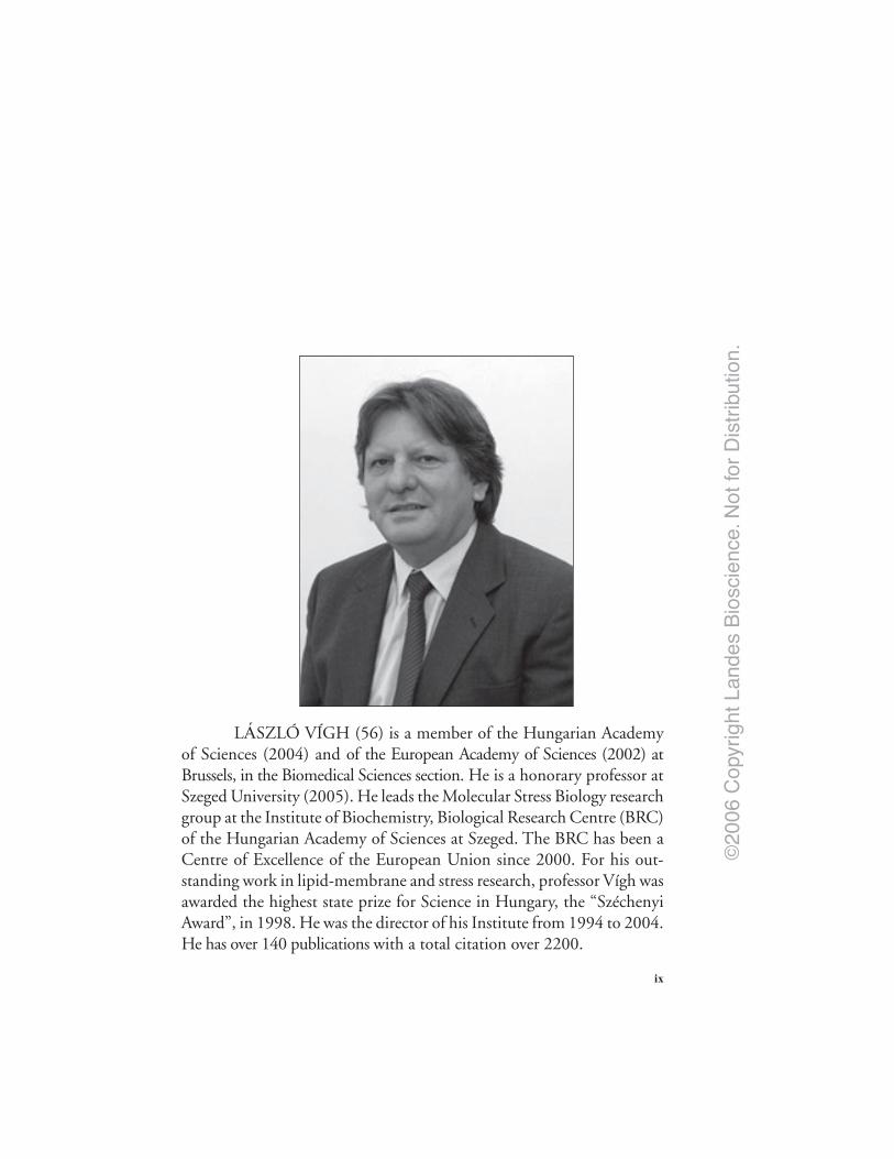
LÁSZLÓ VÍGH (56) is a member of the Hungarian Academyof Sciences (2004) and of the European Academy of Sciences (2002) atBrussels, in the Biomedical Sciences section. He is a honorary professor atSzeged University (2005). He leads the Molecular Stress Biology researchgroup at the Institute of Biochemistry, Biological Research Centre (BRC)of the Hungarian Academy of Sciences at Szeged. The BRC has been aCentre of Excellence of the European Union since 2000. For his out-standing work in lipid-membrane and stress research, professor Vígh wasawarded the highest state prize for Science in Hungary, the “SzéchenyiAward”, in 1998. He was the director of his Institute from 1994 to 2004.He has over 140 publications with a total citation over 2200.

©20
06 C
opyr
ight
Lan
des
Bio
scie
nce.
Not
for
Dis
trib
utio
n.
x
PARTICIPANTS
Julius AnckarDepartment of BiologyÅbo Akademi University and Turku
Centre for BiotechnologyUniversity of Turku and Åbo
Akademi UniversityTurkuFinland
André-Patrick ArrigoLaboratoire Stress OxydantChaperons et ApoptoseCNRS UMR 5534Centre de Génétique Moléculaire
et CellulaireUniversité Claude BernardFrance
Gâbor BaloghInstitute of BiochemistryBiological Research Center of the
Hungarian Academy of SciencesTemesváriHungary
Gregory L. BlatchDepartment of Medical ChemistrySemmelweis UniversityBudapestHungary
Frank BoellmannHSF Pharmaceuticals SAPullySwitzerland
Heather R. BrignullDepartment of Biochemistry,
Molecular Biology and Cell BiologyRice Institute for Biomedical ResearchNorthwestern UniversityEvanston, IllinoisU.S.A.
Andrew CossinsSchool of Biological SciencesLiverpool UniversityThe Biosciences BuildingLiverpoolUK
John H. CroweSection of Molecular
and Cellular BiologyUniversity of CaliforniaDavis, CaliforniaU.S.A.
Peter CsermelyDepartment of Medical ChemistrySemmelweis UniversityBudapestHungary
Editor: Please review participant information. Only the correspondingauther information was provided so the other chapter authors information
was assumed to be the same.

©20
06 C
opyr
ight
Lan
des
Bio
scie
nce.
Not
for
Dis
trib
utio
n.
xiParticipants
R. John EllisDepartment of Biological SciencesUniversity of WarwickCoventry CV4 7ALU.K
Attila GlatzInstitute of BiochemistryBiological Research Center of the
Hungarian Academy of SciencesTemesváriHungary
Pierre GoloubinoffDBMVFaculty of Biology and MedicinUniversity of LausanneLausanneSwitzerland
Andrew Y. GraceySchool of Biological SciencesLiverpool UniversityThe Biosciences BuildingLiverpoolU.K.
Scott A.L. HaywardSchool of Biological SciencesLiverpool UniversityThe Biosciences BuildingLiverpoolUK
Marie-Pierre HinaultDBMVFaculty of Biology and MedicinUniversity of LausanneLausanneSwitzerland
Ibolya HorváthInstitute of BiochemistryBiological Research Center of the
Hungarian Academy of SciencesTemesváriHungary
Walid A. HouryDepartment of BiochemistryUniversity of TorontoToronto, OntarioCanada
Linda M. HendershotDepartment of Genetics and Tumor
Cell BiologySt. Jude Children’s Research HospitalMemphis, TennesseeU.S.A.
Jacques LandryCentre de recherche en cancérologie
de l’Université LavalL’Hôtel-Dieu de QuébecQuébecCanada
Richard I. MorimotoDepartment of Biochemistry,
Molecular Biology and Cell BiologyRice Institute for Biomedical ResearchNorthwestern UniversityEvanston, IllinoisU.S.A.
James F. MorleyDepartment of Biochemistry,
Molecular Biology and Cell BiologyRice Institute for Biomedical ResearchNorthwestern UniversityEvanston, IllinoisU.S.A.

©20
06 C
opyr
ight
Lan
des
Bio
scie
nce.
Not
for
Dis
trib
utio
n.
xii Participants
Patricia A. MurraySchool of Biological SciencesLiverpool UniversityThe Biosciences BuildingLiverpoolUK.
Sébastien Ian NadeauCentre de recherche en cancérologie
de l’Université LavalL’Hôtel-Dieu de QuébecQuébecCanada
Stefano PiottoInstitute of BiochemistryBiological Research Center of the
Hungarian Academy of SciencesTemesváriHungary
Zoltán ProhászkaIIIrd Department of Internal MedicineSemmelweis UniversityBudapest, KútvölgyiHungary
Rosario RizzutoDepartment of Experimental
and Diagnostic MedicineUniversity of FerraraFerraraItaly
Suzannah RutherfordDivision of Basic SciencesFred Hutchinson Cancer
Research CenterSeattle, WashingtonU.S.A.
Yuichiro ShimizuDepartment of Genetics and Tumor
Cell BiologySt. Jude Children’s Research HospitalMemphis, TennesseeU.S.A.
Lea SistonenDepartment of BiologyÅbo Akademi University and Turku
Centre for BiotechnologyUniversity of Turku and Åbo
Akademi UniversityTurkuFinland
Csaba SötiDepartment of Medical ChemistrySemmelweis UniversityBudapestHungary
György SzabadkaiDepartment of Experimental
and Diagnostic MedicineUniversity of FerraraFerraraItaly
Zsolt TörökInstitute of BiochemistryBiological Research Center of the
Hungarian Academy of SciencesTemesváriHungary
László VíghInstitute of BiochemistryBiological Research Center of the
Hungarian Academy of SciencesTemesváriHungary

©20
06 C
opyr
ight
Lan
des
Bio
scie
nce.
Not
for
Dis
trib
utio
n.
xiiiParticipants
Richard VoellmyHSF Pharmaceuticals SAPullySwitzerland
Rongmin ZhaoDepartment of BiochemistryUniversity of TorontoToronto, OntarioCanada

©20
06 C
opyr
ight
Lan
des
Bio
scie
nce.
Not
for
Dis
trib
utio
n.
xiv
CONTENTS
1. PROTEIN MISASSEMBLY: MACROMOLECULAR CROWDINGAND MOLECULAR CHAPERONES ................................................. 1
R. John Ellis
Introduction ......................................................................................................................... 1Inside the Cell ...................................................................................................................... 1The Principle of Protein Self-Assembly: Yesterday and Today ....................................... 3The Molecular Chaperone Concept ................................................................................... 4The Problem of Protein Misassembly ................................................................................ 6Macromolecular Crowding ................................................................................................. 7Stimulation of Misassembly by Crowding Agents .......................................................... 10How do Chaperones Combat Misassembly? ................................................................... 11The Molecular Chaperone Function ................................................................................ 11
2. THE CELLULAR “NETWORKING” OF MAMMALIAN HSP27AND ITS FUNCTIONS IN THE CONTROL OF PROTEINFOLDING, REDOX STATE AND APOPTOSIS .............................. 14
André-Patrick Arrigo
Introduction ....................................................................................................................... 14Hsp27 in Cells Exposed to Heat Shock ............................................................................ 15Hsp27 in Cells Exposed to Oxidative Stress .................................................................... 17Hsp27 in Cells Committed to Apoptosis .......................................................................... 19Conclusions and in Vivo Perspectives .............................................................................. 21
3. MOLECULAR INTERACTION NETWORKOF THE HSP90 CHAPERONE SYSTEM ........................................ 27
Rongmin Zhao and Walid A. Houry
Introduction ....................................................................................................................... 27Mapping the Hsp90 Physical Interaction Network ........................................................ 29Mapping the Hsp90 Genetic Interaction Network ......................................................... 30The Hsp90 Interactome ..................................................................................................... 31Perspectives and Future Directions ................................................................................. 34
©20
06 C
opyr
ight
Lan
des
Bio
scie
nce.
Not
for
Dis
trib
utio
n.
xvContents
4. ORGANIZATION OF THE FUNCTIONS AND COMPONENTSOF THE ENDOPLASMIC RETICULUM ........................................ 37
Yuichiro Shimizu and Linda M. Hendershot
Introduction ....................................................................................................................... 37Overview of Protein Biosynthesis in the ER ................................................................... 37The ER Possesses a Unique Environment for Protein Folding ..................................... 39The ER Quality Control System ...................................................................................... 39Chaperone Selection during Protein Maturation in the ER .......................................... 41Organization of a Subset of Chaperones into Large
Preformed Complexes ............................................................................................... 42Components of the Calnexin/Calreticulin System
and Their Organization ............................................................................................. 43Possible Advantages and Constraints That an Organization
of ER Chaperones Might Imposed ........................................................................... 44
5. MOLECULAR CRIME AND CELLULAR PUNISHMENT:ACTIVE DETOXIFICATION OF MISFOLDEDAND AGGREGATED PROTEINS IN THE CELLBY THE CHAPERONE AND PROTEASE NETWORKS .............. 47
Marie-Pierre Hinault and Pierre Goloubinoff
The Criminal Nature of Protein Aggregation in the Cell ............................................... 47Defence Mechanisms against Protein Aggregation in the Cell ...................................... 48Aging and Conformational Diseases: Failures of Law Enforcement
Leading to Lawlessness ............................................................................................. 52
6. CHAPERONES AS PARTS OF CELLULAR NETWORKS .................... 55
Peter Csermely, Csaba Söti and Gregory L Blatch
Introduction: Cellular Networks and Chaperones ......................................................... 55Chaperones in Cellular Networks .................................................................................... 58Chaperone-Mediated Emergent Properties of Cellular Networks ............................... 59Chaperone Therapies ........................................................................................................ 60Conclusion .......................................................................................................................... 60
7. CHAPERONES AS PARTS OF ORGANELLE NETWORKS ................. 64
György Szabadkai and Rosario Rizzuto
Biogenesis of the ER and Mitochondrial Networks:A Role for Chaperons in Interorganellar Communication? .................................. 65
ER-Mitochondrial Ca2+ Transfer: A Major Example of Organelle Interactions ......... 67Chaperone Control of ER-Mitochondrial Interaction along the Ca2+ Signal
Transmission Pathway .............................................................................................. 68Perspectives: The Role of Chaperone Mediated ER-Mitochondria
Coupling in Cell Death .............................................................................................. 71Conclusions ........................................................................................................................ 73

©20
06 C
opyr
ight
Lan
des
Bio
scie
nce.
Not
for
Dis
trib
utio
n.
xvi Contents
8. HEAT SHOCK FACTOR 1 AS A COORDINATOROF STRESS AND DEVELOPMENTAL PATHWAYS..................... 78
Julius Anckar and Lea Sistonen
Introduction ....................................................................................................................... 78Functional Domains of HSF1 ........................................................................................... 79Activation Mechanisms of HSF1 ...................................................................................... 80Regulation of hsp Gene Transcription by HSF1 ............................................................. 81Stress-Specific Activation of HSF1 ................................................................................... 82HSF1 as a Developmental Regulator ............................................................................... 83HSF1-Mediated Expression of Cytokines ....................................................................... 83Heat Shock Factors Working Together ........................................................................... 84Future Perspectives ........................................................................................................... 85
9. CHAPERONE REGULATION OF THE HEAT SHOCKPROTEIN RESPONSE ....................................................................... 89
Richard Voellmy and Frank Boellmann
Introduction ....................................................................................................................... 89Feedback Regulation of the Heat Shock Protein Response
by Stress-Inducible Chaperones ............................................................................... 90Hsps and Co-Chaperones Repress Activation of HSF1 ................................................. 90HSP90-Containing Multichaperone Complexes Regulate HSF1 Oligomeric
Status and Transcriptional Competence ................................................................. 91Regulation of HSF1 by CHIP as Part of the Protein Quality
Control System........................................................................................................... 93Synopsis .............................................................................................................................. 94
10. MECHANISMS OF ACTIVATION AND REGULATIONOF THE HEAT SHOCK-SENSITIVESIGNALING PATHWAYS ................................................................ 100
Sébastien Ian Nadeau and Jacques Landry
Introduction ..................................................................................................................... 100Major Signaling Pathways Activated Heat Shock ........................................................ 101Molecular Origin of the Heat Shock Signal .................................................................. 106Conclusion ........................................................................................................................ 107
11. MEMBRANE-REGULATED STRESS RESPONSE:A THEORETICAL AND PRACTICAL APPROACH ................... 114
László Vigh, Zsolt Török, Gábor Balogh, Attila Glatz, Stefano Piottoand Ibolya Horváth
Introduction ..................................................................................................................... 114The Evolution of the “Membrane Sensor” Hypothesis with the Aid
of Unicellular Stress Models: The Beauty of Simplicity ...................................... 115
©20
06 C
opyr
ight
Lan
des
Bio
scie
nce.
Not
for
Dis
trib
utio
n.
xviiContents
Evidence Concerning the Operation of Membrane-AssociatedStress Sensing and Signaling Mechanisms in Mammalian Cells.Membrane Lipids May Provide the Molecular Switch for StressSensing and Signaling ............................................................................................. 119
Stress Response Profiling: Can We “Zoom In” on Membrane HyperstructuresEngaded in the Generation of Stress Signal? ........................................................ 122
Can We Point to Lipid Molecular Species Engaged in Stress Sensingand Signaling? .......................................................................................................... 123
Computational Methods for the Design of Subtle Interactionsbetween Lipids and Proteins of Membranes ......................................................... 124
Conclusions ...................................................................................................................... 127
12. BEYOND THE LIPID HYPOTHESIS:MECHANISMS UNDERLYING PHENOTYPICPLASTICITY IN INDUCIBLE COLD TOLERANCE ................. 132
Scott A.L. Hayward, Patricia A. Murray, Andrew Y. Gracey and Andrew R. Cossins
Introduction ..................................................................................................................... 132Cold Adaptation and the Lipid Hypothesis ................................................................... 133Evidence in Prokaryotes ................................................................................................. 133Evidence in Plants ........................................................................................................... 134Evidence in Animals ........................................................................................................ 134Caenorhabditis elegans Cold Tolerance and the Contribution
of Desaturases .......................................................................................................... 135NonLipid Mechanisms of Cold Tolerance ..................................................................... 137Interaction and Compensatory Mechanisms ................................................................ 138Conclusions ...................................................................................................................... 138
13. TREHALOSE AS A “CHEMICAL CHAPERONE”:FACT AND FANTASY ...................................................................... 143
John H. Crowe
Sugars and Stabilization of Biological Materials .......................................................... 143Origins of the Trehalose Myth ........................................................................................ 144The Mechanism of Depression of Tm ............................................................................ 145Trehalose Stabilizes Microdomains in Membranes ...................................................... 145There Is More Than One Way to the Same End ........................................................... 147Trehalose Has Useful Properties, Nevertheless ............................................................. 147Glass Transitions and Stability ....................................................................................... 148NonEnzymatic Browning and Stability of the Glycosidic Bond ................................. 149Sugar Glasses in Plant Anhydrobiotes ........................................................................... 150Lessons from Nature Can Be Used to Preserve Intact Cells
in the Dry State ........................................................................................................ 151Successful Freeze-Drying of Trehalose-Loaded Cells .................................................. 152Can Nucleated Cells Be Stabilized in the Dry State? ................................................... 153What Is the Role of p26 in Stabilizing Dry Nucleated Cells? ...................................... 153Summary and Conclusions ............................................................................................. 154
©20
06 C
opyr
ight
Lan
des
Bio
scie
nce.
Not
for
Dis
trib
utio
n.
xviii Contents
14. CHAPERONES AS PART OF IMMUNE NETWORKS ....................... 159
Zoltán Prohászka
Introduction ..................................................................................................................... 159Activation of Innate Immunity by Heat Shock Proteins .............................................. 159Immunological Protection of Heat Shock Proteins ...................................................... 160Role of Natural Autoantibody Networks in Regulation
of Autoimmunity ...................................................................................................... 161Heat Shock Proteins as Negotiators between Promotion
of Inflammation or Control of Autoimmunity ...................................................... 162Heat Shock Proteins as Elements of Multiple Networks ............................................. 163
15. THE STRESS OF MISFOLDED PROTEINS:C. ELEGANS MODELS FOR NEURODEGENERATIVEDISEASE AND AGING ..................................................................... 167
Heather R. Brignull, James F. Morley and Richard I. Morimoto
Introduction ..................................................................................................................... 167Models of Neurodegenerative Disease ........................................................................... 168C. elegans Model of polyQ Disease ................................................................................. 168The C. elegans polyQ Series in Neurons ........................................................................ 169Biophysical Properties of polyQ Proteins in Neurons of Live Animals ...................... 170PolyQ Length-Dependent Aggregation Correlates
with Neuronal Dysfunction ..................................................................................... 172Dynamic Biophysical Properties of Intermediate polyQ Tracts
in the Ventral Nerve Cord ....................................................................................... 173Neuron-Specific Responses to polyQ Proteins .............................................................. 175The C. elegans polyQ Series in Muscle Cells ................................................................. 175Aging Influences the Threshold for polyQ Aggregation and Toxicity ........................ 176Longevity Genes Influence Aging-Dependent Aggregation
and Toxicity of polyQ Proteins ............................................................................... 178Genome-Wide RNAi Screening Identifies Novel Regulators
of polyQ Aggregation and Toxicity ........................................................................ 179Global Disruption of Folding Homeostasis by polyQ Proteins ................................... 181Conclusion ........................................................................................................................ 185
16. HSP90 AND DEVELOPMENTAL NETWORKS ................................... 190
Suzannah Rutherford, Jennifer R. Knapp and Peter Csermely
Introduction ..................................................................................................................... 190Hidden Genetic Variation ............................................................................................... 191Hsp90 and Signal Transduction Thresholds ................................................................. 193Nonlinearity in Developmental Responses to Signal Transduction ............................ 195A Pivotal Role for Hsp90 in Network Evolvability? ..................................................... 195
INDEX ....................................................................................................... pending
©20
06 C
opyr
ight
Lan
des
Bio
scie
nce.
Not
for
Dis
trib
utio
n.
xix
ACKNOWLEDGMENTS
Editor: Do you want to include any acknowledgmentshere?

©20
06 C
opyr
ight
Lan
des
Bio
scie
nce.
Not
for
Dis
trib
utio
n.

©20
06 C
opyr
ight
Lan
des
Bio
scie
nce.
Not
for
Dis
trib
utio
n.
CHAPTER 1
*R. John Ellis—Department of Biological Sciences, University of Warwick, Coventry CV4 7AL,U.K. Email: [email protected]
Molecular Aspects of the Stress Response: Chaperones, Membranes and Networks,edited by Peter Csermely and Laszlo Vigh. ©2007 Landes Bioscienceand Springer Science+Business Media.
Protein Misassembly:Macromolecular Crowding and Molecular ChaperonesR. John Ellis*
Abstract
The generic tendency of proteins to misassemble into nonfunctional, and sometimescytotoxic, structures poses a universal problem for all types of cell. This problem isexacerbated by the high total concentration of macromolecules found within most in-
tracellular compartments but it is solved by the actions of molecular chaperones. This reviewdiscusses some of the basic evidence and key concepts relating to this conclusion.
IntroductionA recent article in the journal Nature recommended authors to begin their review with a
story, a story that is amusing but also sets the scene. My story concerns a professor of biologywho decided to test the knowledge of his students at the end of his lecture course. He pointedto a young woman in the front row and said to her “There is an organ of the human body thatunder appropriate circumstances can increase in size by a factor of six-fold. What is that organand what are the circumstances?” The young woman blushed and exclaimed that she foundthis question so offensive she would report the professor to the head of the university. Theprofessor ignored this response and repeated the question to the next student “What is theorgan of the body that can increase in size by six-fold?” This student replied that this organ isthe iris of the human eye, which expands from a pinpoint in bright light to wide open in dimlight. “Correct” said the professor, who then returned to the first student. “I have two things tosay to you. Firstly, you clearly have not been paying sufficient attention to my lectures andsecondly, I predict that at some point in the future you are going to be greatly disappointed!”
The point of this story is that the first student made an unwarranted assumption, unwar-ranted in the quantitative sense. This point of this article is to explain why those people whostudy the properties of isolated macromolecules in uncrowded buffers are similarly makingunwarranted assumptions about the quantitative relevance of their measurements to the prop-erties of these molecules inside the cell. They are thereby ignoring the universal cellular featurethat explains the existence of molecular chaperones.
Inside the CellThe term ‘crowded’ is a quick way of referring to the fact that the total concentration of
macromolecules inside cells is very high, a phenomenon called macromolecular crowding. Thecartoon in Figure 1A represents the densities and shapes of the macromolecules in part of thecytosol of an animal cell, as drawn by David Goodsell and it is obvious why the term ‘crowded’

©20
06 C
opyr
ight
Lan
des
Bio
scie
nce.
Not
for
Dis
trib
utio
n.
Molecular Aspects of the Stress Response: Chaperones, Membranes and Networks2
is appropriate. Note that the ribosomes are connected by strands of messenger RNA to formpolysomes. Figure 1B shows an actual polysome, isolated from the salivary gland of an insectand spread out on the grid of an electron microscope. You can see the newly synthesized polypep-tide chains growing longer as each ribosome slides along the messenger RNA, and you willnotice that these chains are close together—within touching distance. You can also see that, asthe chains get longer, they show signs of folding into more compact structures whilst stillattached to the ribosomes.
This proximity of identical, partly folded chains in the crowded environment of the cytosolcreates a danger—a danger that these chains will not fold and assemble correctly into functional
Figure 1. Proteins fold in highly crowded environments. A) representation of part of the cytosolof an animal cell. Reprinted with permission (from ref. 31). B) Electron micrograph of a polysomeisolated from the salivary gland of the insect Chironomus x 140,000. At the bottom is the startof the polysome, at the top the end, the rest looping out of shot. Reprinted with permission fromreference 32, ©1989 Springer Science and Business Media, LLC.

©20
06 C
opyr
ight
Lan
des
Bio
scie
nce.
Not
for
Dis
trib
utio
n.
3Protein Misassembly
proteins but will bind to one another to form nonfunctional misassemblies. It follows thatprotein misassembly is a universal cellular problem created by the advantage of making severalprotein chains at the same time from one molecule of messenger RNA. But the misassemblyproblem is even wider than this, because it affects mature proteins as well, which can partlyunfold as a result of environmental stresses. The most distressing consequences of proteinmisassembly are the human neurodegenerative diseases, characterised by the occurrence ofprotein aggregates called amyloid plaques in the brain. In these diseases misassembly does nottake place at the stage of protein synthesis but when mature proteins partly unfold, but theprinciple is the same—misassembly occurs when partly folded identical chains are close to oneanother.
In the remainder of this article I shall discuss the reasons for believing that the dramaticstimulation of protein misassembly by macromolecular crowding accounts for the existence ofproteins acting as molecular chaperones to combat this problem. This discussion will focus onthe historical origins of the molecular chaperone concept because the fact that most computerdatabases rarely extend back more than about two decades is leading to ignorance of how thisparadigm shift occurred.
The Principle of Protein Self-Assembly: Yesterday and TodayIf we ask why protein chains fold as they do, the answer was provided by the classic refold-
ing experiments initiated by Christian Anfinsen around 1956. In this type of experiment apure native protein is dissolved in dilute buffer and denatured by a high concentration of achaotrope such as urea. This treatment causes the protein to unfold and thereby lose its biologi-cal properties, but if the concentration of chaotrope is lowered by 10 to 50-fold, Anfinsenobserved that many of the chains refold into their original functional conformations.1 Thistype of experiment has been repeated many times with many proteins, and it is clear that themajority of denatured proteins will refold into their original conformations in the absence ofeither an energy source or other macromolecules—in other words, proteins can self-assemblespontaneously by a process requiring only their primary structures. Despite the fact that therates and yields of refolded proteins are often too small to meet biological requirements, as waspointed out by Anfinsen,2 this observation led to the assumption that spontaneous assemblyalso happens inside the cell. This assumption was reasonable because a basic principle of scien-tific enquiry is Occam’s razor, which advises us not to complicate hypotheses unnecessarily, buta series of observations made in the 1980s challenged this view. Two reports appeared thatnewly synthesized polypeptide chains bind transiently to a preexisting protein before they fold.
The first report concerns the synthesis of the photosynthetic enzyme rubisco in higherplants. Rubisco is a chloroplast enzyme, consisting of eight catalytic large subunits made bychloroplast ribosomes and eight structural small subunits made by cytosolic ribosomes. It ishighly water-soluble, occurring at around 300g/l in the chloroplast stroma, but it is also one ofthe few proteins that fails to renature after dilution from a chaotrope. This failure stems fromthe extreme hydrophobicity of the large subunits, which aggregate with one another to form awater-insoluble precipitate. So we were surprised to find that when we allowed isolated chloro-plasts to synthesize large subunits, these accumulate in a water-soluble form, despite the factthat they do not assemble into the holoenzyme until late in the incubation (Fig. 2). Thissolubility arises because newly synthesized large subunits bind transiently to another water-solubleprotein before they assemble with small subunits.3 Sequencing of this large subunit bindingprotein revealed that it is 50% identical to the GroEL protein of Escherichia coli, a proteinrequired for some bacteriophages to assemble in this bacterium but whose role in uninfectedcells was unknown at that time. This identity was interpreted in terms of the molecular chap-erone concept, and it was Sean Hemmingsen who coined the name ‘chaperonin’ for this familyof molecular chaperone.4
The second report of a newly synthesized polypeptide binding to a preexisting proteinbefore folding was found during experiments with a cultured cell line derived from lymphocyte

©20
06 C
opyr
ight
Lan
des
Bio
scie
nce.
Not
for
Dis
trib
utio
n.
Molecular Aspects of the Stress Response: Chaperones, Membranes and Networks4
precursors. These cells synthesize immunoglobin, an oligomeric protein composed of heavyand light chains. Haas and Wabl found that in a cell line of preB lymphocytes that are unableto make light chains, the heavy chains become bound to a preexisting small protein as theyenter the lumen of the endoplasmic reticulum. They called this protein BiP and suggested thatit is involved in the regulation of immunoglobulin assembly.5 BiP was later identified by Munroand Pelham as a member of the heat shock protein 70 family.6 We now know that both thechaperonins and the hsp70 chaperones assist the folding of many newly synthesized proteins inall types of cell.7
The Molecular Chaperone ConceptThe term ‘molecular chaperone’ was coined by Ron Laskey in a Nature paper published in
1978 to describe a nuclear protein that solves a misassembly problem during the assembly ofnucleosomes in amphibian eggs. Nucleosomes are octamers of basic histone protein bound toDNA by electrostatic interactions. Disruption of these interactions by high salt concentrationsenables the histones to be separated from the DNA, but mixing these components together atphysiological salt concentrations results in a failure of self-assembly—an insoluble precipitateforms instead of nucleosomes. Laskey and his coworkers discovered that this failure can beprevented by adding an abundant acidic nuclear protein called nucleoplasmin, which results in
Figure 2. The newly synthesized large subunit of rubisco binds to another protein before assem-bling into the holoenzyme. Intact chloroplasts were isolated from young pea leaves and incu-bated with light as the energy source for protein synthesis and S35-methionine. Samples wereremoved at intervals and stromal extracts prepared by osmotic lysis and centrigugation. Stromalextracts were run on both native and SDS polyacrylamide gels and autoradiographed. Bandscorresponding to rubisco large subunit were excised from the gels and their radioactivity mea-sured. Symbols: crosses, total large subunit from SDS gel; filled circles, large subunit attached tobinding protein from native gel; open circles, large subunit in holoenzyme from native gel.Reprinted from reference 33.
©20
06 C
opyr
ight
Lan
des
Bio
scie
nce.
Not
for
Dis
trib
utio
n.
5Protein Misassembly
the correct assembly of nucleosome cores. The detailed mechanism is unclear but in generalterms nucleoplasmin is thought to solve the misassembly problem by binding its acidic groupsto the positively charged groups on the histones. This binding lowers their overall surfacecharge and allows the intrinsic self-assembly properties of the histones to predominate over theincorrect interactions favoured by the high densities of opposite charge. In the Discussion ofthis paper, the term ‘molecular chaperone’ is used for the first time.8
Control experiments show that nucleoplasmin does not provide steric information requiredfor histones to bind correctly to DNA, nor is it a component of assembled nucleosomes. It isthese latter two features that laid the basic foundation of our current general concept of thechaperone function, as I shall discuss later, but at this point I wish to emphasise that the role ofnucleoplasmin is in the later stages of protein assembly, beyond the folding of newly synthesizedhistone chains, so the current common perception that chaperones are concerned solely withprotein folding is incorrect and has been incorrect since this subject started. I suspect that thiserroneous perception is retarding the search for chaperones that deal with the misassembly offolded subunits rather than with the problems posed by the folding of newly synthesized chains.It was only three years ago that a chaperone was found in red blood cells that mops up the excessof alpha subunits of haemoglobin that would otherwise misassemble and damage these cells.9 Ipredict that more examples of this type of subunit chaperone will be found in the future.
Ron Laskey did not use the term molecular chaperone to describe any other protein ordevelop the idea into a more general concept. This is where I enter the story. I read the Laskeypaper in 1985 and it struck me that his observations with nucleoplasmin could be thought of inthe same functional terms as the observations we had made about the rubisco binding protein,and others such as Hugh Pelham and Jim Rothman had made about hsp70. So I proposed in1987 the existence of a new type of general cellular function, defined as ensuring that the foldingof certain polypeptide chains and their assembly into oligomeric structures occur correctly.10
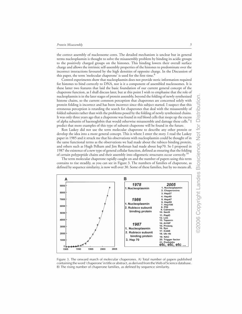
The term molecular chaperone rapidly caught on and the number of papers using this termcontains to rise steadily, as you can see in Figure 3. The numbers of families of chaperone, asdefined by sequence similarity, is now well over 30. Some of these families, but by no means all,
Figure 3. The onward march of molecular chaperones. A) Total number of papers publishedcontaining the word ‘chaperone’ in title or abstract, as derived from the Web of Science database.B) The rising number of chaperone families, as defined by sequence similarity.

©20
06 C
opyr
ight
Lan
des
Bio
scie
nce.
Not
for
Dis
trib
utio
n.
Molecular Aspects of the Stress Response: Chaperones, Membranes and Networks6
are heat shock proteins and this reflects the fact that the need for chaperone function increasesafter proteins have been denatured by environmental stresses. Many chaperones are involved inprotein folding but some are also assist disassembly processes, such as the remodelling of chro-matin during fertilization and transcription, and the resolubilisation of insoluble aggregatesthat have escaped the attention of other chaperones. This accumulation of discoveries supportsa new view of protein assembly. In this new view the principle of self-assembly is retained, butbefore chaperones, this assembly was thought to be spontaneous and require no energy expen-diture, whereas now self-assembly is seen in many instances to require assistance by chaper-ones, some of which hydrolyse ATP. Thus the concept of spontaneous protein self-assembly hasbeen replaced by the concept of assisted protein self-assembly. This new view was first articu-lated in a TiBS article in 1989 (see ref. 11), and has so far stood the test of time.
There is now a huge literature on the details of chaperone structure and function, but whatI wish to concentrate on in this article is why chaperones exist. Why do cells need a chaperonefunction, given that most denatured proteins know how to fold correctly in the absence ofother macromolecules?
The Problem of Protein MisassemblyThere are two answers to this question. The fact that proteins are made by polysomes en-
sures that identical partly folded chains are close together, as I indicated earlier. Moreover, thesechains grow vectorially so they cannot fold correctly until a complete folding domain has beenmade, raising the possibility that incomplete domains may misfold. In addition to these fac-tors, polysomes function within a highly crowded compartment. All these features favour aprocess that competes with folding, the process of misassembly.
Misassembly is defined as the association of two or more polypeptide chains to form non-functional structures; these structures may be as small as dimers or large enough to be in-soluble. This emphasis on function serves to distinguish misassembly from the formation offunctional oligomers, which is thus termed oligomerization. Many authors use the term ‘aggre-gation’ to describe misassembly, and point to the amyloid fibrils found in the brains of peoplesuffering from neurodegenerative disease as typical examples. The problem with this term isthat there are increasing reports of amyloid structures that do have biological functions, so theessential distinction between aggregation and oligomerization has disappeared.12 Misassemblyshould be distinguished from misfolding, which I define as the formation of a conformationwhich cannot proceed to the functional conformation on a biologically relevant time scale.Misassemblies are by definition misfolded, but there are very few reports of renaturing proteinsthat misfold but remain monomeric. It seems likely that the vast majority of primary transla-tion products are capable of folding correctly, provided they can avoid misassembly. It is forthis reason that I suggest that the essential problem that chaperones have evolved to combat ismisassembly rather than misfolding.
Protein misassembly has not been widely studied but two important aspects are established.It has been known for many years that misassembly competes with folding in an Anfinsen-typerefolding experiment (Fig. 4A). Since misassembly is a high order process, it is very sensitive tothe concentration of the unfolded chains, so as this concentration rises, misassembly outcompetesfolding. The molecular basis for this competition is simply that polypeptide chains do notdistinguish between inter- and intra-molecular interactions. Protein chemists traditionally solvethis problem by lowering the concentration of the chains, and/or the temperature, but thissolution is not open to cells.
A more surprising feature of misassembly is that in many cases it is highly specific - chainsmisassemble only if they are identical or very similar. This observation is interpreted to meanthat the aggregating species have a degree of secondary or tertiary structure - in other wordsthey are partly folded. This point was first established by adding total crude extracts of E. colicells to the 8M urea in a standard Anfinsen refolding experiment applied to tryptophanase(Fig. 4B). The numbers marked with asterisks in the box indicate the enzymic activity of the

©20
06 C
opyr
ight
Lan
des
Bio
scie
nce.
Not
for
Dis
trib
utio
n.
7Protein Misassembly
tryptophanase chains that have refolded after dilution of the urea, and you can see that thisactivity is unaffected by the addition of 100 times as much total protein from E. coli to theurea-containing buffer. Thus the several thousand different unfolded polypeptide chains in theE. coli extract do not affect the competition between correct folding and misassembly.13 Whatdoes affect this competition however, is the phenomenon of macromolecular crowding.
Macromolecular CrowdingWe use the term ‘crowded’ rather than ‘concentrated’ because in general no single macro-
molecular species occurs at a high concentration, but taken together, macromolecules occupy asignificant fraction of the total volume. This fraction is in the range 8-40%. That is, 8-40% ofthe total volume is physically occupied by macromolecules and therefore is unavailable to othermolecules, just as in a football crowd most of the space is occupied by people and is unavailableto other people—other people are sterically excluded. This steric exclusion of part of the vol-ume generates considerable energetic consequences, whose magnitude is not generally appreci-ated because it is so counter-intuitive—8-40% reduction in available volume does not soundvery much. You may think that it simply means that the concentration of macromolecules is8-40% larger than it appears to be, but in reality this steric exclusion produces large highlynonlinear effects on the effective concentration of macromolecules. This nonlinearity results inan exquisite sensitivity of macromolecular properties to changes in their environment.
There exists a quantitative theory of crowding developed largely by Allen Minton, a bio-physicist at the National Institutes of Health in America.14-16 This theory predicts two majorconsequences of crowding. The first prediction is that crowding will reduce the diffusion ratesof both large and small molecules. There is good experimental evidence that diffusion is
Figure 4. Two properties of misassembly. A) Misassembly competes with refolding. Lactatedehydrogenase was denatured by acid pH and then diluted into buffer at pH 7.0 and the regainof enzymic activity measured. Reprinted with permission from reference 13, ©1979 AmericanChemical Society. B) Misassembly is highly specific. Tryptophanase was denatured in 8M ureawith and without a crude total extract of E. coli and diluted into refolding buffer. The numbersin the box are the specific activities of the tryptophanase recovered. Reprinted with permissionfrom reference 34, ©1974 Blackwell.

©20
06 C
opyr
ight
Lan
des
Bio
scie
nce.
Not
for
Dis
trib
utio
n.
Molecular Aspects of the Stress Response: Chaperones, Membranes and Networks8
reduced in the range three-four fold in eukaryotic cells and eleven-fold in E. coli relative to therate in water; prokaryotic cells appear to be more crowded than eukaryotic cells.17 Much moreimportant with respect to misassembly however is the large increase in association constantsfor macromolecules. For example, let us consider the association of two 40 kDa monomersinto a dimer. Suppose that the association constant for this oligomerization in water is 1.0.Crowding theory predicts that the value of this constant inside a cell of E. coli will be between8 and 40, depending on what specific volume of the protein is assumed. If we suppose that thedimers can form a homotetramer, the effect is even larger—the association constant is now10,000 (see ref. 18).
This dramatic consequence arises because crowding increases the effective concentration ofmacromolecules—in other words, it increases their thermodynamic activity. This increase inactivity is produced by the reduction in excluded volume when molecules bind to one another.In fact macromolecular crowding is more precisely called ‘the excluded volume effect’. Thisterm emphasises the fact that crowding is a purely physical nonspecific effect based solely onsteric exclusion. As the number and the size of molecules in a solution increase, the less ran-domly they can be distributed. So as the concentration rises, the free energy of the solution alsorises, because the entropy of each molecule becomes less. But if these molecules bind to oneanother, the volume available to them increases, their entropy therefore is greater and the totalfree energy of the solution decreases. In other words, the most favoured state is the state thatexcludes the least volume to other macromolecules.
This is a general conclusion—it applies not just to associating macromolecules, but to allprocesses where a change in excluded volume occurs. So it applies to the folding of newlysynthesized polypeptide chains, the folding of nucleic acid chains, the unfolding of matureproteins as a result of environmental stresses such as high temperature, the condensation ofDNA in chromosomes, the operation of motile systems such as actin filaments and microtu-bules, and, of particular relevance here, to the formation of protein misassemblies.
It is important to grasp that the effect of crowding on thermodynamic activity is highlynonlinear with respect to the concentration of the crowding agent. The term ‘crowding agent’is used to describe the molecules that cause the crowding. Figure 5A shows how the activitycoefficient, that is the ratio of the effective concentration to the actual concentration, varies
Figure 5. A) Activity coefficients increase nonlinearly with cellular concentrations of macromol-ecules. The log of the activity coefficient of hemoglobin is plotted against the concentration ofhemoglobin. Reprinted from reference 19. B) The effect of crowding on activity is exerted by largemolecules on large molecules. The change in activity coefficient of a test molecule introducedinto a solution of hemoglobin as crowding agent was measured with respect to the molecularweight of the test molecule. Reprinted with author permission from reference 20.

©20
06 C
opyr
ight
Lan
des
Bio
scie
nce.
Not
for
Dis
trib
utio
n.
9Protein Misassembly
with the concentration of haemoglobin.19 Note that this is a log/linear plot and that theactivity coefficient rises in a nonlinear fashion with respect to the actual concentration. Theconcentration of hemoglobin in red blood cells is about 340 g/l, so you can see that theactivity of haemoglobin inside the cell is more than two orders of magnitude greater than itis in the dilute buffer where its properties are commonly studied.
Figure 5B plots the activity coefficient against molecular weight for a test molecule placedin a background of hemoglobin at 300 g/l. So in this experiment the crowding agent is haemo-globin and we are asking how the activity of another molecule placed in that solution dependson the molecular weight of that molecule. Note that this is a log/log plot. You can see that theeffect of crowding on activity becomes significant only after about 10,000 molecular weight.20
This is why we use the term ‘macromolecular crowding’ because the effect on the activity ofsmall molecules such as metabolites and inorganic ions is small by comparison.
I have so far talked about the effect of crowding on thermodynamics. What about effects onkinetics? Figure 6 summarises the effects of crowding on reaction rates for a bimolecular associa-tion reaction.21 The vertical axis of the graph is the log of the association rate constant. We canconsider two situations in turn. If the rate-limiting step is the encounter rate of the two compo-nents A and B, then the reaction is diffusion-limited. But crowding reduces diffusion so in thiscase the reaction rate will decrease as the concentration of crowding agent rises. However in theother situation, the rate-limiting step is the conversion of the activated complex (or transitionstate) to the dimer, so in this case the rate depends on the concentration of this complex. Butcrowding increases association, so the equilibrium between A + B and the activated complex isdisplaced to the right. Thus the reaction rate will rise as the concentration of crowding reagentincreases. However, if you think about it, the absolute upper limit of any bimolecular reactionmust be ultimately set by the encounter rate of the two molecules, so even for transitionstate-limited reactions, the rate must eventually come down as the concentration of the crowd-ing agent gets high enough. So we have a resultant curve between two opposing effects.
Thus we see that the effect of crowding on reaction rate is complex; it depends on thenature of the reaction and where you are on the concentration axis. The sad fact however, isthat although these dramatic effects of crowding have been known for at least 25 years, the vastmajority of studies on isolated macromolecules, including those on protein folding, protein
Figure 6. Effects of crowding on reaction rate. The change in association rate constant for thebimolecular reaction A + B = AB* + AB with respect to crowding agent is plotted in two situations:A) where the encounter of A and B is rate-limiting; and B) where the conversion of the activatedcomplex AB* to AB is rate-limiting. Reprinted from reference 21.

©20
06 C
opyr
ight
Lan
des
Bio
scie
nce.
Not
for
Dis
trib
utio
n.
Molecular Aspects of the Stress Response: Chaperones, Membranes and Networks10
misassembly and molecular chaperones, continue to be done in uncrowded buffers. In my viewthis is a mistake. Until the effect of crowding agents becomes as routine to study as the effectsof pH or redox potential, the extrapolation of interpretations made from studies of isolatedsystems to the intact cell must have a question mark hanging over them.22
Stimulation of Misassembly by Crowding AgentsThe first demonstration that crowding agents increase the misassembly of refolding chains
was obtained with hen lysozyme.23,24 Oxidised lysozyme refolds impeccably, achieving almostcomplete recovery of enzymic activity in one or two seconds, but when the two four disulfidebonds are reduced, the chains misassemble in the refolding buffer, the extent depending on theirconcentration. Misassembly occurs because at least two disulfide bonds have to form by slow airoxidation before the chains collapse and during this time the chains have a chance to misassemblewith one another. When several different crowding agents are added to the refolding buffer, allthe reduced chains misassemble but the refolding of oxidised chains is unaffected (Fig. 7).Missasembly can be prevented by adding protein disulfide isomerase (PDI) to the refolding buffer.Low concentrations of PDI act by speeding up the rate of disufide formation, which stabilises thecorrectly folded chains, while higher concentrations act in a chaperone fashion. Further examplesof the stimulation of protein misassembly by crowding are discussed in reference 25.
Figure 7. Crowding stimulates the misassembly of reduced lysozyme. Hen lysozyme was dena-tured under reducing conditions, diluted into refolding buffer containing varying amounts of fourcrowding agents and the regain of enzymic activity measured. In the absence of crowding agent,the yield is about 20% under the conditions used. Symbols: squares, Ficoll 70; triangles, Dextran70; circles, bovine serum albumin; inverted triangles, ovalbumin. Reprinted from reference 23.
©20
06 C
opyr
ight
Lan
des
Bio
scie
nce.
Not
for
Dis
trib
utio
n.
11Protein Misassembly
How do Chaperones Combat Misassembly?If we survey the literature on the roles of many different chaperones in preventing the
misassembly of newly synthesized polypeptides, the picture looks complex, but I suggest thatthe available data can be encompassed within just two basic principles of chaperone operation.7
It is useful to divide chaperones into two classes on the basis of size. Small chaperones aredefined as those less than 200 kDa in size and include hsp70, hsp40, PDI and the very recentlydiscovered cases of membrane chaperones i.e., proteins that occur inside membranes and pre-vent the aggregation of other transmembrane proteins.26 Small chaperones bind transiently toshort hydrophobic sequences as these appear on nascent or just released polypeptide chains,and prevent them from both folding prematurely or misassembling together by binding tothese sequences for a time. These chaperones do not appear to affect protein conformation, butfunction essentially by reducing the time potentially interactive surfaces are exposed by cyclingon and off these surfaces until they are buried by folding. Such a simple mechanism can bethought of as analogous to tossing a hot potato from hand-to-hand until it has cooled, ananalogy suggested by Ulrich Hartl.
Large chaperones, exemplified by the chaperonins such as GroEL, function by a muchmore sophisticated mechanism that uses what I call the Anfinsen cage principle.27 Thechaperonins function essentially by providing a molecular cage made of one oligomer of GroELcapped by one oligomer of GroES. Single partly folded chains are encapsulated one at a timeinside this cage. The enclosed chain continues to fold in the absence of other folded chainsuntil the hydrophobic surfaces that cause misassembly are buried within the final folded struc-ture. The time of folding inside this cage is set by the slow ATPase activity of the GroELsubunits. Completion of ATP hydrolysis allows ATP to bind to the opposite end of the GroEL/ES complex from the enclosed protein, and this binding triggers via allosteric interactions therelease of the folded chain into the cytosol.7
But suppose the released chain has not managed to bury its hydrophobic regions during thetime it is enclosed? It would be dangerous to release such misassembly-prone chains into thecytosol but this problem is prevented by the crowded state of the cytosol. Experiment withcrowding agents added to isolated GroEL/ES complexes by Jorg Martin and Ulrich Hartl showthat crowding ensures that any such released, but partly folded chains, bind back to the sameGroEL molecule from which they have just been released.28,29 So we reach the pleasing conclu-sion that the chaperonins use the crowded state of the cytosol to combat the problem of proteinaggregation that has been created by crowding in the first place.
The Molecular Chaperone FunctionMy current definition of molecular chaperones is that they are a large and diverse group of
proteins that share the property of assisting the noncovalent assembly and/or disassembly ofother macromolecular structures, but which are not permanent components of these structureswhen these are performing their normal biological functions.30 It is important to note that thisdefinition is functional and not structural, but it contains no constraints on the mechanisms bywhich different chaperones may act; this is the reason for the use of the imprecise term ‘assist’.Thus molecular chaperones are not defined by a common mechanism or by sequence similar-ity. In my view only two criteria need be satisfied to designate a macromolecule a molecularchaperone. Firstly, it must in some sense assist the noncovalent assembly or disassembly ofsome other macromolecular structure, the mechanism being irrelevant, and secondly, it mustnot be a component of these structures when they are performing their normal biologicalfunction. Note that the term ‘assembly’ is used in this definition in a very broad sense, andincludes the folding of newly synthesized or stress-denatured proteins, the unfolding of pro-teins during transport across membranes, the association of monomers into oligomers andmacromolecular disassembly processes.
All these considerations can be reduced to a simple unifying concept of the chaperonefunction, defined as the prevention and reversal of incorrect interactions that may occur when
©20
06 C
opyr
ight
Lan
des
Bio
scie
nce.
Not
for
Dis
trib
utio
n.
Molecular Aspects of the Stress Response: Chaperones, Membranes and Networks12
potentially interactive surfaces are exposed to the crowded intracellular environment. Thesesurfaces occur on nascent and newly synthesized polypeptide chains, on mature proteins un-folded by environmental stresses, and also on folded proteins in near-native conformations, asin signalling proteins such as the steroid receptor. Thus the term ‘molecular chaperone’ is not ametaphor, nor an example of academic whimsy, but a precise description. The word ‘chaper-one’ is appropriate because the traditional role of the human chaperone is to prevent incorrectinteractions between pairs of people without either providing the steric information requiredfor their correct interaction, or being present during married life - but often reappearing duringdivorce and remarriage!
AcknowledgementsI thank Robert Freedman for his generous provision of Departmental facilities during my
retirement and Allen Minton for correcting my misconceptions about crowding theory.
References1. Anfinsen CB. Principles that govern the folding of polypeptide chains. Science 1973; 181:223-230.2. Epstein CJ, Goldberg RF, Anfinsen CB. The genetic control of tertiary protein structure: Studies
with model systems. Cold Spring Harbor Symp Quant Biol 1963; 28:439-448.3. Barraclough R, Ellis RJ. Protein synthesis in chloroplasts. IX. Assembly of newly synthesized large
subunits into ribulose bisphosphate carboxylase in isolated intact chloroplasts. Biochim BiophysActa 1980; 608:19-31.
4. Hemmingsen SM, Woolford C, van der Vies SM et al. Homologous plant and bacterial proteinschaperone oligomeric protein assembly. Nature 1988; 333:330-334.
5. Haas IG, Wabl M. Immunoglobulin heavy chain binding protein. Nature 1983; 306:387-389.6. Munro S, Pelham SRB. An hsp70-like protein in the ER: Identity with the 78 kd glucose-regulated
protein and immunoglobulin heavy chain binding protein. Cell 1986; 46:291-300.7. Young JC, Agashe VR, Siegers K et al. Pathways of chaperone-mediated folding in the cytosol.
Nature Revs Mol Cell Biol 2004; 5:781-791.8. Laskey RA, Honda BM, Mills AD et al. Nucleosomes are assembled by an acidic protein which
binds histones and transfers them to DNA. Nature 1978; 275:416-420.9. Kihm AJ, Kong YI, Hong W et al. An abundant erythroid protein that stabilizes free
alpha-hemoglobin. Nature 2002; 417:758-767.10. Ellis RJ. Proteins as molecular chaperones. Nature 1987; 328:378-379.11. Ellis RJ, Hemmingsen SM. Molecular chaperones: Proteins essential for the biogenesis of some
macromolecular structures. Trends Biochem Sci 1989; 14:339-342.12. Fowler DM, Koulov AV, Alory-Jost C. Functional amyloid formation in mammalian tissue. PloS
Biol 2006; 4:0001-0008.13. Zettlmeiss G, Rudolph R, Jaenicke R. Reconstitution of lactic dehydrogenase. Noncovalent aggre-
gation vs. reactivation. 1. Physical properties and kinetics of aggregation. Biochemistry 1979;18:5567-5571.
14. Minton AP. Molecular crowding: Analysis of effects of high concentrations of inert cosolutes onbiochemical equilibria and rates in terms of volume exclusion. Methods Enzym 1988; 295:127-149.
15. Minton AP. Implications of macromolecular crowding for protein assembly. Curr Opin Struct Biol2000; 10:34-39.
16. Minton AP. The influence of macromolecular crowding and macromolecular confinement on bio-chemical reactions in physiological media. J Biol Chem 2001; 276:10577-10580.
17. Ellis RJ. Macromolecular crowding: An important but neglected aspect of the intracellular environ-ment. Curr Opin Struct Biol 2001; 11:114-119.
18. Zimmerman SB, Trach SO. Estimation of macromolecular concentrations and excluded volumeeffects for the cytoplasm of Escherichia coli. J Mol Biol 1991; 222:599-620.
19. Minton AP. The effect of volume occupancy upon the thermodynamic activity of proteins: Somebiochemical consequences. Mol Cell Biochem 1983; 55:119-140.
20. Minton AP, Colclasure GC, Parker JC. Model for the role of macromolecular crowding in regula-tion of cellular volume. Proc Natl Acad Sci USA 1992; 89:10504-10506.
21. Zimmerman SB, Minton AP. Macromolecular crowding: Biochemical, biophysical and physiologi-cal consequences. Annu Rev Biophys Biomol Struct 1993; 22:27-65.
22. Ellis RJ. Macromolecular crowding: Obvious but underappreciated. Trends Biochem Sci 2001;26:597-604.

©20
06 C
opyr
ight
Lan
des
Bio
scie
nce.
Not
for
Dis
trib
utio
n.
13Protein Misassembly
23. van den Berg B, Ellis RJ, Dobson CM. Effects of macromolecular crowding on protein foldingand aggregation. EMBO J 1999; 18:6927-6933.
24. van den Berg B, Wain R, Dobson CM et al. Macromolecular crowding perturbs protein refoldingkinetics: Implications for folding inside the cell. EMBO J 2000; 19:3870-3875.
25. Ellis RJ, Minton AP. Protein aggregation in crowded environments. Biol Chem 2006, (in press).26. Kota J, Ljungdahl PO. Specialized membrane-localized chaperones prevent aggregation of polytopic
proteins in the ER. J Cell Biol 2005; 168:79-88.27. Ellis RJ. Revisiting the Anfinsen cage. Folding and Design 1996; 1:R9-R15.28. Martin J, Hartl FU. The effect of macromolecular crowding on chaperonin-mediated protein fold-
ing. Proc Natl Acad Sci USA 94:1107-1112.29. Martin J. Chaperonin function - Effects of crowding and confinement. J Mol Recog 2004;
17:465-472.30. Ellis RJ. Chaperone function: The orthodox view. In: Henderson B, Pockley AG, eds. Molecular
Chaperones and Cell Signalling. Cambridge University Press, 2005:3-41.31. Goodsell DS. The Machinery of Life. Springer-Verlag, 1992:68.32. Kiseleva EV. Secretory protein synthesis in Chironomus salivary gland cells is not coupled with
protein translocation across endoplasmic reticulum membranes. Electron microscopic evidence. FEBSLett 1989; 257:251-253.
33. Musgrove JE, Ellis RJ. The Rubisco large subunit binding protein. Phil Trans R Soc London B1986; 313:419-428.
34. London J, Skrzynia C, Goldberg ME. Renaturation of E. coli tryptophanase after exposure to 8 Murea. Evidence for the existence of nucleation centers. Eur J Biochem 1974; 47:409-415.

©20
06 C
opyr
ight
Lan
des
Bio
scie
nce.
Not
for
Dis
trib
utio
n.

©20
06 C
opyr
ight
Lan
des
Bio
scie
nce.
Not
for
Dis
trib
utio
n.
©20
06 C
opyr
ight
Lan
des
Bio
scie
nce.
Not
for
Dis
trib
utio
n.
CHAPTER 2
The Cellular “Networking” of MammalianHsp27 and Its Functions in the Control ofProtein Folding, Redox State and ApoptosisAndré-Patrick Arrigo*
Abstract
Cells possess effective mechanisms to cope with chronic or acute disturbance of homeo-stasis. Key roles in maintaining or restoring homeostasis are played by the various heatshock or stress proteins (Hsps). Among the Hsps, the group of proteins characterized
by low molecular masses (between 20 to 30 kDa) and homology to α-crystallin are called smallstress proteins (denoted sHsps). The present chapter summarizes the actual knowledge of theprotective mechanisms generated by the expression of mammalian Hsp27 (also denoted HspB1in human) against the cytotoxicity induced by heat shock and oxidative stress. It also describesthe anti-apoptotic properties of Hsp27 and their putative consequences in different pathologi-cal conditions.
IntroductionsHsps have been described first in Drosophila as being expressed concomitantly with the
high molecular masses heat shock proteins in cells exposed to a heat treament.1 At the molecu-lar level, sHsps are characterized in their C-terminal moeity by a conserved sequence (thealpha-crystallin domain) shared by mammalian alpha-crystallin polypeptide (Fig. 1). sHspshave been described in every eukaryots studied so far2-4 as well as in prokaryots.5 For example,the human genome contains 10 genes encoding different small stress proteins6 (see Table 1).Until recently, the most studied sHsps were the four Drosophila sHsps as well as Hsp27 andαB-crystallin from mammals.7 Hsp27 and αB-crystallin are characterized by a remarkablevariety of cellular functions, the first one being to protect the cell against conditions that wouldotherwise be lethal to the cell. These sHsps increase the cellular resistance to different types ofstress, including heat shock,8 oxidative conditions,7,9-12 exposure to cytotoxic drugs13,14 andapoptotic inducers.15-19 In vitro analysis have demonstrated that Hsp27 and αB-crystallin sharean ATP-independent chaperone activity that counteracts protein denaturation and helps in therefolding of denatured polypeptides.20,21 However, a chaperone activity is not shared by all themembers of the human sHsps family (Table 1). Hsp27 is also known to modulate cell growth,differentiation,10,22 intracellular redox state11 and tumorigenicity.15,23 This sHsp also interacts
*André-Patrick Arrigo—Laboratoire Stress Oxydant, Chaperons et Apoptose, CNRS UMR 5534,Centre de Génétique Moléculaire et Cellulaire, Université Claude Bernard, 16 rue Dubois,69622 Villeurbanne Cedex, France. Email: [email protected]
Molecular Aspects of the Stress Response: Chaperones, Membranes and Networks,edited by Peter Csermely and Laszlo Vigh. ©2007 Landes Bioscienceand Springer Science+Business Media.

©20
06 C
opyr
ight
Lan
des
Bio
scie
nce.
Not
for
Dis
trib
utio
n.
15The Cellular “Networking” of Mammalian Hsp27 and Its Functions
with the cytoskeleton and membranes and is involved in signaling via MAPKAP kinase 224 orestrogen receptors25 pathways.
Hsp27 in Cells Exposed to Heat ShockCells are usually devoid of Hsp27 expression in the absence of heat shock. However, in
human, several types of cells, as for example cancer cells of carcinoma origin, constitutivelyexpress Hsp27 (HspB1). In both types of cells, sub-lethal heat shock treatments up-regulate orinduce the synthesis of Hsp27 as a consequence of the transcriptional activation of hsp27 gene.This transcriptional activation is mediated by HSF-1, a specific transcription factor, that oncetrimerized binds HSE elements located in the promoter region of the genes encoding Hsps.26
The heat induced synthesis of the different Hsps is coordinately regulated. This results in thetransient accumulation of the Hsps during and after the heat stress. In heat shock conditions,human Hsp27 (HspB1) is phosphorylated at three serine sites by the MAPKAPK2/3 kinase.This kinase is itself activated by the P38 stress kinase.27 Analysis of the structural organizationof the constitutively expressed and therefore preexisting Hsp27 polypeptide present in un-stressed HeLa cells has revealed that this protein is in the form of spherical oligomers of heter-ogenous native sizes, ranging from 140 to more than 800 kDa.28,29 No X-ray structure has yetbeen obtained due to the extreme difficulty to obtain pure crystals of mammalian Hsp27. Inheat shock conditions, the oligomerization of preexisting Hsp27 is drastically altered and re-sembles that of the newly made Hsp27.29 Indeed, heat shock induces the rapid formation ofsmall sized oligomers, a phenomenon which is followed by the generation of very large Hsp27containing structures (also called heat shock granules).29 Phosphorylation is though to be re-sponsible of the dissociation of Hsp27 oligomers during heat shock.30 The phenomenon istransient and the structural organization of Hsp27 is back to normal several hours after theheat stress.29 In unstressed cells, the constitutively expressed Hsp27 polypeptide is mainly cyto-plasmic with a fraction being associated with membranous structures.29 In contrast, duringheat shock, Hsp27 is mostly associated with detergent resistant structures and redistributesinside the nucleus if the heat stress is drastic enough (45°C, 30 min, HeLa cells).29 Inthermotolerant cells, the structural organization and localization of Hsp27 is not altered byheat shock.29
Cells, genetically modified to constitutively express Hsp27 or αB-crystallin, are character-ized by an enhanced resistance to the deleterious damages induced by heat shock.8,31 Similarobservations were made in the case of Drosophila Hsp2732 and HspB8, a recently describednew human sHsps33 (see Table 1). Since protein denaturation and aggregation are likely thecause of the noxious effects of heat shock, studies have been performed to test the possibility of
Figure 1. Organization of Hsps polypeptides. Light box: conserved region; black box: WD/EPFdomain; gray box: alpha crystallin domain ; ΛΛΛΛΛ flexible domain ; P: phosphorylated sites.Amino acids number are indicated.

©20
06 C
opyr
ight
Lan
des
Bio
scie
nce.
Not
for
Dis
trib
utio
n.
Molecular Aspects of the Stress Response: Chaperones, Membranes and Networks16
an in vivo chaperone activity associated to Hsp27 that could explain its cellular protectiveactivity. Extinction of thermosensitive luciferase light emission was used as an intracellularmarker of protein aggregation. No effect of Hsp27 was noticed towards the loss of luciferaseactivity during heat shock. In contrast, after the heat shock, Hsp27 was found to accelerate therecovery of luciferase activity. This suggests that, in vivo, Hsp27 accelerates the dissociation ofthe aggregates formed by luciferase.34 In heat shock treated cells, Hsp27 also play a role in theinhibition of translation of non heat shock induced mRNAs. This phenomenon results prob-ably of the ability of Hsp27 to bind eIF4G initiation factor and to facilitate dissociation ofcap-initiation complexes.35 Following a heat shock treatment, Hsp27 stimulates the recoveryof RNA splicing as well as RNA and protein synthesis.36,37 In addition, phosphorylated Hsp27,as it is observed in heat shock treated cells, helps in maintaining cytoskeletal architecture.38,39
Taken together, these phenomena provide the cell with a survival advantage.In vitro analysis have revealed that Hsp27 is a dynamic tetramer of tetramers with a unique
ability to refold and reassembles into its active quaternary structure after denaturation. At hightemperatures, the structures formed by Hsp27 increase in size but in slow equilibrium with thehexadecameric form.40 In heat shock conditions, Hsp27 is in the form of an oligomeric activechaperone that binds and stores non native proteins (molten globules) and prevents their ag-gregation41-44 before subsequent refolding by ATP-dependent protein chaperones (Hsp70,Hsp40, Hsp90 and co-chaperones) (see Fig. 2). Indeed, Hsp27 ability to refold denaturedpolypeptides is poor compared to the high efficiency provided by the ATP-dependent Hsp70/Hsp40 chaperone machinery.45 Further in vitro analysis have demonstrated that phosphorylation
Table 1. The 10 different human sHsps and the proposed new (HspB) names forhuman sHsps. Note that only four sHsps share a chaperone activity
GeneHspB Other Localization on TissueName Name Chromosomes Expression Function(s)
HspB1 Hsp27, 4,7 p12,3 Many tissues ChaperoneHsp28 including muscles Protection against stress,
Tumors apoptosis
HspB2 MKBP 11 q22 Binds myotonic dystrophy protein kinase
HspB3 – 5 q11.2 Muscles Differentiation
HspB4 αA-crystallin 1 q22.3 Eye lens Chaperone
HspB5 αB-crystallin 11 q22.3-q23.1 Many tissues Chaperone including muscles Protection against stress,
apoptosis
HspB6 Hsp20 19 q13.13 Smooth muscles ChaperoneRelaxation of vascular smooth muscle
HspB7 cvHsp 1 p36.2-p34.3 Cardiovascular tissues ?
HspB8 H11/Hsp22 12 q24.23 Muscle Protein Kinase (Thr/ser)
HspB9 – 17 q21.2 Testis ?
HspB10 ODF1 8 q22 Testis ?
Adapted from Kappe et al, Cell Stress and Chaperones 2003; 8:53-61.6

©20
06 C
opyr
ight
Lan
des
Bio
scie
nce.
Not
for
Dis
trib
utio
n.
17The Cellular “Networking” of Mammalian Hsp27 and Its Functions
significantly reduces Hsp27 ability to form reservoirs containing denatured substrates.46 It isnot known whether phosphorylation is a negative regulator of the formation of reservoirs or ispart of a mechanism that promotes their dynamic recycling. On the other hand, the actinpolymerization-inhibiting activity associated to Hsp27 is inhibited when this protein in theform of phosphorylated tetramers.47 This phenomenon therefore favors F-actin reformation.48,49
Indeed, in heat treated cells, Hsp27 forms stable complexes with denatured F-actin preventingits aggregation and facilitating its reformation.50
Hsp27 in Cells Exposed to Oxidative StressOxidative stress is common in eukaryotic cells since the use of molecular oxygen that leads
to respiratory energy production generates side products such as reactive oxygen species (ROS)that become cytotoxic when they are produced at a too high concentration. As mentionedabove in the case of heat shock, the type of death that is triggered depends on the intensity ofthe stress. Apoptosis is usually induced by a moderate oxidative stress, while a drastic exposure
Figure 2. Hsp27 function in cells exposed to agents or conditions that alter protein folding.Denatured or oxidized are either rapidly refolded by ATP-dependent protein chaperones(Hsp70, Hsp40, Hsp90 and co-chaperones Hip, Hop and Hsp90) or recognized by the CHIPco-chaperone and send to degradation by the proteasome machinery. In case of an intensestress, the high loads in denatured or oxidized proteins (molten globules) rapidly saturate theHsp70 refolding machinery, this results in the formation of aggregates (lipofuscin) that are verydeleterious to the cell. These aggregates may activate the PIDDosome machinery (made of thepolypeptides PIDD, RIADD and pro-caspase-2) leading to pro-caspase-2 activation andapoptosis through the activation of downstream caspases. To counteract the problem and todown-regulate the formation of aggregates, Hsp27 oligomers bind non native proteins and trapthem in very high molecular weight structures. These structures act as storage reservoirs.Denatured proteins present in these reservoirs are less prone to aggregate. A more secure wayto process denatured proteins is then created by Hsp27 oligomeric structures. Hsp27 oligo-mers generate also a pro-reduced state in the cells through the modulation of enzymes in-volved in gluthatione-ROS pathway. This pro-reduced state counteracts ROS formation andprobably also aberrant protein interactions and lipofuscin formation. HS: heat shock.
©20
06 C
opyr
ight
Lan
des
Bio
scie
nce.
Not
for
Dis
trib
utio
n.
Molecular Aspects of the Stress Response: Chaperones, Membranes and Networks18
to oxidant conditions induces necrosis.51 In the case of apoptosis, endogenous ROS induce aglutathione depletion which triggers the mitochondrial apoptotic pathway52 (see below). Incontrast, high levels of ROS result in necrosis probably because of irreversible oxidations thatinhibit caspases and decrease ATP intracellular levels.53,54
Sophisticated defenses (i.e., detoxifiant enzymes, vitamines C and E and thiol-containingmolecules, such as glutathione) are induced by the cells to protect themselves in the face of anextracellular oxidizing environment.55 To the list of protectors one can add the anti-apoptoticprotein Bcl256,57 and several sHsps, such as mammalian Hsp27 and αB-crystallin and Droso-phila sHsps.11 Indeed, these sHsps have a cytoprotector effect in cells exposed to oxidativestress induced by agents or conditions such as hydrogen peroxide, menadione, doxorubicinand tumor necrosis factor (TNFα).13,38,58-63 In unstressed cells, devoid of constitutively ex-pressed sHsps, such as murine L929,46,60,61 murine NIH 3T3-ras64 and human colorectal can-cer HT-2965 cells, sHsps expression significantly decreases the basal level of intracellular reac-tive oxygen species (ROS) as well as the burst of ROS generated by oxidative stress. This effectis correlated with decreased lipid peroxidation and protein oxidation59 and F-actin architecturedisruption.38,66,67 Glutathione68 is an important factor of the protective activity of Hsp27against oxidative stress.59 Consequently, Hsp27 does not counteract the oxidative stress gener-ated by either buthionine sulfoximine (BSO) (a specific and essentially irreversible inhibitor ofγ-glutamyl-cysteine synthetase), or diethyl maleate (DEM) which binds the free sulfhydrylgroups of glutathione. In unstressed murine cells that are devoid of endogenous sHsps expres-sion, Hsp27 expression upregulates glutathione level and upholds this redox modulator in itsreduced form.59,60,69,70 However, in cells that already constitutively express high levels of Hsp27(i.e HeLa cells), an expression vector mediated increase in Hsp27 level does not necessarlyupregulates glutathione level (probably because the maximum intracellular level of glutathioneis already reached in normal HeLa cells).59 The expression and/or activity of enzymes involvedin the ROS-glutathione pathway, such as glucose-6-phosphate dehydrogenase, glutathione re-ductase and glutathione transferase is often up-regulated in cells (particularly murine fibro-blasts) expressing Hsp27. This phenomenon could be at the origin of the glutathione mediatedantioxidant power generated by the expression of Hsp27.60 Recently, we have observed that theexpression of Hsp27 or αB-crystallin downregulates iron intracellular level71 (Virot S and ArrigoA.-P., manuscript in preparation). This down-regulation could represent a key factor in theprotective mechanism of Hsp27 since free iron, through Fenton reaction, catalyzes the forma-tion of hydroxyl radicals that oxidize proteins.
In oxidative conditions, Hsp27 mediated rise in reduced glutathione and/or the upholdingof this redox modulator in its reduced form appears directly responsible of the protectionobserved at the level of the cell morphology, cytoskeletal architecture60,67 and mitochondrialmembrane potential.60,70 In addition, the cellular locale, oligomerization and phosphorylationof Hsp27 are also drastically modified in cells undergoing oxdative stress. Formation of dimersis also stimulated in cells exposed to oxidative stress. This occurs through the unique cysteineresidue present in Hsp27 which is essential for the protective activity of the protein.72 In thisrespect, it is intriguing to note that only medium sized oligomers contain cysteine-dependentdimers. In L929 murine fibroblasts genetically modified to express human Hsp27, a one hourexposure to TNFα, shifted, in an ROS-dependent manner, Hsp27 oligomers from an heterog-enous native size distribution to large structures (up to 800 kDa). Thereafter, TNFα decreasedthe size of Hsp27 oligomers to about 200 kDa.73,74 Hsp27 phosphorylated isoforms are recov-ered as small or medium-sized oligomers (<300 kDa). These studies, together with the analysisof Hsp27 mutants, led to the conclusion that the large unphosphorylated oligomers of Hsp27represent the active form of the protein which modulates ROS and glutathione levels,64 prob-ably as a consequence of their in vitro chaperone activity.46 Moreover, Hsp27 large oligomersmay bind irreversibly oxidized polypeptides and stimulate their presentation to theubiquitin-independent 20S proteasome, a protein degradation machine with a high affinity foroxidized proteins.75 This activity may then counteract the accumulation of lipofuscin made ofproteolysis resistant large aggregates of oxidized proteins.
©20
06 C
opyr
ight
Lan
des
Bio
scie
nce.
Not
for
Dis
trib
utio
n.
19The Cellular “Networking” of Mammalian Hsp27 and Its Functions
Hsp27 was also reported to sensitize some cells to oxidative stress in spite of its ability toconfer heat shock resistance.31,76,77 Hence, despite the proeminent role played by the chaper-one activity associated to its large oligomers, the mechanism of action of Hsp27 is probablystress- and cell-specific.
Hsp27 in Cells Committed to ApoptosisDepending on their intensity, heat, oxidative or other types of stress induce at least two
major two types of cell death processes. In the case of cells exposed to mild stress, cells have theopportunity to decide either to repair the damages or to commit suicide through an apoptoticprocess. In contrast, drastic stress usually trigger cell necrosis. The execution phase of apoptosisoccurs through the proteolytic activation of specific proteases called caspases.78,79 Several dif-ferent pathways lead to the activation of caspases, such as those triggered by death receptors80
and those which depend on the mitochondria-apoptosome machinery.81,82 Apoptosis inducedby either heat shock or oxidative stress usually occurs through the mitochondria pathway ofactivation.83
Depending on its intensity heat shock induces first an apoptotic death while necrosis is ob-served when the stress is drastic and impairs Hsps expression. Heat induced apoptotic processrequires cytochrome c release from mitochondria and caspase 3 activation83 and is blocked by thebroad caspase inhibitor z-VAD-fmk.84,85 It has recently been described that apoptosis induced byheat shock is dependent on the PIDDosome complex and pro-caspase-2 activation.86 For ex-ample, caspase-2 (-/-) and RIADD (-/-) cells that are devoid of functional PIDDosome are resis-tant to heat-induced apoptosis. Whether accumulation of denatured proteins, as it occurs duringheat shock, triggers the activation of the PIDDosome complex is an interesting hypothesis to betested (Fig. 2). Moreover, downstream of pro-caspase-2, the multidomain proapoptotic mol-ecules Bax and Bak have been described to be directly activated by heat.87 It is not yet known ifHsp27 or the other Hsps interfere specifically with these pathways.
We and others have reported that mammalian Hsp27, Drosophila Hsp27 as well as α-Bcrystallin counteract apoptosis induced by the kinase C inhibitor staurosporine. Hsp27 fromhuman also protects against apoptosis induced by actinomycin D, camptothecin, Fas/APO-1receptor,88 cisplatin,65 the topoisomerase II inhibitor etoposide,85,89 and doxorubicin.65,90,91
Hsp27 is less efficient in the protection against T antigen/p5392 and stimulated CTL cellsmediated apoptotic cell deaths.93
Concerning death ligands, we and others have shown that Hsp27 or alphaAB-crystallinexpression are negative regulators of TNFα and Fas ligand mediated cell death.9,88,94 Only thephosphorylated dimeric form of Hsp27 has been shown to interact with DAXX, a mediator ofFas-induced apoptosis, preventing the interaction of DAXX with both Fas and apoptotic sig-naling kinase 1 (Ask1) and therefore blocking DAXX-mediated apoptosis.95 In the case of theROS dependent necrotic cell death generated by TNFα, the formation of the largeunphosphorylated oligomers of Hsp27 which bear chaperone activity is observed. However,since Hsp27 protects also against the apoptotic death induced by TNFα in the presence ofactinomycine D, an inhibitory action of this Hsp should also occur at the level of caspases.
We have observed that Hsp27 is a negative regulator of several different pathways whihconverge to mitochondria and trigger the release in the cytosol of apoptogenic factors, such ascytochrome c96 and Smac/Diablo.97 This activity of Hsp27 could be related, at least in part, tothe ability of Hsp27 to protect the integrity of F-actin microfilaments against the damagesinduced by cytochalasin D or other apoptosis inducers (staurosporine, ROS, oxidative stress,heat shock...). This study also led to the discovery of an apoptotic-signaling pathway linkingcytoskeleton damages to mitochondria.96 Hsp27 also acts downstream of mitochondria. Forexample, Hsp27 was shown to interact with cytochrome c once this apoptogenic polypeptide isreleased from mitochondria. Once bound to Hsp27, cytochrome c looses its ability to triggerapoptosome formation.89,98 Cytochrome c probably interacts with the N-terminal part of Hsp27polypeptide, between amino acids 51 and 88;98 a domain which is not related with the

©20
06 C
opyr
ight
Lan
des
Bio
scie
nce.
Not
for
Dis
trib
utio
n.
Molecular Aspects of the Stress Response: Chaperones, Membranes and Networks20
chaperone activity of Hsp27.99 In contrast, Hsp27 does not interfere with the caspase inde-pendent apoptosis inducing factor (AIF) which, once released from mitochondria, triggerschromatin condensation and large-scale DNA fragmentation. Hsp27 has also been reported tobind pro-caspase 3 and to interfere with its activation ; however, the structural organization ofHsp27 responsible of this event is not known.100
Studies performed with genetically manipulated HeLa cells which express reduced levels ofHsp27 have revealed that the constitutive expression of this protein provides apoptotic resis-tance to staurosporine, etoposide, Fas, or cytochalasin D.96 In HeLa cells, inducers-specific changesin the structural organization and phosphorylation of Hsp27 are observed that correlate withthe decreased activation of caspases (Paul C, Arrigo A.-P. in preparation). For example, etoposideinduces the transient formation of large Hsp27 oligomers suggesting that only the chaperone-likestructure of the protein protects against this apoptotic agent. In contrast, staurosporine andcytochalasin D induce the transient formation of two populations of oligomeric Hsp27 struc-tures characterized by large and small molecular masses. As already mentioned above, inhibitionof DAXX-mediated cell death requires only the unphosphorylated dimers of Hsp27.95 Hence,future studies will have to determine which oligomeric form of Hsp27 is active at the differentsteps of the apoptotic pathways and whether activities other than the chaperone function (for-mation of reservoirs) are also involved in the protection (see Fig. 3).
Figure 3. Hsp27 function in the mitochondrial apoptotic pathway. In vitro caspase assay per-formed with cytosolic post-mitochondrial fraction treated with cytochrome c and dATP revealedthat Hsp27, once in the form of large unphosphorylated oligomers, interferes with the activationof pro-caspase-9 and pro-caspase-3 and copurifies with apoptosome containing fractions. Hsp27in the form of medium sized or small oligomers may bind cytochrome c and only small oligomersappear to be active upstream of mitochondria at the level of F-actin. The structural organizationof Hsp27 which is active in the case of the interaction with pro-caspase-3 is unknown.
©20
06 C
opyr
ight
Lan
des
Bio
scie
nce.
Not
for
Dis
trib
utio
n.
21The Cellular “Networking” of Mammalian Hsp27 and Its Functions
Hsp27 is transiently expressed during the early differentiation of different cell types.7,101,102
Inhibition of Hsp27 accumulation results in differentiation failure, a phenomenon often ac-companied by massive apoptosis induction.22,103-105 The mechanism by which Hsp27 controlsthe differentiation program and negatively modulates apoptosis is unknown but correlates withthe formation of chaperone-like large Hsp27 oligomers. These large oligomers may avoid theaccumulation of junk protein structures that trigger abberrant differentiation processes andapoptosis.
Conclusions and in Vivo PerspectivesIt is now well established that the expression of Hsp27 and alphaB-crystallin protect, through
their chaperone activity, cells against the deleterious damages induced by heat or oxidativeinjuries. These proteins also reduce and/or delay different apoptotic processes. However, morework is needed to better understand what is the exact role played by the « classical chaperonefunction » of sHsps (such as the formation of reservoirs containing folding intermediates) intheir anti-apoptotic activity. Indeed, it is conceivable that, in addition to their chaperone func-tion, other activities associated to these proteins may be required to fight against apoptotic celldeath. This hypothesis is strenghtened by the fact that some Hsp27 mutants which protectagainst cytochrome c induced apoptosis are not efficient against oxidative stress induced celldeath.99
In spite of the fact that the knock out of Hsp27 encoding gene is lethal, recent reports nowprovide the evidences that, in vivo, Hsp27 also acts as a negative regulator of programmed celldeath,106 and therefore may play a crucial role in the development and integrity of organisms.It is also well known that several human tumor cells constitutively express high levels ofHsp27.107-110 Moreover, Hsp27 has been described to have a potent tumorigenic activity,23
probably because of its ability to counteracts apoptosis mediated by death ligants, hence allow-ing cancer cells to escape the surveillance mediated by the immune system. In this respect,modification of Hsp27 by the methylglyoxal-arginine adduct argpyrimidine (due to the highlevel of non enzymatic glycation in glycolitic active cancer cells) stimulates its anti-apoptoticactivity111 and may be part of the mechanism triggered by some cancer cells for evasion ofapoptosis.112 It is also intriguing to note that elevated titers of serum antibodies to Hsp27accompany several human diseases such as cancer and glaucoma. Hsp27 auto-immune anti-bodies are associated with improved survival in patients with breast cancer113 probably becauseof their ability to induce apoptosis by inactivating or attenuating the anti-apoptotic activity ofnative Hsp27.114 These observations have led to the conclusion that Hsp27 is a potential thera-peutic target for future clinical studies.110,115-119
There are also several other pathological situations where high levels of Hsp27 are detected,such as in the case of chronic liver damage,120 neurodegenerescence pathologies and asthma.121,122
In these pathologies, the protective mechanism provided by Hsp27 expression is supposed tobe beneficial as it fights against the pathological state. This assumption is supported by the factthat mutations in several human sHsps, such as Hsp27 (HspB1), are associated with inheritedperipheral neuropathologies.122 Hence, more work is needed before future strategies aimed attargetting Hsp27 function(s) in a specific disease could be planned.
AcknowledgementsWork in the author laboratory was supported by the Région Rhône-Alpes and the Associa-
tion pour la Recherche sur le Cancer (ARC).
References1. Tissieres A, Mitchell H, Tracy U. Protein synthesis in salivary glands of Drosophila melanogaster:
Relation to chromosome puffs. J Mol Biol 1974; 84:389-398.2. de Jong W, Leunissen J, Voorter C. Evolution of the alpha-crystallin/small heat-shock protein
family. Mol Biol Evol 1993; 10:103-126.
©20
06 C
opyr
ight
Lan
des
Bio
scie
nce.
Not
for
Dis
trib
utio
n.
Molecular Aspects of the Stress Response: Chaperones, Membranes and Networks22
3. de Jong WW, Caspers GJ, Leunissen JA. Genealogy of the alpha-crystallin—small heat-shock pro-tein superfamily. Int J Biol Macromol 1998; 22:151-162.
4. Kappe G, Verschuure P, Philipsen RL et al. Characterization of two novel human small heat shockproteins: Protein kinase-related HspB8 and testis-specific HspB9. Biochim Biophys Acta 2001;1520:1-6.
5. Kappe G, Leunissen JA, de Jong WW. Evolution and diversity of prokaryotic small heat shockproteins. Prog Mol Subcell Biol 2002; 28:1-17.
6. Kappe G, Franck E, Verschuure P et al. The human genome encodes 10 alpha-crystallin-relatedsmall heat shock proteins: HspB1-10. Cell Stress Chaperones 2003; 8:53-61.
7. Arrigo AP, Landry J. In: Morimoto RI, Tissieres A, Georgopoulos C, eds. The Biology of HeatShock Proteins and Molecular Chaperones. Cold Spring Harbor, NY: Cold Spring Harbor Labora-tory Press, 1994:335-373.
8. Landry J, Chretien P, Lambert H et al. Heat shock resistance confered by expression of the humanHSP 27 gene in rodent cells. J Cell Biol 1989; 109:7-15.
9. Arrigo AP. Small stress proteins: Chaperones that act as regulators of intracellular redox state andprogrammed cell death. Biol Chem 1998; 379:19-26.
10. Arrigo AP, Préville X. In: Latchman DS, ed. Stress Proteins, Vol. 136, Handbook of ExperimentalPharmacology. Springer, 1999:101-132.
11. Arrigo AP. Hsp27: Novel regulator of intracellular redox state. IUBMB Life 2001; 52:303-307.12. Arrigo AP, Paul C, Ducasse C et al. Small stress proteins: Modulation of intracellular redox state
and protection against oxidative stress. Prog Mol Subcell Biol 2002; 28:171-184.13. Huot J, Roy G, Lambert H et al. Increased survival after treatments with anticancer agents of
Chinese hamster cells expressing the human Mr 27,000 heat shock protein. Cancer Res 1991;51:5245-5252.
14. Fuqua SA, Oesterreich S, Hilsenbeck SG et al. Heat shock proteins and drug resistance. BreastCancer Res Treat 1994; 32:67-71.
15. Arrigo AP. sHsp as novel regulators of programmed cell death and tumorigenicity. Pathol Biol(Paris) 2000; 48:280-288.
16. Garrido C, Gurbuxani S, Ravagnan L et al. Heat shock proteins: Endogenous modulators ofapoptotic cell death. Biochem Biophys Res Commun 2001; 286:433-442.
17. Arrigo AP, Paul C, Ducasse C et al. Small stress proteins: Novel negative modulators of apoptosisinduced independently of reactive oxygen species. Prog Mol Subcell Biol 2002; 28:185-204.
18. Concannon CG, Gorman AM, Samali A. On the role of Hsp27 in regulating apoptosis. Apoptosis2003; 8:61-70.
19. Latchman DS. HSP27 and cell survival in neurones. Int J Hyperthermia 2005; 21:393-402.20. Jakob U, Gaestel M, Engels K et al. Small heat shock proteins are molecular chaperones. J Biol
Chem 1993; 268:1517-1520.21. Jakob U, Buchner J. Assisting spontaneity: The role of Hsp90 and small Hsps as molecular chap-
erones. Trends Biochem Sci 1994; 19:205-211.22. Mehlen P, Mehlen A, Godet J et al. hsp27 as a switch between differentiation and apoptosis in
murine embryonic stem cells. J Biol Chem 1997; 272:31657-31665.23. Garrido C, Fromentin A, Bonnotte B et al. Heat shock protein 27 enhances the tumorigenicity of
immunogenic rat colon carcinoma cell clones. Cancer Res 1998; 58:5495-5499.24. Stokoe D, Engel K, Campbell D et al. Identification of MAPKAP kinase 2 as a major enzyme
responsible for the phosphorylation of the small mammalian heat shock proteins. FEBS Lett 1992;313:307-313.
25. Ciocca DR, Luque EH. Immunological evidence for the identity between the hsp27 estrogen-regulated heat shock protein and the p29 estrogen receptor-associated protein in breast and en-dometrial cancer. Breast Cancer Res Treat 1991; 20:33-42.
26. Morimoto RI. Regulation of the heat shock transcriptional response: Cross talk between a familyof heat shock factors, molecular chaperones, and negative regulators. Genes Dev 1998; 12:3788-3796.
27. Rouse J et al. A novel kinase cascade triggered by stress and heat shock that stimulates MAPKAPkinase-2 and phosphorylation of the small heat shock proteins. Cell 1994; 78:1027-1037.
28. Arrigo AP, Welch W. Characterization and purification of the small 28,000-dalton mammalianheat shock protein. J Biol Chem 1987; 262:15359-15369.
29. Arrigo AP, Suhan JP, Welch WJ. Dynamic changes in the structure and intracellular locale of themammalian low-molecular-weight heat shock protein. Mol Cell Biol 1988; 8:5059-5071.
30. Kato K, Hasegawa K, Goto S et al. Dissociation as a result of phosphorylation of an aggregatedform of the small stress protein, hsp27. J Biol Chem 1994; 269:11274-11278.

©20
06 C
opyr
ight
Lan
des
Bio
scie
nce.
Not
for
Dis
trib
utio
n.
23The Cellular “Networking” of Mammalian Hsp27 and Its Functions
31. Trautinger F, Kokesch C, Herbacek I et al. Overexpression of the small heat shock protein, hsp27,confers resistance to hyperthermia, but not to oxidative stress and UV-induced cell death, in astably transfected squamous cell carcinoma cell line. J Photochem Photobiol 1997; 39:90-95.
32. Rollet E, Lavoie J, Landry J et al. Expression of Drosophila’s 27 kDa heat shock protein intorodent cells confers thermal resistance. Biochem Biophys Res Commun 1992; 185:116-120.
33. Carra S, Sivilotti M, Chavez Zobel AT et al. HspB8, a small heat shock protein mutated in hu-man neuromuscular disorders, has in vivo chaperone activity in cultured cells. Hum Mol Genet2005; 14:1659-1669.
34. Kampinga HH, Brunsting JF, Stege GJ et al. Cells overexpressing Hsp27 show accelerated recoveryfrom heat-induced nuclear protein aggregation. Biochem Biophys Res Commun 1994;204:1170-1177.
35. Cuesta R, Laroia G, Schneider RJ. Chaperone Hsp27 inhibits translation during heat shock bybinding eIF4G and facilitating dissociation of cap-initiation complexes. Genes Dev 2000;14:1460-1470.
36. Carper SW, Rocheleau TA, Cimino D et al. Heat shock protein 27 stimulates recovery of RNAand protein synthesis following a heat shock. J Cell Biochem 1997; 66:153-164.
37. Marin-Vinader L, Shin C, Onnekink C et al. Hsp27 enhances recovery of splicing as well asrephosphorylation of SRp38 after heat shock. Mol Biol Cell 2005; 7:7-17.
38. Huot J, Houle F, Spitz DR et al. HSP27 phosphorylation-mediated resistance against actin frag-mentation and cell death induced by oxidative stress. Cancer Res 1996; 56:273-279.
39. Lavoie JN, Lambert H, Hickey E et al. Modulation of cellular thermoresistance and actin filamentstability accompanies phosphorylation-induced changes in the oligomeric structure of heat shockprotein 27. Mol Cell Biol 1995; 15:505-516.
40. Ehrnsperger M, Lilie H, Gaestel M et al. The dynamics of hsp25 quaternary structure. Structureand function of different oligomeric species. J Biol Chem 1999; 274:14867-14874.
41. Ehrnsperger M, Graber S, Gaestel M et al. Binding of nonnative protein to Hsp25 during heatshock creates a reservoir of folding intermediates for reactivation. EMBO J 1997; 16:221-229.
42. Lee GJ, Roseman AM, Saibil HR et al. Small heat shock protein stably binds heat-denatured modelsubstrates and can maintain a substrate in a folding-competent state. EMBO J 1997; 16:659-671.
43. Stromer T, Ehrnsperger M, Gaestel M et al. Analysis of the interaction of small heat shock pro-teins with unfolding proteins. J Biol Chem 2003; 278:18015-18021.
44. Haslbeck M, Franzmann T, Weinfurtner D et al. Some like it hot: The structure and function ofsmall heat-shock proteins. Nat Struct Mol Biol 2005; 12:842-846.
45. Ehrnsperger M, Gaestel M, Buchner J. Analysis of chaperone properties of small Hsp’s. MethodsMol Biol 2000; 99:421-429.
46. Rogalla T, Ehrnsperger M, Preville X et al. Regulation of Hsp27 oligomerization, chaperone func-tion, and protective activity against oxidative stress/tumor necrosis factor alpha by phosphorylation.J Biol Chem 1999; 274:18947-18956.
47. Benndorf R, Hayess K, Ryazantsev S et al. Phosphorylation and supramolecular organization ofmurine small heat shock protein HSP25 abolish its actin polymerization-inhibiting activity. J BiolChem 1994; 269:20780-20784.
48. Guay J, Lambert H, GingrasBreton G et al. Regulation of actin filament dynamics by p38 mapkinase-mediated phosphorylation of heat shock protein 27. J Cell Sci 1997; 110:357-368.
49. Mounier N, Arrigo AP. Actin cytoskeleton and small heat shock proteins: How do they interact?Cell Stress Chaperones 2002; 7:167-176.
50. Pivovarova AV, Mikhailova VV, Chernik IS et al. Effects of small heat shock proteins on thethermal denaturation and aggregation of F-actin. Biochem Biophys Res Commun 2005;331:1548-1553.
51. Vayssier M, Banzet N, Francois D et al. Tobacco smoke induces both apoptosis and necrosis inmammalian cells: Differential effects of HSP70. Am J Physiol 1998; 275:771-779.
52. Zucker B, Hanusch J, Bauer G. Glutathione depletion in fibroblasts is the basis forapoptosis-induction by endogenous reactive oxygen species. Cell Death and Diff 1997; 4:388-395.
53. Samali A, Nordgren H, Zhivotovsky B et al. A comparative study of apoptosis and necrosis inHepG2 cells: Oxidant-induced caspase inactivation leads to necrosis. Biochem Biophys Res Commun1999; 255:6-11.
54. Jacobson MD. Reactive oxygen species and programmed cell death. Trends Biochem Sci 1996;21:83-86.
55. Powis G, Briehl M, Oblong J. Redox signalling and the control of cell growth and death. PharmacolTher 1995; 68:149-173.
56. Hockenbery DM, Oltvai ZN, Yin XM et al. Bcl-2 functions in an antioxidant pathway to preventapoptosis. Cell 1993; 75:241-251.
©20
06 C
opyr
ight
Lan
des
Bio
scie
nce.
Not
for
Dis
trib
utio
n.
Molecular Aspects of the Stress Response: Chaperones, Membranes and Networks24
57. Kane DJ, Sarafian TA, Anton R et al. Bcl-2 inhibition of neural death: Decreased generation ofreactive oxygen species. Science 1993; 262:1274-1277.
58. Mehlen P, Briolay J, Smith L et al. Analysis of the resistance to heat and hydrogen peroxidestresses in COS cells transiently expressing wid type or deletion mutants of the Drosophila 27-kDaheat-shock protein. Eur J Biochem 1993; 215:277-284.
59. Mehlen P, Préville X, Kretz-Remy C et al. Human hsp27, Drosophila hsp27 and human aB-crystallinexpression-mediated increase in glutathione is essential for the protective activity of these proteinagainst TNFα-induced cell death. EMBO J 1996; 15:2695-2706.
60. Preville X, Salvemin F, Giraud S et al. Mammalian small stress proteins protect against oxidativestress through their ability to increase glucose-6-phosphate dehydrogenase activity and by main-taining optimal cellular detoxifying machinery. Exp Cell Res 1999; 247:61-78.
61. Mehlen P, Préville X, Chareyron P et al. Constitutive expression of human hsp27, Drosophilahsp27, or human alpha B-crystallin confers resistance to TNF- and oxidative stress-induced cyto-toxicity in stably transfected murine L929 fibroblasts. J Immunol 1995; 154:363-374.
62. Wang G, Klostergaard J, Khodadadian M et al. Murine cells transfected with human Hsp27 cDNAresist TNF-induced cytotoxicity. J Immunother Emphasis Tumor Immunol 1996; 19:9-20.
63. Park YM, Han MY, Blackburn RV et al. Overexpression of HSP25 reduces the level of TNFalpha-induced oxidative DNA damage biomarker, 8-hydroxy-2'-deoxyguanosine, in L929 cells. JCell Physiol 1998; 174:27-34.
64. Mehlen P, Hickey E, Weber L et al. Large unphosphorylated aggregates as the active form ofhsp27 which controls intracellular reactive oxygen species and glutathione levels and generates aprotection against TNFα in NIH-3T3-ras cells. Biochem Biophys Res Comm 1997; 241:187-192.
65. Garrido C, Ottavi P, Fromentin A et al. HSP27 as a mediator of confluence-dependent resistanceto cell death induced by anticancer drugs. Cancer Res 1997; 57:2661-2667.
66. Huot J, Houle F, Marceau F et al. Oxidative stress-induced actin reorganization mediated by thep38 mitogen-activated protein kinase/heat shock protein 27 pathway in vascular endothelial cells.Circ Res 1997; 80:383-392.
67. Préville X, Gaestel M, Arrigo AP. Phosphorylation is not essential for protection of L929 cells byHsp25 against H2O2-mediated disruption actin cytoskeleton, a protection which appears relatedto the redox change mediated by Hsp25. Cell Stress Chaperones 1998; 3:177-187.
68. Meister A, Anderson ME. Glutathione. Annu Rev Biochem 1983; 52:711-760.69. Baek SH, Min JN, Park EM et al. Role of small heat shock protein HSP25 in radioresistance and
glutathione-redox cycle. J Cell Physiol 2000; 183:100-107.70. Paul C, Arrigo AP. Comparison of the protective activities generated by two survival proteins:
Bcl-2 and Hsp27 in L929 murine fibroblasts exposed to menadione or staurosporine. Exp Gerontol2000; 35:757-766.
71. Arrigo AP, Firdaus WJ, Mellier G et al. Cytotoxic effects induced by oxidative stress in culturedmammalian cells and protection provided by Hsp27 expression. Methods 2005; 35:126-138.
72. Diaz-Latoud C, Buache E, Javouhey E et al. Substitution of the unique cysteine residue of murinehsp25 interferes with the protective activity of this stress protein through inhibition of dimer for-mation. Antioxid Redox Signal 2005; 7:436-445.
73. Mehlen P, Mehlen A, Guillet D et al. Tumor necrosis factor-a induces changes in the phosphory-lation, cellular localization, and oligomerization of human hsp27, a stress protein that confers cel-lular resistance to this cytokine. J Cell Biochem 1995; 58:248-259.
74. Mehlen P, Kretz-Remy C, Briolay J et al. Intracellular reactive oxygen species as apparent modula-tors of heat-shock protein 27 (hsp27) structural organization and phosphorylation in basal andtumour necrosis factor alpha-treated T47D human carcinoma cells. Biochem J 1995; 312:367-375.
75. Sitte N, Merker K, Grune T. Proteasome-dependent degradation of oxidized proteins in MRC-5fibroblasts. FEBS Lett 1998; 440:399-402.
76. Arata S, Hamaguchi S, Nose K. Effects of the overexpression of the small heat shock protein,Hsp27, on the sensitivity of human fibroblast cells exposed to oxidative stress. J Cell Physiol 1995;163:458-465.
77. Mairesse N, Bernaert D, Del Bino G et al. Expression of HSP27 results in increased sensitivity totumor necrosis factor, etoposide, and H2O2 in an oxidative stress-resistant cell line. J Cell Physiol1998; 177:606-617.
78. Nicholson DW, Thornberry NA. Caspases: Killer proteases. Trends Biochem Sci 1997; 22:299-306.79. Thornberry NA, Lazebnik Y. Caspases: Enemies within. Science 1998; 281:1312-1316.80. Scaffidi C, Fulda S, Srinivasan A et al. Two CD95 (APO-1/Fas) signaling pathways. Embo J 1998;
17:1675-1687.81. Reed JC. Cytochrome c: Can’t live with it—can’t live without it. Cell 1997; 91:559-562.82. Green DR, Reed JC. Mitochondria and apoptosis. Science 1998; 281:1309-1312.
©20
06 C
opyr
ight
Lan
des
Bio
scie
nce.
Not
for
Dis
trib
utio
n.
25The Cellular “Networking” of Mammalian Hsp27 and Its Functions
83. Samali A, Robertson JD, Peterson E et al. Hsp27 protects mitochondria of thermotolerant cellsagainst apoptotic stimuli. Cell Stress Chaperones 2001; 6:49-58.
84. Samali A, Cotter TG. Heat shock proteins increase resistance to apoptosis. Exp Cell Res 1996;223:163-170.
85. Samali A, Orrenius S. Heat shock proteins: Regulators of stress response and apoptosis. Cell StressChaperones 1998; 3:228-236.
86. Tu S, McStay GP, Boucher LM et al. In situ trapping of activated initiator caspases reveals a rolefor caspase-2 in heat shock-induced apoptosis. Nat Cell Biol 2006; 8:72-77.
87. Pagliari LJ, Kuwana T, Bonzon C et al. The multidomain proapoptotic molecules Bax and Bak aredirectly activated by heat. Proc Natl Acad Sci USA 2005; 102:17975-17980.
88. Mehlen P, Schulze-Osthoff K, Arrigo AP. Small stress proteins as novel regulators of apoptosis.Heat shock protein 27 blocks Fas/APO-1- and staurosporine-induced cell death. The J Biol Chem1996; 271:16510-16514.
89. Garrido C, Bruey JM, Fromentin A et al. HSP27 inhibits cytochrome c-dependent activation ofprocaspase-9. Faseb J 1999; 13:2061-2070.
90. Garrido C, Mehlen P, Fromentin A et al. Inconstant association between 27-kDa heat-shock protein(Hsp27) content and doxorubicin resistance in human colon cancer cells. The doxorubicin-protectingeffect of Hsp27. Eur J Biochem 1996; 237:653-659.
91. Hansen RK, Parra I, Lemieux P et al. Hsp27 overexpression inhibits doxorubicin-induced apoptosisin human breast cancer cells. Breast Cancer Res Treat 1999; 56:187-196.
92. Guenal I, Sidoti-de Fraisse C, Gaumer S et al. Bcl-2 and Hsp27 act at different levels to suppressprogrammed cell death. Oncogene 1997; 15:347-360.
93. Beresford PJ, Jaju M, Friedman RS et al. A role for heat shock protein 27 in CTL-mediated celldeath. J Immunol 1998; 161:161-167.
94. Andley UP, Song Z, Wawrousek EF et al. Differential protective activity of αA- and{alpha}B-crystallin in lens epithelial cells. J Biol Chem 2000; 30:30-40.
95. Charette SJ, Lavoie JN, Lambert H et al. Inhibition of daxx-mediated apoptosis by heat shockprotein 27. Mol Cell Biol 2000; 20:7602-7612.
96. Paul C, Manero F, Gonin S et al. Hsp27 as a negative regulator of cytochrome C release. Mol CellBiol 2002; 22:816-834.
97. Chauhan D, Li G, Hideshima T et al. Hsp27 inhibits release of mitochondrial protein Smac inmultiple myeloma cells and confers dexamethasone resistance. Blood 2003; 102:3379-3386.
98. Bruey JM, Ducasse C, Bonniaud P et al. Hsp27 negatively regulates cell death by interacting withcytochrome c. Nat Cell Biol 2000; 2:645-652.
99. Arrigo AP, Virot S, Chaufour S et al. Hsp27 consolidates intracellular redox homeostasis by up-holding glutathione in its reduced form and by decreasing iron intracellular levels. Antioxid RedoxSignal 2005; 7:414-422.
100. Pandey P, Farber R, Nakazawa A et al. Hsp27 functions as a negative regulator of cytochromec-dependent activation of procaspase-3. Oncogene 2000; 19:1975-1981.
101. Arrigo AP. Expression of stress genes during development. Neuropathol and Applied Neurobiol1995; 21:488-491.
102. Arrigo AP. In search of the molecular mechanism by which small stress proteins counteract apoptosisduring cellular differentiation. J Cell Biochem 2005; 94:241-246.
103. Chaufour S, Mehlen P, Arrigo AP. Transient accumulation, phosphorylation and changes in theoligomerization of Hsp27 during retinoic acid-induced differentiation of HL-60 cells: Possible rolein the control of cellular growth and differentiation. Cell Stress Chaperones 1996; 1:225-235.
104. Davidson SM, Loones MT, Duverger O et al. The developmental expression of small HSP. ProgMol Subcell Biol 2002; 28:103-128.
105. Mehlen P, Coronas V, Ljubic-Thibal V et al. Small stress protein Hsp27 accumulation duringdopamine-mediated differentiation of rat olfactory neurons counteracts apoptosis. Cell Death Dif-fer 1999; 6:227-233.
106. Brar BK, Stephanou A, Wagstaff MJ et al. Heat shock proteins delivered with a virus vector canprotect cardiac cells against apoptosis as well as against thermal or hypoxic stress. J Mol CellCardiol 1999; 31:135-146.
107. Têtu B, Têtu BB, Lacasse HL et al. Prognostic influence of HSP-27 expression in malignant fi-brous histiocytoma: A clinicopathological and immunohistochemical study. Cancer Res 1992;52:2325-2328.
108. Ciocca DR, Oesterreich S, Chamnes GC et al. Biological and clinical implications of heat shockproteins 27000 (Hsp27): A review. J Natl Cancer Inst 1993; 85:1558-1570.
109. Ciocca DR, Calderwood SK. Heat shock proteins in cancer: Diagnostic, prognostic, predictive,and treatment implications. Cell Stress Chaperones 2005; 10:86-103.

©20
06 C
opyr
ight
Lan
des
Bio
scie
nce.
Not
for
Dis
trib
utio
n.
Molecular Aspects of the Stress Response: Chaperones, Membranes and Networks26
110. Rocchi P, Beraldi E, Ettinger S et al. Increased Hsp27 after androgen ablation facilitatesandrogen-independent progression in prostate cancer via signal transducers and activators of tran-scription 3-mediated suppression of apoptosis. Cancer Res 2005; 65:11083-11093.
111. Sakamoto H, Mashima T, Yamamoto K et al. Modulation of heat-shock protein 27 (Hsp27)anti-apoptotic activity by methylglyoxal modification. J Biol Chem 2002; 277:45770-45775.
112. Heijst JW, Niessen HW, Musters RJ et al. Argpyrimidine-modified Heat Shock Protein 27 inhuman nonsmall cell lung cancer: A possible mechanism for evasion of apoptosis. Cancer Lett2005; 5:5-15.
113. Conroy SE, Sasieni PD, Amin V et al. Antibodies to heat-shock protein 27 are associated withimproved survival in patients with breast cancer. Br J Cancer 1998; 77:1875-1879.
114. Tezel G, Wax MB. The mechanisms of hsp27 antibody-mediated apoptosis in retinal neuronalcells. J Neurosci 2000; 20:3552-3562.
115. Berrieman HK, Cawkwell L, O’Kane SL et al. Hsp27 may allow prediction of the response tosingle-agent vinorelbine chemotherapy in nonsmall cell lung cancer. Oncol Rep 2006; 15:283-286.
116. Feng JT, Liu YK, Song HY et al. Heat-shock protein 27: A potential biomarker for hepatocellularcarcinoma identified by serum proteome analysis. Proteomics 2005; 5:4581-4588.
117. Shin KD, Lee MY, Shin DS et al. Blocking tumor cell migration and invasion with biphenylisoxazole derivative KRIBB3, a synthetic molecule that inhibits Hsp27 phosphorylation. J BiolChem 2005; 280:41439-41448.
118. Mineva I, Gartne W, Hauser P et al. Differential expression of alphaB-crystallin and Hsp27-1 inanaplastic thyroid carcinomas because of tumor-specific alphaB-crystallin gene (CRYAB) silencing.Cell Stress Chaperones 2005; 10:171-184.
119. Thanner F, Sutterlin MW, Kapp M et al. Heat shock protein 27 is associated with decreasedsurvival in node-negative breast cancer patients. Anticancer Res 2005; 25:1649-1653.
120. Federico A, Tuccillo C, Terracciano F et al. Heat shock protein 27 expression in patients withchronic liver damage. Immunobiology 2005; 209:729-735.
121. Merendino AM, Paul C, Costa MA et al. Heat shock protein-27 protects human bronchial epithe-lial cells against oxidative stress-mediated apoptosis: Possible implication in asthma. Cell StressChaperones 2002; 7:269-280.
122. Dierick I, Irobi J, De Jonghe P et al. Small heat shock proteins in inherited peripheral neuropa-thies. Ann Med 2005; 37:413-422.

©20
06 C
opyr
ight
Lan
des
Bio
scie
nce.
Not
for
Dis
trib
utio
n.
CHAPTER 3
*Corresponding Author: Walid A. Houry—Department of Biochemistry, University of Toronto,Toronto, Ontario M5S 1A8, Canada. Email: [email protected]
Molecular Aspects of the Stress Response: Chaperones, Membranes and Networks,edited by Peter Csermely and Laszlo Vigh. ©2007 Landes Bioscienceand Springer Science+Business Media.
Molecular Interaction Networkof the Hsp90 Chaperone SystemRongmin Zhao and Walid A. Houry*
Abstract
Hsp90 is an essential and ubiquitous molecular chaperone that is required for the properfolding of a set of client proteins at a late stage in their folding process. In eukaryotes,cytoplasmic Hsp90 is absolutely essential for cell viability under all growth condi-
tions. The functional cycle of the Hsp90 system requires a cohort of cochaperones and cofac-tors that regulate the activity of this chaperone. Hence, Hsp90 function is highly complex; inorder to understand that complexity, several groups have attempted to map out the interactionnetwork of this chaperone in yeast and mammalian systems using the latest available proteomicand genomic tools. Interaction networks emerging from these large scale efforts clearly demon-strate that Hsp90 plays a central role effecting multiple pathways and cellular processes. Inyeast Saccharomyces cerevisiae, Hsp90 was shown to interact directly or indirectly with at least10% of the yeast ORFs. The systematic application of large scale approaches to map out theHsp90 chaperone network should allow the determination of the mechanisms employed bythis chaperone system to maintain protein homeostasis in the cell.
IntroductionHsp90 is an abundant and essential molecular chaperone present in all eukaryotic cells.
Hsp90 has been implicated in mediating the folding of a specific set of substrate proteins,which are typically called ‘clients’. Hsp90 clients are thought to interact with the chaperone ina nearly native state at the late stage of de novo folding;1 alternatively, Hsp90 can also enhancethe rate of reactivation of heat denatured proteins under stress conditions.2 Although no com-mon sequence or structural features have yet been identified for Hsp90 clients, however, manyof the early clients that were identified fell into two functional classes: transcription factors3
and protein kinases.4 These early observations suggested the involvement of Hsp90 in modu-lating signaling pathways. Hsp90 is, therefore, considered to be particularly important for cellcycle control, organism development, and response to environmental stresses.5,6 Hsp90 hasbecome a novel anti-cancer drug target, and there are several drugs that specifically bind toHsp90 and are in clinical trials.7,8
Hsp90 is a dimeric protein consisting of three domains: an N-terminal ATP-binding do-main, a middle region, and a C-terminal domain involved in homodimerization. The N-terminaldomain has a unique fold termed the Bergerat fold.9 Although the ATPase activity of Hsp90 isweak, ATP binding and hydrolysis is crucial to the chaperone function.10 Several drugs that

©20
06 C
opyr
ight
Lan
des
Bio
scie
nce.
Not
for
Dis
trib
utio
n.
Molecular Aspects of the Stress Response: Chaperones, Membranes and Networks28
have been designed to target Hsp90, such as geldanamycin, bind in the ATP pocket at theN-terminal domain. In eukaryotes, the C-terminal domain of cytosolic Hsp90 ends with aconserved EEVD motif. This motif has been shown to be essential for the interaction of cyto-solic Hsp90 with cofactors that contain α-helical tetratricopeptide repeat domains (TPR).
The Hsp90 functional cycle has been intensively investigated, however, due to the com-plexity of this chaperone system and the diversity of the clients, a full understanding of themolecular aspects of the cycle is still lacking. It is known that the activity of Hsp90 requires theinvolvement of other chaperone systems such as Hsp70 and Hsp40 as well as several cofactors(Fig. 1). The Hsp90 functional cycle has been well-studied for the case of steroid hormonereceptors (Fig. 1).3,11 It is thought that the monomeric inactive receptor is initially recognizedby the Hsp70/40 system and, subsequently, transferred to Hsp90. The formation of a complexbetween Hsp70 and Hsp90 is mediated by the scaffold protein Hop/p60 (Sti1 in yeast). Inyeast, Sti1 has been shown to be an activator of the Hsp70 ATPase and an inhibitor of Hsp90ATPase, although mammalian Hop does not seem to have such an effect.12,13 Subsequently,other cyclophilins (e.g., yeast Cpr6 and Cpr7) and immunophilins (e.g., yeast Cns1) as well asthe cochaperone p23 (Sba1 in yeast) interact with the Hsp70/40-Hop-Hsp90-substrate complex
Figure 1. The Hsp90 functional cycle. A nascent polypeptide chain of a steroid hormonereceptor, for example, is initially recognized by the Hsp70/Hsp40 system. Hop (yeast Sti1)binds both Hsp70 and Hsp90 to form the intermediate complex allowing the transfer of thesubstrate protein to Hsp90. Subsequently, p23 (yeast Sba1) and other cyclophilins andimmunophilins (indicated by PPI) bind Hsp90, releasing Hsp70/Hsp40 and Hop. In the result-ing mature complex, Hsp90 is stabilized in the ATP state by p23. If the substrate is a proteinkinase, Cdc37 is also involved in stabilizing this complex (not shown). Upon entry of thehormone into the cell, the receptor binds the hormone and is released from Hsp90. The releaseis promoted by Aha1 which enhances the ATPase activity of the chaperone and, in mammaliancells, is also possibly promoted by the acetylation of the chaperone.
©20
06 C
opyr
ight
Lan
des
Bio
scie
nce.
Not
for
Dis
trib
utio
n.
29Molecular Interaction Network of the Hsp90 Chaperone System
resulting in the release of Hsp70/40 and Hop. p23 stabilizes the Hsp90-receptor complex inthe ATP state. Upon arrival of the hormone into the cell, the receptor binds the hormone andis released from Hsp90 upon hydrolysis of the chaperone-bound ATP to ADP; the receptor isthen active as a dimer and translocates into the nucleus. The ATPase activity of Hsp90 isenhanced by the cofactor Aha1/Hch1.14,15 It has also been demonstrated that Hsp90 activitycan be regulated by phosphorylation by the phosphatase Ppt116 in yeast and by acetylation/deacetylation by an unknown HAT and by HDAC6 in mammalian cells, respectively.17,18
Several other Hsp90 cofactors are also known, such as Cdc37 and CHIP, and they affect thechaperone activity in different manners. It is generally thought that different Hsp90 cofactorstarget the chaperone to different sets of substrates.
Recently, several systematic genomic and proteomic approaches were employed in order tomap out the Hsp90 molecular interaction network. These approaches provide a global view ofthe influence of the Hsp90 system on multiple processes and complexes in the cell. They alsoshed more light on the mechanism of Hsp90 function. The description of these approachesfollows below.
Mapping the Hsp90 Physical Interaction NetworkThe Hsp90 physical interactors represent a class of proteins that either directly interact with
Hsp90 or that are part of complexes that interact with Hsp90. Most of these Hsp90 physicalinteractors are thought to mainly include Hsp90 cofactors and cochaperones as well as Hsp90clients. Two different methods have been employed to map out such interactors: (1) pull-downor immunoprecipitation of the chaperone to isolate Hsp90-containing complexes followed bymass spectrometry to identify proteins in those complexes, and (2) two-hybrid method toidentify proteins that, predominantly, directly bind to Hsp90.
Isolation of Hsp90 Complexes Followed by Mass SpectrometryIsolation of Hsp90 complexes followed by mass spectrometry to identify components of the
complexes is an effective and straight forward approach to explore the Hsp90 interactors. Ma-trix assisted laser desorption ionization time of flight (MALDI-ToF), tandem electrospray massspectrometry (MS/MS), or liquid chromatography coupled with tandem mass spectrometry(LC-MS/MS) have been extensively used to identify single proteins or protein mixtures19 ingels or in solution. While protein identification might be robust, this approach greatly dependson the quality of purified complexes.
This method has been used to isolate Hsp90 complexes either by affinity pull-down oftagged Hsp90 in yeast or by coimmunoprecipitation of Hsp90 from mammalian cells. Zhao etal20 introduced a TAP-tag to endogenous Hsp90 in yeast, while keeping the gene under thecontrol of its own promoter. The TAP-tag is a tandem affinity purification tag containing twoIgG binding domains from Protein A and a calmodulin binding peptide motif separated by atobacco etch virus (TEV) protease cut site.21 Yeast has two virtually identical HSP90 genes, onetermed HSP82, which is heat shock induced, while the other is termed HSC82, which is con-stitutively expressed. The proteins are localized in the cytoplasm. Strains were constructed inwhich endogenous HSC82 or HSP82 were C-terminally TAP-tagged, in addition, a third strainwas constructed in which HSP82 was deleted, while HSC82 was N-terminally TAP-tagged.Hsp90-containing complexes were then isolated on IgG beads, released using TEV protease,further purified on calmodulin beads in the presence of calcium, and then released by additionof EGTA to chelate the calcium. This approach allowed the isolation of native protein com-plexes that were purified to more than 106 fold to virtual homogeneity. Since the EEVD motifat the C-terminus of Hsp90 is essential for the interaction of Hsp90 cofactors with the chaper-one, most of the useful complexes were obtained from the third strain in which HSC82 wasN-terminally TAP-tagged and HSP82 was deleted. It should be noted that, in this approach,the addition of the tag might affect Hsp90 activity. Indeed, the yeast strain with N-terminallyTAP-tagged Hsp90 had slight temperature sensitivity. Isolated proteins were identified by mass
©20
06 C
opyr
ight
Lan
des
Bio
scie
nce.
Not
for
Dis
trib
utio
n.
Molecular Aspects of the Stress Response: Chaperones, Membranes and Networks30
spectrometry and some ‘hits’ were then verified using reverse pull-downs in which the identi-fied proteins were TAP-tagged and pulled-down.
To identify Hsp90 interactors in mammalian systems, Falsone et al22 used a commerciallyavailable anti-Hsp90 antibody to immunoprecipitate Hsp90 complexes from human embry-onic kidney (HEK293) cells. One of the hits was verified by determining that its levels weresensitive to the inhibition of Hsp90 by the drug geldanamycin. It should be noted that in thehuman genome there are seventeen genes which have been detected to belong to the Hsp90family.23 Six of these genes were recognized as functional, while the other eleven genes wereclassified as pseudogenes. These Hsp90s are present in different compartments of the eukary-otic cell further complicating the studies on mammalian Hsp90.
Two Hybrid ScreensThe yeast two-hybrid (2H) system is one of the most widely used methods to screen or
confirm protein-protein interactions.24 The basic principle of this system is that a protein X,the bait, is fused to a DNA binding domain (DBD), which is commonly derived from theGal4 or LexA transcription factors. Protein Y, the prey, is fused to a transcription activationdomain (AD). If proteins X and Y interact, then AD and DBD are brought close to each other,triggering the activation of a downstream reporter gene.
Despite the popularity of this method, there are several drawbacks that have to be consid-ered. The interactions have to occur in the nucleus and a nuclear targeting sequence is presentin the constructs used. In this regard, yeast Hsp90 is predominantly cytoplasmic and it has beenreported that the chaperone has a cytoplasmic localization signal in its sequence.25 The nuclearlocalization signal in the DBD domain may not be strong enough to bring the large Hsp90 tothe nucleus and this is indeed observed for full-length yeast Hsp90 (R. Zhao and W.A. Houry,unpublished results). Also, the DBD-Hsp90 fusion might not be fully active. It has been re-ported that a C-terminal Hsp90-Gal4 fusion can support yeast growth better than an N-terminalGal4-Hsp90 fusion.26 Also, the interaction between Hsp90 and its client proteins is probablytransient and the interaction might not be strong enough to activate the downstream reportergene. Nevertheless, this approach has been successfully used by the Piper and Houry groups20,27,28
to map out the Hsp90 2H interaction network using an ordered array of about 6000 yeastORFs fused to Gal4 activation domain that was created by Uetz et al.29
Mapping the Hsp90 Genetic Interaction NetworkHsp90 genetic interactors represent a class of proteins that are involved in a pathway where
Hsp90 also plays key functions in parallel. Hsp90 genetic interactors may or may not directlyinteract with the chaperone, but both, the chaperone and its genetic interactor, are required forthe successful function of a particular essential cellular process or pathway, and consequently,for cell viability. The large scale identification of Hsp90 genetic interactors has been success-fully used for yeast Hsp90.20
Synthetic Genetic ArraySynthetic genetic array (SGA) analysis is based on the principle of synthetic lethality devel-
oped for the budding yeast Saccharomyces cerevisiae.30 For this organism, about 80% of theopen reading frames are nonessential.31 Tong et al30 developed automated methods to screenfor synthetic lethality of a given knockout strain against the systematic gene deletion array32,33
consisting of about 4700 haploid strains each of which has a marked deletion of one nonessen-tial gene. A knockout strain is mated with the deletion array to eventually obtain strains withdouble deletions. If these strains are sick or not viable, then the two deleted genes are said to begenetic interactors. Putative hits can be verified by tetrad analysis or random spore analysis.The detailed method and its application has recently been reviewed in reference 34.
This screen is ideally designed for a query of nonessential gene; however, it is also suitablefor an essential gene query if a conditional knockout, such a temperature sensitive mutant of

©20
06 C
opyr
ight
Lan
des
Bio
scie
nce.
Not
for
Dis
trib
utio
n.
31Molecular Interaction Network of the Hsp90 Chaperone System
the essential gene, is available. Hsp90 is essential in all eukaryotic cells. A number of tempera-ture sensitive yeast Hsp82 mutants such as Hsp82(G170D), Hsp82(G313S), andHsp82(E381K) have already been identified and characterized.35 An SGA analysis of Hsp90interactors has recently been carried out using a strain in which hsc82 is deleted and HSP82 isrendered temperature sensitive by introducing the G170D mutation.20 The screen was carriedout at the semi-permissive temperature of 35ºC.
Chemical Genetic Screen/Drug ScreenThe chemical genetic screen or drug screen is based on the principle of synthetic lethality
and takes advantage of the unique bar codes introduced to mark each deletion mutant in thesystematic deletion array.32 These bar codes can be amplified using common primers. In thismethod, the bar-coded haploid deletion mutants of the nonessential genes31 are pooled andgrown in liquid culture containing an Hsp90 specific drug inhibitor, geldanamycin, to chal-lenge Hsp90 function. Strains containing deleted genes that are synthetic lethal or syntheticsick with Hsp90 will die or grow slower compared to other strains. The same experiment isrepeated in the absence of the drug. The common primers are then used to amplify genomicDNA from the pooled strains and the resultant products are hybridized onto specific DNAoligo array chips. Strains that do not grow or that grow slowly in the presence of the drug willnot give a signal or will give a lower signal when compared to the signal obtained in the absenceof the drug. The hits obtained from applying this technique should closely overlap with the hitsobtained from the SGA analysis as both methods depend on the same principle of syntheticlethality. Zhao et al20 employed this method to determine yeast Hsp90 genetic interactorsusing the drug geldanamycin (geldanamycin screen or GS).
The Hsp90 InteractomeYeast is currently the ideal system for large-scale genetic and proteomic studies due to the
ready availability of many tools for such studies in this organism. Figure 2A gives an overviewof the number of hits obtained using the four different methods described above for yeastHsp90 as reported by Zhao et al.20 The Hsp90 interactors (over 600 ORFs) represent about10% of the yeast proteome and are distributed among multiple cellular functional categories(Fig. 3) suggesting that Hsp90 is involved in many processes. One feature that stands out inFigure 2A is that there is little overlap among the different datasets obtained using the differentmethods. This reflects the experimental differences between the methods used as each methodfavors certain types of interactions. The best overlap was obtained between the SGA and thegeldanamycin screen (GS). A total of 451 Hsp90 genetic interactors were identified from thetwo screens with an overlap of 49 hits. These 49 hits represent a high fidelity dataset that couldthen be pursued further for more detailed biochemical studies. The overlap is considered to bereasonable.
On the other hand, the physical interaction screens gave a total of 208 hits with an overlapof only 10 hits between the TAP and 2H screens (Fig. 2A), which is less than expected. Thedifferences might arise because the 2H screen predominantly detects binary interactions, whilethe TAP method detects interactions between the bait and a complex of polypeptides that neednot directly bind to the protein bait. Hence, the 2H screen might reveal weaker interactionsthan the TAP screen.
It is interesting to note that no protein was simultaneously detected by the four methodsused and that only Cpr6, Cpr7, Sti1, and Mon1 were detected by three of the four screens.Cpr6, Cpr7, and Sti1 are all well-established Hsp90 cofactors, while the interaction of Hsp90with Mon1 has not been reported before; therefore, Mon1 is likely an Hsp90 substrate. Mon1is poorly studied; however, it is known to form a complex with another protein termed Ccz1that binds the vacuolar membrane and is involved in homotypic vacuole fusion.36,37 This mightimplicate Hsp90 in vacuolar trafficking.
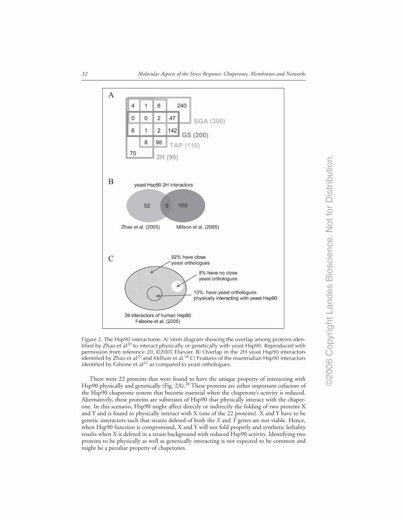
©20
06 C
opyr
ight
Lan
des
Bio
scie
nce.
Not
for
Dis
trib
utio
n.
Molecular Aspects of the Stress Response: Chaperones, Membranes and Networks32
There were 22 proteins that were found to have the unique property of interacting withHsp90 physically and genetically (Fig. 2A).20 These proteins are either important cofactors ofthe Hsp90 chaperone system that become essential when the chaperone’s activity is reduced.Alternatively, these proteins are substrates of Hsp90 that physically interact with the chaper-one. In this scenario, Hsp90 might affect directly or indirectly the folding of two proteins Xand Y and is found to physically interact with X (one of the 22 proteins). X and Y have to begenetic interactors such that strains deleted of both the X and Y genes are not viable. Hence,when Hsp90 function is compromised, X and Y will not fold properly and synthetic lethalityresults when X is deleted in a strain background with reduced Hsp90 activity. Identifying twoproteins to be physically as well as genetically interacting is not expected to be common andmight be a peculiar property of chaperones.
Figure 2. The Hsp90 interactome. A) Venn diagram showing the overlap among proteins iden-tified by Zhao et al20 to interact physically or genetically with yeast Hsp90. Reproduced wtihpermission from reference 20, ©2005 Elsevier. B) Overlap in the 2H yeast Hsp90 interactorsidentified by Zhao et al20 and Millson et al.28 C) Features of the mammalian Hsp90 interactorsidentified by Falsone et al22 as compared to yeast orthologues.

©20
06 C
opyr
ight
Lan
des
Bio
scie
nce.
Not
for
Dis
trib
utio
n.
33Molecular Interaction Network of the Hsp90 Chaperone System
2H screens for yeast Hsp90 have also been carried out by Millson et al.27,28 Initially, thegroup used full length Hsc82 as bait; however, even with DBD domain fused to the C-terminusof Hsc82, which is reported to have similar activity as the wild type chaperone, only fourinteractors were identified including the known Hsp90 cofactors Aha1 and Hch1.27 To detectHsp90 clients that probably only weakly interact with Hsp90, Millson et al repeated the 2Hscreen using a mutant Hsp82(E33A). This form of Hsp90 is barely functional and is thoughtnot to proceed to the final maturation step in the chaperone functional cycle due to failed ATPhydrolysis, which in turn is thought to hold the client proteins tighter rendering them easier todetect by 2H. A total of 177 genes including some known Hsp90 cochaperones and substrateswere identified using this approach.28 In this regard, it is surprising to find that one-third (59out of 177) of the ORFs identified by Millson et al28 are putative membrane proteins, puttinginto question the validity of this screen.
In comparison, Zhao et al20 used full length yeast Hsp82 as well as the different domains ofHsp82 as baits. This approach might be better in identifying physiological substrates and co-factors of Hsp90 as compared to the use of an inactive mutant of the chaperone.28 A total of 90Hsp90 interactors were identified20 mostly by binding to Hsp90 domains as only 8 interactorswere found to bind to full length Hsp82. In this screen, only 6 proteins out of the 90 areputative membrane proteins.
Eight proteins overlap between the 2H datasets of Zhao et al20 and Millson et al28 (Fig. 2B).These proteins are: Aha1, Arr3, Bsd2, Cns1, She3, Slt2, Sor2, and Tah1. This low overlap mightreflect differences in experimental conditions and baits used. The 8 proteins are probably robustinteractors of Hsp90. In fact, it is already known that Aha114,15 and Cns138 are Hsp90 cofactorsthat interact with Hsp90 in vivo and in vitro. Furthermore, Zhao et al20 and Millson et al28
verified the interaction of Hsp90 with Tah1 and Slt2, respectively, using in vitro assays.The high throughput analysis of mammalian Hsp90 interactors is limited. Physical interactors
of mammalian Hsp90 in HEK293 cells were identified using a simple procedure of immuno-precipitation of Hsp90 complexes and the identification of proteins in those complexes using
Figure 3. Hsp90 influences multiple cellular processes. The yeast Hsp90 interactors identifiedby Zhao et al.20 are distributed among a variety of functional categories. Functional categorieswhich contain more than 10% of interactors for each screening method are listed.
©20
06 C
opyr
ight
Lan
des
Bio
scie
nce.
Not
for
Dis
trib
utio
n.
Molecular Aspects of the Stress Response: Chaperones, Membranes and Networks34
nano-LC-MS/MS.22 Falsone et al22 identified 39 proteins as Hsp90 interactors (Fig. 2C). Theseproteins include metabolic enzymes, ribosomal subunits, and components of the cytoskeleton.However, many of the known Hsp90 interactors were not detected. The identified proteins aremostly housekeeping proteins and are abundant in the cell. This reflects the nature of thecoimmunoprecipitation method; that is, most of the weak and transient interactions, espe-cially with low abundance proteins, are missed. However, even for the short list of humanHsp90 physical interactors obtained, it is noticed that 36 out of 39 of the interactors (exceptfor P04075, Q9NR45 and Q96KM8) have close yeast orthologues (Fig. 2C). Some of theseorthologues (yeast Act1, Mdh3, Tdh3, and Yer156c which correspond to human P63261,P00338, P00354, and Q9HB07, respectively) were also found to physically interact with theyeast Hsp90 by TAP20or 2H28 screens (Fig. 2C). This may suggest that not only the Hsp90chaperone machinery itself, but also the core Hsp90 chaperone network may be somewhatconserved across organisms.
Perspectives and Future DirectionsThe integrated approach used by Zhao et al20 could also be used for other chaperone sys-
tems in yeast. The ultimate goal of mapping the chaperone interaction networks in the cell is todetermine the in vivo mechanisms that govern protein homeostasis. In other words, if thesescreens are repeated often enough and if high fidelity data sets are obtained using multiplemethods under different conditions, can a set of rules be eventually derived that would allow usto a priori predict how a given protein will fold inside the cell: what chaperone systems arerecruited to help in the folding process and what types of interactions are important for properde novo folding?
None of the current reported large-scale interaction studies on Hsp90 have been able tofind a consensus sequence or structural motif that is preferentially recognized by this chaper-one. This is in contrast to other simpler systems such as Hsp60 in E. coli (GroEL) in whichpreferential binding of GroEL to αβ folds or TIM barrels has been suggested.39,40 This seemsto point to the functional complexity of the Hsp90 system and the fact that substrate specific-ity is probably dictated, to a certain extent, by the cofactors and cochaperones of Hsp90. There-fore, mapping the chaperone interaction networks must be carried out using multiple geneticand proteomic screens and varying experimental conditions in order to obtain a comprehen-sive view of the cellular protein folding landscape and in order to minimize false negative andfalse positive hits. For example, even with the integrated proteomic and genomic approachemployed by Zhao et al,20 some well-established Hsp90 interactors were missed such as Sba1/p23 and Hap1. The interaction between Hsp90 and Sba1/p23 is stable only in the presence ofATP. In another example, physical interactions with membrane proteins are usually difficult todetect. The application of a recently developed split-ubiquitin membrane yeast two-hybridsystem might allow the detection of such interactors.41,42
With the development of new techniques, the integrated approach used by Zhao et al20
could also be applied for mammalian Hsp90: genetic interactors can be revealed using RNAinterference technology, while physical interactors can be studied using 2H and pull-down/mass spectrometry methods. Furthermore, investigating Hsp90 interactors in a particular tis-sue or organ, such as human liver or kidney, might be more meaningful than a wholegenome-wide screen of Hsp90 interactors. The interactions identified in these organs or tissuescould reveal site-specific roles of Hsp90. Finally, quantitative proteomics has been used as asensitive method to monitor the dynamics of a proteome.43 Future studies should concentrateon examining the change of the Hsp90 interactome under different stress conditions. Hence,the hope is that future chaperone networks will evolve from static pictures to dynamic entities.

©20
06 C
opyr
ight
Lan
des
Bio
scie
nce.
Not
for
Dis
trib
utio
n.
35Molecular Interaction Network of the Hsp90 Chaperone System
AcknowledgementsWork in the author’s laboratory is supported by research grants from the Canadian Insti-
tutes of Health research, Natural Sciences and Engineering Research Council of Canada, andthe National Cancer Institute of Canada.
References1. Buchner J. Hsp90 & Co. - A holding for folding. Trends Biochem Sci 1999; 24(4):136-141.2. Nathan DF, Vos MH, Lindquist S. In vivo functions of the Saccharomyces cerevisiae Hsp90 chap-
erone. Proc Natl Acad Sci USA 1997; 94(24):12949-12956.3. Pratt WB, Galigniana MD, Morishima Y et al. Role of molecular chaperones in steroid receptor
action. Essays Biochem 2004; 40:41-58.4. Sreedhar AS, Soti C, Csermely P. Inhibition of Hsp90: A new strategy for inhibiting protein ki-
nases. Biochim Biophys Acta 2004; 1697(1-2):233-242.5. Young JC, Moarefi I, Hartl FU. Hsp90: A specialized but essential protein-folding tool. J Cell Biol
2001; 154(2):267-273.6. Terasawa K, Minami M, Minami Y. Constantly updated knowledge of Hsp90. J Biochem (Tokyo)
2005; 137(4):443-447.7. Beliakoff J, Whitesell L. Hsp90: An emerging target for breast cancer therapy. Anticancer Drugs
2004; 15(7):651-662.8. Workman P. Altered states: Selectively drugging the Hsp90 cancer chaperone. Trends Mol Med
2004; 10(2):47-51.9. Dutta R, Inouye M. GHKL, an emergent ATPase/kinase superfamily. Trends Biochem Sci 2000;
25(1):24-28.10. Prodromou C, Panaretou B, Chohan S et al. The ATPase cycle of Hsp90 drives a molecular ‘clamp’
via transient dimerization of the N-terminal domains. EMBO J 2000; 19(16):4383-4392.11. Zhao R, Houry WA. Hsp90: A chaperone for protein folding and gene regulation. Biochem Cell
Biol 2005; 83(6):703-710.12. Richter K, Muschler P, Hainzl O et al. Sti1 is a noncompetitive inhibitor of the Hsp90 ATPase.
Binding prevents the N-terminal dimerization reaction during the atpase cycle. J Biol Chem 2003;278(12):10328-10333.
13. Wegele H, Haslbeck M, Reinstein J et al. Sti1 is a novel activator of the Ssa proteins. J Biol Chem2003; 278(28):25970-25976.
14. Panaretou B, Siligardi G, Meyer P et al. Activation of the ATPase activity of hsp90 by thestress-regulated cochaperone aha1. Mol Cell 2002; 10(6):1307-1318.
15. Lotz GP, Lin H, Harst A et al. Aha1 binds to the middle domain of Hsp90, contributes to clientprotein activation, and stimulates the ATPase activity of the molecular chaperone. J Biol Chem2003; 278(19):17228-17235.
16. Wandinger SK, Suhre MH, Wegele H et al. The phosphatase Ppt1 is a dedicated regulator of themolecular chaperone Hsp90. EMBO J 2006; 25(2):367-376.
17. Bali P, Pranpat M, Bradner J et al. Inhibition of histone deacetylase 6 acetylates and disrupts thechaperone function of heat shock protein 90: A novel basis for antileukemia activity of histonedeacetylase inhibitors. J Biol Chem 2005; 280(29):26729-26734.
18. Kovacs JJ, Murphy PJ, Gaillard S et al. HDAC6 regulates Hsp90 acetylation and chaperone-dependentactivation of glucocorticoid receptor. Mol Cell 2005; 18(5):601-607.
19. Lane CS. Mass spectrometry-based proteomics in the life sciences. Cell Mol Life Sci 2005;62(7-8):848-869.
20. Zhao R, Davey M, Hsu YC et al. Navigating the chaperone network: An integrative map of physi-cal and genetic interactions mediated by the hsp90 chaperone. Cell 2005; 120(5):715-727.
21. Puig O, Caspary F, Rigaut G et al. The tandem affinity purification (TAP) method: A generalprocedure of protein complex purification. Methods 2001; 24(3):218-229.
22. Falsone SF, Gesslbauer B, Tirk F et al. A proteomic snapshot of the human heat shock protein 90interactome. FEBS Lett 2005; 579(28):6350-6354.
23. Chen B, Piel WH, Gui L et al. The HSP90 family of genes in the human genome: Insights intotheir divergence and evolution. Genomics 2005; 86(6):627-637.
24. Fields S, Song O. A novel genetic system to detect protein-protein interactions. Nature 1989;340(6230):245-246.
25. Passinen S, Valkila J, Manninen T et al. The C-terminal half of Hsp90 is responsible for itscytoplasmic localization. Eur J Biochem 2001; 268(20):5337-5342.
26. Millson SH, Truman AW, Piper PW. Vectors for N- or C-terminal positioning of the yeast Gal4pDNA binding or activator domains. Biotechniques 2003; 35:60-64.

©20
06 C
opyr
ight
Lan
des
Bio
scie
nce.
Not
for
Dis
trib
utio
n.
Molecular Aspects of the Stress Response: Chaperones, Membranes and Networks36
27. Millson SH, Truman AW, Wolfram F et al. Investigating the protein-protein interactions of theyeast Hsp90 chaperone system by two-hybrid analysis: Potential uses and limitations of this ap-proach. Cell Stress Chaperones (Winter) 2004; 9(4):359-368.
28. Millson SH, Truman AW, King V et al. A two-hybrid screen of the yeast proteome for Hsp90interactors uncovers a novel Hsp90 chaperone requirement in the activity of a stress-activatedmitogen-activated protein kinase, Slt2p (Mpk1p). Eukaryot Cell 2005; 4(5):849-860.
29. Uetz P, Giot L, Cagney G et al. A comprehensive analysis of protein-protein interactions in Sac-charomyces cerevisiae. Nature 2000; 403(6770):623-627.
30. Tong AH, Evangelista M, Parsons AB et al. Systematic genetic analysis with ordered arrays of yeastdeletion mutants. Science 2001; 294(5550):2364-2368.
31. Giaever G, Chu AM, Ni L et al. Functional profiling of the Saccharomyces cerevisiae genome.Nature 2002; 418(6896):387-391.
32. Shoemaker DD, Lashkari DA, Morris D et al. Quantitative phenotypic analysis of yeast deletionmutants using a highly parallel molecular bar-coding strategy. Nat Genet 1996; 14(4):450-456.
33. Winzeler EA, Shoemaker DD, Astromoff A et al. Functional characterization of the S. cerevisiaegenome by gene deletion and parallel analysis. Science 1999; 285(5429):901-906.
34. Tong AH, Boone C. Synthetic genetic array analysis in Saccharomyces cerevisiae. Methods MolBiol 2006; 313:171-192.
35. Nathan DF, Lindquist S. Mutational analysis of Hsp90 function: Interactions with a steroid recep-tor and a protein kinase. Mol Cell Biol 1995; 15(7):3917-3925.
36. Wang CW, Stromhaug PE, Shima J et al. The Ccz1-Mon1 protein complex is required for the latestep of multiple vacuole delivery pathways. J Biol Chem 2002; 277(49):47917-47927.
37. Wang CW, Stromhaug PE, Kauffman EJ et al. Yeast homotypic vacuole fusion requires theCcz1-Mon1 complex during the tethering/docking stage. J Cell Biol 2003; 163(5):973-985.
38. Dolinski KJ, Cardenas ME, Heitman J. CNS1 encodes an essential p60/Sti1 homolog in Saccharo-myces cerevisiae that suppresses cyclophilin 40 mutations and interacts with Hsp90. Mol Cell Biol1998; 18(12):7344-7352.
39. Houry WA, Frishman D, Eckerskorn C et al. Identification of in vivo substrates of the chaperoninGroEL. Nature 1999; 402(6758):147-154.
40. Kerner MJ, Naylor DJ, Ishihama Y et al. Proteome-wide analysis of chaperonin-dependent proteinfolding in Escherichia coli. Cell 2005; 122(2):209-220.
41. Iyer K, Burkle L, Auerbach D et al. Utilizing the split-ubiquitin membrane yeast two-hybrid sys-tem to identify protein-protein interactions of integral membrane proteins. Sci STKE 2005;2005(275):pl3.
42. Miller JP, Lo RS, Ben-Hur A et al. Large-scale identification of yeast integral membrane proteininteractions. Proc Natl Acad Sci USA 2005; 102(34):12123-12128.
43. Yan W, Chen SS. Mass spectrometry-based quantitative proteomic profiling. Brief Funct GenomicProteomic 2005; 4(1):27-38.
©20
06 C
opyr
ight
Lan
des
Bio
scie
nce.
Not
for
Dis
trib
utio
n.
CHAPTER 4
Organization of the Functions andComponents of the Endoplasmic ReticulumYuichiro Shimizu and Linda M. Hendershot*
Abstract
The endoplasmic reticulum is the site of entry into the secretory pathway and representsa major and particularly crowded site of protein biosynthesis. In addition to thecomplexity of protein folding in any organelle, the ER environment poses further
dangers and constraints to the process. A quality control apparatus exists to monitor the matu-ration of proteins in the ER. Nascent polypeptide chains are specifically prevented from travel-ing further along the secretory pathway until they have completed their folding or assembly.Proteins that cannot achieve a proper conformation are recognized and removed from the ERfor degradation by the 26S proteasome. Finally, the homeostasis of the ER is vigilantly moni-tored and changes that impinge upon the proper maturation of proteins in this organelle leadto the activation of a signal transduction cascade that serves to restore balance to the ER.Recent studies suggest that some of these diverse functions may be achieved due to the organi-zation of the ER into functional and perhaps even physical sub-domains.
IntroductionThe endoplasmic reticulum (ER) is a command center of the cell that is second only to the
nucleus in terms to the breath of its influence on other organelles and activities. It is a major siteof protein synthesis, contains the cellular calcium stores that are an essential component ofmany signaling pathways, and is the proximal site of a signal transduction cascade that respondsto cellular stress conditions and serves to maintain homeostasis of the cell. All eucaryotic cellspossess an ER, which can comprise nearly 50% of the membranes of a cell. Its functions can bedivided into those that occur on the cytosolic side of the membrane (where protein translationand degradation occur) and the lumenal space (where most other ER functions take place).
Overview of Protein Biosynthesis in the ERProteins that will populate single membrane bound organelles of the cell, as well as those
destined for expression on the cell surface or secretion, are synthesized on membrane boundpolyribosomes or rough ER, where, in the case of mammalian cells, they will be cotranslationallytranslocated into the lumen of the ER. The nascent polypeptide chain is transported throughthe transolocon, which is composed of Sec61 α, β, and γ and translocation associated protein(TRAM).1-3 Once inside the ER, the nascent chain begins to fold cotranslationally, and disul-fide bonds are formed to stabilize protein folding and assembly of subunits. Most secretory
*Corresponding Author: Linda M. Hendershot—Department of Genetics and Tumor CellBiology, St. Jude Children’s Research Hospital, 332 N. Lauderdale Memphis, Tennessee38105, U.S.A. Email: [email protected]
Molecular Aspects of the Stress Response: Chaperones, Membranes and Networks,edited by Peter Csermely and Laszlo Vigh. ©2007 Landes Bioscienceand Springer Science+Business Media.

©20
06 C
opyr
ight
Lan
des
Bio
scie
nce.
Not
for
Dis
trib
utio
n.
Molecular Aspects of the Stress Response: Chaperones, Membranes and Networks38
pathway proteins are modified cotranslationally by the addition of a complex core of sugarsthat are added in block to asparagine residues within a N-X-S/T sequence as it emerges fromthe translocon.4 The N-linked glycans immediately become the target of enzymes that con-tinuously add and remove sugars from the core. The composition of the glycan moiety is thebasis of systems that recognize the folding state of the protein (see below). This highly chargedmoiety attached to an unfolded protein also limits the folding pathways that are available to thenascent protein. Mutations or drugs that prevent N-linked glycosylation dramatically affectprotein folding in the ER, suggesting that proteins in this organelle may have evolved to placeglycosylation sites at critical regions of the protein to ensure they do not fold improperly.
When protein folding and assembly are completed, the protein can be transferred out of theER through ER exit sites on the smooth face of the ER and transported to the Golgi for furthermodification and sorting along the secretory pathway.5 It is well-accepted that the ER is di-vided into spatially distinct regions for translocation and transport and in fact these regions canbe separated physically by density centrifugation of ER that has been disrupted into vesicles. Ifhowever, the nascent polypeptide does not fold properly, it will be detected by an ER qualitycontrol system and retrotranslocated to the cytosol for degradation by the 26S proteasome. It isnot clear whether this function also exists in a separate region of the ER or how the ER main-tains these three distinct functions that would be expected to have different requirements interms of components and ER conditions (Fig. 1).
Figure 1. The ER houses numerous diverse functions. The ER is a major site of protein synthesis.Proteins enter the ER through a region termed the rough ER (black arrow), which is an oxidizingenvironment that promotes protein folding and assembly. Properly matured proteins exit the ERfor the Golgi from the smooth ER (grey arrow), and it is believed that this region of the ER wouldalso be oxidizing to maintain the disulfide bonds that stabilize protein folding. Proteins that failto mature properly are identified and translocated back to the cytosol. It is not known whichregion of the ER supports this activity (dashed arrow) nor is the oxidizing status of this “region”known, but it is likely to be somewhat more reducing than the other two regions. Finally, thefolding environment is monitored and corrected by activating a signal transduction pathway thatserves to restore ER homeostasis. The “locations” of the signal transducers are not known, butit is anticipated that they would be required in both the folding and the retrotranslocation sites.

©20
06 C
opyr
ight
Lan
des
Bio
scie
nce.
Not
for
Dis
trib
utio
n.
39Organization of the Functions and Components of the Endoplasmic Reticulum
The ER Possesses a Unique Environment for Protein FoldingThe environment of the ER introduces additional dangers for the nascent protein beyond
those encountered by proteins folding in the cytosol, but in some cases it can offer advantages.The concentration of unfolded or partially folded proteins is particularly high in the ER, whichwould be expected to dramatically increase the potential for aggregation or inappropriate inter-and intra-molecular interactions between nascent proteins. This is prevented by a correspond-ingly high concentration of molecular chaperones and folding enzymes in this organelle. Inaddition, the ER lumen has a much higher oxidizing potential.6 This promotes disulfide bondformation between cysteine residues, while few disulfide bonds are formed in proteins synthe-sized in the cytosol due to the low redox state.7,8 Although disulfide bonds serve to stabilize aproperly folded protein or subunit interaction, the fact that the correct disulfide bond oftenmust form between cysteine residues that are far apart on the linear sequence or between cys-teines in other subunits increases the possibility that transient, inappropriate interactions couldbe stabilized by the formation of a disulfide bond. This must be prevented or resolved if itoccurs. The maintenance of an oxidizing environment in the ER is achieved by Ero1, which isoxidized by molecular oxygen. Oxidized Ero1 can then oxidize proteins like protein disulfideisomerase,9,10 which possess oxy-reductase activity. The oxidized forms of these proteins arethen able to facilitate oxidation of the unfolded proteins associated with them, whereas thereduced forms of these proteins are thought to reduce substrate proteins. Thus, the balancebetween formation and disruption of disulfide bonds is regulated by the oxidation status of theER, which in turn contributes to stabilizing or destabilizing the folding of proteins in thisorganelle. The latter is likely to be important for the dislocation of proteins back to the cytosolfor degradation, although it is unclear how both systems might work in the same organelle andraises the possibility of separate domains or subcompartments within the ER (Fig. 1).
In addition, the ER has high concentrations of Ca2+ and ATP, which contribute to thefolding efficiency of the chaperones.11,12 High concentrations of ER calcium are achieved throughthe action of the ER calcium ATPase and ryanodine receptors and are stored via weak associa-tions with several abundant ER chaperones including BiP, GRP94, calnexin and calreticulin.The binding of calcium to acidic residues on nascent proteins can affect side chain interactionsthat are a fundamental basis of protein folding. Thus, ER proteins have evolved to fold in acalcium rich environment and decreases in ER calcium levels adversely affect folding in thisorganelle.13 Although it would appear that the environment of the ER could interfere withprotein folding, some dedicated secretory cells (i.e., hepatocytes, pancreatic β islet cells, andplasma cells) are able to successfully fold and export very large quantities of proteins. For ex-ample, a single plasma cell is able to produce and secrete thousands of immunoglobulin (Ig)molecules per second, which requires the formation of ~100,000 disulfide bonds and additionof ~50,000 N-linked glycans.14
The ER Quality Control SystemA critical checkpoint in the protein maturation process in the ER is the identification of
proteins that have not completed folding, have folded improperly, or have not acquired theproper subunit complement. An elaborate quality control system exists, which is able to distin-guish between these intermediates based on the identification of hydrophobic residues thathave not been properly buried, N-linked sugars that are not properly modified, and unpairedor mispaired cysteines that result in exposed thiols or improper disulfide bond formations,respectively. For the most part, these systems are not specific for individual proteins but insteadidentify features that are common to unfolded proteins, although protein-specific quality con-trol mechanisms do exist. The proper conformational maturation of most nascent secretorypathway proteins is both aided and monitored by a large number of ER chaperones, theircofactors, and folding enzymes.15 The components and mechanisms of action of two majorchaperone systems have been best studied. The first ER chaperone to be identified in anyorganism was BiP,16,17 the ER Hsp70 family member, which recognizes hydrophobic side chains

©20
06 C
opyr
ight
Lan
des
Bio
scie
nce.
Not
for
Dis
trib
utio
n.
Molecular Aspects of the Stress Response: Chaperones, Membranes and Networks40
that are exposed on unfolded regions of a nascent protein.18 BiP can bind to nascent chainscotranslationally and the binding is usually transient, except in the case of unpartnered sub-units or mutant proteins. Like all Hsp70 proteins, BiP binds both ADP and ATP, which serveto regulate its binding and release from nascent chains.19,20 The ATPase cycle of Hsp70 pro-teins is both positively and negatively regulated by a number of chaperones and cofactors. Todate five DnaJ homologues have been identified (ERdj1-5) that stimulate the ATPase activityof BiP15 and at least one nucleotide releasing factor, BAP/Sls1/Sil1p.21 ERdj3 binds directly tosome secretory pathway proteins and is thought to recruit BiP and stabilize its binding. Byreleasing ADP and allowing ATP to bind, BAP accelerates the release of BiP from the unfoldedprotein and allows folding to occur.
The processing of N-linked glycans is closely linked to the folding status of the proteinthrough the action of glucosidases I and II, UDP-glucosyltransferase (UGT), and ER-localizedα-mannosidase.22,23 Glucosidases I and II sequentially trim the outer two glucoses from aprecursor high mannose glycan (Glc3Man9GlcNAc2) that is added en bloc to the translocatingpolypeptide chain reducing it to a mono-glucosylated state (Glc1Man9GlcNAc2). Calnexin(CNX) and calreticulin (CRT), which constitute the second major chaperone system in theER, bind specifically to this mono-glucosylated glycan until glucosidase II removes the finalglucose from the core resulting in a Man9GlcNAc2 structure.24,25 UGT recognizes both un-folded regions on a protein and the Man9GlcNAc2 glycan,26 and as long as unfolded regionsremain on the protein, UGT can reglucosylate the glycan moiety, thus recreating the CNX/CRT binding motif. If however, the substrate has completed folding and is ready for furthertransport to the Golgi complex; one mannose is trimmed before the protein exits the ER. Thisabolishes the ability of UGT to recognize the protein and stops the CNX/CRT cycle.
Finally, oxy-reductases, like protein disulfide isomerase (PDI) and ERp57, catalyze disul-fide bond formation between unpaired cysteine residues in proteins as they fold and in somecases break these bonds to either achieve proper folding or to prepare a partially folded proteinfor retrotranslocation.27,28 The activity of these enzymes are supported by specific interactionwith chaperones (i.e., PDI-BiP or ERp57-CNX/CRT), which bind and consequently stabilizethe substrate so that the reductases can access it efficiently.
Once folding and disulfide bond formation are complete and the features recognized bythese systems are no longer exposed, the protein in many cases is free to move along the secre-tory pathway to the Golgi. However, proteins that fail ER quality control inspection are recog-nized, translocated back through the ER membrane and targeted for degradation by the 26Sproteasome (Fig. 2). The components of this “retrotranslocon” are not completely understoodbut recent studies suggest that it is composed of Derlin, a multi-pass transmembrane protein,p97 a member of the AAA ATPase family that provides energy for extracting the protein,Vimp, Hrd1, an E3 ubiquitin ligase, and Herp.29,30 Proteins that are in the process of foldingmust somehow be distinguished from proteins that are unable to fold completely as they sharethe same common features. In the case of glycoproteins, this involves recognition of a mannosetrimmed form of N-linked glycans (Glc0-3Man8GlcNAc2) by EDEM, and this interaction re-moves the protein from a “folding” pathway and puts it on a “degradation” pathway.31,32 How-ever, the Man8 form is commonly produced during the normal maturation process beforetransport of the folded protein to the Golgi complex by the action of a resident mannosidase.Therefore it is not entirely clear how the substrate’s native and nonnative states are distin-guished. This could be explained if the site for retrotranslocation was separate from the sites ofexit for Golgi transport. The mechanism for targeting nonglycosylated proteins for degrada-tion is even less well understood.
A balance exists between protein folding and degradation in cells under normal physiologicalconditions that in most cases favors protein folding. However, changes in the cellular environmentcan often impact on protein folding in the ER by either altering the redox potential, the abilityto glycosylate proteins or the storage of calcium in this organelle. These changes in ER homeo-stasis can shift the balance and lead to the accumulation of unfolded proteins in the ER.33,34

©20
06 C
opyr
ight
Lan
des
Bio
scie
nce.
Not
for
Dis
trib
utio
n.
41Organization of the Functions and Components of the Endoplasmic Reticulum
The cell has the ability to recognize this change via three resident ER transmembrane proteins,Ire1, PERK and ATF6 that activate a signal transduction cascade termed the “unfolded proteinresponse”.35 Together these transducers up-regulate ER chaperones and folding enzymes toprevent aggregation of unfolded proteins, transiently inhibit translation to limit the accumula-tion of proteins, and increase the degradative capacity of the cell to reduce the load of unfoldedproteins. These responses serve to protect the cell and maintain ER homeostasis, but in ex-treme or prolonged conditions of ER stress, proapoptotic responses, including caspase 12/4,CHOP, and JNK, are activated to eliminate the cell and protect the organism.36,37
Chaperone Selection during Protein Maturation in the ERThe nascent polypeptide moves through the ribosome channel and translocon in a prima-
rily unfolded state that does not appear to progress beyond α-helix formation.38,39 It is largelyprotected against degradation while in these two channels.40,41 As the growing polypeptide
Figure 2. ER functions require a distinct complement of proteins. Secretory pathway proteinsenter the ER through a membrane channel known as the translocon (1). This proteinaceous poreis comprised of the Sec61 complex (light blue), a DnaJ family member (red) that serves to recruitBiP and to pull the nascent polypeptide into the ER and oligosaccharyl transferase (purple), whichadds complex N-linked sugars to the extended chain. The protein begins to fold (2), a processthat is both aided and monitored through the action of a large number of resident ER chaperonesand folding enzymes. If a protein achieves the proper tertiary structure, it is no longer a substratefor molecular chaperones and will leave the ER for the Golgi through ER exit sites (3). However,proteins that fail to fold properly are identified, unfolded, and translocated back to the cytosolfor degradation by the 26S proteasome (dark pink) (4). The nature of the retrotranslocon is notwell understood, but appears to be formed by several transmembrane proteins including Derlin(yellow), which is thought to form the channel, VIMP (purple), which binds to both Derlin andp97 (blue) an ATPase that provides energy for extracting the protein, and Hrd1, an E3 ligase thatadds ubiquitin to the substrate. Finally, in order to maintain ER homeostasis in the face ofincreased demands on the ER or adverse conditions in the ER, 3 transmembrane proteins, Ire1(green), PERK (dark blue), and ATF6 (yellow) exist that sense ER stress via interactions with BiP(turquoise) and upon activation signal the UPR (5). A color version of this figure is available onlineat http://www.Eurekah.com.

©20
06 C
opyr
ight
Lan
des
Bio
scie
nce.
Not
for
Dis
trib
utio
n.
Molecular Aspects of the Stress Response: Chaperones, Membranes and Networks42
chain enters inside the ER a choice is usually made between the two major chaperone systemsdepending on certain features of the nascent polypeptide chain. If an N-linked glycan is addedwithin the first ~50 residues of the protein, the calnexin system will be engaged, whereas if noglycan is present, BiP will bind.42-44 In addition to protecting the nascent unfolded proteinfrom aggregation, it is thought that the binding of chaperones very early can further serve topull the substrate into the lumen or prevent it from slipping back to the cytosol. However, thelargely cotranslational nature of translocation in higher eukaryotes is likely to have less of arequirement for the latter function.
Proteins that remain in the ER for prolonged times in an unfolded state become sub-strates for ER associated degradation (ERAD).45-47 This requires first distinguishing a pro-tein that will not fold from those that are in the process of folding. In the case of substratesusing the CNX/CRT system, part of the identification of terminally misfolded proteins in-volves EDEM, which can recognize glycan structures that are formed due to a competitionbetween mannosidases and UGT.48 In many cases the protein is likely to have partially foldedand must be unfolded for efficient extraction from the ER. In the case of BACE457 and atemperature-sensitive mutant of the VSV protein G, ts045, it appears these CNX substratesare transferred to BiP/PDI prior to degradation for further unfolding.49,50 Based on the factthat glucosidase I and II can remove glycans irrespective of substrate’s conformational state,4
while UGT only reglucosylates nearly native glycoprotein folding intermediates,51 Molinariet al (ref. 50.) have hypothesized that if glucosidase II acted on the monoglucosylated glycanof a more unfolded glycoprotein, UGT would no longer bind and reglucosylate the glycan.This would cause the protein to exit the CNX/CRT cycle and allow BiP to bind. The factthat UGT is a component of the BiP chaperone complex52 (see below) makes this a particu-larly appealing idea, since competition between neighboring BiP and UGT could possiblydetermine the fate of the substrate. In addition, the presence of PDI in the BiP complexcould serve to break disulfide bonds if it was in a partially oxidized state.
Organization of a Subset of Chaperones into LargePreformed Complexes
In a number of studies, multiple ER chaperones were shown to associate with a given na-scent protein. For example, both thyroglobulin53 and HCGβ54 can be cross-linked to BiP,GRP94, and ERp72 during their maturation. In addition, multiple chaperones includingcalnexin, calreticulin, BiP and GRP94 were found to interact with newly synthesized influenzahemagglutinin protein,55 while unoxidized Ig LC formed transient disulfide bonds with bothPDI and ERp72, probably in order to trap the immature (unoxidized) protein in the ER untilit is properly processed.56 However, it was not clear from these studies if the different chaper-ones were binding to distinct regions of the unfolded protein, if each bound to somewhatdifferent folding states of a protein, or if the chaperones reacted as a complex to the sameexposed hydrophobic region on the nascent protein.
In the case of Ig heavy chains, a preformed complex including BiP, GRP94, CaBP1, PDI,ERdj3, Hsp40, cyclophilin B, ERp72, GRP170, UGT, and SDF2-L1 is formed and bindstogether to the CH1 domain of the heavy chain.52 Factors involved in the CNX/CRT systemwere largely absent from the complex, with the exception of UGT, which might serve as a linkbetween the two chaperone systems. The complex existed in multiple cell types where it had asimilar composition suggesting that this may represent a common organization of the chaper-ones in the ER. This could serve to concentrate a number of folding enzymes and chaperonesonto the nascent protein making them available if needed. The absence of members of theCNX/CRT system from the complex suggests that there is a spatial separation to the twochaperone systems in addition to the functional and temporal separation that has been re-ported for many substrates. However, the fact that several proteins have been found to associatewith both the BiP and the CNX/CRT system means that either the two systems are in closeproximity or that distinct pools of the protein bind to each of the chaperones. Further support

©20
06 C
opyr
ight
Lan
des
Bio
scie
nce.
Not
for
Dis
trib
utio
n.
43Organization of the Functions and Components of the Endoplasmic Reticulum
for a separation between these two chaperone systems comes from studies on what has beentermed the ER quality control compartment. Using fluorescence microscopy, the precursor ofhuman asialoglycoprotein receptor, H2a, and the free heavy chains of MHC class I moleculeswere shown to accumulate in a compartment containing calnexin and calreticulin, but not BiP,PDI, or UDP-GT, when proteasomal degradation was inhibited.57,58
Although it seems reasonable that the organization of chaperones and folding enzymes intoa complex would allow them to work together more efficiently, only limited data are availableto support this idea. Argon and coworkers showed the sequential interaction of BiP and GRP94with Ig LC.59 In a separate study GRP94 remained bound to Ig HC in vitro even after BiP wasreleased with ATP.60 In the first case the results suggest that BiP delivers the substrate to GRP94and then leaves, whereas in the second case, they suggest that both BiP and GRP94 bindsimultaneously, although independently to the substrate. Data showing that BiP andGRP94 exist as a complex that binds together to the CH1 domain of Ig HC appears inconsis-tent with these results at first glance.52 However, it should be noted that neither of these twostudies used cross-linkers to stabilize binding of the various components of the complex. Thus,it is possible in the first study that the chaperone complex can associate with the nascent LCthrough BiP and that as the protein begins to fold that the complex “rolls over” to allow GRP94to bind directly. In the absence of crosslinker this would appear as BiP binding alone first andGRP94 second. In addition to evidence suggesting a collaboration between BiP and GRP94, asynergistic action between BiP and PDI were also suggested in a yeast expression system,61 andin an in vitro system.62 These studies suggested that BiP binds to unfolded or denatured anti-body fragments and keeps them in a conformation in which the cysteine residues are morereadily accessible for PDI to catalyze disulfide bond formation. The association of both BiPand PDI with an aggregated pancreatic isoform of human β-secretase (BACE457), which hadformed nonnative disulfide bonds in vivo, further suggests a cooperation between BiP and PDIin vivo.49
Components of the Calnexin/Calreticulin Systemand Their Organization
CNX and CRT are highly homologous (membrane bound and soluble respectively) lectin-likeproteins that bind to the monoglucosylated N-linked glycans of nascent polypeptide chainstranslocated into the ER.22,63 Although there are redundancies in substrate specificities be-tween CNX and CRT as expected by their similarities,64,65 various studies demonstrated thatCNX and CRT have different properties in substrate binding,66,67 including kinetics and dura-tion of binding during substrate maturation.68,69 These findings led to a number of studiestrying to explain the observed differences. Given that both CNX and CRT possess identicallectin binding specificities and affinities,70 one possible explanation could be distance con-straints that might arise for the membrane anchored CNX that could develop as the polypep-tide elongates in the ER lumen. This idea was examined by expressing either membrane boundCRT or transmembrane deleted CNX in vivo compared with the activity of wild-type CNX orCRT, respectively.71,72 The study revealed that the functions were largely interchangeable, whichconfirmed the importance of their spatial distribution. Also, by expressing translocation inter-mediates of hemagglutinin, Hebert and coworkers showed that CNX binding could be de-tected very soon after the polypeptide entered the ER, while the binding of CRT required thesynthesis of at least 30 additional amino acids.68
Although the association of CXN/CRT with unfolded substrates is functionally dependenton the action of UGT, there are no data to demonstrate that they are in a physical complexwith each other. Thus, it is unclear if their concentrations and affinities for substrates in the ERare high enough to allow them to perform their functions efficiently without forming a com-plex or if they do enjoy an association that has not been revealed. Like BiP, which is associatedwith oxy-reductases (PDI and ERp72), the CXN/CRT system has its own dedicatedoxy-reductase, ERp57, which binds directly to the chaperones at their P domain.73,74

©20
06 C
opyr
ight
Lan
des
Bio
scie
nce.
Not
for
Dis
trib
utio
n.
Molecular Aspects of the Stress Response: Chaperones, Membranes and Networks44
Possible Advantages and Constraints That an Organization of ERChaperones Might Imposed
Both the BiP and CXN/CRT systems are associated with a “cofactor” that can either formor reduce disulfide bonds in the substrate protein depending on its oxidation status. This wouldprovide the chaperone systems with the ability to aid in both the folding of nascent proteins aswell as the disposal of misfolded ones. How the necessary function is specifically called upon isnot known, but could be achieved if there were sub-regions of the ER that varied in terms oftheir oxidizing state. This could be achieved by regulating the concentration of molecular oxy-gen, Ero1, or even as yet to be identified reductases within subregions of the ER. However,there are currently no data to support this idea or to clarify how the ER is able to separate itsessential functions of folding and retaining nascent proteins in the ER, releasing proteins thathave matured properly for transport to the Golgi, and identifying proteins that fail to fold andtargeting them for degradation. Recent studies have begun to reveal an organization to the ERthat was not previously appreciated. It is likely that studies in the near future will furtherilluminate how the diverse functions of this complicated organelle are balanced to allow asecretory cell to produce large quantities of properly matured proteins efficiently and with ahigh degree of fidelity. It is equally likely that the organization of the ER will be at the heart of this.
References1. Nunnari J, Walter P. Protein targeting to and translocation across the membrane of the endoplas-
mic reticulum. Curr Opin Cell Biol 1992; 4:573-580.2. Johnson AE, van Waes MA. The translocon: A dynamic gateway at the ER membrane. Annu Rev
Cell Dev Biol 1999; 15:799-842.3. Meacock SL, Greenfield JJ, High S. Protein targeting and translocation at the endoplasmic reticu-
lum membrane — Through the eye of a needle? Essays Biochem 2000; 36:1-13.4. Kornfeld R, Kornfeld S. Assembly of asparagine-linked oligosaccharides. Annu Rev Biochem 1985;
54:631-664.5. LaPointe P, Gurkan C, Balch WE. Mise en place-this bud’s for the Golgi. Mol Cell 2004; 14:413-414.6. Hwang C, Sinskey AJ, Lodish HF. Oxidized redox state of glutathione in the endoplasmic reticu-
lum. Science 1992; 257:1496-1502.7. Fahey RC, Hunt JS, Windham GC. On the cysteine and cystine content of proteins. Differences
between intracellular and extracellular proteins. J Mol Evol 1977; 10:155-160.8. Freedman R. Native disulphide bond formation in protein biosynthesis: Evidence for the role of
protein disulfide isomerase. Trends Biochem Sci 1984; 9:438-441.9. Tu BP, Weissman JS. The FAD- and O(2)-dependent reaction cycle of Ero1-mediated oxidative
protein folding in the endoplasmic reticulum. Mol Cell 2002; 10:983-994.10. Mezghrani A, Fassio A, Benham A et al. Manipulation of oxidative protein folding and PDI redox
state in mammalian cells. EMBO J 2001; 20:6288-6296.11. Clairmont CA, De Maio A, Hirschberg CB. Translocation of ATP into the lumen of rough endo-
plasmic reticulum-derived vesicles and its binding to luminal proteins including BiP (GRP 78) andGRP 94. J Biol Chem 1992; 267:3983-3990.
12. Michalak M, Robert Parker JM, Opas M. Ca(2+) signaling and calcium binding chaperones of theendoplasmic reticulum. Cell Calcium 2002; 32:269-278.
13. Lee AS. Coordinated regulation of a set of genes by glucose and calcium ionophores in mammaliancells. Trends Biochem Sci 1987; 12:20-23.
14. Hendershot LM, Sitia R. Antibody synthesis and assembly. In: Alt FW, Honjo T, Neuberger MS,eds. Molecular Biology of B Cells. Elsevier Science, 2004:261-73.
15. Ma Y, Hendershot LM. ER chaperone functions during normal and stress conditions. J ChemNeuro 2004; 28:51-65.
16. Haas IG, Wabl M. Immunoglobulin heavy chain binding protein. Nature 1983; 306:387-389.17. Bole DG, Hendershot LM, Kearney JF. Posttranslational association of immunoglobulin heavy
chain binding protein with nascent heavy chains in nonsecreting and secreting hybridomas. J CellBiol 1986; 102:1558-1566.
18. Blond-Elguindi S, Cwirla SE, Dower WJ et al. Affinity panning of a library of peptides displayedon bacteriophages reveals the binding specificity of BiP. Cell 1993; 75:717-728.
19. Kassenbrock CK, Kelly RB. Interaction of heavy chain binding protein (BiP/GRP78) with adeninenucleotides. EMBO J 1989; 8:1461-1467.
©20
06 C
opyr
ight
Lan
des
Bio
scie
nce.
Not
for
Dis
trib
utio
n.
45Organization of the Functions and Components of the Endoplasmic Reticulum
20. Wei JY, Gaut JR, Hendershot LM. In vitro dissociation of BiP: Peptide complexes requires aconformational change in BiP after ATP binding but does not require ATP hydrolysis. J BiolChem 1995; 270:26677-26682.
21. Chung KT, Shen Y, Hendershot LM. BAP, a mammalian BiP associated protein, is a nucleotideexchange factor that regulates the ATPase activity of BiP. J Biol Chem 2002; 277:47557-47563.
22. Helenius A, Aebi M. Intracellular functions of N-linked glycans. Science 2001; 291:2364-2369.23. Chevet E, Cameron PH, Pelletier MF et al. The endoplasmic reticulum: Integration of protein fold-
ing, quality control, signaling and degradation. Curr Opin Struct Biol 2001; 11:120-124.24. Ware FE, Vassilakos A, Peterson PA et al. The molecular chaperone calnexin binds Glc1Man9GlcNAc2
oligosaccharide as an initial step in recognizing unfolded glycoproteins. J Biol Chem 1995;270:4697-4704.
25. Hammond C, Braakman I, Helenius A. Role of N-linked oligosaccharide recognition, glucose trim-ming, and calnexin in glycoprotein folding and quality control. Proc Natl Acad Sci USA 1994;91:913-917.
26. Sousa M, Parodi AJ. The molecular basis for the recognition of misfolded glycoproteins by theUDP-Glc: Glycoprotein glucosyltransferase. EMBO J 1995; 14:4196-4203.
27. Rietsch A, Beckwith J. The genetics of disulfide bond metabolism. Annu Rev Genet 1998; 32:163-184.28. Frand AR, Cuozzo JW, Kaiser CA. Pathways for protein disulphide bond formation. Trends Cell
Biol 2000; 10:203-210.29. Ye Y, Shibata Y, Yun C et al. A membrane protein complex mediates retro-translocation from the
ER lumen into the cytosol. Nature 2004; 429:841-847.30. Schuberth C, Buchberger A. Membrane-bound Ubx2 recruits Cdc48 to ubiquitin ligases and their
substrates to ensure efficient ER-associated protein degradation. Nat Cell Biol 2005; 7:999-1006.31. Oda Y, Hosokawa N, Wada I et al. EDEM as an acceptor of terminally misfolded glycoproteins
released from calnexin. Science 2003; 299:1394-1397.32. Molinari M, Calanca V, Galli C et al. Role of EDEM in the release of misfolded glycoproteins
from the calnexin cycle. Science 2003; 299:1397-1400.33. Kozutsumi Y, Segal M, Normington K et al. The presence of malfolded proteins in the endoplas-
mic reticulum signals the induction of glucose-regulated proteins. Nature 1988; 332:462-464.34. Lee AS. Mammalian stress response: Induction of the glucose-regulated protein family. Curr Opin
Cell Biol 1992; 4:267-273.35. Kaufman RJ. Stress signaling from the lumen of the endoplasmic reticulum: Coordination of gene
transcriptional and translational controls. Genes and Development 1999; 13:1211-1233.36. Ma Y, Hendershot LM. The role of the unfolded protein response in tumour development: Friend
or foe? Nat Rev Cancer 2004; 4:966-977.37. Rao RV, Ellerby HM, Bredesen DE. Coupling endoplasmic reticulum stress to the cell death pro-
gram. Cell Death Differ 2004; 11:372-380.38. Mingarro I, Nilsson I, Whitley P et al. Different conformations of nascent polypeptides during
translocation across the ER membrane. BMC Cell Biol 2000; 1:3.39. Johnson AE. Functional ramifications of FRET-detected nascent chain folding far inside the
membrane-bound ribosome. Biochem Soc Trans 2004; 32:668-672.40. Chen W, Helenius A. Role of ribosome and translocon complex during folding of influenza he-
magglutinin in the endoplasmic reticulum of living cells. Mol Biol Cell 2000; 11:765-772.41. Kowarik M, Kung S, Martoglio B et al. Protein folding during cotranslational translocation in the
endoplasmic reticulum. Mol Cell 2002; 10:769-778.42. Hammond C, Helenius A. Folding of VSV G protein: Sequential interaction with BiP and calnexin.
Science 1994; 266:456-458.43. Molinari M, Helenius A. Chaperone selection during glycoprotein translocation into the endoplas-
mic reticulum. Science 2000; 288:331-333.44. Wang N, Daniels R, Hebert DN. The cotranslational maturation of the type I membrane glyco-
protein tyrosinase: The heat shock protein 70 system hands off to the lectin-based chaperone sys-tem. Mol Biol Cell 2005; 16:3740-3752.
45. Werner ED, Brodsky JL, McCracken AA. Proteasome-dependent endoplasmic reticulum-associatedprotein degradation: An unconventional route to a familiar fate. Proc Natl Acad Sci USA 1996;93:13797-13801.
46. Meusser B, Hirsch C, Jarosch E et al. ERAD: The long road to destruction. Nat Cell Biol 2005;7:766-772.
47. Hampton RY. ER-associated degradation in protein quality control and cellular regulation. CurrOpin Cell Biol 2002; 14:476-482.
48. Hosokawa N, Wada I, Hasegawa K et al. A novel ER alpha-mannosidase-like protein acceleratesER-associated degradation. EMBO Rep 2001; 2:415-422.
©20
06 C
opyr
ight
Lan
des
Bio
scie
nce.
Not
for
Dis
trib
utio
n.
Molecular Aspects of the Stress Response: Chaperones, Membranes and Networks46
49. Molinari M, Galli C, Piccaluga V et al. Sequential assistance of molecular chaperones and transientformation of covalent complexes during protein degradation from the ER. J Cell Biol 2002;158:247-257.
50. Molinari M, Galli C, Vanoni O et al. Persistent glycoprotein misfolding activates the glucosidaseII/UGT1-driven calnexin cycle to delay aggregation and loss of folding competence. Mol Cell 2005;20:503-512.
51. Trombetta ES, Helenius A. Conformational requirements for glycoprotein reglucosylation in theendoplasmic reticulum. J Cell Biol 2000; 148:1123-1129.
52. Meunier L, Usherwood YK, Chung KT et al. A subset of chaperones and folding enzymes formmultiprotein complexes in endoplasmic reticulum to bind nascent proteins. Mol Biol Cell 2002;13:4456-4469.
53. Kuznetsov G, Chen LB, Nigam SK. Multiple molecular chaperones complex with misfolded largeoligomeric glycoproteins in the endoplasmic reticulum. J Biol Chem 1997; 272:3057-3063.
54. Feng W, Matzuk MM, Mountjoy K et al. The asparagine-linked oligosaccharides of the humanchorionic gonadotropin beta subunit facilitate correct disulfide bond pairing. J Biol Chem 1995;270:11851-11859.
55. Tatu U, Helenius A. Interactions between newly synthesized glycoproteins, calnexin and a networkof resident chaperones in the endoplasmic reticulum. J Cell Biol 1997; 136:555-565.
56. Reddy P, Sparvoli A, Fagioli C et al. Formation of reversible disulfide bonds with the proteinmatrix of the endoplasmic reticulum correlates with the retention of unassembled Ig light chains.EMBO J 1996; 15:2077-2085.
57. Kamhi-Nesher S, Shenkman M, Tolchinsky S et al. A novel quality control compartment derivedfrom the endoplasmic reticulum. Mol Biol Cell 2001; 12:1711-1723.
58. Frenkel Z, Shenkman M, Kondratyev M et al. Separate roles and different routing of calnexin andERp57 in endoplasmic reticulum quality control revealed by interactions with asialoglycoproteinreceptor chains. Mol Biol Cell 2004; 15:2133-2142.
59. Melnick J, Dul JL, Argon Y. Sequential interaction of the chaperones BiP and Grp94 with immu-noglobulin chains in the endoplasmic reticulum. Nature 1994; 370:373-375.
60. Rosser MF, Trotta BM, Marshall MR et al. Adenosine nucleotides and the regulation ofGRP94-client protein interactions. Biochemistry 2004; 43:8835-8845.
61. Shusta EV, Raines RT, Pluckthun A et al. Increasing the secretory capacity of Saccharomycescerevisiae for production of single-chain antibody fragments. Nat Biotechnol 1998; 16:773-777.
62. Mayer M, Kies U, Kammermeier R et al. BiP and PDI cooperate in the oxidative folding ofantibodies in vitro. J Biol Chem 2000; 275:29421-29425.
63. Bedard K, Szabo E, Michalak M et al. Cellular functions of endoplasmic reticulum chaperonescalreticulin, calnexin, and ERp57. Int Rev Cytol 2005; 245:91-121.
64. Otteken A, Moss B. Calreticulin interacts with newly synthesized human immunodeficiency virustype 1 envelope glycoprotein, suggesting a chaperone function similar to that of calnexin. J BiolChem 1996; 271:97-103.
65. Peterson JR, Ora A, Van PN et al. Transient, lectin-like association of calreticulin with foldingintermediates of cellular and viral glycoproteins. Mol Biol Cell 1995; 6:1173-1184.
66. Halaban R, Cheng E, Zhang Y et al. Aberrant retention of tyrosinase in the endoplasmic reticulummediates accelerated degradation of the enzyme and contributes to the dedifferentiated phenotypeof amelanotic melanoma cells. Proc Natl Acad Sci USA 1997; 94:6210-6215.
67. Pipe SW, Morris JA, Shah J et al. Differential interaction of coagulation factor VIII and factor Vwith protein chaperones calnexin and calreticulin. J Biol Chem 1998; 273:8537-8544.
68. Daniels R, Kurowski B, Johnson AE et al. N-linked glycans direct the cotranslational folding path-way of influenza hemagglutinin. Mol Cell 2003; 11:79-90.
69. Van Leeuwen JE, Kearse KP. The related molecular chaperones calnexin and calreticulin differen-tially associate with nascent T cell antigen receptor proteins within the endoplasmic reticulum. JBiol Chem 1996; 271:25345-25349.
70. Vassilakos A, Michalak M, Lehrman MA et al. Oligosaccharide binding characteristics of the mo-lecular chaperones calnexin and calreticulin. Biochemistry 1998; 37:3480-3490.
71. Danilczyk UG, Cohen-Doyle MF, Williams DB. Functional relationship between calreticulin, calnexin,and the endoplasmic reticulum luminal domain of calnexin. J Biol Chem 2000; 275:13089-13097.
72. Wada I, Imai S, Kai M et al. Chaperone function of calreticulin when expressed in the endoplas-mic reticulum as the membrane-anchored and soluble forms. J Biol Chem 1995; 270:20298-20304.
73. Frickel EM, Riek R, Jelesarov I et al. TROSY-NMR reveals interaction between ERp57 and thetip of the calreticulin P-domain. Proc Natl Acad Sci USA 2002; 99:1954-1959.
74. Pollock S, Kozlov G, Pelletier MF et al. Specific interaction of ERp57 and calnexin determined byNMR spectroscopy and an ER two-hybrid system. EMBO J 2004; 23:1020-1029.

©20
06 C
opyr
ight
Lan
des
Bio
scie
nce.
Not
for
Dis
trib
utio
n.
CHAPTER 5
*Corresponding Author: Pierre Goloubinoff—DBMV, Faculty of Biology and Medicine,University of Lausanne, CH-1015 Lausanne, Switzerland. Email: [email protected]
Molecular Aspects of the Stress Response: Chaperones, Membranes and Networks,edited by Peter Csermely and Laszlo Vigh. ©2007 Landes Bioscienceand Springer Science+Business Media.
Molecular Crime and Cellular Punishment:Active Detoxification of Misfolded and Aggregated Proteinsin the Cell by the Chaperone and Protease NetworksMarie-Pierre Hinault and Pierre Goloubinoff*
Abstract
Labile or mutation-sensitised proteins may spontaneously convert into aggregation-proneconformations that may be toxic and infectious. This hazardous behavior, whichcan be described as a form of “molecular criminality”, can be actively counteracted in the
cell by a network of molecular chaperone and proteases. Similar to law enforcement agents,molecular chaperones and proteases can specifically identify, apprehend, unfold and thus neu-tralize “criminal” protein conformers, allowing them to subsequently refold into harmless func-tional proteins. Irreversibly damaged polypeptides that have lost the ability to natively refoldare preferentially degraded by highly controlled ATP-consuming proteases. Damaged proteinsthat escape proteasomal degradation can also be “incarcerated” into dense amyloids, “evicted”from the cell, or internally “exiled” to the lysosome to be hydrolysed and recycled. Thus, re-markable parallels exist between molecular and human forms of criminality, as well as in thecellular and social responses to various forms of crime. Yet, differences also exist: whereas pro-grammed death is the preferred solution chosen by aged and aggregation-stressed cells, collec-tive suicide is seldom chosen by lawless societies. Significantly, there is no cellular equivalentfor the role of familial care and of education in general, which is so crucial to the propershaping of functional persons in the society. Unlike in the cell, humanism introduces a biasagainst radical solutions such as capital punishment, favouring crime prevention, reeducationand social reinsertion of criminals.
The Criminal Nature of Protein Aggregation in the CellBecause of their structural and functional complexity, proteins are particularly fragile macro-
molecules, especially under environmental stress. Heat-shock, light, oxidation, dehydration orpathogen attacks may cause labile proteins to partially unfold in the cell, resulting in their loss offunction, misfolding and aggregation. Anfinsen has shown that the primary sequence of allproteins should, in principle, contain all the information needed to spontaneously fold, or re-fold, into natively functional proteins.1 However, when nascent polypeptides exit the ribosomein the unfolded state, or when labile proteins are partially unfolded by stress, they may fault andinstead of folding properly, achieve misfolded conformations that abnormally expose hydropho-bic surfaces and are enriched in β-strands.2 Seeking stabilization, newly exposed hydrophobic
©20
06 C
opyr
ight
Lan
des
Bio
scie
nce.
Not
for
Dis
trib
utio
n.
Molecular Aspects of the Stress Response: Chaperones, Membranes and Networks48
surfaces tend to spontaneously associate to form stable protein aggregates,3 or to combine withcellular membranes, thereby disturbing vital membranal functions (for a review, see ref. 4).
The toxicity of aggregates from proteins such as SOD-1, α-synuclein and PrPsc, respec-tively causing familial amyotrophic lateral sclerosis, Parkinson’s and prion diseases, may resultfrom several factors:5 (1) loss of function, (2) filling of precious cytoplasm space, (3) unlawfulinteractions with native and misfolded proteins and (4) disruption of membranes. Notice-ably, the aggregated form of α-synuclein can spontaneously insert into the plasma membranesof neurones and form pores6 and thus induce programmed cell death and Parkinsonian symp-toms (for a review, see ref. 7). Some protein aggregates may induce the aggregation of otherproteins, an intra- and inter-cellular self-feeding process known in its most extreme form asprion propagation.8
One could thus describe the misfolding and aggregation events that may take place withina small minority of stress- or mutation-labile proteins in the cell, as being a form of molecularcriminality. Whereas after a stress, some labile proteins may still choose the native refoldingpathway, others may choose a “wrong” or “criminal” path of refolding, leading to confronta-tional interactions with the surrounding proteins and membranes, with strong negative effectson cell fitness and survival.
Each protein in the cell has its own intrinsic propensity to unfold and misfold spontane-ously, a tendency, which increases with mutations and variations of environmental conditions.Thus, a continuous flux of toxic misfolded proteins, which because of cumulative damagesmay increase with age, is expected to spontaneously form during the lifetime of a cell. Depend-ing on their cellular concentration, misfolded species tend to assemble into stable protein ag-gregates in the cytoplasm, which in itself is extremely dense and viscous, with 350 mg nativeproteins per mL.9 Thus, the cytoplasm resembles the centre of a large city, in which denselycrowded proteins, each with a different complementing function, need to move randomly inorder to meet and timely interact with rare specific partners. Most native proteins expose repul-sive negative charges on their surfaces and refrain from exposing hydrophobic segments. Thusnative proteins can optimally move in the highly promiscuous environment of the cytoplasmwhile avoiding each other.
In this context, the spontaneous conversion of a native protein into a “criminal” misfoldedone, exposing positive charges and new hydrophobic surfaces, will greatly increase the frictionbetween the macromolecules and thus the viscosity of the cytoplasm. Increased cytoplasmicviscosity is expected to reduce the freedom of movements and consequently generally impairthe function of many cytoplasmic proteins, in addition to the other above-mentioned cyto-toxic effects of aggregates.6,10,11 Similarly, near native protein subunits, on their way to assem-bly into native oligomers, may also present problematic surfaces to the surrounding, whichwould need to be neutralised, for example by transient association with binding chaperones(see below).
Defence Mechanisms against Protein Aggregation in the CellDuring their lifetime, cells have to maintain a battery of defences that can reduce the
concentration of the toxic misfolded protein species as they form, and maintain them belowcritical toxic concentrations. Very early in evolution, bacteria and eukaryotes have developeddefence mechanisms against “criminal” protein aggregation, in the form of two main classesof proteins: the molecular chaperones (typically Hsp90, Hsp70, Hsp60, Hsp27) and theATP-dependant proteases (typically Lon, ClpC/X/P, FtsH, HslU/V, and the 26Sproteasome),12 which can be paralleled in the human society to the police and judicial sys-tems, respectively (Fig. 1).
Without stress, the molecular chaperones and the proteases are expressed in the cytoplasmat low concentrations, which are sufficient to carry the physiological and housekeeping func-tions, and to remove sporadically-forming misfolded protein species. In contrast, during astress such as heat shock, many molecular chaperones and proteases are massively synthetized

©20
06 C
opyr
ight
Lan
des
Bio
scie
nce.
Not
for
Dis
trib
utio
n.
49Molecular Crime and Cellular Punishment
by the cell.13 The stress-inducible nature of many molecular chaperones has led to their earlyclassification among the heat shock proteins (HSPs), according to their apparent molecularweights in gels: Hsp100, Hsp90, Hsp70 (Hsp40, Hsp20), Hsp60 (Hsp10) and Hsp22/27 ineukaryotes (co-chaperones in brackets), corresponding in bacteria to ClpB, HtpG, DnaK (DnaJ,GrpE), GroEL (GroES), IbpA/B, respectively. Different chaperones display mutually nonex-clusive properties. Some “binding” chaperones, such as Hsp90, Hsp70, Hsp60, Hsp40 andHsp22/27 can provide adhesive surfaces, which upon interaction with partially denaturedpolypeptides or oligomerizing subunits, can passively reduce the extent of aggregation.14,15
Unfolding chaperones, such as Hsp100, Hsp70 and Hsp60 (possibly also Hsp90), are involvedin the ATP-dependent unfolding (followed by the spontaneous native refolding) of denaturedpolypeptides16,17 (for a review, see ref. 18). Misfolded polypeptides may be transferred from“binding” to “unfolding” chaperones, thereby allowing optimal cooperation in the recovery ofnative proteins by the various chaperone systems.
The common denominator to all classes of molecular chaperones is their ability to identifyrare misfolded (delinquent) proteins within a large crowd of native functional ones. Like effi-cient police officers, molecular chaperones can discriminate between potentially toxic misfoldedproteins and apprehend them without disturbing the “law abiding” surrounding functionalproteins. Hence, Hsp70, Hsp90, Hsp100 and the ATPases guarding the entrance to theproteasome can interact with abnormally exposed hydrophobic structures at the surface ofmisfolded proteins.19,20 Molecular chaperones have a high affinity for misfolded proteins -typi-cal of an enzyme-to-substrate relationship-, and a low affinity for natively folded proteins -typicalto an enzyme-to-product relationship.18 We therefore suggest to define the molecular chaper-ones as enzymes that can accelerate the unfolding of their misfolded protein substrates andassist the later to reach a state from which they can spontaneously convert into low affinity,natively folded products. To overcome the energetic barrier between the stable misfolded ag-gregated state of the substrate and the unstable transiently unfolded intermediate leading to the
Figure 1. Consecutive lines of defence against the formation of toxic protein conformers in thecell. NT: Non Toxic conformers, LT: Less Toxic conformers, CT: Cytotoxic conformers.

©20
06 C
opyr
ight
Lan
des
Bio
scie
nce.
Not
for
Dis
trib
utio
n.
Molecular Aspects of the Stress Response: Chaperones, Membranes and Networks50
low affinity chaperone product, the energy of ATP hydrolysis is used by some ATPase chaper-ones to apply an unfolding force on the misfolded substrate.
The capture of an unfolding protein by a molecular chaperone is only the beginning of anevaluation process between a possible subsequent native refolding and a costly degradation,followed by an even costlier resynthesis of the irreversibly damaged molecules. Thus, the un-folding ATPase chaperones Hsp100, Hsp70 and GroEL use the energy of ATP hydrolysis toforcefully unfold transiently bound (apprehended) misfolded and aggregated polypeptides,allowing them, once released, to refold into native functional proteins.17 This approachresembles that of social reeducation programs offered to minor criminals, leading to their pos-sible social reinsertion.
Laskey has first proposed the term “molecular chaperone” for nucleoplasmin.21 John Ellisthereafter suggested extending the definition of a social chaperone to describe the molecularmechanism of GroEL-related proteins,22 whereby a molecular chaperone would become a (ma-ture) protein escorting (young) nascent proteins to (social) gatherings, to prevent improperassociations (aggregation). Nearly two decades later, we find that this definition, implying arather passive, energetically inexpensive mechanism, is still applicable to some simple bindingchaperones, such as the small HSPs. However, it has since been demonstrated that ATPasechaperones, such as GroEL,23 Hsp70 and Hsp100 carry much more active functions:18 theycan convert part of the energy of ATP hydrolysis to repair structural damages in stably misfoldedand already aggregated proteins (see Fig. 1). Unfolding chaperones can forcefully disentanglestably associated partners of a protein aggregate and, moreover, unfold (“re-educate”) formerlystable “criminal” misfolded polypeptides into “born again” native, law-abiding functional mem-bers of the society of proteins in the cell. Unless plagued by recidivist criminal proteins, thissolution is apparently less costly, in terms of ATP-consumption, than the alternative of havingto degrade and resynthesize indiscriminately all the misfolded proteins that form during thelifetime of a cell. Molecular recidivism is possible when a misfolded protein becomes irrevers-ibly damaged, for example by oxidation or breakage and has lost its intrinsic ability to regain itsnative functional structure. As in the case of social reeducation and reinsertion services, theproblem with recidivism is that “criminal” molecules may block the unfolding chaperone sys-tems and prevent potentially recoverable proteins from being reactivated.
To this aim, the cells have placed the repairing system carried by the chaperones in compe-tition with more radical systems carried by proteases, leading to the elimination and recyclingof the toxic protein conformers. Thus, misfolded polypeptides that remain misfolded too long,despite the recurrent futile unfolding attempts by ATPase chaperones, may sooner or laterinteract with a protease and be degraded.
There are extreme situations in nature where the chaperone and protease systems becomeoverloaded by toxic protein forms. This is the case when mutant proteins are overexpressed, orwhen the molecular chaperones and proteases are insufficiently produced by aging mammaliancell, which poorly react to inflammation and environmental stresses (for a review, see ref. 24).Then, an excess of toxic misfolded proteins in the cell can be compacted into less toxic inclu-sion bodies by the aggresome, an active ATP-consuming mechanism involving molecular mo-tors and the microtubule cytoskeleton25 (Fig. 1). The social equivalent to the aggresome wouldbe incarceration, and to the ATP needed for the process, the salary of the jail keepers (Table 1).Others toxic aggregates can be actively secreted from the cell, socially equivalent to banishment.This is the case of PrPSc prions and the Aβ-peptides that accumulate as compact protein depositsoutside the cell.26,27 Active secretion of toxic aggregates to the lysosome for degradation by non-specific proteolytic enzymes could be compared to active deportation to death camps.
By far, the most frequent strategy used by eukaryotic cells to eliminate “recidivist” toxicprotein conformers from the cytoplasm is controlled (ATP-consuming) degradation by theproteasome, whose social analogy would be that of a tribunal ruling for capital punishments.The decision to destroy proteins in the cell depends on a very sophisticated molecular systemthat, in a first step, must discriminate nonrecoverable delinquent molecules from the recoverable

©20
06 C
opyr
ight
Lan
des
Bio
scie
nce.
Not
for
Dis
trib
utio
n.
51Molecular Crime and Cellular Punishment
Table 1. Equivalent terms between molecular and social organization levels of life
Protein in the Cell Human in the Society
Various problems leading to criminalityNatively folded functional protein Law abiding mature functional citizenFolding of a simple nascent protein Maturing adolescent without problemsFolding of a multi-domain nascent protein Maturing adolescent with potential problemsFolding of a mutant protein Maturing individual with an inherited propensity
to criminalityFolding of a damaged protein Maturing individual who has been abused in his
childhoodNegative repulsive charges on native proteins Politeness, courtesy, civilityStress, environmental pressures on proteins Stress, environmental pressures on functional
individualsNatively folded stable protein under stress Functional (employed) individual under pressureOsmolytes that stabilize native proteins Social security, welfare that stabilize labile proteinsProtein unfolding and inactivation (stress) Dismissal of a functional employeeProteins misfolding Dismissed employee turning into a criminalProtein with exposed hydrophobic surfaces Rude, aggressive beggarProtein aggregation Gang formation, association of criminals, mobsRecoverable toxic protein aggregate, Redeemable criminal causing short-term damagesNonrecoverable toxic protein aggregate Nonredeemable criminal causing long-term
damagesAggregation prone mutant protein Multi-recidivist criminalPropagation of protein aggregates Corruption of functional (key) members
of the societyNo obvious equivalent in the cell Parental careNo obvious equivalent in the cell Affection, love, empathyNo obvious equivalent in the cell Education, TeacherNo obvious equivalent in the cell SchoolNo obvious equivalent in the cell Morality, empathy, humanism
Various solutions to criminalityInflammation Establishment of a state of emergencyFever Recruitment of law enforcement agentsExcessive inflammation Abolition of civil rightsEmission of Reactive Oxygen species (ROS) Smoking, drugs abuse, gambling,
self-destructive behaviourPeroxidases, catalases, glutathione Drug detoxification and rehabilitation programmesApoptosis Collective suicideCompacting aggregates by the aggresome Active imprisonment of criminalsCompacted amyloids and fibres Partially neutralized criminals (prisoners)Exocytosis, secretion of toxic aggregates Eviction of criminalsSecretion of aggregates to the lysosome Deportation of criminals to death campsMolecular chaperones Law enforcement officersBinding chaperone (sHsps, Hsp40) Social worker, community educator,
counsellor, priestUnfolding chaperones (Hsp70, Hsp100) Psychotherapist, rehabilitation services, probation
officers, correctional treatment specialists.E3-Ubiquitin ligase, lid of the proteasome JudgesClpA, ClpX, HslU JudgesATP Salaries of policemen, social workers and judgesProteasome chamber, ClpP, HslV Execution chamberTricorn proteases, aminopeptidases Funeral and burial services

©20
06 C
opyr
ight
Lan
des
Bio
scie
nce.
Not
for
Dis
trib
utio
n.
Molecular Aspects of the Stress Response: Chaperones, Membranes and Networks52
ones and from the majority of “law abiding” native proteins in the cytoplasm, and in a secondstep, must deliver these delinquent molecules to the death chamber of the proteasome.20 Thishigh level of scrutiny is essential to prevent costly judicial errors in the cell.12 A blind execu-tioner cannot be allowed to ram freely within a dense crowd of law-abiding citizens, waving hisaxe indiscriminately. Again, the molecular chaperones, in particular Hsp70, as zealous policeofficers, are the first to point out the damaged delinquent proteins and, in close collaborationwith molecular “judges”, direct them either to reinsertion programs or to elimination.Poly-Ubiquitin proteins which will be destined to proteasomal degradation. Poly-Ubiquitinproteins would be the social equivalent to the infamous nazi mark of the yellow star. TheATPase proteins guarding the lid of the proteolytic chamber of the proteasome, like theE3-Ubiquitin ligases, which designate the proteins doomed to degradation, can be consideredas “molecular judges” (Table 1).
Figure 1 summarizes the various lines of defences that can be used by the eukaryotic cell toprevent the formation of toxic protein conformers and to detoxify those which have alreadyformed. Stress or mutations may cause the partial unfolding of labile native proteins. Unfold-ing can be prevented by the presence of stabilizing osmolytes, such as glycine betaine, prolineand trehalose.28 Partially unfolded species may then convert into cytotoxic, misfolded species,which may further associate into toxic aggregates. Binding chaperones, such as Hsp27, canprevent aggregation. ATP-hydrolysing chaperones, such as Hsp70 and Hsp100 can activelyunfold toxic misfolded and aggregated conformers, converting them into nontoxic native pro-teins. Alternatively, irreversibly damaged, toxic conformers are degraded by ATPase-gated pro-teases. Failing that, they can be compacted into the cytoplasm by the aggresome, or secretedoutside the cell, to be concentrated into compact, less toxic amyloids. Others are targeted tothe lysosome and degraded into amino acids, to be recycled into the energy-consuming resyn-thesis of functional proteins.
Aging and Conformational Diseases: Failures of Law EnforcementLeading to Lawlessness
In aging mammalian cells, the levels of molecular chaperones and proteases are significantlydecreased and, in parallel, irreversibly damaged proteins accumulate.29,30 In addition to theirgeneral cytotoxic effect, irreversibly damaged proteins can inhibit the activity of the remainingminority of active chaperones and proteases. At this stage, old or mutant cells often choosesuicide, which may be advantageous to some forms of life, as in the case of cancer cells, or ofinfected plant tissues that prevent the propagation of the pathogen to the whole plant.
Interestingly, Hsp70 has been shown to protect against cell death by directly interferingwith the mitochondrial apoptosis pathway.31 This is exemplified in the case of the acute respi-ratory distress syndrome (ARDS), which is an inflammatory response in the lungs culminatingin necrosis and fatal apoptosis. Sepsis-induced ARDS in rats correlates with the specific failureof Hsp70 expression in alveolar tissues.32 In contrast, in a rat model of sepsis-induced ARDS,adenovirus-mediated transient expression of Hsp70 in the lungs effectively prevented apoptosis,lung failure and dramatically improved survival.33 The overproduction of molecular chaper-ones observed following treatments with various nonsteroidal anti-inflammatory drugs(NSAIDs), such as sodium salicylate,13 ibuprofen,34 and less classical Hsp-inducers such ascelastrol,35 resveratrol (the French paradox),36 geranylgeranylacetone,37 may be responsible forthe observed reduction of damages related to reactive oxygen species and induced programmedcell death in various damaging contexts, such as ARDS,38 post-ischemic reperfusions,39 exces-sive inflammation, toxic protein aggregation and protein-conformation neurodegenerative dis-eases and aging.40
Human societies seldom choose collective suicide in response to lawlessness, which is anoticeable difference between cellular and social levels of crime responses. Yet, as in the cell, amassive build up of law-enforcement forces, when energetically still possible, may graduallydecrease lawlessness and restore cellular functions in societies, as well as in cells otherwise doomed

©20
06 C
opyr
ight
Lan
des
Bio
scie
nce.
Not
for
Dis
trib
utio
n.
53Molecular Crime and Cellular Punishment
to self destruction. This is observed in the case of apoptotic tissues treated with NSAIDs or lowdoses of Hsp-inducing poisons.24,40 Another major difference between criminal molecules inthe cell and social criminals is the extreme dependence of human newborns toward their par-ents and siblings. Unlike proteins, the successful formation of a functional member of thehuman society demands a massive, long-term investment from the progenitors and the educa-tion system. This phenomenon, which has no obvious parallel in the cell, could not withstandselective pressures of evolution, unless the existence of powerful ties between relatives in thehuman society, namely affective ties, that do not readily disappear once the person has becomea functional unit of the society, or a criminal. In contrast to children, nascent proteins exit theribosome with all the information necessary for their spontaneous folding into functional unitsof the cell. Unlike parental education, no new information is to be transmitted to a foldingprotein by the molecular chaperones. Chaperones may only passively prevent proteins fromleaving, or actively return astrayed proteins on their innate native folding path.
The resilient affective ties between humans, especially from the same family or clan, havegradually influenced policies of crime management by human societies, especially in recenthistory, with the appearance of humanism. Whereas ribosomes have apparently no particularfeelings towards the proteins they synthesize, siblings of convicted criminals and less-relatedhumanists have become increasingly reluctant to accept irreversible radical solutions to socialcriminality, such as torture, amputations and executions, and encouraged solutions wherebycriminals may be reeducated, pardoned, reinserted as functional members of the society, and inthe worst case, incarcerated for a half life period, at most.
Nonetheless, many remarkable parallels, sometimes ethically questionable, but didacticallyappealing, do exist between the proteins in the cell, and humans in the society (see Table 1).These parallels were initially expressed by the adequate, albeit incomplete, social term “chaper-one”, suggesting only a rather passive defence mechanism against toxic protein aggregations inthe cell. They can inspire scientists and politicians alike, in their commons search for solutionsto crime: may it be molecular crime, as in the case of aggregate-induced neurodegeneration andaging, or social crime, as in the case of mobs burning foreign embassies, or terrorists wagingwar on the democracies of the world.
AcknowledgementsThis chapter was inspired, in part, from a review in French by the same authors: Hinault,
M-P. and Goloubinoff, P. L’agrégation toxique des protéines: une forme de “délinquancemoléculaire” activement combattue dans la cellule par les chaperones moléculaires et les protéases.Annales de cardiologie et d’angéiologie 2006. In Press.
References1. Anfinsen CB. Principles that govern the folding of protein chains. Science 1973; 181:223-230.2. Uversky VN. Protein folding revisited. A polypeptide chain at the folding-misfolding-nonfolding
cross-roads: Which way to go? Cell Mol Life Sci 2003; 60:1852-1871.3. Dobson CM. Protein misfolding, evolution and disease. Trends Biochem Sci 1999; 24:329-332.4. Torok Z, Goloubinoff P, Horvath I et al. Synechocystis HSP17 is an amphitropic protein that
stabilizes heat-stressed membranes and binds denatured proteins for subsequent chaperone-mediatedrefolding. Proc Natl Acad Sci USA 2001; 98:3098-3103.
5. Muchowski PJ, Wacker JL. Modulation of neurodegeneration by molecular chaperones. Nat RevNeurosci 2005; 6:11-22.
6. Lashuel HA, Hartley D, Petre BM et al. Neurodegenerative disease: Amyloid pores from patho-genic mutations. Nature 2002; 418:291.
7. Dawson TM, Dawson VL. Molecular pathways of neurodegeneration in Parkinson’s disease. Sci-ence 2003; 302:819-822.
8. Prusiner SB. Scrapie prions. Annu Rev Microbiol 1989; 43:345-374.9. Ellis RJ. Macromolecular crowding: Obvious but underappreciated. Trends Biochem Sci 2001;
26:597-604.10. Satyal SH, Schmidt E, Kitagawa K et al. Polyglutamine aggregates alter protein folding homeosta-
sis in Caenorhabditis elegans. Proc Natl Acad Sci USA 2000; 97:5750-5755.

©20
06 C
opyr
ight
Lan
des
Bio
scie
nce.
Not
for
Dis
trib
utio
n.
Molecular Aspects of the Stress Response: Chaperones, Membranes and Networks54
11. Ben-Zvi AP, Goloubinoff P. Proteinaceous infectious behavior in nonpathogenic proteins is con-trolled by molecular chaperones. J Biol Chem 2002; 277:49422-49427.
12. Tomoyasu T, Mogk A, Langen H et al. Genetic dissection of the roles of chaperones and proteasesin protein folding and degradation in the Escherichia coli cytosol. Mol Microbiol 2001; 40:397-413.
13. Westerheide SD, Morimoto RI. Heat shock response modulators as therapeutic tools for diseasesof protein conformation. J Biol Chem 2005; 280:33097-33100.
14. Chatellier J, Hill F, Fersht AR. From minichaperone to GroEL 2: Importance of avidity of themultisite ring structure. J Mol Biol 2000; 304:883-896.
15. Mogk A, Tomoyasu T, Goloubinoff P et al. Identification of thermolabile Escherichia coli pro-teins: Prevention and reversion of aggregation by DnaK and ClpB. Embo J 1999; 18:6934-6949.
16. Goloubinoff P, Christeller JT, Gatenby AA et al. Reconstitution of active dimeric ribulosebisphosphate carboxylase from an unfoleded state depends on two chaperonin proteins and Mg-ATP.Nature 1989; 342:884-889.
17. Goloubinoff P, Mogk A, Zvi AP et al. Sequential mechanism of solubilization and refolding ofstable protein aggregates by a bichaperone network. Proc Natl Acad Sci USA 1999; 96:13732-13737.
18. Ben-Zvi AP, Goloubinoff P. Review: Mechanisms of disaggregation and refolding of stable proteinaggregates by molecular chaperones. J Struct Biol 2001; 135:84-93.
19. Mayer MP, Bukau B. Hsp70 chaperones: Cellular functions and molecular mechanism. Cell MolLife Sci 2005; 62:670-684.
20. Wolf DH, Hilt W. The proteasome: A proteolytic nanomachine of cell regulation and waste dis-posal. Biochim Biophys Acta 2004; 1695:19-31.
21. Dingwall C, Laskey RA. Nucleoplasmin: The archetypal molecular chaperone. Semin Cell Biol1990; 1:11-17.
22. Ellis RJ, van der Vies SM. Molecular chaperones. Annu Rev Biochem 1991; 60:321-347.23. Shtilerman M, Lorimer GH, Englander SW. Chaperonin function: Folding by forced unfolding.
Science 1999; 284:822-825.24. Hinault MP, Goloubinoff P. Molecular chaperones and proteases: Cellular fold-controlling factors
of proteins in neurodegenerative diseases and aging. J Mol Neurosci 2006, (In Press).25. Kopito RR. Aggresomes, inclusion bodies and protein aggregation. Trends Cell Biol 2000;
10:524-530.26. Fevrier B, Vilette D, Archer F et al. Cells release prions in association with exosomes. Proc Natl
Acad Sci USA 2004; 101:9683-9688.27. Turner RS, Suzuki N, Chyung AS et al. Amyloids beta40 and beta42 are generated intracellularly
in cultured human neurons and their secretion increases with maturation. J Biol Chem 1996;271:8966-8970.
28. Diamant S, Eliahu N, Rosenthal D et al. Chemical chaperones regulate molecular chaperones invitro and in cells under combined salt and heat stresses. J Biol Chem 2001; 276:39586-39591.
29. Heydari AR, You S, Takahashi R et al. Age-related alterations in the activation of heat shocktranscription factor 1 in rat hepatocytes. Exp Cell Res 2000; 256:83-93.
30. Soti C, Csermely P. Aging and molecular chaperones. Exp Gerontol 2003; 38:1037-1040.31. Saleh A, Srinivasula SM, Balkir L et al. Negative regulation of the Apaf-1 apoptosome by Hsp70.
Nat Cell Biol 2000; 2:476-483.32. Weiss YG, Bouwman A, Gehan B et al. Cecal ligation and double puncture impairs heat shock
protein 70 (HSP-70) expression in the lungs of rats. Shock 2000; 13:19-23.33. Weiss YG, Maloyan A, Tazelaar J et al. Adenoviral transfer of HSP-70 into pulmonary epithelium
ameliorates experimental acute respiratory distress syndrome. J Clin Invest 2002; 110:801-806.34. Wang Q, Mosser DD, Bag J. Induction of HSP70 expression and recruitment of HSC70 and
HSP70 in the nucleus reduce aggregation of a polyalanine expansion mutant of PABPN1 in HeLacells. Hum Mol Genet 2005; 14:3673-3684.
35. Westerheide SD, Bosman JD, Mbadugha BN et al. Celastrols as inducers of the heat shock re-sponse and cytoprotection. J Biol Chem 2004; 279:56053-56060.
36. Delmas D, Jannin B, Latruffe N. Resveratrol: Preventing properties against vascular alterations andageing. Mol Nutr Food Res 2005; 49:377-395.
37. Ooie T, Takahashi N, Saikawa T et al. Single oral dose of geranylgeranylacetone induces heat-shockprotein 72 and renders protection against ischemia/reperfusion injury in rat heart. Circulation 2001;104:1837-1843.
38. Haddad JJ. Science review: Redox and oxygen-sensitive transcription factors in the regulation ofoxidant-mediated lung injury: Role for nuclear factor-kappaB. Crit Care 2002; 6:481-490.
39. Lopez-Neblina F, Toledo AH, Toledo-Pereyra LH. Molecular biology of apoptosis in ischemia andreperfusion. J Invest Surg 2005; 18:335-350.
40. Hinault MP, Goloubinoff P. L’agrégation toxique des protéines: Une forme de “délinquancemoléculaire” activement combattue dans la cellule par les chaperones moléculaires et les protéases.Annales de cardiologie et d’angéiologie 2006, (In Press).

©20
06 C
opyr
ight
Lan
des
Bio
scie
nce.
Not
for
Dis
trib
utio
n.
CHAPTER 6
*Corresponding Author: Peter Csermely—Department of Medical Chemistry, SemmelweisUniversity, Puskin street 9, H-1088 Budapest, Hungary. Email: [email protected]
Molecular Aspects of the Stress Response: Chaperones, Membranes and Networks,edited by Peter Csermely and Laszlo Vigh. ©2007 Landes Bioscienceand Springer Science+Business Media.
Chaperones as Parts of Cellular NetworksPeter Csermely,* Csaba Söti and Gregory L Blatch
Abstract
The most important interactions between cellular molecules have a high affinity, areunique and specific, and require a network approach for a detailed description.Molecular chaperones usually have many first and second neighbors in protein-protein
interaction networks and they play a prominent role in signaling and transcriptional regula-tory networks of the cell. Chaperones may uncouple protein, signaling, membranous, organellarand transcriptional networks during stress, which gives an additional protection for the cell atthe network-level. Recent advances uncovered that chaperones act as genetic buffers stabiliz-ing the phenotype of various cells and organisms. This chaperone effect on the emergentproperties of cellular networks may be generalized to proteins having a specific, central posi-tion and low affinity, weak links in protein networks. Cellular networks are preferentiallyremodeled in various diseases and aging, which may help us to design novel therapeutic andanti-aging strategies.
Introduction: Cellular Networks and ChaperonesMost of the molecular interactions of our cells have a low affinity, are rather unspecific,
and can be described in general terms. An example for this is the self-association of lipids tomembranes. However, the most important interactions between cellular molecules have ahigh affinity, are unique and specific, and require a network approach for a better understand-ing and prediction of their changes after various environmental changes, like stress.1-3 One ofthe good examples for the network description of unique cellular interactions between mol-ecules is the protein-protein interaction network (Fig. 1), where the elements of the networkare proteins, and the links between them are permanent or transient bonds.4-6 At a higherlevel of complexity we have networks of protein complexes (sometimes built together withlipid membranes), where the individual complexes are modules of the protein-protein net-work. The cytoskeletal network and the membranous, organellar network are good examplesof these, larger networks. In the cytoskeletal network, we have individual cytoskeletal fila-ments, like actin, tubulin filaments, or their junctions as the elements of the network, and thebonds between them are the links. In the membranous, organellar network various membranesegments (membrane vesicles, domains, rafts, of cellular membranes) and cellular organelles(mitochondria, lysosomes, segments of the endoplasmic reticulum, etc.) are the elements, andthey are linked by protein complexes and/or membrane channels. Both the membranes andthe organelles contain large protein-protein interaction networks. In signaling networks theelements are proteins or protein complexes and the links are highly specific interactions be-tween them, which undergo a profound change (either activation or inhibition), when a

©20
06 C
opyr
ight
Lan
des
Bio
scie
nce.
Not
for
Dis
trib
utio
n.
Molecular Aspects of the Stress Response: Chaperones, Membranes and Networks56
specific signal reaches the cell.7 In metabolic networks the network elements are metabolites,such as glucose, or adenine, and the links between them are the enzyme reactions, whichtransform one metabolite from the other.8 Finally, gene transcription networks have two typesof elements, transcriptional factor-complexes and the DNA gene sequences, which they regu-late. Here the transcriptional factor-complexes may initiate or block the transcription of thegene’s messenger RNA. The links between these elements are the functional (and physical)interactions between the proteins (sometimes RNAs) and various parts of the gene sequencesin the cellular DNA.9
Cellular networks are often small worlds, where from a given element any other elements ofthe network can be reached via only a few other elements. Networks of our cells usually have ascale-free degree distribution, which means that these networks have hubs, i.e., elements, whichhave a large number of neighbors. These networks are rich in motifs, which are regularly ap-pearing combinations of a few adjacent network elements, and contain hierarchical modules,or in other words: are forming hierarchical communities.1-3 The complex architecture of cellu-lar networks is needed to fulfill four simultaneous tasks (Fig. 2). (1) The first task is the localdissipation of the perturbations/noise coming from outside the cell, and from the stochasticelements of intracellular reactions. (2) The second task is the efficient and reliable global trans-mission of signals from one distant element of the cell to another. (3) The third task is the
Figure 1. Cellular networks. The figure illustrates the most important networks in our cells. Theprotein-protein interaction network, the cytoskeletal network and the membranous, organellarnetworks provide a general scaffold of the cell containing the physical interactions betweencellular proteins. Other networks, like the signaling, transcriptional, or metabolic networks arefunctionally defined. In the signaling network elements of various signaling pathways arelinked by the interactions between them. In the transcriptional regulatory network the ele-ments are the transcription factors and the genes, and the connecting links are functionalinteractions between them. In the metabolic network we have the various metabolites aselements and the enzyme reactions as links. All these networks highly overlap with each other,and some of them contain modules of other networks.

©20
06 C
opyr
ight
Lan
des
Bio
scie
nce.
Not
for
Dis
trib
utio
n.
57Chaperones as Parts of Cellular Networks
discrimination between signals and noise via the continuous remodeling of these networksduring the evolutionary learning process of the cell. (4) The fourth task is the protection againstthe continuous random damage of free radicals and other harmful effects during stress andaging. During the execution of these tasks the assembly of network elements produces a vastnumber of emergent properties of networks, which can only be understood, if we study thewhole network and cannot be predicted knowing the behavior of any of its elements.
The cellular networks use all their features mentioned above to solve their tasks. As a rela-tively simplified view, hubs help to confine most of the perturbations to a local environment,while the small world character allows the global propagation of signals. Motifs and hierarchi-cal modules help both the discrimination between the two, and provide stability at the networklevel (which is helped by a number of repair functions at the molecular level).3 However, thissummary of the major features of cellular networks is largely a generalization, and needs to bevalidated through critical scrutiny of the datasets, sampling procedures and methods of dataanalysis at each network examined.10-12
Molecular chaperones mostly form low affinity, dynamic temporary interactions (weak links)in cellular networks (Table 1).13,14 Chaperones generally have a large number of partners, thusthey behave like hubs of protein-protein interaction or transcriptional regulatory networks.15,16
Moreover, many chaperone effects (like cell survival, changes in the phenotype diversity, etc.)are typical emergent network properties, which can rarely be understood by studying exclu-sively the individual chaperone/client interactions. Thus the network approach is a promisingtool to explain some key aspects of chaperone function.17
Figure 2. Major tasks of cellular networks. 1) Local dissipation of the perturbations/noisecoming from outside the cell, and from the stochastic elements of intracellular reactions. 2)Efficient and reliable global transmission of signals from one element of the cell to another. 3)Discrimination between signals and noise via the continuous remodeling of these networksduring the evolutionary learning process of the cell. 4) Protection against the continuousrandom damage of free radicals and other harmful effects during stress and aging.
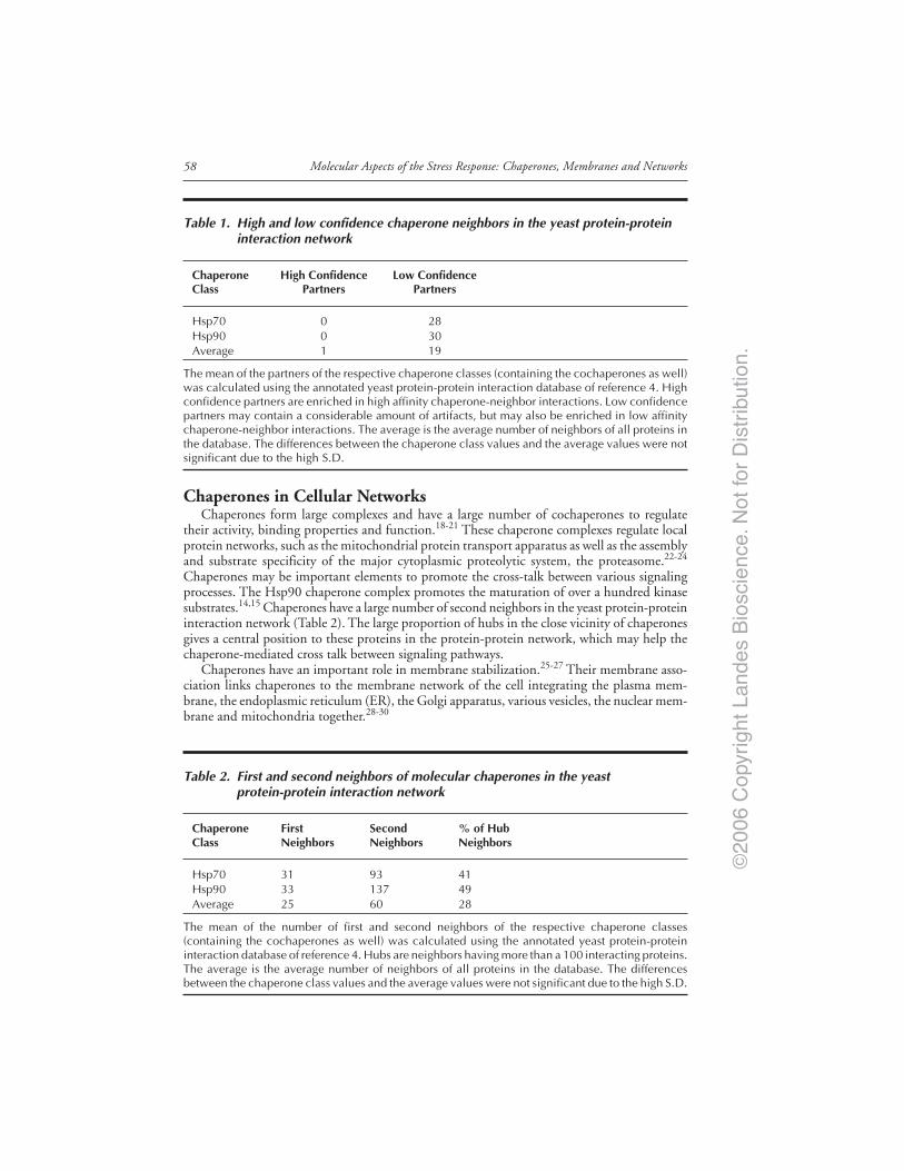
©20
06 C
opyr
ight
Lan
des
Bio
scie
nce.
Not
for
Dis
trib
utio
n.
Molecular Aspects of the Stress Response: Chaperones, Membranes and Networks58
Chaperones in Cellular NetworksChaperones form large complexes and have a large number of cochaperones to regulate
their activity, binding properties and function.18-21 These chaperone complexes regulate localprotein networks, such as the mitochondrial protein transport apparatus as well as the assemblyand substrate specificity of the major cytoplasmic proteolytic system, the proteasome.22-24
Chaperones may be important elements to promote the cross-talk between various signalingprocesses. The Hsp90 chaperone complex promotes the maturation of over a hundred kinasesubstrates.14,15 Chaperones have a large number of second neighbors in the yeast protein-proteininteraction network (Table 2). The large proportion of hubs in the close vicinity of chaperonesgives a central position to these proteins in the protein-protein network, which may help thechaperone-mediated cross talk between signaling pathways.
Chaperones have an important role in membrane stabilization.25-27 Their membrane asso-ciation links chaperones to the membrane network of the cell integrating the plasma mem-brane, the endoplasmic reticulum (ER), the Golgi apparatus, various vesicles, the nuclear mem-brane and mitochondria together.28-30
Table 1. High and low confidence chaperone neighbors in the yeast protein-proteininteraction network
Chaperone High Confidence Low ConfidenceClass Partners Partners
Hsp70 0 28Hsp90 0 30Average 1 19
The mean of the partners of the respective chaperone classes (containing the cochaperones as well)was calculated using the annotated yeast protein-protein interaction database of reference 4. Highconfidence partners are enriched in high affinity chaperone-neighbor interactions. Low confidencepartners may contain a considerable amount of artifacts, but may also be enriched in low affinitychaperone-neighbor interactions. The average is the average number of neighbors of all proteins inthe database. The differences between the chaperone class values and the average values were notsignificant due to the high S.D.
Table 2. First and second neighbors of molecular chaperones in the yeastprotein-protein interaction network
Chaperone First Second % of HubClass Neighbors Neighbors Neighbors
Hsp70 31 93 41Hsp90 33 137 49Average 25 60 28
The mean of the number of first and second neighbors of the respective chaperone classes(containing the cochaperones as well) was calculated using the annotated yeast protein-proteininteraction database of reference 4. Hubs are neighbors having more than a 100 interacting proteins.The average is the average number of neighbors of all proteins in the database. The differencesbetween the chaperone class values and the average values were not significant due to the high S.D.
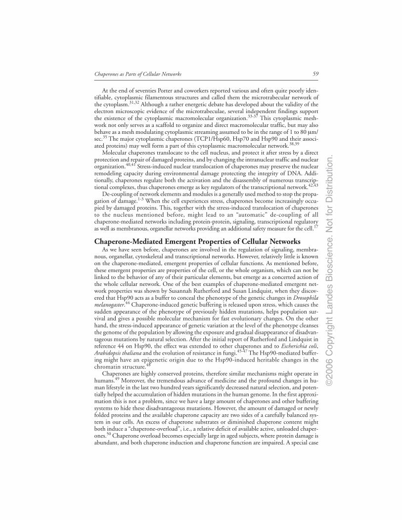
©20
06 C
opyr
ight
Lan
des
Bio
scie
nce.
Not
for
Dis
trib
utio
n.
59Chaperones as Parts of Cellular Networks
At the end of seventies Porter and coworkers reported various and often quite poorly iden-tifiable, cytoplasmic filamentous structures and called them the microtrabecular network ofthe cytoplasm.31,32 Although a rather energetic debate has developed about the validity of theelectron microscopic evidence of the microtrabeculae, several independent findings supportthe existence of the cytoplasmic macromolecular organization.33-37 This cytoplasmic mesh-work not only serves as a scaffold to organize and direct macromolecular traffic, but may alsobehave as a mesh modulating cytoplasmic streaming assumed to be in the range of 1 to 80 µm/sec.35 The major cytoplasmic chaperones (TCP1/Hsp60, Hsp70 and Hsp90 and their associ-ated proteins) may well form a part of this cytoplasmic macromolecular network.38,39
Molecular chaperones translocate to the cell nucleus, and protect it after stress by a directprotection and repair of damaged proteins, and by changing the intranuclear traffic and nuclearorganization.40,41 Stress-induced nuclear translocation of chaperones may preserve the nuclearremodeling capacity during environmental damage protecting the integrity of DNA. Addi-tionally, chaperones regulate both the activation and the disassembly of numerous transcrip-tional complexes, thus chaperones emerge as key regulators of the transcriptional network.42,43
De-coupling of network elements and modules is a generally used method to stop the propa-gation of damage.1-3 When the cell experiences stress, chaperones become increasingly occu-pied by damaged proteins. This, together with the stress-induced translocation of chaperonesto the nucleus mentioned before, might lead to an “automatic” de-coupling of allchaperone-mediated networks including protein-protein, signaling, transcriptional regulatoryas well as membranous, organellar networks providing an additional safety measure for the cell.17
Chaperone-Mediated Emergent Properties of Cellular NetworksAs we have seen before, chaperones are involved in the regulation of signaling, membra-
nous, organellar, cytoskeletal and transcriptional networks. However, relatively little is knownon the chaperone-mediated, emergent properties of cellular functions. As mentioned before,these emergent properties are properties of the cell, or the whole organism, which can not belinked to the behavior of any of their particular elements, but emerge as a concerted action ofthe whole cellular network. One of the best examples of chaperone-mediated emergent net-work properties was shown by Susannah Rutherford and Susan Lindquist, when they discov-ered that Hsp90 acts as a buffer to conceal the phenotype of the genetic changes in Drosophilamelanogaster.44 Chaperone-induced genetic buffering is released upon stress, which causes thesudden appearance of the phenotype of previously hidden mutations, helps population sur-vival and gives a possible molecular mechanism for fast evolutionary changes. On the otherhand, the stress-induced appearance of genetic variation at the level of the phenotype cleansesthe genome of the population by allowing the exposure and gradual disappearance of disadvan-tageous mutations by natural selection. After the initial report of Rutherford and Lindquist inreference 44 on Hsp90, the effect was extended to other chaperones and to Escherichia coli,Arabidopsis thaliana and the evolution of resistance in fungi.45-47 The Hsp90-mediated buffer-ing might have an epigenetic origin due to the Hsp90-induced heritable changes in thechromatin structure.48
Chaperones are highly conserved proteins, therefore similar mechanisms might operate inhumans.49 Moreover, the tremendous advance of medicine and the profound changes in hu-man lifestyle in the last two hundred years significantly decreased natural selection, and poten-tially helped the accumulation of hidden mutations in the human genome. In the first approxi-mation this is not a problem, since we have a large amount of chaperones and other bufferingsystems to hide these disadvantageous mutations. However, the amount of damaged or newlyfolded proteins and the available chaperone capacity are two sides of a carefully balanced sys-tem in our cells. An excess of chaperone substrates or diminished chaperone content mightboth induce a “chaperone-overload”, i.e., a relative deficit of available active, unloaded chaper-ones.50 Chaperone overload becomes especially large in aged subjects, where protein damage isabundant, and both chaperone induction and chaperone function are impaired. A special case

©20
06 C
opyr
ight
Lan
des
Bio
scie
nce.
Not
for
Dis
trib
utio
n.
Molecular Aspects of the Stress Response: Chaperones, Membranes and Networks60
of chaperone-overload occurs in folding diseases including various forms of neurodegeneration,such as Alzheimer’s disease, Parkinson’s disease or Hungtington’s disease, where a misfoldedand usually not degraded protein sequesters most chaperones.50-53 In consequence of the chap-erone overload, the protein products harboring the “hidden mutations” may be released, andmay contribute to the development of civilization diseases, such as cancer, atherosclerosis anddiabetes. Since relatively few generations have been passed from the beginning of the medicalrevolution and lifestyle changes in the 19th century, this effect is most probably negligibletoday. However, it increases with each generation. Still, we probably have many hundreds ofyears to think about a possible solution, which gives us time to learn much more and reconcilethe serious ethical concerns with a possible solution.50
In recent years the scientific community has became increasingly aware of the idea thatnot only chaperones but a large number of other proteins may also regulate the diversity ofthe phenotype.3,14,54-56 Though a relatively small number of regulators were uncovered yet, acommon molecular mechanism, such as the involvement in signaling or modifications ofhistones and DNA structure seems to be an unlikely explanation for all the effects observed.If a general explanation is sought, it is more likely to be related to the network properties ofthe cell. In this context, chaperones are typical weak linkers, providing low affinity, lowprobability contacts with other proteins (for a grossly simplified illustration, see Table 1).Weak links are known to help system stability in a large variety of networks from macromol-ecules to social networks and ecosystems, which may be a general network-level phenom-enon explaining many of the genetic buffering effects. Currently we do not know, whatposition is required, if any, in the network for these ‘weak links’ besides their low affinity andtransient interactions. The central position of chaperones demonstrated in Table 2 may bean additional hallmark of stabilizing weak links.3,14
Chaperone TherapiesCellular networks are remodeled in various diseases and after stress. Proper interventions to
push the equilibrium towards the original state may not be limited to single-target drugs,which have a well-designed, high affinity interaction with one of the cellular proteins. In agree-ment with this general assumption, several examples show that multi-target therapy may besuperior to the usual single-target approach. The best known examples of multi-target drugsinclude Aspirin, Metformin or Gleevec as well as combinatorial therapy and natural remedies,such as herbal teas.57 Due to the multiple regulatory roles of chaperones, chaperone-modulatorsprovide additional examples for multi-target drugs. Indeed, chaperone substitution (in theform of chemical chaperones), the help of chaperone induction and chaperone inhibition areall promising therapeutic strategies.58-61
ConclusionChaperones regulate cellular functions at two levels. In several cases they interact with a
specific target protein, and become mandatory to its folding as well as for the assistance in theformation of specific protein complexes (and in the prevention of the assembly of others).These specific interactions make chaperones important parts of the core of cellular networks,such as the protein net, the signaling network, the membranous and organellar network as wellas the transcriptional network. However, in most cases chaperones have only a low affinity,temporary interactions, i.e., ‘weak links’ with most of their targets. Changes of these interac-tions do not affect the general behavior of the whole network, the cell. However, an inhibitionof these weak links might lead to a rise in cellular noise, the destabilization and des-integrationof the whole network. By this complex version of the ‘error-catastrophe’, chaperone inhibitorshelp us to combat cancer. In contrast, chaperone activation may decrease cellular noise, stabi-lize and integrate cells and thus give a general aid against aging and diseases. Thus, besidesslowing the development or reversing protein folding diseases, chaperone-therapies may alsogenerally benefit the aging organism by stabilizing its cells and functions. Properly working
©20
06 C
opyr
ight
Lan
des
Bio
scie
nce.
Not
for
Dis
trib
utio
n.
61Chaperones as Parts of Cellular Networks
chaperones may be key players to help us to reach improved life conditions in an advanced age.The assessment of the multiple roles of chaperones in the context of cellular networks is onlyjust beginning.
AcknowledgementsWork in the authors’ laboratory was supported by research grants from the EU (FP6506850,
FP6-016003), Hungarian Science Foundation (OTKA-T37357 and OTKA-F47281), Hun-garian Ministry of Social Welfare (ETT-32/03), Hungarian National Research Initiative (1A/056/2004 and KKK-0015/3.0) and from the South African National Research Foundation(2067467 and the South African-Hungarian Collaborative Program 2053542). C. Söti is aBolyai research Scholar of the Hungarian Academy of Sciences.
References1. Barabasi AL, Oltvai ZN. Network biology: Understanding the cell’s functional organization. Nat
Rev Genet 2004; 5:101-113.2. Albert R. Scale-free networks in cell biology. J Cell Sci 2005; 118:4947-4957.3. Csermely P. Weak links: Stabilizers of complex systems from proteins to social networks. Heidel-
berg: Springer Verlag, 2006.4. von Mering C, Krause R, Snel B et al. Comparative assessment of large-scale data sets of
protein-protein interactions. Nature 2002; 417:399-403.5. Rual JF, Venkatesan K, Hao T et al. Towards a proteome-scale map of the human protein-protein
interaction network. Nature 2005; 437:1173-1178.6. Stelzl U, Worm U, Lalowski M et al. A human protein-protein interaction network: A resource for
annotating the proteome. Cell 2005; 122:957-968.7. White MA, Anderson RG. Signaling networks in living cells. Annu Rev Pharmacol Toxicol 2005;
45:587-603.8. Borodina I, Nielsen J. From genomes to in silico cells via metabolic networks. Curr Opin Biotechnol
2005; 16:350-355.9. Blais A, Dynlacht BD. Constructing transcriptional regulatory networks. Genes Dev 2005;
19:1499-1511.10. Arita M. The metabolic world of Escherichia coli is not small. Proc Natl Acad Sci USA 2004;
101:1543-1547.11. Ma HW, Zeng AP. Reconstruction of metabolic networks from genome data and analysis of their
global structure for various organisms. Bioinformatics 2003; 19:220-277.12. Tanaka R, Yi TM, Doyle J. Some protein interaction data do not exhibit power law statistics.
FEBS Lett 2005; 579:5140-5144.13. Tsigelny IF, Nigam SK. Complex dynamics of chaperone-protein interactions under cellular stress.
Cell Biochem Biophys 2004; 40:263-276.14. Csermely P. Strong links are important - But weak links stabilize them. Trends Biochem Sci 2004;
29:331-334.15. Zhao R, Davey M, Hsu YC et al. Navigating the chaperone network: An integrative map of physi-
cal and genetic interactions mediated by the hsp90 chaperone. Cell 2005; 120:715-727.16. Nardai G, Vegh E, Prohaszka Z et al. Chaperone-related immune dysfunctions: An emergent prop-
erty of distorted chaperone-networks. Trends Immunol 2006; 27:74-7917. Soti C, Pal C, Papp B et al. Chaperones as regulatory elements of cellular networks. Curr Op Cell
Biol 2005; 17:210-215.18. Frydman J. Folding of newly translated proteins in vivo: The role of molecular chaperones. Annu
Rev Biochem 2001; 70:603-647.19. Kleizen B, Braakman I. Protein folding and quality control in the endoplasmic reticulum. Curr
Opin Cell Biol 2004; 16:343-349.20. Young JC, Agashe VR, Siegers K et al. Pathways of chaperone-mediated protein folding in the
cytosol. Nat Rev Mol Cell Biol 2004; 5:781-791.21. Blatch GL, ed. Networking of Chaperones by Co-Chaperones. Georgetown: Landes Bioscience,
2006.22. Young JC, Hoogenraad NJ, Hartl FU. Molecular chaperones Hsp90 and Hsp70 deliver preproteins
to the mitochondrial import receptor Tom70. Cell 2003; 112:41-50.23. Imai J, Maruya M, Yashiroda H et al. The molecular chaperone Hsp90 plays a role in the assem-
bly and maintenance of the 26S proteasome. EMBO J 2003; 22:3557-3567.

©20
06 C
opyr
ight
Lan
des
Bio
scie
nce.
Not
for
Dis
trib
utio
n.
Molecular Aspects of the Stress Response: Chaperones, Membranes and Networks62
24. Whittier JE, Xiong Y, Rechsteiner MC et al. Hsp90 enhances degradation of oxidized calmodulinby the 20 S proteasome. J Biol Chem 2004; 279:46135-46142.
25. Tsvetkova NM, Horvath I, Torok Z et al. Small heat-shock proteins regulate membrane lipidpolymorphism. Proc Natl Acad Sci USA 2002; 99:13504-13509.
26. Torok Z, Horvath I, Goloubinoff P et al. Evidence for a lipochaperonin: Association of activeprotein-folding GroESL oligomers with lipids can stabilize membranes under heat shock condi-tions. Proc Natl Acad Sci USA 1997; 94:2192-2197.
27. Torok Z, Goloubinoff P, Horvath I et al. Synechocystis HSP17 is an amphitropic protein thatstabilizes heat-stressed membranes and binds denatured proteins for subsequent chaperone-mediatedrefolding. Proc Natl Acad Sci USA 2001; 98:3098-3103.
28. Filippin L, Magalhaes PJ, Di Benedetto G et al. Stable interactions between mitochondria andendoplasmic reticulum allow rapid accumulation of calcium in a subpopulation of mitochondria. JBiol Chem 2003; 278:39224-39234.
29. Aon MA, Cortassa S, O’Rourke B. Percolation and criticality in a mitochondrial network. ProcNatl Acad Sci USA 2004; 101:4447-4452.
30. Szabadkai G, Simoni AM, Chami M et al. Drp-1-dependent division of the mitochondrial net-work blocks intraorganellar Ca2+ waves and protects against Ca2+-mediated apoptosis. Mol Cell2004; 16:59-68.
31. Wolosewick JJ, Porter KR. Microtrabecular lattice of the cytoplasmic ground substance. Artifact orreality. J Cell Biol 1979; 82:114-139.
32. Schliwa M, van Blerkom J, Porter KR. Stabilization of the cytoplasmic ground substance indetergent-opened cells and a structural and biochemical analysis of its composition. Proc Natl AcadSci USA 1981; 78:4329-4333.
33. Clegg JS. Properties and metabolism of the aqueous cytoplasm and its boundaries. Am J Physiol1984; 246:R133-R151.
34. Luby-Phelps K, Lanni F, Taylor DL. The submicroscopic properties of cytoplasm as a determinantof cellular function. Annu Rev Biophys Biophys Chem 1988; 17:369-396.
35. Hochachka PW. The metabolic implications of intracellular circulation. Proc Natl Acad Sci USA1999; 96:12233-12239.
36. Verkman AS. Solute and macromolecule diffusion in cellular aqueous compartments. Trends BiochemSci 2002; 27:27-33.
37. Spitzer JJ, Poolman B. Electrochemical structure of the crowded cytoplasm. Trends Biochem Sci2005; 30:536-541.
38. Csermely P. A nonconventional role of molecular chaperones: Involvement in the cytoarchitecture.News Physiol Sci 2001; 16:123-126.
39. Sreedhar AS, Mihaly K, Pato B et al. Hsp90 inhibition accelerates cell lysis. Anti-Hsp90 ribozymereveals a complex mechanism of Hsp90 inhibitors involving both superoxide- and Hsp90-dependentevents. J Biol Chem 2003; 278:35231-35240.
40. Michels AA, Kanon B, Konings AW et al. Hsp70 and Hsp40 chaperone activities in the cytoplasmand the nucleus of mammalian cells. J Biol Chem 1997; 272:33283-33289.
41. Nollen EA, Salomons FA, Brunsting JF et al. Dynamic changes in the localization of thermallyunfolded nuclear proteins associated with chaperone-dependent protection. Proc Natl Acad Sci USA2001; 98:12038-12043.
42. Guo Y, Guettouche T, Fenna M et al. Evidence for a mechanism of repression of heat shock factor1 transcriptional activity by a multichaperone complex. J Biol Chem 2001; 276:45791-45799.
43. Freeman BC, Yamamoto KR. Disassembly of transcriptional regulatory complexes by molecularchaperones. Science 2002; 296:2232-2235.
44. Rutherford SL, Lindquist S. Hsp90 as a capacitor for morphological evolution. Nature 1998;396:336-342.
45. Fares MA, Ruiz-González MX, Moya A et al. Endosymbiotic bacteria: GroEL buffers against del-eterious mutations. Nature 2002; 417:398.
46. Queitsch C, Sangster TA, Lindquist S. Hsp90 as a capacitor of phenotypic variation. Nature 2002;417:618-624.
47. Cowen LE, Lindquist S. Hsp90 potentiates the rapid evolution of new traits: Drug resistance indiverse fungi. Science 2005; 309:2185-2189.
48. Sollars V, Lu X, Xiao L et al. Evidence for an epigenetic mechanism by which Hsp90 acts as acapacitor for morphological evolution. Nat Genet 2003; 33:70-74.
49. Whitesell L, Lindquist SL. HSP90 and the chaperoning of cancer. Nat Rev Cancer 2005; 5:761-772.50. Csermely P. Chaperone-overload as a possible contributor to “civilization diseases”: Atherosclerosis,
cancer, diabetes. Trends Genet 2001; 17:701-704.

©20
06 C
opyr
ight
Lan
des
Bio
scie
nce.
Not
for
Dis
trib
utio
n.
63Chaperones as Parts of Cellular Networks
51. Nardai G, Csermely P, Söti C. Chaperone function and chaperone overload in the aged. ExpGerontol 2002; 37:1255-1260.
52. Söti C, Csermely P. Chaperones and aging: Their role in neurodegeneration and other civilizationaldiseases. Neurochem International 2002; 41:383-389.
53. Papp E, Száraz P, Korcsmáros T et al. Changes of endoplasmic reticulum chaperone complexes,redox state and impaired protein disulfide reductase activity in misfolding alpha-1-antitrypsintransgenic mice. FASEB J 2006, (in press).
54. Bergman A, Siegal ML. Evolutionary capacitance as a general feature of complex gene networks.Nature 2003; 424:549-552.
55. True HL, Berlin I, Lindquist SL. Epigenetic regulation of translation reveals hidden genetic varia-tion to produce complex traits. Nature 2004; 431:184-187.
56. Sangster TA, Lindquist S, Queitsch C. Under cover: Causes, effects and implications ofHsp90-mediated genetic capacitance. Bioessays 2004; 26:348-362.
57. Csermely P, Agoston V, Pongor S. The efficiency of multi-target drugs: The network approachmight help drug design. Trends Pharmacol Sci 2005; 26:178-182.
58. Vigh L, Literati PN, Horvath I et al. Bimoclomol: A nontoxic, hydroxylamine derivative withstress protein-inducing activity and cytoprotective effects. Nat Med 1997; 3:1150-1154.
59. Bernier V, Lagace M, Bichet DG et al. Pharmacological chaperones: Potential treatment for con-formational diseases. Trends Endocrinol Metab 2004; 15:222-228.
60. Neckers L, Neckers K. Heat-shock protein 90 inhibitors as novel cancer chemotherapeutics - Anupdate. Expert Opin Emerg Drugs 2005; 10:137-149.
61. Söti C, Nagy E, Giricz Z et al. Heat shock proteins as emerging therapeutic targets. Br J Pharmacol2005; 146:769-780.

©20
06 C
opyr
ight
Lan
des
Bio
scie
nce.
Not
for
Dis
trib
utio
n.

©20
06 C
opyr
ight
Lan
des
Bio
scie
nce.
Not
for
Dis
trib
utio
n.

©20
06 C
opyr
ight
Lan
des
Bio
scie
nce.
Not
for
Dis
trib
utio
n.
CHAPTER 7
*Corresponding Author: György Szabadkai—Department of Experimental and DiagnosticMedicine, University of Ferrara, Via Borsari 46, Ferrara, 44100, Italy. Email: [email protected]
Molecular Aspects of the Stress Response: Chaperones, Membranes and Networks,edited by Peter Csermely and Laszlo Vigh. ©2007 Landes Bioscienceand Springer Science+Business Media.
Chaperones as Parts of Organelle NetworksGyörgy Szabadkai* and Rosario Rizzuto
Abstract
The efficiency, divergence and specificity of virtually all intracellular metabolic andsignalling pathways largely depend on their compartmentalized organization. Acorollary of the requirement of compartmentalization is the dynamic structural parti-
tion of the intracellular space by endomembrane systems. A branch of these membranes com-municate with the extracellular space through the endo- and exocytotic processes. Others, likethe mitochondrial and endoplasmic reticulum networks accomplish a further role, being fun-damental for the maintenance of cellular energy balance and for determination of cell fateunder stress conditions. Recent structural and functional studies revealed that the interactionof these networks and the connectivity state of mitochondria controls metabolic flow, proteintransport, intracellular Ca2+ signalling, and cell death. Moreover, reflecting the fact that theabove processes are accomplished in a microdomain between collaborating organelle mem-branes, the existence of macromolecular complexes at their contact sites have also been re-vealed. Being not only assistants of nascent protein folding, chaperones are proposed to partici-pate in assembling and maintaining the function of the above complexes. In this chapter wediscuss recently found examples of such an assembly of protein interactions driven by chaper-one proteins, and their role in regulating physiological and pathological processes.
IntroductionThe functionality of eukaryotic cells lies on their complex and highly dynamic endomembrane
system, composed of the endocytotic machinery, the secretory membranes and the mitochon-drial network. Membranes at the interface of these systems display dynamic interactions witheach other, ensuring protein transport, signal transmission between compartments, and regula-tion of network shape/connectivity. A long time known function of chaperone proteins is toregulate the assembly of local protein complexes involved in specific organelle function (e.g.,mitochondrial or ER protein import). Recently, a novel concept arose, pointing to a morecomplex, integrating role of these proteins, i.e., to stabilize larger intracellular morphologicaland functional networks through weak but numerous links.1-3 One of the most importantpredictions of such kind of organization is that while under normal conditions chaperoningelements ensure interactions between cellular organelles, during cellular stress theirhyperactivation is needed for coping with stress, leading to more intense communication in theregulated networks. Along these lines, exhaustion of this ‘network chaperoning’ function willlead to decomposition of normal cellular function, thus it will lie at the base of a wide range ofpathophysiological conditions. Indeed, chaperones in the endoplasmic reticulum (ER) havenow a well established role in sensing and signalling stress conditions and executing defence
©20
06 C
opyr
ight
Lan
des
Bio
scie
nce.
Not
for
Dis
trib
utio
n.
65Chaperones as Parts of Organelle Networks
responses such as the unfolded protein response (UPR).4-7 A similar, but less characterizedstress response has been also observed in the mitochondrial network, following insults leadingimpairment of protein folding in the organelle. Moreover, following ER stress, mitochondriaseems to be directly activated by ER-derived signals and they are the target of numerous pro-and anti- cell death mechanisms.
To discuss the potential role of chaperones in the interaction between organellar networkswe will use the above described paradigm as an example and we will refer to eventual similari-ties to mechanisms described in other interacting organelle networks. First, we will brieflydescribe the regulation of dynamics how ER-mitochondria interaction evolved, presumably bythe help of numerous chaperoning activities; then, through the example of recent data onER-mitochondrial Ca2+ metabolic and cell death signalling, we will discuss the eventual spe-cific functions which chaperons might fulfil in microdomains between these organelles; finally,we will describe the perspectives related to chaperone assisted organellar interactions underpathophysiological conditions.
Biogenesis of the ER and Mitochondrial Networks:A Role for Chaperons in Interorganellar Communication?
The endoplasmic reticulum (ER), adjoining to the nuclear envelope, forms a continuousnetwork in virtually all cell types that is the major site of lipid biosynthesis, protein folding andthe entry point into the secretory pathway. Thus the ER can be considered the common ances-tor of all membranes downstream in this pathway, including the Golgi, secretory vesicles, thelysosomes, and the plasma membrane.8-11 The biogenesis of the ER is a result of coordinateactivation of protein and lipid biosynthesis, as deduced from experiments showing the interde-pendency of the unfolded protein response (UPR) and inositol response pathways.8 Theseworks also provided the clue that chaperoning inside the ER lumen plays an important role inorganelle biogenesis and network formation. Indeed, during UPR, accumulation of unfoldedproteins (e.g., by a disequilibrium between the amount of proteins processed in the secretorypathway and the actual requirement of protein synthesis at a given developmental stage of thecell) leads to activation of a transcriptome including ER chaperones (to ensure proper foldingin the ER lumen), and concerted activation of genes involved in phospholipid synthesis canalso be observed during this process.12,13 Similar mechanisms operate during ‘ER stress’ condi-tions, which now seems to be involved in pathogenesis of several diseases (see below).4-7
In contrast to the ER, the mitochondrial network* is apparently shaped in a more variableand complex way, depending on cell type and metabolic state. These features of mitochondrialmorphology seems to reflect a complex evolutionary path evolving from their α-proteobacterialancestor, since, to the best of our knowledge, it had a quite standard ovoid shape.14-16 Themitochondrial proteome consists of about 500-1500 proteins of which only a minor fraction(8 proteins in yeast and 13 in humans) is encoded by mitochondrial DNA. Thus, duringmitochondrial biogenesis, virtually all mitochondrial proteins are synthesized as precursorpolypeptides in the cytosol and are imported post-translationally into the organelle.17 Defec-tive protein folding and processing in the organelle, e.g., due to the impairment of the assem-bly of multi-subunit mitochondrial complexes or downregulation of genes encoding mito-chondrial chaperones or proteases, lead to a mitochondria-specific (ER independent) ‘stress’response, comprising transcriptional upregulation of matrix chaperones such as Hsp60, mtHsp70(also known as Grp-75/mortalin).18-20
* Whether mitochondria form a ‘real’ network, is still a matter of debate. E.g., from a structuralpoint of view, there has never been documentation of a network of mitochondria in the neuronsor glia of brain tissue. Although, a mitochondrial network is not disputed in lower organisms,such as yeast or in most cultured cell lines. On the other hand, from a functional point of viewwe can define mitochondria as a communication “network” wherein the mitochondria of a cellrespond in a concerted fashion to a stimulus, then “network” is probably the right term to usefor mitochondria of all eukaryotic cells.
©20
06 C
opyr
ight
Lan
des
Bio
scie
nce.
Not
for
Dis
trib
utio
n.
Molecular Aspects of the Stress Response: Chaperones, Membranes and Networks66
In this manner, the ER and mitochondrial network appear to function in an analogous wayin utilizing chaperones to maintain correct organelle function. Moreover, the biogenesis ofboth organelles relies on protein import from the cytosolic compartment, marking out a cross-road for newly synthesized proteins in the cytosol.21 The proper protein sorting into the or-ganelles is driven by a complex protein interaction network.22 The general model for proteinrecognition and transport at the mitochondrial outer membrane and the ER are fascinatinglysimilar and relies on a series of chaperones interacting with the ER and mitochondrial mem-branes: first, either cytosolic chaperones of the Hsp70 family, or occupancy of the C-terminalregion of nascent chains in the ribosomal exit site prevents premature stable folding of preproteins.Membrane-bound receptors (such as Tom70 and Tom20 of the mitochondrial outer mem-brane translocase complex [TOM], and the Signal recognition particle [Srp] of the ER mem-brane) bind preproteins and transfer them into the translocation channel (Tom40 of the OMMand the Sec61 complex of the ER). Mitochondria have to operate with further complexes ofIMM to ensure translocation and insertion of matrix and IMM proteins (Tim22 and Tim23,respectively).17,23 Finally, protein motors and chaperones in the matrix and the ER lumen(mtHsp70/Grp-75 and BiP/Grp-78 in the ER and mitochondria, respectively) complete thetransport task. This protein sorting task of the cytosolic chaperone network requires severalother elements, such as Hsp90 and its co-chaperone interactors.24
A recent meticulous proteomic analysis of the different mitochondrial compartments shedlight on the existence of a conserved subset of matrix preproteins, which reside also on theOMM surface.25 Among them, mitochondrial matrix chaperones (Grp-75, Hsp60) were alsofound, indicating that they may localize along the whole protein translocation pathway, andthat they could play a role in interacting with the cytosolic or ER protein sorting machinery,thus also ER membrane proteins. A corollary of the latter assumption is that the ER andmitochondria are located close enough to mediate these interactions. Indeed, recently signifi-cant progress has been achieved in exploring the morphological characteristics of their interac-tion. This topic is extensively covered and illustrated in recent reviews (see refs. 26-28), here wejust mention that a close, synaptic-like apposition of the organelles was clearly demonstrated ina broad variety of cell types using light and electron microscopy studies (Fig. 1).29-32
It is now generally accepted that even if the ER forms a structurally cohesive network, itcontains functionally and structurally separate sub-domains, specialized to a defined function(for a recent see review ref. 9). Indeed, the ER-mitochondria interface seems to be one thesespecialized domains, containing smooth ER membrane, surrounded by attached ribosomes.33
This specific ER compartment involved in interactions with mitochondria can be isolated as alight mitochondrial fraction by density gradient centrifugation and is called asmitochondria-associated membranes (MAM).34 Originally it was recognized as a fraction en-riched of phospholipid synthetic and transfer activities, but now it has been shown to have arole in other signaling pathways, like Ca2+ and apoptotic signaling following ER stress.35,36
A further implication of chaperones at the ER-mitochondria interface came from the de-scription of an alternative protein sorting pathway, independent of the one described above. Inthis path, Ydj1, the yeast homologue of HDJ2 (a DJ-domain containing co-chaperone, knownto function as regulator of ATPase activity of hsp70 family proteins), assists the Hsc70 (Ssa inyeast)-mediated post-translational targeting of some proteins to both mitochondria and theendoplasmic reticulum by transiently associating with ER and mitochondria membranes throughits covalently attached lipid moiety.37 Similarly, other reports, by analysing alternative mito-chondrial and ER targeting sequences (e.g., tail anchors), showed that the two organelles mayshare particular proteins, including also integral membrane proteins.38-40
In summary, the biogenesis of the ER and mitochondria requires a continuous flow of theirprotein components in a tight microdomain between the organelles, through alternative routeswhich are delineated by a cluster of chaperones. Along these lines, a further avenue to exploreis how these processes might provide a dynamic framework to maintain organelle interactions.In the next section, we will discuss this prospect, through the example of ER-mitochondrialCa2+ signalling machinery.

©20
06 C
opyr
ight
Lan
des
Bio
scie
nce.
Not
for
Dis
trib
utio
n.
67Chaperones as Parts of Organelle Networks
ER-Mitochondrial Ca2+ Transfer: A Major Exampleof Organelle Interactions
A significant part of our current view on ER-mitochondrial interactions arose from theinvestigation of Ca2+ signal transmission between these organelles; an affirmation which mightreflect its importance in cellular signalling but also the availability of technical approaches tostudy Ca2+ signals. Introduction of protein based Ca2+ sensitive probes like aequorins andGFP/YFP fusion proteins with genetically engineered Ca2+ binding sites, targeted to differentsubcellular locations facilitated profoundly the development of this field.41,42
The outer mitochondrial membrane (OMM) is permeable to ions of a limited size, owingto the abundance of the large conductance channel VDAC (for the role of the channel inmetabolic channelling, see refs. 43, 44). Indeed, recent data shows that the availability andselective placement of VDAC channels at ER/mitochondria contact sites facilitates mito-chondrial Ca2+ accumulation, in keeping with the idea that the latter process requires the fastand efficient transfer of Ca2+ microdomains from the mouth of the Ca2+ channels located inneighbouring ER to the transporters of the inner mitochondrial membrane (IMM).45,46 TheIMM is an ion impermeable membrane, forming foldings into the internal space, known ascristae.32 The activity of respiratory chain complexes allows the translocation of H+ into the
Figure 1. Visualization of the three dimensional structure and colocalization of the ER andmitochondrial network. HeLa cells were transfected with ER targeted mRFP1 and mitochondriallytargeted GFP (as described in refs. 76, 101), and digital microscopy, 3D deconvolution andimage reconstruction was applied.76 The left panel shows the original images of one focalplane, overlayed on the middle panel (ER—red; mitochondria—green). The right panels show3D surface rendered images using the same colour code. The top right panel shows theinterconnected network, rendered transparent on the right middle panel. The bottom rightpanel shows the mitochondrial network with the surface colocalizing with the ER (shown inred). A color version of this figure is available online at www.Eurekah.com.

©20
06 C
opyr
ight
Lan
des
Bio
scie
nce.
Not
for
Dis
trib
utio
n.
Molecular Aspects of the Stress Response: Chaperones, Membranes and Networks68
intermembrane space (IMS), with consequent generation of an electrochemical gradient (∆µH),composed of a chemical (∆pH) and electrical (∆Ψm) component, the latter being about -180mV, negative inside, thus providing a huge driving force for Ca2+ entry into the organelle. Asto the uptake route, its molecular identity is still unknown. It is suggested to be a gatedchannel rather than a carrier, showing a Ca2+ activated second order kinetics, with a separateCa2+ activation and a Ca2+ transport site.47,48 Based on these properties, the rate of mitochon-drial Ca2+ uptake through the channel was shown to be significant only above an ‘activatory’threshold (200- 300 nM), also known as ‘set point’. A known inhibitor of Ca2+ uptake intomitochondria is ruthenium red (RuR), of which the binding site however, was shown to be onthe OMM located VDAC channel.46,49 This latter finding indicates that, similarly to theprotein translocation mechanisms, Ca2+ transport through the two mitochondrial membranesis mediated by interacting protein complexes of both the OMM and IMM, presumably at thecontact sites between the two membranes.50 Interestingly, at these contact sites a non specificion- and metabolite-channel activity of major pathophysiological interest has been character-ized in the past years, commonly referred to the permeability transition pore (PTP).51 Thishigh-conductance channel was shown to be a multiprotein complex (the putatively essentialcomponents being VDAC, the adenine-nucleotide translocase [ANT] of the IMM andcyclophilin D from the matrix side), and is activated by [Ca2+] increase in the mitochondrialmatrix and oxidation of critical cysteins of its protein components.52-54 Since this channelwas shown to display several conductance state, and the Ca2+ uniporter mechanism is sensi-tive to RuR acting on VDAC, one can imagine that this or a similar protein complex mediatesCa2+ uptake under physiological conditions. Importantly, reflecting the relatively low affinityof Ca2+ activation of the uniporter, efficient mitochondrial Ca2+ uptake in intact cells wasshown to be dependent on the close apposition of mitochondria to Ca2+ release sites of theER or the sarcoplasmic reticulum.55,56 Here, at mouth of Ca2+release channels (inositol 1,4,5trisphosphate receptor, IP3R or the ryanodine receptor, RyR, respectively), during Ca2+ re-lease, a high [Ca2+] microdomain is formed, which in turn is instantly transferred to themitochondrial matrix.
Taken together, the ER-mitochondria Ca2+ transmission machinery represents a transportsystem for which necessary is (i) the interaction of two organelles, (ii) and the interaction ofchannelling proteins of three adjacent membranes. Augmenting the complexity of the system,these interactions do not seem to be static, considering the continuous dynamic structuralremodelling of both the ER and mitochondrial networks in living cells.9,57 Moreover,ER-mitochondrial interactions and mitochondrial mobility were shown to be regulated byCa2+ itself.58
Chaperone Control of ER-Mitochondrial Interaction along the Ca2+
Signal Transmission PathwayThe morphological and functional description of the above detailed complex machineries
logically led to the question: what are the molecular components that bring together this path-way? To answer this challenge, first, biochemical studies, exploring protein interactions withthe already known components provided some clues. Surprisingly, a two-hybrid study, usingVDAC as a bait (see ref. 59), has identified mtHsp70/Grp75, the above mentioned mitochon-drial matrix chaperone as a VDAC interactor, which not only attached to VDAC, located inthe OMM, but was able also to change its conductance properties, proving the physiologicalsignificance of the interaction. This study called to mind that several lines of recent evidenceshowed the presence of different classes of chaperone proteins along the ER-mitochondrialCa2+ transfer route. Indeed, the IP3R and RyR Ca2+ release channels, as well as VDAC wereshown to be important nodes of protein interaction networks (described in detail in the re-views refs. 43, 60-63), among which several exhibit chaperone activity.61 The broad interactionmodules of the IP3R and VDAC, and the particular role of chaperones in that network areshown on (Fig. 2) and are described in more detail in the figure legend. Seeing the vast presence

©20
06 C
opyr
ight
Lan
des
Bio
scie
nce.
Not
for
Dis
trib
utio
n.
69Chaperones as Parts of Organelle Networks
Figure 2. Please see figure legend on next page.

©20
06 C
opyr
ight
Lan
des
Bio
scie
nce.
Not
for
Dis
trib
utio
n.
Molecular Aspects of the Stress Response: Chaperones, Membranes and Networks70
of chaperones in this system, again an apparent question arises: what can be their function inthe Ca2+ signal transmission machinery, apparently unrelated to protein folding and transfer?
The first clue emerges from an analogous system at the plasmamembrane-ER interface,described in hippocampal neurons. In their postsynaptic density the Homer and Shank pro-teins play a scaffolding role to maintain the interaction between the glutamatergic receptors ofthe postsynaptic membrane and the Ca2+ signalling components of the ER: the IP3R and ERCa2+ ATPases (SERCAs).64-66 Strikingly, overexpression of Homer1b and Shank1b inducesrelocation to plasmamembrane not only of the above Ca2+ transport proteins but also ofcalreticulin and calbindin, Ca2+ binding chaperones of the ER lumen and membrane, respec-tively.65 Similarly, we can propose that the chaperones found at the ER-mitochondrial interfacemay structurally bring together the building blocks of the Ca2+ transfer system. A noticeabledifference between the two systems would be the ATP dependent dynamic binding/scaffoldingof the Hsp70/90 family chaperones, which might represent a target for unlocking the interac-tion under conditions when the ATP supply is not sufficient, i.e., under stress conditions (seealso below).
A second plausible role for chaperone association to ER and mitochondrial Ca2+ channels isthe modulation of the ion channel function itself. Indeed, both the peptydil-prolyl isomerase/immunophilin FK506 binding protein (FKBP12) binding to the IP3Rs and RyRs, and mtHsp70/Grp75 binding to VDAC was shown to modify their conductance.59 While the effect on theRyRs of FKBP12 was shown not to be related to its enzyme activity (ref. 67), it appears tostabilize the receptor homo- and heterodimers through a conformational effect.68 Direct, du-rable binding of FKBP12 to the IP3R was failed to be shown (ref. 69), still it is able to inhibitCa2+ release through both Ca2+ channels.70 Another member of the FKBP family of proteins,FKBP52 binds to Hsp90 (see protein sorting to mitochondria above), and through binding tothe dynein motor on the microtubule network it directs translocation of glucocorticoid recep-tors and p53 to the nucleus.71,72 The finding that mtHsp70/Grp75 also interacts with the dyneinlight chain component TCTEL and p53, and that VDAC docks both former proteins (refs. 59,73), raise the possibility that also the presence of FKBP (family member 12) close to the IP3Rand RyR, plays a similar role, i.e., it recruits regulatory factors on the surface of mitochondriaand the ER. In this case, mtHsp70/Grp75 can be a regulatory protein by its plain interaction,since its ATPase domain was shown to be dispensable for modifying VDAC conductance.59
Our recent findings along the ER-mitochondria Ca2+ signal transmission axis provided fur-ther confirmation of chaperone mediated coupling between these two organelles. We have foundthat the IP3R and VDAC-1 reside in a common macromolecular complex, localized in theabove described mitochondria-associated subfraction of the ER (MAM). Moreover, in this com-plex the N-terminal ligand binding domain of the IP3R was found to directly enhance mito-chondrial Ca2+ accumulation. Since the downregulation of the OMM localized mtHsp70/Grp75
Figure 2, viewed on previous page. Components of the Ca2+ signal transmission axis betweenthe ER and mitochondria, and its interaction points with chaperone networks. The left panelsshow the cellular interaction network of the IP3R (upper) and VDAC proteins (lower) as browsedfrom the HiMAP human protein-protein interaction map (www.himap.org), based on two-hybriddatasets, literature-confirmed interactions (Human Protein Reference Database, www.hprd.org)and prediction algorithms.102 Gene names are shown as defined in the OMIM database(www.ncbi.nlm.nih.gov). Highlighted genes represent subsets of proteins with similar functionor subcellular localization. The right panel shows literature based interactions of chaperoneproteins (showed in ovals, blue: ER origin; red: cytosolic origin; green: mitochondrial origin) withthe Ca2+ signal transmission machinery from the endoplasmic reticulum (ER) to the mitochondria(OMM: outer mitochondrial membrane; IMM: inner mitochondrial membrane). Abbreviationsand citations proving interaction not mentioned in the text: cbd: calbindin; cnx: calnexin;103 crt:calreticulin;104 cyp-D: cyclophilin-D;105 FKBP: FK506 binding protein;69 mtc: mitocalcin.106
Note the multiple localization of Hsp60 and Grp75.88,107 A color version of this figure is availableonline at www.Eurekah.com.
©20
06 C
opyr
ight
Lan
des
Bio
scie
nce.
Not
for
Dis
trib
utio
n.
71Chaperones as Parts of Organelle Networks
abolished this effect, we concluded that a chaperone-mediated direct coupling between theorganelles is mediated by these three proteins (Szabadkai, Bianchi et al, manuscript submitted).
To summarize the above described scenario, we can embed these findings in the conceptthat chaperones, through their interaction with the elements of a signal transduction pathwaybetween the ER and mitochondria, build up and maintain the integrity of the interactingsurface the two organelles. Pending on results and new experimental approaches that will clarifythe nature and strength of interaction of chaperones with the Ca2+ signalling protein compo-nents, they might also underlie the emerging concept of stabilization of networks by weak,transient links.1,2
Here we considered Ca2+ merely as an example of a substance transferred from the ER tomitochondria, but we have also to mention that Ca2+, as a messenger released from the ER, wasshown to determine functional properties and integrity of the mitochondrial network. El-evated [Ca2+] in the cytosol and the ER-mitochondrial interface may induce two differentresponses from the mitochondrial remodelling apparatus. The Ca2+ induced translocation ofthe mitochondrial fission factor Drp-1 induces mitochondrial fragmentation, i.e., disintegrationof the continuous mitochondrial network.74,75 The functional consequence of the breakdownof the network is well illustrated by the block of intramitochondrial Ca2+ waves, which in thenormal mitochondrial network was shown to be initiated close to the ER Ca2+ release sites andto be responsible for the complete upload of the integral mitochondrial network.76,77
Thus, in summary, the chaperone mediated interaction between the ER and mitochondriaappears as a novel way to integrate organellar networks and by regulating signalling betweenthe organelles, might be responsible for controlling organellar and cellular function. Apartfrom massive biochemical work to identify interacting partners of the chaperons located at theorganellar interface, detailed mathematical modelling of the network properties will be neces-sary to show the exact role of these proteins.
Perspectives: The Role of Chaperone Mediated ER-MitochondriaCoupling in Cell Death
Mitochondrial Ca2+ overload, originating from Ca2+ release from the ER, has been shownto play a crucial role in apoptosis induction caused by certain proapoptotic stimuli, such as C2ceramide.78-80 C2 ceramide was shown to directly induce Ca2+ release from the ER Ca2+ store(ref. 79) and also to sensitize mitochondria to Ca2+ impulses from InsP3 or ryanodine receptor(InsP3R, RyR) mediated Ca2+ release, leading to OMM permeabilization (OMP), mitochon-drial permeability transition (MPT) and depolarization.78 This is followed by the release ofproapoptotic factors (e.g., cytochrome c) activating the effector caspases and finally triggeringapoptotic cell death.81 Importantly, mitochondrial depolarization in these cases is propagatedas a wave throughout the cells, pointing to a fundamental role of mitochondrial network integ-rity in apoptotic signaling.82 The overall picture emerging from a series of recent studies is thatan important mechanism underlying the sensitivity of cells to apoptosis is the up- anddownregulation of steady state [Ca2+]er by pro- and antiapoptotic factors, respectively. Theresulting increase or decrease of Ca2+ release from the ER and Ca2+ uptake into mitochondriain turn is pivotal in triggering apoptotic signals through the mitochondrial pathway.83 Indeed,Bcl-2 was shown to reduce steady state [Ca2+]er, leading to a marked inhibition in apoptoticdeath and preservation of the mitochondrial structure.79 Similarly, following overexpression ofcalreticulin (an abundant luminal ER Ca2+ buffer) which does not raise [Ca2+]er, but doesincrease the releasable Ca2+ pool, cell survival is drastically reduced upon C2-ceramide treat-ment.79 Furthermore, cell lines derived from calreticulin knock-out mice are more resistant toapoptosis, indicating that the crucial requirement is the amount of released Ca2+ and not[Ca2+]er.84 Scorrano and coworkers provided further evidence in favor of the hypothesis thatCa2+ movement from the ER to mitochondria is a key process in the activation of apoptosis bya number of stimuli.85 They showed that mouse embryonic fibroblasts deficient of the twoproapoptotic proteins Bax and Bak double knock out (DKO) cells, are markedly resistant to a
©20
06 C
opyr
ight
Lan
des
Bio
scie
nce.
Not
for
Dis
trib
utio
n.
Molecular Aspects of the Stress Response: Chaperones, Membranes and Networks72
variety of apoptotic stimuli and have a much reduced Ca2+ concentration in the ER. If the ERCa2+ levels are restored by recombinantly overexpressing the sarco-endoplasmic reticulum Ca2+
ATPase (SERCA), not only mitochondrial Ca2+ uptake in response to stimulation is reestab-lished, but the cells regain sensitivity to apoptotic stimuli such as arachidonic acid, C2-ceramideand oxidative stress. An important positive feed-back loop was recently added to this picture,by showing that cytochrome c, released from the mitochondria, binds to the IP3R and furtherstimulates Ca2+ release from the ER even at low IP3 concentrations, leading to PTP opening,ulterior breakdown of mitochondrial network, and metabolic failure.86
Several heat shock proteins have been demonstrated to directly interact with various com-ponents of the cell death machinery, both upstream and downstream of the above describedmitochondrial events. Indeed, as extensively (reviewed in refs. 87-89), chaperones (the cytoso-lic Hsp40, Hsp70 and Hsp90) promote cell survival by preventing the Bax/Bad and tBid in-duced OMP, or by promoting the antiapoptotic action of Bcl-2 (FKPB38, ref. 90). In a broadercontext it means that they help to maintain the rheostat between the action of pro- andantiapoptotic Bcl-2 family members.91 As for the pathways downstream of mitochondria, Hsp70and Hsp27 were shown to bind to proapoptotic factors (AIF and cytochrome c, respectively)released from the intermembrane space, thus inhibiting the activation of the apoptosome.92,93
Apart from identifying the role of the individual chaperone proteins in the complex apoptoticpathways, a more general question arises: how the above described role of chaperones in inte-grating organellar networks could be applied to the cell death process? The answer appears tobe rather complex. Above we concluded that the links provided by different chaperones arenecessary for efficient signalling between the ER and mitochondria. Indeed, downregulation ofany of these proteins (Grp78, Grp75, Hsp60) leads to increased cell death.88,89,94 A reasonableexplanation for this effect is the uncoupling of organelle interaction, disrupting normal or-ganelle biogenesis, e.g., due to reduced protein import and metabolic disturbances, potentialmechanisms, still awaiting experimental demonstration. On the other hand, several line ofevidence show that massive signaling from the ER to mitochondria is necessary also to inducethe mitochondrial pathway of cell death. This include the above described Ca2+ signaling path-way (refs. 83, 95), but unrelated pathways were also demonstrated, such as Bax translocationfollowing cleavage of the ER membrane protein Bap31 (ref. 75), or ROS or RNS signalingduring ER stress.96 In these cases, overexpression of chaperones are mostly protective. Forinstance, Grp75-transfected cells show resistance to cell death induction by different proapoptoticagents (refs. 97, 98), leading to extended life span and in certain cases cell transformation andtumorigenesis.97 Similar protective mechanisms were described for other chaperones (Grp78,Hsp60), participating in organelle communication.87,89
Thus, to rationalize the impact of chaperone linkage between the ER and mitochondria oncell death we can propose two scenarios. First, chaperones may not only maintain the commu-nication between these organelles, but they can have a buffering role, i.e., they can also protectfrom excessive death signal transduction. This hypothesis is validated by the finding that Grp75,apart from maintaining contact between the IP3R and VDAC (Szabadkai, Bianchi et al., manu-script in preparation) reduces also the cation selectivity (is that also of Ca2+) of VDAC, thusprotecting from Ca2+ overload and mitochondrial permeability transition.59 An increasedcolocalization between the ER and mitochondria and Ca2+ induced cell death was shown byapplying recombinant linkers between the ER and OMM, or by inducing ER stress (Csordasand Hajnoczky; Chami and Paterlini-Brechot, manuscripts in preparation). Moreover, to sub-stantiate the role of chaperones in this ER-mitochondrial signalling process, Grp78, whichnormally reside in the ER lumen, during ER stress was shown to translocate even to the mito-chondria.99 The exact role of Grp78 in mitochondria was not described in this work, but ourfinding that Grp78 interacts with VDAC (in a yeast two-hybrid screen) indicates, that it mightalso control VDAC conductance.
A second scenario may envisage that chaperones, by recognizing multiple targets in thediverse death signalling pathways, discriminate between ‘normal’ and ‘dangerous’ targets, and

©20
06 C
opyr
ight
Lan
des
Bio
scie
nce.
Not
for
Dis
trib
utio
n.
73Chaperones as Parts of Organelle Networks
thus they can counter the vicious effect of death inducing signals. Hsp27, a small cytoplasmicchaperone, binds to cytochrome c released from the mitochondria, preventing cytochrome cinduced apoptosome activation (ref. 93). In the context of ER-mitochondria communication,it might also inhibit the positive feedback loop induced by cytochrome c on the ER surface byinteracting with the IP3R (ref. 86, see also above). Similarly, as recently proposed by Lemastersand He, chaperones (e.g., Hsp25 of the mitochondrial matrix, ref. 100) may recognize theunfolded state of the protein components of permeability transition pore, and thus might beable protect mitochondria by blocking conductance through these misfolded protein clusters.52
ConclusionsIn summary, the authors of this chapter, by performing an uncountable number of experi-
ments exploring the role of Grp75 on mitochondrial Ca2+ signaling and cell death, recognizedthat chaperones, by interacting the actual bait (VDAC) in the actual two-hybrid screen, maynot only represent an artefact of the experimental approach, but can also have a significantcontribution to the mechanisms determining cell signalling and cell fate under stress conditions.During this route, Katiuscia Bianchi and Diego De Stefani gave us an immense contribution,and we hope that conserving our interactions, we will describe the above outlined picture inmore details, leading to innovations to alleviate stress induced impairment of organelle, celland organism function.
AcknowledgementsThis work was supported by grants from Telethon-Italy, the Italian Association for Cancer
Research (AIRC), the Italian University Ministry (PRIN, FIRB and local research grants), theEmilia-Romagna PRRIITT program and the Italian Space Agency (ASI) to R. Rizzuto Part ofthe work by G. Szabadkai was supported by a Marie-Curie individual fellowship.
References1. Csermely P. Strong links are important, but weak links stabilize them. Trends Biochem Sci 2004;
29:331-334.2. Soti C, Pal C, Papp B et al. Molecular chaperones as regulatory elements of cellular networks.
Curr Opin Cell Biol 2005; 17:210-215.3. Barabasi AL, Oltvai ZN. Network biology: Understanding the cell’s functional organization. Nat
Rev Genet 2004; 5:101-113.4. Szczesna-Skorupa E, Chen CD, Liu H et al. Gene expression changes associated with the endoplas-
mic reticulum stress response induced by microsomal cytochrome p450 overproduction. J BiolChem 2004; 279:13953-13961.
5. Xu C, Bailly-Maitre B, Reed JC. Endoplasmic reticulum stress: Cell life and death decisions. JClin Invest 2005; 115:2656-2664.
6. Rao RV, Bredesen DE. Misfolded proteins, endoplasmic reticulum stress and neurodegeneration.Curr Opin Cell Biol 2004; 16:653-662.
7. Paschen W, Mengesdorf T. Endoplasmic reticulum stress response and neurodegeneration. CellCalcium 2005; 38:409-415.
8. Federovitch CM, Ron D, Hampton RY. The dynamic ER: Experimental approaches and currentquestions. Curr Opin Cell Biol 2005; 17:409-414.
9. Levine T, Rabouille C. Endoplasmic reticulum: One continuous network compartmentalized byextrinsic cues. Curr Opin Cell Biol 2005; 17:362-368.
10. Baumann O, Walz B. Endoplasmic reticulum of animal cells and its organization into structuraland functional domains. Int Rev Cytol 2001; 205:149-214.
11. Voeltz GK, Rolls MM, Rapoport TA. Structural organization of the endoplasmic reticulum. EMBORep 2002; 3:944-950.
12. Cox JS, Chapman RE, Walter P. The unfolded protein response coordinates the production ofendoplasmic reticulum protein and endoplasmic reticulum membrane. Mol Biol Cell 1997;8:1805-1814.
13. Powell KS, Latterich M. The making and breaking of the endoplasmic reticulum. Traffic 2000;1:689-694.
14. Bereiter-Hahn J. Behavior of mitochondria in the living cell. Int Rev Cytol 1990; 122:1-63.
©20
06 C
opyr
ight
Lan
des
Bio
scie
nce.
Not
for
Dis
trib
utio
n.
Molecular Aspects of the Stress Response: Chaperones, Membranes and Networks74
15. Yaffe MP. The machinery of mitochondrial inheritance and behavior. Science 1999; 283:1493-1497.16. Gray MW, Burger G, Lang BF. Mitochondrial evolution. Science 1999; 283:1476-1481.17. Truscott KN, Brandner K, Pfanner N. Mechanisms of protein import into mitochondria. Curr
Biol 2003; 13:R326-R337.18. Yoneda T, Benedetti C, Urano F et al. Compartment-specific perturbation of protein handling
activates genes encoding mitochondrial chaperones. J Cell Sci 2004; 117:4055-4066.19. Zhao Q, Wang J, Levichkin IV et al. A mitochondrial specific stress response in mammalian cells.
EMBO J 2002; 21:4411-4419.20. Martinus RD, Garth GP, Webster TL et al. Selective induction of mitochondrial chaperones in
response to loss of the mitochondrial genome. Eur J Biochem 1996; 240:98-103.21. Deshaies RJ, Koch BD, Werner-Washburne M et al. A subfamily of stress proteins facilitates trans-
location of secretory and mitochondrial precursor polypeptides. Nature 1988; 332:800-805.22. Wickner W, Schekman R. Protein translocation across biological membranes. Science 2005;
310:1452-1456.23. Neupert W, Brunner M. The protein import motor of mitochondria. Nat Rev Mol Cell Biol
2002; 3:555-565.24. Young JC, Agashe VR, Siegers K et al. Pathways of chaperone-mediated protein folding in the
cytosol. Nat Rev Mol Cell Biol 2004; 5:781-791.25. Zahedi RP, Sickmann A, Boehm AM et al. Proteomic analysis of the yeast mitochondrial outer
membrane reveals accumulation of a subclass of preproteins. Mol Biol Cell 2006; E05-E08.26. Frey TG, Mannella CA. The internal structure of mitochondria. Trends Biochem Sci 2000;
25:319-324.27. Mannella CA, Pfeiffer DR, Bradshaw PC et al. Topology of the mitochondrial inner membrane:
Dynamics and bioenergetic implications. IUBMB Life 2001; 52:93-100.28. Hajnoczky G, Csordas G, Yi M. Old players in a new role: Mitochondria-associated membranes,
VDAC, and ryanodine receptors as contributors to calcium signal propagation from endoplasmicreticulum to the mitochondria. Cell Calcium 2002; 32:363-377.
29. Rizzuto R, Pinton P, Carrington W et al. Close contacts with the endoplasmic reticulum as deter-minants of mitochondrial Ca2+ responses. Science 1998; 280:1763-1766.
30. Mannella CA, Buttle K, Rath BK et al. Electron microscopic tomography of rat-liver mitochondriaand their interaction with the endoplasmic reticulum. Biofactors 1998; 8:225-228.
31. Marsh BJ, Mastronarde DN, Buttle KF et al. Organellar relationships in the Golgi region of thepancreatic beta cell line, HIT-T15, visualized by high resolution electron tomography. Proc NatlAcad Sci USA 2001; 98:2399-2406.
32. Frey TG, Renken CW, Perkins GA. Insight into mitochondrial structure and function from elec-tron tomography. Biochim Biophys Acta 2002; 1555:196-203.
33. Wang HJ, Guay G, Pogan L et al. Calcium regulates the association between mitochondria and asmooth subdomain of the endoplasmic reticulum. J Cell Biol 2000; 150:1489-1498.
34. Vance JE. Phospholipid synthesis in a membrane fraction associated with mitochondria. J BiolChem 1990; 265:7248-7256.
35. Simmen T, Aslan JE, Blagoveshchenskaya AD et al. PACS-2 controls endoplasmicreticulum-mitochondria communication and Bid-mediated apoptosis. EMBO J 2005.
36. Filippin L, Magalhaes PJ, Di Benedetto G et al. Stable interactions between mitochondria andendoplasmic reticulum allow rapid accumulation of calcium in a subpopulation of mitochondria. JBiol Chem 2003; 278:39224-39234.
37. Becker J, Walter W, Yan W et al. Functional interaction of cytosolic hsp70 and a DnaJ-relatedprotein, Ydj1p, in protein translocation in vivo. Mol Cell Biol 1996; 16:4378-4386.
38. Colombo S, Longhi R, Alcaro S et al. N-myristoylation determines dual targeting of mammalianNADH-cytochrome b5 reductase to ER and mitochondrial outer membranes by a mechanism ofkinetic partitioning. J Cell Biol 2005; 168:735-745.
39. Miyazaki E, Kida Y, Mihara K et al. Switching the sorting mode of membrane proteins fromcotranslational endoplasmic reticulum targeting to posttranslational mitochondrial import. Mol BiolCell 2005; 16:1788-1799.
40. van Herpen RE, Oude Ophuis RJ, Wijers M et al. Divergent mitochondrial and endoplasmicreticulum association of DMPK splice isoforms depends on unique sequence arrangements in tailanchors. Mol Cell Biol 2005; 25:1402-1414.
41. Chiesa A, Rapizzi E, Tosello V et al. Recombinant aequorin and green fluorescent protein as valu-able tools in the study of cell signalling. Biochem J 2001; 355:1-12.
42. Bianchi K, Rimessi A, Prandini A et al. Calcium and mitochondria: Mechanisms and functions ofa troubled relationship. Biochim Biophys Acta 2004; 1742:119-131.
©20
06 C
opyr
ight
Lan
des
Bio
scie
nce.
Not
for
Dis
trib
utio
n.
75Chaperones as Parts of Organelle Networks
43. Colombini M. VDAC: The channel at the interface between mitochondria and the cytosol. MolCell Biochem 2004; 256-257:107-115.
44. Lemasters JJ, Holmuhamedov E. Voltage-dependent anion channel (VDAC) as mitochondrialgovernator-Thinking outside the box. Biochim Biophys Acta 2006; 1762:181-190.
45. Rapizzi E, Pinton P, Szabadkai G et al. Recombinant expression of the voltage-dependent anionchannel enhances the transfer of Ca2+ microdomains to mitochondria. J Cell Biol 2002;159:613-624.
46. Gincel D, Zaid H, Shoshan-Barmatz V. Calcium binding and translocation by the voltage-dependentanion channel: A possible regulatory mechanism in mitochondrial function. Biochem J 2001;358:147-155.
47. Bernardi P. Mitochondrial transport of cations: Channels, exchangers, and permeability transition.Physiol Rev 1999; 79:1127-1155.
48. Kirichok Y, Krapivinsky G, Clapham DE. The mitochondrial calcium uniporter is a highly selec-tive ion channel. Nature 2004; 427:360-364.
49. Israelson A, Arzoine L, Abu-hamad S et al. A photoactivable probe for calcium binding proteins.Chem Biol 2005; 12:1169-1178.
50. Crompton M, Barksby E, Johnson N et al. Mitochondrial intermembrane junctional complexesand their involvement in cell death. Biochimie 2002; 84:143-152.
51. Bernardi P, Colonna R, Costantini P et al. The mitochondrial permeability transition. Biofactors1998; 8:273-281.
52. He L, Lemasters JJ. Regulated and unregulated mitochondrial permeability transition pores: A newparadigm of pore structure and function? FEBS Lett 2002; 512:1-7.
53. Halestrap AP, McStay GP, Clarke SJ. The permeability transition pore complex: Another view.Biochimie 2002; 84:153-166.
54. Crompton M. Mitochondrial intermembrane junctional complexes and their role in cell death. JPhysiol 2000; 529(Pt 1):11-21.
55. Rizzuto R, Brini M, Murgia M et al. Microdomains with high Ca2+ close to IP3-sensitive chan-nels that are sensed by neighboring mitochondria. Science 1993; 262:744-747.
56. Csordas G, Thomas AP, Hajnoczky G. Calcium signal transmission between ryanodine receptorsand mitochondria in cardiac muscle. Trends Cardiovasc Med 2001; 11:269-275.
57. Yaffe MP. Dynamic mitochondria. Nat Cell Biol 1999; 1:E149-E150.58. Yi M, Weaver D, Hajnoczky G. Control of mitochondrial motility and distribution by the calcium
signal: A homeostatic circuit. J Cell Biol 2004; 167:661-672.59. Schwarzer C, Barnikol-Watanabe S, Thinnes FP et al. Voltage-dependent anion-selective channel
(VDAC) interacts with the dynein light chain Tctex1 and the heat-shock protein PBP74. Int JBiochem Cell Biol 2002; 34:1059-1070.
60. Bosanac I, Michikawa T, Mikoshiba K. Structural insights into the regulatory mechanism of IP3receptor. 2004; 1742:89-102.
61. Patterson RL, Boehning D, Snyder SH. Inositol 1,4,5-trisphosphate receptors as signal integrators.Annu Rev Biochem 2004; 73:437-465.
62. Rostovtseva TK, Tan W, Colombini M. On the role of VDAC in apoptosis: Fact and fiction. JBioenerg Biomembr 2005; 37:129-142.
63. Vyssokikh M, Brdiczka D. VDAC and peripheral channelling complexes in health and disease.Mol Cell Biochem 2004; 256-257:117-126.
64. Brakeman PR, Lanahan AA, O’Brien R et al. Homer: A protein that selectively binds metabotropicglutamate receptors. Nature 1997; 386:284-288.
65. Sala C, Roussignol G, Meldolesi J et al. Key role of the postsynaptic density scaffold proteinsshank and homer in the functional architecture of Ca2+ homeostasis at dendritic spines in hippoc-ampal neurons. J Neurosci 2005; 25:4587-4592.
66. Xiao B, Tu JC, Worley PF. Homer: A link between neural activity and glutamate receptor func-tion. Curr Opin Neurobiol 2000; 10:370-374.
67. Timerman AP, Onoue H, Xin HB et al. Selective binding of FKBP12.6 by the cardiac ryanodinereceptor. J Biol Chem 1996; 271:20385-20391.
68. Lehnart SE, Huang F, Marx SO et al. Immunophilins and coupled gating of ryanodine receptors.Curr Top Med Chem 2003; 3:1383-1391.
69. Bultynck G, de Smet P, Rossi D et al. Characterization and mapping of the 12 kDa FK506-bindingprotein (FKBP12)-binding site on different isoforms of the ryanodine receptor and of the inositol1,4,5-trisphosphate receptor. Biochem J 2001; 354:413-422.
70. MacMillan D, Currie S, Bradley KN et al. In smooth muscle, FK506-binding protein modulatesIP3 receptor-evoked Ca2+ release by mTOR and calcineurin. J Cell Sci 2005; 118:5443-5451.

©20
06 C
opyr
ight
Lan
des
Bio
scie
nce.
Not
for
Dis
trib
utio
n.
Molecular Aspects of the Stress Response: Chaperones, Membranes and Networks76
71. Galigniana MD, Radanyi C, Renoir JM et al. Evidence that the peptidylprolyl isomerase domainof the hsp90-binding immunophilin FKBP52 is involved in both dynein interaction and glucocor-ticoid receptor movement to the nucleus. Journal of Biological Chemistry 2001; 276:14884-14889.
72. Galigniana MD, Harrell JM, O’Hagen HM et al. Hsp90-binding immunophilins link p53 to dyneinduring p53 transport to the nucleus. Journal of Biological Chemistry 2004; 279:22483-22489.
73. Wadhwa R, Yaguchi T, Hasan MK et al. Hsp70 family member, mot-2/mthsp70/GRP75, binds tothe cytoplasmic sequestration domain of the p53 protein. Exp Cell Res 2002; 274:246-253.
74. Bleazard W, McCaffery JM, King EJ et al. The dynamin-related GTPase Dnm1 regulates mito-chondrial fission in yeast. Nat Cell Biol 1999; 1:298-304.
75. Breckenridge DG, Stojanovic M, Marcellus RC et al. Caspase cleavage product of BAP31 inducesmitochondrial fission through endoplasmic reticulum calcium signals, enhancing cytochrome c re-lease to the cytosol. J Cell Biol 2003; 160:1115-1127.
76. Szabadkai G, Simoni AM, Chami M et al. Drp-1 dependent division of the mitochondrial networkblocks intraorganellar Ca2+ waves and protects against Ca2+ mediated apoptosis. Mol Cell 2004;16:59-68.
77. Gerencser AA, Adam-Vizi V. Mitochondrial Ca2+ dynamics reveals limited intramitochondrial Ca2+diffusion. Biophys J 2005; 88:698-714.
78. Szalai G, Krishnamurthy R, Hajnoczky G. Apoptosis driven by IP(3)-linked mitochondrial calciumsignals. EMBO J 1999; 18:6349-6361.
79. Pinton P, Ferrari D, Rapizzi E et al. The Ca2+ concentration of the endoplasmic reticulum is akey determinant of ceramide-induced apoptosis: Significance for the molecular mechanism of Bcl-2action. EMBO J 2001; 20:2690-2701.
80. Walter L, Hajnoczky G. Mitochondria and endoplasmic reticulum: The lethal interorganellecross-talk. J Bioenerg Biomembr 2005; 37:191-206.
81. Orrenius S, Zhivotovsky B, Nicotera P. Regulation of cell death: The calcium-apoptosis link. NatRev Mol Cell Biol 2003; 4:552-565.
82. Pacher P, Hajnoczky G. Propagation of the apoptotic signal by mitochondrial waves. EMBO J2001; 20:4107-4121.
83. Szabadkai G, Rizzuto R. Participation of endoplasmic reticulum and mitochondrial calcium han-dling in apoptosis: More than just neighborhood? FEBS Lett 2004; 567:111-115.
84. Nakamura K, Bossy-Wetzel E, Burns K et al. Changes in endoplasmic reticulum luminal environ-ment affect cell sensitivity to apoptosis. J Cell Biol 2000; 150:731-740.
85. Scorrano L, Oakes SA, Opferman JT et al. BAX and BAK regulation of endoplasmic reticulumCa2+: A control point for apoptosis. Science 2003; 300:135-139.
86. Boehning D, Patterson RL, Sedaghat L et al. Cytochrome c binds to inositol (1,4,5) trisphosphatereceptors, amplifying calcium-dependent apoptosis. Nat Cell Biol 2003; 5:1051-1061.
87. Beere HM. Death versus survival: Functional interaction between the apoptotic and stress-inducibleheat shock protein pathways. J Clin Invest 2005; 115:2633-2639.
88. Gupta S, Knowlton AA. HSP60, Bax, apoptosis and the heart. J Cell Mol Med 2005; 9:51-58.89. Sreedhar AS, Csermely P. Heat shock proteins in the regulation of apoptosis: New strategies in
tumor therapy: A comprehensive review. Pharmacol Ther 2004; 101:227-257.90. Shirane M, Nakayama KI. Inherent calcineurin inhibitor FKBP38 targets Bcl-2 to mitochondria
and inhibits apoptosis. Nat Cell Biol 2003; 5:28-37.91. Korsmeyer SJ, Shutter JR, Veis DJ et al. Bcl-2/Bax: A rheostat that regulates an anti-oxidant path-
way and cell death. Semin Cancer Biol 1993; 4:327-332.92. Ravagnan L, Gurbuxani S, Susin SA et al. Heat-shock protein 70 antagonizes apoptosis-inducing
factor. Nat Cell Biol 2001; 3:839-843.93. Bruey JM, Ducasse C, Bonniaud P et al. Hsp27 negatively regulates cell death by interacting with
cytochrome c. Nat Cell Biol 2000; 2:645-652.94. Wadhwa R, Takano S, Taira K et al. Reduction in mortalin level by its antisense expression causes
senescence-like growth arrest in human immortalized cells. J Gene Med 2004; 6:439-444.95. Szabadkai G, Simoni AM, Rizzuto R. Mitochondrial Ca2+ uptake requires sustained Ca2+ release
from the endoplasmic reticulum. J Biol Chem 2003; 278:15153-15161.96. Xu W, Liu L, Charles IG et al. Nitric oxide induces coupling of mitochondrial signalling with the
endoplasmic reticulum stress response. Nat Cell Biol 2004; 6:1129-1134.97. Kaul SC, Yaguchi T, Taira K et al. Overexpressed mortalin (mot-2)/mthsp70/GRP75 and hTERT
cooperate to extend the in vitro lifespan of human fibroblasts. Exp Cell Res 2003; 286:96-101.98. Liu Y, Liu W, Song XD et al. Effect of GRP75/mthsp70/PBP74/mortalin overexpression on intra-
cellular ATP level, mitochondrial membrane potential and ROS accumulation following glucosedeprivation in PC12 cells. Mol Cell Biochem 2005; 268:45-51.

©20
06 C
opyr
ight
Lan
des
Bio
scie
nce.
Not
for
Dis
trib
utio
n.
77Chaperones as Parts of Organelle Networks
99. Sun FC, Wei S, Li CW et al. Localization of GRP78 to mitochondria in response to unfoldedprotein response. Biochem J 2006.
100. He L, Lemasters JJ. Heat shock suppresses the permeability transition in rat liver mitochondria. JBiol Chem 2003; 278:16755-16760.
101. Varnai P, Balla A, Hunyady L et al. Targeted expression of the inositol 1,4,5-triphosphate receptor(IP3R) ligand-binding domain releases Ca2+ via endogenous IP3R channels. Proc Natl Acad SciUSA 2005; 102:7859-7864.
102. Rhodes DR, Tomlins SA, Varambally S et al. Probabilistic model of the human protein-proteininteraction network. Nat Biotechnol 2005; 23:951-959.
103. Zuppini A, Groenendyk J, Cormack LA et al. Calnexin deficiency and endoplasmic reticulumstress-induced apoptosis. Biochemistry 2002; 41:2850-2858.
104. Enyedi P, Szabadkai G, Krause KH et al. Inositol 1,4,5-trisphosphate binding sites copurify withthe putative Ca-storage protein calreticulin in rat liver. Cell Calcium 1993; 14:485-492.
105. Crompton M, Virji S, Ward JM. Cyclophilin-D binds strongly to complexes of the voltage-dependentanion channel and the adenine nucleotide translocase to form the permeability transition pore. EurJ Biochem 1998; 258:729-735.
106. Tominaga M, Kurihara H, Honda S et al. Molecular characterization of mitocalcin, a novel mito-chondrial Ca2+-binding protein with EF-hand and coiled-coil domains. J Neurochem 2006;96:292-304.
107. Ran Q, Wadhwa R, Kawai R et al. Extramitochondrial localization of mortalin/mthsp70/PBP74/GRP75. Biochem Biophys Res Commun 2000; 275:174-179.

©20
06 C
opyr
ight
Lan
des
Bio
scie
nce.
Not
for
Dis
trib
utio
n.

©20
06 C
opyr
ight
Lan
des
Bio
scie
nce.
Not
for
Dis
trib
utio
n.
©20
06 C
opyr
ight
Lan
des
Bio
scie
nce.
Not
for
Dis
trib
utio
n.
CHAPTER 8
Heat Shock Factor 1 as a Coordinatorof Stress and Developmental PathwaysJulius Anckar and Lea Sistonen*
Abstract
The transition from normal growth conditions to stressful conditions is accompanied bya robust upregulation of heat shock proteins, which dampen the cytotoxicity caused bymisfolded and denatured proteins. The most prominent part of this transition occurs
on the transcriptional level. In mammals, protein-damaging stress leads to the activation ofheat shock factor 1 (HSF1), which binds to upstream regulatory sequences in the promoters ofheat shock genes. The activation of HSF1 proceeds through a multi-step pathway, involving amonomer-to-trimer transition, nuclear accumulation and extensive posttranslational modifi-cations. In addition to its established role as the main regulator of heat shock genes, new datalink HSF1 to developmental pathways. In this chapter, we examine the established stress-relatedfunctions and prospect the intriguing role of HSF1 as a developmental coordinator.
IntroductionOne year after the identification of mRNA in 1961, Ferruccio Ritossa reported a different
puffing pattern in the polytene chromosomes of Drosophila after an accidental upshift in tem-perature of an incubator containing Drosophila larvae.1,2 Subsequent analysis of the heat-inducedpuffs verified that the loci were transcriptionally active regions, in which mRNAs coding forheat shock proteins (Hsps) were synthesized.2 The transcriptional response to hyperthermia andother protein-damaging stresses has later been shown to be a highly regulated process in whichthe transcription of most nonstress genes is repressed, but that of heat shock genes rapidlyupregulated.3 Hence, the cell possesses a machinery for the privileged transcription and transla-tion of stress-related genes and mRNA. In invertebrates such as yeast, nematode and fruit fly, thetranscriptional activation of the heat shock response is regulated by a single heat shock factor(HSF). Vertebrates, on the other hand, have evolved a family of HSF members, HSF1-4, ofwhich HSF3 is specific for avian species and HSF4 for mammals.4 HSF1 is the mammaliancounterpart of the single invertebrate HSF and cannot be replaced by other HSF members,since the heat shock response is obliterated in mice and fibroblasts lacking hsf1.5,6 In contrast,HSF2 and HSF4 are known to play important roles in differentiation and development. Forexample, hsf2 knockout mice have an intact heat shock response, but disrupted spermatogenesisand neuronal development.7,8 HSF4, on the other hand, is expressed in only a few tissues and
*Corresponding Author: Lea Sistonen—Department of Biology, Åbo Akademi University andTurku Centre for Biotechnology, University of Turku and Åbo Akademi University, P.O. Box123 FI-20521 Turku, Finland. Email: [email protected]
Molecular Aspects of the Stress Response: Chaperones, Membranes and Networks,edited by Peter Csermely and Laszlo Vigh. ©2007 Landes Bioscienceand Springer Science+Business Media.

©20
06 C
opyr
ight
Lan
des
Bio
scie
nce.
Not
for
Dis
trib
utio
n.
79Heat Shock Factor 1 as a Coordinator of Stress and Developmental Pathways
required for proper eye development.9,10 These differences notwithstanding, the different HSFsmay have overlapping roles in target gene regulation, as discussed later in this chapter.
Functional Domains of HSF1Although different HSFs play distinct biological roles, they share certain conserved func-
tional domains, of which the N-terminal helix-loop-helix DNA-binding domain is best pre-served (see Fig. 1). HSFs bind to DNA as trimers, in which an individual DNA-binding do-main recognizes the pentameric sequence nGAAn in the major groove of the DNA doublehelix.11 The concurrent binding of all DNA-binding domains of the trimer to three adjacentnGAAn repeats is required for stable binding.11 Thus, the minimal functional upstream regu-latory elements recognized by HSF1 contain at least three nGAAn repeats, and are called heatshock elements (HSEs). The promoters of most hsp genes contain more than one HSE, allow-ing for multiple HSF1 trimers to bind simultaneously. It has been convincingly shown thatHSF1 binds to HSEs in a cooperative manner, in which one DNA-bound trimer facilitates thebinding of the next trimer.12,13 Such cooperativity may be involved in regulating the magni-tude of HSF1-mediated transcription, although some HSEs are not required for downstreamgene expression.11 Furthermore, the precise architecture of the HSE is an important determi-nant in gene-specific expression. For example, yeast HSF adopts different conformations whenbound to typical and atypical HSEs and displays distinct requirements for temperature andposttranslational modifications depending on HSE architecture.14,15
The regions involved in regulating HSF1 trimerization have been thoroughly investigatedin mutagenesis studies. The oligomerization domain is found adjacent to the DNA-bindingdomain and is characterized by hydrophobic amino acids at every seventh residue.16 In thetrimeric HSF1, these heptad repeats (HR-A/B; Fig. 1) are proposed to form an unusualtriple-stranded coiled-coil configuration through the interactions of the hydrophobic residues.17
In addition, most members of the HSF family contain an additional C-terminal heptad repeat(HR-C). Whereas a deletion of HR-A/B abolishes the HSF1 trimer formation, the deletion ofHR-C generates a constitutively trimeric HSF1, suggesting that HR-C negatively regulatesHSF1 trimerization.18 In accordance, HSF of Saccharomyces cerevisiae and Kluyveromyces lactisas well as mammalian HSF4, which lack a conserved HR-C, are constitutively trimeric.19,20
These data have impelled a model in which the HR-C represses HSF1 trimerization by foldingback and directly interacting with the HR-A/B. The hydrophobic contacts between HR-A/Band HR-C are thought to restrain the HR-A/B, forming an intramolecular coiled-coil andthereby maintaining the monomeric HSF1 in a closed inactive state in the absence of stress.11
Figure 1. Functional regions of HSF family members. The helix-loop-helix DNA-binding domain(DBD) is the most conserved region within the HSF family. DNA-binding activity is achievedthrough homo- or heterotrimerization, and regulated by three leucin-zipper-like heptad-repeatdomains (HR-A/B/C). The centrally located regulatory domain (RD) of HSF1 is extensively modifiedposttranslationally and regulates HSF1 transactivation capacity, conveyed by the C-terminalactivation domain (AD). HSF of S. cerevisiae has both an N-terminal and a C-terminal activationdomain. Note that the regulatory and activation domains of dHSF1, cHSF3 and hHSF4 are yetpoorly characterized. The numbers indicate the last amino acid of the protein. Abbreviations: h:human; c: chicken; Sc: S. cerevisiae; d: Drosophila. Modified from reference 83.
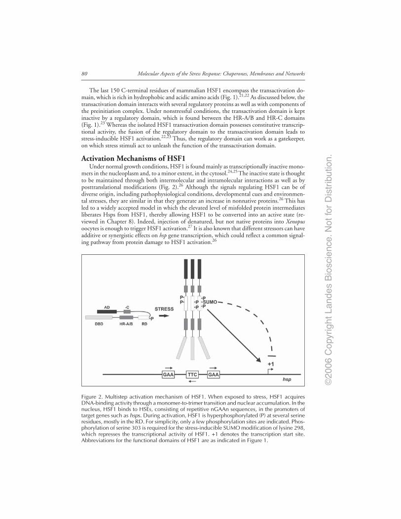
©20
06 C
opyr
ight
Lan
des
Bio
scie
nce.
Not
for
Dis
trib
utio
n.
Molecular Aspects of the Stress Response: Chaperones, Membranes and Networks80
The last 150 C-terminal residues of mammalian HSF1 encompass the transactivation do-main, which is rich in hydrophobic and acidic amino acids (Fig. 1).21,22 As discussed below, thetransactivation domain interacts with several regulatory proteins as well as with components ofthe preinitiation complex. Under nonstressful conditions, the transactivation domain is keptinactive by a regulatory domain, which is found between the HR-A/B and HR-C domains(Fig. 1).23 Whereas the isolated HSF1 transactivation domain possesses constitutive transcrip-tional activity, the fusion of the regulatory domain to the transactivation domain leads tostress-inducible HSF1 activation.22,23 Thus, the regulatory domain can work as a gatekeeper,on which stress stimuli act to unleash the function of the transactivation domain.
Activation Mechanisms of HSF1Under normal growth conditions, HSF1 is found mainly as transcriptionally inactive mono-
mers in the nucleoplasm and, to a minor extent, in the cytosol.24,25 The inactive state is thoughtto be maintained through both intermolecular and intramolecular interactions as well as byposttranslational modifications (Fig. 2).26 Although the signals regulating HSF1 can be ofdiverse origin, including pathophysiological conditions, developmental cues and environmen-tal stresses, they are similar in that they generate an increase in nonnative proteins.26 This hasled to a widely accepted model in which the elevated level of misfolded protein intermediatesliberates Hsps from HSF1, thereby allowing HSF1 to be converted into an active state (re-viewed in Chapter 8). Indeed, injection of denatured, but not native proteins into Xenopusoocytes is enough to trigger HSF1 activation.27 It is also known that different stressors can haveadditive or synergistic effects on hsp gene transcription, which could reflect a common signal-ing pathway from protein damage to HSF1 activation.26
Figure 2. Multistep activation mechanism of HSF1. When exposed to stress, HSF1 acquiresDNA-binding activity through a monomer-to-trimer transition and nuclear accumulation. In thenucleus, HSF1 binds to HSEs, consisting of repetitive nGAAn sequences, in the promoters oftarget genes such as hsps. During activation, HSF1 is hyperphosphorylated (P) at several serineresidues, mostly in the RD. For simplicity, only a few phosphorylation sites are indicated. Phos-phorylation of serine 303 is required for the stress-inducible SUMO modification of lysine 298,which represses the transcriptional activity of HSF1. +1 denotes the transcription start site.Abbreviations for the functional domains of HSF1 are as indicated in Figure 1.

©20
06 C
opyr
ight
Lan
des
Bio
scie
nce.
Not
for
Dis
trib
utio
n.
81Heat Shock Factor 1 as a Coordinator of Stress and Developmental Pathways
The activation mechanism seems to be more multifaceted than depicted above, since HSF1also possesses an intrinsic ability to sense stresses such as elevated temperatures and changedredox states. Recombinant HSF1 from different species can be activated by various stressors,such as heat, H202 and low pH.28-31 Ahn and Thiele noted that two conserved cysteine residuesin the DNA-binding domain of HSF1 were crucial for HSF1 trimer formation in vitro, andsuggested that disulfide bonds between these cysteines would function as a stress sensor forHSF1 trimerization.31 This built-in sensor capacity of HSF1 might also explain the extremerapidity of HSF1 activation in response to severe stress. In fact, binding of HSF to the Droso-phila hsp70 promoter is detected already within seconds after the onset of heat shock, and thenumber of HSF molecules on the promoter reaches saturated levels in about one minute.32
Although little is known about how HSF1 is converted from a latent monomer to a transcrip-tionally active trimer, the rapidity of the process strongly implies the involvement of posttrans-lational modifications in the transcriptional onset of HSF1 target genes. Concurrently with thecloning of HSF from yeast, HSF was found to be extensively phosphorylated, and a recentmass spectrometric analysis revealed that human HSF1 is phosphorylated on at least 12 sites,most of which are located in the regulatory domain.33,34 Therefore, stress-induced phosphory-lation of the regulatory domain likely acts as a switch that relieves the inhibitory effect of theregulatory domain on HSF1 transactivation capacity.
It is interesting to note that although HSF1 is rapidly hyperphosphorylated upon variousstresses and in a manner that correlates with its transactivation capacity, only a few studieshave been able to directly link phosphorylation to HSF1 transcriptional activity.34-38 On thecontrary, most phosphorylation events have been shown to repress the transcriptional activityof HSF1. A particularly interesting example is the phosphorylation of serines 303 and 307,located in the regulatory domain. The phosphorylation of these residues maintains HSF1 ina transcriptionally inactive state under nonstress conditions, but this inhibitory effect can beoverridden by heat shock.35,39 A putative mechanism for this regulatory role was presented byWang and coworkers, who reported that phosphorylation of S303 and S307 mediated aninteraction between HSF1 and the scaffolding protein 14-3-3ε followed by a sequestration ofHSF1 in the cytoplasm.40,41 The phosphorylation of the serines might be a hierarchical event,in which ERK-primed phosphorylation of S307 would be required for GSK3β-mediatedphosphorylation of S303.42,43 Nevertheless, mutation of S303 alone leads to a dramatic dere-pression of HSF1 activity, demonstrating a functional independence between the two phos-phorylation events.35,44 The mechanism behind the repressive function of S303 phosphory-lation was revealed when it was shown to be a prerequisite for yet another posttranslationalmodification, the covalent attachment of a protein called SUMO (small ubiquitin-like modi-fier) to the adjacent lysine 298.45 SUMO is a member of the ubiquitin-like protein superfam-ily of modifiers and an established regulator of transcription.46 For most transcriptional regu-lators, such as Sp3, Elk-1 and HSF2, sumoylation negatively regulates the transactivationcapacity of the substrate.47,48 Accordingly, the phosphorylation-dependent sumoylation in-hibits the transcriptional activity of HSF1 in vivo (Fig. 2).44 SUMO modification usuallyrequires the consensus tetrapeptide (I/V/L)-K-X-E, in which X denotes any amino acid andK the target lysine to which SUMO becomes attached.46 Interestingly, the identification ofphosphorylation-dependent sumoylation of HSF1 was the preamble to the recent discoverythat several transcriptional regulators contain a corresponding phosphorylation-dependentsumoylation motif (IVL)-K-X-E-X-X-S-P, called PDSM, and that this regulatory mechanismis a conserved feature for a number of transcriptional players.44
Regulation of hsp Gene Transcription by HSF1The hsp70 promoter is an established model system for inductive transcriptional responses
and by far the best studied among the HSF/HSF1-responsive promoters. The details of howHSF1 triggers hsp70 transcription are, however, not fully understood. The uninduced hsp70promoter is primed for transcription in that it is occupied by a transcriptionally engaged, but

©20
06 C
opyr
ight
Lan
des
Bio
scie
nce.
Not
for
Dis
trib
utio
n.
Molecular Aspects of the Stress Response: Chaperones, Membranes and Networks82
stalled RNA polymerase II (RNAP II).49,50 Upon heat shock, RNAP II is released from itsdormant state and the transcript is rapidly elongated. In vitro, the stalling of RNAP II is greatlyenhanced by nucleosome formation, suggesting that chromatin remodeling is pertinent for therelease of the paused RNAP II.51 Subsequently, certain hydrophobic residues in the HSF1activation domain were shown to stimulate RNAP II release, and directly interact with BRG1,the ATPase subunit of the chromatin-remodeller SWI/SNF.52-54 The maturation of RNAP IIinto an active elongating complex is accompanied by site-specific hyperphosphorylation of theC-terminal domain of RNAP II. In Drosophila, heat shock induces the recruitment of P-TEFbkinase to heat shock loci. The P-TEFb kinase is composed of a Cdk9 subunit and a cyclin Tsubunit, and known to hyperphosphorylate the C-terminal domain of RNAP II.55 WhenP-TEFb is artificially recruited to the hsp70 promoter, hsp70 is transcribed also under nonstressfulconditions, suggesting that heat-induced recruitment of P-TEFb to heat shock loci could act asa switch in stress-induced transcription of hsp genes.56 Accordingly, the translocation of P-TEFbto heat shock loci is abrogated in the absence of Drosophila HSF, but the recruitment does notseem to be due to a direct interaction between these two proteins.56 Interestingly, inhibition ofP-TEFb kinase activity does not affect the maturation of the stalled RNAP II complex, butinstead leads to defect processing of the 3' end of hsp70 and hsp26 transcripts.57 In addition,mammalian HSF1 interacts with symplekin, a scaffold for polyadenylation factors, and thisinteraction is required for efficient polyadenylation of hsp70 transcripts.58 Together, these in-triguing findings that HSF1 not only activates hsp gene transcription but also participates inmRNA processing broaden the repertoire of mechanisms by which HSF1 regulates the expres-sion of heat shock genes.
Upon heat shock, HSF recruits the Mediator coactivator to heat shock loci.59 Mediatorcomplexes regulate a wide range of genes through relaying activating signals from transcriptionfactors to the basal transcription machinery. In contrast to P-TEFb, the recruitment of Mediatoroccurs through a direct interaction with HSF, since the activation domain of Drosophila HSFbinds to the TRAP80 subunit of the Mediator complex.59 HSF1 activity may further be con-veyed to the preinitiation complex more directly, which is supported by findings that the activa-tion domain of HSF1 binds to the TATA-binding protein TBP as well as to other componentsof the preinitiation complex, including TFIIB in vitro.60,61 These results may indicate that aspatial constraint regulates the interaction between HSF1 and the general transcription machin-ery and possibly explain why most HSEs lie in close proximity to the transcription start site.11
Stress-Specific Activation of HSF1Activation of HSF1 by stresses that are greatly different in nature raises the important ques-
tion whether the HSF1-activating stressors simply signal through a common pathway or whetherdistinct stimuli differentially modulate HSF1-dependent transcriptional processes. Several re-ports have provided evidence for the latter, showing that the mechanism of HSF1 activationand HSF1-dependent transcription can proceed in a stimulus-specific manner. For example,studies in S. cerevisiae have demonstrated that both glucose starvation and heat shock lead toHSF-mediated transcription of the CUP1 gene, but through distinct mechanisms.62 Specifi-cally, during glucose starvation HSF interacts with and is phosphorylated by the Snf1 kinase,which is required for HSF activation. However, HSF activation by heat shock is not Snf1dependent, demonstrating the involvement of stress-specific signal transduction pathways inHSF activation.63 Similarly, HSF activated by oxidative stress shows a phosphorylation patterndistinct from HSF activated by heat.64 Although phosphorylation is involved in a stress-specificactivation mechanism of HSF, it may also specify the subset of HSF target genes that are to betranscribed in response to a particular stress. A mutation that inhibits HSF phosphorylationimpairs transcription of HSF targets which have short or mildly dispersed nGAAn elements intheir promoter regions, but does not significantly affect transcription of genes with multiplecopies of nGAAn pentamers.15

©20
06 C
opyr
ight
Lan
des
Bio
scie
nce.
Not
for
Dis
trib
utio
n.
83Heat Shock Factor 1 as a Coordinator of Stress and Developmental Pathways
In mammals, little is known how HSF1 activation can be adjusted according to differentsignals. By introduction of dominant-negative forms of the SWI/SNF ATPases BRG1 andBRM, de la Serna and coworkers noticed a differential SWI/SNF dependency of HSF1-mediatedtranscription. Whereas the transcriptional activation of hsp70 by arsenite or cadmium wasseverely impaired by the disruption of SWI/SNF activity, hsp70 transcription by heat shockwas unaffected.65 Further studies are necessary, since chromatin immunoprecipitation and re-striction enzyme accessibility of the hsp70 promoter demonstrated that the direct recruitmentof SWI/SNF by HSF1 is essential for chromatin accessibility and efficient hsp70 transcriptionduring heat shock.54 The differential activation of hsp70 by heat and arsenite was further eluci-dated when Thomson and coworkers noticed that hsp70 induction by arsenite, but not by heatshock, was dependent on the p38 MAP kinase pathway, perhaps due to specific histone phos-phorylation mediated by p38 signaling.66 Moreover, during both stresses HSF1 was foundresponsible for targeting histone H4 acetylation to hsp70 chromatin, highlighting the impor-tance of epigenetic modifications in HSF1-mediated stimulus-specific transcription.66
HSF1 as a Developmental RegulatorAlbeit HSF1 plays its most prominent role in protecting cells and organisms from proteotoxic
stresses, several lines of genetic evidence support a versatile role for HSF1 in regulation ofdevelopment. In yeast, HSF is essential for viability also under normal growth conditions, andmore recently, a genome-wide analysis of S. cerevisiae HSF targets showed that HSF partici-pates in diverse biological pathways, such as energy metabolism, synthesis of cytoskeletal com-ponents and vesicular transport.33,67,68 In higher invertebrates and vertebrates, HSF/HSF1 isnot required for survival, but its absence causes severe reproductive disturbances, a reduction inbody size and deficient embryonic development. For example, the deletion of the hsf gene leadsto defect oogenesis and larvae development in Drosophila.69 hsf1 knockout mice undergo nor-mal oogenesis but have malformed chorioallantoic placenta, resulting in high prenatal lethality.Moreover, hsf1-/- females are infertile, which is due to HSF1 being a maternal factor.70 In thetestis, HSF1 plays a dual role in the protection against harmful signals. Whereas HSF1 acts asa survival factor of more immature germ cells in testes exposed to hyperthermia, it is alsorequired for apoptosis of pachytene spermatocytes in response to thermal insults.71 Accord-ingly, the expression of a constitutively active form of HSF1 inhibits the progression of sper-matogenesis and leads to apoptosis of spermatocytes at the pachytene stage.72 It is interesting tonote that the threshold for HSF1 activation is reduced in pachytene spermatocytes relative tosomatic cells, implying that HSF1 could play a role in monitoring the quality control of malegerm cells.73,74
HSF1-Mediated Expression of CytokinesThe requirement for HSF1 in developmental pathways does not seem to be explicitly due
to altered Hsp levels, emphasizing the importance of other HSF1 targets. An interesting excep-tion is seen in the mouse heart, where the disruption of hsf1 causes decreased constitutiveexpression of several Hsps, which is intimately associated with impaired redox homeostasis andmitochondrial damage.75 Transcriptional profiling of hsf1-deficient fibroblasts has revealed thatHSF1 regulates several genes also under nonstressful conditions. Surprisingly, the transcriptionof many of these genes is not enhanced by heat shock, suggesting that the stress-dependentactivation of HSF1 may favor the expression of only a subset of its target genes.76,77 Among thenonstress HSF1 targets, locally acting signal molecules, including cytokines and chemokines,make up a significant part.76 One of these targets, IL-6 was shown to be under direct control ofHSF1 in spleen cells. IL-6 is a multi-functional cytokine that participates in immune responsesand inflammation and is important for B cell differentiation and maturation into plasma cells.78
Accordingly, in response to immunization, the hsf1 knockout mice show an impaired produc-tion of immunoglobulins, especially IgG2a.76

©20
06 C
opyr
ight
Lan
des
Bio
scie
nce.
Not
for
Dis
trib
utio
n.
Molecular Aspects of the Stress Response: Chaperones, Membranes and Networks84
In the olfactory epithelium of pubescent mice, HSF1 acquires DNA-binding activity dur-ing post-natal week 4. One of the targets of this activation is another member of the IL-6family, leukemia inhibitory factor (LIF), a pleiotropic cytokine implicated in numerous devel-opmental pathways.79,80 Interestingly, HSF1 seems to repress the expression of LIF, therebyallowing the differentiation of the olfactory epithelium.80 Negative regulation by HSF1 is alsoinvolved in modulating the expression of the proinflammatory tumor necrosis factor alpha(TNFα). When hsf1/- mice were injected with an LD50 dose of bacterial lipopolysaccharideLPS, they displayed an increased mortality and abnormally high expression of TNFα.6 Thefascinating concept of HSF1 functioning both as a transcriptional activator and as a transcrip-tional repressor in a context-dependent manner markedly broadens the biological role of HSF1.The repressive function of HSF1 was originally introduced by Cahill and coworkers, whoreported that the LPS-induced interleukin 1β gene expression could be inhibited by HSF1activation.81 This phenomenon was subsequently shown to be mediated through the directinteraction between HSF1 and C/EBPβ, a regulator of IL-1β transcription.82 In most cases,however, the mechanism of HSF1-mediated repression is poorly understood. Moreover, pos-sible indirect effects of HSF1 activity may be emphasized through the complexity of cytokinesignaling networks, accentuating the need for further studies to elucidate the intriguing func-tion of HSF1 in cytokine expression.
Heat Shock Factors Working TogetherSince the DNA-binding and oligomerization domains are particularily well conserved within
the HSF family, it is not surprising that different HSFs display similar fundamental properties.This is especially evident in vitro, where all HSFs are able to form trimers and bind to the sameHSEs, strongly suggesting that different members of the HSF family could share a subset oftarget genes.83 Nevertheless, as HSF1 cannot be substituted by other HSF members, and thehsf1, hsf2 and hsf4 knockout mice have clearly distinct phenotypes, the HSFs might be re-stricted to different fields of action in vivo.6-8,10,84 Recently, however, HSF1 was found toconverge with HSF4 in the regulation of eye development. In certain Chinese and Danishfamilies, mutations in the HSF4 DNA-binding domain were found to be the cause of autoso-mal dominant lamellar and Marnier cataract, and shortly thereafter, hsf4 null mice were shownto develop cataract during the early postnatal period.9,10,84 The hsf4-/- lens contains inclusion-likestructures of protein aggregates, perhaps due to an increased expression of fibroblast growthfactors (FGFs) and a decrease in protective γ-crystallins and Hsps, particularly Hsp27, in lenscells.10,84 Surprisingly, HSF1 and HSF4 have opposing effects on FGF expression in the lens.Both HSF1 and HSF4 bind to the FGF7 promoter, but whereas the expression of FGF7 isincreased in hsf4-/-, it is decreased in hsf1-/- and returned to normal levels in the hsf1/hsf4 doubleknockout mice.10 Similarly, the increased LIF expression in the absence of hsf1 is partiallyrelieved by the simultaneous disruption of hsf4, demonstrating an intimate relationship be-tween these two factors in the maintenance of sensory systems.80
The generation of double knockout mice has provided valuable information for a func-tional relationship also between HSF1 and HSF2. While spermatogenesis is normal in hsf1-/-
mice and only modestly impaired in hsf2-/- mice, the simultaneous disruption of both hsf1 andhsf2 leads to male infertility.7,8,85 A gene expression analysis of the hsf1/hsf2 double knockouttestis revealed that many genes involved in spermatogenesis were downregulated, suggestingthat HSF1 and HSF2 could have complementary or interdependent functions in the regula-tion of male fertility.85 The notion that HSF1 and HSF2 could share a subset of target geneswas initially proposed when HSF2 was found to bind to the hsp70 promoter upon hemin-inducederythroid differentiation of K562 erythroleukemia cells.86 More recently, an extensive pro-moter analysis in K562 cells by Trinklein and coworkers demonstrated that both HSF1 andHSF2 are recruited to several hsp promoters in response to hemin treatment, implying thatHSF2 could be involved in regulating the expression of a broad repertoire of hsp genes.87 Anelegant mechanism by which HSF2 could influence the transcription of heat shock genes was
©20
06 C
opyr
ight
Lan
des
Bio
scie
nce.
Not
for
Dis
trib
utio
n.
85Heat Shock Factor 1 as a Coordinator of Stress and Developmental Pathways
recently presented by Xing and coworkers, who reported that HSF2 maintains chromatin ac-cessibility of the inducible hsp70 gene during the M phase in mitosis, a phenomenon calledbookmarking, and that this function is required for HSF1-mediated transcription of hsp70 inthe G1 phase.88 The putative modulatory role of HSF2 in HSF1-mediated transcription couldalso result from heterocomplex formation, since the direct interaction between HSF1 and HSF2as well as the colocalization of HSF1 and HSF2 in nuclear stress bodies is dependent on intactoligomerization domains.89,90 This hypothesis is further supported by the recent observationthat upon proteasome inhibition, both HSF1 and HSF2 bind to the clusterin promoter in thecontext of chromatin and to oligonucleotides containing binding sites for only one HSF tri-mer.91 Although the fundamental question of how HSF2 modulates HSF1-mediated tran-scription remains to be answered, the prospect of cooperative heterogenous HSF complexescertainly adds a novel flavor to the transcriptional regulation of heat shock genes as well asother targets.
Future PerspectivesAlthough the heat shock response was initially described already four decades ago, the mo-
lecular details of this strictly regulated process are only beginning to emerge. For example, theessential questions how cells are able to sense stress signals and how these signals are subse-quently conveyed to HSF1 for transcription of hsp genes need to be explored more thoroughly.While genome-wide analyses may provide further understanding of target gene selectivity inresponse to specific stress stimuli, it is becoming evident that distinct stressors employ differentsignaling pathways for activation of the same HSF1 target genes. The elucidation of howstress-specific networks are organized poses an intriguing challenge for future studies. More-over, the involvement of HSF1 in several developmental pathways brings unforeseen complex-ity to the mechanisms of HSF1 action. The recent discoveries of HSF1 functioning in tightcooperation with other HSFs during development propose that such cross talk might occuralso upon exposure to stress stimuli. To this end, analyses of the posttranscriptional regulation,to which HSF2 and HSF4 are subjected under stressful conditions, likely provide novel in-sights into how these factors contribute to the heat shock response.
AcknowledgementsWe apologize to our many colleagues whose work could only be cited indirectly due to
space limitations. We are grateful to Aura Kaunisto, Annika Meinander, Pia Roos-Mattjus andAnton Sandqvist for their valuable comments on the manuscript. The work done in our labo-ratory is financially supported by the Academy of Finland, the Finnish Cancer Organizations,the Sigrid Jusélius Foundation, the Finnish Life and Pension Insurance Companies, and ÅboAkademi University. J. Anckar is supported by the Turku Graduate School for BiomedicalSciences (TuBS).
References1. Brenner S, Jacobson F, Meselson M. An unstable intermediate carrying information from genes to
ribosomes for protein synthesis. Nature 1961; 190:576-577.2. Ritossa F. A new puffing pattern induced by temperature shock and DNP in Drosophila.
Experimentia 1962; 18:571-573.3. Lindquist S. The heat-shock response. Annu Rev Biochem 1986; 55:1151-1191.4. Nakai A. New aspects in the vertebrate heat shock factor system: Hsf3 and Hsf4. Cell Stress Chap
1999; 2:86-93.5. McMillan DR, Xiao X, Shao L et al. Targeted disruption of heat shock transcription factor 1
abolishes thermotolerance and protection against heat-inducible apoptosis. J Biol Chem 1998;273:7523-7528.
6. Xiao X, Zuo X, Davis AA et al. HSF1 is required for extra-embryonic development, postnatalgrowth and protection during inflammatory responses in mice. EMBO J 1999; 18:5943-5952.
7. Kallio M, Chang Y, Manuel M et al. Brain abnormalities, defective meiotic chromosome synapsisand female subfertility in HSF2 null mice. EMBO J 2002; 21:2591-2601.
©20
06 C
opyr
ight
Lan
des
Bio
scie
nce.
Not
for
Dis
trib
utio
n.
Molecular Aspects of the Stress Response: Chaperones, Membranes and Networks86
8. Wang G, Zhang J, Moskophidis D et al. Targeted disruption of the heat shock transcription factor(hsf)-2 gene results in increased embryonic lethality, neuronal defects, and reduced spermatogen-esis. Genesis 2003; 36:48-61.
9. Bu L, Jin Y, Shi Y et al. Mutant DNA-binding domain of HSF4 is associated with autosomaldominant lamellar and Marner cataract. Nat Genet 2002; 31:276-278.
10. Fujimoto M, Izu H, Seki K et al. HSF4 is required for normal cell growth and differentiationduring mouse lens development. EMBO J 2004; 23:4297-4306.
11. Wu C. Heat shock transcription factors: Structure and regulation. Annu Rev Cell Dev Biol 1995;11:441-469.
12. Xiao H, Perisic O, Lis JT. Cooperative binding of Drosophila heat shock factor to arrays of aconserved 5 bp unit. Cell 1991; 64:585-593.
13. Kroeger PE, Morimoto RI. Selection of new HSF1 and HSF2 DNA-binding sites reveals differencein trimer cooperativity. Mol Cell Biol 1994; 14:7592-7603.
14. Santoro N, Johansson N, Thiele DJ. Heat shock element architecture is an important determinantin the temperature and transactivation domain requirements for heat shock transcription factor.Mol Cell Biol 1998; 18:6340-6352.
15. Hashikawa N, Sakurai H. Phosphorylation of the yeast heat shock transcription factor is impli-cated in gene-specific activation dependent on the architecture of the heat shock element. Mol CellBiol 2004; 24:3648-3659.
16. Sorger PK, Nelson HCM. Trimerization of a yeast transcriptional activator via a coiled-coil motif.Cell 1989; 59:807-813.
17. Peteranderl R, Nelson HCM. Trimerization of the heat shock transcription factor by a triple-strandedalpha-helical coiled-coil. Biochemistry 1992; 31:12272-12276.
18. Rabindran SK, Haroun RI, Clos J et al. Regulation of heat shock factor trimer formation: Role ofa conserved leucine zipper. Science 1993; 259:230-234.
19. Chen Y, Barlev NA, Westergaard O et al. Identification of the C-terminal activator domain inyeast heat shock factor: Independent control of transient and sustained transcriptional activity.EMBO J 1993; 12:5007-5018.
20. Nakai A, Tanabe M, Kawazoe Y et al. HSF4, a new member of the human heat shock factorfamily which lacks properties of a transcriptional activator. Mol Cell Biol 1997; 17:469-481.
21. Shi Y, Kroeger PE, Morimoto RI. The carboxyl-terminal transactivation domain of heat shockfactor 1 is negatively regulated and stress responsive. Mol Cell Biol 1995; 15:4309-4318.
22. Newton EM, Knauf U, Green M et al. The regulatory domain of human heat shock factor 1 issufficient to sense heat stress. Mol Cell Biol 1996; 16:839-846.
23. Green M, Schuetz TJ, Sullivan EK et al. A heat shock-responsive domain of human HSF1 thatregulates transcription activation domain function. Mol Cell Biol 1995; 15:3354-3362.
24. Mercier PA, Winegarden NA, Westwood JT. Human heat shock factor 1 is predominantly a nuclearprotein before and after heat stress. J Cell Sci 1999; 112:2765-2774.
25. Vujanac M, Fenaroli A, Zimarino V. Constitutive nuclear import and stress-regulated nucleocyto-plasmic shuttling of mammalian heat-shock factor 1. Traffic 2005; 6:214-229.
26. Morimoto RI. Regulation of the heat shock transcriptional response: Cross talk between a familyof heat shock factors, molecular chaperones, and negative regulators. Genes Dev 1998; 12:3788-3796.
27. Ananthan J, Goldberg AL, Voellmy R. Abnormal proteins serve as eukaryotic stress signals andtrigger the activation of heat shock genes. Science 1986; 232:522-524.
28. Goodson ML, Sarge KD. Heat-inducible DNA binding of purified heat shock transcription factor1. J Biol Chem 1995; 270:2447-2450.
29. Farkas T, Kutskova YA, Zimarino V. Intramolecular repression of mouse heat shock factor 1. MolCell Biol 1998; 18:906-918.
30. Zhong M, Orosz A, Wu C. Direct sensing of heat and oxidation by Drosophila heat shock tran-scription factor. Mol Cell 1998; 2:101-108.
31. Ahn SG, Thiele DJ. Redox regulation of mammalian heat shock factor 1 is essential for Hsp geneactivation and protection from stress. Genes Dev 2003; 17:516-528.
32. Boehm AK, Saunders A, Werner J et al. Transcription factor and polymerase recruitment, modifi-cation, and movement on dhsp70 in vivo in the minutes following heat shock. Mol Cell Biol2003; 23:7628-7637.
33. Sorger PK, Pelham HRB. Yeast heat shock factor is an essential DNA-binding protein that exhibitstemperature-dependent phosphorylation. Cell 1988; 54:855-864.
34. Guettouche T, Boellmann F, Lane WS et al. Analysis of phosphorylation of human heat shockfactor 1 in cells experiencing a stress. BMC Biochem 2005; 6:4.
35. Kline MP, Morimoto RI. Repression of the heat shock factor 1 transcriptional activation domain ismodulated by constitutive phosphorylation. Mol Cell Biol 1997; 17:2107-2115.

©20
06 C
opyr
ight
Lan
des
Bio
scie
nce.
Not
for
Dis
trib
utio
n.
87Heat Shock Factor 1 as a Coordinator of Stress and Developmental Pathways
36. Holmberg CI, Hietakangas V, Mikhailov A et al. Phosphorylation of serine 230 promotes induc-ible transcriptional activity of heat shock factor 1. EMBO J 2001; 20:3800-3810.
37. Holmberg CI, Tran SEF, Eriksson JE et al. Multisite phosphorylation provides sophisticated regu-lation of transcription factors. Trends Biochem Sci 2002; 27:619-627.
38. Boellmann F, Guettouche T, Guo Y et al. DAXX interacts with heat shock factor 1 during stressactivation and enhances its transcriptional activity. Proc Natl Acad Sci USA 2004; 101:4100-4105.
39. Knauf U, Newton EM, Kyriakis J et al. Repression of human heat shock factor 1 activity at con-trol temperature by phosphorylation. Genes Dev 1996; 10:2782-2793.
40. Wang X, Grammatikakis N, Siganou A et al. Regulation of molecular chaperone gene transcriptioninvolves the serine phosphorylation, 14-3-3 epsilon binding, and cytoplasmic sequestration of heatshock factor 1. Mol Cell Biol 2003; 23:6013-6026.
41. Wang X, Grammatikakis N, Siganou A et al. Interactions between extracellular signal-regulatedprotein kinase 1, 14-3-3epsilon, and heat shock factor 1 during stress. J Biol Chem 2004;279:49460-49469.
42. Chu B, Soncin F, Price BD et al. Sequential phosphorylation by mitogen-activated protein kinaseand glycogen synthase kinase 3 represses transcriptional activation by heat shock factor-1. J BiolChem 1996; 271:30847-20857.
43. Chu B, Zhong R, Soncin F et al. Transcriptional activity of heat shock factor 1 at 37 degrees C isrepressed through phosphorylation on two distinct serine residues by glycogen synthase kinase 3and protein kinases Calpha and Czeta. J Biol Chem 1998; 273:18640-18646.
44. Hietakangas V, Anckar J, Blomster HA et al. PDSM, a motif for phosphorylation-dependent SUMOmodification. Proc Natl Acad Sci USA 2006; 103:45-50.
45. Hietakangas V, Ahlskog JK, Jakobsson AM et al. Phosphorylation of serine 303 is a prerequisitefor the stress-inducible SUMO modification of heat shock factor 1. Mol Cell Biol 2003; 23:2953-68.
46. Johnson ES. Protein modification by SUMO. Annu Rev Biochem 2004; 73:355-382.47. Gill G. Something about SUMO inhibits transcription. Curr Opin Genet Dev 2005; 15:536-541.48. Anckar J, Hietakangas V, Denessiouk K et al. Inhibition of DNA binding by differential sumoylation
of heat shock factors. Mol Cell Biol 2006; 26:955-964.49. Rougvie AE, Lis JT. The RNA polymerase II molecule at the 5' end of the uninduced hsp70 gene
of D. melanogaster is transcriptionally engaged. Cell 1988; 54:795-804.50. Lis JT. Promoter-associated pausing in promoter architecture and postinitiation transcriptional regu-
lation. Cold Spring Harb Symp Quant Biol 1998; 63:347-56.51. Brown SA, Imbalzano AN, Kingston RE. Activator-dependent regulation of transcriptional pausing
on nucleosomal templates. Genes Dev 1996; 10:1479-1490.52. Brown SA, Weirich CS, Newton EM et al. Transcriptional activation domains stimulate initiation
and elongation at different times and via different residues. EMBO J 1998; 17:3146-3154.53. Sullivan EK, Weirich CS, Guyon JR et al. Transcriptional activation domains of human heat shock
factor 1 recruit human SWI/SNF. Mol Cell Biol 2001; 21:5826-5837.54. Corey LL, Weirich CS, Benjamin IJ et al. Localized recruitment of a chromatin-remodeling activ-
ity by an activator in vivo drives transcriptional elongation. Genes Dev 2003; 17:1392-1401.55. Marshall, NF, Peng J, Xie Z et al. Control of RNA polymerase II elongation potential by a novel
carboxyl-terminal domain kinase. J Biol Chem 1996; 271:27176-27183.56. Lis JT, Mason P, Peng J et al. P-TEFb kinase recruitment and function at heat shock loci. Genes
Dev 2000; 14:792-803.57. Ni Z, Schwartz BE, Werner J et al. Coordination of transcription, RNA processing, and surveil-
lance by P-TEFb kinase on heat shock genes. Mol Cell 2004; 13:55-65.58. Xing H, Mayhew CN, Cullen KE et al. HSF1 modulation of Hsp70 mRNA polyadenylation via
interaction with symplekin. J Biol Chem 2004; 279:10551-10555.59. Park JM, Werner J, Kim JM et al. Mediator, not holoenzyme, is directly recruited to the heat
shock promoter by HSF upon heat shock. Mol Cell 2001; 8:9-19.60. Mason Jr PB, Lis JT. Cooperative and competitive protein interactions at the hsp70 promoter. J
Biol Chem 1997; 272:33227-33233.61. Yuan CX, Gurley WB. Potential targets for HSF1 within the preinitiation complex. Cell Stress
Chap 2000; 5:229-242.62. Tamai KT, Liu X, Silar P et al. Heat shock transcription factor activates yeast metallothionein
gene expression in response to heat and glucose starvation via distinct signalling pathways. MolCell Biol 1994; 14:8155-8165.
63. Hahn JS, Thiele DJ. Activation of the Saccharomyces cerevisiae heat shock transcription factorunder glucose starvation conditions by Snf1 protein kinase. J Biol Chem 2004; 279:5169-5176.
64. Liu XD, Thiele DJ. Oxidative stress induced heat shock factor phosphorylation and HSF-dependentactivation of yeast metallothionein gene transcription. Genes Dev 1996; 10:592-603.

©20
06 C
opyr
ight
Lan
des
Bio
scie
nce.
Not
for
Dis
trib
utio
n.
Molecular Aspects of the Stress Response: Chaperones, Membranes and Networks88
65. de la Serna IL, Carlson KA, Hill DA et al. Mammalian SWI-SNF complexes contribute to activa-tion of the hsp70 gene. Mol Cell Biol 2000; 20:2839-2851.
66. Thomson S, Hollis A, Hazzalin CA et al. Distinct stimulus-specific histone modifications at hsp70chromatin targeted by the transcription factor heat shock factor-1. Mol Cell 2004; 15:585-594.
67. Gallo GJ, Prentice H, Kingston RE. Heat shock factor is required for growth at normal tempera-tures in the fission yeast Schizosaccharomyces pombe. Mol Cell Biol 1993; 13:749-761.
68. Hahn JS, Hu Z, Thiele DJ et al. Genome-wide analysis of the biology of stress responses throughheat shock transcription factor. Mol Cell Biol 2004; 24:5249-5256.
69. Jedlicka P, Mortin MA, Wu C. Multiple functions of Drosophila heat shock transcription factor invivo. EMBO J 1997; 16:2452-2462.
70. Christians E, Davis AA, Thomas SD et al. Maternal effect of Hsf1 on reproductive success. Nature2000; 407:693-694.
71. Izu H, Inouye S, Fujimoto M et al. Heat shock transcription factor 1 is involved in quality-controlmechanisms in male germ cells. Biol Reprod 2004; 70:18-24.
72. Nakai A, Suzuki M, Tanabe M. Arrest of spermatogenesis in mice expressing an active heat shocktranscription factor 1. EMBO J 2000; 19:1545-1554.
73. Sarge KD. Male germ cell-specific alteration in temperature set point of the cellular stress response.J Biol Chem 1995; 270:18745-18748.
74. Sarge KD, Bray AE, Goodson ML. Altered stress response in testis. Nature 1995; 374:126.75. Yan LJ, Christians ES, Liu L et al. Mouse heat shock transcription factor 1 deficiency alters car-
diac redox homeostasis and increases mitochondrial oxidative damage. EMBO J 2002; 21:5164-5172.76. Inouye S, Izu H, Takaki E et al. Impaired IgG production in mice deficient for heat shock tran-
scription factor 1. J Biol Chem 2004; 279:38701-38709.77. Trinklein ND, Murray JI, Hartman SJ et al. The role of heat shock transcription factor 1 in the
genome-wide regulation of the mammalian heat shock response. Mol Biol Cell 2004; 15:1254-1261.78. Heinrich PC, Behrmann I, Haan S et al. Principles of interleukin (IL)-6-type cytokine signalling
and its regulation. Biochem J 2003; 374:1-20.79. Metcalf D. The unsolved enigmas of leukemia inhibitory factor. Stem Cells 2003; 21:5-14.80. Takaki E, Fujimoto M, Sugahara K et al. Maintenance of olfactory neurogenesis requires HSF1, a
major heat shock transcription factor in mice. J Biol Chem 2006; 281:4931-4937.81. Cahill CM, Waterman WR, Xie Y et al. Transcriptional repression of the prointerleukin 1beta
gene by heat shock factor 1. J Biol Chem 1996; 271:24874-24879.82. Xie Y, Chen C, Stevenson MA et al. Heat shock factor 1 represses transcription of the IL-1beta
gene through physical interaction with the nuclear factor of interleukin 6. J Biol Chem 2002;277:11802-11810.
83. Pirkkala L, Nykänen P, Sistonen L. Roles of the heat shock transcription factors in regulation ofthe heat shock response and beyond. FASEB J 2001; 15:1118-1131.
84. Min JN, Zhang Y, Moskophidis D et al. Unique contribution of heat shock transcription factor 4in ocular lens development and fiber cell differentiation. Genesis 2004; 40:205-217.
85. Wang G, Ying Z, Jin X et al. Essential requirement for both hsf1 and hsf2 transcriptional activityin spermatogenesis and male fertility. Genesis 2004; 38:66-80.
86. Sistonen L, Sarge KD, Phillips B et al. Activation of heat shock factor 2 during hemin-induceddifferentiation of human erythroleukemia cells. Mol Cell Biol 1992; 12:4104-4111.
87. Trinklein ND, Chen WC, Kingston RE et al. Transcriptional regulation and binding of heat shockfactor 1 and heat shock factor 2 to 32 human heat shock genes during thermal stress and differen-tiation. Cell Stress Chap 2004; 9:21-28.
88. Xing H, Wilkerson DC, Mayhew CN et al. Mechanism of hsp70i gene bookmarking. Science2005; 307:421-423.
89. Alastalo TP, Hellesuo M, Sandqvist A et al. Formation of nuclear stress granules involves HSF2and coincides with the nucleolar localization of Hsp70. J Cell Sci 2003; 116:3557-3570.
90. He H, Soncin F, Grammatikakis N et al. Elevated expression of heat shock factor (HSF) 2Astimulates HSF1-induced transcription during stress. J Biol Chem 2003; 278:35465-35475.
91. Loison F, Debure L, Nizard P et al. Up-regulation of the clusterin gene after proteotoxic stress:implication of HSF1-HSF2 heterocomplexes. Biochem J 2006; 395:223-231.
©20
06 C
opyr
ight
Lan
des
Bio
scie
nce.
Not
for
Dis
trib
utio
n.
CHAPTER 9
*Corresponding Author: Richard Voellmy—HSF Pharmaceuticals SA, Avenue des Cerisiers39B, 1009 Pully, Switzerland. Email: [email protected]
Molecular Aspects of the Stress Response: Chaperones, Membranes and Networks,edited by Peter Csermely and Laszlo Vigh. ©2007 Landes Bioscienceand Springer Science+Business Media.
Chaperone Regulation of the Heat ShockProtein ResponseRichard Voellmy* and Frank Boellmann
Abstract
The heat shock protein response appears to be triggered primarily by nonnative proteinsaccumulating in a stressed cell and results in increased expression of heat shock proteins(HSPs). Many heat shock proteins prevent protein aggregation and participate in re-
folding or elimination of misfolded proteins in their capacity as chaperones. Even thoughseveral mechanisms exist to regulate the abundance of cytosolic and nuclear chaperones, activa-tion of heat shock transcription factor 1 (HSF1) is an essential aspect of the heat shock proteinresponse. HSPs and co-chaperones that are assembled into multichaperone complexes regulateHSF1 activity at different levels. HSP90-containing multichaperone complexes appear to bethe most relevant repressors of HSF1 activity. Because HSP90-containing multichaperone com-plexes interact not only specifically with client proteins including HSF1 but also genericallywith nonnative proteins, the concentration of nonnative proteins influences assembly on HSF1of HSP90-containing complexes that repress activation, and may play a role in inactivation, ofthe transcription factor. Proteins that are unable to achieve stable tertiary structures and remainchaperone substrates are targeted for proteasomal degradation through polyubiquitination byco-chaperone CHIP. CHIP can activate HSF1 to regulate the protein quality control systemthat balances protection and degradation of chaperone substrates.
IntroductionThe term “heat shock protein response” relates to the induction of heat shock or stress
protein (HSP) synthesis that occurs in eukaryotic cells subsequent to heat treatment or expo-sure to other proteotoxic stress. The transcriptional response to heat and other proteotoxicstress is mediated by so-called heat shock transcription factors (HSF).1,2 Vertebrate animalsand plants express several different but related HSFs. Among the different vertebrate HSFs, thefactor termed HSF1 is essentially required for stress regulation of HSP expression.3-6 However,it is noted that other HSF family members also may contribute and, in some cases, play criticalroles in regulating the response.7-9 Disruption of HSF in invertebrates abolishes the heat shockresponse in a similar way as disruption of HSF1 in vertebrates.10-15
In this chapter we are primarily concerned with the regulation of vertebrate, in particularhuman, HSF1 and the single HSF species of invertebrate organisms such as Saccharomycescerevisiae and Drosophila melanogaster that perform the function of vertebrate HSF1 in theseorganisms (also referred to herein as HSF1 for simplicity). Sequence and functional features of

©20
06 C
opyr
ight
Lan
des
Bio
scie
nce.
Not
for
Dis
trib
utio
n.
Molecular Aspects of the Stress Response: Chaperones, Membranes and Networks90
a typical HSF1 are outlined in Figure 1, using human HSF1 as the example. A DNA-bindingdomain comprising a winged, helix-turn-helix motif is located near the amino terminus.16-19
The domain is capable of interacting specifically with so-called heat shock element (HSE)sequences present in the promoters of heat shock protein (hsp) genes.20,21 A long, interrupted,hydrophobic repeat sequence (HR-A/B) is situated adjacent to the DNA-binding domain. Thepresence of at least a portion of this sequence is required for oligomerization of the factor.16,22,23
HSF1 includes an additional hydrophobic repeat sequence (HR-C) in the carboxy-terminalthird of its sequence. Studies on mammalian and Drosophila HSF1 identified transactivationdomains near the carboxy ends of these factors.24-27 Regulation of HSF1 activity is largelycontrolled posttranslationally and not at the level of synthesis/degradation of the transcriptionfactor.26,28-31
Feedback Regulation of the Heat Shock Protein Responseby Stress-Inducible Chaperones
In their early studies of cultured Drosophila cells, Lindquist and colleagues observed that,under conditions of moderate heat stress, the heat shock protein response was self-regulated,meaning that HSP expression increased rapidly after initiation of a heat treatment, continuedfor some time and then decreased to a low rate approximately corresponding to the prestressrate (a phenomenon later called attenuation).32,33 Reduction of rates of heat-induced tran-scription or translation resulted in an extension of the period of elevated hsp gene transcriptionduring stress recovery, suggesting that one or more newly synthesized HSPs feedback-regulatedthe heat shock protein response. Since activation of hsp gene transcription did not require newprotein synthesis, it was also recognized that the heat shock protein response is regulated bothtranscriptionally and post-transcriptionally. Attenuation of the stress protein response was sub-sequently also observed and studied in mammalian cells.34 The signal that triggers the heatshock protein response appears to be accumulation of nonnative proteins resulting from chemicalor physical denaturation.35-41 Many HSPs can bind such nonnative proteins, assist in theirrefolding or target them for degradation, preventing cytotoxic protein aggregation. Proteinsthat display these capabilities are known as molecular chaperones.
Hsps and Co-Chaperones Repress Activation of HSF1Homotrimerization of HSF1 was found to be essential for DNA binding and, therefore,
absolutely required for its function as a transcriptional activator.3,4,23,42-44 Heat or hydrogenperoxide exposure can induce this trimerization in vitro, arguing for an inherent ability ofHSF1 to sense these stresses.45-47 Several observations, however, suggested the existence of acellular mechanism(s) that keeps HSF1 in a nonhomooligomeric state in the absence of astress. When human HSF1 was introduced into Drosophila or Xenopus laevis cells, the factor
Figure 1. Linear map of human HSF1. Numbers refer to positions in the sequence of the HSF1protein. Functions of individual domains/regions are indicated above and below the map.

©20
06 C
opyr
ight
Lan
des
Bio
scie
nce.
Not
for
Dis
trib
utio
n.
91Chaperone Regulation of the Heat Shock Protein Response
was induced to trimerize at the low heat shock temperatures characteristic of the latter cells,demonstrating that it is not an absolute temperature that results in activation of the transcrip-tion factor.26,48 Another observation in favor of a cellular mechanism negatively regulating thetranscription factor was that overexpression of HSF1 from transduced genes resulted in accu-mulation of trimeric HSF1 even in the absence of a stress. This observation suggested theexistence of a cellular repressor of trimerization that was titrated in these experiments.26 Whenit was found that the transcriptional activity of homotrimeric HSF1 derived from overexpressionwas negligible compared to that of stress-activated factor, it became obvious that additionallayers of regulation exist. It is noted that regulation at multiple levels had been postulatedearlier based on observations that compounds such as salicylates, menadione and hydrogenperoxide induced HSE DNA-binding activity but not expression of inducible hsp genes.49,50
Even prior to the latter observations, heat exposure of an erythroleukemia cell line was reportedto result in HSE DNA-binding activity but not in increased HSP expression.51
The search for factors that repress HSF1 homooligomerization initially focused on HSP70after heterocomplexes with monomeric HSF1 could be shown to exist in unstressed cells.52,53
However, several eukaryotic species contain very low levels of endogenous HSP70 and areperfectly capable of suppressing HSF1 activity in the absence of a stress.54-56 To develop ageneral model of HSF1 regulation, the possible involvement of other chaperones was exam-ined as well. Several observations that will be reviewed in more detail in the next sectionpointed to HSP90 and HSP90-containing multichaperone complexes as the key regulators ofHSF1 activity.
HSP90-Containing Multichaperone Complexes Regulate HSF1Oligomeric Status and Transcriptional Competence
Repression of Oligomerization of HSF1 in the Absence of a StressExposure of cells to benzoquinone ansamycins herbimycin A and geldanamycin was found
to induce homotrimerization of HSF1 and result in an increased rate of expression of HSPs.41,57-60
Geldanamycin is known to bind in the ATP-binding pocket of HSP90 with apparent specific-ity and to alter HSP90 function.61-63 Together, these findings suggested that HSP90, alone orin concert with other factors, repressed HSF1 oligomerization in the absence of a stress. Theobservations of Nadeau et al and Nair et al of in vitro interactions between HSF1 and HSP90were consistent with this hypothesis.64,65 To obtain more definitive information about theinvolvement of HSP90 in HSF1 regulation, an in vitro system derived from HeLa cells wasdeveloped by Zou et al that was capable of reproducing important aspects of the in vivo regu-lation of HSF1 oligomeric status: HSF1 oligomerization and HSE DNA-binding activity wereinduced upon exposure to elevated temperature, or addition of geldanamycin or chemicallydenatured proteins.60 Immunodepletion of HSP90 induced specific HSE DNA-binding activ-ity in this system. Back addition of purified HSP90 immediately following HSP90immunodepletion prevented an increase in HSE DNA-binding activity. Ali et al carried outcorresponding experiments in vivo, using Xenopus oocytes that were microinjected with anti-bodies and/or HSP90.66 Results obtained essentially mirrored those of the Zou et al study.Genetic evidence for a functional role of HSP90 in HSF1 regulation in yeast was reported byDuina et al.67 In a complementary type of study, Zhao et al demonstrated that overexpressionof HSP90 desensitized heat induction of HSE DNA-binding activity in mammalian cells.68
Other experiments examined the abundance of HSF1-HSP90 complexes in HeLa cells prior toand during a heat stress using in situ cross-linking techniques.69 Results revealed that the levelof HSF1-HSP90 decreased dramatically in the course of a 15-min exposure of the cells tomoderately severe heat. Another study demonstrated that administration to cardiac cells ofgeldanamycin also reduced the concentration of HSF1-HSP90 complexes.70 Taken together,these results provide strong evidence that HSF1 oligomerization is inhibited by HSP90 thatbinds to HSF1 polypeptide in the absence of a stress.

©20
06 C
opyr
ight
Lan
des
Bio
scie
nce.
Not
for
Dis
trib
utio
n.
Molecular Aspects of the Stress Response: Chaperones, Membranes and Networks92
Typically, Hsp90 does not bind its client proteins alone but as part of an HSP90-containingmultichaperone complex. In the case of steroid receptors, where this has been studied exten-sively, “mature” multichaperone complexes comprise HSP90, P23 and an immunophilin.71
Assembly of this type of complexes appears to be the end result of several intermediate assem-bly steps, involving initial binding by HSP70 and HSP40, introduction of HSP90 throughadapter protein HOP and replacement of the resulting HSP90-containing multichaperonecomplex by above-mentioned mature HSP90-containing complex. Results obtained byBharadwaj et al and Duina et al suggested that a similar mature HSP90-containingmultichaperone complex assembled on HSF1 polypeptide.67,72 The study by Baradwaj et alshowed that immunodepletion not only of HSP90 but also of P23 enhanced HSE DNA-bindingactivity in Xenopus oocytes.72 Duina et al provided evidence for an involvement of animmunophilin, a CYP40-like cyclophilin, in repression of HSF1 activity in yeast.67 Finally,Marchler and Wu obtained data supporting the notion that HSF1 polypeptide undergoes asimilar series of chaperone complex assembly reactions as steroid receptors.73 The latter authorsreported that siRNA depletion of HSP70, DROJ1 (HSP40) or HSP90 increased the HSEDNA-binding activity of Drosophila HSF1. Codepletion of HSP70 and HSP40 or HSP90 andHSP40 produced synergistic effects. In conclusion, the information available suggests that amature HSP90-containing multichaperone complex similar to that associating with steroidreceptors assembles on HSF1 polypeptide and prevents oligomerization of the HSF1 in theabsence of a stress.
Repression of Transcriptional Competence of HSF1 in the Absenceof a Stress
Transcriptional competence of HSF1, i.e., the ability of the factor to transactivate hsp genes,is regulated independent from oligomeric status. A so-called regulatory domain was defined inthe HSF1 polypeptide that is required for repression of transcriptional competence in theabsence of a stress.24,26 Deletion of this sequence or parts thereof results in a factor that effec-tively transactivates hsp genes when overexpressed in the absence of a stress. As discussed be-fore, overexpression of wildtype HSF1 titrates the mechanism that represses oligomerizationbut not that controlling transcriptional competence. Nair et al and Guo et al found thatHsp90-containing multichaperone complexes (HSP90-P23-FKBP52 and others) assembledon trimeric HSF1 in vitro and that components of these complexes also associated with trim-eric HSF1 in vivo.65,69 Interestingly, FKBP52 appeared to only interact with trimeric but not(or differently) with nontrimeric HSF1 in human HeLa cells. Therefore, FKBP52 could beused as a marker for HSP90-containing complexes associating with trimeric HSF1. Using thistool and working with HeLa cells transfected to overexpress an HSF1 form (a LexA-HSF1chimera), Guo et al were able to examine in parallel HSF1 transcriptional competence andrelative concentration of HSP90-containing complexes associated with trimeric HSF1.69 Theauthors reported that, in different experimental situations that caused concentration of chaper-one substrates to rise, concentration of HSP90-containing complexes found in associationwith trimeric HSF1 declined and transcriptional competence of HSF1 increased. Further-more, they observed that mutations in the HSF1 regulatory domain that increased transcrip-tional competence impaired the assembly of HSP90-containing complexes. These findingsstrongly suggest the existence of a mechanism that can prevent inadvertently formed HSF1trimers from becoming active transcription factors in the absence of a stress and involves as-sembly of HSP90-containing multichaperone complexes such as HSP90-P23-FKBP52 on suchHSF1 trimers. The mechanism may also serve to ensure that HSF1 activity increases propor-tionally with the level of stress a cell is exposed to. This function of the mechanism becomesplausible if one considers that mammalian HSF1 trimers are relatively stable to dissociation, atleast in vitro. Consequently, homooligomerization of HSF1 cannot be expected to maintainproportionality with the stress level, except at relatively low stress intensities. The notion thatassembly of HSP90-containing multichaperone complexes on HSF1 trimers serves to

©20
06 C
opyr
ight
Lan
des
Bio
scie
nce.
Not
for
Dis
trib
utio
n.
93Chaperone Regulation of the Heat Shock Protein Response
downmodulate HSF1 activity under all but the most extreme stress conditions is also sup-ported by the observation that, upon exposure of human cells to a moderately severe stress, theHSF1-FKBP52 interaction increased during the exposure and remained prominent even afteressentially all HSF1 molecules had been incorporated in homotrimers.69 It is conceivable thatthe mechanism also plays a role in inactivation and, possibly, even dissociation of HSF1 trimersthat occurs subsequent to a stress.
Another mechanism for repression of HSF1 transcriptional competence was described byShi et al.74 These authors observed that HSP70 and HSP40 were capable of binding to thetranscriptional activation domain region of HSF1. Overexpression of HSP70 or HSP40 re-pressed transactivation by a GAL4-HSF1 transcriptional activation domain chimera. Theseresults support the notion that transcriptional competence of HSF1 may be suppressed duringrecovery from a stress by binding of HSP70 or HSP40, and, possibly, subsequent assembly ofa multichaperone complex in the HSF1 transcriptional activation domain region.
Regulation of HSF1 by CHIP as Part of the Protein QualityControl System
HSPs and chaperones in general are directly responsible for the establishment, protectionand restoration of protein functionality. However, when a cell is unable to salvage proteins thatare irreparably damaged, trapped in a nonnative conformation or unable to participate in theformation of their native complexes, it needs to eliminate these proteins to prevent unregulatedprotein aggregation. Chaperones are an integral part of this process. Eukaryotic cells targetintracellular proteins for degradation by the 26S proteasome complex through covalent attach-ment of polyubiquitin side chains.75,76 The interplay and partitioning of proteins betweenprotection/refolding and proteasomal degradation is known as the protein quality control sys-tem.77 At this date, only one protein of higher eukaryotes is known that is able to target chap-erone substrates (i.e., nonnative or misfolded proteins) for proteasomal degradation by facili-tating their polyubiquitination. This co-chaperone has been named CHIP (C-terminus ofHSP70-interacting protein).78 CHIP is able to target nonnative proteins to the proteasomebecause it contains not only a tetratricopeptide repeat (TPR) domain but also a U-Box domainthat has E3 ubiquitin ligase activity.79,80 The TPR domain is responsible for binding the con-served C-terminus of several chaperones (HSC70, HSP70 and HSP90), and the E3 ubiquitinligase activity of CHIP is able to attach polyubiquitin side chains to a chaperone-bound non-native protein. The N-terminal TPR domain is separated from the C-terminal U-Box domainby a charged domain that is required for homodimerization of the protein.81 Inactivation ofany of the TPR, charged or U-Box domains abolishes the ability of CHIP to ubiquitinatechaperone-bound proteins.
Under normal growth conditions, the cell apparently achieves a dynamic equilibrium be-tween chaperone-dependent protein protection and chaperone-directed proteasomal proteindegradation. This equilibrium manifests itself in the stringency of the protein quality control,or in how many of the naturally occurring chaperone substrates (e.g., spontaneously unfoldedproteins) are targeted for degradation. In the case of the cystic fibrosis transmembrane receptor,it has been clearly demonstrated that raising the concentration of CHIP increases the strin-gency of the protein quality control system.82,83 Another model substrate used to illustrate theability of CHIP to influence protein quality control was p53.84 Not only could endogenouslevels of p53 be reduced by CHIP overexpression, but p53 protein levels increased after deple-tion of CHIP by specific siRNA transfection. The protein level of CHIP therefore seems todetermine whether a chaperone substrate either achieves its final conformation and seizes to bea chaperone substrate or whether it will be polyubiquitinated and subsequently degraded. Howan appropriate CHIP level and, hence, level of protein quality control is established and main-tained is not well understood at this time. One aspect of the mechanism that contributes to abalanced protein quality control is the activation of HSF1 and, consequently, the heat shockprotein response by increased levels of CHIP.85 Activation of HSF1 and the ensuing increase in

©20
06 C
opyr
ight
Lan
des
Bio
scie
nce.
Not
for
Dis
trib
utio
n.
Molecular Aspects of the Stress Response: Chaperones, Membranes and Networks94
chaperone concentration occurring at elevated levels of CHIP should counteract an undueincrease in stringency of protein quality control. Experimental results suggest direct and/orindirect mechanisms for this functional interaction between HSF1 and CHIP. In order toactivate HSF1, CHIP needs to be able to bind chaperones via its TPR domain.85 Hence, CHIPappears to interfere with the assembly of Hsp90-containing chaperone complexes on HSF1 ormay force dissassembly of these chaperone complexes, resulting in activation of the transcrip-tion factor. Other results favor a direct involvement of CHIP in HSF1 activation, suggesting adirect physical interaction between HSF1 and CHIP in vivo and in vitro.85,86 Genomic inacti-vation of endogenous CHIP in mouse cells led to an impairment of the heat shock proteinresponse.85 This finding was interpreted as evidence for a direct connection between HSF1 andCHIP. However, cells lacking CHIP can be expected to have a chronically elevated level ofnonnative proteins that may attenuate the stress-induced heat shock protein response. Directand indirect explanations for the activation of HSF1 by CHIP are not mutually exclusive; bothmay contribute to balancing the stringency of the protein quality control.
The only known transcriptional mechanism that can influence the protein level of CHIP istissue-specific induction of its mRNA expression. Interestingly, elevated levels of CHIP tran-scription occur in tissues that are known for their elevated oxygen consumption, such as heart,pancreas and skeletal muscle, and are perhaps more prone to oxidative damage.85,87 In suchtissues, in which both CHIP and heat shock protein levels may be elevated when compared toother tissues, protein protection and degradation should remain balanced, but the cells shouldhave acquired a higher capacity for partitioning damaged proteins between chaperones and theproteasome. Recent evidence demonstrates that CHIP in fact plays an important role in maxi-mal cardioprotection after myocardial infarction.87
SynopsisA graphic representation of interactions between HSF1, chaperone complexes (prominently
including HSP90-containing complexes), nonnative proteins and CHIP is provided in Figure 2.HSP90-containing multichaperone complexes (and, possibly, HSP90 alone) appear to re-
press oligomerization of nonhomotrimeric and transcriptional competence of homotrimericHSF1. These complexes exert their repressing effects by dynamically interacting with HSF1.When a cell is exposed to a proteotoxic stress, cellular proteins are denatured at an elevated rateand begin to accumulate in the cell. Because such nonnative proteins are substrates of chaper-ones, total concentration of client proteins of chaperones increases, and the rate of assembly ofHSP90-containing chaperone complexes on HSF1 and, hence, the concentration ofchaperone-bound HSF1 decrease. Unbound HSF1 homotrimerizes, acquires transcriptionalcompetence and then transactivates hsp genes. HSP concentration in the cell begins to rise.During and subsequent to the proteotoxic stress, HSPs and co-chaperones engage in the dis-posal of stress-unfolded proteins, either by chaperoning their refolding or their proteolyticdegradation. The newly synthesized HSPs accelerate these processes (observed as feedback regu-lation). The proper balance between the cell’s capacities for refolding and degrading nonnativeproteins is maintained by CHIP that modulates HSF1 activity prior to (background HSF1activity) and during the stress. Presumably, when the concentration of nonnative proteins inthe cell has returned to a level that is close to the prestress level, HSF1 activity is suppressed,trimeric factor is induced to dissociate, and nonhomooligomeric HSF1 reassociates withHSP90-containing chaperone complex that inhibits its reoligomerization. Although little isknown about the type and number of reactions involved in returning active HSF1 to its inac-tive form, we believe that most of them will be found to be chaperone-mediated. We know thisto be true for at the least the final reaction that consists of reassembly of HSP90-containingchaperone complex on HSF1 polypeptide.
Although the interactions with chaperones and chaperone complexes discussed herein areimportant aspects of the control of HSF1 activity, the transcription factor is exposed to severalother regulatory influences. It is sumoylated, phosphorylated, and associates with regulatory

©20
06 C
opyr
ight
Lan
des
Bio
scie
nce.
Not
for
Dis
trib
utio
n.
95Chaperone Regulation of the Heat Shock Protein Response
factors such as DAXX and 14-3-3epsilon, other transcription factors such as STAT-1 andNF-IL6, chromatin, splicing factors, chromatin-remodeling factors and components of thetranscriptional machinery.88 DAXX is a nuclear protein that in a stressed cell appears to inter-act with trimeric HSF1 and enhance activation of its transcriptional competence.89 As dis-cussed before, HSF1 trimers resulting from overexpression from transduced hsf1 genes possessonly marginal transcriptional competence in an unstressed cell. Coexpression of DAXX dra-matically enhances the activity of these HSF1 trimers. Considering that transcriptional com-petence of the latter HSF1 trimers is repressed by dynamically bound HSP90-containing chap-erone complexes, the finding suggests the possibility that DAXX may function to assist trimericHSF1 in shedding its repressive HSP90 complex(es) in a stressed cell.
Functional activation of HSF1 after stress exposure correlates with elevated levels of serineand threonine phosphorylation of the factor.88 Only a small number of the 12 phosphorylationsites of human HSF1 identified to date (Ser121, Ser230, Ser292, Ser303, Ser307, Ser314, Ser319,Ser326, Ser344, Ser363, Ser419 and Ser444) appears to play a role in activation of the factor during astress (Ser230, Ser326) or inactivation subsequent to a stress (Ser303, Ser307, Ser363).88,90,91 A gen-eralized connection between chaperone binding or release from binding and phosphorylation ofHSF1 was not established. However, Wang et al recently reported results suggesting thatphosphorylation of Ser121 of human HSF1 suppresses activation by stabilizing the interaction
Figure 2. Network of functional interactions relevant to regulation of the heat shock proteinresponse and protein quality control. The illustration shows how chaperone complexes (and/or chaperones) are shared between nonnative proteins and client protein HSF1, and how CHIPaffects HSF1 activity through inactivation of chaperone complexes and protein quality throughubiquitination of chaperone complex-bound, nonnative proteins.
©20
06 C
opyr
ight
Lan
des
Bio
scie
nce.
Not
for
Dis
trib
utio
n.
Molecular Aspects of the Stress Response: Chaperones, Membranes and Networks96
between nonhomotrimeric factor and HSP90 complex.92 Phosphorylation of Ser121 increasesunder proinflammatory conditions but not during proteotoxic stress (i.e., heat). It remains to beexamined whether stress-induced phosphorylation of HSF1 destabilizes (e.g., in the case ofSer230 and/or Ser326) or stabilizes (e.g., in the case of Ser303, Ser307, and/or Ser363) interactionsbetween HSF1 and HSP90-containing chaperone complexes. It is noted that such auxiliaryroles for HSF1 phosphorylation would be consistent with the finding that phosphorylation ofnone of the known sites is absolutely required for activation of HSF1 in response to a stress andsubsequent inactivation in the wake of the stress.
Much needs to be learned about the interplay between chaperone complexes and otherregulatory factors. The small amount of information available today and presented above hope-fully will trigger further investigation.
References1. Wu C. Heat shock transcription factors: Structure and regulation. Annu Rev Cell Dev Biol 1995;
11:441-469.2. Pirkkala L, Nykanen P, Sistonen L. Roles of the heat shock transcription factors in regulation of
the heat shock response and beyond. FASEB J 2001; 15(7):1118-1131.3. Sarge KD, Murphy SP, Morimoto RI. Activation of heat shock gene transcription by heat shock
factor 1 involves oligomerization, acquisition of DNA-binding activity, and nuclear localizationand can occur in the absence of stress. Mol Cell Biol 1993; 13(3):1392-1407.
4. Baler R, Dahl G, Voellmy R. Activation of human heat shock genes is accompanied by oligomer-ization, modification, and rapid translocation of heat shock transcription factor HSF1. Mol CellBiol 1993; 13(4):2486-2496.
5. McMillan DR, Xiao X, Shao L et al. Targeted disruption of heat shock transcription factor 1abolishes thermotolerance and protection against heat-inducible apoptosis. J Biol Chem 1998;273(13):7523-7528.
6. Zhang Y, Huang L, Zhang J et al. Targeted disruption of hsf1 leads to lack of thermotoleranceand defines tissue-specific regulation for stress-inducible Hsp molecular chaperones. J Cell Biochem2002; 86(2):376-393.
7. He H, Soncin F, Grammatikakis N et al. Elevated expression of heat shock factor (HSF) 2Astimulates HSF1-induced transcription during stress. J Biol Chem 2003; 278(37):35465-35475.
8. Tanabe M, Kawazoe Y, Takeda S et al. Disruption of the HSF3 gene results in the severe reduc-tion of heat shock gene expression and loss of thermotolerance. EMBO J 1998; 17(6):1750-1758.
9. Inouye S, Katsuki K, Izu H et al. Activation of heat shock genes is not necessary for protection byheat shock transcription factor 1 against cell death due to a single exposure to high temperatures.Mol Cell Biol 2003; 23(16):5882-5895.
10. Lohmann C, Eggers-Schumacher G, Wunderlich M et al. Two different heat shock transcriptionfactors regulate immediate early expression of stress genes in Arabidopsis. Mol Genet Genomics2004; 271(1):11-21.
11. Wiederrecht G, Seto D, Parker CS. Isolation of the gene encoding the S. cerevisiae heat shocktranscription factor. Cell 1988; 54(6):841-853.
12. Sorger PK, Pelham HR. Yeast heat shock factor is an essential DNA-binding protein that exhibitstemperature-dependent phosphorylation. Cell 1988; 54(6):855-864.
13. Walker GA, Lithgow GJ. Lifespan extension in C. elegans by a molecular chaperone dependentupon insulin-like signals. Aging Cell 2003; 2(2):131-139.
14. Gallo GJ, Prentice H, Kingston RE. Heat shock factor is required for growth at normal tempera-tures in the fission yeast Schizosaccharomyces pombe. Mol Cell Biol 1993; 13(2):749-761.
15. Jedlicka P, Mortin MA, Wu C. Multiple functions of Drosophila heat shock transcription factor invivo. EMBO J 1997; 16(9):2452-2462.
16. Clos J, Westwood JT, Becker PB et al. Molecular cloning and expression of a hexameric Droso-phila heat shock factor subject to negative regulation. Cell 1990; 63(5):1085-1097.
17. Vuister GW, Kim SJ, Orosz A et al. Solution structure of the DNA-binding domain of Drosophilaheat shock transcription factor. Nat Struct Biol 1994; 1(9):605-614.
18. Schultheiss J, Kunert O, Gase U et al. Solution structure of the DNA-binding domain of thetomato heat-stress transcription factor HSF24. Eur J Biochem 1996; 236(3):911-921.
19. Damberger FF, Pelton JG, Harrison CJ et al. Solution structure of the DNA-binding domain ofthe heat shock transcription factor determined by multidimensional heteronuclear magnetic reso-nance spectroscopy. Protein Sci 1994; 3(10):1806-1821.

©20
06 C
opyr
ight
Lan
des
Bio
scie
nce.
Not
for
Dis
trib
utio
n.
97Chaperone Regulation of the Heat Shock Protein Response
20. Amin J, Ananthan J, Voellmy R. Key features of heat shock regulatory elements. Mol Cell Biol1988; 8(9):3761-3769.
21. Xiao H, Lis JT. Germline transformation used to define key features of heat-shock response ele-ments. Science 1988; 239(4844):1139-1142.
22. Peteranderl R, Rabenstein M, Shin YK et al. Biochemical and biophysical characterization of thetrimerization domain from the heat shock transcription factor. Biochemistry 1999; 38(12):3559-3569.
23. Sorger PK, Nelson HC. Trimerization of a yeast transcriptional activator via a coiled-coil motif.Cell 1989; 59(5):807-813.
24. Green M, Schuetz TJ, Sullivan EK et al. A heat shock-responsive domain of human HSF1 thatregulates transcription activation domain function. Mol Cell Biol 1995; 15(6):3354-3362.
25. Shi Y, Kroeger PE, Morimoto RI. The carboxyl-terminal transactivation domain of heat shockfactor 1 is negatively regulated and stress responsive. Mol Cell Biol 1995; 15(8):4309-4318.
26. Zuo J, Rungger D, Voellmy R. Multiple layers of regulation of human heat shock transcriptionfactor 1. Mol Cell Biol 1995; 15(8):4319-4330.
27. Wisniewski J, Orosz A, Allada R et al. The C-terminal region of Drosophila heat shock factor(HSF) contains a constitutively functional transactivation domain. Nucleic Acids Res 1996;24(2):367-374.
28. Rabindran SK, Giorgi G, Clos J et al. Molecular cloning and expression of a human heat shockfactor, HSF1. Proc Natl Acad Sci USA 1991; 88(16):6906-6910.
29. Zimarino V, Wu C. Induction of sequence-specific binding of Drosophila heat shock activatorprotein without protein synthesis. Nature 1987; 327(6124):727-730.
30. Wu C, Wilson S, Walker B et al. Purification and properties of Drosophila heat shock activatorprotein. Science 1987; 238(4831):1247-1253.
31. Kline MP, Morimoto RI. Repression of the heat shock factor 1 transcriptional activation domain ismodulated by constitutive phosphorylation. Mol Cell Biol 1997; 17(4):2107-2115.
32. Lindquist S. Varying patterns of protein synthesis in Drosophila during heat shock: Implicationsfor regulation. Dev Biol 1980; 77(2):463-479.
33. DiDomenico BJ, Bugaisky GE, Lindquist S. The heat shock response is self-regulated at both thetranscriptional and posttranscriptional levels. Cell 1982; 31(3 Pt 2):593-603.
34. Abravaya K, Phillips B, Morimoto RI. Attenuation of the heat shock response in HeLa cells ismediated by the release of bound heat shock transcription factor and is modulated by changes ingrowth and in heat shock temperatures. Genes Dev 1991; 5(11):2117-2127.
35. Kelley PM, Schlesinger MJ. The effect of amino acid analogues and heat shock on gene expressionin chicken embryo fibroblasts. Cell 1978; 15(4):1277-1286.
36. Hightower LE. Cultured animal cells exposed to amino acid analogues or puromycin rapidly syn-thesize several polypeptides. J Cell Physiol 1980; 102(3):407-427.
37. Ananthan J, Goldberg AL, Voellmy R. Abnormal proteins serve as eukaryotic stress signals andtrigger the activation of heat shock genes. Science 1986; 232(4749):522-524.
38. Freeman ML, Borrelli MJ, Syed K et al. Characterization of a signal generated by oxidation ofprotein thiols that activates the heat shock transcription factor. J Cell Physiol 1995; 164(2):356-366.
39. Liu H, Lightfoot R, Stevens JL. Activation of heat shock factor by alkylating agents is triggered byglutathione depletion and oxidation of protein thiols. J Biol Chem 1996; 271(9):4805-4812.
40. McDuffee AT, Senisterra G, Huntley S et al. Proteins containing nonnative disulfide bonds gener-ated by oxidative stress can act as signals for the induction of the heat shock response. J CellPhysiol 1997; 171(2):143-151.
41. Zou J, Salminen WF, Roberts SM et al. Correlation between glutathione oxidation and trimerizationof heat shock factor 1, an early step in stress induction of the Hsp response. Cell Stress Chaper-ones 1998; 3(2):130-141.
42. Westwood JT, Clos J, Wu C. Stress-induced oligomerization and chromosomal relocalization ofheat-shock factor. Nature 1991; 353(6347):822-827.
43. Zuo J, Baler R, Dahl G et al. Activation of the DNA-binding ability of human heat shock tran-scription factor 1 may involve the transition from an intramolecular to an intermoleculartriple-stranded coiled-coil structure. Mol Cell Biol 1994; 14(11):7557-7568.
44. Westwood JT, Wu C. Activation of Drosophila heat shock factor: Conformational change associ-ated with a monomer-to-trimer transition. Mol Cell Biol 1993; 13(6):3481-3486.
45. Goodson ML, Sarge KD. Heat-inducible DNA binding of purified heat shock transcription factor1. J Biol Chem 1995; 270(6):2447-2450.
46. Larson JS, Schuetz TJ, Kingston RE. In vitro activation of purified human heat shock factor byheat. Biochemistry 1995; 34(6):1902-1911.
47. Zhong M, Orosz A, Wu C. Direct sensing of heat and oxidation by Drosophila heat shock tran-scription factor. Mol Cell 1998; 2(1):101-108.

©20
06 C
opyr
ight
Lan
des
Bio
scie
nce.
Not
for
Dis
trib
utio
n.
Molecular Aspects of the Stress Response: Chaperones, Membranes and Networks98
48. Clos J, Rabindran S, Wisniewski J et al. Induction temperature of human heat shock factor isreprogrammed in a Drosophila cell environment. Nature 1993; 364(6434):252-255.
49. Jurivich DA, Sistonen L, Kroes RA et al. Effect of sodium salicylate on the human heat shockresponse. Science 1992; 255(5049):1243-1245.
50. Bruce JL, Price BD, Coleman CN et al. Oxidative injury rapidly activates the heat shock transcrip-tion factor but fails to increase levels of heat shock proteins. Cancer Res 1993; 53(1):12-15.
51. Hensold JO, Hunt CR, Calderwood SK et al. DNA binding of heat shock factor to the heat shockelement is insufficient for transcriptional activation in murine erythroleukemia cells. Mol Cell Biol1990; 10(4):1600-1608.
52. Baler R, Zou J, Voellmy R. Evidence for a role of Hsp70 in the regulation of the heat shockresponse in mammalian cells. Cell Stress Chaperones 1996; 1(1):33-39.
53. Rabindran SK, Wisniewski J, Li L et al. Interaction between heat shock factor and hsp70 isinsufficient to suppress induction of DNA-binding activity in vivo. Mol Cell Biol 1994;14(10):6552-6560.
54. Velazquez JM, Sonoda S, Bugaisky G et al. Is the major Drosophila heat shock protein present incells that have not been heat shocked? J Cell Biol 1983; 96(1):286-290.
55. Milner CM, Campbell RD. Structure and expression of the three MHC-linked HSP70 genes. Im-munogenetics 1990; 32(4):242-251.
56. Jaattela M, Wissing D, Kokholm K et al. Hsp70 exerts its anti-apoptotic function downstream ofcaspase-3-like proteases. EMBO J 1998; 17(21):6124-6134.
57. Murakami Y, Uehara Y, Yamamoto C et al. Induction of hsp 72/73 by herbimycin A, an inhibitorof transformation by tyrosine kinase oncogenes. Exp Cell Res 1991; 195(2):338-344.
58. Hegde RS, Zuo J, Voellmy R et al. Short circuiting stress protein expression via a tyrosine kinaseinhibitor, herbimycin A. J Cell Physiol 1995; 165(1):186-200.
59. Conde AG, Lau SS, Dillmann WH et al. Induction of heat shock proteins by tyrosine kinaseinhibitors in rat cardiomyocytes and myogenic cells confers protection against simulated ischemia.J Mol Cell Cardiol 1997; 29(7):1927-1938.
60. Zou J, Guo Y, Guettouche T et al. Repression of heat shock transcription factor HSF1 activationby HSP90 (HSP90 complex) that forms a stress-sensitive complex with HSF1. Cell 1998;94(4):471-480.
61. Whitesell L, Mimnaugh EG, De Costa B et al. Inhibition of heat shock protein HSP90-pp60v-srcheteroprotein complex formation by benzoquinone ansamycins: Essential role for stress proteins inoncogenic transformation. Proc Natl Acad Sci USA 1994; 91(18):8324-8328.
62. Grenert JP, Sullivan WP, Fadden P et al. The amino-terminal domain of heat shock protein 90(hsp90) that binds geldanamycin is an ATP/ADP switch domain that regulates hsp90 conforma-tion. J Biol Chem 1997; 272(38):23843-23850.
63. Prodromou C, Roe SM, O’Brien R et al. Identification and structural characterization of the ATP/ADP-binding site in the Hsp90 molecular chaperone. Cell 1997; 90(1):65-75.
64. Nadeau K, Das A, Walsh CT. Hsp90 chaperonins possess ATPase activity and bind heat shocktranscription factors and peptidyl prolyl isomerases. J Biol Chem 1993; 268(2):1479-1487.
65. Nair SC, Toran EJ, Rimerman RA et al. A pathway of multi-chaperone interactions common todiverse regulatory proteins: Estrogen receptor, Fes tyrosine kinase, heat shock transcription factorHsf1, and the aryl hydrocarbon receptor. Cell Stress Chaperones 1996; 1(4):237-250.
66. Ali A, Bharadwaj S, O’Carroll R et al. HSP90 interacts with and regulates the activity of heatshock factor 1 in Xenopus oocytes. Mol Cell Biol 1998; 18(9):4949-4960.
67. Duina AA, Kalton HM, Gaber RF. Requirement for Hsp90 and a CyP-40-type cyclophilin innegative regulation of the heat shock response. J Biol Chem 1998; 273(30):18974-18978.
68. Zhao C, Hashiguchi A, Kondoh K et al. Exogenous expression of heat shock protein 90kDa re-tards the cell cycle and impairs the heat shock response. Exp Cell Res 2002; 275(2):200-214.
69. Guo Y, Guettouche T, Fenna M et al. Evidence for a mechanism of repression of heat shock factor1 transcriptional activity by a multichaperone complex. J Biol Chem 2001; 276(49):45791-45799.
70. Knowlton AA, Sun L. Heat-shock factor-1, steroid hormones, and regulation of heat-shock proteinexpression in the heart. Am J Physiol Heart Circ Physiol 2001; 280(1):H455-H464.
71. Pratt WB, Toft DO. Steroid receptor interactions with heat shock protein and immunophilin chap-erones. Endocr Rev 1997; 18(3):306-360.
72. Bharadwaj S, Ali A, Ovsenek N. Multiple components of the HSP90 chaperone complex functionin regulation of heat shock factor 1 In vivo. Mol Cell Biol 1999; 19(12):8033-8041.
73. Marchler G, Wu C. Modulation of Drosophila heat shock transcription factor activity by themolecular chaperone DROJ1. EMBO J 2001; 20(3):499-509.
74. Shi Y, Mosser DD, Morimoto RI. Molecular chaperones as HSF1-specific transcriptional repres-sors. Genes Dev 1998; 12(5):654-666.

©20
06 C
opyr
ight
Lan
des
Bio
scie
nce.
Not
for
Dis
trib
utio
n.
99Chaperone Regulation of the Heat Shock Protein Response
75. Goldberg AL. Protein degradation and protection against misfolded or damaged proteins. Nature2003; 426(6968):895-899.
76. Ciechanover A, Iwai K. The ubiquitin system: From basic mechanisms to the patient bed. IUBMBLife 2004; 56(4):193-201.
77. Wickner S, Maurizi MR, Gottesman S. Posttranslational quality control: Folding, refolding, anddegrading proteins. Science 1999; 286(5446):1888-1893.
78. Ballinger CA, Connell P, Wu Y et al. Identification of CHIP, a novel tetratricopeptiderepeat-containing protein that interacts with heat shock proteins and negatively regulates chaper-one functions. Mol Cell Biol 1999; 19(6):4535-4545.
79. Jiang J, Ballinger CA, Wu Y et al. CHIP is a U-box-dependent E3 ubiquitin ligase: Identificationof Hsc70 as a Target for Ubiquitylation. J Biol Chem 2001; 276(46):42938-42944.
80. Murata S, Minami Y, Minami M et al. CHIP is a chaperone-dependent E3 ligase that ubiquitylatesunfolded protein. EMBO Rep 2001; 2(12):1133-1138.
81. Nikolay R, Wiederkehr T, Rist W et al. Dimerization of the human E3 ligase CHIP via a coiled-coildomain is essential for its activity. J Biol Chem 2004; 279(4):2673-2678.
82. Meacham GC, Patterson C, Zhang W et al. The Hsc70 co-chaperone CHIP targets immatureCFTR for proteasomal degradation. Nat Cell Biol 2001; 3(1):100-105.
83. Cyr DM, Hohfeld J, Patterson C. Protein quality control: U-box-containing E3 ubiquitin ligasesjoin the fold. Trends Biochem Sci 2002; 27(7):368-375.
84. Esser C, Scheffner M, Hohfeld J. The chaperone-associated ubiquitin ligase CHIP is able to targetp53 for proteasomal degradation. J Biol Chem 2005; 280(29):27443-27448.
85. Dai Q, Zhang C, Wu Y et al. CHIP activates HSF1 and confers protection against apoptosis andcellular stress. EMBO J 2003; 22(20):5446-5458.
86. Kim SA, Yoon JH, Kim DK et al. CHIP interacts with heat shock factor 1 during heat stress.FEBS Lett 2005; 579(29):6559-6563.
87. Zhang C, Xu Z, He XR et al. CHIP, a co-chaperone/ubiquitin ligase that regulates protein qualitycontrol, is required for maximal cardioprotection after myocardial infarction in mice. Am J PhysiolHeart Circ Physiol 2005; 288(6):H2836-H2842.
88. Voellmy R. Transcriptional regulation of the metazoan stress protein response. Prog Nucleic AcidRes Mol Biol 2004; 78:143-185.
89. Boellmann F, Guettouche T, Guo Y et al. DAXX interacts with heat shock factor 1 during stressactivation and enhances its transcriptional activity. Proc Natl Acad Sci USA 2004;101(12):4100-4105.
90. Holmberg CI, Hietakangas V, Mikhailov A et al. Phosphorylation of serine 230 promotes induc-ible transcriptional activity of heat shock factor 1. EMBO J 2001; 20(14):3800-3810.
91. Guettouche T, Boellmann F, Lane WS et al. Analysis of phosphorylation of human heat shockfactor 1 in cells experiencing a stress. BMC Biochem 2005; 6(1):4.
92. Wang X, Khaleque MA, Zhao MJ et al. Phosphorylation of HSF1 by MAPK-activated proteinkinase 2 on Serine 121, inhibits transcriptional activity and promotes HSP90 binding. J Biol Chem2006; 281(2):782-791.

©20
06 C
opyr
ight
Lan
des
Bio
scie
nce.
Not
for
Dis
trib
utio
n.

©20
06 C
opyr
ight
Lan
des
Bio
scie
nce.
Not
for
Dis
trib
utio
n.

©20
06 C
opyr
ight
Lan
des
Bio
scie
nce.
Not
for
Dis
trib
utio
n.
CHAPTER 10
Mechanisms of Activationand Regulation of the HeatShock-Sensitive Signaling PathwaysSébastien Ian Nadeau and Jacques Landry*
Abstract
Heat shock (HS), like many other stresses, induces specific and highly regulatedsignaling cascades that promote cellular homeostasis. The three majormitogen-activated protein kinases (MAPK) and protein kinase B (PKB/Akt) are the
most notable of these HS-stimulated pathways. Their activation occurs rapidly and soonerthan the transcriptional upregulation of heat shock proteins (Hsp), which generate a transientstate of extreme resistance against subsequent thermal stress. The direct connection of thesesignaling pathways to cellular death or survival mechanisms suggests that they contribute im-portantly to the HS response. Some of them may counteract early noxious effects of heat, whileothers may bolster key apoptosis events. The triggering events responsible for activating thesepathways are unclear. Protein denaturation, specific and nonspecific receptor activation, mem-brane alteration and chromatin structure perturbation are potential initiating factors.
IntroductionThe cellular ability to resist and rapidly adapt to hazardous environmental conditions was
one of the main prerequisites for the development of living organisms during evolution. Sud-den changes in temperature are among the earliest and most ubiquitous insults that cells had tocope with to preserve their structural and enzymatic integrity. Accordingly, the HS response isone of the most ancient and conserved cellular stress responses. This response is characterizedby the transcriptional activation and accumulation of a set of proteins known as heat shockproteins (Hsp), which provide a state of extreme thermal resistance to the cells.1-5 The com-mon view on the protective function of Hsp relies on their chaperone activity, i.e., their capac-ity to bind unfolded or damaged proteins and prevent their aggregation as well as enhancingrefolding or degradation.6,7
The accumulation of heat-denatured proteins appears to be the main trigger of the Hsptranscriptional response.8 At normal temperatures, most Hsp are expressed at a low basal level,keeping the HS transcriptional factor (HSF) in a repressed conformation. Relief of repressionoccurs via the titration of the Hsp by the stress-induced denaturation of native proteins. Thisleads to the activation of HSF by homotrimerization and to the accumulation of the Hsp
*Corresponding Author: Jacques Landry—Centre de recherche en cancérologie de l’UniversitéLaval, L’Hôtel-Dieu de Québec, 9, rue McMahon, Québec, Canada G1R 2J6.Email: [email protected]
Molecular Aspects of the Stress Response: Chaperones, Membranes and Networks,edited by Peter Csermely and Laszlo Vigh. ©2007 Landes Bioscienceand Springer Science+Business Media.

©20
06 C
opyr
ight
Lan
des
Bio
scie
nce.
Not
for
Dis
trib
utio
n.
101Mechanisms of Activation and Regulation of the Heat Shock-Sensitive Signaling Pathways
which, in mammals, occurs over a few hours and lasts for 2-3 days. The cellular increase in Hspenhances cell resistance against subsequent protein-denaturing stresses and turns off HSF in anegative feedback loop.9-13
The attention directed towards the transcriptional activation and function of Hsp has over-shadowed an aspect of the HS response that might be fundamental for cell adaptation. Prior tothe accumulation of Hsp, HS rapidly activates many distinct signaling pathways. The exactmechanisms of activation of these pathways and their cellular functions during thermal stressare still ill-defined. However, studies on other cellular stress responses have indicated that thesetransducing pathways regulate many aspects of survival or death, and therefore are primordialdeterminants of cell fates.
Major Signaling Pathways Activated Heat ShockHS activates in minutes an array of signal-transducing kinases, which comprise the three
mitogen-activated protein kinase (MAPK) pathways, namely the extracellular signal-regulatedkinase (ERK), c-jun N-terminal kinase (JNK) and p38.14-18 MAPK cascades are organizedhierarchically into three-layered phosphorylation modules, where each MAPK is the specifictarget of a restricted number of MAPK kinases (MAP2K), which in turn are phosphorylatedby stimulus-specific MAP2K kinases (MAP3K).19-21 Protein kinase B (PKB/Akt) is anotherimportant kinase rapidly stimulated by heat stress.22 The early induction of the pathwaysleading to MAPK and PKB induction can either serve to trigger adaptative responses or beused to signal cell death. As discussed below, many studies have shown that most of thesesignaling cascades can have both pro-survival and pro-death functions. This plasticity in me-diating diverses responses depends on many factors, such as the nature and intensity of thestimulus applied and the cellular context (i.e., cell-cycle, cell type and extracellular condi-tions). Such adverse consequences highlight the need for a tight control over the activation ofthese pathways in order to elicit adequate responses. Current experimental data effectivelyindicate that HS-induced pathways are highly regulated and rely upon very specific and dis-tinct mechanisms of induction.
Activation of the ERK Pathway (Fig. 1)The ERK kinases ERK1 and ERK2 (also known as p44/p42) are preferentially activated by
growth-promoting stimuli such as growth factors and oncogenes.23 However, many stressfulagents including HS also activate them.16,24-26 It is still not totally clear how HS induces ERK.It was initially found that ERK activation by HS requires the phosphorylation and activationof the epidermal growth factor receptor (EGFR), probably in conjunction with another ty-rosine kinase such as c-Src, which is also induced by HS.27 Activation of EGFR by HS likelyinduces the downstream canonical ERK pathways involving the MAP3K Raf-1 and the MAP2KMEK1/2, since the expression of a dominant negative mutant of Raf-1 inhibited HS-inducedERK activity.27 Interestingly, the activation of EGFR by HS is ligand-independent. This modeof induction resembles the activation of the ERK pathway by ultraviolet (UV) light,hyperosmolarity and hydrogen peroxide (H2O2), which all hijack growth factor receptors fromtheir normal physiological function to initiate stress signals.28-31 In contrast, other reports claimedthat ERK activation by HS is independent of both EGFR and Raf-1, but still depends on theaction of the MAP2K MEK1/2.32,33 However, the use of suramin, an extracellular antagonistof several membrane receptors, reduced significantly this activation, therefore confirming thatsome cell surface receptors are indeed involved in the activation of ERK by HS.27,33 A possibleexplanation for these different results was provided from another study that showed that themechanisms of ERK activation are different depending on the strength of the HS.34 ERKactivation appears to result not only from the stimulation of the MAP2K MEK1/2 and up-stream pathways, but also from the inhibition of ERK dephosphorylation. Under mild HSconditions, ERK is strongly activated by MEK1/2. As the heat treatment becomes more severe,

©20
06 C
opyr
ight
Lan
des
Bio
scie
nce.
Not
for
Dis
trib
utio
n.
Molecular Aspects of the Stress Response: Chaperones, Membranes and Networks102
the activation of MEK1/2 diminishes gradually, whereas the ERK dual specificity phosphatasesMKP3 and MKP1 are rapidly insolubilized and inactivated by heat-aggregation. Thus, duringa prolonged or acute HS, the inhibition of phosphatases and the consequent lack of dephos-phorylation may be more important than the action of an upstream activator to explain thesustained induction of ERK. Interestingly, Hsp70 overexpression prevents activation of ERKby two distinct mechanisms. It can stabilize the ERK phosphatases through a chaperonedependent-mechanism and block MEK1/2 activation in a chaperone-independent mechanism.34
The inhibitory effect of Hsp70 on MEK1/2 may be through the regulation of Raf-1 activity bythe Hsp70 cofactor Bag-1.35
Although a sustained activation of ERK in a variety of stress conditions can lead to celldeath, ERK is generally considered as a pro-survival kinase.36-39 Accordingly, preventing theactivation of ERK during HS by the MEK inhibitor PD98059 or overexpression of adominant-negative construct is sufficient to greatly reduce cell survival.33,40 The exact mecha-nisms by which ERK promotes long term cell survival following hyperthermia are not under-stood. One consequence of ERK activation under both growth and HS conditions is the directphosphorylation of HSF1 on serine 307.41 Phosphorylation on some specific residues modu-lates the transcriptional activity of HSF1.42 Intriguingly, the HSF1 phosphorylation by ERKrather promotes its repression.41,43 The phosphorylation of HSF1 on serine 307 is a necessarystep for its binding to the scaffold protein 14-3-3ε.44,45 During serum stimulation, this associa-tion negatively regulates HSF1 activity by favoring its cytoplasmic sequestration, thereby in-suring a low basal activity under nonstressful conditions.44 This physical interaction also takesplace during HS and might be implicated in the progressive shut-down of HSF1 during theHS recovery period.45 However, it is not clear how HSF1 phosphorylation/repression can con-tribute to the protective activity of ERK during HS.
Figure 1. Major players in the HS-activation of the PKB and ERK pathways. See text for details.

©20
06 C
opyr
ight
Lan
des
Bio
scie
nce.
Not
for
Dis
trib
utio
n.
103Mechanisms of Activation and Regulation of the Heat Shock-Sensitive Signaling Pathways
Activation of the JNK Pathway (Fig. 2)The HS activation of the JNK pathway shows major similarities with the activation of
ERK. Although its induction does not initially proceed from a receptor-born signal, it alsoarises from the heat-inactivation of a JNK phosphatase.46,47 Indeed, JNK activation is mainlycaused by the heat-induced insolubilization and inactivation of the dual specificity JNK phos-phatase M3/6 in a manner akin to MKP1 and MKP3 in the ERK pathway.47,48 The basalactivities of the MAP2K MKK4 is nonetheless essential to initiate this pathway since the ab-sence of an activable form of this kinase abrogates JNK activation.46,49-52 Hence, without anyproper phosphatase activity to counteract the accumulation of active JNK, only a slight in-crease in the activities of upstream signaling elements is sufficient to augment drastically theamount of hyperphosphorylated JNK. This model agrees with the observation that MKK4 isindeed minimally activated by HS, as compared to other stresses such as UV light, but stillcauses a strong induction of JNK.46,53 However, little is known about the mechanisms of acti-vation of MKK4 during HS. The small GTPase Rac1 might play a role since the presence of adominant negative form of this protein reduces partly the heat activation of JNK.54
In contrast to the ERK pathway, JNK is generally considered as an apoptosis-promotingpathway. JNK can mediate cell death signals initiated by a variety of stress factors including UVlight, TNFα and HS.55-57 Under these stresses, JNK activation can cause the efflux of cyto-chrome c from mitochondria, either via the phosphorylation/inactivation of the anti-apoptoticproteins Bcl-2 and Bcl-x or the cleavage/activation of the pro-apoptotic protein Bid.55,56,58,59
The release of cytochrome c from mitochondria leads to the execution of the apoptotic cascadetriggered by activated caspases. Stabilization of the tumor suppressor p53 is another mecha-nism by which JNK can promote apoptosis. In unstressed cells, JNK can bind p53 and targetsit to ubiquitylation and proteasomal degradation.60 Upon stress-activation, JNK phosphorylatesp53, thus reducing its degradation.61 The subsequent increased stability and accumulation ofp53 may contribute to HS-induced cell-cycle arrest and/or apoptosis. Apart from its apoptosispromoting activities, JNK can also causes the phosphorylation of HSF1. Two opposing effectshave been reported. On one hand, the phosphorylation of serine 363 of HSF1 by JNK sup-presses its transcriptional activity, in a manner similar to ERK phosphorylation.62 Under moresevere HS conditions, JNK phosphorylation at an unknown site seems to enhance HSF1 activ-ity and stabilization.63
In some cells, blocking JNK activation during HS is sufficient for protection and accord-ingly, the state of thermotolerance that develops after a priming HS closely correlates with thedownregulation of JNK.64,65 As in the case of ERK, Hsp70 negatively regulates JNK activa-tion. This inhibitory action arises either through the direct binding of Hsp70 to JNK, whichprevents its activation, or by the Hsp70-mediated protection of the heat-denatured JNK phos-phatase.47,66,67 The mechanism involved in this protection is unclear since the ATPase activity(hence the refolding activity) of Hsp70 is not required for blocking the JNK pathway.68 Thismight explain the surprising finding that in some cells, a chaperone-defective mutant of Hsp70can still give protection against HS.69,70 It is intriguing that the regulation of the ERK andJNK pathways following heat stress are quite similar, whereas their respective function in thefate of heat-shocked cells is antagonistic.
Activation of the p38 Pathway (Fig. 2)HS activates the p38 pathway by a mechanism that is both distinct from ERK and JNK and
very specific to HS. In contrast to ERK, p38 activation is not antagonized by a dominantnegative mutant of EGFR nor a specific EGFR inhibitor, tyrphostin AG1478.27 Moreover,suramin does not block its activation whereas it completely prevents the activation of ERK byHS and the activation of JNK by UV light and hyperosmotic shock.27,28,30,53 Unlike ERK andJNK, HS-induced p38 is not due to the inactivation of a phosphatase.46 The p38 activation byHS rather needs the successive induction of a specific set of kinases, namely, the MAP3Kapoptosis signal-regulating kinase-1 (ASK1) followed by the MAP2K MKK3/6.53,71 ASK1 is

©20
06 C
opyr
ight
Lan
des
Bio
scie
nce.
Not
for
Dis
trib
utio
n.
Molecular Aspects of the Stress Response: Chaperones, Membranes and Networks104
the upstream MAP3K also responsible for relaying the oxidative stress signal to JNK and p38via the dissociation from Ask1 of the redox sensible inhibitor thioredoxin.72,73 HS-activationof ASK1 instead involves the dissociation of another repressor, the glutathione S-transferase-µ1(GSTM1).71 Accordingly, the overexpression of GSTM1 inhibits the activation of p38 by HS.71
However, the specific factors that lead to the dissociation of GSTM1 from ASK1 upon HS arestill unknown. One hypothesis is that GSTM1, which is known to bind hydrophobic mol-ecules, could be titrated by the production of sphingosine and ceramide during HS, thus re-leasing the basal repression on ASK1.74-77
The importance of p38 activation during HS is underscored by the fact that one down-stream target of the pathway is a major thermoprotective Hsp, Hsp27/HspB1.78,79 Hsp27 isphosphorylated within minutes of HS by MAPKAP kinase 2 (MAPKAPK-2), a direct target ofp38.18,80 Hsp27 phosphorylation is transient, last typically 2 to 3 h and vanishes before theprotein starts to increase its concentration as a result of the transcriptional activation of the HSgenes. Phosphorylation induces major structural changes in the supramolecular organizationof Hsp27, shifting it from large oligomers towards dimers, and appears to have a thermoprotectiverole since nonphosphorylatable mutants of Hsp27 are much less active in thermoprotection.81,82
Whereas induction of p38 may be very useful in the first minutes of HS to rapidly promote,through Hsp27, intrinsic protective activity, its inappropriate activation can also be very harm-ful. In fact, numerous studies have characterized pro-apoptotic functions for p38 that are verysimilar to those of JNK.83-87 Paradoxically, even Hsp27 phosphorylation can become harmfulin some circumstances, particularly when Hsp27 is present at high concentration or when theERK pathway is not activated at the same time.88 Hsp27 is an actin polymerization factor andits phosphorylation enhances the stability and even promotes the polymerization of actin fila-ments under stressful conditions, such as HS and oxidative stress.81,89 Overstimulated or badlytimed phosphorylation of Hsp27 can lead to extensive cell blebbing and apoptosis by mediating
Figure 2. Major players in the HS-activation of the p38 and JNK pathways. See text for details.

©20
06 C
opyr
ight
Lan
des
Bio
scie
nce.
Not
for
Dis
trib
utio
n.
105Mechanisms of Activation and Regulation of the Heat Shock-Sensitive Signaling Pathways
inappropriate actin polymerization.83,88 For this reason, activation of p38 is tightly regulated.Like JNK, p38 is not reactivated during the induced-state of thermotolerance and its capacityto induce Hsp27 phosphorylation inversely correlates with cell thermoresistance. Indeed, afirst HS treatment totally desensitizes cell to reinduction of Hsp27 phosphorylation by anysubsequent HS in the time frame corresponding to the thermotolerant state.79 During thisperiod, all the abovementioned elements of the p38 pathway become completely insensitive toanother heat treatment.71,90 In contrast to ERK and JNK, p38 desensitization is not aHsp70-related effect.90 Observations inferred from classical homologous and heterologous de-sensitization experiments strongly suggest that the mechanism of activation of p38 by HS ishighly specific. Indeed, the HS-desensitized p38 pathway remains fully activable by other stressessuch as hyperosmolarity, arsenite and H2O2, and by many agonists such as EGF, platelet-derivedgrowth factor (PDGF), tumor necrosis factor (TNF), fresh serum, and tetradecanoylphorbol-13acetate (TPA; unpublished results).90 The mechanisms responsible for this desensitization areunknown. Indirect evidence suggested the existence of an upstream element of the p38 path-way, which is rapidly inactivated by heat stress.90
Activation of the PKB Pathway (Fig. 1)Concomitantly to MAPK stimulation, thermal stress also induces the well-known pro-survival
pathway composed of phosphatidylinositol 3-kinase (PI3K) and PKB. Probably by the actionof a tyrosine kinase, HS causes the rapid activation of PI3K that generates the formation of thelipid-bound phosphatidylinositol 3,4-diphosphate (PIP2) and phosphatidylinositol 3,4,5-triph-osphate (PIP3).27 The accumulation of PIP3 favors the membrane translocation of PKB that issubsequently activated by phosphoinositide-dependent kinase 1 and 2 (PDK1/2).91 The acti-vation of PKB during HS needs the prior activation of PI3K since the pretreatment with any ofthe PI3K inhibitors, namely wortmannin, LY294002 and caffeine (unpublished results) com-pletely block this induction.92,93 As for the ERK pathway, PKB activation mediates a primor-dial survival response in heat-stressed cells. Thus, the inhibition of PKB augments drasticallythe apoptosis of heat-shocked cells and increases significantly their susceptibility to cell deatheven under mild HS conditions.64,94,95 Unpublished data from our lab suggest that, in contrastto the MAPK, the activation of PKB is not desensitized by a prior HS treatment. This suggeststhat activation of PKB is important for the survival of heat-shocked cells even in the course ofthermotolerance when Hsp are fully expressed. However, how this survival function is medi-ated during HS has not been investigated.
Since PKB is known to inhibit many proteins closely implicated in apoptosis, its rapidactivation during HS likely dampers many coinduced pro-death mechanisms. For example,PKB activation can restrict the induction of MKK4/JNK pathway, inactivates the pro-apoptoticprotein Bad, influence the stability of p53 and affects the transcriptional response to apoptoticstimuli by acting on Forkhead factors.96-100 Inhibition of glycogen synthase kinase-3 β (GSK3β)by PKB phosphorylation is another event that could play an important role in the regulation ofthe HS response. Indeed, GSK3β overexpression impairs HSF1 activation by HS and results inreduction of Hsp70 production.93 This effect seems to be mediated by the GSK3βdirected-phosphorylation of HSF1 on serine 303.41 This phosphorylation, in conjunction tothe ERK-mediated phosphorylation of serine 307, is another essential step for the binding of14-3-3ε to HSF1.41,44 Thus, by inhibiting GSK3β, PKB might indirectly modulates HSF1stimulation.
Like the MAPK, the Hsp also seem to modulate the activity of PKB. Hsp90 has a majorimpact on PKB regulation since both PDK1 and PKB are clients of Hsp90 and are rapidlydegraded in the presence of Hsp90 inhibitors.101,102 PKB also interact with Hsp27, however,the significance of this interaction is not clear, neither for the protective activity of Hsp27 norfor that of PKB.103 It was shown that under oxidative stress, PKB can also negatively regulateASK1, thereby attenuating the activation of the p38 pathway.104 These data suggest that PKBmight play a role in the activation/desensitization of the p38 pathway by HS. However, the HS

©20
06 C
opyr
ight
Lan
des
Bio
scie
nce.
Not
for
Dis
trib
utio
n.
Molecular Aspects of the Stress Response: Chaperones, Membranes and Networks106
activation of the p38 pathway is not impaired by the inhibition of PKB, nor does the chemicalinhibition of p38 by SB203580 interferes with the activation of PKB by HS.92,93 Thus, despitethe possible links between the two pathways, current evidence indicates that there is no directcross-talk involved during heat stress.
Molecular Origin of the Heat Shock SignalWhat is (are) the initial event(s) that trigger(s) the activation of these signaling pathways? As
a global physical stress, HS profoundly alters all cellular structures and metabolic processes.Heat rapidly induces structural changes in cytoskeletal integrity: intermediate filaments aredestabilized and form aggregates around the nucleus, the actin network is reorganized andmicrotubules are disintegrated.105-107 In some cell types, the Golgi apparatus is fragmented andmitochondria become swollen.107 The respiratory chain reaction of mitochondria is greatlyreduced by the decoupling of oxidative phosphorylation with the electron chain transport.108
Perturbations at the initiation and elongation steps impaired strongly DNA replication, whereassplicing of RNA is inhibited and protein synthesis is strongly reduced and redirected towardthe preferential translation of Hsp messenger RNA.109,110 In theory, activation of kinase cas-cades may occur as a feedback reaction to the accumulation of any of these damages. Anotherpossibility, which intuitively would appear better for cell homeostasis, is that the signal mightbe generated before any damage occurs,. For this, one needs to postulate the existence of anefficient HS-sensor.
HS is a potent proteotoxic stress and as such, a compelling hypothesis would be that dena-tured proteins provide the initial signal for the activation of the MAPK and PKB pathways. Asmentioned before, accumulation of denatured proteins, by titrating the basally expressed Hsp,are responsible in a large part for the activation of HSF. The role of protein denaturation is,however, much less clear in the case of kinase activation. The finding that Hsp70 overexpressioncan block the HS-activation of these pathways could support this view.34,111 However, theresults are difficult to interpret since chaperone dead mutants of Hsp70 work as well as wildtype.68 It was also demonstrated that p38 and JNK are induced upon the accumulation andaggregation of polyglutamine-containing proteins such as huntingtin and androgen receptor,which are responsible for the Huntington and Kennedy’s diseases, respectively.47,112 However,in contrast to HS where denatured proteins accumulate in minutes, the actual aggregation ofpolyglutamine is a slow process that takes days and thus it is not possible to determine whetherthe activation process is direct. Finally, we have found that some pathways such as the PKBpathway are activated in thermotolerant cells (HS-primed cells that have accumulated the fullcomplement of Hsp) as well as in control cells, suggesting that the bulk denaturation of pro-teins, which is expected to occur at a much reduced rate in thermotolerant cells, is not the earlysignaling event for activation of these pathways. More subtle changes in the conformation(activation) of specific molecules are more likely to be at the origin of the signal for at leastsome of these pathways.
Many pieces of indirect evidence suggest a model in which the initial HS signal wouldoriginate from the cellular membrane and involve receptors or receptor-like molecules.113 Thisconcept is not unique to thermal stress since activation of the ERK and JNK pathways by UVlight and osmotic shock also involves agonist-independent receptor activation.28-30 It has beenshown repeatedly that HS causes an overall gradual increase in the fluidity of the cell mem-brane as the temperature increases.114,115 Interestedly, the hyperfluidization of cell membranesinduces an Hsp response very similar to that induced by HS.116-118 (see also the chapter byLaszlo Vigh in this volume for more details). HS-induced increase in membrane fluidity mightalso accelerate the clustering of receptors into lipid rafts, thereby changing the activation thresholdof certain receptors leading to agonist-independent activation. The ERK pathway is alreadyknown to be heat-activated by the agonist-independent activation of a receptor, probablyEGFR.27 PKB is commonly activated via PI3K downstream of membrane receptors and maybe similarly activated by HS.119 Furthermore, the HS desensitization of the p38 pathway is

©20
06 C
opyr
ight
Lan
des
Bio
scie
nce.
Not
for
Dis
trib
utio
n.
107Mechanisms of Activation and Regulation of the Heat Shock-Sensitive Signaling Pathways
reminiscent of the growth factor receptor desensitization that follows their stimulation. Inter-estingly, clathrin, one of the major components of the machinery involved in the internaliza-tion and desensitization of these receptors, is tyrosine-phosphorylated in response to HS (Dorion,S and Landry J, unpublished results) and H2O2. Under oxidative stress, this phosphorylationseems to affect positively the endocytosis of certain molecules, such as transferrin.120 Moreover,the phosphorylation of clathrin by the tyrosine kinase Src has already been shown to favor theuptake of the agonist-stimulated EGFR.121 The possibility that heat might activate some spe-cific membrane receptor is further accredited by the discovery of a receptor for noxious heatstimuli in nociceptive neurons.122 Curiously, this receptor was first described as a cation chan-nel activated by capsaicin, the irritating ingredient of chili peppers. In the case of HS, a modi-fication in the structure or the gating potential of the receptor is responsible for activation.123,124
Whether such HS receptor exists in all cell types is not clear. However, capsaicin does activatep38 in many cells types (Dorion, S and Landry J, unpublished results).
In addition to facilitating receptor activation, HS-induced changes in the fluidity of thebilayer lipid membrane might alter membrane tension and stretching. Membrane stretchingleads to the activation of the MAPK pathways in a variety of cell types.125,126 Furthermore, inyeast, the cell wall mechanosensor Wsc1 is required for HS-activation of a specific stress kinasepathway composed of PKC1 and Mpk1 and is a critical determinant for the induction ofthermotolerance.127,128 Yeast strains defective in Wsc1 or any of the proteins of the down-stream HS-activated pathway are characterized by increased cell lysis at high temperatures, aphenotype very similar to that observed in HSF1 mutant strains.128-130 The plasma membranemay still contribute in another way to the early HS-induced cell responses. HS induces theproduction of certain lipid signaling molecules derived from sphingolipids, such as ceramide.75-77
These lipids play pivotal roles in the stimulation of signaling pathways under a variety of stressconditions.131 For example, the formation of ceramide following exposure to environmentalstresses can lead to the activation of the JNK pathway and promotes apoptosis.132 An earlierstudy described that ceramide could induce the enhancement of gene transcription forαB-crystallin, one of the small Hsp.75 Interestedly, another product of ceramide metabolism,sphingosine 1-phosphate, might contribute to the induction of the p38 pathway.133
A final possibility is that the chromatin acts as a thermosensor and is at the origin of theearly HS signal. This hypothesis is suggested from the observation that ataxia-telangiectasiamutated (ATM) is activated during HS and that two probable targets of this kinase, the histoneH2AX and p53 are rapidly phosphorylated in the course of HS (our unpublished observa-tion).134-137 ATM is a member of the phosphoinositide-3-kinase-related protein kinase (PIKK)family considered as major players in the initiation of the genotoxic stress response and phos-phorylation of H2AX is generally considered as a marker of DNA damage. It is unlikely thatHS induces significant DNA damage.138 It appears more likely that, as shown for hyperosmoticstress, induction of H2AX and ATM occur as a response to HS-induced perturbations in thestructure of chromatin.139 A large molecular organization such as the chromatin is in all likelinessa very sensitive sensor for small changes in temperature. This is particularly interesting consid-ering that a molecular connection has already been suggested and partly characterized betweenPIKK and MAPK and PKB activation.38,140-142
ConclusionThe rapidity of activation, specificity and tight regulation of the signaling pathways acti-
vated by HS suggest an important role in the early cell response. One can envision that thesepathways not only connect to cell death and survival pathways but also to numerous cellularprocesses allowing cells to rapidly react and thereby keeping an optimal functional state undermild variation in temperature. An important aspect in future work will be to integrate all theinformation obtained on individual pathways, in individual cell lines and at different tempera-tures into a global picture of the HS response.

©20
06 C
opyr
ight
Lan
des
Bio
scie
nce.
Not
for
Dis
trib
utio
n.
Molecular Aspects of the Stress Response: Chaperones, Membranes and Networks108
AcknowledgementsThe research in the authors’ laboratory was supported by the Canadian Institutes of Health
Research and the Canada Research Chair in Stress Signal Transduction. S.I. Nadeau was recipi-ent of a studentship from the Fonds de recherche en santé du Québec.
References1. Gerner EW, Schneider MJ. Induced thermal resistance in HeLa cells. Nature 1975; 256:500-502.2. Landry J, Bernier D, Chretien P et al. Synthesis and degradation of heat shock proteins during
development and decay of thermotolerance. Cancer Res 1982; 42:2457-2461.3. Li GC, Werb Z. Correlation between synthesis of heat shock proteins and development of
thermotolerance in Chinese hamster fibroblasts. Proc Natl Acad Sci USA 1982; 79:3218-3222.4. Subjeck JR, Sciandra JJ, Chao CF et al. Heat shock proteins and biological response to hyperther-
mia. Br J Cancer Suppl 1982; 45:127-131.5. Lindquist S. The heat-shock response. Annu Rev Biochem 1986; 55:1151-1191.6. Hohfeld J, Cyr DM, Patterson C. From the cradle to the grave: Molecular chaperones that may
choose between folding and degradation. EMBO Rep 2001; 2:885-890.7. Friant S, Meier KD, Riezman H. Increased ubiquitin-dependent degradation can replace the essen-
tial requirement for heat shock protein induction. EMBO J 2003; 22:3783-3791.8. Ananthan J, Goldberg AL, Voellmy R. Abnormal proteins serve as eukaryotic stress signals and
trigger the activation of heat shock genes. Science 1986; 232:522-524.9. Voellmy R. On mechanisms that control heat shock transcription factor activity in metazoan cells.
Cell Stress Chaperones 2004; 9:122-133.10. Sarge KD, Murphy SP, Morimoto RI. Activation of heat shock gene transcription by heat shock
factor 1 involves oligomerization, acquisition of DNA-binding activity, and nuclear localizationand can occur in the absence of stress. Mol Cell Biol 1993; 13:1392-1407.
11. Marchler G, Wu C. Modulation of Drosophila heat shock transcription factor activity by themolecular chaperone DROJ1. EMBO J 2001; 20:499-509.
12. Morimoto RI. Cells in stress: Transcriptional activation of heat shock genes. Science 1993;259:1409-1410.
13. Zou J, Guo Y, Guettouche T et al. Repression of heat shock transcription factor HSF1 activation byHSP90 (HSP90 complex) that forms a stress-sensitive complex with HSF1. Cell 1998; 94:471-480.
14. Huot J, Lambert H, Lavoie JN et al. Characterization of 45-kDa/54-kDa HSP27 kinase, astress-sensitive kinase which may activate the phosphorylation-dependent protective function of mam-malian 27-kDa heat-shock protein HSP27. Eur J Biochem 1995; 227:416-427.
15. Guay J, Lambert H, Gingras-Breton G et al. Regulation of actin filament dynamics by p38 mapkinase-mediated phosphorylation of heat shock protein 27. J Cell Sci 1997; 110:357-368.
16. Dubois MF, Bensaude O. MAP kinase activation during heat shock in quiescent and exponentiallygrowing mammalian cells. FEBS Lett 1993; 324:191-195.
17. Adler V, Schaffer A, Kim J et al. UV irradiation and heat shock mediate JNK activation via alter-nate pathways. J Biol Chem 1995; 270:26071-26077.
18. Rouse J, Cohen P, Trigon S et al. A novel kinase cascade triggered by stress and heat shock thatstimulates MAPKAP kinase-2 and phosphorylation of the small heat shock proteins. Cell 1994;78:1027-1037.
19. Widmann C, Gibson S, Jarpe MB et al. Mitogen-activated protein kinase: Conservation of athree-kinase module from yeast to human. Physiol Rev 1999; 79:143-180.
20. Garrington TP, Johnson GL. Organization and regulation of mitogen-activated protein kinase sig-naling pathways. Curr Opin Cell Biol 1999; 11:211-218.
21. Chang L, Karin M. Mammalian MAP kinase signalling cascades. Nature 2001; 410:37-40.22. Konishi H, Matsuzaki H, Tanaka M et al. Activation of RAC-protein kinase by heat shock and
hyperosmolarity stress through a pathway independent of phosphatidylinositol 3-kinase. Proc NatlAcad Sci USA 1996; 93:7639-7643.
23. Kyriakis JM. Making the connection: Coupling of stress-activated ERK/MAPK (extracellular-signal-regulated kinase/mitogen-activated protein kinase) core signalling modules to extracellularstimuli and biological responses. Biochem Soc Symp 1999; 64:29-48.
24. Chen F, Torres M, Duncan RF. Activation of mitogen-activated protein kinase by heat shocktreatment in Drosophila. Biochem J 1995; 312:341-349.
25. Aikawa R, Komuro I, Yamazaki T et al. Oxidative stress activates extracellular signal-regulatedkinases through Src and Ras in cultured cardiac myocytes of neonatal rats. J Clin Invest 1997;100:1813-1821.

©20
06 C
opyr
ight
Lan
des
Bio
scie
nce.
Not
for
Dis
trib
utio
n.
109Mechanisms of Activation and Regulation of the Heat Shock-Sensitive Signaling Pathways
26. Chen W, Martindale JL, Holbrook NJ et al. Tumor promoter arsenite activates extracellularsignal-regulated kinase through a signaling pathway mediated by epidermal growth factor receptorand Shc. Mol Cell Biol 1998; 18:5178-5188.
27. Lin RZ, Hu ZW, Chin JH et al. Heat shock activates c-Src tyrosine kinases and phosphatidylinositol3-kinase in NIH3T3 fibroblasts. J Biol Chem 1997; 272:31196-31202.
28. Sachsenmaier C, Radler-Pohl A, Zinck R et al. Involvement of growth factor receptors in themammalian UVC response. Cell 1994; 78:963-972.
29. Coffer PJ, Burgering BM, Peppelenbosch MP et al. UV activation of receptor tyrosine kinase activ-ity. Oncogene 1995; 11:561-569.
30. Rosette C, Karin M. Ultraviolet light and osmotic stress: Activation of the JNK cascade throughmultiple growth factor and cytokine receptors. Science 1996; 274:1194-1197.
31. Rao GN. Hydrogen peroxide induces complex formation of SHC-Grb2-SOS with receptor ty-rosine kinase and activates Ras and extracellular signal-regulated protein kinases group ofmitogen-activated protein kinases. Oncogene 1996; 13:713-719.
32. Kataoka K, Miura M. Insulin-like growth factor I receptor does not contribute to heat shock-inducedActivation of Akt and extracellular signal-regulated kinase (ERK) in mouse embryo fibroblasts. JRadiat Res (Tokyo) 2004; 45:141-144.
33. Ng DC, Bogoyevitch MA. The mechanism of heat shock activation of ERK mitogen-activatedprotein kinases in the interleukin 3-dependent ProB cell line BaF3. J Biol Chem 2000;275:40856-40866.
34. Yaglom J, O’Callaghan-Sunol C, Gabai V et al. Inactivation of dual-specificity phosphatases isinvolved in the regulation of extracellular signal-regulated kinases by heat shock and hsp72. MolCell Biol 2003; 23:3813-3824.
35. Song J, Takeda M, Morimoto RI. Bag1-Hsp70 mediates a physiological stress signalling pathwaythat regulates Raf-1/ERK and cell growth. Nat Cell Biol 2001; 3:276-282.
36. Wang X, Martindale JL, Holbrook NJ. Requirement for ERK activation in cisplatin-inducedapoptosis. J Biol Chem 2000; 275:39435-39443.
37. Bacus SS, Gudkov AV, Lowe M et al. Taxol-induced apoptosis depends on MAP kinase pathways(ERK and p38) and is independent of p53. Oncogene 2001; 20:147-155.
38. Tang D, Wu D, Hirao A et al. ERK activation mediates cell cycle arrest and apoptosis after DNAdamage independently of p53. J Biol Chem 2002; 277:12710-12717.
39. Ballif BA, Blenis J. Molecular mechanisms mediating mammalian mitogen-activated protein kinase(MAPK) kinase (MEK)-MAPK cell survival signals. Cell Growth Differ 2001; 12:397-408.
40. Woessmann W, Meng YH, Mivechi NF. An essential role for mitogen-activated protein kinases,ERKs, in preventing heat-induced cell death. J Cell Biochem 1999; 74:648-662.
41. Chu B, Soncin F, Price BD et al. Sequential phosphorylation by mitogen-activated protein kinaseand glycogen synthase kinase 3 represses transcriptional activation by heat shock factor-1. J BiolChem 1996; 271:30847-30857.
42. Holmberg CI, Tran SE, Eriksson JE et al. Multisite phosphorylation provides sophisticated regula-tion of transcription factors. Trends Biochem Sci 2002; 27:619-627.
43. He B, Meng YH, Mivechi NF. Glycogen synthase kinase 3beta and extracellular signal-regulatedkinase inactivate heat shock transcription factor 1 by facilitating the disappearance of transcription-ally active granules after heat shock. Mol Cell Biol 1998; 18:6624-6633.
44. Wang X, Grammatikakis N, Siganou A et al. Regulation of molecular chaperone gene transcriptioninvolves the serine phosphorylation, 14-3-3 epsilon binding, and cytoplasmic sequestration of heatshock factor 1. Mol Cell Biol 2003; 23:6013-6026.
45. Wang X, Grammatikakis N, Siganou A et al. Interactions between extracellular signal-regulatedprotein kinase 1, 14-3-3epsilon, and heat shock factor 1 during stress. J Biol Chem 2004;279:49460-49469.
46. Meriin AB, Yaglom JA, Gabai VL et al. Protein-damaging stresses activate c-Jun N-terminal kinasevia inhibition of its dephosphorylation: A novel pathway controlled by HSP72. Mol Cell Biol1999; 19:2547-2555.
47. Merienne K, Helmlinger D, Perkin GR et al. Polyglutamine expansion induces a protein-damagingstress connecting heat shock protein 70 to the JNK pathway. J Biol Chem 2003; 278:16957-16967.
48. Palacios C, Collins MK, Perkins GR. The JNK phosphatase M3/6 is inhibited by protein-damagingstress. Curr Biol 2001; 11:1439-1443.
49. Meriin AB, Gabai VL, Yaglom J et al. Proteasome inhibitors activate stress kinases and induceHsp72. Diverse effects on apoptosis. J Biol Chem 1998; 273:6373-6379.
50. Zanke BW, Boudreau K, Rubie E et al. The stress-activated protein kinase pathway mediates celldeath following injury induced by cis-platinum, UV irradiation or heat. Curr Biol 1996; 6:606-613.

©20
06 C
opyr
ight
Lan
des
Bio
scie
nce.
Not
for
Dis
trib
utio
n.
Molecular Aspects of the Stress Response: Chaperones, Membranes and Networks110
51. Nishina H, Fischer KD, Radvanyi L et al. Stress-signalling kinase Sek1 protects thymocytes fromapoptosis mediated by CD95 and CD3. Nature 1997; 385:350-353.
52. Yang D, Tournier C, Wysk M et al. Targeted disruption of the MKK4 gene causes embryonicdeath, inhibition of c-Jun NH2-terminal kinase activation, and defects in AP-1 transcriptional ac-tivity. Proc Natl Acad Sci USA 1997; 94:3004-3009.
53. Maroni P, Bendinelli P, Zuccorononno C et al. Cellular signalling after in vivo heat shock in theliver. Cell Biol Int 2000; 24:145-152.
54. Han SI, Oh SY, Woo SH et al. Implication of a small GTPase Rac1 in the activation of c-JunN-terminal kinase and heat shock factor in response to heat shock. J Biol Chem 2001;276:1889-1895.
55. Tournier C, Hess P, Yang DD et al. Requirement of JNK for stress-induced activation of thecytochrome c-mediated death pathway. Science 2000; 288:870-874.
56. Deng Y, Ren X, Yang L et al. A JNK-dependent pathway is required for TNFalpha-inducedapoptosis. Cell 2003; 115:61-70.
57. Enomoto A, Suzuki N, Liu C et al. Involvement of c-Jun NH2-terminal kinase-1 in heat-inducedapoptotic cell death of human monoblastic leukaemia U937 cells. Int J Radiat Biol 2001;77:867-874.
58. Yamamoto K, Ichijo H, Korsmeyer SJ. BCL-2 is phosphorylated and inactivated by an ASK1/JunN-terminal protein kinase pathway normally activated at G(2)/M. Mol Cell Biol 1999;19:8469-8478.
59. Kharbanda S, Saxena S, Yoshida K et al. Translocation of SAPK/JNK to mitochondria and inter-action with Bcl-x(L) in response to DNA damage. J Biol Chem 2000; 275:322-327.
60. Fuchs SY, Adler V, Buschmann T et al. JNK targets p53 ubiquitination and degradation innonstressed cells. Genes Dev 1998; 12:2658-2663.
61. Fuchs SY, Adler V, Pincus MR et al. MEKK1/JNK signaling stabilizes and activates p53. ProcNatl Acad Sci USA 1998; 95:10541-10546.
62. Dai R, Frejtag W, He B et al. c-Jun NH2-terminal kinase targeting and phosphorylation of heatshock factor-1 suppress its transcriptional activity. J Biol Chem 2000; 275:18210-18218.
63. Park J, Liu AY. JNK phosphorylates the HSF1 transcriptional activation domain: Role of JNK inthe regulation of the heat shock response. J Cell Biochem 2001; 82:326-338.
64. Gabai VL, Yaglom JA, Volloch V et al. Hsp72-mediated suppression of c-Jun N-terminal kinase isimplicated in development of tolerance to caspase-independent cell death. Mol Cell Biol 2000;20:6826-6836.
65. Gabai VL, Meriin AB, Mosser DD et al. Hsp70 prevents activation of stress kinases. A novelpathway of cellular thermotolerance. J Biol Chem 1997; 272:18033-18037.
66. Park HS, Lee JS, Huh SH et al. Hsp72 functions as a natural inhibitory protein of c-Jun N-terminalkinase. EMBO J 2001; 20:446-456.
67. Volloch V, Gabai VL, Rits S et al. HSP72 can protect cells from heat-induced apoptosis by ac-celerating the inactivation of stress kinase JNK. Cell Stress Chaperones 2000; 5:139-147.
68. Yaglom JA, Gabai VL, Meriin AB et al. The function of HSP72 in suppression of c-Jun N-terminalkinase activation can be dissociated from its role in prevention of protein damage. J Biol Chem1999; 274:20223-20228.
69. Li GC, Li L, Liu RY et al. Heat shock protein hsp70 protects cells from thermal stress even afterdeletion of its ATP-binding domain. Proc Natl Acad Sci USA 1992; 89:2036-2040.
70. Volloch V, Gabai VL, Rits S et al. ATPase activity of the heat shock protein hsp72 is dispensablefor its effects on dephosphorylation of stress kinase JNK and on heat-induced apoptosis. FEBS Lett1999; 461:73-76.
71. Dorion S, Lambert H, Landry J. Activation of the p38 signaling pathway by heat shock involvesthe dissociation of glutathione S-transferase Mu from Ask1. J Biol Chem 2002; 277:30792-30797.
72. Ichijo H, Nishida E, Irie K et al. Induction of apoptosis by ASK1, a mammalian MAPKKK thatactivates SAPK/JNK and p38 signaling pathways. Science 1997; 275:90-94.
73. Saitoh M, Nishitoh H, Fujii M et al. Mammalian thioredoxin is a direct inhibitor of apoptosissignal-regulating kinase (ASK) 1. EMBO J 1998; 17:2596-2606.
74. Hayes JD, Pulford DJ. The glutathione S-transferase supergene family: Regulation of GST and thecontribution of the isoenzymes to cancer chemoprotection and drug resistance. Crit Rev BiochemMol Biol 1995; 30:445-600.
75. Chang Y, Abe A, Shayman JA. Ceramide formation during heat shock: A potential mediator ofalpha B-crystallin transcription. Proc Natl Acad Sci USA 1995; 92:12275-12279.
76. Chung N, Jenkins G, Hannun YA et al. Sphingolipids signal heat stress-induced ubiquitin-dependentproteolysis. J Biol Chem 2000; 275:17229-17232.
©20
06 C
opyr
ight
Lan
des
Bio
scie
nce.
Not
for
Dis
trib
utio
n.
111Mechanisms of Activation and Regulation of the Heat Shock-Sensitive Signaling Pathways
77. Dickson RC, Nagiec EE, Skrzypek M et al. Sphingolipids are potential heat stress signals in Sac-charomyces. J Biol Chem 1997; 272:30196-30200.
78. Chrétien P, Landry J. Enhanced constitutive expression of the 27-kDa heat shock proteins inheat-resistant variants from Chinese hamster cells. J Cell Physiol 1988; 137:157-166.
79. Landry J, Chretien P, Laszlo A et al. Phosphorylation of HSP27 during development and decay ofthermotolerance in Chinese hamster cells. J Cell Physiol 1991; 147:93-101.
80. Huot J, Lambert H, Lavoie JN et al. Characterization of 45-kDa/54-kDa HSP27 kinase, astress-sensitive kinase which may activate the phosphorylation-dependent protective function of mam-malian 27-kDa heat-shock protein HSP27. Eur J Biochem 1995; 227:416-427.
81. Lavoie JN, Lambert H, Hickey E et al. Modulation of cellular thermoresistance and actin filamentstability accompanies phosphorylation-induced changes in the oligomeric structure of heat shockprotein 27. Mol Cell Biol 1995; 15:505-516.
82. Lambert H, Charette SJ, Bernier AF et al. HSP27 multimerization mediated by phosphorylation-sensitive intermolecular interactions at the amino terminus. J Biol Chem 1999; 274:9378-9385.
83. Deschesnes RG, Huot J, Valerie K et al. Involvement of p38 in apoptosis-associated membraneblebbing and nuclear condensation. Mol Biol Cell 2001; 12:1569-1582.
84. Brenner B, Koppenhoefer U, Weinstock C et al. Fas- or ceramide-induced apoptosis is mediatedby a Rac1-regulated activation of Jun N-terminal kinase/p38 kinases and GADD153. J Biol Chem1997; 272:22173-22181.
85. Toyoshima F, Moriguchi T, Nishida E. Fas induces cytoplasmic apoptotic responses and activationof the MKK7-JNK/SAPK and MKK6-p38 pathways independent of CPP32-like proteases. J CellBiol 1997; 139:1005-1015.
86. Frasch SC, Nick JA, Fadok VA et al. p38 mitogen-activated protein kinase-dependent and -inde-pendent intracellular signal transduction pathways leading to apoptosis in human neutrophils. JBiol Chem 1998; 273:8389-8397.
87. Desbiens KM, Deschesnes RG, Labrie MM et al. c-Myc potentiates the mitochondrial pathway ofapoptosis by acting upstream of apoptosis signal-regulating kinase 1 (Ask1) in the p38 signallingcascade. Biochem J 2003; 372:631-641.
88. Huot J, Houle F, Rousseau S et al. SAPK2/p38-dependent F-actin reorganization regulates earlymembrane blebbing during stress-induced apoptosis. J Cell Biol 1998; 143:1361-1373.
89. Guay J, Lambert H, Gingras-Breton G et al. Regulation of actin filament dynamics by p38 mapkinase-mediated phosphorylation of heat shock protein 27. J Cell Sci 1997; 110:357-368.
90. Dorion S, Berube J, Huot J et al. A short lived protein involved in the heat shock sensing mecha-nism responsible for stress-activated protein kinase 2 (SAPK2/p38) activation. J Biol Chem 1999;274:37591-37597.
91. Vanhaesebroeck B, Alessi DR. The PI3K-PDK1 connection: More than just a road to PKB. BiochemJ 2000; 346:561-576.
92. Shaw M, Cohen P, Alessi DR. The activation of protein kinase B by H2O2 or heat shock ismediated by phosphoinositide 3-kinase and not by mitogen-activated protein kinase-activated pro-tein kinase-2. Biochem J 1998; 336:241-246.
93. Bijur GN, Jope RS. Opposing actions of phosphatidylinositol 3-kinase and glycogen synthasekinase-3beta in the regulation of HSF-1 activity. J Neurochem 2000; 75:2401-2408.
94. Bang OS, Ha BG, Park EK et al. Activation of Akt is induced by heat shock and involved insuppression of heat-shock-induced apoptosis of NIH3T3 cells. Biochem Biophys Res Commun2000; 278:306-311.
95. Ma N, Jin J, Lu F et al. The role of protein kinase B (PKB) in modulating heat sensitivity in ahuman breast cancer cell line. Int J Radiat Oncol Biol Phys 2001; 50:1041-1050.
96. Park HS, Kim MS, Huh SH et al. Akt (protein kinase B) negatively regulates SEK1 by means ofprotein phosphorylation. J Biol Chem 2002; 277:2573-2578.
97. Levresse V, Butterfield L, Zentrich E et al. Akt negatively regulates the cJun N-terminal kinasepathway in PC12 cells. J Neurosci Res 2000; 62:799-808.
98. Kennedy SG, Kandel ES, Cross TK et al. Akt/Protein kinase B inhibits cell death by preventingthe release of cytochrome c from mitochondria. Mol Cell Biol 1999; 19:5800-5810.
99. Ogawara Y, Kishishita S, Obata T et al. Akt enhances Mdm2-mediated ubiquitination and degra-dation of p53. J Biol Chem 2002; 277:21843-21850.
100. Rena G, Guo S, Cichy SC et al. Phosphorylation of the transcription factor forkhead family mem-ber FKHR by protein kinase B. J Biol Chem 1999; 274:17179-17183.
101. Sato S, Fujita N, Tsuruo T. Modulation of Akt kinase activity by binding to Hsp90. Proc NatlAcad Sci USA 2000; 97:10832-10837.
102. Fujita N, Sato S, Ishida A et al. Involvement of Hsp90 in signaling and stability of3-phosphoinositide-dependent kinase-1. J Biol Chem 2002; 277:10346-10353.

©20
06 C
opyr
ight
Lan
des
Bio
scie
nce.
Not
for
Dis
trib
utio
n.
Molecular Aspects of the Stress Response: Chaperones, Membranes and Networks112
103. Konishi H, Matsuzaki H, Tanaka M et al. Activation of protein kinase B (Akt/RAC-protein ki-nase) by cellular stress and its association with heat shock protein Hsp27. FEBS Lett 1997;410:493-498.
104. Zhang R, Luo D, Miao R et al. Hsp90-Akt phosphorylates ASK1 and inhibits ASK1-mediatedapoptosis. Oncogene 2005; 24:3954-3963.
105. Glass JR, DeWitt RG, Cress AE. Rapid loss of stress fibers in Chinese hamster ovary cells afterhyperthermia. Cancer Res 1985; 45:258-262.
106. Iida K, Iida H, Yahara I. Heat shock induction of intranuclear actin rods in cultured mammaliancells. Exp Cell Res 1986; 165:207-215.
107. Welch WJ, Suhan JP. Morphological study of the mammalian stress response: Characterization ofchanges in cytoplasmic organelles, cytoskeleton, and nucleoli, and appearance of intranuclear actinfilaments in rat fibroblasts after heat-shock treatment. J Cell Biol 1985; 101:1198-1211.
108. Patriarca EJ, Maresca B. Acquired thermotolerance following heat shock protein synthesis preventsimpairment of mitochondrial ATPase activity at elevated temperatures in Saccharomyces cerevisiae.Exp Cell Res 1990; 190:57-64.
109. Warters RL, Stone OL. The effects of hyperthermia on DNA replication in HeLa cells. Radiat Res1983; 93:71-84.
110. Yost HJ, Petersen RB, Lindquist S. RNA metabolism: Strategies for regulation in the heat shockresponse. Trends Genet 1990; 6:223-227.
111. Mosser DD, Caron AW, Bourget L et al. Role of the human heat shock protein hsp70 in protec-tion against stress-induced apoptosis. Mol Cell Biol 1997; 17:5317-5327.
112. LaFevre-Bernt MA, Ellerby LM. Kennedy’s disease. Phosphorylation of the polyglutamine-expandedform of androgen receptor regulates its cleavage by caspase-3 and enhances cell death. J Biol Chem2003; 278:34918-34924.
113. Vigh L, Escriba PV, Sonnleitner A et al. The significance of lipid composition for membraneactivity: New concepts and ways of assessing function. Prog Lipid Res 2005; 44:303-344.
114. Dynlacht JR, Fox MH. The effect of 45 degrees C hyperthermia on the membrane fluidity of cellsof several lines. Radiat Res 1992; 130:55-60.
115. Mejia R, Gomez-Eichelmann MC, Fernandez MS. Membrane fluidity of Escherichia coli duringheat-shock. Biochim Biophys Acta 1995; 1239:195-200.
116. Carratu L, Franceschelli S, Pardini CL et al. Membrane lipid perturbation modifies the set pointof the temperature of heat shock response in yeast. Proc Natl Acad Sci USA 1996; 93:3870-3875.
117. Horvath I, Glatz A, Varvasovszki V et al. Membrane physical state controls the signaling mecha-nism of the heat shock response in Synechocystis PCC 6803: Identification of hsp17 as a “fluiditygene”. Proc Natl Acad Sci USA 1998; 95:3513-3518.
118. Balogh G, Horvath I, Nagy E et al. The hyperfluidization of mammalian cell membranes acts as asignal to initiate the heat shock protein response. FEBS J 2005; 272:6077-6086.
119. Burgering BM, Coffer PJ. Protein kinase B (c-Akt) in phosphatidylinositol-3-OH kinase signaltransduction. Nature 1995; 376:599-602.
120. Ihara Y, Yasuoka C, Kageyama K et al. Tyrosine phosphorylation of clathrin heavy chain underoxidative stress. Biochem Biophys Res Commun 2002; 297:353-360.
121. Wilde A, Beattie EC, Lem L et al. EGF receptor signaling stimulates SRC kinase phosphorylationof clathrin, influencing clathrin redistribution and EGF uptake. Cell 1999; 96:677-687.
122. Caterina MJ, Schumacher MA, Tominaga M et al. The capsaicin receptor: A heat-activated ionchannel in the pain pathway. Nature 1997; 389:816-824.
123. Clapham DE. TRP channels as cellular sensors. Nature 2003; 426:517-524.124. Voets T, Droogmans G, Wissenbach U et al. The principle of temperature-dependent gating in
cold- and heat-sensitive TRP channels. Nature 2004; 430:748-754.125. Li C, Xu Q. Mechanical stress-initiated signal transductions in vascular smooth muscle cells. Cell
Signal 2000; 12:435-445.126. Sadoshima J, Izumo S. Mechanical stretch rapidly activates multiple signal transduction pathways
in cardiac myocytes: Potential involvement of an autocrine/paracrine mechanism. EMBO J 1993;12:1681-1692.
127. Gray JV, Ogas JP, Kamada Y et al. A role for the Pkc1 MAP kinase pathway of Saccharomycescerevisiae in bud emergence and identification of a putative upstream regulator. EMBO J 1997;16:4924-4937.
128. Verna J, Lodder A, Lee K et al. A family of genes required for maintenance of cell wall integrityand for the stress response in Saccharomyces cerevisiae. Proc Natl Acad Sci USA 1997;94:13804-13809.

©20
06 C
opyr
ight
Lan
des
Bio
scie
nce.
Not
for
Dis
trib
utio
n.
113Mechanisms of Activation and Regulation of the Heat Shock-Sensitive Signaling Pathways
129. Kamada Y, Jung US, Piotrowski J et al. The protein kinase C-activated MAP kinase pathway ofSaccharomyces cerevisiae mediates a novel aspect of the heat shock response. Genes Dev 1995;9:1559-1571.
130. Imazu H, Sakurai H. Saccharomyces cerevisiae heat shock transcription factor regulates cell wallremodeling in response to heat shock. Eukaryot Cell 2005; 4:1050-1056.
131. Jenkins GM. The emerging role for sphingolipids in the eukaryotic heat shock response. Cell MolLife Sci 2003; 60:701-710.
132. Verheij M, Bose R, Lin XH et al. Requirement for ceramide-initiated SAPK/JNK signalling instress-induced apoptosis. Nature 1996; 380:75-79.
133. Kozawa O, Tanabe K, Ito H et al. Sphingosine 1-phosphate regulates heat shock protein 27 induc-tion by a p38 MAP kinase-dependent mechanism in aortic smooth muscle cells. Exp Cell Res1999; 250:376-380.
134. Kaneko H, Igarashi K, Kataoka K et al. Heat shock induces phosphorylation of histone H2AX inmammalian cells. Biochem Biophys Res Commun 2005; 328:1101-1106.
135. Takahashi A, Matsumoto H, Nagayama K et al. Evidence for the involvement of double-strandbreaks in heat-induced cell killing. Cancer Res 2004; 64:8839-8845.
136. Miyakoda M, Suzuki K, Kodama S et al. Activation of ATM and phosphorylation of p53 by heatshock. Oncogene 2002; 21:1090-1096.
137. Guan J, Stavridi E, Leeper DB et al. Effects of hyperthermia on p53 protein expression and activ-ity. J Cell Physiol 2002; 190:365-374.
138. Kampinga HH, Laszlo A. DNA double strand breaks do not play a role in heat-induced cell kill-ing. Cancer Res 2005; 65:10632-10633.
139. Bakkenist CJ, Kastan MB. DNA damage activates ATM through intermolecular autophosphorylationand dimer dissociation. Nature 2003; 421:499-506.
140. Fritz G, Kaina B. Late activation of stress kinases (SAPK/JNK) by genotoxins requires the DNArepair proteins DNA-PKcs and CSB. Mol Biol Cell 2006; 17:851-861.
141. Viniegra JG, Martinez N, Modirassari P et al. Full activation of PKB/Akt in response to insulin orionizing radiation is mediated through ATM. J Biol Chem 2005; 280:4029-4036.
142. Wang X, McGowan CH, Zhao M et al. Involvement of the MKK6-p38gamma cascade ingamma-radiation-induced cell cycle arrest. Mol Cell Biol 2000; 20:4543-4552.

©20
06 C
opyr
ight
Lan
des
Bio
scie
nce.
Not
for
Dis
trib
utio
n.

©20
06 C
opyr
ight
Lan
des
Bio
scie
nce.
Not
for
Dis
trib
utio
n.

©20
06 C
opyr
ight
Lan
des
Bio
scie
nce.
Not
for
Dis
trib
utio
n.
CHAPTER 11
*Corresponding Author: László Vígh—Institute of Biochemistry, Biological Research Centerof the Hungarian Academy of Sciences, H-6726, Szeged, Temesvári Krt. 62, Hungary.Email: [email protected]
Molecular Aspects of the Stress Response: Chaperones, Membranes and Networks,edited by Peter Csermely and Laszlo Vigh. ©2007 Landes Bioscienceand Springer Science+Business Media.
Membrane-Regulated Stress Response:A Theoretical and Practical ApproachLászló Vigh,* Zsolt Török, Gábor Balogh, Attila Glatz, Stefano Piottoand Ibolya Horváth
Abstract
A number of observations have lent support to a model in which thermal stress is trans-duced into a signal at the level of the cellular membranes. Our alternative, but notexclusive, approach is based on the concept that the initial stress-sensing events are
associated with the physical state and lipid composition of cellular membranes, i.e., the subtlealteration(s) of membrane fluidity, phase state, and/or microheterogeneity may operate as acellular thermometer. In fact, various pathological states and aging are associated with typical“membrane defects” and simultaneous dysregulation of heat shock protein synthesis. The dis-covery of nonproteotoxic membrane-lipid interacting compounds, capable of modulatingmembrane microdomains engaged in primary stress sensing may be of paramount importancefor the design of new drugs with the ability to induce or attenuate the level of particular heatshock proteins.
IntroductionDuring recent decades, a mounting volume of evidence has demonstrated that, when sub-
jected to a wide variety of environmental assaults, ranging from extreme temperatures to meta-bolic poisons, cells of widely differing origins respond in a highly conserved and universal man-ner, producing stress proteins. Functional analysis of the minimal stress proteome of all cellularorganisms has revealed at least 6 distinct categories, providing information concerning key as-pects of the cellular stress response.1 Seemingly different stress stimuli always led to an increasedexpression of the same group of proteins, and many of the agents that induced the stress proteinresponse were protein denaturants. As a landmark result of these studies, the injection of dena-tured proteins into living cells was sufficient to induce a stress protein response.2 These observa-tions gave rise to the conclusion, that the stress response is universally initiated by the accumu-lation of denatured or misfolded proteins.3 Following the establishment of a novel role forcertain stress proteins as molecular chaperones in unstressed cells, from the early 1990s most ofthe investigators in this field turned their attention to chaperone research. Scientists began torealize the enormous impact to be expected from the pharmacological manipulation of theexpression of stress proteins. Paradoxically, in parallel with efforts aimed at the development ofheat shock protein (HSP)-inducing or inhibiting agents, the basic question of the molecularmechanism of stress sensing and signaling again became timely and of enormous interest.4,5

©20
06 C
opyr
ight
Lan
des
Bio
scie
nce.
Not
for
Dis
trib
utio
n.
115Membrane-Regulated Stress Response
The discovery of compounds such as bimoclomol and derivatives that can upregulate HSPswithout evidence of proteotoxicity6,7 stimulated us to reinvestigate this question. We discusshere a model in which the initial stress signal may originate from the cellular membranes,related to the fluidity and microdomain organization and may involve lipases, receptors,receptor-like molecules. Theoretically, pharmacologic methods are applicable to correct mem-brane defects and normalize the dysregulated stress protein response in disease states.8 Thestudy of “membrane-based” cellular stress poses a number of unique intellectual and technicalquestions, which will also be discussed. The discovery of new drugs is a process generally basedon the knowledge of an exactly defined target molecule. In the literature, drug discovery is oftenreferred to as involving high-throughput methods that utilize combinatorial chemistry, genomicsand proteomics information as starting point. Changes in the lipid organization of the plasmamembrane can modify cell signaling events, but lipid membranes are not a conventional targetfor the design of new drugs.9 Consequently, computer simulations are generally confined to theinvestigation of drug-substrate interactions. In this context, we set out to explore some recentadvances in the computer simulation of lipid membranes.
The Evolution of the “Membrane Sensor” Hypothesis with the Aidof Unicellular Stress Models: The Beauty of Simplicity
It has long been well known that, during abrupt temperature fluctuations, membranes proveto be among the most thermally sensitive macromolecular structures within cells. Not surpris-ingly, therefore, membranes are one of the main cellular targets of temperature adaptation.10,11
Many organisms are known to adapt via the extent of unsaturation of their constituent mem-brane lipids.12 The readily transformable unicellular cyanobacteria have proved to be particu-larly suitable for studies of temperature stress response at the molecular level. The generalfeatures of their plasma and thylakoid membranes resemble those of the chloroplasts of higherplants in terms of both lipid composition and the assembly of membranes.13
As it is totally nonspecific in its effects on the cellular metabolism, temperature is a bluntinstrument with which to dissect the mechanisms involved in this response. Nevertheless, ap-plication of the technique of homogeneous catalytic hydrogenation14-18 allowed the controlledin vivo in situ hydrogenation of unsaturated membrane lipids with apparently no effects otherthan ordering of the membrane interior.
Catalytic hydrogenation has been demonstrated to be a useful method for investigation ofthe molecular basis of cellular chilling sensitivity in various in vivo unicellular model systems,such as cyanobacteria,14,15 the green alga Dunaliella salina19 or the ciliated protozoan Tetrahy-mena mimbres,20 and also in vitro, using isolated membranes of very different origins.18 Thephase behavior of membrane lipids of the cyanobacterium Anacystis nidulans was believed to bethe primary factor underlying the chilling sensitivity of this organism, and catalytic hydrogena-tion studies were undertaken to examine this hypothesis. With appropriate selection of thereaction conditions, the initial phase of in vivo membrane lipid hydrogenation was confined tothe outer cell surface membrane. Such a cytoplasmic membrane-selective in vivo hydrogena-tion was associated with an increased leakiness to solutes, and fluid-to-gel-phase separation ofthe membrane components occurred at relatively high temperatures.15 As judged by the K+
release, (reflecting the leakiness of the cytoplasmic membrane), and the rate of O2 evolution(an index of the integrity of the inner, thylakoid membrane), 28˚C-grown cells subjected tosurface membrane-specific hydrogenation displayed a susceptibility to chilling injury similar tothat of their 38˚C-grown counterparts. This finding furnished the first support for the hypoth-esis that the phase behavior of the cytoplasmic membrane, as modulated by the degree ofunsaturation of the constituent membrane lipids, is directly related to the susceptibility of thecells to chilling damage.
The surface membrane-selective hydrogenation of another blue-green alga, Synechocystis,stimulated the transcription of the desaturase desA gene in the same way21 as cooling.22 Onceagain, only the plasma membrane was hydrogenated, and it was therefore proposed that this
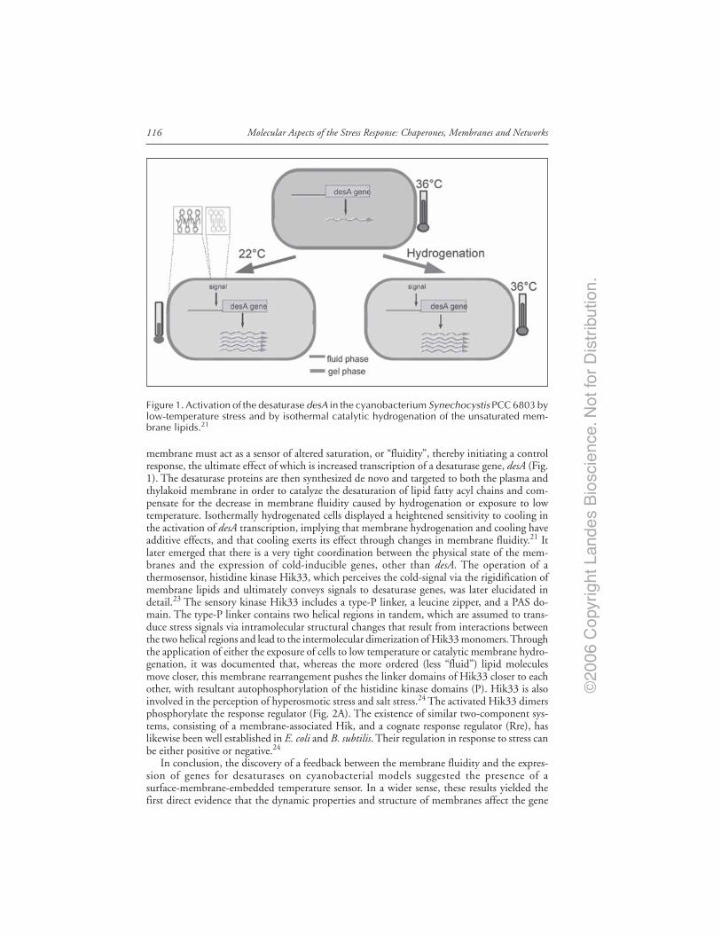
©20
06 C
opyr
ight
Lan
des
Bio
scie
nce.
Not
for
Dis
trib
utio
n.
Molecular Aspects of the Stress Response: Chaperones, Membranes and Networks116
membrane must act as a sensor of altered saturation, or “fluidity”, thereby initiating a controlresponse, the ultimate effect of which is increased transcription of a desaturase gene, desA (Fig.1). The desaturase proteins are then synthesized de novo and targeted to both the plasma andthylakoid membrane in order to catalyze the desaturation of lipid fatty acyl chains and com-pensate for the decrease in membrane fluidity caused by hydrogenation or exposure to lowtemperature. Isothermally hydrogenated cells displayed a heightened sensitivity to cooling inthe activation of desA transcription, implying that membrane hydrogenation and cooling haveadditive effects, and that cooling exerts its effect through changes in membrane fluidity.21 Itlater emerged that there is a very tight coordination between the physical state of the mem-branes and the expression of cold-inducible genes, other than desA. The operation of athermosensor, histidine kinase Hik33, which perceives the cold-signal via the rigidification ofmembrane lipids and ultimately conveys signals to desaturase genes, was later elucidated indetail.23 The sensory kinase Hik33 includes a type-P linker, a leucine zipper, and a PAS do-main. The type-P linker contains two helical regions in tandem, which are assumed to trans-duce stress signals via intramolecular structural changes that result from interactions betweenthe two helical regions and lead to the intermolecular dimerization of Hik33 monomers. Throughthe application of either the exposure of cells to low temperature or catalytic membrane hydro-genation, it was documented that, whereas the more ordered (less “fluid”) lipid moleculesmove closer, this membrane rearrangement pushes the linker domains of Hik33 closer to eachother, with resultant autophosphorylation of the histidine kinase domains (P). Hik33 is alsoinvolved in the perception of hyperosmotic stress and salt stress.24 The activated Hik33 dimersphosphorylate the response regulator (Fig. 2A). The existence of similar two-component sys-tems, consisting of a membrane-associated Hik, and a cognate response regulator (Rre), haslikewise been well established in E. coli and B. subtilis. Their regulation in response to stress canbe either positive or negative.24
In conclusion, the discovery of a feedback between the membrane fluidity and the expres-sion of genes for desaturases on cyanobacterial models suggested the presence of asurface-membrane-embedded temperature sensor. In a wider sense, these results yielded thefirst direct evidence that the dynamic properties and structure of membranes affect the gene
Figure 1. Activation of the desaturase desA in the cyanobacterium Synechocystis PCC 6803 bylow-temperature stress and by isothermal catalytic hydrogenation of the unsaturated mem-brane lipids.21

©20
06 C
opyr
ight
Lan
des
Bio
scie
nce.
Not
for
Dis
trib
utio
n.
117Membrane-Regulated Stress Response
transcriptional activity . On the other hand, this observation served as a landmark in studies ofthe potential roles of the lipid phase and fluidity changes of membranes in high-temperaturesensing and concomitant activation of heat shock (HS) genes.25
In fact, the genetic modification of the lipid unsaturation and thereby the membrane physi-cal state in the yeast Saccharomyces cerevisiae obtained by over-expression of a desaturase gene,was shown to reset the optimal temperature of heat shock response (HSR) and altered theexpression of the hsp70 and hsp82 genes.26 In agreement with the above findings, in a series ofstudies, Curran and coworkers demonstrated that the heat sensitivity of the HSR27 and thegeneral stress response28 (GSR) pathways depend critically on the fatty acid composition of themembrane lipids present in the yeast cells. Furthermore, these studies excluded direct thermaldenaturation of the cellular proteins as the trigger for the activation of these major stress-signalingpathways since the addition of different fatty acids to the growth media led to different thresh-olds of the HSR. It was suggested that HS is detected by a membrane-linked thermostat(s),whose activation is a consequence not only of the elevated temperature, but also of the specificcomposition and physical state of the membrane lipids.
Figure 2. A) Cold activation of a primary low-temperature sensor in the cyanobacterium, themembrane-associated histidine kinase, Hik33. Via response regulator(s), Rre(s), Hik33 is ulti-mately linked to fatty acid desaturases and can be stimulated by membrane rigidification andsimultaneous formation of the gel lipid phase, attained either by low- temperature shift or bycatalytic membrane lipid hydrogenation.21 B) A heat shock (shear stress, membrane fludizers,etc.) induced increase in membrane fluidity can activate growth factor receptors (GFRs) even ina ligand-independent manner. Membrane hyperfluidization may accelerate the clustering ofGFRs into lipid microdomains and their autophosphorylation. This leads to a down-stream signalcascade through RAF and PI3K, leading to ERK and GSK3 phosphorylation, respectively, withconcomitant upregulation of HSP synthesis (typically as described by Khalegue et al62 for ErbBactivation in tumor cells). Note the striking similarity of the low-temperature induced (probablyfluid-to-gel phase transition) membrane changes on the activation of HIK33, leading to upregulateddesaturases, and, the high-temperature-induced upregulation of HS genes via GFR dimerization.Both end-products, i.e., lipids with more unsaturated acyl chains and certain subpopulations ofHSPs, are capable of stabilizing membranes under extreme temperature stress.
©20
06 C
opyr
ight
Lan
des
Bio
scie
nce.
Not
for
Dis
trib
utio
n.
Molecular Aspects of the Stress Response: Chaperones, Membranes and Networks118
HS promotes at least two MAPK-driven signal transduction pathways in Schizosaccharomycespombe. Interestingly, their high-temperature-induced phosphorylations display striking differ-ences. The high-temperature-induced phosphorylation of MAPK Sty1/Spc1 appears to de-pend on the inhibitory phosphatases, Pyp1,2, rather than the downstream MAPKK Wis1,29
similarly as for mammalian ERK kinase (for details, see the chapter by Nadeau and Landry). Incontrast, the HS- (and other)-induced phosphorylation of the “cell-integrity” responsive MAPK,Pmk1 (p42/44), seems to be completely dependent on the donwnstream MAPK kinases. Fur-thermore, the time course of phosphorylation and the cellular localization of the two MAPKsdisplay intriguing differences,30 holding out the promise of an understanding of their distinct,stress-stimulated operation in real-time measurements. Besides the above-mentioned alteredfeatures, both sty1+ and pmk1+ proved to be dispensable under normal conditions, but themutants exhibit a heat-sensitive phenotype.30,31 Deletion of any of the two MAPKs causedosmosensitivity,32 whereas pmk1 is involved in the “cell-integrity” pathway.33 In contrast withthese MAPKs, the HS factor (HSF) is required for growth even in the absence of HS. TheDrosophila melanogaster HSF can rescue the lethality, and the S. pombe HSF is able to bind tothe HS element (HSE) of the human hsp70 gene.34 It has also been demonstrated that strainsbearing different hsf1 deletions exhibit an altered growth capability when exposed to differentkinds of stress (e.g., heat or cadmium treatment).35 The striking similarities of the human andS. pombe signal transduction pathways suggest that S. pombe might serve as a powerful andcomplementary model with which to explore membrane-associated stress sensors and stresssignaling in mammalian cells.
A close correlation has also been observed between the physical state of the thylakoid mem-brane and the threshold temperatures required for maximal activation of HS-inducible genes(i.e., dnaK, groESL, cpn60 and hsp17)36,13 in Synechocystis cells. Overproduction of a Synechocystis∆12 desaturase in Salmonella typhi, which is inactive enzymatically under the experimentalconditions, but which inserts into the membranes, was also able to cause resetting of the HSRin parallel with a considerable effect on the virulence of this pathogen. A substantially highermembrane protein content (i.e., an unbalanced protein/phospholipid ratio) was found in themembranes of the transformed cells. As evidence that the desaturase-transformed cells are un-able to accommodate the extra membrane protein properly, they displayed an elevated perme-ability in their outer membrane even under nonstressed conditions (Colonna-Romano S, ElettoAM, Török Zs et al unpublished). An unbalanced membrane phospholipid composition wasshown to affect the expression of several regulatory genes in E. coli.37 Overproduction of themembrane-bound sn-glycerol-acyltransferse in E. coli triggered a HSR.38 Treatment of E. coliwith nonlethal doses of heat or of the membrane-perturbing agent benzyl alcohol caused tran-sient membrane fluidization and permeabilization, and induced the transcription of the entirearray of heat shock genes in a sigma-32 (σ32)-dependent manner. This early response was fol-lowed by a rapid adaptation (priming) of the cells to an otherwise lethal elevated temperature,in strong correlation with an observed remodeling of the composition and alkyl chainunsaturation of the membrane lipids. Just like the activation of hs genes, however, the acquisi-tion of cellular thermotolerance in benzyl alcohol-primed E. coli cells was unrelated to proteindenaturation.39 The benzyl alcohol-mediated induction of chaperones in E. coli improves thenative folding of aggregation-prone recombinant proteins, thereby substituting the more de-manding approach of chaperone coexpression.40 When heat treatment is to be avoided, as inthe case of heterologously expressing heat-sensitive proteins, the membrane fluidizer-inducedexpression of recombinant proteins by a conditional HS promoter may serve as a novel andefficient biotechnological tool.41
We assume that, both in E. coli and in Salmonella, a high-temperature signal is transduced,in part, via the CpxA-CpxR phospho-relay system. CpxA is a histidine kinase that contains twotransmembrane regions, and CpxR is a response regulator that functions as a transcriptionfactor to regulate the expression of heat-inducible genes.42 Since the activity of CpxA is greatlyinfluenced by the composition of membrane lipids,43 it is tempting to suggest that CpxA
©20
06 C
opyr
ight
Lan
des
Bio
scie
nce.
Not
for
Dis
trib
utio
n.
119Membrane-Regulated Stress Response
might also sense changes in the physical state of the membrane lipids of E. coli and Salmonellacells exposed to high-temperature stress.39
A reporter system based on the highly thermostable enzyme lichenase was recently designedfor the analysis of Synechocystis HS promoters when heterologously induced in E. coli. Suprisingly,the cyanobacterial groESL promoter was not only recognized by the E. coli transcriptional appa-ratus, but was regulated by changes in the membrane fluidity of the host attained by benzylalcohol administration. In E. coli, most HS genes are controlled by σ,32 the product of the rpoHgene, which is completely absent in cyanobacteria. Instead, a CIRCE (Controling Inverted Re-peat of Chaperone Expression) element is present upstream of the Synechocystis groESL operon,which is controled by the repressor hrcA (heat regulation at CIRCE).13,44 Whereas its mecha-nism is unknown, the data demonstrate that the membrane physical properties of a Gram-negativebacterium, such as E. coli are capable of regulating the HS promoter from a distantly relatedphotosynthetic cyanobacterium, apparently in a σ32- independent manner (Shigapova N, BalogiZs, Fodor E et al, unpublished). Overall, a common denominator for the effects of cold stressand HS, i.e., a change in membrane fluidity, clearly appears to play the primary role in theperception of temperature changes causing the transcriptional induction of desaturases at coldand a reset of HS gene transcription at high temperature in these unicellular models.
Evidence Concerning the Operation of Membrane-AssociatedStress Sensing and Signaling Mechanisms in Mammalian Cells.Membrane Lipids May Provide the Molecular Switch for StressSensing and Signaling
There is a large body of evidence, much of which was earlier ignored or simply overlooked,that membrane-associated stress sensing and signaling mechanisms operate in mammalian cells.The formation of isofluid membrane states by the administration of benzyl alcohol and heptanol,which corresponded to mild HS-induced hyperfluidization, resulted in almost identical down-shifts in the temperature thresholds of the HSR, accompanied by the synthesis of stress pro-teins at the growth temperature in human erythroleukemic (K562) cells.45 Like thermal stress,membrane fluidizers elicited nearly identical rises of cytosolic Ca2+, in both Ca2+-containingand Ca2+-free media and closely similar extents of increase in mitochondrial hyperpolarization.Most importantly, neither of the membrane fluidizers caused any detectable protein denatur-ation at concentrations that induced the HSP response. Again, the induction of HSP70, HSP40and HSP27 by paeoniflorin, a major active constituent of a herbal medicine, was shown to bemediated by HSF1, in the complete absence of proteotoxicity.46 The coinducing effect of thehydroxylamine derivative bimoclomol on HSP expression6 was also demonstrated to be medi-ated via HSF1,47 and the presence of bimoclomol did not affect protein denaturation in vari-ous mammalian cells. Instead, the compound (and its analogs) specifically interacts with andsignificantly increases the fluidity of negatively-charged membrane lipids. In accordance withthis, the HSP-coinducing activity of bimoclomol appeared susceptible to the fatty acid compo-sition and membrane physical state of the target cells (Vigh L., unpublished). Additionally,bimoclomol was revealed to be an efficient inhibitor of bilayer-to-nonbilayer lipid-phase tran-sitions.7 It has been suggested that, while sensitizing the cellular membranes under mild HSconditions, the drug and its analogs ensure simultaneous protection against irreversible mem-brane damage at higher temperatures.7 Whereas bimoclomol was observed to have potentialtherapeutic value in the treatment of diabetes, a cardiac dysfunction and cerebrovascular disor-ders,5,7 another, closely-related bimoclomol analog, arimoclomol, delayed the disease progres-sion in amyotrophic lateral sclerosis.48 As reviewed elsewhere,5,8 pharmacological activation ofthe HSP response with lipid-interacting HSP coinducers may be a successful therapeutic ap-proach for the treatment of most varied diseases.
It was recently suggested that the mode of action of non proteotoxic membrane perturbants(e.g., benzyl alcohol) resembles mild, “fever-like” HS49 and in its underlying mechanism can be

©20
06 C
opyr
ight
Lan
des
Bio
scie
nce.
Not
for
Dis
trib
utio
n.
Molecular Aspects of the Stress Response: Chaperones, Membranes and Networks120
analogous to shear stress. The fact that hemodynamic shear stress, an important determinant ofvascular remodeling and atherogenesis, is able to increase HSP expression in cells under isother-mal conditions, is well established and has significant clinical consequences. HSP70 inductionhas been demonstrated in the arterial wall in response to acute hypertension, balloon angioplastyand advanced lesions of atherosclerosis.50 The mechanical stress-induced Hsp70 expression inarterial smooth muscle cells was shown to be regulated by the small G proteins Ras and Rac viaPI3K.51 The key shear stress responses, including the HSP response, are apparently linked withina single, integrin-mediated pathway. A fluorescence resonance energy transfer (FRET) studylocalized activated Rac1 in the direction of flow during shear stress. It was shown that, in parallelwith increased membrane fluidity in the upstream side of the cells, shear stress results in theremodeling of focal adhesions, with new adhesions forming preferentially toward the down-stream edge of the cells. Active Rac1 binds preferentially to low-density, cholesterol-rich mem-branes (rafts) and this binding step is specifically determined, at least in part, by the compositionand physical state of the membrane lipids.52
Analogously to shear stress or oxidative stress, moderate HS has been observed to inducemembrane translocation of Rac1 and membrane ruffling in a Rac1-dependent manner, whereasincreased HSP expression is paralleled by the activation of HSF1.53 In favor of a membranestress sensor model, it has been suggested that the potential sensing mechanism of mild HS isbased on membrane fluidization and/or rearrangement,1 closely resembling the HSP induc-tion attained by nonproteotoxic membrane fluidizers. Membrane rearrangement by mild HSor membrane hyperfluidization may activate growth factor receptor tyrosine kinases by causingtheir nonspecific clustering.8 As described earlier, the activation of such cell surface receptorshas other potential consequences, including the activation of PI3K, which in turn activatesRac1. Rac1 may stimulate NADPH oxidase which produces H2O2 and therefore thestress-stimulated nonspecific clustering of cell surface receptors, providing a possible avenuefor the oxidative burst and activation of the H2O2-induced stress signaling mechanism.1 ARac1-dependent HS signal pathway very probably plays an important role in physiologicalthermal stress responses such as fever. Rather than acting as a proteotoxic stress, fever mayfunction as a key, membrane-mediated signal required for resetting the body conditions. Incontrast with the poikilotherms, mammalian acclimation is limited to a very narrow range ofchanges in body temperature; accordingly, little is known about membrane and lipid remodel-ing under such acclimation conditions. The marked changes in the affinity of variousG-protein-coupled receptors and Na-K-ATPase activity during the course of heat acclimationin mammals imply the possibility of significant and specific changes in membrane lipid com-position,54 since alterations in the functions of these proteins have been documented to bestrongly membrane lipid-dependent.9
In conclusion, while many inducers of the HSR may function through a protein unfoldingmechanism, the above observations clearly suggest that some inducers may work through adistinct mechanism.55-57 Moreover, a membrane sensor model could explain specific HSP ex-pression patterns with different sensors. This model predicts that the plasma membrane, whichis the barrier to the external environment and well suited for sensing thermal stress, acts as animportant regulatory interface. Hence, even subtle alterations in the lipid phase of surfacemembranes (“membrane defects”) may influence membrane-initiated stress-signaling processesby causing changes in fluidity, membrane thickness or the clustering of receptors or otherproteins localized in the surface membranes.
It was suggested recently that in such prominent disease states as insulin-resistant diabetesand cancer, where the directions of HSP dysregulation and membrane fluidity run in parallel,but strikingly opposite manners, there must exist a conserved signaling cascade which is uni-formly controlled by membrane hyperstructures and ultimately affects the level of HSP expres-sion as well (Vigh L, Horvath I, Maresca B et al, unpublished). We believe that the signalingfrom the transmembrane growth factor receptors to HS genes fulfills such a criterion. PI3K,Akt and GSK3, a negative regulator of HSF1 activation,58 are central components of this
©20
06 C
opyr
ight
Lan
des
Bio
scie
nce.
Not
for
Dis
trib
utio
n.
121Membrane-Regulated Stress Response
signaling cascade . Diabetes is associated with a low HSP level, and with decreased PI3K andenhanced GSK3 activities.59 Correction of a low HSP state improves insulin resistance. A closechemical relative of bimoclomol, BRX-220, has been reported to increase the insulin sensitiv-ity of both Zucker diabetic fatty rats and streptozotocin-diabetic rats.60 Another bimoclomolanalog, the HSP coinducer BGP-15, has passed successfully a Phase IIa human clinical trial asan insulin-sensitizer compound. Similarly to several other insulin sensitizers,61 BGP-15upregulated Akt kinase and acted as an GSK3 inhibitor. However, the operation of such asignaling cascade also provides a potential mechanism for the widespread elevation in HSPexpression in cancer (Fig. 2B). As recently documented by Khaleque et al,62 an HSP elevationin tumor cells can be induced by the highly malignant growth factor heregulin β1 (HRGβ1),which causes homo- and heterodimerization between each member of the four ErbB receptormolecules via “horizontal signaling” in the plane of the plasma membrane. Just like the inter-molecular dimerization of membrane-associated Hik33, which serves as the first key step inlow-temperature sensing and signaling, with resultant upregulation of desaturases in prokary-otes (Fig. 2A), the formation of raft-associated GFR dimers is followed by tyrosine phosphory-lation of their intracellular domains in mammalian cells (Fig. 2B). The major downstreamsignaling pathways include the Ras-Raf1-Mek-ERK and PI3K-PDK1-Akt pathways.62 HRGβ1appears to be linked to HSP expression by its activation of HSF1 through inhibition of theconstitutive kinase GSK3.
It may be noted that the transmission of a signal from the cell surface to HS genes followingmild HS, shear stress or growth factors, with the involvement of the activation of receptors orreceptor-like molecules and their clustering into lipid rafts, is uniformly dependent on preciseregulation by the lipid composition of the membranes. The localization of growth factor recep-tors to distinct microdomains appears to modulate both their ligand binding and tyrosinekinase activities.63 Moreover, the major signal termination mechanism, i.e., the lateral move-ment of dimerized receptors in the plane of the surface membrane and the delivery of activatedreceptor-containing endosomes, is totally lipid-dependent.64,65
The precise role played by lipid microdomains in membrane-directed stress sensing andsignaling is far from clear. Their studies have been hampered by the lack of suitable physicalmethods for the visualization of membrane microdomains in intact cells.8,66 As comparedwith unicellular model organisms, the membrane structures in mammalian cells display enor-mous lipid variety, multiple functions, locations, associations and intimate links withneighbouring membranes, and this makes their study extremely difficult. It is tempting tospeculate that one of the major roles of the more than one thousand lipid molecular species inmammalian membranes is to provide an on-off switch for signaling events at the membranelevel.66,67 Membrane lipids are among the molecules that adapt best in response to variousperturbations. Even subtle changes in the compositions of acyl chains or head groups can alterthe packing arrangements of lipids within a bilayer. As a chain reaction, altered lipid packingproperties change the balance between bilayer and nonbilayer lipids, affect the bilayer stabil-ity and fluidity, and also the lipid-protein interactions and microdomain organizations. Ex-ternal factors, including temperature, chemicals, ions, radiation, pressure, nutrients, the growthphase of the culture, etc., are all capable of changing the membrane packing order and lipidcomposition.8 Finally, it should be born in mind that the lipid-selective association of certainHSPs may also lead to an increased membrane molecular order and may result indownregulation of the HS gene expression.68-70
In recent years, our understanding of the plasma membrane has changed considerably asour knowledge of lipid microdomains has expanded. These include structures known as lipidrafts and caveolae, which are readily identified by their unique lipid constituents. Cholesterol,sphingolipids and specific phospholipids with more saturated fatty acyl chain moieties aretypically highly enriched in these lipid microdomains. Since lipid microdomains have beenwidely shown to play important roles in the compartmentalization, modulation and integra-tion of cell signaling, we suggest that these microdomains may additionally have an influential

©20
06 C
opyr
ight
Lan
des
Bio
scie
nce.
Not
for
Dis
trib
utio
n.
Molecular Aspects of the Stress Response: Chaperones, Membranes and Networks122
role in stress sensing and signaling. Favorable interactions between cholesterol and the satu-rated lipids result in patches of liquid-ordered (Lo) or raft domains from the remainingliquid-disordered (Ld) membranes. Specific signaling proteins are targeted to or concentratedin rafts in consequence of the greater solubility of their lipid anchors in Lo than in Ld compart-ments.71 Since the raft structure is also dependent on the thermally controlled lipid-phasebehavior, even mild changes in temperature could result in a fundamentally altered solubilityand consequently redistribution of these proteins in the rafts. More severe heat may result incomplete raft dissolution and disruption of the signaling activities.8 It was recently documentedby means of single-molecule microscopy that diffusional trapping through protein-protein in-teractions does indeed create plasma membrane microdomains that concentrate or exclude cellsurface proteins, facilitating cell signalling.72 Different fluorescence imaging techniques, add-ing the beauty of visualization to the scientific information,73 in conjunction with computa-tional modeling techniques (see later), can promote a better understanding of the network ofcomplex molecular interactions taking place within membranes under stress conditions.
Stress Response Profiling: Can We “Zoom In” on MembraneHyperstructures Engaded in the Generation of Stress Signal?
Most of the experiments targeted toward an understanding of stress signaling are per-formed on one or just a few cells, or on many cells, but without a knowledge of the prehis-tory of each individual cell. It is critical that cells in culture can be quite heterogeneous andpresent a wide variety of phenotypes. This heterogeneity can result from the cells being indifferent stages of the cell cycle, or from other intrinsic differences between the cells. Arecent study of transcription activation in a serum starvation assay highlighted the widevariety of responses among individual cells, even though the large differences between thecontrol and treatment groups were scored in a population assay such as an immunoblot.74
For this reason, it is necessary to record data from many individual cells so as to aquire astatistically significant sampling of cellular behavior and dynamics. This quest depends heavilyon molecular imaging, which shows when and where genetically or biochemically definedmolecules, signals or processes appear, interact and disappear, in time and space. Withhigh-content cellular analysis, it is necessary to perform high-throughput phenotype profil-ing, linking gene expression to biochemical signaling pathways in the cell and, ultimately, tocell behavior.74 During or after the imposition of stress on cells, it becomes possible to zoomin on individual cells or individual fluorescently tagged molecules, using an ultrasensitive,high-speed camera to observe what happens as each cell reacts to the particular treatment.75
A change may occur in the topology of a particular membrane domain, a candidate fromwhich an initial HS signal could originate, or a membrane receptor may be activated, allow-ing the monitoring in real time as a receptor complex responds and activates signaling path-ways. An intracellular signaling molecule tagged with a fluorescent protein could light up asit interacts with another component of the signaling pathway, perhaps relaying instructionsto the nucleus to activate or deactivate a particular stress defensive gene. The particularlyvaluable aspect of this methodology is not the astounding visual images it produces, butrather the abundant and diverse data that can be extracted from those images—data thatafford a better understanding of what is happening in the cell in response to stress. Imageanalysis of this nature requires efficient imaging algorithms, such as the open source CellProfiler (www.cellprofiler.org), which provides a selection of tools for automatic analysis ofthe cell morphology, and the localization and intensity distribution of fluorescent markers(Carpenter AE, Jones TR, Lamprecht M et al, unpublished). Investigators can exploit theflexibility of the data analysis software to design their own algorithms from the buildingblocks provided, customizing the data-mining tools to optimize the data output. The poten-tial applications and ultimate value of high content screening and cellular image analysis arelimited only by the imagination and expertise of the investigator who is using them to probestress sensing and cell behavior.
©20
06 C
opyr
ight
Lan
des
Bio
scie
nce.
Not
for
Dis
trib
utio
n.
123Membrane-Regulated Stress Response
Can We Point to Lipid Molecular Species Engaged in Stress Sensingand Signaling?
The diversity of the membrane constituent phospholipids originates from the differences inhead groups, acyl chains, and the degree of unsaturation, resulting in several hundred coexistinglipid molecular species within a given cell. This polymorphism permits the regulation of mem-brane fluidity in poikilothermal organisms. The organized self-assembly of functionalsignal-generating protein-lipid complexes leads to a wide range of chemical and physical mem-brane properties, through which the membrane-associated events are regulated and coordinated.
By means of a combination of thin-layer separation techniques with MS in thecyanobacterium Synechocystis, a highly saturated monoglucosyldiacylglycerol (MGlcDG) mo-lecular species (“heat shock lipid”) was identified; this has the ability to cause membranerigidification and to counteract the formation of nonbilayer structures during heat/light stress.Moreover, MGlcDG expressed the strongest interaction with the membrane-stabilizing smallHSP, HSP17.76 Specific HSP-lipid interactions may be unrecognized means for the spatialseparation and distinct compartmentalization of HSPs to lipid domains, which are thought tobe involved in various signaling pathways.8 Another HS lipid, cholesteryl glucoside (CGl), wasshown to accumulate rapidly in cells from molds to humans following exposure to environ-mental stress. CGl production is followed by the activation of certain PKCs and the inductionof HSPs.77,78
Lipids and lipid-derived metabolites may also contribute to the early HS-induced cellresponses. Among others, arachidonate, certain prostaglandins, IP3 and ceramides have beenproved to be second messengers of the HSR.8 The source lipids of signaling molecules arelocalized in different compartments of the cell. The mapping of time and stimulus-dependentchanges in the lipidome of plasmamembrane, rafts, mitochondria ER, Golgi or nuclear mem-branes requires appropriate subcellular fractionation and lipid isolation. In principle, com-plete characterization of the lipid components of these membranes or membranemicrodomains is essential for an understanding of how the membrane senses and copes withenvironmental stress.
The cross-talk between the raft proteins and lipids would trigger a remodeling of thenewly-formed raft to optimize the stability of the new structure. This might be accomplishedby excluding some proteins and lipids from the raft, while recruiting others. It is tempting tospeculate that, in response to stress conditions, chemical (e.g., oxidation) and physical (e.g.,fluidity) changes in these lipids might result in the recruitment/exclusion of other proteinsand/or lipids, leading to a unique remodeled composition, thereby provoking the generation ofstress signals.
However, it should be pointed out that no two rafts or membrane preparations yield thesame analytical result, and there is no isolation procedure that does not involve loss of severalcomponents during “disassembly” of the cells. These caveats provide the impulse for the devel-opment of whole-cell or organ analysis with the aim of the creation of overall lipid maps afterdifferent perturbations. The ESI/MS of lipids is the most sensitive, discriminating and directmethod for identification of the complete set of lipid molecular species.79-81
Furthermore, lipidomics today is extended toward the more polar extraction proceduresand the inspection of unknown peaks of possible new moieties. In this manner, the entirespectrum of new metabolites can be identified and the potential signaling functions of thesemolecules can be determined. The question arises as to how the observed lipidomic andmetabolic fingerprints will help in the elucidation of the sensing and signaling of stress. Theplethora of new data requires different statistical approaches, which are not limited to simplestatistical metrics. Supervised and unsupervised classification methods, such as discriminant,cluster and principal component analysis, multidimensional scaling or other pattern recog-nition techniques, will be important82,83 for the identification of significant and reproduc-ible changes in the patterns of lipid species changes in response to stress or other stimuli. Anunderstanding of the potential signaling function of these metabolites or the importance of

©20
06 C
opyr
ight
Lan
des
Bio
scie
nce.
Not
for
Dis
trib
utio
n.
Molecular Aspects of the Stress Response: Chaperones, Membranes and Networks124
a specific lipid composition of a membrane domain requires the correlation of the alterationswith physiological data with the aid of system-biological methods.
In order to illustrate the power of these statistical procedures, the lipidome changes of B16mouse melanoma cells under different circumstances are presented in Figure 3. B16 cells weresubjected to different stimuli or plated with low or high numbers of cells. Besides the choles-terol level, the molecular species compositions of several lipid classes were determined. Figure 3shows the principal components represented in a two-dimensional scaling model. The princi-pal component P1 extracted those molecular species which can be clearly attributed to the lipidchanges due to the distinct initial cell densities, while the lipid molecular species belonging incomponent P2 were mainly altered by other stimuli. Our parallel investigations on the geneexpression patterns by the chip method and the induction of HSPs by RT-PCR revealed thatthe lipid status plays a regulatory role in the stress response (Balogh G. and Vigh L, unpub-lished). The combination of gene expression and lipidome data by appropriate statistical meth-ods will trigger the postulation of new theories with higher explanatory relevance.
Computational Methods for the Design of Subtle Interactionsbetween Lipids and Proteins of Membranes
The interest of computational biologists has recently focused on the active roles of phospho-lipids in affecting the behavior of membrane proteins,84 the assembly of protein-lipid arrays andthe modulation of protein-protein interactions.85 Lipid and protein-lipid- phase equilibria arealso believed to be relevant for membrane fusion and raft formation. Computational methodscan provide useful information on the inner organization of membranes, which are normallyinaccessible by direct experimental investigations. During the past twenty years, atomistic simu-lation and, in particular, full atom Molecular Dynamics (MD), have been developed to a degree
Figure 3. The lipidome of the cells was determined by direct-injection ESI tandem massspectrometry (Balogh and Liebisch, unpublished) and the molecular species compositions ofPC (phosphatidylcholine), PE (phosphatidylethanolamine), PEP (Phoshaptidyl- ethanolamineplasmalogen), PS (Phosphatidylserine), PI (Phosphatidylinositol) , SPM (Sphingomyelin), LPC(Lysophosphatydilcholine), Cer (Ceramide), Glucer (Glucosyl ceramide) , Chol (Cholesterol)and Chol-e (Cholesterol esther) were calculated. First, the meaningful components (based onmeasurement accuracy and the relative quantity) were selected (approximately 100 of 300species) and the values were transformed to Z-scores. For multidimensional scaling, the Eu-clidean distance was used. Two principal components (P1 and P2) were extracted. The groupsrepresent the positive or negative participation associated with a given component.

©20
06 C
opyr
ight
Lan
des
Bio
scie
nce.
Not
for
Dis
trib
utio
n.
125Membrane-Regulated Stress Response
that allowss prediction of the structural characteristics with acceptable precision. Besides MD,computer modeling also utilizes coarse grain (CG) dynamics, quantitative structure analysisrelationship methods (QSAR) and quantum mechanics/molecular mechanics (QM/MM), whichhave a large potential for the investigation of biological structures (Fig. 4).
A number of atomistic simulations have been performed to reproduce and predict manyfundamental properties of lipid membranes.86,87 However, with today’s computers and algo-rithms, the size of the systems and the time scales for which phospholipid bilayers can bestudied preclude the scrupulous examination of many phenomena such as the cooperativemotion in phase transitions, or the interaction of proteins with the membrane or the flip-flop.For these reasons, numerous strategies have been conceived to extend the temporal and spa-tial limits of MD, which are capable of covering a large spatial and time scale, but with areduced level of detail in the representation of the membrane. CG models are an improve-ment toward the study of collective phenomena in membranes88 which are not accessible bythe MD approach.
CG simulation are extremely effective in simulating both the formation and the be-havior of a mixed bilayer. In Figure 5 the spontaneous formation of a bilayer ofdimirystoyl-phospahtidylcholine/dimirystoyl-phosphatidylethanolamine (DMPC/DMPE) wascalculated through the use of CG dynamics. The simplification introduced with CG can stronglyaffect the possibilities of investigations of protein folding and protein-membrane interactions.
Figure 4. Simulation methods schematically ordered as functions of the time and length scales.Some relevant membrane events are indicated.

©20
06 C
opyr
ight
Lan
des
Bio
scie
nce.
Not
for
Dis
trib
utio
n.
Molecular Aspects of the Stress Response: Chaperones, Membranes and Networks126
The continuous development of new parameterization techniques will hopefully permitthe simulation of ever more complicated mixtures of lipids, and consequently a better mim-icking of membranes. Evidently, simulations and fast computers are not sufficient for thegeneration of new understanding: new models are necessary as well. A model is a simple wayof describing and/or predicting scientific results, which is known to yield an incorrect orincomplete description. Models may be simple mathematical descriptions or completelynonmathematical. In recent decades, continuous efforts have been made to reformulate bio-logical concepts into a more formal, mathematical language, allowing the prediction andunderstanding of phenomena without the performance of difficult experiments. The mostdifficult problem in the simulation of lipid membrane behavior is the lack of correct models.Implicit in most membrane studies is the idea of an “average membrane” or, in other words,the idea that a membrane with a given lipid composition exists for each cell (which is nottrue, of course). As described earlier, a cell membrane is organized in discrete domains, andthe knowledge of the lipid composition is therefore necessary for the simulation of mem-brane suprastructures.89 A tremendous improvement of the computer simulations of lipidrafts will be possible in the coming years with the accumulation of experimental data onlipid composition and with the availability of faster computers. The design of newmembrane-interacting drugs (such as the stress protein coinducer bimoclomol and its ana-logs) will be assisted by the molecular simulation of membranes and proteins. It will bepossible to highlight slight conformational changes induced in a membrane protein inducedin the target raft by a chemical, physical (e.g., temperature) or biological alteration of thebilayer. Nevertheless, the generation of new models for membrane behavior is a task whichcannot be performed via molecular (or CG) dynamics, since these are not intrinsically ableto observe properties which are not encoded in the simulation. There are computationalmethods, which to some extent, are able to generate models that can ultimately be validatedvia MD. Examples come from genetic algorithms (GAs) and neural networks.
GAs permit computers to generate new, random data. In combination with a fitness func-tion, only the best data are randomly exchanged and mutated until a new generation of data isproduced. In all cells, the genetic material is DNA, which is described in terms of base pairs. Astring of three base pairs makes up a codon, which corresponds to a specific amino acid in thecoded protein. As in real genes, GAs operate on a sort of genetic material, but as a substitute ofDNA; the “genetic material” is a linear string of symbols. The computer equivalent of the basepairs and codon is replaced by a symbol or numbers that represent any structural unit.
We recently (Piotto S, Bianchino E., unpublished) extracted information on the antimicro-bial activities of short peptides (Fig. 6) through the successful application of GA techniques.
Figure 5. Spontaneous organization of DMPC/DMPE and water into a bilayer structure (CGsimulation).
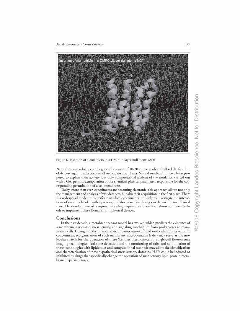
©20
06 C
opyr
ight
Lan
des
Bio
scie
nce.
Not
for
Dis
trib
utio
n.
127Membrane-Regulated Stress Response
Natural antimicrobial peptides generally consist of 10-20 amino acids and afford the first lineof defense against infections in all metazoans and plants. Several mechanisms have been pro-posed to explain their activity, but only computational analysis of the similarity, carried outwith a GA, permits extrapolation of the chemical-physical parameters responsible for the cor-responding perturbation of a cell membrane.
Today, more than ever, experiments are becoming electronic; this approach allows not onlythe management and analysis of vast data sets, but also their acquisition in the first place. Thereis a widespread tendency to perform in silico experiments, not only to investigate the interac-tions of small molecules with a protein, but also to analyze changes in the membrane physicalstate. The development of computer modeling requires both new formalisms and new meth-ods to implement these formalisms in physical devices.
ConclusionsIn the past decade, a membrane sensor model has evolved which predicts the existence of
a membrane-associated stress sensing and signaling mechanism from prokaryotes to mam-malian cells. Changes in the physical state or composition of lipid molecular species with theconcomitant reorganization of such membrane microdomains (rafts) may serve as the mo-lecular switch for the operation of these “cellular thermometers”. Single-cell fluorescenceimaging technologies, real-time detection and the monitoring of rafts and combination ofthese technologies with lipidomics and computational methods may allow the identificationand characterization of these hypothetical stress-sensory domains. HSPs could be induced orinhibited by drugs that specifically change the operation of such sensory lipid-protein mem-brane hyperstructures.
Figure 6. Insertion of alamethicin in a DMPC bilayer (full atoms MD).

©20
06 C
opyr
ight
Lan
des
Bio
scie
nce.
Not
for
Dis
trib
utio
n.
Molecular Aspects of the Stress Response: Chaperones, Membranes and Networks128
AcknowledgementsThis work was supported by grants from the Hungarian National Scientific Research Foun-
dation (OTKA: TS 044836, T 038334) and Agency for Research Fund Management andResearch Exploitation (RET OMFB00067/2005 and Bio- 00120/2003 KPI).
References1. Kultz D. Molecular and evolutionary basis of the cellular stress response. Annu Rev Physiol 2005;
67:225-257.2. Ananthan J, Goldberg AL, Voellmy R. Abnormal proteins serve as eukaryotic stress signals and
trigger the activation of heat shock genes. Science 1986; 232:522-524.3. Lepock JR. Measurement of protein stability and protein denaturation in cells using differential
scanning calorimetry. Methods 2005; 35:117-125.4. Pockley GA. Heat shock proteins in health and disease: Therapeutic targets or therapeutic agents?
Expert Rev Mol Med 2001; 2001:1-21.5. Soti C, Nagy E, Giricz Z et al. Heat shock proteins as emerging therapeutic targets. Br J Pharmacol
2005; 146:769-780.6. Vigh L, Literati NP, Horvath I et al. Bimoclomol: A novel nontoxic, hyroxylamine derivative with
stress protein inducing activity and wide cytoprotective effects. Nature Med 1997; 3:1150-1154.7. Török Z, Tsvetkova NM, Balogh G et al. Heat shock protein coinducers with no effect on protein
denaturation specifically modulate the membrane lipid phase. Proc Natl Acad Sci USA 2003;100:3131-3136.
8. Vigh L, Escriba P, Sonnleitner A et al. The significance of lipid composition for membrane activ-ity: New concepts and ways of assessing function. Progr Lipid Res 2005; 44:303-344.
9. Escriba PV. Membrane-lipid therapy: A new approach in molecular medicine. Trends Mol Med2006; 12:34-43.
10. Yatvin MB, Cramp WA. Role of cellular membranes in hyperthermia: Some observations and theoriesreviewed. Int J Hyperthermia 1993; 9:165-185.
11. Hazel JR, Williams EE, Livermore R et al. Thermal adaptation in biological membranes: Func-tional significance of changes in phospholipids molecular species composition. Lipids 1991;26:277-282.
12. Vigh L, Horváth I, van Hasselt PR et al. Effect of frost hardening on lipid and fatty acid compo-sition of chloroplast thylakoid membranes in two wheat varieties of contrasting hardiness. PlantPhysiol 1985; 79:756-759.
13. Glatz A, Vass I, Los DA et al. The Synechocystis model of stress: From molecular chaperones tomembranes. Plant Physiol Biochem 1999; 37:1-12.
14. Vigh L, Joó F. Modulation of membrane fluidity by catalytic hydrogenation affects the chillingsusceptibility of the blue-green alga, Anacystis nidulans. FEBS L 1983; 162:423-426.
15. Vigh L, Gombos Z, Joó F. Selective modification of cytoplasmic membrane fluidity by catalytichydrogenation provides evidence on its primary role in chilling susceptibility of the blue-greenalga, Anacystis nidulans. FEBS L 1985; 191:200-204.
16. Benkö S, Hilkmann H, Vigh L et al. Catalytic hydrogenation of fatty acyl chains in plasma mem-branes; effect of membrane fluidity and expression of cell surface antigens. Biochim Biophys Acta1987; 896:129-135.
17. Quinn PJ, Joó F, Vigh L. The role of unsaturated lipids in membrane structure and stability.Progr Biophys Mol Biol 1989; 53:71-103.
18. Joó F, Balogh N, Horváth I et al. Complex hydrogenation/oxidation reactions of the water-solublehydrogenation catalyst palladium di(sodium alizarinmonosulfonate) and details of homogeneoushydrogenation of lipids in isolated membranes and living cells. Anal Biochem 1991; 194:34-40.
19. Vigh L, Horváth I, Thomspon GA. Recovery of Dunaliella salina cells following hydrogenation oflipids in specific membranes by a homogeneous palladium catalyst. Biochim Biophys Acta 1988;937:42-50.
20. Pak Y, Joó F, Vigh L. Action of a homogeneous hydrogenation catalyst on living Tetrahymenamimbres cells. Biochim Biophys Acta 1990; 1023:230-238.
21. Vigh L, Los DA, Horváth I et al. The primary signal in the biological perception of temperature:Pd-catalyzed hydrogenation of membrane lipids stimulated the expression of the desA gene inSynechocystis PCC 6803. Proc Natl Acad Sci USA 1993; 90:9090-9094.
22. Los D, Horváth I, Vigh L et al. The temperature-dependent expression of the desaturase gene desAin Synechocystis PCC6803. FEBS L 1993; 318:57-60.
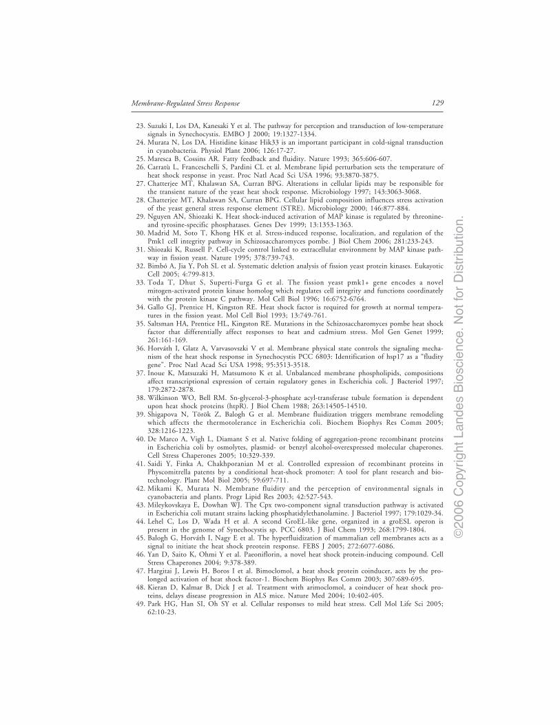
©20
06 C
opyr
ight
Lan
des
Bio
scie
nce.
Not
for
Dis
trib
utio
n.
129Membrane-Regulated Stress Response
23. Suzuki I, Los DA, Kanesaki Y et al. The pathway for perception and transduction of low-temperaturesignals in Synechocystis. EMBO J 2000; 19:1327-1334.
24. Murata N, Los DA. Histidine kinase Hik33 is an important participant in cold-signal transductionin cyanobacteria. Physiol Plant 2006; 126:17-27.
25. Maresca B, Cossins AR. Fatty feedback and fluidity. Nature 1993; 365:606-607.26. Carratù L, Franceschelli S, Pardini CL et al. Membrane lipid perturbation sets the temperature of
heat shock response in yeast. Proc Natl Acad Sci USA 1996; 93:3870-3875.27. Chatterjee MT, Khalawan SA, Curran BPG. Alterations in cellular lipids may be responsible for
the transient nature of the yeast heat shock response. Microbiology 1997; 143:3063-3068.28. Chatterjee MT, Khalawan SA, Curran BPG. Cellular lipid composition influences stress activation
of the yeast general stress response element (STRE). Microbiology 2000; 146:877-884.29. Nguyen AN, Shiozaki K. Heat shock-induced activation of MAP kinase is regulated by threonine-
and tyrosine-specific phosphatases. Genes Dev 1999; 13:1353-1363.30. Madrid M, Soto T, Khong HK et al. Stress-induced response, localization, and regulation of the
Pmk1 cell integrity pathway in Schizosaccharomyces pombe. J Biol Chem 2006; 281:233-243.31. Shiozaki K, Russell P. Cell-cycle control linked to extracellular environment by MAP kinase path-
way in fission yeast. Nature 1995; 378:739-743.32. Bimbó A, Jia Y, Poh SL et al. Systematic deletion analysis of fission yeast protein kinases. Eukayotic
Cell 2005; 4:799-813.33. Toda T, Dhut S, Superti-Furga G et al. The fission yeast pmk1+ gene encodes a novel
mitogen-activated protein kinase homolog which regulates cell integrity and functions coordinatelywith the protein kinase C pathway. Mol Cell Biol 1996; 16:6752-6764.
34. Gallo GJ, Prentice H, Kingston RE. Heat shock factor is required for growth at normal tempera-tures in the fission yeast. Mol Cell Biol 1993; 13:749-761.
35. Saltsman HA, Prentice HL, Kingston RE. Mutations in the Schizosaccharomyces pombe heat shockfactor that differentially affect responses to heat and cadmium stress. Mol Gen Genet 1999;261:161-169.
36. Horváth I, Glatz A, Varvasovszki V et al. Membrane physical state controls the signaling mecha-nism of the heat shock response in Synechocystis PCC 6803: Identification of hsp17 as a “fluditygene”. Proc Natl Acad Sci USA 1998; 95:3513-3518.
37. Inoue K, Matsuzaki H, Matsumoto K et al. Unbalanced membrane phospholipids, compositionsaffect transcriptional expression of certain regulatory genes in Escherichia coli. J Bacteriol 1997;179:2872-2878.
38. Wilkinson WO, Bell RM. Sn-glycerol-3-phosphate acyl-transferase tubule formation is dependentupon heat shock proteins (htpR). J Biol Chem 1988; 263:14505-14510.
39. Shigapova N, Török Z, Balogh G et al. Membrane fluidization triggers membrane remodelingwhich affects the thermotolerance in Escherichia coli. Biochem Biophys Res Comm 2005;328:1216-1223.
40. De Marco A, Vigh L, Diamant S et al. Native folding of aggregation-prone recombinant proteinsin Escherichia coli by osmolytes, plasmid- or benzyl alcohol-overexpressed molecular chaperones.Cell Stress Chaperones 2005; 10:329-339.
41. Saidi Y, Finka A, Chakhporanian M et al. Controlled expression of recombinant proteins inPhyscomitrella patents by a conditional heat-shock promoter: A tool for plant research and bio-technology. Plant Mol Biol 2005; 59:697-711.
42. Mikami K, Murata N. Membrane fluidity and the perception of environmental signals incyanobacteria and plants. Progr Lipid Res 2003; 42:527-543.
43. Mileykovskaya E, Dowhan WJ. The Cpx two-component signal transduction pathway is activatedin Escherichia coli mutant strains lacking phosphatidylethanolamine. J Bacteriol 1997; 179:1029-34.
44. Lehel C, Los D, Wada H et al. A second GroEL-like gene, organized in a groESL operon ispresent in the genome of Synechocystis sp. PCC 6803. J Biol Chem 1993; 268:1799-1804.
45. Balogh G, Horváth I, Nagy E et al. The hyperfluidization of mammalian cell membranes acts as asignal to initiate the heat shock preotein response. FEBS J 2005; 272:6077-6086.
46. Yan D, Saito K, Ohmi Y et al. Paeoniflorin, a novel heat shock protein-inducing compound. CellStress Chaperones 2004; 9:378-389.
47. Hargitai J, Lewis H, Boros I et al. Bimoclomol, a heat shock protein coinducer, acts by the pro-longed activation of heat shock factor-1. Biochem Biophys Res Comm 2003; 307:689-695.
48. Kieran D, Kalmar B, Dick J et al. Treatment with arimoclomol, a coinducer of heat shock pro-teins, delays disease progression in ALS mice. Nature Med 2004; 10:402-405.
49. Park HG, Han SI, Oh SY et al. Cellular responses to mild heat stress. Cell Mol Life Sci 2005;62:10-23.

©20
06 C
opyr
ight
Lan
des
Bio
scie
nce.
Not
for
Dis
trib
utio
n.
Molecular Aspects of the Stress Response: Chaperones, Membranes and Networks130
50. Bornfeldt KE. Stressing Rac, Ras, and downstream heat shock protein 70. Circ Res 2000;86:1101-1103.
51. Xu Q, Schett G, Li C et al. Mechanical stress-induced heat shock protein 70 expression in vascularsmooth muscle cells is regulated by Rac and Ras small G proteins but not mitogen-activated pro-tein kinases. Circ Res 2000; 86:1124-1130.
52. Del Pozo MA, Alderson NB, Kiosses WB et al. Integrins regulate Rac targeting by internalizationof membrane domains. Science 2004; 303:839-842.
53. Han SI, Oh SY, Woo SH et al. Implication of a small GTPase Rac1 in the activation of c-JunN-terminal kinase and heat shock factor in response to heat shock. J Biol Chem 2001;276:1889-1895.
54. Kaspler P, Horowitz M. Heat acclimation and heat stress have different effects on cholinergicmuscarinic receptors. Ann Ny Acad Sci 1997; 813:620-627.
55. Vigh L, Maresca B, Harwood J. Does the membrane physical state control the expression of heatshock and other genes? Trends Biochem Sci 1998; 23:369-373.
56. Vigh L, Maresca B. Dual role of membranes in heat stress: As thermosensors they modulate theexpression of stress genes and, by interacting with stress proteins, reorganize their own lipid orderand functionality. In: Storey KB, Storey JM, eds. Cell and Molecular Responses to Stress. Amsterdam:Elsevier, 2002:173-188.
57. Westerheide SD, Morimoto RI. Heat shock response modulators as therapeutic tools for diseasesof protein conformation. J Biol Chem 2005; 280:33097-33100.
58. Bijur GN, Jope RS. Opposing actions of phosphatidylinositol 3-kinase and glycogen synthasekinase-3beta in the regulation of HSF-1 activity. J Neurochem 2000; 75:2401-2408.
59. Hooper PL, Hooper JJ. Loss of defense against stress: Diabetes and heat shock proteins. DiabetesTechnol Ther 2005; 7:204-208.
60. Kürthy M, Mogyorósi T, Nagy K et al. Effect of BRX-220 against peripheral neuropathy andinsulin resistance in diabetic rat models. Ann NY Acad Sci 2002; 967:482-489.
61. Meijer L, Flajolet M, Greengard P. Pharmacological inhibitors of glycogen synthase kinase 3. TrendsPharmacol Sci 2004; 25:471-480.
62. Khaleque MA, Bharti A, Sawyer D et al. Induction of heat shock proteins by heregulin beta1 leadsto protection from apoptosis and anchorage-independent growth. Oncogene 2005; 24:6554-6573.
63. Pike LJ, Han X, Gross RW. Epidermal growth factor receptors are localized to lipid rafts thatcontain a balance of inner and outer leaflet lipids: A shotgun lipidomics study. J Biol Chem 2005;280:26796-26804.
64. Gur G, Yarden Y. Enlightened receptor dynamics. Nature Biotechnology 2004; 22:169-170.65. Koijman EE, Chupin V, Fuller NL et al. Spontaneous curvature of phosphatidic acid and
lysophosphatidic acid. Biochemistry 2005; 44:2097-2102.66. Vereb G, Szöllösi J, Matko J et al. Dynamic, yet structured: The cell membrane three decades after
the Singer-Nicolson model. Proc Natl Acad Sci USA 2003; 100:8053-8058.67. Hinderliter A, Biltonen RL, Almeida PF. Lipid modulation of protein-induced membrane domains
as a mechanism for controlling signal transduction. Biochemistry 2004; 43:7102-7110.68. Török Z, Horváth I, Goloubinoff P et al. Evidence for a lipochaperonin: Association of active
protein-folding GroESL oligomers with lipids can stabilize membranes under heat shock condi-tions. Proc Natl Acad Sci USA 1997; 94:2192-2197.
69. Török Z, Goloubinoff P, Horváth I et al. Synechocystis HSP17 is an amphitropic protein thatstabilizes heat-stressed membranes and binds denatured proteins for subsequent chaperone-mediatedrefolding. Proc Natl Acad Sci USA 2001; 98:3098-3103.
70. Tsvetkova NM, Horváth I, Török Z et al. Small heat shock proteins regulate membrane lipidpolymorphism. Proc Natl Acad Sci USA 2002; 99:13504-13509.
71. Harder T. Formation of functional cell membrane domains: The interplay of lipid- andprotein-mediated interactions. Phil Trans R Soc Lond B 2003; 358:863-868.
72. Douglass AD, Vale RD. Single-molecule microscopy reveals plasma membrane microdomains cre-ated by protein-protein networks that exclude or trap signaling molecules in T cells. Cell 2005;121:937-950.
73. Sanchez SA, Gratton E. Lipid-protein interactions revealed by two-photon microscopy and fluores-cence correlation spectroscopy. Acc Chem Res 2005; 6:469-477.
74. Levsky JM, Shenoy SM, Pezo RC et al. Single-cell gene expression profiling. Science 2002;297:836-840.
75. Sako Y, Yanagida T. Single-molecule visualization in cell biology. Nature Reviews Mol Cell Biol2003; 4:SS1-SS5.

©20
06 C
opyr
ight
Lan
des
Bio
scie
nce.
Not
for
Dis
trib
utio
n.
131Membrane-Regulated Stress Response
76. Balogi Z, Török Z, Balogh G et al. “Heat shock lipid” in cyanobacteria during heat/light-acclimation.Arch Biochem Biophys 2005; 436:346-354.
77. Kunimoto S, Murofushi W, Kai H et al. Steryl glucoside is a lipid mediator in stress-responsivesignal transduction. Cell Struct Funct 2002; 27:157-162.
78. Kunimoto S, Murofushi W, Yamatsu I et al. Cholesteryl glucoside-induced protection against gas-tric ulcer. Cell Struct Funct 2003; 28:179-186.
79. van Meer G. Cellular lipidomics. EMBO J 2005; 24:3159-3165.80. Han X, Gross RW. Shotgun lipidomics: Multidimensional MS analysis of cellular lipidomes. Ex-
pert Rev Proteomics 2005; 2:253-264.81. Pulfer M, Murphy RC. Electrospray mass spectrometry of phospholipids. Mass Spectrom Rev 2003;
22:332-364.82. van der Greef J, Stroobant P, van der Heijden R. The role of analytical sciences in medical systems
biology. Curr Opin Chem Biol 2004; 8:559-565.83. Forrester JS, Milne SB, Ivanova PT et al. Computational lipidomics: A multiplexed analysis of
dynamic changes in membrane lipid composition during signal transduction. Mol Pharmacol 2004;65:813-821.
84. de Kruijff B. Biomembranes. Lipids beyond the bilayer. Nature 1997; 386:129-130.85. Mouritsen OG, Sperotto MM et al. Computational approach to lipid-protein interactions in mem-
branes. Advances in Computational Biology 1996; 2:15-64.86. Tieleman DP, Marrink SJ, Berendsen HJ. A computer perspective of membranes: Molecular dy-
namics studies of lipid bilayer systems. Biochim Biophys Acta 1997; 1331:235-27087. Feller SE. Molecular dynamics simulations of lipid bilayers. Curr Op Coll Int Sc 2000; 5:217-223.88. Goetz R, Lipowsky R. Computer simulations of bilayer membranes: Self-assembly and interfacial
tension. J Chem Phys 1998; 108:7397-7409.89. Pralle A, Keller P, Florin EL. Sphingolipid-cholesterol rafts diffuse as small entities in the plasma
membrane of mammalian cells. J Cell Biol 2000; 148:997-1007.

©20
06 C
opyr
ight
Lan
des
Bio
scie
nce.
Not
for
Dis
trib
utio
n.

©20
06 C
opyr
ight
Lan
des
Bio
scie
nce.
Not
for
Dis
trib
utio
n.

©20
06 C
opyr
ight
Lan
des
Bio
scie
nce.
Not
for
Dis
trib
utio
n.
CHAPTER 12
*Corresponding Author: Andrew Cossins—School of Biological Sciences, Liverpool University,The Biosciences Building, Crown St., Liverpool, L69 7ZB, UK. Email: [email protected]
Molecular Aspects of the Stress Response: Chaperones, Membranes and Networks,edited by Peter Csermely and Laszlo Vigh. ©2007 Landes Bioscienceand Springer Science+Business Media.
Beyond the Lipid Hypothesis:Mechanisms Underlying Phenotypic Plasticity in InducibleCold ToleranceScott A.L. Hayward, Patricia A. Murray, Andrew Y. Gracey and Andrew R.Cossins*
Abstract
The physiological adjustment of organisms in response to temperature variation is acrucial part of coping with environmental stress. An important component of the coldresponse is the increase in membrane lipid unsaturation, and this has been linked to an
enhanced resistance to the debilitating or lethal effects of cold. Underpinning the lipid re-sponse is the upregulation of fatty acid desaturases (des), particularly those introducing doublebonds at the 9-10 position of saturated fatty acids. For plants and microbes there is goodgenetic evidence that regulation of des genes, and the consequent changes in lipid saturation,are causally linked to generation of a cold-tolerant phenotype. In animals, however, supportingevidence is almost entirely limited to correlations of saturation with cold conditions. We de-scribe our recent attempts to provide a direct test of this relationship by genetic manipulationof the nematode Caenorhabditis elegans. We show that this species displays a strong cold toler-ant phenotype induced by prior conditioning to cold, and that this is directly linked toupregulated des activity. However, whilst genetic disruption of des activity and lipid unsaturationsignificantly reduced cold tolerance, animals retained a substantial component of their stresstolerant phenotype produced by cold conditioning. This indicates that mechanisms other thanlipid unsaturation play an important role in cold adaptation.
IntroductionCold presents a major problem for all living organisms by reducing the rates of molecular
processes that underpin life, and by causing damaging and lethal effects at low extremes.1,2
Poikilotherms in particular, whose tissues, cells and molecules are exposed to the full effects ofany environmental temperature variation, often display powerful adaptive responses to cold,which can be classified into two broad groups: (i) capacity adaptations, which occur over thenormal range of temperatures and promote the constancy of life functions; and (ii) resistanceadaptations, which enhance resistance to the debilitating and even lethal effects of extremecold.1 Resistance adaptations thus underlie the adaptive transition to an enhanced stress toler-ant phenotype and can occur within minutes/hours - rapid cold hardening, or take days/weeksof conditioning to sub-lethal temperatures - acclimation. Understanding the magnitude andlimitations of these responses, their impact upon fitness and identifying the underlying

©20
06 C
opyr
ight
Lan
des
Bio
scie
nce.
Not
for
Dis
trib
utio
n.
133Beyond the Lipid Hypothesis
mechanisms is central to ecophysiology. In addition, the study of stress adaptation provides thebasis for addressing environmental health problems, toxicological risk assessment, utilizingbioindication processes to monitor climate change, and the improvement of clinical techniques.3
A historically important and central concept in cold adaptation research is the idea thatbodily lipids alter their physical properties in response to cold exposure, and that this mediatescold adaptation in its various forms. This originated over a century ago, when Henriques andHansen4 noted the higher melting point of subcutaneous fats in pigs raised wearing clothing,which resulted in increased tissue temperature. From these somewhat bizarre beginnings, the‘lipid hypothesis of temperature adaptation’ has developed to encompass the idea of an optimalphysical condition (variously termed fluidity or hydrocarbon physical ordering) of cellular andsub-cellular membranes for supporting critical membrane-associated functions, and a homeo-static mechanism for preserving this property in the face of environmental disturbance. Thus,acute cold causes some disturbance to membrane properties which leads to disrupted functionor injury, but prior conditioning alters physical properties to offer protection.5 While the pri-mary lesion inflicted through cold exposure has not been identified with any great precision, itis thought to involve either a disruption in the performance of membrane-bound proteins, ora disturbance to normal membrane permeability. The process of conserving membrane physi-cal state under fluctuating environmental conditions has been termed ‘homeoviscous adapta-tion’6 and has found application in a wide variety of organisms from microbes to mammals.The lipid hypothesis has also been invoked in a variety of other phenomena including environ-mentally adaptive dormancies, such as insect diapause,7 and mechanisms of anesthesia.8
Here we review evidence in favour of the lipid hypothesis and explore the extent to whichthis mechanism accounts for inducible increases in cold tolerance, and for the inducible coldresistant phenotype of the whole organism. We highlight recent gene manipulation work inplants and microorganisms that strongly support the hypothesis, and comment on the paucityof equivalent data in animals.
Cold Adaptation and the Lipid HypothesisThe recognition that lipid compositional adjustments underpin the acquired protection to
chilling (i.e., nonfreeze), through preconditioning or acclimation, originates from the earlywork of Heilbrunn,9 and membrane adaptation is now widely regarded as a central contributorto low temperature survival of all organisms, including bacteria,10 plants11 and poikilothermicanimals.12 Prior exposure to cool conditions typically leads to an increase in the proportion ofunsaturated fatty acids in membrane phospholipids, at the expense of saturated fatty acids, theincreased hydrocarbon disorder compensating for the rigidifying effects of cooling.12 There isnow a huge literature since the 1950s supporting this basic observation using progressivelymore sophisticated analytical techniques. As a result, the syndrome now also includes changesin phospholipid headgroup class,13 molecular species composition,14 cholesterol content,15 aswell as a distinction between lipid raft and nonraft regions.16 Most attention however hasfocused on the mechanisms of changing fatty acid unsaturation, and evidence has accumulatedin all domains of life for a key role of fatty acid desaturases, since they alone insert doublebonds at specific positions of the fatty acyl chain. Evidence that acyl desaturase activity con-tributes directly to chilling resistance is strong, at least for prokaryotes and plants, where ge-netic or gene expression manipulation has allowed this hypothesis and its implications to beevaluated. In contrast, while considerable circumstantial evidence exists supporting the role ofmembrane lipid unsaturation in acquired cold tolerance in animals, clear indications of a causalrelationship are at present limited.
Evidence in ProkaryotesThe unicellular cyanobacterium Synechosystis sp. PCC 6803 (hereafter Synechosystis) is per-
haps the most intensively studied prokaryote with respect to membrane adaptation in responseto cold. Systematic mutagenesis of potential cold sensors and signal transducers, in combination

©20
06 C
opyr
ight
Lan
des
Bio
scie
nce.
Not
for
Dis
trib
utio
n.
Molecular Aspects of the Stress Response: Chaperones, Membranes and Networks134
with DNA microarrays, have advanced considerably our understanding of the cold stress re-sponse17 and have also highlighted a critical homeoviscous function for fatty acid desaturases.
Inactivation of the ∆9 desaturase gene in Synechosystis is lethal.18 In contrast, ∆12-desaturasemutants are viable, but have a dramatically different fatty acid composition relative to wildtype cells.19-21 The resultant strains were characterised by a considerable increase in the level ofoleic acid, at the expense of polyunsaturated fatty acids, and by a strong sensitivity to coldexposure.18 Combined inactivation of ∆12 and ∆6 genes resulted in the complete inability tosynthesise polyunsaturated fatty acids, and cells were unable to recover from cold-inducedphotosystem damage.22 Further evidence supporting the importance of polyunsaturated fattyacids to cold tolerance was provided by experiments with Synechococcus sp PCC 7942, whichhas a single ∆9 desaturase and can only synthesize monounsaturated fatty acids. When ma-nipulated to express the ∆12 gene of Synechocystis, considerable amounts of di-unsaturatedfatty acids were produced and the cold tolerance of cells increased.23 In contrast, targetedinactivation of the ω-3 desaturase in Synechocystis, which introduces a double bond into fattyacids that already have a double bond at the ∆12 position, had a limited influence on coldtolerance,20 unless combined with conditions of nutrient limitation.21 Thus cold protectionwas dependent upon position-specific desaturase reactions. Experimentally increasing mem-brane rigidity, through a catalyst-mediated hydrogenation reaction, provided direct supportthat desaturase transcript induction was linked to membrane physical condition through di-minishing quantities of polyunsaturated fatty acids.24 Vigh and colleagues24 thus suggestedthat membrane fluidity is the first signal in the perception of cold, and that desaturase induc-tion occurs when this change exceeds a critical threshold.
Evidence in PlantsThe use of compounds which inhibit the synthesis of polyunsaturated fatty acids provided
perhaps the first direct evidence of their importance cold tolerance in plants.25 However, cleargenetic proof of the relationship between desaturase enzyme activity and cold tolerance inplants did not emerge until the isolation of fatty acid desaturase (fad) mutants in Arabidopsisthaliana.26 Several mutants, defective in the desaturation of membrane lipids, have been iden-tified,27-29 each having reduced amounts of polyunsaturated fatty acids and greater chillingsusceptibility relative to wild type plants.30,31 fad genes from A. thaliana, e.g., fad7 (the genefor chloroplast ω-3 fatty acid desaturase), have also been successfully introduced into tobaccoplants with a resultant increase in trienoic fatty acids and enhanced cold tolerance.32 In addi-tion, transgenic tobacco plants expressing ∆9-desaturase genes from A. thaliana33 orcyanobacterium34 have greatly reduced saturated fatty acid content and significantly increasedchilling resistance. Other genes involved in fatty acid desaturation pathways have generatedsimilar results, for example glycerol-3-phosphate acyltransferase, a key enzyme determiningthe extent of cis-unsaturation in the phosphatidylglycerol. Plants overexpressing this gene hadincreased levels of saturated and trans-monounsaturated molecular species ofphosphatidylglycerol chloroplast membranes35,36 and demonstrated reduced recovery fromchilling in both the photosynthetic machinery37 and reproductive organs.36 Interestingly, si-lencing of chloroplast ω-3 fatty acid desaturase in tobacco plants enhanced their ability tophotosynthesize at high temperatures, while mutants lacking ω-3 fatty acid desaturase in theendoplasmic reticulum showed wild type photosynthetic activity.38 Thus, adaptation to tem-perature change in the chloroplast fatty acids may have a greater influence on survival of tem-perature stress than in nonchloroplast lipids in plants.38
Evidence in AnimalsThe principal evidence in favour of the lipid hypothesis in animals comes from correlated
changes in lipid unsaturation and cold tolerance. Examples of this relationship continue toemerge from the literature and, for invertebrates at least, a reasonable diversity of organismsand tissues have been studied to date.39-44 In contrast, studies investigating homeoviscous ad-aptation in poikilothermic vertebrates appear to be exclusively restricted to fish.45

©20
06 C
opyr
ight
Lan
des
Bio
scie
nce.
Not
for
Dis
trib
utio
n.
135Beyond the Lipid Hypothesis
Typically these works have mainly been directed at following the effects of cold acclimation,but Logue and colleagues46 demonstrated a clear cut increase in unsaturation of membranephosphoglycerides from fish living at high latitudes, including in polar waters, compared towarm water fish species and homeotherms. This effect was linked particularly to the sn-2 posi-tion of the ethanolamine phosphoglycerides with the replacement of a saturate with a∆9-monunsaturate, a change that has the greatest effect upon membrane physical properties.Wodtke and colleagues47 first convincingly showed that a hepatic ∆9-desaturase in fish wassubstantially induced in the 5-7 days following transfer to the cold. Wodtke and Cossins48 laterfound that the time course of induction was broadly similar to that of changes in membranephysical structure, the implication being that homeoviscous adaptation was a direct result ofthe changing expression of this gene. Tiku et al49 cloned a ∆9-desaturase from carp and dem-onstrated that cold induction of enzyme activity was caused by both post-translational andtranscriptional mechanisms, which together accounted for the graded, but transient, responseto increasing cold. Polley et al50 subsequently identified a second hepatic isoform and showedthat only one of the two hepatic isoforms was induced transcriptionally by cold.
Using cDNA microarrays to screen for changing gene expression in multiple tissues, Graceyet al51 detected desaturase transcripts in a variety of tissues other than liver in the commoncarp. Thus, the key gene accounting for increased unsaturation in the cold has a widespreaddistribution in animals and earlier ideas on its restriction to hepatic tissues are giving way to amuch wider tissue distribution. Most recently, the Cossins group has discovered new 9-desaturaseisoforms in fugu and the zebrafish, which arose from an ancient duplication event that pre-ceded that of the carp hepatic isoforms (Evans H, Cossins AR, Berenbrink M, unpublishedstudies). One of the new isoforms was brain-specific but at present its cold-inducibility is notknown.
In animals the most direct evidence in favour of a role for desaturases enzymes in establish-ing the cold tolerant phenotype comes from genetic manipulation of Drosophila melanogaster.Greenberg et al52 used site-directed gene replacement to transfer a geographically differentiateddesaturase allele between ‘African’ and ‘Cosmopolitan’ races of Drosophila within an otherwiseidentical genetic background. A significant decrease in cold tolerance was noted when the‘African’ allele was artificially introduced into ‘Cosmopolitan’ flies, and it was suggested the lossof this allele facilitated the spread of this species out of African to more temperate latitudes.Recent studies with Drosophila have also suggested an important role for homeoviscous adapta-tion during rapid cold hardening.53
Caenorhabditis elegans Cold Tolerance and the Contributionof Desaturases
Thus, whilst persuasive, the evidence to date in favour of the lipid hypothesis in animals isgenerally restricted to compositional studies and new information on the cold induction ofdesaturase enzymes. Together these support the hypothesis but do not constitute strong tests,since their relationship may be correlative rather than causative. We sought to address thisproblem by seeking to manipulate the expression of the desaturase gene(s), and thus enzymaticactivity, to control the saturation of membrane lipids. We then determined whether this treat-ment influenced cold tolerance.
The nematode C. elegans offers many well-appreciated advantages as a subject for experi-mental study,54 particularly the ability to control gene expression using interfering doublestranded RNA.55 In unpublished work we have established that this species possesses a strongcold tolerance phenotype, with an increase in the time to 50% mortality (LT50) at 0°C from~20h, when reared at 25°C, to almost 80h after 12 days acclimation to 10°C (Fig. 1). Of the 7acyl CoA desaturases in C. elegans only fat-7 transcripts were up-regulated (20-fold) upontransfer of worms to 10°C.56 This was blocked by amanitin, indicating that increasedtranscript levels resulted from de novo transcription rather than just the increased stabilizationof the mRNA. Knockdown of fat-7 by RNAi was >90% effective, and this partially ablated the
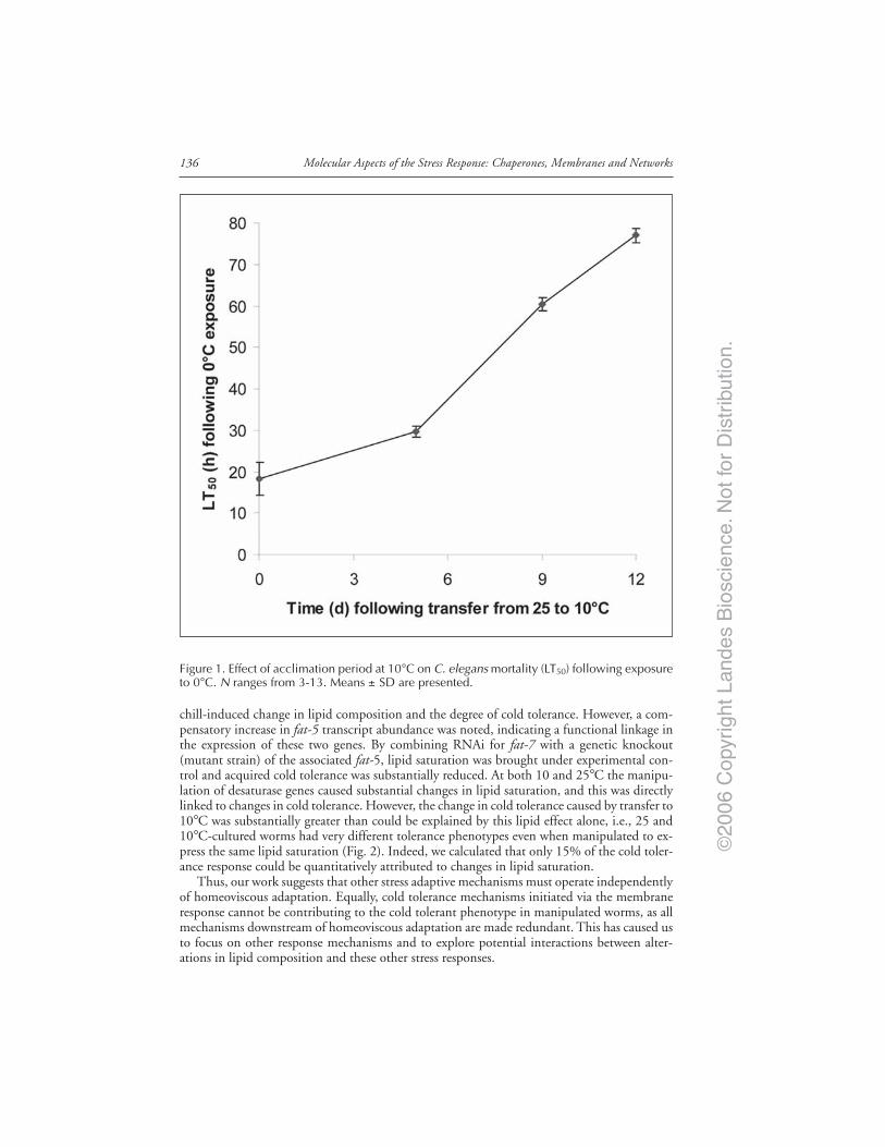
©20
06 C
opyr
ight
Lan
des
Bio
scie
nce.
Not
for
Dis
trib
utio
n.
Molecular Aspects of the Stress Response: Chaperones, Membranes and Networks136
chill-induced change in lipid composition and the degree of cold tolerance. However, a com-pensatory increase in fat-5 transcript abundance was noted, indicating a functional linkage inthe expression of these two genes. By combining RNAi for fat-7 with a genetic knockout(mutant strain) of the associated fat-5, lipid saturation was brought under experimental con-trol and acquired cold tolerance was substantially reduced. At both 10 and 25°C the manipu-lation of desaturase genes caused substantial changes in lipid saturation, and this was directlylinked to changes in cold tolerance. However, the change in cold tolerance caused by transfer to10°C was substantially greater than could be explained by this lipid effect alone, i.e., 25 and10°C-cultured worms had very different tolerance phenotypes even when manipulated to ex-press the same lipid saturation (Fig. 2). Indeed, we calculated that only 15% of the cold toler-ance response could be quantitatively attributed to changes in lipid saturation.
Thus, our work suggests that other stress adaptive mechanisms must operate independentlyof homeoviscous adaptation. Equally, cold tolerance mechanisms initiated via the membraneresponse cannot be contributing to the cold tolerant phenotype in manipulated worms, as allmechanisms downstream of homeoviscous adaptation are made redundant. This has caused usto focus on other response mechanisms and to explore potential interactions between alter-ations in lipid composition and these other stress responses.
Figure 1. Effect of acclimation period at 10°C on C. elegans mortality (LT50) following exposureto 0°C. N ranges from 3-13. Means ± SD are presented.
©20
06 C
opyr
ight
Lan
des
Bio
scie
nce.
Not
for
Dis
trib
utio
n.
137Beyond the Lipid Hypothesis
NonLipid Mechanisms of Cold ToleranceWhile heat stress induces a highly conserved heat shock protein (Hsp) response in all organ-
isms, a similar set of cold inducible proteins have not been identified. Indeed, the applicationof microarrays to study genome-wide responses to cold in different organisms has highlightedconsiderable diversity,51,57-60 and a conserved cold response, certainly in eukaryotes, is notclearly evident. Despite this, several common mechanisms appear to underpin low tempera-ture adaptation at the cellular level, and represent the most likely candidates contributing to astress tolerant phenotype in conjunction with homeoviscous adaptation.
One well studied component of cold adaptation in prokaryotes is the preservation of ribo-some function, with strong evidence suggesting the cold shock response is initiated by an inhi-bition of translation.61 In the bacterium Escherichia coli a family of structurally related pro-teins, CspA, CspB and CspG show the highest induction response to cold, and act as RNAchaperones.61 Homologues in other prokaryotes and eukaryotes have been identified and, whilethese are not always cold inducible,62,63 clearly the preservation of ribosomal function is criticalto organism survival post cold exposure.
Another common component of the cold stress response is the accumulationlow-molecular-mass polyhydric alcohols and sugars, such as glycerol and trehalose.64 Whilethese have primarily been studied with respect to protection against freezing, i.e., ascryoprotectants,64 they can also contribute to chill tolerance.65 In addition, microarrays haveconsistently identified genes regulating the synthesis or transport of cryoprotectants as coldshock stimulons.58,60
The role of HSPs during cold acclimation has received limited attention, but HSP-inducedcross-tolerance between different environmental stressors66 suggests that some underlying mecha-nisms of cold adaptation may be similar to those of heat stress. To date, however, the results aresomewhat equivocal with evidence for both the cold downregulation58 and up-regulation59 ofHsp transcripts.
Figure 2. Relationship between LT50 and SFA content following exposure to 0°C; r2 = 0.813 for25°C worms and 0.997 for 10°C worms. Means ± SD are presented, with N ranging from 3 to10.Control is filled circle, fat-5(tm420): fat-7 RNAi is open triangle.
©20
06 C
opyr
ight
Lan
des
Bio
scie
nce.
Not
for
Dis
trib
utio
n.
Molecular Aspects of the Stress Response: Chaperones, Membranes and Networks138
Thus, multiple mechanisms underpin the phenotypic transition within acquired cold toler-ance. Furthermore, these mechanisms probably do not operate in isolation, but interact closelyin generating the stress tolerant phenotype. Understanding their relative importance and theextent to which they interact, and so offer a redundant capacity, remains an important goal.
Interaction and Compensatory MechanismsAs outlined earlier, interactions between physiological mechanisms of stress adaptation can
result in compensatory responses when one mechanism is perturbed, e.g., between fat-5 andfat-7 in C. elegans. Such interactions and compensatory responses have received limited atten-tion, especially in multi-cellular organisms, but we now know that they are important, andidentifying their complexity is fundamental to understanding ecophysiological adaptability.Studies targeting these interactions could also identify mechanisms of cross-tolerance betweenstress mechanisms, and highlight the best gene targets for the more complete disruption ofstress responses.
There is growing evidence to indicate that membrane lipid order, as affected by lipid satu-ration, is a critical parameter regulating Hsp gene expression. For example, manipulation ofmembrane composition and/or fluidity altered the threshold temperature of Hsp expression inboth yeast67 and bacteria.68 The yeast study also noted that the ∆9-desaturase activity directlyinfluenced Hsp transcription. Recent, as yet unpublished, work from our lab has generatedsimilar results with C. elegans. As described earlier, combining RNAi for fat-7 with a geneticknockout (mutant strain) of the associated fat-5 allowed lipid saturation to be controlled, andthe resultant worms had distinctly lower Hsp70 transcript abundance under heat stress, whencompared to wild type animals (Fig. 3). Circumstantial evidence supporting this relationship isprovided by data from a variety of organisms indicating that temperature acclimation, whichinfluences membrane lipid composition, also changes Hsp expression thresholds.69 Amembrane-Hsp association is further supported by data suggesting that lipid interactions dif-ferentiate the constitutive (Hsc70) and stress-induced (Hsp70) heat shock protein response.70
Recent studies have shown that Hsps, in turn, may be involved in a feedback process by theirstabilizing effects on membrane proteins and lipids.71
Interestingly, trehalose and glycerol are also likely to influence both membrane adaptationand Hsp transcription. Trehalose interacts with, and directly protects, lipid membranes andproteins during cold stress,72 and glycerol can also influence Hsp expression thresholds.73 Thusany alteration in levels of these cryoprotectants during acclimation may interfere withhomeoviscous adaptation and/or the Hsp response.
ConclusionsThe question of cold tolerance in animals remains enigmatic. Much of the work over the
past 50 years has focused on changing lipid composition in the cold, yet this has never beenunequivocally linked to changes in cold tolerance. The evidence is much more persuasive inmicrobes and plants where genetic manipulation has established that lipid biosynthetic genes,notably the desaturases, mediate this effect. In animals, recent work has identified a network ofdesaturase genes expressed in many different tissues, and that they are cold-inducible. Ourwork with the genetic manipulation of ∆9-desaturase in C. elegans offers the first direct test inan animal species of the relationship between lipid saturation and the cold tolerance pheno-type; and our results indicate that, at best, the lipid response accounts for only a small fractionof acquired cold tolerance. C. elegans in many ways is an ideal organism to study the mecha-nisms underpinning environmental adaptation, with access to RNAi libraries and mutant strainsetc., providing the opportunity to quantify the impact of any gene upon environmentally-relevantstress tolerance phenotypes. We can also manipulate genes in groups to overcome the problemof redundant or overlapping mechanisms to study how interactions impact on the phenotype.The key and as yet unanswered question is whether the outcome of this work is directly trans-latable to other animal species, and particularly to vertebrates.
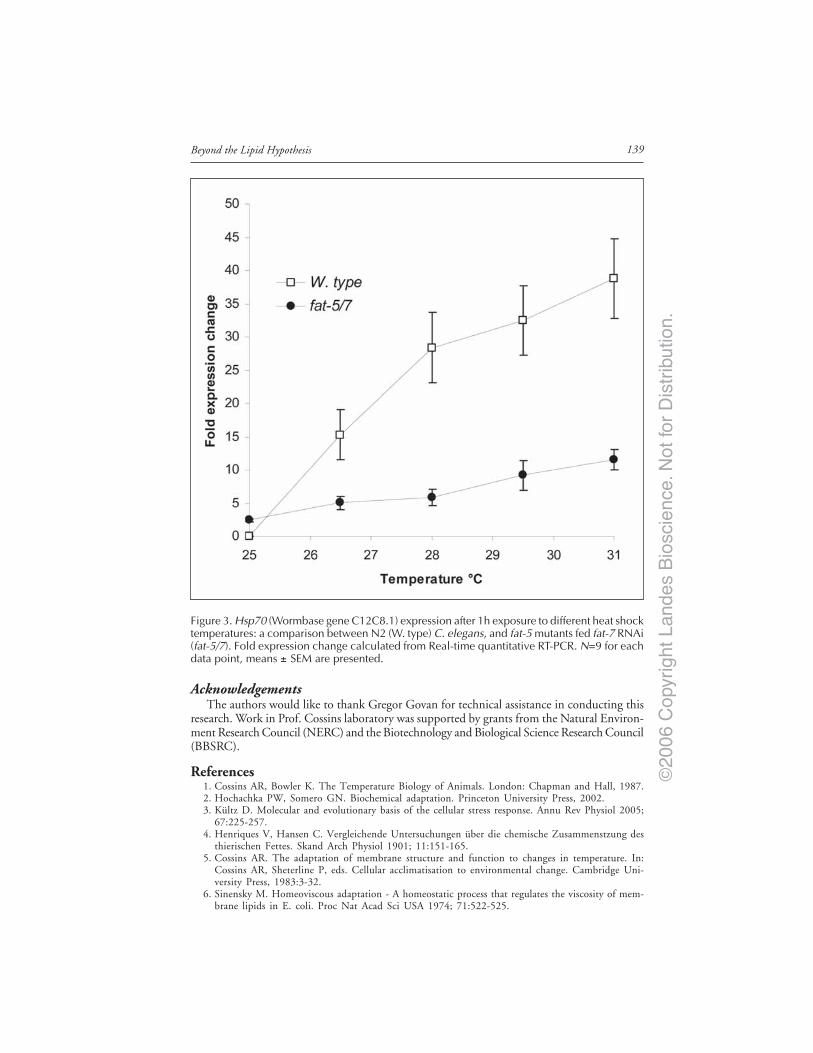
©20
06 C
opyr
ight
Lan
des
Bio
scie
nce.
Not
for
Dis
trib
utio
n.
139Beyond the Lipid Hypothesis
AcknowledgementsThe authors would like to thank Gregor Govan for technical assistance in conducting this
research. Work in Prof. Cossins laboratory was supported by grants from the Natural Environ-ment Research Council (NERC) and the Biotechnology and Biological Science Research Council(BBSRC).
References1. Cossins AR, Bowler K. The Temperature Biology of Animals. London: Chapman and Hall, 1987.2. Hochachka PW, Somero GN. Biochemical adaptation. Princeton University Press, 2002.3. Kültz D. Molecular and evolutionary basis of the cellular stress response. Annu Rev Physiol 2005;
67:225-257.4. Henriques V, Hansen C. Vergleichende Untersuchungen über die chemische Zusammenstzung des
thierischen Fettes. Skand Arch Physiol 1901; 11:151-165.5. Cossins AR. The adaptation of membrane structure and function to changes in temperature. In:
Cossins AR, Sheterline P, eds. Cellular acclimatisation to environmental change. Cambridge Uni-versity Press, 1983:3-32.
6. Sinensky M. Homeoviscous adaptation - A homeostatic process that regulates the viscosity of mem-brane lipids in E. coli. Proc Nat Acad Sci USA 1974; 71:522-525.
Figure 3. Hsp70 (Wormbase gene C12C8.1) expression after 1h exposure to different heat shocktemperatures: a comparison between N2 (W. type) C. elegans, and fat-5 mutants fed fat-7 RNAi(fat-5/7). Fold expression change calculated from Real-time quantitative RT-PCR. N=9 for eachdata point, means ± SEM are presented.

©20
06 C
opyr
ight
Lan
des
Bio
scie
nce.
Not
for
Dis
trib
utio
n.
Molecular Aspects of the Stress Response: Chaperones, Membranes and Networks140
7. Hodkova M, Simek P, Zahradnickova H et al. Seasonal changes in the phospholipid compositionin thoracic muscles of a heteropteran, Pyrrhocoris apterus. Insect Biochem Mol Biol 1999;29:367-376.
8. In: Aloia RC, Curtain CC, Gordon LM eds. Drug and anesthetic effects on membrane structureand function. New York: Wiley, 1991.
9. Heilbrunn LV. The colloid chemistry of protoplasm. IV. The heat coagulation of protoplasm. AmJ Physiol 1924; 69:190-199.
10. Suzuki I, Los DA, Kanesaki Y et al. The pathway for perception and transduction of low-temperaturesignals in Synechocystis. EMBO J 2000; 19:1327-1334.
11. Orr GR, Raison JK. The effect of changing the composition of phosphatidylglycerol from thyla-koid polar lipids of oleander and cucumber on the temperature of the transition related to chillinginjury. Planta 1990; 181:137-143.
12. Cossins AR. Homeoviscous adaptation of biological membranes and its functional significance. In:Cossins AR, ed. Temperature adaptation of biological membranes. London: Portland Press,1994:63-75.
13. Hazel JR, Landrey SR. Time course of thermal adaptation in plasma membranes of trout kidney.I. Headgroup composition. Am J Physiol Regul Int Comp Physiol 1988; 255:R622-627.
14. Hazel JR, Zerba E. Adaptation of biological membranes to temperature: Molecular species compo-sitions of phosphatidylcholine and phosphatidylethanolamine in mitochondrial and microsomalmembranes of liver from thermally-acclimated rainbow trout. J Comp Physiol B Biochem SystEnviron Physiol 1986; 156:665-674.
15. Robertson JC, Hazel JR. Cholesterol content of trout plasma membranes varies with acclimationtemperature. Am J Physiol Regul Int Comp Physiol 1995; 269:R1113-1119.
16. Zehmer JK, Hazel JR. Thermally induced changes in lipid composition of raft and nonraft regionsof hepatocyte plasma membranes of rainbow trout. J Exp Biol 2005; 208:4283-4290.
17. Murata N, Suzuki I. Exploitation of genomic sequences in a systematic analysis to access howcyanobacteria sense environmental stress. J Exp Bot 2006; 57:235-247.
18. Los DA, Murata N. Structure and expression of fatty acid desaturases. Biochim Biophys Acta 1998;1394:3-15.
19. Murata N, Wada H. Acyl-lipid desaturases and their importance in the tolerance and acclimatiza-tion to cold of cyanobacteria. Biochem J 1995; 308:1-8.
20. Tasaka Y, Gombos Z, Nishiyama Y et al. Targeted mutagenesis of acyl-lipid desaturases inSynechosystis: Evidence for the important roles of polyunsaturated membrane lipids in growth,respiration and photosynthesis. Embo J 1996; 15:6416-6425.
21. Sakamoto T, Shen GZ, Higashi S et al. Alteration of low-temperature susceptibility of thecyanobacterium Synechosystis sp. PCC 7002 by genetic manipulation of membrane lipidunsaturation. Arch Microbiol 1997; 169:20-28.
22. Kanervo E, Tasaka Y, Murata N et al. Membrane lipid unsaturation modulates processing of thephotosystem II reaction-center protein D1 at low temperatures. Plant Physiol 1997; 114:841-849.
23. Wada H, Gombos Z, Murata N. Enhancement of chilling tolerance of a cyanobacterium by ge-netic manipulation of fatty acid desaturation. Nature 1990; 347:200-203.
24. Vigh L, Los DA, Horvath I et al. The primary signal in the biological perception of temperature:Pd-catalysed hydrogenation of membrane lipids stimulates the expression of the desA gene inSynechosystis PCC 6803. Proc Nat Acad Sci USA 1993; 90:9090-9094.
25. St. John JB, Christiansen MN, Ashworth EN et al. Effect of BASF 13-338, a substitutedpyridazinone, on linolenic acid levels and winterhardiness of cereals. Crop Sci 1979; 19:65-69.
26. Somerville C, Browse J. Plant lipids: Metabolism, mutants and membranes. Science 1991; 252:80-87.27. Browse J, Kunst L, Anderson S et al. A mutant of Arabidopsis deficient in the chloroplast 16:1/
18:1 desaturase. Plant Physiol 1989; 90:522-529.28. Kunst L, Browse J, Somerville C. A mutant of Arabidopsis deficient in desaturation of palmitic
acid in leaf lipids. Plant Physiol 1989; 90:943-947.29. Lemieux B, Miquel M, Somerville C et al. Mutants of Arabidopsis with alterations in seed lipid
fatty acid composition. Theor Appl Genet 1990; 80:234-240.30. Hugly S, Somerville C. A role for membrane lipid polyunsaturation in chloroplast biogenesis at
low temperature. Plant Physiol 1992; 99:197-202.31. Miquel M, James D, Dooner H et al. Arabidopsis requires polyunsaturated lipids for low-temperature
survival. Proc Nat Acad Sci USA 1993; 90:6208-6212.32. Kodama H, Hamada T, Horiguchi G et al. Genetic enhancement of cold tolerance by expression
of a gene for chloroplast ω-3 fatty acid desaturase in transgenic tobacco. Plant Physiol 1994;105:601-605.

©20
06 C
opyr
ight
Lan
des
Bio
scie
nce.
Not
for
Dis
trib
utio
n.
141Beyond the Lipid Hypothesis
33. Ishizaki-Nishizawa O, Fujii T, Azuma M et al. Low-temperature resistance of higher plants issignificantly enhanced by nonspecific cyanobacterial desaturase. Nat Biotechnol 1996; 14:1003-1006.
34. Orlova IV, Serebriiskaya TS, Popov V et al. Transformation of tobacco with a gene for the ther-mophilic acyl-lipid desaturase enhances the chilling tolerance of plants. Plant cell Physiol 2003;44:447-450.
35. Murata N, Ishizaki-Nishizawa O, Higashi S et al. Genetically engineered alteration in the chillingsensitivity of plants. Nature 1992; 356:710-713.
36. Sakamoto A, Sulpice R, Hou CX et al. Genetic modification of the fatty acid unsaturation ofphosphatidylglycerol in chloroplasts alters the sensitivity of tobacco plants to cold stress. Plant CellEnviron 2003; 27:99-105.
37. Moon BY, Higashi S, Gombos Z et al. Unsaturation of the membrane lipids of chloroplasts stabi-lizes the photosynthetic machinery against low-temperature photoinhibition in transgenic tobaccoplants. Proc Nat Acad Sci USA 1995; 92:6219-6223.
38. Murakami Y, Tsuyama M, Kobayashi Y et al. Tienoic fatty acids and plant tolerance of hightemperature. Science 2000; 287:476-479.
39. Eguchi E, Ogawa Y, Okamoto K et al. Fatty acid compositions of arthropod and cephalopodphotoreceptors - Interspecific, seasonal and development studies. J Comp Physiol B 1994;164:94-102.
40. Kostal V, Simek P. Changes in fatty acid composition of phospholipids and triacylglycerols aftercold-acclimation of an aestivating insect prepupa. J Comp Physiol B 1998; 168;453-460.
41. Cuculescu M, Pearson T, Hyde D et al. Heterothermal acclimation: An experimental paradigm forstudying the control of thermal acclimation in crabs. Proc Nat Acad Sci USA 1999; 96:6501-6505.
42. Lahdes E, Balogh G, Fodor E et al. Adaptation of composition and biophysical properties of phos-pholipids to temperature by the crustacean, Gammarus spp. Lipds 2000; 35:1093-1098.
43. Bayley M, Peterson SO, Knigge T et al. Drought acclimation confers cold tolerance in the soilcollembolan Folsomia candida. J Insect Physiol 2001; 47:1197-1204.
44. Hall JM, Parrish CC, Thompson RJ. Eicosapentaenoic acid regulates scallop (Placopectenmagellanicus) membrane fluidity in response to cold. Biol bull 2002; 202:201-203.
45. Cossins AR, Crawford DL. Fish as models for environmental genomics. Nat Rev Gen 2005;6:324-333.
46. Logue JA, de Vries AL, Fodor E et al. Lipid compositional correlates of temperature-adaptive in-terspecific differences in membrane physical structure. J Exp Biol 2000; 203:2105-2115.
47. Wodtke E, Teichert T, Konig, A. Control of membrane fluidity in carp upon cold stress: Studieson fatty acid desaturases. In: Heller HC, ed. Living in the Cold: Physiological and BiochemicalAdaptations. Elsevier, 1986:35-42.
48. Wodtke E, Cossins AR. Rapid cold-induced changes of membrane order and ∆9-desaturase activityin endoplasmic reticulum of carp liver: A time-course study of thermal acclimation. Biochim BiophysActa 1991; 1064:343-350.
49. Tiku PE, Gracey AY, Macartney AI et al. Cold-induced expression of ∆9-desaturase by transcrip-tional and post-translational mechanisms. Science 1996; 271:815-818.
50. Polley SD, Tiku PE, Trueman RT et al. Differential expression of cold- and diet-specific genesencoding two carp liver ∆9-acyl-CoA desaturase isoforms. Am J Physiol 2003; 284:R41-R50.
51. Gracey AY, Fraser EJ, Li W et al. Coping with cold: An integrative, multi-tissue analysis of thetranscriptome of a poikilothermic vertebrate. Proc Nat Acad Sci USA 2004; 101:6970-16975.
52. Greenberg AJ, Moran JR, Coyne JA et al. Ecological adaptation during incipient speciation re-vealed by precise gene replacement. Science 2003; 302:1754-1757.
53. Overgaard J, Sørensen JG, Petersen SO et al. Changes in membrane lipid composition followingrapid cold hardening in Drosophila melanogaster. J Insect Physiol 2005; 51:1173-1182.
54. In: C. elegans II. Riddle DL, Blumenthal T, Meyer BJ, Priess JR, eds. Cold Spring Harbor Labo-ratory Press, 1997.
55. Fire A, Xu SQ, Montgomery MK et al. Potent and specific genetic interference by double-strandedRNA in Caenorhabditis elegans. Nature 1998; 391:806-811.
56. Murray PA, Hayward SAL, Gracey AY et al. Proc Natl Acad Sci USA, (In submission).57. Phadtare S, Inouye M. Genome-wide transcriptional analysis of the cold shock response in wild-type
and cold-sensitive, quadrupole-csp-deletion strains of Escherichia coli. J Bacteriol 2004;186:7007-7014.
58. Han Y, Zhou D, Pang X et al. DNA microarray analysis of the heat- and cold-shock stimulons inYersinia pestis. Microb Infect 2005; 7:335-348.
59. Qin W, Neal SJ, Robertson RM et al. Cold hardening and transcriptional change in Drosophilamelanogaster. Insect Mol Biol 2005; 14:607-613.
©20
06 C
opyr
ight
Lan
des
Bio
scie
nce.
Not
for
Dis
trib
utio
n.
Molecular Aspects of the Stress Response: Chaperones, Membranes and Networks142
60. Hannah MA, Heyer AG, Hincha DK. A global survey of gene regulation during cold acclimationin Arabidopsis thaliana. PLoS Gen 2005; 1:179-196.
61. Thieringer HA, Jones PG, Inouye M. Cold shock and adaptation. BioEssays 1998; 20:49-57.62. Schroder K, Zuber P, Willimsky G et al. Mapping of the Bacillus subtilis cspB gene and cloning
of its homologs in thermophilic, mesophilic and psychrotropic bacilli. Gene 1993; 136:277-280.63. Wolffe AP. Structural and functional properties of the evolutionary ancient Y-box family of nucleic
acid binding protein. BioEssays 1994; 16:245-250.64. Denlinger DL, Lee RE. Physiology of cold sensitivity. In: Hallman GJ, Denlinger DL, eds. Tem-
perature sensitivity in insects and application in integrated pest management. Boulder: WestviewPress, 1998:55-95.
65. Slachta M, Vambera J, Zahradnickova H et al. Entering diapause is a prerequisite for successfulcold-acclimation in adult Graphosoma lineatum (Heteroptera: Pentatomidae). J Insect Physiol 2002;48:1031-1039.
66. Sabehat A, Weiss D, Lurie S. Heat-shock proteins and cross tolerance in plants. Physiol Plant1998; 103:437-441.
67. Carratù L, Franceschelli S, Pardini CL et al. Membrane lipid perturbation modifies the set pointof the temperature of heat shock response in yeast. Proc Nat Acad Sci USA 1996; 93:3870-3875.
68. Horváth I, Glatz A, Varvasovszki V et al. Membrane physical state controls the signaling mecha-nism of the heat shock response in Synechocystis PCC 6803: Identification of hsp17 as a “fluiditygene”. Proc Nat Acad Sci USA 1998; 95:3513-3518.
69. Barua D, Heckathorn SA. Acclimation of the temperature set-points of the heat-shock response. JTherm Biol 2004; 29:185-193.
70. Arispe N, Doh M, De Maio A. Lipid interaction differentiates the constitutive and stress-inducedheat shock proteins Hsc70 and Hsp70. Cell Stress Chap 2002; 7:330-338.
71. Vigh L, Maresca B, Harwood JL. Does the membrane’s physical state control the expression ofheat shock and other genes? Trends Biochem Sci 1998; 23:369-374.
72. Behm CA. The role of trehalose in the physiology of Nematodes. Int J Parasitol 1997; 27:215-229.73. Brown CR, Hong-Brown LQ, Doxsey SJ et al. Molecular chaperones and the centrosome. J Biol
Chem 1996; 271:833-840.

©20
06 C
opyr
ight
Lan
des
Bio
scie
nce.
Not
for
Dis
trib
utio
n.
CHAPTER 13
*Corresponding Author: Dr. John H. Crowe—Section of Molecular and Cellular Biology,University of California, Davis, California 95616, U.S.A. Email: [email protected]
Molecular Aspects of the Stress Response: Chaperones, Membranes and Networks,edited by Peter Csermely and Laszlo Vigh. ©2007 Landes Bioscienceand Springer Science+Business Media.
Trehalose As a “Chemical Chaperone”:Fact and FantasyJohn H. Crowe*
Abstract
Trehalose is a disaccharide of glucose that is found at high concentrations in a widevariety of organisms that naturally survive drying in nature. Many years ago wereported that this molecule has the remarkable ability to stabilize membranes and pro-
teins in the dry state. A mechanism for the stabilization rapidly emerged, and it was sufficientlyattractive that a myth grew up about trehalose as a universal protectant and chemical chaper-one. Many of the claims in this regard can be explained by what is now known about thephysical properties of this interesting sugar. It is emerging that these properties may make itunusually useful in stabilizing intact cells in the dry state.
Sugars and Stabilization of Biological MaterialsWe reported two decades ago that biomolecules and molecular assemblages such as mem-
branes and proteins can be stabilized in the dry state in the presence of a sugar found at highconcentrations in many anhydrobiotic organisms, trehalose.1 We also showed that trehalosewas clearly superior to other sugars in this regard.2 This effect seemed so clear it quickly led towide-spread, and often uncritical, use of the sugar for preservation and other purposes. In fact,an array of applications for trehalose have been reported, ranging from stabilization of vaccinesand liposomes to hypothermic storage of human organs.3 Other workers showed that it mighteven be efficacious in treatment of dry eye syndrome or dry skin in humans.4,5 Trehalose isprominently listed as an ingredient in cosmetics, apparently because it is reputed to inhibitoxidation of certain fatty acids in vitro that might be related to body odor.6 Trehalose has beenshown by several groups to suppress free radical damage, protect against anoxia, inhibit dentalcaries, enhance ethanol production during fermentation, stabilize the flavor in foods, and toprotect plants against physical stress.7-13 According to one group, trehalose inhibits bone re-sorption in ovariectomized mice, apparently by suppressing osteoclast differentiation; the sug-gestion followed that trehalose might be used to treat osteoporosis in humans.14,15 More re-cently, Tanaka et al16 reported that trehalose could be used to inhibit the protein aggregationassociated with Huntington’s disease in vivo in a rat model for this disease. That report that hasalready led to an unorthodox clinical trial in humans.17
A myth has grown up about trehalose and its properties, as a result of which it is beingapplied, sometimes rather uncritically, to a myriad of biological and clinical problems. Thus,we are making special efforts in the literature to clarify the properties of trehalose that make ituseful for stabilization of biomaterials and to dispel the most misleading aspects of this myth.
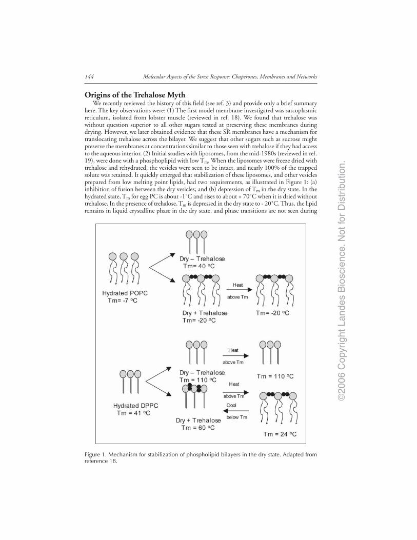
©20
06 C
opyr
ight
Lan
des
Bio
scie
nce.
Not
for
Dis
trib
utio
n.
Molecular Aspects of the Stress Response: Chaperones, Membranes and Networks144
Origins of the Trehalose MythWe recently reviewed the history of this field (see ref. 3) and provide only a brief summary
here. The key observations were: (1) The first model membrane investigated was sarcoplasmicreticulum, isolated from lobster muscle (reviewed in ref. 18). We found that trehalose waswithout question superior to all other sugars tested at preserving these membranes duringdrying. However, we later obtained evidence that these SR membranes have a mechanism fortranslocating trehalose across the bilayer. We suggest that other sugars such as sucrose mightpreserve the membranes at concentrations similar to those seen with trehalose if they had accessto the aqueous interior. (2) Initial studies with liposomes, from the mid-1980s (reviewed in ref.19), were done with a phosphoplipid with low Tm. When the liposomes were freeze dried withtrehalose and rehydrated, the vesicles were seen to be intact, and nearly 100% of the trappedsolute was retained. It quickly emerged that stabilization of these liposomes, and other vesiclesprepared from low melting point lipids, had two requirements, as illustrated in Figure 1: (a)inhibition of fusion between the dry vesicles; and (b) depression of Tm in the dry state. In thehydrated state, Tm for egg PC is about -1˚C and rises to about + 70˚C when it is dried withouttrehalose. In the presence of trehalose, Tm is depressed in the dry state to - 20˚C. Thus, the lipidremains in liquid crystalline phase in the dry state, and phase transitions are not seen during
Figure 1. Mechanism for stabilization of phospholipid bilayers in the dry state. Adapted fromreference 18.

©20
06 C
opyr
ight
Lan
des
Bio
scie
nce.
Not
for
Dis
trib
utio
n.
145Trehalose as a “Chemical Chaperone”: Fact and Fantasy
rehydration. The significance of this phase transition during rehydration is that, when phos-pholipids pass through such transitions, the bilayer becomes transiently leaky. The physicalbasis for this leakiness has recently been investigated in some detail.20 These effects were re-ported first for trehalose (reviewed in ref. 19). When we compared the effects of other sugarsand polymers on the preservation, we found that, with vesicles made from lipids with low Tm,trehalose appeared to be significantly superior to the best of the additives tested. Oligosaccha-rides larger than trisaccharides did not work at all.21 Other sugars, particularly disaccharides,did provide good stabilization of egg PC vesicles in the dry state, but much higher concentra-tions than trehalose were required, at least according to initial reports. However, as freeze-dryingtechnology improved, the differences between disaccharides tended to disappear, and the mytheventually got modified to encompass disaccharides in general. Nevertheless, the observationthat trehalose was significantly more effective at low concentrations under suboptimal condi-tions for freeze drying requires explanation, which we provide later. (3) At first it appeared thatthe ability to preserve liposomes in the dry state is restricted to disaccharides. Subsequently, wefound this is not the case. For example, DPPC is a lipid with saturated acyl chains and thus anelevated Tm (41˚C). When it is dried without trehalose Tm rises to about 110oC; with trehalosepresent Tm rises to about 65oC (reviewed in ref. 22). Thus, DPPC is in gel phase at all stages ofthe freeze-drying and rehydration process, and one would expect that inhibition of fusionmight be sufficient for the stabilization. In other words, any inert solute that would separatethe vesicles in the dry state and thus prevent aggregation and fusion should stabilize the dryvesicles. That appears to be the case; a high molecular weight (450,000) HES has no effect onTm in dry DPPC, but preserves the vesicles, nevertheless.
The Mechanism of Depression of TmThe Mechanism of Depression of Tm has received a great deal of attention since the discov-
ery of this effect.23 Three main hypotheses have emerged: The water replacement hypothesissuggests that sugars can replace water molecules by forming hydrogen binds with polar resi-dues, thereby stabilizing the structure in the absence of water.3,22,24-26 The water entrapmenthypothesis suggests that sugars concentrate water near surfaces, thereby preserving its salva-tion.27-29 The vitrification hypothesis suggests that the sugars form amorphous glasses, thusreducing structural fluctuations.30-31
A consensus has emerged that these three mechanisms are not mutually exclusive (reviewedin ref. 3). Vitrification may occur simultaneously with direct interactions between the sugarand polar residues. Direct interaction, on the other hand, has been demonstrated by a widevariety of physical techniques, including infrared spectroscopy, NMR, and X-ray.23,32-36
Theoretical analyses have contributed greatly to this field in recent years. Chandrasekharand Gaber37 and Rudolph et al38 in the earliest studies, showed that trehalose can form ener-getically stable conformations with phospholipids, binding three adjacent phospholipids in thedry state. Similarly, trehalose-protein interactions have been studied by simulations, with simi-lar conclusions.28,29 More recently, Sum et al39 showed by molecular simulations that the sug-ars adapt molecular conformations that permit them to fit onto the surface topology of thebilayer through hydrogen bonds. The sugars interact with up to three adjacent phospholipids.Pereira et al40 produced complementary results from molecular dynamics simulations, withcomparable conclusions.
Trehalose Stabilizes Microdomains in MembranesPhase separation is segregation of membrane components in the plane of the bilayer. Al-
though there are lingering doubts about whether or not phase separated domains in native mem-branes are real (see refs. 41-43) or artifacts (see refs. 44-46), there is abundant evidence that thesedomains, known as “rafts”, are involved in such processes (among others) as signaling, endocytosi,and viral assembly.47-51 Although several forces are involved, one of the main driving forces forphase separation is the hydrophobic mismatch, which arises from a difference in membrane
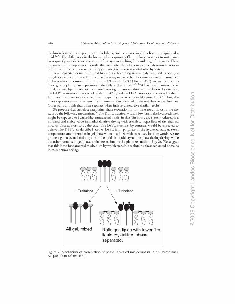
©20
06 C
opyr
ight
Lan
des
Bio
scie
nce.
Not
for
Dis
trib
utio
n.
Molecular Aspects of the Stress Response: Chaperones, Membranes and Networks146
thickness between two species within a bilayer, such as a protein and a lipid or a lipid and alipid.52,53 The differences in thickness lead to exposure of hydrophobic residues to water and,consequently, to a decrease in entropy of the system resulting from ordering of the water. Thus,the assembly of components of similar thickness into relatively homogeneous domains is entropi-cally driven. The net increase in entropy driving the process is contributed by water.
Phase separated domains in lipid bilayers are becoming increasingly well understood (seeref. 54 for a recent review). Thus, we have investigated whether the domains can be maintainedin freeze-dried liposomes. DLPC (Tm = 0˚C) and DSPC (Tm = 50˚C) are well known toundergo complete phase separation in the fully hydrated state.55,56 When these liposomes weredried, the two lipids underwent extensive mixing. In samples dried with trehalose, by contrast,the DLPC transition is depressed to about -20˚C, and the DSPC transition increases by about10˚C and becomes more cooperative, suggesting that it is more like pure DSPC. Thus, thephase separation—and the domain structure—are maintained by the trehalose in the dry state.Other pairs of lipids that phase separate when fully hydrated give similar results.
We propose that trehalose maintains phase separation in this mixture of lipids in the drystate by the following mechanism.36 The DLPC fraction, with its low Tm in the hydrated state,might be expected to behave like unsaturated lipids, in that Tm in the dry state is reduced to aminimal and stable value immediately after drying with trehalose, regardless of the thermalhistory. That appears to be the case. The DSPC fraction, by contrast, would be expected tobehave like DPPC, as described earlier. DSPC is in gel phase in the hydrated state at roomtemperature, and it remains in gel phase when it is dried with trehalose. In other words, we areproposing that by maintaining one of the lipids in liquid crystalline phase during drying, whilethe other remains in gel phase, trehalose maintains the phase separation (Fig. 2). We suggestthat this is the fundamental mechanism by which trehalose maintains phase separated domainsin membranes drying.
Figure 2. Mechanism of preservation of phase separated microdomains in dry membranes.Adapted from reference 54.

©20
06 C
opyr
ight
Lan
des
Bio
scie
nce.
Not
for
Dis
trib
utio
n.
147Trehalose as a “Chemical Chaperone”: Fact and Fantasy
There Is More Than One Way to the Same EndAlthough the occurrence of trehalose at high concentrations is common in anhydrobiotic
animals, some such animals have only small amounts of trehalose or none at all.57-60 It is tempt-ing to construe these findings as evidence against a role for sugars in anhydrobiosis.61 We suggestthat it is not the sugars per se that are of interest in this regard, but rather the physical principlesof the requirements for stabilization, as described above. There are multiple ways to achieve suchstabilization: (1) Hydroxyethyl starch (HES) alone will not stabilize dry membrane vesicles com-posed of lipids with low Tm, but a combination of a low molecular weight sugar such as glucoseand HES can be effective.62 Here is the apparent mechanism: glucose depresses Tm in the drylipid, but has little effect on inhibiting fusion, except at extremely high concentrations. On theother hand, the polymer has no effect on the phase transition, but inhibits fusion. Thus, thecombination of the two meets both requirements, while neither alone does so.62 It seems likelythat such combinations of molecules might be found in anhydrobiotes in nature. (2) In fact, aglycan isolated from the desiccation tolerant alga Nostoc apparently works in conjunction witholigosaccharides.63 Similarly, certain proteins have been shown to affect the phase state of thesugars and either enhance or are required for stabilization (reviewed in ref. 64). (3) Hincha etal65 have shown that fructans from desiccation tolerant higher plants will by themselves bothinhibit fusion and reduce Tm in dry phospholipids such as egg PC. The mechanism behind thiseffect is still unclear. The interaction is similar to that shown by sugars, but it is also specific tofructans and is not shown by other polymers.66 In a related study, Hincha et al67 reported that aseries of raffinose family oligosaccharides are all capable of stabilizing dry liposomes. (4) Hinchaand Hagemann68 recently studied effects of other compatible solutes on stabilization of lipo-somes by sugars. This approach is in its earliest stages, but those authors found that some com-patible solutes improve the stabilization in the presence of sugars, suggesting that the solutesmight decrease the amount of sugar required in vivo. (5) Hoekstra and Golovina69 have reportedthat amphiphiles that are free in the cytoplasm in fully hydrated cells of anhydrobiotes appar-ently insert into membranes during dehydration. The role of this phenomenon in stabilizationis uncertain, but presumably the amphiphiles alter the order of the acyl chains. (6) Goodrich etal70 reported that disaccharides tethered to the bilayer surface by a flexible linker esterified tocholesterol has an effect on membrane stability similar to that seen in the free sugar. Such mol-ecules could provide stability in anhydrobiotes, although they have not yet been reported. How-ever, Popova and Hincha71 recently found that digalactosyl diacylglycerol depressed Tm in dryphospholipids, perhaps in keeping with this suggestion.
The point is there are many ways to achieve stability. Once an understanding of the physicalrequirements for preservation was achieved, it became apparent that many routes can lead tothe same end. Similar observations on the stability of dry proteins have been made by Carpen-ter and his group, with similar conclusions.24,25
Trehalose Has Useful Properties, NeverthelessWe implied above that trehalose works well for freeze-drying liposomes under less than
optimal conditions. The same applies for storage under conditions that would normally de-grade the biomaterial. Bacteria freeze-dried in the presence of trehalose showed remarkablyhigh survival immediately after freeze drying. Furthermore, t the bacteria freeze-dried withtrehalose retained high viability even after long exposure to moist air.72 By contrast, when thebacteria were freeze-dried with sucrose they showed lower initial survival, and when they wereexposed to moist air viability deceased rapidly. Further, when immunoconjugates were freezedried with trehalose or other disaccharides all the sugars provided reasonable levels of preserva-tion. However, when the dry samples were stored at high relative humilities and temperatures,those dried with trehalose were stable for much longer than those dried with other sugars.73
This finding is of some considerable significance since there is a need for shippingimmunoconjugates, vaccines, antisera and the like to locales where they would be exposed tohigh temperatures and humidities as soon as they are exposed to air.
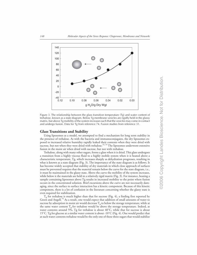
©20
06 C
opyr
ight
Lan
des
Bio
scie
nce.
Not
for
Dis
trib
utio
n.
Molecular Aspects of the Stress Response: Chaperones, Membranes and Networks148
Glass Transitions and StabilityUsing liposomes as a model, we attempted to find a mechanism for long term stability in
the presence of trehalose. As with the bacteria and immunoconjugates, the dry liposomes ex-posed to increased relative humidity rapidly leaked their contents when they were dried withsucrose, but not when they were dried with trehalose.31,74 The liposomes underwent extensivefusion in the moist air when dried with sucrose, but not with trehalose.
Trehalose, along with many other sugars, forms a glass when it is dried. This glass undergoesa transition from a highly viscous fluid to a highly mobile system when it is heated above acharacteristic temperature, Tg, which increases sharply as dehydration progresses, resulting inwhat is known as a state diagram (Fig. 3). The importance of the state diagram is as follows. Ithas become widely accepted that stability of dry materials in which close approach of surfacesmust be prevented requires that the material remain below the curve for the state diagram, i.e.,it must be maintained in the glassy state. Above the curve the mobility of the system increases,while below it the materials are held in a relatively rigid matrix (Fig. 3). For instance, heating asample containing liposomes above Tg results in increased mobility to the point where fusionoccurs in the concentrated solution. Brief excursions above the curve are not necessarily dam-aging, since the surface to surface interaction has a kinetic component. Because of this kineticcomponent, there is a lot of confusion in the literature concerning whether the glassy state iseven required for stabilization.
Tg for trehalose is much higher than that for sucrose (Fig. 4), a finding first reported byGreen and Angell.75 As a result, one would expect that addition of small amounts of water tosucrose by adsorption in moist air would decrease Tg to below the storage temperature, while atthe same water content Tg for trehalose would be above the storage temperature. Indeed, atwater contents around 5%, Tg for trehalose is about 40˚C, while that for sucrose is about15˚C. Tg for glucose at a similar water content is about -10˚C (Fig. 4). One would predict thatat such water contents trehalose would be the only one of these three sugars that would stabilize
Figure 3. The relationship between the glass transition temperature (Tg) and water content oftrehalose, known as a state diagram. Below Tg membrane vesicles are rigidly held in the glassymatrix, but above Tg mobility of the system increases such that the vesicles may come in contactand undergo fusion. Data for Tg from reference 74. Fusion studies from reference 31.

©20
06 C
opyr
ight
Lan
des
Bio
scie
nce.
Not
for
Dis
trib
utio
n.
149Trehalose as a “Chemical Chaperone”: Fact and Fantasy
the sample, and this appears to be the case. This would seem to provide an explanation for thesuperior stability of, for example, the immunoconjugates stored in sucrose or trehalose de-scribed above. We stress, however, that the elevated Tg seen in trehalose is not anomalous.Indeed, trehalose lies at the end of a continuum of sugars that show increasing Tg (40), al-though the basis for this effect is not understood.
NonEnzymatic Browning and Stability of the Glycosidic BondThe Maillard (browning) reaction between reducing sugars and proteins in the dry state has
often been invoked as a major source of damage,76 and the fact that both sucrose and trehaloseare nonreducing sugars may explain at least partly why they are the natural products accumu-lated by anhydrobiotic organisms. However, the glycosidic bonds linking the monomers insucrose and trehalose have very different susceptibilities to hydrolysis.77,78 For instance, theactivation energy for acid hydrolysis in aqueous solution is nearly twice that that for otherdisaccharides.77 When O’Brien77 and subsequently Schebor et al78 incubated a freeze driedmodel system (albumin, with the addition of lysine) with sucrose, trehalose, and glucose atrelative humidities in excess of 20%, the rate of browning seen with sucrose approached that ofglucose—as much as 2000 times faster than that with trehalose, although they observed adistinct lag in the onset of browning (Fig. 5). Schebor et al found that a peak in the appearanceof monosaccharides occurs prior to the onset of browning, after which free monosaccharidesdecline, coincidentally with the onset of browning (Fig. 5). These observations strongly suggestthat the browning seen with sucrose—but not with trehalose—is due to hydrolysis of theglycosidic bond during storage.
The glassy state is undoubtedly related to these effects; if the samples are stored at very lowhumidities only minimal amounts of hydrolysis and subsequent browning were seen in thesucrose preparations.77,78 Nevertheless, since sucrose is the major sugar associated with desicca-tion tolerance in higher plants, consideration of the mechanisms by which devitrification atmoderate water contents and hydrolysis of the glycosidic bond in sucrose glasses are obviated inanhydrobiotic plants is instructive.
Figure 4. Comparison of state diagrams for trehalose, sucrose, and glucose. Data fromreference 74.
©20
06 C
opyr
ight
Lan
des
Bio
scie
nce.
Not
for
Dis
trib
utio
n.
Molecular Aspects of the Stress Response: Chaperones, Membranes and Networks150
Sugar Glasses in Plant AnhydrobiotesBuitink has published an elegant series of studies of the properties of glasses in vivo in
anhydrobiotic plants (see refs. 79,80), along with a superb review of the work (see ref. 81).Briefly, Koster82 found that mixtures of sugars similar to those found in desiccation tolerant(corn) embryonic axes (85% (w/w) sucrose, 15% (w/w) raffinose) formed glasses at tempera-tures above 0oC, while sugar mixtures similar to those found in desiccation-sensitive axes (75%(w/w) glucose, 25% (w/w) sucrose) formed glasses only at subzero temperatures. These andsimilar data suggested that sugar mixtures form glasses in plant anhydrobiotes, but subsequentstudies indicated that the intracellular glasses are not composed of sugars alone. The statediagram for intact pollen of cat tail at first glance seems to agree reasonably well with that forsucrose (the major sugar in these pollen grains). However, subtle differences can be seen that
Figure 5. Browning of dry proteins (top) and release of monosaccharides by trehalose and sucroseduring storage. Adapted from reference 78.

©20
06 C
opyr
ight
Lan
des
Bio
scie
nce.
Not
for
Dis
trib
utio
n.
151Trehalose as a “Chemical Chaperone”: Fact and Fantasy
have turned out to be significant: at low water contents Tg in the intact pollen is higher thanpredicted based on the state diagram for the sugar, while at low water contents it is lower thanpredicted.79 Furthermore, the temperature at which the glass collapses (Tc), which occurs sev-eral degrees above Tg, is elevated by as much as 40˚C in intact pollen and otheranhydrobiotes.80,81 The outcome of these studies is a clear indication that glasses in intactanhydrobiotic plants are not composed simply of mixtures of sugars.
Wolkers et al83 developed a powerful approach based on infrared spectroscopy that permit-ted characterization of cytoplasmic glasses. The measurement—vibrational frequency of the-OH stretch in sugars—permitted an estimate of the length and strength of hydrogen bondswithin the glass. Using this technique, Wolkers et al84,85 found that the molecular density ofthe cytoplasm resembled that of protein glasses more than that of sucrose, a finding that ini-tially suggested that sucrose may be a relatively minor player in formation of the cytoplasmicglass. However, studies on molecular motion in protein glasses have shown that rotationalmobility of the proteins is almost twice that seen in the cytoplasmic glass.80 The conclusion isthat the cytoplasmic glass is likely to consist of a mixture of sucrose and proteins. The mostlikely candidates for the protein component are the late embryogenesis abundant (LEA) pro-teins that are accumulated in seeds and pollen late in development, and there is some evidencesuggesting that this is the case; when extracts are made from wheat embryos a sucrose in largeamounts was coisolated with the LEA proteins.86 Exhaustive dialysis removed only a fractionof the sucrose, indicating that it is tightly bound to the protein.
The conclusion from these studies is that at least in plants cytoplasmic glasses consist ofsugar-protein mixtures. The apparent elevation of Tg and the collapse temperature by additionof the protein to the glass is likely to lead to increased stability of the kind seen in trehalosealone in vitro, owing to its elevated Tg. Thus, devitrification at moderate water contents isobviated. The problem of stability of the glycosidic bond in sucrose during storage in the drystate is somewhat more problematic, but it seems likely that the association with the proteinfraction, leading to the elevated Tg, could limit accessibility of water to the bond, thus limitinghydrolysis.
Lessons from Nature Can Be Used to Preserve Intact Cellsin the Dry State
Clearly, trehalose must be introduced into the cytoplasm of a cell if it is to be effective atstabilizing intracellular proteins and membranes during dehydration. Previous efforts centeredaround this fundamental problem involving molecular engineering have not been particularlysuccessful.87,88 More recently, Wolkers et al89 made the surprising discovery that when humanblood platelets are placed in the presence of modest amounts of trehalose, they take it up byfluid phase endocytosis, and the intact sugar ends up in the cytoplasm. Wolkers et al were ableto show only indirectly that trehalose is in the cytoplasm, but subsequent studies have shownthat this is so. Oliver et al90 have followed the fate of the sugar once it enters the endocytoticpathway in a stem cell line, using fluorescence microscopy. The fluorescence initially appears invesicles, but with time it becomes diffuse, suggesting that the sugar is released into the cyto-plasm. The mechanism of release is not entirely clear, but we have proposed that the sugarsfollow the pathway described in Figure 6. The endocytotic vesicles progress through the nor-mal pathway to lysosomes. It is well established that low molecular weight compounds such asglucose readily cross the lysosomal membrane into the cytoplasm, but there is very little evi-dence concerning the fate of disaccharides in lysosomes.91 Incubation of cells in sucrose led topersistent vesiculation, suggesting that, apparently unlike trehalose, sucrose is retained in thelysosomes. This seems surprising because even though lysosomes lack invertase and thus can-not break the glycosidic bond in sucrose enzymatically, the glycosidic bond in sucrose shouldbe hydrolyzed at the pH known to occur in lysosomes, while that of trehalose should not.91
Thus, if anything, one would expect that the sucrose, broken down into component glucoseand fructose, should cross the lysosomal membrane into the cytosol by means of the glucose
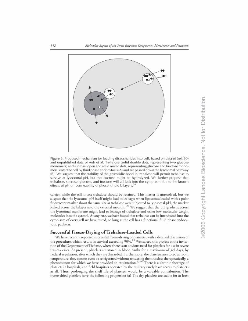
©20
06 C
opyr
ight
Lan
des
Bio
scie
nce.
Not
for
Dis
trib
utio
n.
Molecular Aspects of the Stress Response: Chaperones, Membranes and Networks152
carrier, while the still intact trehalose should be retained. This matter is unresolved, but wesuspect that the lysosomal pH itself might lead to leakage; when liposomes loaded with a polarfluorescent marker about the same size as trehalose were subjected to lysosomal pH, the markerleaked across the bilayer into the external medium.20 We suggest that the pH gradient acrossthe lysosomal membrane might lead to leakage of trehalose and other low molecular weightmolecules into the cytosol. At any rate, we have found that trehalose can be introduced into thecytoplasm of every cell we have tested, so long as the cell has a functional fluid phase endocy-totic pathway.
Successful Freeze-Drying of Trehalose-Loaded CellsWe have recently reported successful freeze-drying of platelets, with a detailed discussion of
the procedure, which results in survival exceeding 90%.89 We started this project at the invita-tion of the Department of Defense, where there is an obvious need for platelets for use in severetrauma cases. At present, platelets are stored in blood banks for a maximum of 3-5 days, byFederal regulation, after which they are discarded. Furthermore, the platelets are stored at roomtemperature; they cannot even be refrigerated without rendering them useless therapeutically, aphenomenon for which we have provided an explanation.92-97 There is a chronic shortage ofplatelets in hospitals, and field hospitals operated by the military rarely have access to plateletsat all. Thus, prolonging the shelf life of platelets would be a valuable contribution. Thefreeze-dried platelets have the following properties: (a) The dry platelets are stable for at least
Figure 6. Proposed mechanism for loading disaccharides into cell, based on data of (ref. 90)and unpublished data of Auh et al. Trehalose (solid double dots, representing two glucosemonomers) and sucrose (open and solid mixed dots, representing glucose and fructose mono-mers) enter the cell by fluid phase endocytosis (A) and are passed down the lysosomal pathway(B). We suggest that the stability of the glycosidic bond in trehalose will permit trehalose tosurvive at lysosomal pH, but that sucrose might be hydrolyzed. We further propose thattrehalose, sucrose, glucose, and fructose will all leak into the cytoplasm due to the knowneffects of pH on permeability of phospholipid bilayers.20

©20
06 C
opyr
ight
Lan
des
Bio
scie
nce.
Not
for
Dis
trib
utio
n.
153Trehalose as a “Chemical Chaperone”: Fact and Fantasy
two years when stored at room temperature, under vacuum. During that time we have seen noloss of platelets. (b) The freeze-dried, rehydrated cells respond to normal platelet agonists in-cluding thrombin, ADP, collagen, and ristocetin. (c) Studies on the morphology of thetrehalose-loaded, freeze-dried, and rehydrated platelets show that they are affected by the dry-ing, but are morphologically similar to fresh platelets. When they were dried without trehalose,on the other hand, most of the platelets disintegrated during the rehydration event, but of thesmall number that were left, most had fused with adjacent cells, forming an insoluble clump.(d) We have extended the freeze-drying to mouse and pig platelets as animal models for in vivotesting.
The rehydrated platelets are far from perfect, but they nevertheless show surprisingly goodregulation of key elements of cellular physiology such as intracellular calcium (Auh et al, 2004).98
For instance, when fresh platelets are challenged with thrombin they show an increase in [Cai]that is dose dependent. The rehydrated platelets show a similar response, although it is stronglyattenuated. Nevertheless, the increase in [Cai] appears to be sufficient to trigger morphologicaland physiological changes necessary for coagulation.98
Can Nucleated Cells Be Stabilized in the Dry State?Platelets are admittedly specialized cellular fragments, so it seemed likely at the outset that
the single perturbation of adding trehalose might not be sufficient to stabilize more complexliving cells. Indeed, this appears to be the case. When 293 cells were dried without prior load-ing with trehalose by the fluid phase endocytosis described previously they all died at fairlyhigh water contents.99 When they were loaded with trehalose survival was extended consider-ably, but the cells nevertheless died when water content was reduced below about 0.3 g H2O/g dry wgt. Thus, we have begun studies on effects of stress proteins on improving survival ofnucleated cells at lower water contents and settled on p26 a small α-crystallin stress proteinfrom desiccation tolerant Artemia cysts, discovered by Clegg and his colleagues.100-103 In Artemia,p26 protects against many different stresses.104-107 In addition, p26 has been shown to protectsynergistically with trehalose in vitro conditions or when loaded artificially into mammaliancells.108,109 Along the same lines, Singer and Lindquist110,111 previously showed that trehaloseacts synergistically with heat shock proteins in protein folding.
Sun et al107 isolated and cloned the gene for this protein and transfected 293 cells with it.They generously supplied us with the transfected cells. To our surprise, this protein signifi-cantly improved the survival to low water contents, even though the levels of expression havebeen very low—much less than that reported by Clegg for Artemia cysts.109 The protein alonedoes nothing to improve survival; trehalose is required as well, and the two appear to act syner-gistically. The effects of expression of this gene become even more pronounced in the timefollowing rehydration; the cells expressing p26 showed a ten fold increase in colony growthover those without the protein. Metabolism, expressed in terms of Alamar blue reduction,improved at least five fold compared with cells dried with trehalose alone.
What Is the Role of p26 in Stabilizing Dry Nucleated Cells?One hypothesis is that p26 participates in modulating the structure of the sugar glass, as
suggested from the findings of Wolkers83 and Buitink and Leprince.81 However, the expressionlevels are so low in this case that we doubt that the protein would have much effect on the glass.We favor instead the catalytic-like functions ascribed to stress proteins. A possible role for heatshock proteins in the protection of mammalian cells during dehydration stress has not beenaddressed, but there are indications from other organisms implicating HSPs in this regard. Forinstance, a drought-resistant form of maize expresses a 45 Kd HSP that is not found indrought-sensitive lines.112 Further, crossing the drought-resistant and sensitive lines led to F2plants in which tolerance to soil drying was associated with expression of the 45Kd HSP.112 Inaddition, the flesh fly Sarcophaga crassipalpis expresses two inducible HSPs (HSP23 and HSP70)during dehydration of nondiapausing pupae.113 In diapausing pupae, these proteins are al-ready highly expressed, and desiccation does not cause a further increase in expression.

©20
06 C
opyr
ight
Lan
des
Bio
scie
nce.
Not
for
Dis
trib
utio
n.
Molecular Aspects of the Stress Response: Chaperones, Membranes and Networks154
The most important mechanism by which heat shock proteins protect cells from variousstresses has traditionally been considered the protein chaperone function, assisting nascent andmisfolded proteins to gain their proper folded configuration.114,115 However, an association ofstress proteins with membranes has more recently been described.116-120 In fact, the “mem-brane trigger” hypothesis suggests that the membrane may serve as an indicator, sensing theinitial stress and leading to the expression of heat shock proteins within the cell.121,122 Finally,in addition to the protein and membrane effects, heat shock proteins have also been implicatedin the inhibition of apoptosis and oxidative damage.123-128 Finally, even more recent resultshave shown that arbutin, a glycosylated hydroquinone found at high concentrations in certainresurrection plants, also enhances survival of mammalian cells, in concert with trehalose.129
This small molecule is known to have antioxidant properties and to interact with membranesin the dry state, both of which observations seem consistent with the suggestions above.
Summary and ConclusionsUnder ideal conditions for drying and storage, trehalose is probably no more effective than
other oligosaccharides at preserving biomaterials. However, under suboptimal conditions itcan be very effective and is thus still a preferred excipient. There is growing evidence thatadditional modifications to the cellular milieu will probably be required if we are to achieve astable, freeze-dried mammalian cell, including expression of stress proteins, as reported here,and administration of antioxidants and inhibitors of enzyme activity, as described elsewhere.130
Thus, we have come full circle over the past decades on the requirements for stabilization ofcells in the dry state; 34 years ago, we suggested that survival of living cells in the dry state is acomplex phenomenon that is likely to involve multiple adaptations.131 With the discovery thatmembranes and proteins, and human platelets can be stabilized by the single perturbation ofadding trehalose, we suggested that this single lesson from nature might be sufficient, at leastunder ideal storage conditions. However, the studies on nucleated cells summarized here indi-cate that the original viewpoint on this matter131 is most likely the correct one.
AcknowledgmentsThis work was supported by grants HL57810 and HL98171 from NIH, 98171 from ONR,
and N66001-00-C-8048 from DARPA.
References1. Crowe JH, Crowe LM, Jackson SA. Preservation and functional activity in lyophilized sarcoplasmic
reticulum. Arch Biochem Biophys 1983; 220:477-484.2. Crowe M, Mouradian R, Crowe JH et al. Effects of carbohydrates on membrane stability at low
water activities. Biochim Biophys Acta 1984; 769:141-150.3. Crowe JH, Crowe LM, Oliver AE et al. The trehalose myth revisited: Introduction to a sympo-
sium on stabilization of cells in the dry state. Cryobiology 2001; 43:89-105.4. Matsuo T. Trehalose protects corneal epithelial cells from death by drying. British J Ophthalmol
2001; 85:610-612.5. Norcia M. Compositions and methods for wound management. Off Gaz US Patent and Trade-
mark Office 2000; 1232:424-448.6. Higashiyama T. Novel functions and applications of trehalose. Pure Appl Chem 2002; 74:1263-1269.7. Benaroudj N, Lee DL, Goldberg AL. Trehalose Accumulation during cellular stress protects cells
and cellular proteins from damage by oxygen radicals. J Biol Chem 2001; 276:24261-24267.8. Chen Q, Haddad GG. Role of trehalose phosphate synthase and trehalose during hypoxia: From
flies to mammals. J Exp Biol 2004; 207:3125-3129.9. Neta T, Takada K, Hirasawa M. Low-cariogenicity of trehalose as a substrate. Dent 2000; 28:571-6.
10. Gimeno-Alcañiz JV, Pèrez-Ortìn JE, Matallana E. Differential pattern of trehalose accumulation inwine yeast strains during the microvinification process. Biotechnology Lett 1999; 21:271-274.
11. Pataro C, Guerra JB, Gomes FCO et al. Trehalose accumulation, invertase activity and physiologi-cal characteristics of yeasts isolated from 24 h fermentative cycles during the production of artisanalBrazilian cachaça. Brazilian J Microbiol 2002; 33:202-208.
12. Komes D, Lovri T, Kovaevi Gani T et al. Study of trehalose addition on aroma retention indehydrated strawberry puree. Food Technol Biotechnol 2003; 41:111-119.
©20
06 C
opyr
ight
Lan
des
Bio
scie
nce.
Not
for
Dis
trib
utio
n.
155Trehalose as a “Chemical Chaperone”: Fact and Fantasy
13. Garg AK, Kim JK, Owens TG et al. Trehalose accumulation in rice plants confers high tolerancelevels to different abiotic stresses. Proc Nat Acad Sci 2002; 99:15898-15903.
14. Nishizaki Y, Yoshizane C, Toshimori Y et al. Disaccharide-trehalose inhibits bone resorption inovariectomized mice. Nutr Res 2000; 20:653-664.
15. Yoshizane C, Arai N, Arai C et al. Trehalose suppresses osteoclast differentiation in ovariectomizedmice: Correlation with decreased in vitro interleukin-6 production by bone marrow cells. Nutr Res2000; 20:1485-1491.
16. Tanaka M, Machida Y, Niu S et al. Trehalose alleviates polyglutamine-mediated pathology in amouse model of Huntington disease. Nat Med 2004; 10:148-54.
17. Couzin J. Huntington’s disease. Unorthodox clinical trials meld science and care. Science 2004;304:816-817.
18. Crowe JH, Crowe LM, Carpenter JF et al. Stabilization of dry phospholipid bilayers and proteinsby sugars. Biochem J 1987; 242:1-10.
19. Crowe JH, Crowe LM. Preservation of liposomes by freeze drying. In: Gregoriadis G, ed. Lipo-some Technology, 2nd ed. CRC Press Inc., 1992.
20. Hays LM, Crowe JH, Wolkers W et al. Factors affecting leakage of trapped solutes from phospho-lipid vesicles during thermotropic phase transitions. Cryobiology 2001; 42:88-102.
21. Crowe JH, Crowe LM. Factors affecting the stability of dry liposomes. Biochim Biophys Acta1988; 939:327-334.
22. Crowe JH, Carpenter JF, Crowe LM. The role of vitrification in anhydrobiosis. Annu Rev Physiol1998; 6:73-103.
23. Crowe JH, Crowe LM, Chapman D. Preservation of membranes in anhydrobiotic organisms: Therole of trehalose. Science 1984; 223:701-703.
24. Allison SD, Manning MC, Randolph TW et al. Optimization of storage stability of lyophilizedactin using combinations of disaccharides and dextran. J Pharm Sci 2000; 89:199-214.
25. Anchordoquy TJ, Izutsu KI, Randolph TW et al. Maintenance of quaternary structure in the frozenstate stabilizes lactate dehydrogenase during freeze-drying. Arch Biochem Biophys 2001; 390:35-41.
26. Cleland JL, Lam X, Kendrick B et al. A specific molar ratio of stabilizer to protein is required forstorage stability of a lyophilized monoclonal antibody. J Pharm Sci 2000; 90:310-321.
27. Belton PS, Gil AH. IR and Raman spectroscopic studies of the interaction of trehalose with henegg lysozyme. Biopolymers 1994; 34:957-961.
28. Cottone G, Cicotti G, Cordone L. Protein-trehalose-water structures in trehalose coatedcarboxy-myoglobn. J Cell Phys 2002; 117:9862-9866.
29. Lins RD, Pereira CS, Hunenberger PH. Trehalose-protein interactions in aqueous solutions. Pro-teins 2004; 55:177-186.
30. Sun WQ, Leopold AC. Cytoplasmic vitrification and survival of anhydrobiotic organisms. Comp.Biochem Physiol 1997; 117A:327-333.
31. Sun WQ, Leopold AC, Crowe LM et al. Stability of dry liposomes in sugar glasses. Biophys J1996; 70:1769-1776.
32. Lee CWB, Waugh JS, Griffin RG. Solid-state NMR study of trehalose/1,2-dipalmitoyl-sn-phosphatidylcholine interactions. Biochemistry 1986; 25:3737-3742.
33. Nakagaki M, Nagase H, Ueda H. Stabilization of the lamellar structure of phosphatidylcholine bycomplex-formation with trehalose. J Mem Sci 1992; 73:173-180.
34. Tsvetkova NM, Phillips BL, Crowe LM et al. Effect of sugars on headgroiup mobility in freeze-drieddipalmitoylphosphatidylcholine bilayers: Solid-state P-31 NMR and FTIR studies. Biophys J 1998;75:2947-2955.
35. Luzardo MD, Amalfa F, Nunez AM et al. Effect of trehalose and sucrose on the hydration anddipole potential of lipid bilayers. Biophys J 2000; 78:2452-2458.
36. Ricker JV, Tsvetkova NM, Wolkers WF et al. Trehalose maintains phase separation in an air-driedbinary lipid mixture. Biophys J 2003; 84:3045-3051.
37. Chandrasekhar I, Gaber BP. Stabilization of the biomembrane by small molecules: Interaction oftrehalose with the phospholipid bilayer. J Biomol Struct Dyn 1988; 5:1163-1171.
38. Rudolph BR, Chandrasekhar I, Gaber BP et al. Molecular modeling of saccharide-lipid interac-tions. Chem Phys Lipids 1990; 53:243-261.
39. Sum AK, Faller F, de Pablo JJ. Molecular simulation of phospholipid bilayers and insights of theinteractions with disaccharides. Biophys J 2003; 85:2830-2844.
40. Pereira CS, Lins RD, Chandrasekhar I et al. Interaction of the disaccharide trehalose with a phos-pholipid bilayer: A molecular dynamics study. Biophys J 2004; 86:2272-2285.
41. Brown DA. Seeing is believing: Visualization of rafts in model membranes. Proc Nat Acad Sci2001; 98:10517.
42. Brown DA, London E. Structure and origin of ordered lipid domains in biological membranes. JMem Biol 1998; 164:103-114.
©20
06 C
opyr
ight
Lan
des
Bio
scie
nce.
Not
for
Dis
trib
utio
n.
Molecular Aspects of the Stress Response: Chaperones, Membranes and Networks156
43. London E. Insights into lipid raft structure and formation from experiments in model membranes.Curr Opinion Struct Biol 2002; 12:480.
44. Horejs V. Membrane rafts in immunoreceptor signaling: New doubts, new proofs? Trends Immunol2002; 23:562-564.
45. Heerklotz H. Triton promotes domain formation in lipid raft mixtures. Biophys J 2002;83:2693-2697.
46. Shogomori H, Brown DA. Use of detergents to study membrane rafts: The good, the bad, and theugly. Biol Chem 2003; 384:1259-1263.
47. Draber P, Draberova L. Lipid rafts in mast cell signaling. Mol Immunol 2002; 38:1247-1253.48. Horejsi V. The roles of membrane microdomains (rafts) in T cell activation. Immunol Rev 2003;
191:148-154.49. FukI IV, Meyer ME, Williams KJ. Transmembrane and cytoplasmic domains of syndecan mediate
a multi-step endocytic pathway involving detergent-insoluble membrane rafts. Biochem J 2001;351:607-613.
50. Ono A, Freed EO. Plasma membrane rafts play a critical role in HIV-1 assembly and release.Proceedings of the National Academy of Sciences 2001; 98:13925-13929.
51. Vincent S, Gerlier D, Manié SN. Measles virus assembly within membrane rafts. J Virology 2000;74:9911-9916.
52. Gil T, Ipsen JH, Mouritsen OG et al. Theoretical analysis of protein organization in lipid mem-branes. Biochim Biophys Acta 1998; 1376:245-262.
53. Killian JA. Hydrophobic mismatch between proteins and lipids in membranes. Biochim BiophysActa 1998; 137:401-416.
54. Leidy C, Gousset K, Ricker JV et al. Lipid phase behavior and stabilization of domains in mem-branes of platelets. Cell Biochem Biophys 2004; 40:123-135.
55. Mabrey S, Sturtevant JM. Investigation of phase transitions of lipids and lipid mixtures by sensitiv-ity differential scanning calorimetry. Proc Nat Acad Sci 1976; 73:3862-3879.
56. Mabrey S, Mateo PL, Sturtevant JM. High-sensitivity scanning calorimetric study of mixtures ofcholesterol with dimyristoyl- and dipalmitoylphosphatidylcholines. Biochemistry 1978; 17:2464-2468.
57. Womersley C. Dehydration survival and anhydrobiotic potential of entomopathogenic nematodes.In: Gaugler R, Kaya HK, eds. Entomopathogenic Nematodes in Biological Control. Boca Raton,FL: CRC Press, 1990:117-130.
58. Westh P, Ramløv H. Trehalose accumulation in the tardigrade Adorybiotus coronifer duringanhydrobiosis. J Exp Zool 1991; 258:303-311.
59. Lapinski J, Tunnacliffe A. Anhydrobiosis without trehalose in bdelloid rotifers. FEBS Lett 2003;553:387-390.
60. Caprioli M, Katholm AK, Melone G et al. Trehalose in desiccated rotifers: A comparison betweena bdelloid and a monogonont species. Comp Biochem Physiol A 2004; 139:527-532.
61. Tunnacliffe A, Lapinski J. Resurrecting Van Leeuwenhoek’s rotifers: A reappraisal of the role ofdisaccharides in anhydrobiosis. Philos Trans R Soc Lond B 2003; 358:1755-1771.
62. Crowe JH, Oliver AE, Hoekstra FA et al. Stabilization of dry membranes by mixtures of hydroxyethylstarch and glucose: The role of vitrification. Cryobiology 1997; 3:20-30.
63. Hill DR, Keenan TW, Helm RF et al. Extracellular polysaccharide of Nostoc commune (Cyanobacteria)inhibits fusion of membrane vesicles during desiccation. J Applied Phycol 1997; 9:237-248.
64. Buitink J, Walters-Vertucci C, Hoekstra FA et al. Calorimetric properties of dehydrating pollen:Analysis of a desiccation-tolerant and an -intolerant species. Plant Physiol 1996; 111:235-242.
65. Hincha DK, Hellwege EM, Meyer AG et al. Plant fructans stabilize phosphatidylcholine liposomesduring freeze-drying. Eur J Biochem 2000; 267:535-540.
66. Vereyken IJ, Chupin V, Hoekstra FA et al. The effect of fructan on membrane lipid organizationand dynamics in the dry state. Biophys J 2003; 84:3759-3766.
67. Hincha DK, Zuther E, Heyer AG. The preservation of liposomes by raffinose family oligosaccha-rides during drying is mediated by effects on fusion and lipid phase transitions. Biochim BiophysActa 2003; 1612:172-177.
68. Hincha DK, Hagemann M. Stabilization of model membranes during drying by compatible solutesinvolved in the stress tolerance of plants and microorganisms. Biochem J 2004; 383:277-83.
69. Hoekstra FA, Golovina EA. The role of amphiphiles. Comp Biochem Physiol 2002; 131A:527-533.70. Goodrich RP, Crowe JH, Crowe LM et al. Alteration in membrane surfaces induced by attach-
ment of carbohydrates. Biochemistry 1991; 30:2313-2318.71. Popova AV, Hincha DK. Effects of the sugar headgroup of a glyoglycerolipid on the phase behav-
ior of phospholipid model membranes in the dry state. Glycobiology 2005; 15:1150-1155.72. Leslie SB, Israeli E, Lighthart B et al. Trehalose and sucrose protect both membranes and proteins
in intact bacteria during drying. Econ Env Microbiol 1995; 61:3592-3597.73. Esteves MI, Quintilio W, Sato RA et al. Stabilisation of immunoconjugates by trehalose. Biotechnol
Lett 2001; 22:417-420.

©20
06 C
opyr
ight
Lan
des
Bio
scie
nce.
Not
for
Dis
trib
utio
n.
157Trehalose as a “Chemical Chaperone”: Fact and Fantasy
74. Crowe LM, Reid DS, Crowe JH. Is trehalose special for preserving dry biomaterials? Biophys J1996; 71:2087-2093.
75. Green JL, Angell CA. Phase relations and vitrification in saccharide-water solutions and the treha-lose anomaly. J Phys Chem 1989; 93:2880-2882.
76. Li S, Patapoff TW, Overcashier et al. Effects of reducing sugars on the chemical stability of humanrelaxin in the lyophilized state. J Pharm Sci 1996; 85:873-877.
77. O’Brien J. Stability of trehalose, sucrose and glucose to nonenzymatic browning in model systems.J Food Sci 1996; 61:679-682.
78. Schebor C, Burin L, del Pilar Bueras M et al. Stability to hydrolysis and browning of trehalose,sucrose and raffinose in low-moisture systems in relation to their use as protectants of drybiomaterials. Lebensm-Wiss u-Technol 1999; 32:481-485.
79. Buitink J, van den Dries IJ, Hoekstra FA et al. High critical temperature above Tg may contributeto the stability of biological systems. Biophys J 2000; 79:1119-1128.
80. Buitink J, Heminga MA, Hoekstra FA. Is there a role for oligosaccharides in seed longevity? Anassessment of intracellular glass stability. Plant Physiology 2000; 122:1217-1224.
81. Buitink J, Leprince O. Glass formation in plant anhydrobiotes: Survival in the dry state. Cryobiol-ogy 2004; 48:215-228.
82. Koste KL. Glass formation and desiccation tolerance in seeds. Plant Physiol 1991; 96:302-304.83. Wolkers WF, Tetteroo FAA, Alberda M et al. Changed properties of the cytoplasmic matrix asso-
ciated with desiccation tolerance of dried carrot somatic embryos. An in situ Fourier transforminfrared spectroscopic study. Plant Physiol 1999; 120:153-163.
84. Wolkers WF, Alberda M, Koornneef M et al. Properties of proteins and the glassy matrix inmaturation-defective mutant seeds of Arabidopsis thaliana. Plant J 1998; 16:133-143.
85. Wolkers WF, Oldenhof H, Alberda M et al. A Fourier transform infrared study of sugar glasses:Application to anhydrobiotic higher plant cells. Biochim Biophys Acta 1998; 1379:83-96.
86. Walters C, Reid JL, Walker-Simmons MK. Heat soluble proteins extracted from wheat embryoshave tightly bound sugars and unusual hydration properties. Seed Sci Res 1997; 7:125-134.
87. Eroglu A, Russo MJ, Bieganski R et al. Intracellular trehalose improves the survival of cryopreservedmammalian cells. Nat Biotechnol 2000; 18:163-167.
88. Guo NI, Puhlev DR, Brown J et al. Trehalose expression confers desiccation tolerance on humancells. Nat Biotechnol 2000; 18:168-171.
89. Wolkers WF, Walker NJ, Tablin F et al. Human platelets loaded with trehalose survive freeze-drying.Cryobiology 2001; 42:79-87.
90. Oliver AE, Jamil K, Crowe JH et al. Loading human mesenchymal stem cells with trehalose byfluid-phase endocytosis. Cell Preservation Tech 2004; 2:35-49.
91. Lloyd JB. Lysosome membrane permeability: Implications for drug delivery. Advanced Drug Deliv-ery Reviews 2000; 41:189-200.
92. Tablin F, Oliver AE, Walker NJ et al. Membrane phase transitions of intact human platelets:Correlation with cold-induced activation. J Cell Physiol 1996; 168:305-313.
93. Tablin F, Wolkers WF, Walker NJ et al. Membrane reorganization during chilling: Implicationsfor long term storage. Cryobiology 2001; 43:114-123.
94. Oliver AE, Tablin F, Walker NJ et al. The internal calcium concentration of human plateletsincreases during chilling. Biochim Biophys Acta 1999; 1416:349-60.
95. Crowe JH, Tablin F, Tsvetkova NM et al. Are lipid phase transitions responsible for chilling dam-age in human platelets? Cryobiology 1999; 38:180-191.
96. Crowe JH, Tablin F, Wolkers WF et al. Stabilization of membranes in human platelets freeze-driedwith trehalose. Chem Phys Lipids 2003; 122:41-52.
97. Tsvetkova NM, Walker NJ, Crowe JH et al. Lipid phase separation correlates with activation inplatelets during chilling. Mol Mem Biol 2001; 17:209-218.
98. Auh JH, Wolkers WF, Looper SA et al. Calcium mobilization in freeze-dried human platelets. CellPreservation Tech 2004; 2:180-187.
99. Ma X, Jamil K, MacRae TH et al. A small stress protein acts synergistically with trehalose toconfer desiccation tolerance on mammalian cells. Cryobiology 2005; 51:15-28.
100. Clegg JS, Jackson SA, Warner AH. Extensive intracellular translocations of a major protein accom-pany anoxia in embryos of Artemia franciscana. Exp Cell Res 1994; 212:77-83.
101. Liang P, Amons R, Clegg HS et al. Purification, structure and in vitro molecular-chaperone activ-ity of Artemia p26, a small heat- shock/alpha-crystallin protein. Eur J Biochem1997; 243:225-32.
102. Liang P, Amons R, Clegg HS et al. Molecular characterization of a small heat shock/alpha-crystallinprotein in encysted Artemia embryos. J Biol Chem 1997; 272:19051-19058.
103. Liang P, MacRae TH. The synthesis of a small heat shock/α-crystallin protein in Artemia and itsrelationship to stress tolerance during development. Devel Biol 1999; 207:445-456.
©20
06 C
opyr
ight
Lan
des
Bio
scie
nce.
Not
for
Dis
trib
utio
n.
Molecular Aspects of the Stress Response: Chaperones, Membranes and Networks158
104. MacRae TH. Molecular chaperones, stress resistance and development in Artemia franciscana. SeminCell Dev Biol 2003; 14:251-258.
105. Willsie JK, Clegg JS. Nuclear p26, a small heat shock/α-crystallin protein, and its relationship tostress resistance in Artemia franciscana embryos. J Exp Biol 2001; 204:2339-2350.
106. Day RM, Gupta JS, MacRae TH. A small heat shock/alpha-crystallin protein from encysted Artemiaembryos suppresses tubulin denaturation. Cell Stress and Chaperones 2003; 8:183-193.
107. Sun Y, Mansour M, Crack JA et al. Oligomerization, chaperone activity, and nuclear localizationof p26, a small heat shock protein from Artemia franciscana. J Biol Chem 2004; 279:39999-40006.
108. Viner RI, Clegg JS. Influence of trehalose on the molecular chaperone activity of p26, a small heatshock/α-crystallin protein. Cell Stress and Chaperones 2001; 6:126-135.
109. Collins CH, Clegg JS. A small heat-shock protein, p26, from the crustacean Artemia protectsmammalian cells (Cos-1) against oxidative damage. Cell Biol Intl 2004; 28:449-455.
110. Singer MA, Lindquist S. Multiple effects of trehalose on protein folding in vitro and in vivo. MolCell 1998; 1:639-648.
111. Singer MA, Lindquist S. Thermotolerance in Saccharomyces cerevisiae: The Yin and Yang of tre-halose. Trends Biotech 1998; 1:460-468.
112. Ristic Z, Williams G, Yang G et al. Dehydration, damage to cellular membranes, and heat-shockproteins in maize hybrids from different climates. J Plant Physiol 1996; 149:424-432.
113. Hayward SAL, Rinehart JP, Denlinger DL. Desiccation and rehydration elicit distinct heat shockprotein transcript responses in the flesh fly pupae. J Exp Biol 2004; 207:963-971.
114. Hartl FU, Hayer-Hartl M. Molecular chaperones in the cytosol: From nascent chain to foldedprotein. Science 2002; 295:1852-1858.
115. Barral JM, Broadley SA, Schaffar G et al. Roles of molecular chaperones in protein misfoldingdiseases. Sem Cell Devel Biol 2004; 15:17-29.
116. Trent JD, Kagawa HK, Paavola CD et al. Intracellular localization of a group II chaperonin indi-cates a membrane-related function. Proc Natl Acad Sci 2003; 100:15589-15594.
117. Torok Z, Horvath I, Goloubinoff P et al. Evidence for a liposhaperonin: Association of activeprotein-folding GroESLj oligomers with lipids can stabilize membranes under heat shock condi-tions. Proc Natl Acad Sci 1997; 94:2192-2197.
118. Torok Z, Tsvetkova NM, Balogh G et al. Heat shock protein coinducers with no effect on proteindenaturation specifically modulate the membrane lipid phase. Proc Nat Acad Sci 2003; 100:3131-3136.
119. Torok Z, Goloubinoff P, Horvath I et al. Synechocystis HSP17 is an amphitropic protein thatstabilizes heat-stressed membranes and binds denatured proteins for subsequent chaperone-mediatedrefolding. Proc Natl Adad Sci 2001; 98:3098-3103.
120. Tsvetkova NM, Horvath I, Torok Z et al. Small heat-shock proteins regulate membrane lipidpolymorphism. Proc Natl Acad Sci 2002; 99:13504-13509.
121. Vigh L, Maresca B, Harwood JL. Does the membrane’s physical state control the expression ofheat shock and other genes? Trends Bio Sci1998; 23:369-374.
122. Horvath I, Glatz A, Varvasovszki V et al. Membrane physical state controls the signaling mecha-nism of the heat shock response in Synechocystis PCC 6803: Identification of hsp17 as a “fluiditygene”. Proc Nat Acad Sci 1998; 95:3513-3518.
123. Beere HM, Green DR. Stress management - Heat shock protein-70 and the regulation of apoptosis.Trends Cell Biol 2001; 11:6-10.
124. Concannon CG, Gorman AM, Samali A. On the role of Hsp27 in regulating apoptosis. Apoptosis2003; 8:61-70.
125. Samali A, Orrenius S. Heat shock proteins: Regulators of stress response and apoptosis. Cell Stressand Chaperones 1998; 3:228-236.
126. Downs CA, Jones LR, Heckathorn SA. Evidence for a novel set of small heat-shock proteins thatassociates with the mitochondria of murine PC12 cells and protects NADH:ubiquinone oxidoreduc-tase from heat and oxidative stress. Arch Biochem Biophys 1999; 365:344-350.
127. Gill RR, Gbur CJ, Fisher BJ et al. Heat shock provides delayed protection against oxidative injuryin cultured human umbilical nein endothelial cells. J Molec Cell Cardiol 1998; 30:2739-2749.
128. Park YM, Han MY, Blackburn RV et al. Overexpression of HSP25 reduces the level ofTNFalpha-induced oxidative DNA damage biomarker, 8-hydroxy-2'-deoxyguanosine, in L929 cells.J Cell Physiol 1998; 174:27-34.
129. Jamil K, Crowe JH, Tablin F et al. Arbutin enhances recovery and osteogenic differentiation indried and rehydrated human mesenchymal stem cells. Cell Preservation Tech 2005; 3:244-255.
130. Oliver AE, Hincha DK, Crowe JH. Looking beyond sugars: The role of amphiphilic solutes inpreventing adventitious reactions in anhydrobiotes at low water contents. Comp Biochem Physiol2002; 131A:515-525.
131. Crowe JH. Anhydrobiosis: An unsolved problem. Am Nat 1971; 105:563-573.

©20
06 C
opyr
ight
Lan
des
Bio
scie
nce.
Not
for
Dis
trib
utio
n.
CHAPTER 14
*Zoltán Prohászka—IIIrd Department of Internal Medicine, Semmelweis University, H-1125Budapest, Kútvölgyi st. 4, Hungary. Email: [email protected]
Molecular Aspects of the Stress Response: Chaperones, Membranes and Networks,edited by Peter Csermely and Laszlo Vigh. ©2007 Landes Bioscienceand Springer Science+Business Media.
Chaperones As Part of Immune NetworksZoltán Prohászka*
Abstract
Network theory is increasingly accepted as a basic regulatory mechanism in diverseimmunological functions. Heat shock proteins (Hsps) are involved in multiplenetworks in the immune system. Hsps themselves (foreign or endogenous) acti-
vate innate immunity and play important roles to deliver self or nonself materials to antigenpresenting cells. However, Hsps are immunodominant antigens during infectious diseases makingself Hsps endangered targets of autoimmunity by cross-reactive clones. Therefore, it is notsurprising that the mechanism of protection of self Hsps is not clonal deletion in natural selftolerance; rather, self Hsps are protected by active regulating natural autoimmunity. The activeregulatory/protective immunity is accomplished by natural autoantibodies and regulatory Tcells, both recognizing Hsps. The multiple involvements of Hsps in immune networks makethem ideal targets of therapy in autoimmune diseases. Indeed, immunotherapy with Hsps wasrecently reported to be effective treatment modality against cancer, arthritis or diabetes mellitus.
IntroductionOwing to their highly conserved and inducible nature stress proteins are ideal messengers of
cellular stress. Nearly all pathogen microorganisms studied till now possess heat inducible stressprotein genes and respond to the thermal (and other) stresses of infection with increased heatshock protein (Hsp) expression. Higher organisms have innate sense, by means of innate im-munity, to respond stress signals, on one hand. On the other hand, basically the same mol-ecules may messenger stress of the host organism, making altered self ‘dangerous’, in case, forexample, of cell necrosis.1 However, the innate role of Hsps may become problematic in thenew world of the adaptive immunity. With the appearance of specific receptors (i.e., antibodiesand T cell receptors) the overexpressed and conserved stress proteins turn into first class targetsof autoimmunity by means of infection induced molecular mimicry. The highly conservedHsps, which are present in all mammalian cells, need therefore special protection in the ‘adap-tive world’. In this Chapter recent data on the relationship of Hsps with innate immunity, theirprotection by regulating autoimmunity will be summarized and a new concept on their em-bedment in multiple networks of the organism will be presented.
Activation of Innate Immunity by Heat Shock ProteinsMammalian heat shock proteins were described to activate innate immunity by nonclonal
receptors belonging to the family of pattern recognition receptors such us Toll-like receptors.Members belonging to the 70- and 60-kDa Hsp family were mostly investigated and TLR2and TLR4 were reported as important players in the transduction of signals. However, the

©20
06 C
opyr
ight
Lan
des
Bio
scie
nce.
Not
for
Dis
trib
utio
n.
Molecular Aspects of the Stress Response: Chaperones, Membranes and Networks160
binding receptors for Hsps are not yet completely known or debated (see ref. 2) and some ofthe effects Hsps on the target cells seemed to be indistinguishable from effects of endotoxins(LPS). This similarity between the effects of Hsps and LPS lend credence to the hypothesis thatthe so-called cytokine effects of Hsps are due to contaminants copurified with the recombinantproteins used in such experiments (reviewed in ref. 3). In some recent studies this hypothesiswas investigated in details and Gao and Tsan pinpointed out the importance for caution whileinterpreting the results of studies with recombinant Hsps.4 We learned from the works of Gaoand Tsan that usually applied methods to control endotoxin contamination (polymyxin Btreatment and boiling of proteins) might be inappropriate and much more attention has to bepaid to control endotoxin effects in studies on innate immunity. However, several recent inde-pendent lines of evidence obtained in well conducted and controlled experiments suggest thatHsps, nevertheless, do manifest innate immune activities that are not due to bacterial contami-nants. These include: activation of DCs and macrophages by mammalian Hsp70 and gp96purified by a pyrogen-free method (see ref. 5); activation of macrophages via TLR2- andTLR4-signaling by mammalian cells displaying Hsp70 on their surface.6 Furthermore, Habichet al reported results of elegant binding studies using fluorochrome labeled Hsp60 prepara-tions. In these studies specific epitopes of Hsp60 were identified for binding to primary mac-rophages.7 Importantly, these epitopes overlap with B-cell epitopes of the molecule (see below).In addition, the same group determined the binding site of Hsp60 for lipopolysaccharide aswell (see ref. 8), indicating that endotoxin is not just a simple contaminant in recombinantHsp60 preparation, rather, it might reflect an inherent property of this protein as a sensor andmessenger of danger. Interestingly, Hsp70 and Hsp90 have also been reported to act as recep-tors for LPS.9,10 In conclusion, all of the above experiments indicate that Hsps play importantroles in the activation of innate immunity, but critical works rightly call our attention to thefact that Hsps are likely found in complexes with self or nonself material.
Immunological Protection of Heat Shock ProteinsAs mentioned above, Hsps are structurally highly conserved molecules from eubacteria till
humans what is true for their molecular structure, biochemical properties but partly even fortheir immunological (epitope) structure. It is not surprising, therefore, that several autoantigenscharacterized in different autoimmune conditions share some of their epitopes with Hsps.11
These findings indeed support the long lasting observation that infections may induce autoim-mune processes. More surprising is the fact, however, that autoimmune diseases are rare dis-eases and are characterized by well defined autoimmune reaction to only a few number ofautoantigens in a conserved nature. Furthermore, according to the idea that the source of selftolerance is the clonal deletion, one may speculate that the closer a molecule is to self, the lessimmunogenic it should be. It is surprising, therefore, to find that among the major antigensrecognized during a wide variety of infections many belong to conserved protein families shar-ing extensive sequence identity with host’s molecules, for example Hsps. One interesting hy-pothesis, dealing with the above listed controversies, was presented by Irun Cohen and co-workers, called ‘the immunological homunculus’.12 The idea is that some, perhaps all, majorautoantigens are indeed dominant because each of them is encoded in the organizational struc-ture of the immune system. Each dominant self antigen is served by an interacting set of T andB cells that includes cells with receptors for the antigen (antigen-specific) and cells with recep-tors for the antigen-specific receptors (anti-idiotypic). Some of these lymphocytes suppress andothers stimulate. Owing to the mutual connections between the various interacting lympho-cytes in the network, some lymphocytes become activated even without being driven by con-tact with specific antigen in an immunogenic form. The state of autonomous activity definedby a pattern of interconnected lymphocytes constitutes a functional representation of the par-ticular self antigen around which the network is organized. In other words, the picture of theself antigen is encoded within a cohort of lymphocytes.

©20
06 C
opyr
ight
Lan
des
Bio
scie
nce.
Not
for
Dis
trib
utio
n.
161Chaperones as Part of Immune Networks
At the time of the presentation of the immunological homunculus, the information thatcells of innate immunity, and even B cells, possess more abundant and effective receptors (ascompared to the specific ones), the pattern recognition receptors (PRRs, i.e., Toll-like recep-tors), for the conserved antigens, for example for Hsps, was not available. Most probable therecognition of the conserved molecules by PRRs is also a fundamental mechanism why thesemolecules became immunodominant during infection. Although the idea on ‘immunologicalhomunculus’ was debated in the meantime and the conception is far from a general acceptancein the immunologists’ community, some recent results support the existence of regulating naturalautoimmunity. Heat shock proteins seem to be an important contributor to, and target of,these regulatory mechanisms.
Role of Natural Autoantibody Networks in Regulationof Autoimmunity
Natural antibodies refer to antibodies that are present in the serum of healthy individualsin the absence of deliberate immunization with the target antigen.13 Autoantibodies areimmunoglobulins that react with at least one self antigen, whether they originate form healthyindividuals or patients with autoimmune disease. The importance of natural antibodies reac-tive with self antigens (natural autoantibodies, NAA) has earlier been neglected, as toleranceto self was thought to be primarily dependent on the deletion of autoreactive clones duringontogeny. It is now established, that NAAs are present in almost all vertebrates investigateduntil now.14 NAAs may belong to IgM, IgG or IgA isotypes, whereas in humans IgG domi-nates. Natural autoreactive B cells are endowed with some switching ability in the absence ofcognate interactions with T cells. NAAs are characterized by polyreactivity resulting in spe-cific reactivity patterns. NAAs are characterized by a broad range of affinities having mainlydissociation constants in the micromolar range. The notion that an antibody has to be ofhigh affinity in order to be biologically relevant originates primarily from the analysis of therequirements for an efficient immune response against pathogens. This concept does notnecessarily apply to natural antibodies. Specific polyreactive recognition patterns of low af-finity antibodies may result in novel biological properties that emerge from the organizationof networks. The biological activities of such networks may not be predicted from activitiesof individual members of the network.
Evidence for the intrinsic network regulation of autoreactive antibody repertoires include(reviewed in ref. 15): (i) the V-region-dependent connectivity between NAA; (ii) the restrictedpattern of reactivity to a set of self antigens that is conserved among healthy individuals andthroughout life; (iii) the spontaneous fluctuations of concentrations of NAAs over time; and iv,the suppression of pathogenic autoreactive clones by infusions of normal polyvalent IgG (IVIG)in patients with autoimmune diseases.
Polyreactivity and V-region-dependent connectivity are basic features of NAAs comprisinginteractions with other IgG- and immunological active molecules. Reactivities of NAAs to-wards cytokines and cytokine receptors, cell surface receptors (such as CD4 or MHC class Imolecules), adhesion molecules, coagulation factors and heat shock proteins have been reported.15
Anti-Hsp90 reacting antibodies were recently shown to be part of the natural autoantibodyrepertoire and were characterized as broadly cross-reacting antibodies mainly belonging to theIgG2 subclass.16 Furthermore, in the study of Pashov and coworkers,16 although the anti-Hsp90antibodies bound to the same set of antigens in the patients, there were quantitative differencesamong the antibodies tested. Thus, the natural autoantibody repertoire qualitatively seems tobe invariable, directed against a highly conserved set of immunodominant antigens includingHsp90. Work in our laboratory in the past years was focused on the analysis of anti-Hsp60 andanti-Hsp70 IgG autoantibodies. In a recent review paper supporting evidence was summarizedon the view that anti-Hsp60 IgG autoantibodies belong to NAAs.17 These antibodies are presentin all healthy individuals investigated and in IVIG preparations as well.18 In our recent studiesanti-Hsp60 autoantibody level-associated genetic factors were determined. Allelic variants of

©20
06 C
opyr
ight
Lan
des
Bio
scie
nce.
Not
for
Dis
trib
utio
n.
Molecular Aspects of the Stress Response: Chaperones, Membranes and Networks162
promoter polymorphism -174 of interleukine-6 (IL-6) were shown to associate with anti-Hsp60concentrations in two independent populations (ref. 19 and unpublished studies from theauthor’s lab). Furthermore, allelic determinants of immunoglobulin constant regions (KM de-terminants) have also been shown to associate with increased autoantibody levels.20 Most im-portantly, IL-6 and Ig KM determinants were in epistatic interaction in the regulation ofanti-Hsp60 levels.21 IgM and IgD natural autoantibodies to Hsp70 have earlier been reportedin normal mice.22 This observation is strengthened by recent results on the presence ofanti-Hsp70 IgG reactivity in all healthy human serum samples tested (see refs. 23,24) and inIVIG preparation (Fig. 1).
Taken together natural autoantibodies to different families of Hsps are present in sera ofhealthy individuals. These anti-Hsp antibodies—although directly not yet investigated—mostprobably operate in networks like other NAAs. Since Hsp70 and Hsp60 has been reported tobe present in the circulation of healthy individuals (see refs. 24-26) and heat shock proteinsmight be expressed on the surface of stressed or transformed cells, this setting preferentiallygive rise to the formation of self regulating networks. Importantly, B cell- and receptor bindingepitopes of Hsp60 are partly overlapping (see refs. 7,18) indicating functional relevance ofNAAs in the possible regulation of biological activities of Hsp60.
Heat Shock Proteins as Negotiators between Promotionof Inflammation or Control of Autoimmunity
Heat shock proteins present in the circulation of healthy individuals and patients mightoriginate from cells of the organism or from colonizing or pathogenic microorganisms. Thesource of Hsps, their complexes with self- or foreign materials and the nature of the host’sregulating immune network is decisive on the shape of subsequent immunological events.
Hsps originating from self tissues (endogenous Hsps) are present in healthy and diseasedsubjects.24-26 The source of these Hsps is likely cellular necrosis (passive release), but active
Figure 1. IgG anti-Hsp70 autoantibodies are present in the normal immunoglobulin prepara-tion (IVIG) and in normal human serum (NHS). ELISA plates were coated with human heatshock protein 70 (2 µg/ml) and incubated with serial dilutions of IVIG (A) or NHS (B). Uncoatedwells were used as control. Anti-Hsp70 IgG antibodies were determined as described earlier.23
Means of two parallel measurements and SD are plotted, the experiment was repeated fourtimes with identical results.
©20
06 C
opyr
ight
Lan
des
Bio
scie
nce.
Not
for
Dis
trib
utio
n.
163Chaperones as Part of Immune Networks
release (i.e., secretion of small membrane vesicles) can not be excluded. Indeed, exosomes haverepeatedly been shown to contain cytoplasmic members of Hsp families.27 These moleculesmight carry endogenous peptides and deliver them to antigen presenting cells thus signallingthe state of self to other cells. Srivastava and colleagues recently reviewed the potential of Hspseliciting anti-tumor immune responses.28 Furthermore, endogenous Hsps may stimulate regu-latory T-cells that control pathogenic T cells for self antigens other than Hsps.29 However,Hsps originating from infecting agents might be complexed with endotoxins and other bacte-rial materials making these complexes highly active in initiating inflammation (see above).Thus, depending on the source of Hsp material and the surrounding of recognition processesimmunological responses of the host may be very diverse. In other words Hsps may bothpromote or regulate inflammatory processes having fundamental relevance to pathologi-cal conditions.
The best studied examples in this field are adjuvant arthritis (see ref. 30) and autoimmunediabetes mellitus type I.31 In adjuvant arthritis a pathogenic role of T cell reactivity to anonconserved epitope of Hsp65 (AA180-188) has been shown.32 Later studies confirmed thatthe self antigen recognized by this clone is not Hsp60 but the cartilage proteoglycan. Further-more, Hsp65 can be used effectively to prevent different experimental arthritis types, and thisis based on a general anti-inflammatory pathway induced by Hsp65.33-34 The protective effectsof Hsp65 depend on cell mediated processes that induce regulatory cells turning down inflam-mation. In case of diabetes mellitus a beta-cell target antigen in nonobese diabetic (NOD)mice is a molecule cross-reactive with the Hsp65. The importance of hsp65 in the pathogenesisof the disease was confirmed by the ability of clones of anti-hsp65 T cells to cause insulitis andhyperglycemia in young NOD/Lt mice. Moreover, hsp65 antigen could be used either to in-duce diabetes or to vaccinate against diabetes, depending on the form of its administration toprediabetic NOD/Lt mice.35 Subsequent research recognized the minimum structure(AA437-460 called p277) of Hsp60 arresting the destruction of the beta-cells in NOD mice.36
The mechanism of protection against diabetes in the NOD mice is characterized by the shift ofTh1/Th2 balance towards Th2, by the downregulation of T-cell and IgG responses against selfantigens but no modification of spontaneous Th1 cytokine secretion to Hsp65 peptide. Thesuccessful formulation of the p277 peptide (DiaPep277) led to the initiation of phase I and IIhuman studies with prosperous results.37
Taken together the above summarized experimental data strongly support the view that theconserved heat shock protein (self or foreign) molecules are indeed negotiators between pro-motion of inflammation or control of autoimmunity.38 Translation of this knowledge intopractical usage in human health will most probably help to control some of the autoimmunediseases.
Heat Shock Proteins as Elements of Multiple NetworksJerne suggested some 25 years ago that immunoglobulin idiotypes are organized as net-
work of complementary shapes.39 Network regulation in immunology was neglected forlonger time and tolerance was regarded as result of the elimination of all self-reactive clones.However, accumulating data suggested that basic functions of regulatory T cells and naturalautoantibodies are related to network operation, thus more and more light was shed ontothis area of immunology.40-41 Conventional immune responses and much of their regulationare satisfactorily explained by clonal selection principles, such as somatic mutation and ‘in-duced fit’ and clonal selection also contributes the rational basis for anti-infectious protec-tion. However, core operation of immune networks has little to do with immune responses,but is fundamental to the understanding of questions that were not solved by the clonalselection theory. These unresolved areas include internal lymphocyte activities, natural anti-body production in unimmunized animals, preimmune repertoire selection, tolerance andself-nonself discrimination, memory and the evolution of immune systems.42 Basic featuresof Hsps make them core elements (or using the network term: hubs) in immunological
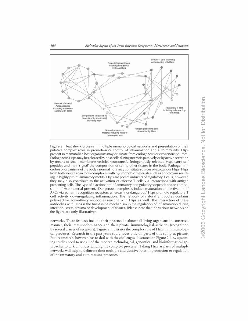
©20
06 C
opyr
ight
Lan
des
Bio
scie
nce.
Not
for
Dis
trib
utio
n.
Molecular Aspects of the Stress Response: Chaperones, Membranes and Networks164
networks. These features include their presence in almost all living organisms in conservedmanner, their immunodominance and their pivotal immunological activities (recognitionby several classes of receptors). Figure 2 illustrates the complex role of Hsps in immunologi-cal processes. Research in the past years could focus only on parts of this complex picture.Future research, however, has to deal with the challenges illustrated on Figure 2, i.e., upcom-ing studies need to use all of the modern technological, genomical and bioinformatical ap-proaches to task on understanding the complete processes. Taking Hsps as parts of multiplenetworks will help to delineate their multiple and decisive roles in promotion or regulationof inflammatory and autoimmune processes.
Figure 2. Heat shock proteins in multiple immunological networks and presentation of theirputative complex roles in promotion or control of inflammation and autoimmunity. Hspspresent in mammalian host organisms may originate from endogenous or exogenous sources.Endogenous Hsps may be released by host cells during necrosis passively or by active secretionby means of small membrane vesicles (exosomes). Endogenously released Hsps carry selfpeptides and may ‘signal’ the composition of self to other tissues in the body. Pathogen mi-crobes or organisms of the body’s normal flora may constitute sources of exogenous Hsps. Hspsfrom both sources can form complexes with hydrophobic materials such as endotoxins result-ing in highly proinflammatory motifs. Hsps are potent inducers of regulatory T cells, however,they may also contribute to the activation of effector T cells via interactions with antigenpresenting cells. The type of reaction (proinflammatory or regulatory) depends on the compo-sition of Hsp material present. ‘Dangerous’ complexes induce maturation and activation ofAPCs via pattern recognition receptors whereas ‘nondangerous’ Hsps promote regulatory Tcell activity downregulating inflammation. The network of natural antibodies containspolyreactive, low-affinity antibodies reacting with Hsps as well. The interaction of theseantibodies with Hsps is the fine-tuning mechanism in the regulation of inflammation duringinfection, stress, trauma or development of tissues. (Please note that the various networks onthe figure are only illustrative).

©20
06 C
opyr
ight
Lan
des
Bio
scie
nce.
Not
for
Dis
trib
utio
n.
165Chaperones as Part of Immune Networks
AcknowledgementsWork in the authors’ laboratory was supported by research grant from the National Re-
search Fund of Hungary (OTKA) T46837. The skillful technical assistance of Szigeti Antalnéis acknowledged with many thanks.
References1. Matzinger P. The danger model: A renewed sense of self. Science 2002; 296:301-305.2. Binder RJ, Vatner R, Srivastava PK. The heat-shock protein receptors: Some answers and more
questions. Tissue Antigens 2004; 64:442-451.3. Tsan MF, Gao B. Endogenous ligands of Toll-like receptors. J Leukoc Biol 2004; 76:514-519.4. Gao B, Tsan MF. Induction of cytokines by heat shock proteins and endotoxin in murine mac-
rophages. Biochem Biophys Res Commun 2004; 317:1149-1154.5. Panjwani NN, Popova L, Srivastava PK. Heat shock proteins gp96 and hsp70 activate the release
of nitric oxide by APCs. J Immunol 2002; 168:2997-3003.6. Korbelik M, Sun J, Cecic I. Photodynamic therapy induced cell surface expression and release of
heat shock proteins: Relevance for tumor response. Cancer Res 2002; 65:1018-1026.7. Habich C, Kempe K, Gomez FJ et al. Heat shock protein 60: Identification of specific epitopes for
binding to primary macrophages. FEBS Lett 2006; 580:115-120.8. Habich C, Kempe K, van der Zee R et al. Heat shock protein 60: Specific binding of lipopolysac-
charide. J Immunol 2005; 174:1298-1305.9. Byrd CA, Bornmann H, Erdjument-Bromage P et al. Heat shock protein 90 mediates macrophage
activation by taxol and bacterial lipopolysaccharide. Proc Natl Acad Sci USA 1999; 96:5645-5651.10. Triantafilou K, Triantafilou M, Dedrick RL. A CD14-independent LPS receptor complex. Nat
Immunol 2001; 2:338-342.11. Jones DB, Coulson AF, Duff GW. Sequence homologies between Hsp60 and autoantigens. Immunol
Today 1993; 14:115-8.12. Cohen IR, Young DB. Autoimmunity, microbial immunity and the immunological homunculus.
Immunol Today 1992; 12:105-110.13. Coutinho A, Kazatchkine MD, Avrameas S. Natural autoantibodies. Curr Opin Immunol 1995;
7:812-821.14. Lacroix-Desmazes S, Kaveri SV, Mouthon L et al. Self-reactive antibodies (natural autoantibodies)
in healthy individuals. J Immunol Meth 1998; 216:117-137.15. Lacroix-Desmazes S, Mouthon L, Spalter SH et al. Immunoglobulins and the regulation of au-
toimmunity through the immune network. Clin Exp Rheumatol 1996; 14:S9-S15.16. Pashov A, Kenderov A, Kyurkchiev S et al. Autoantibodies to heat shock protein 90 in the human
natural antibody repertoire. Int Immunol 2002; 14:453-461.17. Prohászka Z, Füst G. Immunological aspects of heat shock proteins - The optimum stress of life.
Mol Immunol 2004; 41:29-44.18. Uray K, Hudecz F, Füst G et al. Comparative analysis of linear antibody epitopes on human and
mycobacterial 60 kDa heat shock proteins using samples of healthy blood donors. Int Immunol2003; 15:1229-1236.
19. Veres A, Prohászka Z, Kilpinen S et al. The promoter polymorphism of the IL-6 gene is associatedwith levels of antibodies to 60 kDa heat shock proteins. Immunogenetics 2002; 53:851-856.
20. Pandey JP, Prohászka Z, Veres A et al. Epistatic effects of genes encoding immunoglobulin GMallotypes and interleukin-6 on the production of autoantibodies to 60-kDa and 65 kDa heat shockproteins. Genes and Immunity 2004; 5:68-71.
21. Prohászka Z. Gene-gene interactions in immunology as exemplified with studies on autoantibodiesagainst 60 kDa heat shock protein. In: Falus A, ed. Immunogenomics. London: Wiley and Sons,2005.
22. Menoret A, Chandawarkar RY, Srivastava PK. Natural autoantibodies against heat-shock proteinshsp70 and gp96: Implications for immunotherapy using heat-shock proteins. Immunology 2000;101:364-370.
23. Kocsis J, Veres A, Vatay Á et al. Antibodies against the human heat shock protein hsp70 in pa-tients with severe coronary artery disease. Immunol Invest 2002; 31:219-231.
24. Pockley AG, Shepherd J, Corton JM. Detection of heat shock protein 70 (Hsp70) and anti-Hsp70antibodies in the serum of normal individuals. Immunol Invest 1998; 27:367-377.
25. Pockley AG, Bulmer J, Hanks BM et al. Identification of human heat shock protein 60 (Hsp60)and anti-Hsp60 antibodies in the peripheral circulation of normal individuals. Cell Stress Chap1999; 4:29-35.

©20
06 C
opyr
ight
Lan
des
Bio
scie
nce.
Not
for
Dis
trib
utio
n.
Molecular Aspects of the Stress Response: Chaperones, Membranes and Networks166
26. Dybdahl B, Wahba A, Lien E et al. Inflammatory response after open heart surgery: Release ofheat-shock protein 70 and signaling through toll-like receptor-4. Circulation 2002; 105:685-90.
27. Clayton A, Turkes A, Navabi H et al. Induction of heat shock protein sin B-cell exosomes. J CellSci 2005; 118:3631-3638.
28. Srivastava PK. Immunotherapy of human cancer: Lessons from mice. Nat Immunol 2000; 1:363-6.29. Quintana FJ, Cohen IR. Heat shock proteins as endogenous adjuvants in sterile and septic inflam-
mation. J Immunol 2005; 175:2777-2782.30. Prakken BJ, Roord S, Ronaghy A et al. Heat shock protein 60 and adjuvant arthritis: A model for
T cell regulation in human arthritis. Springer Semin Immunopathol 2003; 25:47-63.31. Cohen IR. Peptide therapy for Type I diabetes: The immunological homunculus and the rationale
for vaccination. Diabetologia 2002; 45:1468-1474.32. van Eden W, Thole JE, van der Zee R et al. Cloning of the mycobacterial epitope recognized by T
lymphocytes in adjuvant arthritis. Nature 1988; 331:171-173.33. Anderton SM, van der Zee R, Noordzij A et al. Differential mycobacterial 65-kDa heat shock
protein T cell epitope recognition after adjuvant arthritis-inducing or protective immunization pro-tocols. J Immunol 1994; 152:3656-3664.
34. Paul AG, van Kooten PJ, van Eden W et al. Highly autoproliferative T cells specific for 60-kDaheat shock protein produce IL-4/IL-10 and IFN-gamma and are protective in adjuvant arthritis. JImmunol 2000; 165:7270-7277.
35. Elias D, Markovits D, Reshef T et al. Induction and therapy of autoimmune diabetes in the nonobesediabetic (NOD/LT) mouse by a 65-kDa heat shock protein. Proc Natl Acad Sci USA 1990;87:576-1580.
36. Elias D, Reshef T, Birk OS et al. Vaccination against autoimmune mouse diabetes with a T-cellepitope of the human 65 kD heat shock protein. Proc Natl Acad Sci USA 1991; 88:3088-3091.
37. Raz I, Elias D, Avron A et al. Beta-cell function in new-onset type 1 diabetes and immunomodulationwith heat-shock protein peptide (DiaPep277): A randomized, double-blind, phase II trial. Lancet2001; 358:1749-1753.
38. van Eden W, Koets A, van Kooten P et al. Immunopotentiating heat shock proteins: Negotiatiorsbetween innate danger and control of autoimmunity. Vaccine 2003; 21:897-901.
39. Jerne N. Towards a network theory of the immune system. Ann Inst Pasteur Immunol 1974;125C:435-441.
40. Varela FJ, Coutinho A. Second generation immune networks. Immunol Today 1991; 12:159-166.41. Avrameas S. Natural autoantibodies: From ‘horror autotoxicus’ to ‘gnothi seauton’. Immunol Today
1991; 12:154-158.42. Coutinho A. Will the idiotypic network help to solve natural tolerance? Trends Immunol 2003;
24:53-54.
©20
06 C
opyr
ight
Lan
des
Bio
scie
nce.
Not
for
Dis
trib
utio
n.
CHAPTER 15
The Stress of Misfolded Proteins:C. elegans Models for Neurodegenerative Disease and AgingHeather R. Brignull, James F. Morley and Richard I. Morimoto*
Abstract
A growing number of human neurodegenerative diseases are associated with the expres-sion of misfolded proteins that oligomerize and form aggregate structures. Over time,accumulation of misfolded proteins leads to the disruption of cellular protein folding
homeostasis and eventually to cellular dysfunction and death. To investigate the relationshipbetween misfolded proteins, neuropathology and aging, we have developed models utilizingthe nematode C. elegans. In addition to being genetically tractable, C. elegans have rapid growthrates and short life-cycles, providing unique advantages for modeling neurodegenerative dis-eases of aging caused by the stress of misfolded proteins. The C. elegans models described hereexpress polyglutamine expansion-containing proteins, as occur in Huntington’s Disease. Throughthe use of tissue-specific expression of different lengths of fluorescently tagged polyglutaminerepeats, we have examined the dynamics of aggregate formation both within individual cellsand over time throughout the lifetime of individual animals, identifying aging and other ge-netic modifiers as an important physiologic determinant of aggregation and toxicity.
IntroductionMisfolded proteins, aggregates, and inclusion bodies are hallmarks of a range of
neurodegenerative disorders including Alzheimer’s disease (AD), Parkinson’s disease (PD), priondisorders, amyotrophic lateral sclerosis (ALS), and polyglutamine (polyQ) diseases that in-clude Huntington’s disease (HD) and related ataxias.1-3 Each of these disorders exhibitsaging-dependent onset and selective neuronal vulnerability despite widespread expression ofthe related proteins, and a progressive, usually fatal clinical course. The deposition of intra- orextracellular protein aggregates is a well-conserved pathological feature and has been the focusof extensive investigation.3 Despite differences in the underlying genes involved, inheritanceand clinical presentation, the similarities observed have led to the proposal of shared patho-genic mechanisms and the hope that insights into one process may be generalized to others.
In support of this premise is growing evidence that the cellular protein quality controlsystem appears to be an underlying common denominator of these diseases.4 For example,genes involved in protein folding and degradation, including molecular chaperones and com-ponents of the proteasome, have been shown to modulate onset, development and progressionin models of multiple neurodegenerative diseases.5-7 Further, it has been suggested that despite
*Corresponding Author: Richard I. Morimoto—Department of Biochemistry, Molecular Biology,and Cell Biology. Rice Institute for Biomedical Research, Northwestern University, 2153 NorthCampus Drive, Evanston, Illinois 60208, U.S.A. Email: [email protected]
Molecular Aspects of the Stress Response: Chaperones, Membranes and Networks,edited by Peter Csermely and Laszlo Vigh. ©2007 Landes Bioscience.comand Springer Science+Business Media.
©20
06 C
opyr
ight
Lan
des
Bio
scie
nce.
Not
for
Dis
trib
utio
n.
Molecular Aspects of the Stress Response: Chaperones, Membranes and Networks168
the absence of sequence homology, different disease-related proteins share a common ability toadopt similar proteotoxic conformations and that these might be used as therapeutic targets.8,9
Models of Neurodegenerative DiseaseSome of these disorders, including the polyQ diseases, exhibit familial inheritance, allowing
the use of genetic studies and positional cloning to identify single gene alterations underlyingthe disorders.10-13 Other diseases are sporadic, but rare familial forms have allowed the identi-fication of candidate genes that could reveal insights into pathology. These include mutationsof amyloid precursor protein in AD, parkin and α-synuclein in PD and superoxide dismutasein ALS.14-19 Identification of these genes has led to the development of many model systems toinvestigate the underlying pathology and to identify factors and pathways that modify thedisease process.
In addition to transgenic mouse and cell culture models has been the development of inver-tebrate models using Drosophila and C. elegans for the study of neurodegenerative disease.20-24
As described here for C. elegans, these systems provide a genetic approach for identification ofmodifiers of both cellular and behavioral phenotypes which together with a relative ease oftechnical manipulation facilitates rapid, high-throughput testing of hypotheses.
In a number of cases, disease gene orthologs have been identified and their loss of functionphenotypes observed. For example, inactivation of the fly parkin gene results in a degenerativephenotype.25,26 However, pathogenic mechanisms in neurodegeneration often involve a gainof function toxicity allowing these disorders to be modeled using transgenic overexpression ofhuman disease-related proteins regardless of whether a clear ortholog can be identified. Expres-sion of polyQ containing proteins is neurotoxic in both Drosophila retinal neurons and C.elegans chemosensory or mechanosensory neurons despite the absence of clear disease geneorthologs.27-30 Similar strategies have been used to examine the toxicity of APP, SOD, α-synucleinin flies and worms.21,31-36 Despite the idiosyncrasies of different models, each provides uniqueinsights that clearly validate the general approach.
C. elegans Model of polyQ DiseaseOur studies have focused initially on polyQ expansions as occur in Huntington’s Disease
and related disorders including several spinocerebellar ataxias and Kennedy’s Disease.37-40 Atleast nine human neurodegenerative diseases are associated with polyQ expansions within oth-erwise unrelated genes. In addition to a shared pathogenic motif, gene products associated withthis class of neurodegenerative diseases are ubiquitously expressed but affect only neurons.Even within neuronal tissues, diseases show subset-specific aggregation, toxicity, and death.40,41
Expression of expanded polyQ, with or without flanking sequences from the endogenousproteins—or when inserted into an unrelated protein—is sufficient to recapitulate pathologi-cal features of the diseases in multiple model systems including the appearance of proteinaggregates, loss of cell function, and cell death.40,42-45 This suggests a central role for the polyQexpansion in these disorders and supports an approach whereby expression of isolated polyQexpansions without flanking sequences could lend insight into shared features of these disorders.
In vivo, pathogenesis is length-dependent and results from a toxic gain of function.37,40,46,47
Genetic studies have established that Huntingtin alleles from normal chromosomes containfewer than 30-34 CAG repeats, whereas those from affected chromosomes contain greater than35-40 repeats.48 Analysis of patient databases has established a strong inverse correlation be-tween repeat length and age of onset.48,49 Similar length dependencies are seen for the otherpolyQ repeat diseases suggesting a 35-40 residue threshold at which the disease gene productsare converted to a proteotoxic state.
C. elegans, as a model has certain advantages for the study polyQ expansions. C. elegans isa roundworm that in its free-living form can be found in a variety of soil habitats. In thelaboratory, the animals can be readily cultured in large numbers on agar plates seeded with alawn of E. coli, as wild-type adult animals reach a maximum length of approximately 1mm.

©20
06 C
opyr
ight
Lan
des
Bio
scie
nce.
Not
for
Dis
trib
utio
n.
169The Stress of Misfolded Proteins
The adult stage is preceded by progression through embryonic development, four stereo-typed larval stages and an additional alternative quiescent form—termed the dauer larva—that can be accessed under conditions of low food, high population density or conditionsotherwise unfavorable for growth. This life cycle is completed in approximately 2 days undertypical growth conditions (20-25˚C).
At all stages of development, C. elegans are transparent, permitting easy detection of fluores-cent proteins in live animals. The hermaphrodite body plan is relatively simple, comprised of959 somatic cells of which 302 cells are neurons. Despite the small number of cells C. eleganshave multiple complex tissues types including intestine, muscle, hypodermis and a fully differ-entiated nervous system. Furthermore, C. elegans display conservation of basic molecular mecha-nisms enabling comparison with vertebrate models.50-52 Thus, despite its simplicity and ease ofhandling, studies in C. elegans can offer insight into processes unique to complex multicellularorganisms.
The C. elegans polyQ Series in NeuronsTo determine the effect of polyQ proteins in the neurons of C. elegans, we took a broad
approach and expressed polyQ proteins throughout the nervous system. Phenotypes such asaggregation and neurotoxicity would therefore be the result of an integrated, system wide stressresponse rather than the stress response of a few isolated cells. Transgenic lines were generatedusing a range of polyQ expansion proteins (Q0, Q19, Q35, Q40, Q67, Q86) tagged with afluorescent protein, and then expressed in neurons.
In young adult animals, expression of Q19 resulted in a pattern of diffuse neuronal distri-bution that persisted throughout the life of the animal, similar to control animals expressingthe fluorescent marker (Q0). In Q19 animals cell bodies of commissural neurons (Fig. 1B,arrow) and the dorsal nerve cord (DNC) (Fig. 1B, open arrow), can be observed. The DNC isformed almost exclusively of neuronal processes and therefore displays a smooth, diffuse fluo-rescent intensity in Q19 animals. By comparison, the ventral nerve cord (VNC) contains vis-ible cell bodies scattered along its length (Fig. 1B, triangle).
Figure 1. Poly-Q length dependent changes in protein distribution patterns. Pan-neuronalQ19::CFP has a soluble distribution pattern (A,B) while Q86::CFP is distributed into discreetfoci (C,D). Arrows indicate commissural neurons. Open arrow indicates DNC, triangle indi-cates VNC, scale bar = 100 µm. E) Flattened z-stacks of C. elegans head, scale bar = 50 µm.Expressing a range of polyQ lengths reveals that proteins with tracts of ≤Q40 maintain a solubledistribution pattern while those ≥Q67 form foci. All animals depicted are young adults, fourdays post-hatch.
©20
06 C
opyr
ight
Lan
des
Bio
scie
nce.
Not
for
Dis
trib
utio
n.
Molecular Aspects of the Stress Response: Chaperones, Membranes and Networks170
Distribution of Q86 protein was strikingly different from the diffuse distribution of Q19protein. Throughout the nervous system, Q86 protein was limited to discrete foci, indicatingpolyQ length-dependent aggregation in C. elegans neurons (Fig. 1C-D). Q86 foci were de-tected in the earliest larval stage and persisted throughout the lifespan of the animal suggestingthat length-dependent changes in distribution of proteins with long polyQ expansions oc-curred independent of neuronal subtype in C. elegans.
To determine the threshold length required to change the distribution pattern from diffuse(Q19) to foci (Q86), we expressed Q35, Q40, and Q67 fused to a fluorescent reporter. Ani-mals expressing proteins with ≤Q40 display clearly delineated neuronal cell bodies and pro-cesses, suggesting the presence of soluble proteins. The soluble distribution pattern of Q0,Q19, Q35 and Q40 can be clearly observed in the head of C. elegans where the distinctiveneuroanatomy of chemosensory processes (Fig. 1E, arrow) and the circumpharyngeal nervering can be identified (Fig. 1E, triangle). Q67 formed foci exclusively, similar to those observedin neurons of Q86 C. elegans (Fig. 1E). In contrast, the distribution pattern of proteins ≤Q40never resembled that of Q67 or Q86. The polyQ length-dependent changes in protein distri-bution shown here, together with a threshold for foci formation of >Q40, recapitulate two ofthe major features of most polyQ-repeat diseases.
Biophysical Properties of polyQ Proteins in Neurons of Live AnimalsThe soluble distribution pattern of protein with ≤Q40 was visually distinct from Q67 and
Q86. To assess whether the visual changes in distribution correspond to changes in proteinsolubility, we employed fluorescence recovery after photobleaching (FRAP) analysis. Deter-mining the rate of recovery after photobleaching in an individual neuron provides a directmeasure of protein solubility and mobility and therefore enabled us to discriminate betweenchanges in distribution due to protein aggregation, in which interacting proteins are stablyassociated and immobile, or changes due to restricted subcellular localization.53,54
FRAP experiments on live animals that expressed a soluble control, Q0 or Q19, showedrapid recovery from photobleaching (Fig. 2A,B) consistent with soluble protein (Fig. 2D).However, foci bleached in Q86 neurons did not recover, indicating that the protein was immo-bile, from which we concluded that protein aggregates had formed (Fig. 2C,D). FRAP resultswere confirmed by Western blot analysis showing that Q86 aggregates were resistant to 5%SDS treatment, characteristic of polyQ aggregates.55,56 Therefore, aggregates in our analysis ofC. elegans neurons were visually distinct foci formed by stably associated, immobile proteins.Q86 aggregates failed to recover from photobleaching in all neurons examined, a result whichsuggests that polyQ aggregates form independently of cell-type. This observation is consistentwith the extensive distribution of Q86 proteins into foci as described (Fig. 1C-E).
It has been proposed that polyQ expansions result in the formation of β-sheet structuresthat self-associate and lead to aggregation.57,58 This hypothesis predicts that in addition tobeing immobile and resistant to SDS, polyQ proteins in aggregates would be closely associatedin an ordered structure. Fluorescence Resonance Energy Transfer (FRET) is a technique devel-oped for in vitro biochemical studies and applied to cell culture to determine in vivo, theproximity of two proteins. The resolution of this technique is in the nanometer range; FRET ismaximal at 50Å and will not occur if proteins are more than 100Å apart. This technique hasbeen widely employed to show protein interactions in vitro and in cell culture.59,60 We there-fore performed FRET experiments to determine whether polyQ proteins in C. elegans neuronsare sufficiently ordered, and in molecular proximity for energy transfer between CFP and YFP.
FRET efficiencies were determined for neurons of a live animals using the acceptorphotobleaching technique in which donor (CFP) intensity is compared before and after accep-tor (YFP) photobleaching.61 In C. elegans expressing both Q19::CFP and Q19::YFP, acceptorphotobleaching had no effect on donor intensity, similar to negative control animals (Fig.3C-F). These results show that, indirectly, FRET was not occurring between Q19 proteins. Incontrast, FRET was observed in neuronal aggregates of expanded polyQ proteins. C. elegans
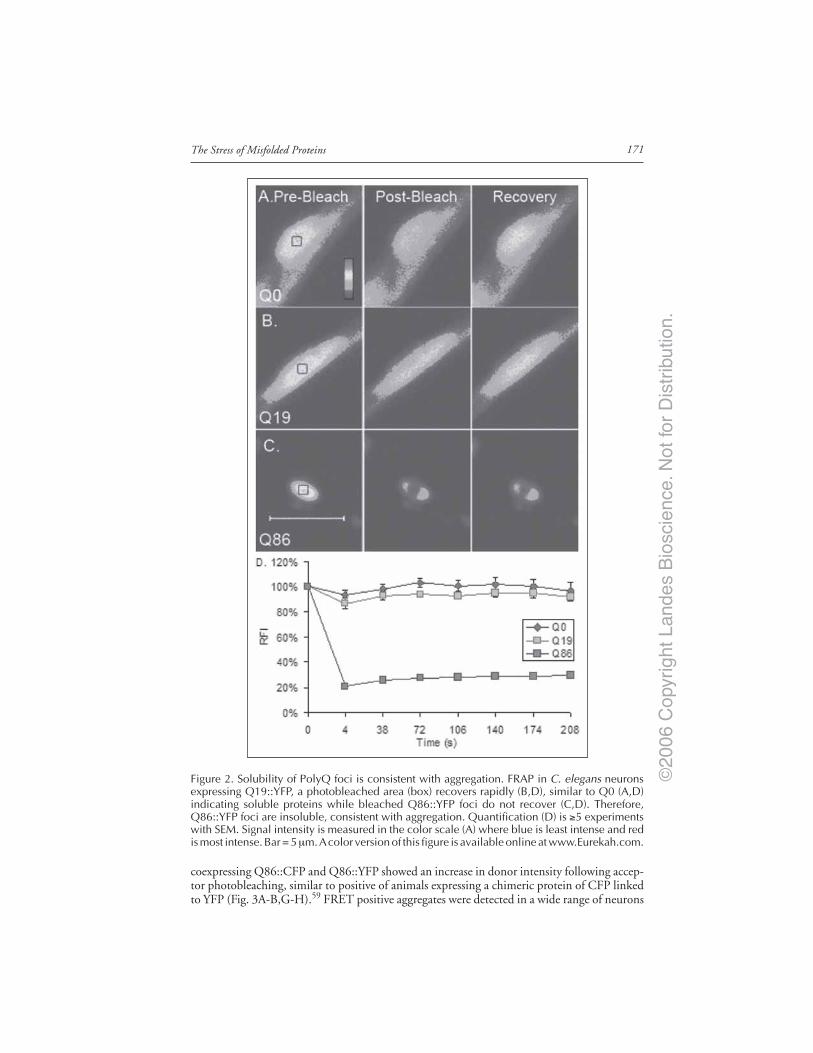
©20
06 C
opyr
ight
Lan
des
Bio
scie
nce.
Not
for
Dis
trib
utio
n.
171The Stress of Misfolded Proteins
coexpressing Q86::CFP and Q86::YFP showed an increase in donor intensity following accep-tor photobleaching, similar to positive of animals expressing a chimeric protein of CFP linkedto YFP (Fig. 3A-B,G-H).59 FRET positive aggregates were detected in a wide range of neurons
Figure 2. Solubility of PolyQ foci is consistent with aggregation. FRAP in C. elegans neuronsexpressing Q19::YFP, a photobleached area (box) recovers rapidly (B,D), similar to Q0 (A,D)indicating soluble proteins while bleached Q86::YFP foci do not recover (C,D). Therefore,Q86::YFP foci are insoluble, consistent with aggregation. Quantification (D) is ≥5 experimentswith SEM. Signal intensity is measured in the color scale (A) where blue is least intense and redis most intense. Bar = 5 µm. A color version of this figure is available online at www.Eurekah.com.

©20
06 C
opyr
ight
Lan
des
Bio
scie
nce.
Not
for
Dis
trib
utio
n.
Molecular Aspects of the Stress Response: Chaperones, Membranes and Networks172
with no clear correlation between FRET intensity and cell-type. The visible redistribution ofQ67 and Q86 into foci, combined with SDS resistance, FRAP and FRET data, shows thatlarge polyQ expansions form insoluble, ordered aggregates in neurons throughout the nervoussystem of C. elegans with no evidence of cell-specific solubility.
PolyQ Length-Dependent Aggregation Correlateswith Neuronal Dysfunction
In human disease, and in mouse, Drosophila, and cell culture studies of polyQ pathogenesis,aggregation is often accompanied by cellular dysfunction, although it remains unknown whetherthe toxic species are polyQ aggregates visible by light microscopy or some intermediate oligo-meric species along the pathway to aggregate formation.62 To test whether polyQ aggregationin C. elegans neurons was accompanied by neurotoxicity, we examined behavioral phenotypesregulated by the nervous system. The most striking phenotype was a polyQ length-dependentloss of coordinated movement leading to nearly complete paralysis. More than 60 neurons inC. elegans enervate muscle cells. Dysfunction or loss of these neurons results in lack of coordi-nation or paralysis.63-65 Q0 or Q19 animals with no visible polyQ aggregates showed rapidmovement similar to wild type (N2) animals and Q19 showed a slight decrease. Animals withvisible aggregates, Q67 and Q86, had limited capacity for coordinated movement exhibiting adecrease of 85% and 89% respectively, relative to Q0 (Fig. 4). These results suggest that forma-tion of visible polyQ aggregates correlates with neuronal dysfunction.
Unexpectedly, pan-neuronal expression of intermediate polyQ lengths, Q35 and Q40, alsoaffected behavior. In contrast to the clear phenotypic differences observed between transgenicanimals that expressed short or long polyQ proteins, individual Q35 and Q40 animals dis-played heterogeneity in behavioral phenotypes (Fig. 4). These results did not seem to correlatewith the soluble distribution pattern of Q35 and Q40 proteins, visually distinct from theextensive foci formation in Q67 and Q86 animals (Fig. 1E). Behavioral toxicity in intermedi-ate animals may have resulted from aggregation in a sub-set of neurons, or Q35 and Q40proteins may display subtle differences in biochemical state, both of which could result inneurotoxicity without overt foci formation.
Figure 3. Q86 protein in neuronal aggregates exhibits FRET, indicating close and roughlyordered interactions at the molecular level. YFP photobleaching is seen on the YFP channel,(panels A,C,E,G) and its effect on CFP donor intensity in the same cell (panels B,D,F,H). Controlanimals expressing CFP::YFP FRET (A,B), Ei = 0.248(± 0.089) while CFP and YFP coexpression(C,D) do not FRET, Ei = 0.001 (± 0.025). Neurons coexpressing Q19::CFP and Q19::YFP (E,F)do not FRET, Ei = -0.090(± -0.057) while coexpression of Q86::CFP and Q86::YFP (G,H) doesproduce FRET, Ei = 0.224(± 0.076). Cells shown are representative of FRET experiments.Intensity is by a color scale (G) where blue is least intense and red is most intense. Scale bar= 2 µm. A color version of this figure is available online at www.Eurekah.com.
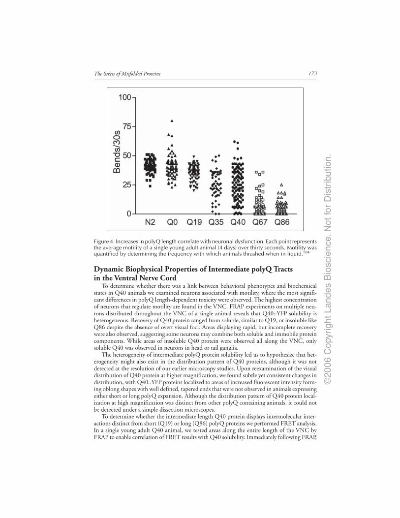
©20
06 C
opyr
ight
Lan
des
Bio
scie
nce.
Not
for
Dis
trib
utio
n.
173The Stress of Misfolded Proteins
Dynamic Biophysical Properties of Intermediate polyQ Tractsin the Ventral Nerve Cord
To determine whether there was a link between behavioral phenotypes and biochemicalstates in Q40 animals we examined neurons associated with motility, where the most signifi-cant differences in polyQ length-dependent toxicity were observed. The highest concentrationof neurons that regulate motility are found in the VNC. FRAP experiments on multiple neu-rons distributed throughout the VNC of a single animal reveals that Q40::YFP solubility isheterogeneous. Recovery of Q40 protein ranged from soluble, similar to Q19, or insoluble likeQ86 despite the absence of overt visual foci. Areas displaying rapid, but incomplete recoverywere also observed, suggesting some neurons may combine both soluble and immobile proteincomponents. While areas of insoluble Q40 protein were observed all along the VNC, onlysoluble Q40 was observed in neurons in head or tail ganglia.
The heterogeneity of intermediate polyQ protein solubility led us to hypothesize that het-erogeneity might also exist in the distribution pattern of Q40 proteins, although it was notdetected at the resolution of our earlier microscopy studies. Upon reexamination of the visualdistribution of Q40 protein at higher magnification, we found subtle yet consistent changes indistribution, with Q40::YFP proteins localized to areas of increased fluorescent intensity form-ing oblong shapes with well defined, tapered ends that were not observed in animals expressingeither short or long polyQ expansion. Although the distribution pattern of Q40 protein local-ization at high magnification was distinct from other polyQ containing animals, it could notbe detected under a simple dissection microscopes.
To determine whether the intermediate length Q40 protein displays intermolecular inter-actions distinct from short (Q19) or long (Q86) polyQ proteins we performed FRET analysis.In a single young adult Q40 animal, we tested areas along the entire length of the VNC byFRAP to enable correlation of FRET results with Q40 solubility. Immediately following FRAP,
Figure 4. Increases in polyQ length correlate with neuronal dysfunction. Each point representsthe average motility of a single young adult animal (4 days) over thirty seconds. Motility wasquantified by determining the frequency with which animals thrashed when in liquid.104
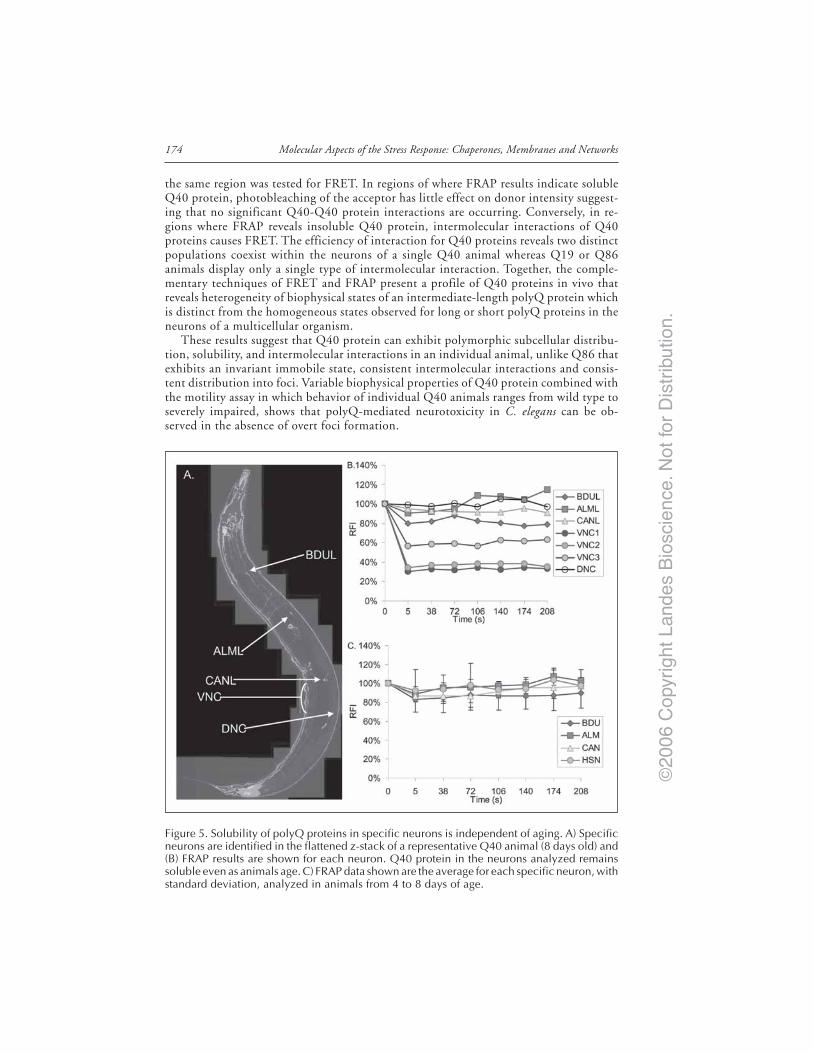
©20
06 C
opyr
ight
Lan
des
Bio
scie
nce.
Not
for
Dis
trib
utio
n.
Molecular Aspects of the Stress Response: Chaperones, Membranes and Networks174
the same region was tested for FRET. In regions of where FRAP results indicate solubleQ40 protein, photobleaching of the acceptor has little effect on donor intensity suggest-ing that no significant Q40-Q40 protein interactions are occurring. Conversely, in re-gions where FRAP reveals insoluble Q40 protein, intermolecular interactions of Q40proteins causes FRET. The efficiency of interaction for Q40 proteins reveals two distinctpopulations coexist within the neurons of a single Q40 animal whereas Q19 or Q86animals display only a single type of intermolecular interaction. Together, the comple-mentary techniques of FRET and FRAP present a profile of Q40 proteins in vivo thatreveals heterogeneity of biophysical states of an intermediate-length polyQ protein whichis distinct from the homogeneous states observed for long or short polyQ proteins in theneurons of a multicellular organism.
These results suggest that Q40 protein can exhibit polymorphic subcellular distribu-tion, solubility, and intermolecular interactions in an individual animal, unlike Q86 thatexhibits an invariant immobile state, consistent intermolecular interactions and consis-tent distribution into foci. Variable biophysical properties of Q40 protein combined withthe motility assay in which behavior of individual Q40 animals ranges from wild type toseverely impaired, shows that polyQ-mediated neurotoxicity in C. elegans can be ob-served in the absence of overt foci formation.
Figure 5. Solubility of polyQ proteins in specific neurons is independent of aging. A) Specificneurons are identified in the flattened z-stack of a representative Q40 animal (8 days old) and(B) FRAP results are shown for each neuron. Q40 protein in the neurons analyzed remainssoluble even as animals age. C) FRAP data shown are the average for each specific neuron, withstandard deviation, analyzed in animals from 4 to 8 days of age.
©20
06 C
opyr
ight
Lan
des
Bio
scie
nce.
Not
for
Dis
trib
utio
n.
175The Stress of Misfolded Proteins
Neuron-Specific Responses to polyQ ProteinsOur observation that solubility of Q40 proteins in C. elegans can vary between different
areas of the nervous system suggests that there are modifiers acting on simple polyQ proteinsindependent of protein context. The observed variability in polyQ protein solubility could bestochastic rather than a neuron-specific response. To address this, we performed FRAP experi-ments on several specific neurons in addition to the VNC. We reasoned that if solubility ofQ40 protein is stochastic, protein solubility in a specific neuron will vary between animals.However, if polyQ protein solubility is neuron-specific, FRAP results should be consistent inall animals tested. FRAP analysis of specific neurons in multiple animals and show that Q40protein is consistently soluble whereas the VNC displays polymorphic Q40 solubility in all C.elegans examined (Fig. 5A,B). These results suggest that polyQ solubility is consistent in spe-cific classes of neurons. We hypothesize that polyQ solubility is modulated by some property ofthe neuron in which it is expressed, such as neuronal function, connectivity, activity levels, orexpression levels of the polyQ protein.
Another characteristic that could have a major effect on polyQ solubility in neurons is theage of the animal. Aging is well established as a major contributor to the onset of polyQ repeatdiseases in both humans and in model systems. We therefore examined specific neurons inanimals of different ages show that in specific neurons, Q40 protein remains soluble in animalsup to 10 days of age (Fig. 5C). In contrast, the VNC displays insoluble protein as early as threedays and polymorphic solubility of Q40 persists as the animals age. These results showed thatQ40 can exhibit polymorphic biochemical properties and remained soluble in some neuronswhile becoming aggregated in the VNC. It also suggested that some neurons displaying solubleQ40 protein are able to maintain solubility as they age. These properties of Q40 are in contrastto the complete aggregation of Q67 or Q86 proteins observed in C. elegans nervous system(Fig. 1) and suggest that for intermediate Q lengths, age is not the only modifier of polyQprotein solubility.
C. elegans model provides a unique tool for investigating the basis of neuron-specific toxic-ity associated with the expression of polyQ proteins. Behavioral and molecular characterizationof polyQ expansions in a pan-neuronal system is the first step in establishing a model in whichneuron-specific, novel modifiers of polyQ-mediated pathogenesis can be identified. Addition-ally, establishing this system in neurons provides the baseline for future studies to examine oneof the major modifiers of polyQ pathogenesis: tissue type. Development of a neuronal modelin C. elegans opens the way to comparative studies on how nonneuronal tissue, such as muscle,responds to polyQ proteins.
The C. elegans polyQ Series in Muscle CellsIn addition to providing a second tissue for comparative studies, C. elegans muscle cells have
some advantages for studying the basic properties of misfolded proteins in vivo. Muscle cellsare significantly larger than neurons facilitating work under comparatively low magnification.In addition, C. elegans muscle cells are sensitive to RNA interference (RNAi) while neurons arerefractory. Therefore, a muscle cell model is ideal for rapid, genome wide screening.
Using a similar approach to that described above, fluorescently tagged polyQ proteinswere expressed in muscle cells of C. elegans. We generated a series of transgenic animalsexpressing polyQ repeats of different lengths, with greater resolution at the intermediatelengths (Q0, Q19, Q29, Q33, Q35, Q40, Q44, Q64, and Q82). In young adult animalsexpressing repeats of Q35 or fewer, we observed diffuse fluorescence in all expressing cellswhile animals expressing muscle Q44, Q64 or Q82 exhibited fluorescence in discreet focisimilar to that observed in neurons (Fig. 6A, Fig. 1). Muscle Q40 animals displayed a poly-morphic distribution with diffuse fluorescence in some cells and foci in others, similar toneuronal Q40 but more easily observed due to the size of muscle cells. In the C. elegansmuscle cell model, there is a shift in the cellular distribution of polyQ protein in young adult

©20
06 C
opyr
ight
Lan
des
Bio
scie
nce.
Not
for
Dis
trib
utio
n.
Molecular Aspects of the Stress Response: Chaperones, Membranes and Networks176
animals between 35-40 repeats, reminiscent of the pathogenic threshold, of 30-40 repeats,observed in the human diseases.
To determine whether there is polyQ-mediated dysfunction in muscle, we examined motil-ity and found that Q19, Q29 or Q35 animals behaved similarly to wild type (N2) while Q82animals displayed severely decreased motility (Fig. 6B). The intermediate Q40 animals, whichhad aggregates in some cells but not in others, exhibited an intermediate motility defect with ahigh degree of variation. The polyQ-dependent motility phenotype observed in the muscle cellmodel was parallel to that of the neuronal model. However, the larger size of body wall musclecells made it possible to examine how polyQ aggregation correlates with cellular dysfunction.When individual animals were assayed for motility and then scored for the number of Q40aggregates, it became clear that in muscle cells, aggregation is directly correlation with cellulardysfunction (Fig. 6C).66
Aging Influences the Threshold for polyQ Aggregation and ToxicityDespite the expression of aggregation-prone proteins throughout the lifetime of the or-
ganism, pathology associated with diseases such as Huntington’s, Alzheimer’s and Parkinson’sare typically not manifest until late in life. This motivated us to study the behavior of polyQproteins during aging. We performed experiments in which individual Q0, Q29, Q33, Q35,Q40, and Q82 animals were examined daily for the appearance of protein aggregates andmotility. Relative to Q40 and Q82 animals that quickly accumulated aggregates and exhib-ited a rapid decline in motility, Q33 and Q35 animals exhibited an initial lag prior to thegradual accumulation of aggregates, however to levels much lower than for Q40 or Q82
Figure 6. Expression of different polyQ::YFP lengths in C. elegans body-wall muscle cellsshows a length-dependent aggregation phenotype. Fluorescence images of young adult C.elegans expressing different lengths of polyQ::YFP (Q0, Q19, Q29, Q33, Q35, Q40, Q44,Q64, Q82). Animals expressing repeats of ≤Q35::YFP show a diffuse fluorescence in all musclecells; in contrast, animals expressing ≥Q44 repeats exhibited punctate distribution of fluores-cence Animals expressing muscle Q40 showed a polymorphic phenotype with both diffuseand punctate fluorescence distribution. (Bar = 0.1 mm). B) Quantification of motility index foryoung adult Q0, Q19, Q29, Q35, Q40, Q82, and unc-54(r293) animals. Data are mean ± SDfor at least 50 animals of each type as percentage of N2 motility. C) Correlation between thenumber of Q40 foci in the muscle cells of an individual animal and their motility.
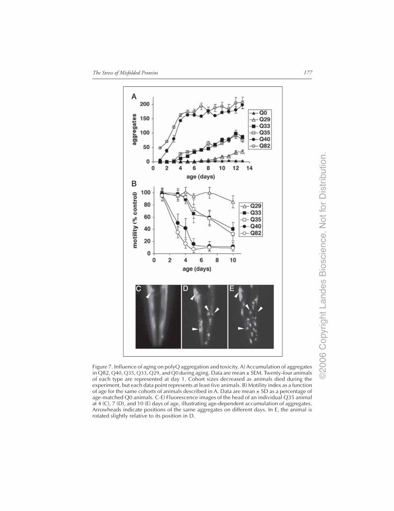
©20
06 C
opyr
ight
Lan
des
Bio
scie
nce.
Not
for
Dis
trib
utio
n.
177The Stress of Misfolded Proteins
Figure 7. Influence of aging on polyQ aggregation and toxicity. A) Accumulation of aggregatesin Q82, Q40, Q35, Q33, Q29, and Q0 during aging. Data are mean ± SEM. Twenty-four animalsof each type are represented at day 1. Cohort sizes decreased as animals died during theexperiment, but each data point represents at least five animals. B) Motility index as a functionof age for the same cohorts of animals described in A. Data are mean ± SD as a percentage ofage-matched Q0 animals. C-E) Fluorescence images of the head of an individual Q35 animalat 4 (C), 7 (D), and 10 (E) days of age, illustrating age-dependent accumulation of aggregates.Arrowheads indicate positions of the same aggregates on different days. In E, the animal isrotated slightly relative to its position in D.
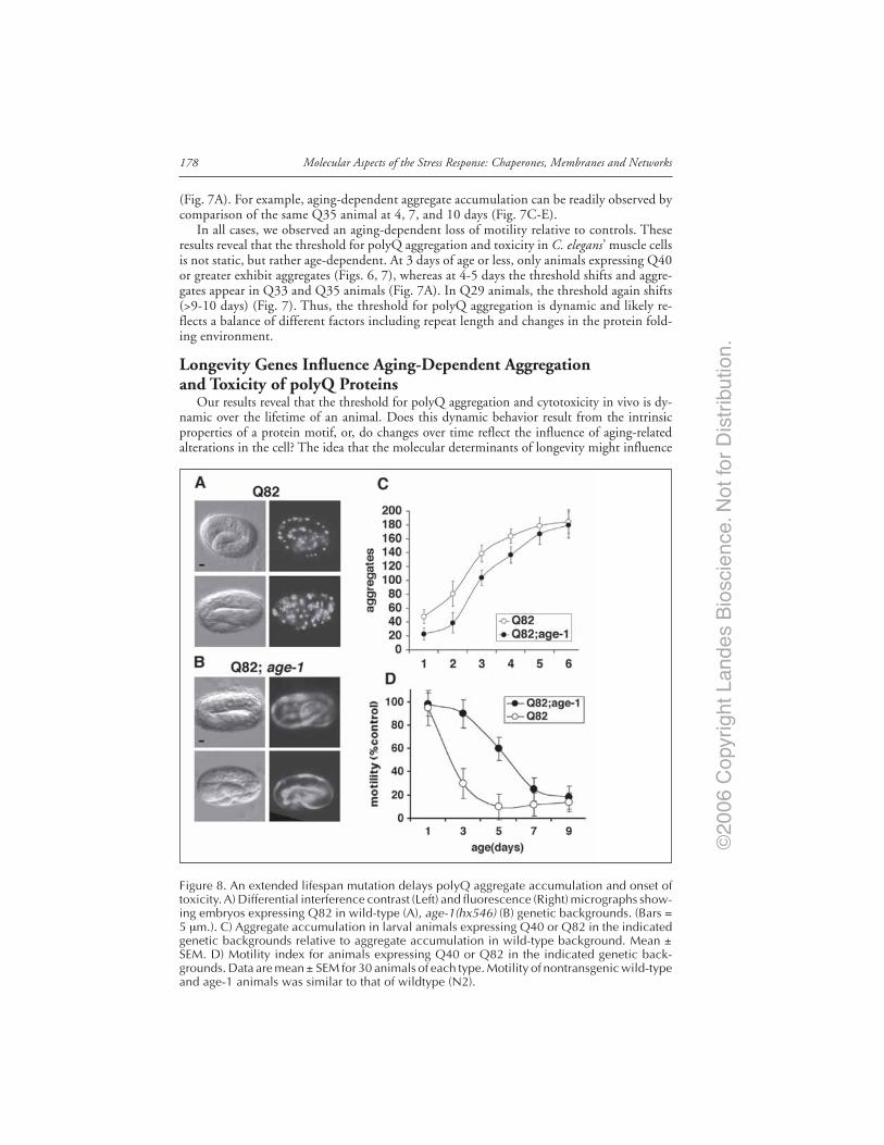
©20
06 C
opyr
ight
Lan
des
Bio
scie
nce.
Not
for
Dis
trib
utio
n.
Molecular Aspects of the Stress Response: Chaperones, Membranes and Networks178
(Fig. 7A). For example, aging-dependent aggregate accumulation can be readily observed bycomparison of the same Q35 animal at 4, 7, and 10 days (Fig. 7C-E).
In all cases, we observed an aging-dependent loss of motility relative to controls. Theseresults reveal that the threshold for polyQ aggregation and toxicity in C. elegans’ muscle cellsis not static, but rather age-dependent. At 3 days of age or less, only animals expressing Q40or greater exhibit aggregates (Figs. 6, 7), whereas at 4-5 days the threshold shifts and aggre-gates appear in Q33 and Q35 animals (Fig. 7A). In Q29 animals, the threshold again shifts(>9-10 days) (Fig. 7). Thus, the threshold for polyQ aggregation is dynamic and likely re-flects a balance of different factors including repeat length and changes in the protein fold-ing environment.
Longevity Genes Influence Aging-Dependent Aggregationand Toxicity of polyQ Proteins
Our results reveal that the threshold for polyQ aggregation and cytotoxicity in vivo is dy-namic over the lifetime of an animal. Does this dynamic behavior result from the intrinsicproperties of a protein motif, or, do changes over time reflect the influence of aging-relatedalterations in the cell? The idea that the molecular determinants of longevity might influence
Figure 8. An extended lifespan mutation delays polyQ aggregate accumulation and onset oftoxicity. A) Differential interference contrast (Left) and fluorescence (Right) micrographs show-ing embryos expressing Q82 in wild-type (A), age-1(hx546) (B) genetic backgrounds. (Bars =5 µm.). C) Aggregate accumulation in larval animals expressing Q40 or Q82 in the indicatedgenetic backgrounds relative to aggregate accumulation in wild-type background. Mean ±SEM. D) Motility index for animals expressing Q40 or Q82 in the indicated genetic back-grounds. Data are mean ± SEM for 30 animals of each type. Motility of nontransgenic wild-typeand age-1 animals was similar to that of wildtype (N2).

©20
06 C
opyr
ight
Lan
des
Bio
scie
nce.
Not
for
Dis
trib
utio
n.
179The Stress of Misfolded Proteins
polyQ-mediated toxicity is supported by observations that the time until pathology develops -days in C. elegans, weeks in Drosophila, months in mice, and years in humans - correlatesapproximately with the lifespan of the organism. Here, again, the C. elegans model was a tre-mendous advantage as the availability of mutants with extended lifespans allowed us to testthese ideas directly.
To accomplish this, we generated transgenic animals expressing Q82 in the background ofthe age-1(hx546) mutation or age-1 RNAi. age-1 encodes a phosphoinositide-3 kinase thatfunctions in an insulin-like signaling (ILS) pathway, and mutations in this gene can extendlifespan by 1.5-2 fold.67,68 Q82 in the age-1 background (Q82;age-1) exhibited reduced aggre-gate formation in embryos relative to Q82 in the wild type background (Fig. 8). Q82 aggregateformation was also reduced 30-50% during larval stages (1-2 days old) in age-1 animals com-pared to wild type background and remained significantly lower through 4-5 days of age.66
Parallel motility assays also demonstrated a delay in onset of the motility defect, consistentwith slower aggregate accumulation in Q82;age-1 animals (Fig. 8).
In wild type animals, the kinase activity of AGE-1 is required in a signaling cascade thatresults in constitutive repression of the fork head transcription factor DAF-16 leading to nor-mal lifespan.67,69,70 Derepression of DAF-16 in age-1 animals results in extended lifespan, anddaf-16 mutations suppress the longevity phenotype. To examine whether age-1 effects on lon-gevity and polyQ aggregation and toxicity are mediated through similar regulatory pathways,we tested whether age-1 suppression of Q82 phenotypes was affected by inactivation of daf-16using RNAi. Q82;age-1;daf-16 animals exhibited aggregation and motility phenotypes similarto Q82 expressed in wild type background. Thus, the dual effects of age-1 on longevity andpolyQ-mediated toxicity share a common genetic pathway.
Our demonstration that a mutation conferring longevity also delays polyQ aggregation andtoxicity suggests a novel link between the genetic regulation of aging and aging-related disease.In subsequent studies, we and others have demonstrated that the molecular link between thesepathways is regulated, in part, by factors that detect and respond to misfolded proteins - namelyheat shock transcription factor (HSF) and molecular chaperones/heat shock proteins. For ex-ample, it has been shown that inhibition of HSF-1 function leads to decreased lifespan and anaccelerated aging phenotype in C. elegans.71-73 Conversely, overexpression of HSF-1 in C. elegansextends lifespan.72,73 Additionally, inactivation of daf-16, hsf-1 or small heat shock proteins inC. elegans accelerated the aggregation of polyQ expansion proteins supporting the idea that ILScould coordinately influence aging and protein aggregation through the action of DAF-16,HSF-1 and molecular chaperones.
Genome-Wide RNAi Screening Identifies Novel Regulators of polyQAggregation and Toxicity
The results described above identify ILS as a genetic pathway that can influence the courseof polyQ-mediated phenotypes. What other pathways might exert similar effects? Numerousover-expression and genetic studies in mammalian and Drosophila models have identified vari-ous enhancers or suppressors of polyQ-mediated aggregation and toxicity.74-76 This iswell-illustrated by the large number of approaches in which various molecular chaperones ei-ther alone or in combination have been shown to influence polyQ-mediated pheno-types.5,7,74,76-78 While one could argue which of these modifiers is the “key” to determining thefate of aggregation-prone proteins, we interpret these results to suggest that the transition ofpolyQ proteins from a soluble to an aggregated state is the result of a balance in which multiplepathways are likely to cooperate. To identify the complete protein-folding buffer involved inpolyQ transition from a soluble to an aggregated state we used a genome-wide RNAi approach.
RNAi is a commonly used reverse-genetics approach in C. elegans allowing the targeteddown-regulation of specific genes by introduction of small fragments of cognate double-strandedRNAs.79,80 This technique has been adapted to genome-wide screens with a library consistingof 16,757 bacterial clones covering 86% of the predicted C. elegans genome.81,82 RNAi has the

©20
06 C
opyr
ight
Lan
des
Bio
scie
nce.
Not
for
Dis
trib
utio
n.
Molecular Aspects of the Stress Response: Chaperones, Membranes and Networks180
additional advantages of allowing detection of lethal positives and the immediate identificationof the target genes. Although genome-wide RNAi screens may miss certain genes secondary tovariation in mRNA depletion and relative inefficiency of the mechanism in neurons, this ap-proach offers an extremely powerful and rapid tool to identify the set of genes that modify agiven phenotype.
To identify genes that prevent polyQ aggregate formation we used C. elegans strains ex-pressing polyQ lengths close to the aggregation threshold, Q33 and Q35 strains, in an RNAigenetic screen. To validate this strategy, we first tested the full range of polyQ strains with agroup of candidate modifiers, genes containing the TPR domain and therefore likely to actas molecular chaperones. Animals with knock down of candidate modifiers were analyzedfor premature appearance of aggregates upon selective RNAi. Of the 72 candidate gene modi-fiers analyzed only four showed an earlier appearance of foci in Q33 and Q35 animals reveal-ing an unexpected specificity among known modifiers of protein folding.83 These effectswere dependent on the presence of an expanded polyQ motif as no foci were observed forany of the 72 RNAi bacterial clones in either Q0 or Q24 animals. The fluorescent fociinduced by RNAi-treatment were biophysically and biochemically identical to aggregates oflong polyQ stretches as judged by FRAP analysis and SDS-PAGE.
Having validated the reliability of our approach, we screened the C. elegans genome andidentified a total of 186 genes that induced earlier onset of the aggregation phenotype inQ35 animals. These modifiers fall into 5 major classes: genes involved in RNA metabolism,protein synthesis, protein folding, protein trafficking and protein degradation (Table 1).Examples of some of these genes are: RNA helicases, splicing factors and transcription fac-tors for RNA metabolism; initiation and elongation factors and ribosomal subunits for pro-tein synthesis; chaperonins and Hsp70 family members for protein folding; nuclear importand cytoskeletal genes for protein trafficking and proteasomal genes for protein degradation.A common feature is an expected imbalance between protein synthesis, folding and degrada-tion triggered by their disruption. In this context, these 5 classes can then be further groupedin two major categories: genes whose disruption leads to an increase in misfolded proteinproduction and genes that when disrupted lead to a decreased clearance of misfolded pro-teins and proper protein turnover. Our results reveal that the transition between soluble and
Table 1. Genome-wide RNAi screen for modifiers of polyQ aggregation identified186 genes, in five classes. These genes all function normally to suppressaggregation; therefore knockdown by RNAi resulted in increased aggregation.
↑Production Misfolded Proteins ↓Clearance of Misfolded Proteins
RNA synthesis & Protein Protein Protein Proteinprocessing (31) synthesis (59) folding (11) transport (18) degradation (16)Transcription Intitiation (6) Chaperonin (6) Endocytosis (1) 19S (8)regulation (5)T27F2.1 (Skip) Y39G10AR.8 (elF-2-γ) cct-5(TCP-1-ε) Y105E8A.9 (γ-adaptin) rpt-5 (26S 6A)
Transcription (2) Elongation (4) Hsp70 (4) Nuclear import (4) 20S(10)ama-1 (RPB1) eft-3 (eEF1A-2) hsp-1 (Hsc70) C53D5.6 (Importin β -3) pas-4 (α type 7)Splicing (17) Ribosomal subunit (43) DnaJ (1) Cytoskeleton (6) Ub ligase (2)
T13H5.4 (SAP61) rps-26 (S26) F18C12.2A K01G5.7 (Tubulin β -2) C47E12.5 (E1)
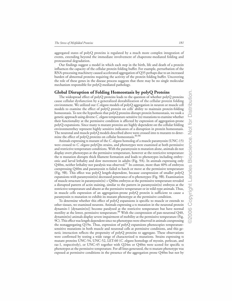
©20
06 C
opyr
ight
Lan
des
Bio
scie
nce.
Not
for
Dis
trib
utio
n.
181The Stress of Misfolded Proteins
aggregated states of polyQ proteins is regulated by a much more complex integration ofevents, extending beyond the immediate involvement of chaperone-mediated folding andproteasomal degradation.
Our findings suggest a model in which each step in the birth, life and death of a proteininfluences the capacity of the cellular protein folding buffer. For example, perturbation of theRNA-processing machinery caused accelerated aggregation of Q35 perhaps due to an increasedburden of abnormal proteins requiring the activity of the protein folding buffer. Uncoveringthe role of these genes in the disease process suggests that there may be no single molecularmechanism responsible for polyQ-mediated pathology.
Global Disruption of Folding Homeostasis by polyQ ProteinsThe widespread effect of polyQ proteins leads to the question of whether polyQ proteins
cause cellular dysfunction by a generalized destabilization of the cellular protein foldingenvironment. We utilized our C elegans models of polyQ aggregation in neuron or muscle cellmodels to examine the effect of polyQ protein on cells’ ability to maintain protein-foldinghomeostasis. To test the hypothesis that polyQ proteins disrupt protein homeostasis, we took agenetic approach using diverse C. elegans temperature-sensitive (ts) mutations to examine whethertheir functionality at the permissive condition is affected by expression of aggregation-pronepolyQ-expansions. Since many ts mutant proteins are highly dependent on the cellular foldingenvironmentthey represent highly sensitive indicators of a disruption in protein homeostasis.The neuronal and muscle polyQ models described above were crossed into ts mutants to deter-mine the effect of polyQ proteins on cellular homeostasis.84-86
Animals expressing ts mutant of the C. elegans homolog of a muscle paramyosin (UNC-15)were crossed to C. elegans polyQm strains, and phenotypes were examined at both permissiveand restrictive temperature conditions. With the paramyosin ts mutation alone, animals do notdisplay overt phenotypes at the permissive temperature, however at the restrictive temperaturethe ts mutation disrupts thick filament formation and leads to phenotypes including embry-onic and larval lethality and slow movement in adults (Fig. 9A). In animals expressing onlyQ40m, neither lethality nor paralysis was observed.87 In contrast, more than 40% of embryoscoexpressing Q40m and paramyosin ts failed to hatch or move at the permissive temperature(Fig. 9B). This effect was polyQ length-dependent, because coexpression of smaller polyQexpansions with paramyosin(ts) decreased penetrance of ts phenotypes (Fig. 9B). Examinationof muscle structure in paramyosin(ts) + Q40m embryos at the permissive temperature revealeda disrupted pattern of actin staining, similar to the pattern in paramyosin(ts) embryos at therestrictive temperature and absent at the permissive temperature or in wild-type animals. Thus,in muscle cells expression of an aggregation-prone polyQ protein is sufficient to cause aparamyosin ts mutation to exhibit its mutant phenotype at the permissive condition.
To determine whether this effect of polyQ expansions is specific to muscle or extends toother tissues, we examined neurons. Animals expressing a ts mutation in the neuronal proteindynamin-1 [dynamin(ts)] become paralyzed at the restrictive temperature but have normalmotility at the lower, permissive temperature.88 With the coexpression of pan-neuronal Q40,dynamin(ts) animals display severe impairment of mobility at the permissive temperature (Fig.9C). This effect was length dependent since no phenotypes were observed in animals coexpressingthe nonaggregating Q19n. Thus, expression of polyQ expansions phenocopies temperature-sensitive mutations in both muscle and neuronal cells at permissive conditions, and this ge-netic interaction reflects the propensity of polyQ proteins to aggregate. These observationswere confirmed by testing a wide range of characterized ts mutations. Strains expressing tsmutant proteins UNC-54, UNC-52, LET-60 (C. elegans homologs of myosin, perlecan, andras-1, respectively), or UNC-45 together with Q24m or Q40m were scored for specific tsphenotypes at the permissive temperature. For all lines generated, the ts mutant phenotype wasexposed at permissive conditions in the presence of the aggregation prone Q40m but not by
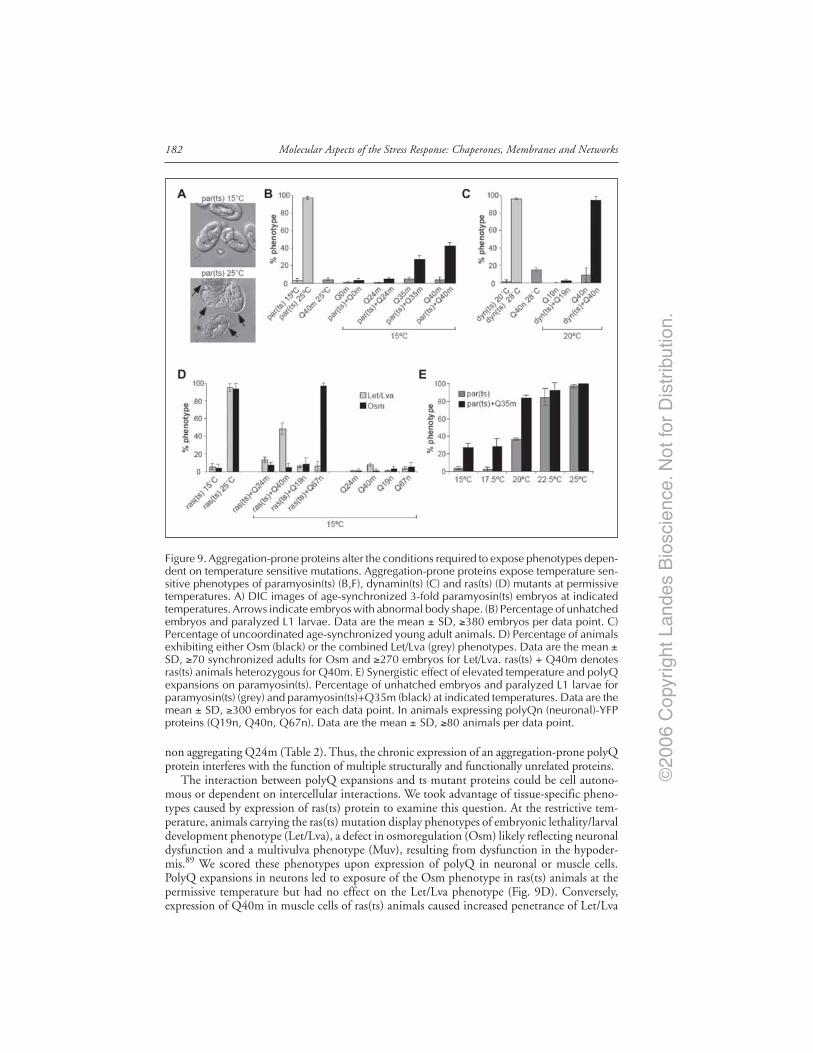
©20
06 C
opyr
ight
Lan
des
Bio
scie
nce.
Not
for
Dis
trib
utio
n.
Molecular Aspects of the Stress Response: Chaperones, Membranes and Networks182
non aggregating Q24m (Table 2). Thus, the chronic expression of an aggregation-prone polyQprotein interferes with the function of multiple structurally and functionally unrelated proteins.
The interaction between polyQ expansions and ts mutant proteins could be cell autono-mous or dependent on intercellular interactions. We took advantage of tissue-specific pheno-types caused by expression of ras(ts) protein to examine this question. At the restrictive tem-perature, animals carrying the ras(ts) mutation display phenotypes of embryonic lethality/larvaldevelopment phenotype (Let/Lva), a defect in osmoregulation (Osm) likely reflecting neuronaldysfunction and a multivulva phenotype (Muv), resulting from dysfunction in the hypoder-mis.89 We scored these phenotypes upon expression of polyQ in neuronal or muscle cells.PolyQ expansions in neurons led to exposure of the Osm phenotype in ras(ts) animals at thepermissive temperature but had no effect on the Let/Lva phenotype (Fig. 9D). Conversely,expression of Q40m in muscle cells of ras(ts) animals caused increased penetrance of Let/Lva
Figure 9. Aggregation-prone proteins alter the conditions required to expose phenotypes depen-dent on temperature sensitive mutations. Aggregation-prone proteins expose temperature sen-sitive phenotypes of paramyosin(ts) (B,F), dynamin(ts) (C) and ras(ts) (D) mutants at permissivetemperatures. A) DIC images of age-synchronized 3-fold paramyosin(ts) embryos at indicatedtemperatures. Arrows indicate embryos with abnormal body shape. (B) Percentage of unhatchedembryos and paralyzed L1 larvae. Data are the mean ± SD, ≥380 embryos per data point. C)Percentage of uncoordinated age-synchronized young adult animals. D) Percentage of animalsexhibiting either Osm (black) or the combined Let/Lva (grey) phenotypes. Data are the mean ±SD, ≥70 synchronized adults for Osm and ≥270 embryos for Let/Lva. ras(ts) + Q40m denotesras(ts) animals heterozygous for Q40m. E) Synergistic effect of elevated temperature and polyQexpansions on paramyosin(ts). Percentage of unhatched embryos and paralyzed L1 larvae forparamyosin(ts) (grey) and paramyosin(ts)+Q35m (black) at indicated temperatures. Data are themean ± SD, ≥300 embryos for each data point. In animals expressing polyQn (neuronal)-YFPproteins (Q19n, Q40n, Q67n). Data are the mean ± SD, ≥80 animals per data point.

©20
06 C
opyr
ight
Lan
des
Bio
scie
nce.
Not
for
Dis
trib
utio
n.
183The Stress of Misfolded Proteins
Tabl
e 2.
Poly
Q e
xpan
sion
s af
fect
the
func
tion
alit
y of
unr
elat
ed t
s m
utan
t pr
otei
ns % A
nim
als
Dis
play
ing
Phen
otyp
e 15˚C
Prot
eins
Exp
ress
edTS
Alle
lePh
enot
ype
Scor
ed (
N V
alue
)15
˚C25
˚CQ
24Q
40
Poly
Qm
-sl
ow m
ovem
ent (
n >
300
)6.
0 ±
5.3
4.6
± 4
.2m
yosi
n(ts
)e1
301
5.6
± 2
.684
.7 ±
13.
5m
yosi
n(ts
) + Q
me1
301
11.2
± 6
.751
.9 ±
19.
3m
yosi
n(ts
)e1
157
5 ±
498
.7 ±
1.4
myo
sin(
ts) +
Qm
e115
75.
9 ±
1.5
55 ±
6
Poly
Qm
-ab
norm
al b
ody
shap
e0
0pe
rlec
an(ts
)su
250
(stif
f par
alys
is) (
n >
100
)1
± 1
.297
.6 ±
2.2
perl
ecan
(ts) +
Qm
su25
00.
8 ±
1.1
48.4
± 6
.5
Poly
Qm
-eg
g la
ying
def
ect (
n >
85)
00
UN
C-4
5(ts
)e2
868.
4 ±
2.1
93.8
± 4
.9U
NC
-45(
ts) +
Qm
e286
5.1
± 7
.387
.7 ±
8.1
Poly
Qm
-em
bryo
nic
leth
ality
+ la
rval
1 ±
0.1
57.
9 ±
1.8
deve
lopm
ent a
rres
t (n
> 2
70)
ras(
ts)
ga89
5.6
± 3
.495
.2 ±
4.3
ras(
ts) +
Qm
ga89
13.3
± 3
100*
Spec
ific
phen
otyp
e of
eac
h ts
mut
atio
n al
one
or in
pol
yQm
bac
kgro
und
was
scor
ed a
t ind
icat
ed te
mpe
ratu
res.
Dat
a ar
e th
e m
ean
± S
D fo
r at l
east
the
indi
cate
d nu
mbe
r(n
) of a
nim
als
for
each
phe
noty
pe s
core
d. D
oubl
e ho
moz
ygou
s ra
s(ts
) + Q
40m
ani
mal
s co
uld
not b
e sc
ored
, as
they
did
not
rea
ch m
atur
e ad
ulth
ood.

©20
06 C
opyr
ight
Lan
des
Bio
scie
nce.
Not
for
Dis
trib
utio
n.
Molecular Aspects of the Stress Response: Chaperones, Membranes and Networks184
phenotype but did not expose the neuronal Osm phenotype (Fig. 9D, Table 2). Neither neu-ronal nor muscle cell expression of polyQ expansions caused the hypodermal Muv phenotype.A similar control was performed with paramyosin(ts),and was not affected by polyQ expan-sions in neurons (Q67n). Likewise, polyQ expansions did not affect phenotypes that did notinvolve ts proteins (caused either by RNAi or gene deletion). Thus, the effect of polyQ expan-sions on mutant ts proteins reflects specific genetic interactions within the same cell type anddoes not result from decreased fitness of the organism.
To understand the nature of this interaction, we examined the cellular localization ofparamyosin(ts) protein when coexpressed with Q40m. The paramyosin ts mutation affectsprotein interactions which, at restrictive temperature, results in mislocalization into foci (Fig.10A).87 When paramyosin(ts) protein is coexpressed with Q40m, it mislocalizes into foci atthe permissive temperature, distinct from Q40m aggregates (Fig. 10B). Thus, expression ofQ40m uncovers the protein folding defect in paramyosin(ts) mutant. In view of this, the dif-ferential penetrance of ts phenotypes (Table 2) may reflect the sensitivity of each ts mutation todisruption of the folding environment.
Aggregation-prone proteins may exert their destabilizing effects by placing a stress on thefolding capacity of the cell. If so, additional stress, such as elevated temperature may act in
Figure 10. PolyQ expansions affect the folding of a ts mutant of paramyosin. A,B) Confocalimages of anti-paramyosin immunostained (red) body wall muscle cells of synchronized youngadults expressing indicated proteins. Arrows: normal muscle sarcomeres, arrowheads: abnormalparamyosin(ts) assemblies. Green color is Q40m-YFP fluorescent protein. Scale bar is 10 µm.

©20
06 C
opyr
ight
Lan
des
Bio
scie
nce.
Not
for
Dis
trib
utio
n.
185The Stress of Misfolded Proteins
synergy with polyQ aggregates to further destabilize ts mutants. Indeed, expression of an inter-mediate (Q35m) expansion shifted the permissive temperature at which paramyosin(ts) wasfully inactivated (Fig. 9E). Furthermore, we observed increased penetrance of ts phenotypes inhomozygous Q40 animals as compared to heterozygous, consistent with the earlier onset ofaggregation in homozygous Q40 animals (Table 2). If the levels of polyQ affect the folding ofthe ts protein, does the misfolding of the ts protein, in turn, intensify misfolding of polyQ? Toanswer that question, we quantified mQ40 aggregation in ts backgrounds and found that ag-gregation was enhanced dramatically. In contrast, mutations that cause a loss of function ratherthan a ts phenotype did not enhance aggregation. From a genetic perspective, temperature-sensitive mutations in proteins unrelated to cellular folding or clearance pathways behaved asmodifiers of polyQ aggregation. This suggests that a positive feedback mechanism exists toenhance the disruption of cellular folding homeostasis.
The appearance of misfolded protein in the cell normally activates a stress response thatincreases protein refolding and turnover and thus rebalances the folding environment.90,91 Incontrast, our results point to the unexpected sensitivity of cellular folding homeostasis to thechronic expression of misfolded proteins under physiological conditions. It is possible that thelow flux of misfolded protein in conformational diseases may alone lack the capacity to activatethe homeostatic stress response. This suggests that the stress response fails to compensate forthe chronic expression of misfolded proteins in human disease.
One potential interpretation of our results is that the protein folding capacity of the cell,integrated at a systems level, is a reflection of expressed protein polymorphisms and randommutations,92 which in themselves do not lead to disease because of the balance achieved byfolding and clearance mechanisms. However, these proteins may misfold and in turn contrib-ute to the progressive disruption of the folding environment when this balance becomes over-whelmed, e.g., by the expression of an aggregation-prone protein in conformational diseases.
ConclusionIn our C. elegans model for the expression of isolated polyQ repeats we focused on the
effects of polyQ-containing proteins in neurons or muscle cells. Expression of isolated polyQmotifs rather than full-length disease-related proteins enables us to uncover conserved featuresunderlying a range of neurodegenerative diseases. A genome wide RNAi screen identified mul-tiple molecular chaperones that have been implicated in polyQ diseases, as well as AD and PD.These studies have revealed a common set of factors that link the genetic regulation of proteinhomeostasis, stress responsiveness and longevity. Thus, longevity and fitness may be, at least inpart, a consequence of the efficient detection, capture and resolution of misfolded andaggregation-prone proteins. The presence of marginally stable or folding-defective proteins inthe genetic background of conformational diseases reveals potent extrinsic factors that canmodify aggregation and toxicity. Given the prevalence of polymorphisms in the human ge-nome,93 the expression of metastable proteins could contribute to variability of disease onsetand progression.94 This interpretation also provides a mechanistic basis to the notion that thelate onset of protein misfolding diseases may be due to gradual accumulation of damagedproteins, resulting in a compromise in folding capacity.95 Cellular degeneration in diseases ofprotein conformation is unlikely to be due to a single defect. Thus, the many toxic effects onvarious cellular processes attributed to misfolded proteins7,59,96-103 could in fact be an integralpart of the global disruption of protein homeostasis.
AcknowledgementsWe thank members of the laboratory past and present who contributed to this work both
intellectually and technically. Support for this research was provided by grants from theNational Institute of General Medical Science, the National Institutes for Aging, theHuntington Disease Society of America Coalition for the Cure, and the Daniel F. and Ada L.Rice Foundation.
©20
06 C
opyr
ight
Lan
des
Bio
scie
nce.
Not
for
Dis
trib
utio
n.
Molecular Aspects of the Stress Response: Chaperones, Membranes and Networks186
References1. Kakizuka A. Protein precipitation: A common etiology in neurodegenerative disorders? Trends Genet
1998; 14:396-402.2. Kopito RR, Ron D. Conformational disease. Nat Cell Biol 2000; 2:E207-209.3. Stefani M, Dobson CM. Protein aggregation and aggregate toxicity: New insights into protein
folding, misfolding diseases and biological evolution. J Mol Med 2003; 81:678-699.4. Dobson CM. Protein folding and its links with human disease. Biochem Soc Symp 2001; 68:1-26.5. Bonini NM. Chaperoning brain degeneration. Proc Natl Acad Sci USA 2002; 99:16407-16411.6. Chan HY, Warrick JM, Andriola I et al. Genetic modulation of polyglutamine toxicity by protein
conjugation pathways in Drosophila. Hum Mol Genet 2002; 11:2895-2904.7. Cummings CJ, Mancini MA, Antalffy B et al. Chaperone suppression of aggregation and altered
subcellular proteasome localization imply protein misfolding in SCA1. Nat Genet 1998; 19:148-154.8. Kayed R, Head E, Thompson JL et al. Common structure of soluble amyloid oligomers implies
common mechanism of pathogenesis. Science 2003; 300:486-489.9. O’Nuallain B, Wetzel R. Conformational Abs recognizing a generic amyloid fibril epitope. Proc
Natl Acad Sci USA 2002; 99:1485-1490.10. Kawaguchi Y, Okamoto T, Taniwaki M et al. CAG expansions in a novel gene for Machado-Joseph
disease at chromosome 14q32.1. Nat Genet 1994; 8:221-228.11. Koide R, Ikeuchi T, Onodera O et al. Unstable expansion of CAG repeat in hereditary
dentatorubral-pallidoluysian atrophy (DRPLA). Nat Genet 1994; 6:9-13.12. La Spada AR, Wilson EM, Lubahn DB et al. Androgen receptor gene mutations in X-linked spinal
and bulbar muscular atrophy. Nature 1991; 352:77-79.13. Orr HT, Chung MY, Banfi S et al. Expansion of an unstable trinucleotide CAG repeat in spinoc-
erebellar ataxia type 1. Nat Genet 1993; 4:221-226.14. Kamino K, Orr HT, Payami H et al. Linkage and mutational analysis of familial Alzheimer disease
kindreds for the APP gene region. Am J Hum Genet 1992; 51:998-1014.15. Laing NG, Siddique T. Cu/Zn superoxide dismutase gene mutations in amyotrophic lateral sclero-
sis: Correlation between genotype and clinical features. J Neurol Neurosurg Psychiatry 1997; 63:815.16. Lucking CB, Durr A, Bonifati V et al. Association between early-onset Parkinson’s disease and
mutations in the parkin gene. French Parkinson’s Disease Genetics Study Group. N Engl J Med2000; 342:1560-1567.
17. Mizuno Y, Hattori N, Kitada T et al. Familial Parkinson’s disease. Alpha-synuclein and parkin.Adv Neurol 2001; 86:13-21.
18. Polymeropoulos MH, Lavedan C, Leroy E et al. Mutation in the alpha-synuclein gene identified infamilies with Parkinson’s disease. Science 1997; 276:2045-2047.
19. Rosen DR, Siddique T, Patterson D et al. Mutations in Cu/Zn superoxide dismutase gene areassociated with familial amyotrophic lateral sclerosis. Nature 1993; 362:59-62.
20. Driscoll M, Gerstbrein B. Dying for a cause: Invertebrate genetics takes on humanneurodegeneration. Nat Rev Genet 2003; 4:181-194.
21. Link CD. Transgenic invertebrate models of age-associated neurodegenerative diseases. Mech Age-ing Dev 2001; 122:1639-1649.
22. Thompson LM, Marsh JL. Invertebrate models of neurologic disease: Insights into pathogenesisand therapy. Curr Neurol Neurosci Rep 2003; 3:442-448.
23. Westlund B, Stilwell G, Sluder A. Invertebrate disease models in neurotherapeutic discovery. CurrOpin Drug Discov Devel 2004; 7:169-178.
24. Merry DE. Animal models of Kennedy disease. NeuroRx 2005; 2:471-479.25. Greene JC, Whitworth AJ, Andrews LA et al. Genetic and genomic studies of Drosophila parkin
mutants implicate oxidative stress and innate immune responses in pathogenesis. Hum Mol Genet2005; 14:799-811.
26. Greene JC, Whitworth AJ, Kuo I et al. Mitochondrial pathology and apoptotic muscle degenera-tion in Drosophila parkin mutants. Proc Natl Acad Sci USA 2003; 100:4078-4083.
27. Faber PW, Alter JR, MacDonald ME et al. Polyglutamine-mediated dysfunction and apoptoticdeath of a Caenorhabditis elegans sensory neuron. Proc Natl Acad Sci USA 1999; 96:179-184.
28. Faber PW, Voisine C, King DC et al. Glutamine/proline-rich PQE-1 proteins protect Caenorhabditiselegans neurons from huntingtin polyglutamine neurotoxicity. Proc Natl Acad Sci USA 2002;99:17131-17136.
29. Parker JA, Connolly JB, Wellington C et al. Expanded polyglutamines in Caenorhabditis eleganscause axonal abnormalities and severe dysfunction of PLM mechanosensory neurons without celldeath. Proc Natl Acad Sci USA 2001; 98:13318-13323.

©20
06 C
opyr
ight
Lan
des
Bio
scie
nce.
Not
for
Dis
trib
utio
n.
187The Stress of Misfolded Proteins
30. Warrick JM, Paulson HL, Gray-Board GL et al. Expanded polyglutamine protein forms nuclearinclusions and causes neural degeneration in Drosophila. Cell 1998; 93:939-949.
31. Feany MB, Bender WW. A Drosophila model of Parkinson’s disease. Nature 2000; 404:394-398.32. Link CD. Expression of human beta-amyloid peptide in transgenic Caenorhabditis elegans. Proc
Natl Acad Sci USA 1995; 92:9368-9372.33. Oeda T, Shimohama S, Kitagawa N et al. Oxidative stress causes abnormal accumulation of famil-
ial amyotrophic lateral sclerosis-related mutant SOD1 in transgenic Caenorhabditis elegans. HumMol Genet 2001; 10:2013-2023.
34. Lee VM, Kenyon TK, Trojanowski JQ. Transgenic animal models of tauopathies. Biochim BiophysActa 2005; 1739:251-259.
35. Marsh JL, Thompson LM. Can flies help humans treat neurodegenerative diseases? Bioessays 2004;26:485-496.
36. Shulman JM, Shulman LM, Weiner WJ et al. From fruit fly to bedside: Translating lessons fromDrosophila models of neurodegenerative disease. Curr Opin Neurol 2003; 16:443-449.
37. Orr HT. Beyond the Qs in the polyglutamine diseases. Genes Dev 2001; 15:925-932.38. Ross CA. Polyglutamine pathogenesis: Emergence of unifying mechanisms for Huntington’s disease
and related disorders. Neuron 2002; 35:819-822.39. Trottier Y, Lutz Y, Stevanin G et al. Polyglutamine expansion as a pathological epitope in
Huntington’s disease and four dominant cerebellar ataxias. Nature 1995; 378:403-406.40. Zoghbi HY, Orr HT. Glutamine repeats and neurodegeneration. Annu Rev Neurosci 2000;
23:217-247.41. Ross CA. When more is less: Pathogenesis of glutamine repeat neurodegenerative diseases. Neuron
1995; 15:493-496.42. Margolis RL, Ross CA. Expansion explosion: New clues to the pathogenesis of repeat expansion
neurodegenerative diseases. Trends Mol Med 2001; 7:479-482.43. Davies SW, Turmaine M, Cozens BA et al. Formation of neuronal intranuclear inclusions under-
lies the neurological dysfunction in mice transgenic for the HD mutation. Cell 1997; 90:537-548.44. Mangiarini L, Sathasivam K, Seller M et al. Exon 1 of the HD gene with an expanded CAG
repeat is sufficient to cause a progressive neurological phenotype in transgenic mice. Cell 1996;87:493-506.
45. Ordway JM, Tallaksen-Greene S, Gutekunst CA et al. Ectopically expressed CAG repeats causeintranuclear inclusions and a progressive late onset neurological phenotype in the mouse. Cell 1997;91:753-763.
46. Gusella JF, MacDonald ME. Molecular genetics: Unmasking polyglutamine triggers inneurodegenerative disease. Nat Rev Neurosci 2000; 1:109-115.
47. Sherman MY, Muchowski PJ. Making yeast tremble: Yeast models as tools to study neurodegenerativedisorders. Neuromolecular Med 2003; 4:133-146.
48. Andrew SE, Goldberg YP, Kremer B et al. The relationship between trinucleotide (CAG) repeatlength and clinical features of Huntington’s disease. Nat Genet 1993; 4:398-403.
49. Brinkman RR, Mezei MM, Theilmann J. The likelihood of being affected with Huntington dis-ease by a particular age, for a specific CAG size. Am J Hum Genet 1997; 60:1202-1210.
50. Bargmann CI. Neurobiology of the Caenorhabditis elegans genome. Science 1998; 282:2028-2033.51. Bargmann CI, Kaplan JM. Signal transduction in the Caenorhabditis elegans nervous system. Annu
Rev Neurosci 1998; 21:279-308.52. Brownlee DJ, Fairweather I. Exploring the neurotransmitter labyrinth in nematodes. Trends Neurosci
1999; 22:16-24.53. Lippincott-Schwartz J, Patterson GH. Development and use of fluorescent protein markers in liv-
ing cells. Science 2003; 300:87-91.54. White J, Stelzer E. Photobleaching GFP reveals protein dynamics inside live cells. Trends Cell Biol
1999; 9:61-65.55. Scherzinger E, Sittler A, Schweiger K et al. Self-assembly of polyglutamine-containing huntingtin
fragments into amyloid-like fibrils: Implications for Huntington’s disease pathology. Proc Natl AcadSci USA 1999; 96:4604-4609.
56. DiFiglia M, Sapp E, Chase KO et al. Aggregation of huntingtin in neuronal intranuclear inclu-sions and dystrophic neurites in brain. Science 1997; 277:1990-1993.
57. Perutz MF, Johnson T, Suzuki M et al. Glutamine repeats as polar zippers: Their possible role ininherited neurodegenerative diseases. Proc Natl Acad Sci USA 1994; 91:5355-5358.
58. Ross CA, Poirier MA, Wanker EE et al. Polyglutamine fibrillogenesis: The pathway unfolds. ProcNatl Acad Sci USA 2003; 100:1-3.
59. Kim S, Nollen EA, Kitagawa K et al. Polyglutamine protein aggregates are dynamic. Nat Cell Biol2002; 4:826-831.

©20
06 C
opyr
ight
Lan
des
Bio
scie
nce.
Not
for
Dis
trib
utio
n.
Molecular Aspects of the Stress Response: Chaperones, Membranes and Networks188
60. Tsien RY. The green fluorescent protein. Annu Rev Biochem 1998; 67:509-544.61. Berney C, Danuser G. FRET or no FRET: A quantitative comparison. Biophys J 2003;
84:3992-4010.62. Ross CA, Poirier MA. Protein aggregation and neurodegenerative disease. Nat Med 2004; 10:S10-17.63. White JG, Southgate E, Thomson JN et al. The Structure of the nervous system of the nematode
Caenorhabditis elegans. Phil Trans Royal Soc London Series B Biol Scien 1986; 314:1-340.64. White JG, Southgate E, Thomson JN et al. The structure of the ventral nerve cord of Caenorhabditis
elegans. Philos Trans R Soc Lond B Biol Sci 1976; 275:327-348.65. Rand JB, Nonet ML. Chapter 22. Synatptic Transmission. In: Riddle DL, Blumenthal T, Meyer
BJ, Priess JR, eds. C. Elegans II. Plainview, NY: Cold Spring Harbor Laboratory Press, 1997.66. Morley JF, Brignull HR, Weyers JJ et al. The threshold for polyglutamine-expansion protein ag-
gregation and cellular toxicity is dynamic and influenced by aging in Caenorhabditis elegans. ProcNatl Acad Sci USA 2002; 99:10417-10422.
67. Guarente L, Kenyon C. Genetic pathways that regulate ageing in model organisms. Nature 2000;408:255-262.
68. Morris JZ, Tissenbaum HA, Ruvkun G. A phosphatidylinositol-3-OH kinase family member regu-lating longevity and diapause in Caenorhabditis elegans. Nature 1996; 382:536-539.
69. Lin K, Dorman JB, Rodan A et al. daf-16: An HNF-3/forkhead family member that can functionto double the life-span of Caenorhabditis elegans. Science 1997; 278:1319-1322.
70. Ogg S, Paradis S, Gottlieb S et al. The Fork head transcription factor DAF-16 transduces insulin-likemetabolic and longevity signals in C. elegans. Nature 1997; 389:994-999.
71. Garigan D, Hsu AL, Fraser AG et al. Genetic analysis of tissue aging in Caenorhabditis elegans: Arole for heat-shock factor and bacterial proliferation. Genetics 2002; 161:1101-1112.
72. Hsu AL, Murphy CT, Kenyon C. Regulation of aging and age-related disease by DAF-16 andheat-shock factor. Science 2003; 300:1142-1145.
73. Morley JF, Morimoto RI. Regulation of longevity in Caenorhabditis elegans by heat shock factorand molecular chaperones. Mol Biol Cell 2004; 15:657-664.
74. Cummings CJ, Sun Y, Opal P et al. Over-expression of inducible HSP70 chaperone suppressesneuropathology and improves motor function in SCA1 mice. Hum Mol Genet 2001; 10:1511-1518.
75. Fernandez-Funez P, Nino-Rosales ML, de Gouyon B et al. Identification of genes that modifyataxin-1-induced neurodegeneration. Nature 2000; 408:101-106.
76. Warrick JM, Chan HY, Gray-Board GL et al. Suppression of polyglutamine-mediatedneurodegeneration in Drosophila by the molecular chaperone HSP70. Nat Genet 1999; 23:425-428.
77. Carmichael J, Chatellier J, Woolfson A et al. Bacterial and yeast chaperones reduce both aggregateformation and cell death in mammalian cell models of Huntington’s disease. Proc Natl Acad SciUSA 2000; 97:9701-9705.
78. Chai Y, Koppenhafer SL, Bonini NM et al. Analysis of the role of heat shock protein (Hsp)molecular chaperones in polyglutamine disease. J Neurosci 1999; 19:10338-10347.
79. Fire A, Xu S, Montgomery MK et al. Potent and specific genetic interference by double-strandedRNA in Caenorhabditis elegans. Nature 1998; 391:806-811.
80. Wang J, Barr MM. RNA interference in Caenorhabditis elegans. Methods Enzymol 2005; 392:36-55.81. Fraser AG, Kamath RS, Zipperlen P et al. Functional genomic analysis of C. elegans chromosome
I by systematic RNA interference. Nature 2000; 408:325-330.82. Kamath RS, Fraser AG, Dong Y et al. Systematic functional analysis of the Caenorhabditis elegans
genome using RNAi. Nature 2003; 421:231-237.83. Nollen EA, Garcia SM, van Haaften G et al. Genome-wide RNA interference screen identifies
previously undescribed regulators of polyglutamine aggregation. Proc Natl Acad Sci USA 2004;101:6403-6408.
84. Brown CR, Hong-Brown LQ, Welch WJ. Correcting temperature-sensitive protein folding defects.J Clin Invest 1997; 99:1432-1444.
85. Pedersen CB, Bross P, Winter VS et al. Misfolding, degradation, and aggregation of variant pro-teins. The molecular pathogenesis of short chain acyl-CoA dehydrogenase (SCAD) deficiency. JBiol Chem 2003; 278:47449-47458.
86. Van Dyk TK, Gatenby AA, LaRossa RA. Demonstration by genetic suppression of interaction ofGroE products with many proteins. Nature 1989; 342:451-453.
87. Gengyo-Ando K, Kagawa H. Single charge change on the helical surface of the paramyosin roddramatically disrupts thick filament assembly in Caenorhabditis elegans. J Mol Biol 1991;219:429-441.
88. Clark SG, Shurland DL, Meyerowitz EM et al. A dynamin GTPase mutation causes a rapid andreversible temperatureinducible locomotion defect in C. elegans. Proc Natl Acad Sci USA 1997;94:10438-10443.

©20
06 C
opyr
ight
Lan
des
Bio
scie
nce.
Not
for
Dis
trib
utio
n.
189The Stress of Misfolded Proteins
89. Eisenmann DM, Kim SK. Mechanism of activation of the Caenorhabditis elegans ras homologuelet-60 by a novel, temperature-sensitive, gain-of-function mutation. Genetics 1997; 146:553-565.
90. Goff SA, Goldberg AL. Production of abnormal proteins in E. coli stimulates transcription of lonand other heat shock genes. Cell 1985; 41:587-595.
91. Morimoto RI. Regulation of the heat shock transcriptional response: Cross talk between a familyof heat shock factors, molecular chaperones, and negative regulators. Genes Dev 1998; 12:3788-3796.
92. Rutherford SL, Lindquist S. Hsp90 as a capacitor for morphological evolution. Nature 1998;396:336-342.
93. Sachidanandam R, Weissman D, Schmidt SC et al. A map of human genome sequence variationcontaining 1.42 million single nucleotide polymorphisms. Nature 2001; 409:928-933.
94. Wexler NS, Lorimer J, Porter J et al. Venezuelan kindreds reveal that genetic and environmentalfactors modulate Huntington’s disease age of onset. Proc Natl Acad Sci USA 2004; 101:3498-3503.
95. Oliver CN, Ahn BW, Moerman EJ et al. Age-related changes in oxidized proteins. J Biol Chem1987; 262:5488-5491.
96. McCampbell A, Taylor JP, Taye AA et al. CREB-binding protein sequestration by expandedpolyglutamine. Hum Mol Genet 2000; 9:2197-2202.
97. Muchowski PJ, Schaffar G, Sittler A et al. Hsp70 and hsp40 chaperones can inhibit self-assemblyof polyglutamine proteins into amyloid-like fibrils. Proc Natl Acad Sci USA 2000; 97:7841-7846.
98. Perez MK, Paulson HL, Pendse SJ et al. Recruitment and the role of nuclear localization inpolyglutamine-mediated aggregation. J Cell Biol 1998; 143:1457-1470.
99. Ii K, Ito H, Tanaka K et al. Immunocytochemical colocalization of the proteasome in ubiquitinatedstructures in neurodegenerative diseases and the elderly. J Neuropathol Exp Neurol 1997;56:125-131.
100. Bence NF, Sampat RM, Kopito RR. Impairment of the ubiquitin-proteasome system by proteinaggregation. Science 2001; 292:1552-1555.
101. Gabai VL, Meriin AB, Yaglom JA et al. Role of Hsp70 in regulation of stress-kinase JNK: Impli-cations in apoptosis and aging. FEBS Lett 1998; 438:1-4.
102. Gu M, Gash MT, Mann VM et al. Mitochondrial defect in Huntington’s disease caudate nucleus.Ann Neurol 1996; 39:385-389.
103. Holmberg CI, Staniszewski KE, Mensah KN et al. Inefficient degradation of truncated polyglutamineproteins by the proteasome. EMBO J 2004; 23:4307-4318.
104. Miller KG, Alfonso A, Nguyen M et al. A genetic selection for Caenorhabditis elegans synaptictransmission mutants. Proc Natl Acad Sci USA 1996; 93:12593-12598.

©20
06 C
opyr
ight
Lan
des
Bio
scie
nce.
Not
for
Dis
trib
utio
n.

©20
06 C
opyr
ight
Lan
des
Bio
scie
nce.
Not
for
Dis
trib
utio
n.

©20
06 C
opyr
ight
Lan
des
Bio
scie
nce.
Not
for
Dis
trib
utio
n.
CHAPTER 16
Hsp90 and Developmental NetworksSuzannah Rutherford,* Jennifer R. Knapp and Peter Csermely
Abstract
The most abundant cytoplasmic chaperone of eukaryotic cells, Hsp90 is a hubin developmental regulatory networks and the first example described of thephenomenon of molecular buffering. As a chaperone for many different signaling
proteins, Hsp90 maintains the clarity and strength of communication within and betweencells, concealing developmental and stochastic variations that otherwise cause abrupt morpho-logical changes in a large variety of organisms, including Drosophila and Arabidopsis. The chap-ter provides a framework for understanding how Hsp90 controls the sudden appearance ofnovel morphologies. We start with a discussion of the longstanding problem of hidden poly-genic variation and then introduce the idea of signal transduction thresholds in mediating theeffect of Hsp90 on the expression of phenotypic variation. This leads to a discussion of the roleof nonlinearity in creating thresholds for sudden changes in cellular responses to developmen-tal signals. We end with speculation on the potentially pivotal role of Hsp90 in controlling thedevelopmental networks that determine morphological stasis and change in evolution.
IntroductionHsp90 is a hub in developmental regulatory networks. By maintaining the activity of over
150 signal transduction proteins in many different developmental pathways, the Hsp90 chap-erone controls the strength of signaling, not only by its client proteins, but through the path-ways in which they reside. Hsp90 target pathways regulate multiple processes including cellcycle and transcriptional control, as well as chromatin remodeling, growth control, apoptosis,stress responses and response to differentiation signals.1-3 Many Hsp90-dependent pathwaysare both ancient and conserved. For example, Hsp90 controls the activity of four of eightancient signaling pathways that arose before the protostome-deuterostome split. These path-ways form a “basic evolutionary toolkit”4 that is thought to have enabled the evolution of mostof biletarian diversity (Table 1). It is not yet understood how the same genes and pathwaysproduce such dramatically different morphologies. One possibility is that evolution occurs viasmall changes in the structure and connectivity of signal transduction networks, the intricateweb of cellular communications and responses that orchestrate development.
Perhaps surprising given their intricacy, biological networks are by nature highly error toler-ant and self-correcting. Part of this stability has to do with positive and negative feedback andthe complexity of inter-connections. In addition, biological networks are “scale-free”. The numberof interactions between nodes (genes or proteins) follows a power-law distribution - meaningthat a few percent of nodes (such as Hsp90) have huge numbers of interactions, but most
*Corresponding Author: Suzannah Rutherford—Division of Basic Sciences, Fred HutchinsonCancer Research Center, Mailstop A2-168, 1100 Fairview Avenue North, Seattle, Washington98109-1024, U.S.A. Email: [email protected]
Molecular Aspects of the Stress Response: Chaperones, Membranes and Networks, edited byPeter Csermely and Laszlo Vigh. ©2007 Landes Bioscienceand Springer Science+Business Media.

©20
06 C
opyr
ight
Lan
des
Bio
scie
nce.
Not
for
Dis
trib
utio
n.
191Hsp90 and Developmental Networks
nodes having very few interactions (~2).5 The resulting behavior of scale-free networks istherefore robust to the random loss of a high percent of nodes.6 However rare, highly-connectednodes such as Hsp90 are control points whose loss is devastating to network function. The“structural stability, dynamic behavior, robustness and error and attack tolerance” depend onthe integrity of highly connected nodes (hubs), particularly those positioned high in the hier-archy of network structure (Fig. 1).7,8 For example, hubs such as Hsp90 provide a connectionbetween many different modules of the network (meaning clusters of nodes more intercon-nected among themselves than to nodes in other modules).
Several years ago, one of the authors (S. Rutherford) discovered that modest (≤50%) reduc-tion of Hsp90 uncovers a remarkable reserve of previously hidden morphogenic variation inDrosophila.9 Hsp90-buffered changes are highly dependent on genetic background and resultsfrom the segregation, in normal populations, of likely hundreds to thousands of alleles withgenerally small effects on the strength of signaling through Hsp90-dependent pathways. Nor-mally this genetic variation is hidden, and therefore has neutral effects on fitness. However, whenthe strength of Hsp90 signaling pathways is reduced beyond critical threshold levels, normallystable phenotypes can abruptly change.10 Waddington called the error tolerance and self-correctingbehavior of developmental processes canalization.11,12 In line with Waddington’s original con-cept of canalization, Hsp90-dependent changes in morphology are not quantitative in nature,but are either highly discrete or qualitative (Rutherford et al, submitted for publication). Indeed,a characteristic and almost unifying feature of life is its highly discrete nature. From species toindividuals, organs, cell types and physiological and sub-cellular systems: perturbations changeprobabilities of alternative types, but rarely result in the generation of intermediate forms.
Here we present a framework for understanding Hsp90 buffering and variation in develop-ment. We begin with a discussion of the longstanding problem of hidden genetic variation. Wethen cover Hsp90 and signal transduction thresholds, the role of nonlinearity in creating thresh-olds in development, and end with speculation on Hsp90 potentially pivotal role in control-ling the networks that control the balance of developmental stasis and change.
Hidden Genetic Variation“We are largely ignorant of the answer to a question of fundamental importance: How much
[and which] variation has direct phenotypic or functional effects that influence the survival andreproduction of individuals?” Quote from special committee charged with evaluating the Hu-man Genome Diversity Project.13
Table 1. A conserved group of signaling pathways, believed to have arisen before theprotostome-deuterostome split, has been deployed to different endsthroughout bilateran development. Shown are the eight major pathways andexamples of Hsp90 client proteins imbedded in each.
Pathway Hsp90 Targets References
Hedgehog (Hh) – –Wingless related (Wnt) – –Transforming growth factor B Tak1* 50Receptor tyrosine kinase (RTK) IKK; ERB2; AKT 51-54Notch – –JAK/STAT Stat1, Stat3 55Nuclear hormone pathways Nuclear hormonre receptors 56Apoptosis Caspase 9, Bid 57,58
*Tak1 is a noncanonical effector of TGFB signaling.
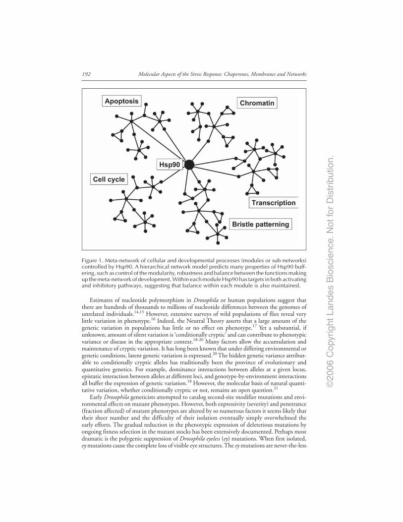
©20
06 C
opyr
ight
Lan
des
Bio
scie
nce.
Not
for
Dis
trib
utio
n.
Molecular Aspects of the Stress Response: Chaperones, Membranes and Networks192
Estimates of nucleotide polymorphism in Drosophila or human populations suggest thatthere are hundreds of thousands to millions of nucleotide differences between the genomes ofunrelated individuals.14,15 However, extensive surveys of wild populations of flies reveal verylittle variation in phenotype.16 Indeed, the Neutral Theory asserts that a large amount of thegenetic variation in populations has little or no effect on phenotype.17 Yet a substantial, ifunknown, amount of silent variation is ‘conditionally cryptic’ and can contribute to phenotypicvariance or disease in the appropriate context.18-20 Many factors allow the accumulation andmaintenance of cryptic variation. It has long been known that under differing environmental orgenetic conditions, latent genetic variation is expressed.20 The hidden genetic variance attribut-able to conditionally cryptic alleles has traditionally been the province of evolutionary andquantitative genetics. For example, dominance interactions between alleles at a given locus,epistatic interaction between alleles at different loci, and genotype-by-environment interactionsall buffer the expression of genetic variation.18 However, the molecular basis of natural quanti-tative variation, whether conditionally cryptic or not, remains an open question.21
Early Drosophila geneticists attempted to catalog second-site modifier mutations and envi-ronmental effects on mutant phenotypes. However, both expressivity (severity) and penetrance(fraction affected) of mutant phenotypes are altered by so numerous factors it seems likely thattheir sheer number and the difficulty of their isolation eventually simply overwhelmed theearly efforts. The gradual reduction in the phenotypic expression of deleterious mutations byongoing fitness selection in the mutant stocks has been extensively documented. Perhaps mostdramatic is the polygenic suppression of Drosophila eyeless (ey) mutations. When first isolated,ey mutations cause the complete loss of visible eye structures. The ey mutations are never-the-less
Figure 1. Meta-network of cellular and developmental processes (modules or sub-networks)controlled by Hsp90. A hierarchical network model predicts many properties of Hsp90 buff-ering, such as control of the modularity, robustness and balance between the functions makingup the meta-network of development. Within each module Hsp90 has targets in both activatingand inhibitory pathways, suggesting that balance within each module is also maintained.

©20
06 C
opyr
ight
Lan
des
Bio
scie
nce.
Not
for
Dis
trib
utio
n.
193Hsp90 and Developmental Networks
extremely sensitive to environmental effects, and are easily suppressed by the rapid accumula-tion of ubiquitous and abundant polygenic modifiers that restore a normal eye structure andvision.22 Outcrossing these stocks again restores the ey phenotype and shows that the mutatationwas still present, but “hidden” by modifier alleles. As eye structure is normally invariant, thesemodifier alleles normally constitute cryptic genetic variation with hidden potential to affecteye development. Perhaps most astounding, ectopic expression of the ey/Pax-6 transcriptionfactor drives the formation of Drosophila eye structures in multiple tissues of the adult fly (forexample on wings, legs, antennae).23 Because Pax-6 function is both necessary and sufficientfor eye development, and is highly conserved throughout evolution, it has been called the“master control gene” for eye development.24 While Pax-6 may be the one of the best examplesof a master control gene, its singularity and importance is questionable as its leading role in thisprocess can be by-passed by polygenic variants and environmental effects.10 These studies dem-onstrate the abundance of developmental variation and alternate routes to the development ofindistinguishable morphological endpoints.
Conversely, the increased variation in phenotype revealed by major developmental muta-tions is common and well-documented, and is a form of gene-interaction or epistasis.10,25
Reducing Hsp90 either genetically, or with an inhibitor, has pleiotropic effects on many mor-phogenic processes, uncovering variation depending on both Hsp90 reduction and its interac-tion with trait-specific genetic backgrounds. The diversion of Hsp90 to proteins damaged byheat or other stress provides environmental control of Hsp90-dependent genetic andgenotype-by-environment variation. Recent work has shown that other chaperones involved inprotein folding, such as Hsp70 and Hsp60 family members, also buffer genetic variation, likelythrough a direct molecular effect on sequence variants that cannot fold normally without theirassistance.26,27 By contrast, we believe Hsp90 effects are largely indirect.28 Rather than buffer-ing variant client proteins directly, allowing them to fold, accumulating evidence indicates thatHsp90 acts through enhancing and suppressing effects of genetic variation and the environ-ment on the strength and fidelity of developmental signaling pathways.
Hsp90 and Signal Transduction ThresholdsOur thinking about Hsp90-buffered variation centers on the idea of thresholds for the
expression of phenotypes in response to continuously varying strengths of signaling throughHsp90 target pathways. The genetic interactions of Hsp90 with client proteins in signal trans-duction pathways demonstrate the existence of thresholds, and are easily understood in light ofthe large body of previous work on the evolutionarily conserved biochemical and genetic re-quirement for Hsp90 by specific client proteins.1-3 When Hsp90 levels are decreased by muta-tion or by inhibitors, signal transduction clients begin to loose activity, and the strength oftarget pathways becomes severely reduced.29-31 Threshold trait models were developed in thecontext of quantitative and population genetics and are often used to describe the genetics ofhuman disease.32 In development, thresholds are transitions between continuous inputs anddiscrete outputs. Many binary cell-fate decisions are controlled by thresholds. Specific geneticinteractions between Hsp90 and signaling pathways dominantly reveal phenotypic thresholds.Loss of Hsp90 function is lethal to most cells in most organisms, in Drosophila heterozygous(recessive) Hsp90 mutations dominantly enhance and suppress sensitizing (cryptic) mutationsin many Hsp90 client pathways. Hsp90 heterozygotes fail to complement Cdc37 heterozygousmutations,33 suppress an over-activated phenotype of the Torso receptor tyrosine kinase,34 en-hance Cdc2 mutant heterozygotes35 and enhance or suppress under- and over-activated com-ponents of the sevenless (sev) mitogen activated protein kinase (MAPK) pathway.
The well-characterized MAPK signaling pathways initiated at the Drosophila sev receptor isa model for Hsp90 interactions with signaling near trait thresholds. High-level activation ofthe sev pathway promotes the normal photoreceptor cell fate, while lower-level activation ofthe pathway results in a default nonneuronal fate and loss of a photoreceptor. This decision isan all-or-none switch; no intermediate cell-types are found. If signaling is too high or in the

©20
06 C
opyr
ight
Lan
des
Bio
scie
nce.
Not
for
Dis
trib
utio
n.
Molecular Aspects of the Stress Response: Chaperones, Membranes and Networks194
wrong place, extra photoreceptors are produced, giving flies a gain-of-function rough eye phe-notype. A series of molecular genetic screens were instrumental in identifying the componentsof receptor tyrosine kinase signaling pathways. Endogenous members of the sev cascade werereplaced with marginally over- or under-activated analogs and the temperature adjusted so thatflies have normal or gain-of-function phenotypes.35-37 Heterozygous mutations in other com-ponents of the pathway (m/+), which are normally ‘cryptic’ recessive mutations, behave likedominant enhancer or suppressor mutations in the appropriate genetic background. Tellingly,when sev signaling is reduced toward a lower threshold, Hsp90 mutations also reduce signalingfurther, resulting in the loss-of-function phenotype seen with heterozygous mutations in otherpathway members.35,36 Similarly, when the sev pathway is over-activated beyond an upperthreshold (gain of function phenotype), heterozygous Hsp90 mutations reduce activity andrestore a normal, smooth eye phenotype.38,39 The genetic interaction of Hsp90 in many clientpathways shows that it can reduce signaling near thresholds for the expression of mutant phe-notypes. If target pathways are sensitive to reduction of Hsp90 client functions, the same resultis produced simply reducing Hsp90 activity.
The level of extrinsic or intrinsic noise in signaling and the cell sensitivity could also modu-late the expression of phenotypes near signal transduction thresholds. This phenomenon iscalled either stochastic resonance or stochastic focusing, depending on the whether the noise isextrinsic or intrinsic, respectively. If noise is low, a low level signal may not pass the signaltransduction threshold. On the contrary, if the noise is high, the same low level signal will passthe same threshold a higher fraction of the time (Fig. 2).40,41 By decreasing the “noisiness” ofcellular signals, molecular chaperones would also modulate the sensitivities of signal transduc-tion near trait thresholds. When Hsp90 is inhibited, increased developmental and/or purelyenvironmental noise may allow normally-lower signals to pass the same sensitivity thresholds,increasing phenotypic diversity at the whole-cell, or whole-organism level.42
Figure 2. Stochastic resonance as a possible reason for chaperone-induced buffering in thediversity of developmental changes. Stochastic resonance is the phenomenon, where intrinsicor extrinsic noise helps a low level signal to surpass a signal transduction threshold. If molecu-lar chaperones decrease the noise of cellular processes, this may also contribute to theirbuffering effects of developmental changes by not allowing low-level signals to act.
©20
06 C
opyr
ight
Lan
des
Bio
scie
nce.
Not
for
Dis
trib
utio
n.
195Hsp90 and Developmental Networks
Nonlinearity in Developmental Responses to Signal TransductionThe ability to produce all-or-none or switch like behavior in response to continuous
variation in the strength of the underlying developmental signal may be a common theme inbiology.40,43 For example, the addition of upstream kinases in MAPK signaling creates asigmoidal (threshold) response over a narrow range.44 The addition of positive feedback cansharply amplify a graded input to produce a steep and sudden (“switch-like”) change inoutput.45 We suggest that such nonlinearity in the response of phenotype to underlyingdevelopmental signals is a common feature of development, and produces much of the ob-served discrete behavior of biological systems. Given nonlinearity in Hsp90 target pathways,when Hsp90 is reduced, signaling is reduced. We believe that the sudden changes in mor-phology, or changes in previously invariant quantitative traits reflects the fact that thresholdshave been crossed.
A Pivotal Role for Hsp90 in Network Evolvability?The activity of signal transduction clients and therefore client pathways decrease sharply
with modest decreases in Hsp90 function. Through these target proteins, Hsp90 simulta-neously controls output of multiple target pathways imbedded in many different develop-mental processes such as regulation of cell-cycle, apoptosis or differentiation responses. Eachof these processes is controlled by a complex sub-network of signaling pathways that inte-grate developmental cues to generate appropriate responses. Hsp90 may be uniquely posi-tioned to balance the stability and modularity of the ‘meta-network’ of processes that makeup development, linking network sensitivity and behavior to the environment.
If the meta-network of development were a mobile with groups of hanging objects (nodes),we suggest that Hsp90 would be at the center of the mobile, supporting several groups ofobjects (‘sub-networks’ or modules). Linkages between each sub network and Hsp90 arerepresented by the strings that support the mobile. The effective concentration of Hsp90available for signal transduction determines the overall height (activity) of the meta-network,and the connectivity or separation of the modules—complete loss of Hsp90 world fragmentthe modules into isolated sub-networks. By reducing or increasing several target proteinfunctions simultaneously, we suggest that Hsp90 maintains the balance of the system. Theoverall height (activity) between the subnetworks is shifted ensemble with the degree ofenvironmental stress.
Pushing the metaphor a little further, we suggest that Hsp90 also maintains balance withineach sub-network. For example, both cdc28 (a key activator of the cell cycle) and wee1 (aninhibitory kinase that acts on cdc28) are Hsp90 targets. Decreases in both activating and in-hibiting pathways would balance out the effect of varying Hsp90 on the rate of the cell cycle. Itseems reasonable to expect that in most cases selection has sculpted the set of targets in activat-ing and inhibitory pathways such that the balance of their function is maintained. Normalchanges in Hsp90 availability would have no net effect. However, if natural variation or theenvironment has already destabilized the network balance, bringing an activating or inhibitorypathway close to its threshold, further reduction of signaling by loss of Hsp90 function couldresult in thresholds being breached and previously hidden phenotypes revealed.
Extending our thoughts beyond the role of Hsp90 to a general molecular buffering effect ofchaperones, we have to note that chaperones provide low affinity, transient contacts with otherproteins. Weak links are known to help system stability in a large variety of networks frommacromolecules to social networks and ecosystems, which may be a general network-level phe-nomenon explaining many of the genetic buffering effects.42,46 Besides molecular buffering byHsp90 and other chaperones, a large number of other mechanisms may also control the diver-sity of the phenotype.25,47-49 A single common mechanism, such as involvement in signaling orepigenetic modifications of histones and DNA structure, seems unlikely to explain all of theeffects observed. If a general explanation is sought, we believe it is more likely to be related tothe overall structure of developmental networks and their emergent properties.
©20
06 C
opyr
ight
Lan
des
Bio
scie
nce.
Not
for
Dis
trib
utio
n.
Molecular Aspects of the Stress Response: Chaperones, Membranes and Networks196
AcknowledgementsWork in the authors’ laboratory was supported by research grants from NIH R01 GM068873
the EU (FP6506850, FP6-016003), Hungarian Science Foundation (OTKA-T37357 andOTKA-F47281), Hungarian Ministry of Social Welfare (ETT-32/03), Hungarian NationalResearch Initiative (1A/056/2004 and KKK-0015/3.0).
References1. Csermely P, Schnaider T, Soti C et al. The 90-kDa molecular chaperone family: Structure, func-
tion, and clinical applications. A comprehensive review. Pharmacol Ther 1998; 79:129-168.2. Pratt WB, Toft DO. Regulation of signaling protein function and trafficking by the hsp90/
hsp70-based chaperone machinery. Exp Biol Med (Maywood) 2003; 228:111-133.3. Zhao R, Davey M, Hsu YC et al. Navigating the chaperone network: An integrative map of physi-
cal and genetic interactions mediated by the hsp90 chaperone. Cell 2005; 120:715-727.4. Knoll AH, Carroll SB. Early animal evolution: Emerging views from comparative biology and ge-
ology. Science 1999; 284:2129-2137.5. Jeong H, Tombor B, Albert R et al. The large-scale organization of metabolic networks. Nature
2000; 407:651-654.6. Albert R, Jeong H, Barabasi AL. Error and attack tolerance of complex networks. Nature 2000;
406:378-382.7. Ravasz E, Somera AL, Mongru DA et al. Hierarchical organization of modularity in metabolic
networks. Science 2002; 297:1551-1555.8. Barabási AL. Linked: The New Science of Networks. Cambridge: Perseus Pub, 2002:280.9. Rutherford SL, Lindquist S. Hsp90 as a capacitor for morphological evolution. Nature 1998;
396:336-342.10. Rutherford SL. From genotype to phenotype: Buffering mechanisms and the storage of genetic
information. Bioessays 2000; 22:1095-1105.11. Waddington CH. The Strategy of the Genes; a Discussion of Some Aspects of Theroetical Biology.
Vol. ix. New York: Macmillan, 1957:262.12. Waddington C. Evolutionary systems - Animal and human. Nature 1950; 183:1634-1638.13. Evaluating Human Genetic Diversity. Commission on Life Sciences, National Research Council.
Washington, DC: National Academy Press, 1997.14. Begun DJ, Aquadro CF. Levels of naturally occurring DNA polymorphism correlate with recombi-
nation rates in D. melanogaster. Nature 1992; 365:519-520.15. Moriyama EN, Powell JR. Intraspecific nuclear DNA variation in Drosophila. Mol Biol Evol 1996;
13:261-277.16. Powell JR. Progress and Prospects in Evolutionary Biology: The Drosophila Model. Vol. xiv. New
York: Oxford University Press, 1997:562.17. Kimura M. The Neutral Theory of Molecular Evolution. Vol. xv. Cambridge [Cambridgeshire],
New York: Cambridge University Press, 1983:367.18. Lynch M, Walsh B. Genetics and Analysis of Quantitative Traits. Vol. xvi. Sunderland: Sinauer,
1998:980.19. Hartman JL, Garvik B, Hartwell L. Principles for the buffering of genetic variation. Science 2001;
291:1001-1004.20. Perera FP. Environment and cancer: Who are susceptible? Science 1997; 278:1068-1073.21. Mackay TF. Quantitative trait loci in Drosophila. Nat Rev Genet 2001; 2:11-20.22. Morgan TH. Variability of eyeless. Publs Carnegie Instn 1929; 399:139-168.23. Halder G, Callaerts P, Gehring WJ. Induction of ectopic eyes by targeted expression of the eyeless
gene in Drosophila. Science 1995; 267:1788-1792.24. Gehring WJ. The master control gene for morphogenesis and evolution of the eye. Genes Cells
1996; 1:11-15.25. Bergman A, Siegal ML. Evolutionary capacitance as a general feature of complex gene networks.
Nature 2003; 424:549-552.26. Fares MA, Ruiz-Gonzalez MX, Moya A et al. Endosymbiotic bacteria: groEL buffers against delete-
rious mutations. Nature 2002; 417:398.27. Chow KC. Hsp70 (DnaK) — An evolution facilitator? Trends Genet 2000; 16:484-485.28. Rutherford SL. Between genotype and phenotype: Protein chaperones and evolvability. Nat Rev
Genet 2003; 4:263-274.29. Sreedhar AS, Soti C, Csermely P. Inhibition of Hsp90: A new strategy for inhibiting protein ki-
nases. Biochim Biophys Acta 2004; 1697:233-242.30. Whitesell L, Mimnaugh EG, De Costa B et al. Inhibition of heat shock protein HSP90-pp60v-src
heteroprotein complex formation by benzoquinone ansamycins: Essential role for stress proteins inoncogenic transformation. Proc Natl Acad Sci USA 1994; 91:8324-8328.
©20
06 C
opyr
ight
Lan
des
Bio
scie
nce.
Not
for
Dis
trib
utio
n.
197Hsp90 and Developmental Networks
31. Zhang H, Burrows F. Targeting multiple signal transduction pathways through inhibition of Hsp90.J Mol Med 2004; 82:488-499.
32. Falconer DS, Mackay TFC. Introduction to Quantitative Genetics. Vol. xiii. Essex, England:Longman, 1996:464.
33. Kimura Y, Rutherford SL, Miyata Y et al. Cdc37 is a molecular chaperone with specific functionsin signal transduction. Genes Dev 1997; 11:1775-1785.
34. Doyle H, Bishop J. Torso, a receptor tyrosine kinase required for embryonic pattern formation,shares substrates with the sevenless and EGF-R pathways in Drosophila. Genes Dev 1993; 7:633-646.
35. Cutforth T, Rubin GM. Mutations in Hsp83 and cdc37 impair signaling by the sevenless receptortyrosine kinase in Drosophila. Cell 1994; 77:1027-1036.
36. Daga A, Banerjee U. Resolving the sevenless pathway using sensitized genetic backgrounds. CellMol Biol Res 1994; 40:245-251.
37. Simon M, Bowtell D, Dodson G et al. Ras1 and a putative guanine nucleotide exchange factorperform crucial steps in signaling by the sevenless protein tyrosine kinase. Cell 1991; 67:701-716.
38. Dickson BJ, van der Straten A, Dominguez M et al. Mutations Modulating Raf signaling in Droso-phila eye development. Genetics 1996; 142:163-171.
39. van der Straten A, Rommel C, Dickson B et al. The heat shock protein 83 (Hsp83) is required forRaf-mediated signalling in Drosophila. EMBO J 1997; 16:1961-1969.
40. Hasty J, Pradines J, Dolnik M et al. Noise-based switches and amplifiers for gene expression. ProcNatl Acad Sci USA 2000; 97:2075-2080.
41. Paulsson J, Berg OG, Ehrenberg M. Stochastic focusing: Fluctuation-enhanced sensitivity of intra-cellular regulation. Proc Natl Acad Sci USA 2000; 97:7148-7153.
42. Csermely P. Weak Links: Stabilizers of Complex Systems from Proteins to Social Networks. Heidel-berg: Springer Verlag, 2006.
43. Gardner TS, Cantor CR, Collins JJ. Construction of a genetic toggle switch in Escherichia coli.Nature 2000; 403:339-342.
44. Goldbeter A, Koshland Jr DE. Ultrasensitivity in biochemical systems controlled by covalent modi-fication. Interplay between zero-order and multistep effects. J Biol Chem 1984; 259:14441-14447.
45. Ferrell Jr JE, Machleder EM. The biochemical basis of an all-or-none cell fate switch in Xenopusoocytes. Science 1998; 280:895-898.
46. Csermely P. Strong links are important, but weak links stabilize them. Trends Biochem Sci 2004;29:331-334.
47. True HL, Berlin I, Lindquist SL. Epigenetic regulation of translation reveals hidden genetic varia-tion to produce complex traits. Nature 2004; 431:184-187.
48. Wade MJ, Johnson NA, Jones R et al. Genetic variation segregating in natural populations ofTribolium castaneum affecting traits observed in hybrids with T. freemani. Genetics 1997;147:1235-1247.
49. Sangster TA, Lindquist S, Queitsch C. Under cover: Causes, effects and implications ofHsp90-mediated genetic capacitance. Bioessays 2004; 26:348-362.
50. Bouwmeester T, Bauch A, Ruffner H et al. A physical and functional map of the human TNF-alpha/NF-kappa B signal transduction pathway. Nat Cell Biol 2004; 6:97-105.
51. Broemer M, Krappmann D, Scheidereit C. Requirement of Hsp90 activity for IkappaB kinase(IKK) biosynthesis and for constitutive and inducible IKK and NF-kappaB activation. Oncogene2004; 23:5378-5386.
52. Xu W, Mimnaugh E, Rosser MF et al. Sensitivity of mature Erbb2 to geldanamycin is conferredby its kinase domain and is mediated by the chaperone protein Hsp90. J Biol Chem 2001;276:3702-3708.
53. Chiosis G, Timaul MN, Lucas B et al. A small molecule designed to bind to the adenine nucle-otide pocket of Hsp90 causes Her2 degradation and the growth arrest and differentiation of breastcancer cells. Chem Biol 2001; 8:289-299.
54. Fujita N, Sato S, Ishida A et al. Involvement of Hsp90 in signaling and stability of3-phosphoinositide-dependent kinase-1. J Biol Chem 2002; 277:10346-10353.
55. Sehgal PB. Plasma membrane rafts and chaperones in cytokine/STAT signaling. Acta Biochim Pol2003; 50:583-594.
56. Pratt WB, Welsh MJ. Chaperone functions of the heat shock proteins associated with steroid re-ceptors. Semin Cell Biol 1994; 5:83-93.
57. Pandey P, Saleh A, Nakazawa A et al. Negative regulation of cytochrome c-mediated oligomeriza-tion of Apaf-1 and activation of procaspase-9 by heat shock protein 90. EMBO J 2000;19:4310-4322.
58. Zhao C, Wang E. Heat shock protein 90 suppresses tumor necrosis factor alpha induced apoptosisby preventing the cleavage of Bid in NIH3T3 fibroblasts. Cell Signal 2004; 16:313-321.

©20
06 C
opyr
ight
Lan
des
Bio
scie
nce.
Not
for
Dis
trib
utio
n.