molecular and genetic evidence for a tetrapolar mating ... · besides the genes encoding pheromones...
TRANSCRIPT
Molecular and Genetic Evidence for a Tetrapolar Mating System inthe Basidiomycetous Yeast Kwoniella mangrovensis and Two NovelSibling Species
Marco A. Guerreiro,a Deborah J. Springer,b Joana A. Rodrigues,a* Laura N. Rusche,b* Keisha Findley,b* Joseph Heitman,b
Álvaro Fonsecaa
Centro de Recursos Microbiológicos (CREM), Departamento de Ciências da Vida, Faculdade de Ciências e Tecnologia, Universidade Nova de Lisboa, Caparica, Portugala;Department of Molecular Genetics and Microbiology, Duke University Medical Center, Durham, North Carolina, USAb
Kwoniella mangrovensis has been described as a sexual species with a bipolar mating system. Phylogenetic analysis of multiplegenes places this species together with Kwoniella heveanensis in the Kwoniella clade, a sister clade to that containing two patho-genic species of global importance, Cryptococcus neoformans and Cryptococcus gattii, within the Tremellales. Recent studiesdefining the mating type loci (MAT) of species in these clades showed that, with the exception of C. neoformans and C. gattii,which are bipolar with a single biallelic multigene MAT locus, several other species feature a tetrapolar mating system with twounlinked loci (homeodomain [HD] and pheromone/receptor [P/R] loci). We characterized several strains from the originalstudy describing K. mangrovensis; two MAT regions were amplified and sequenced: the STE20 gene (P/R locus) and the diver-gently transcribed SXI1 and SXI2 genes (HD locus). We identified five different mating types with different STE20/SXI allelecombinations that together with results of mating experiments demonstrate that K. mangrovensis is not bipolar but instead hasa tetrapolar mating system. Sequence and gene analysis for a 43-kb segment of the K. mangrovensis type strain MAT locus re-vealed remarkable synteny with the homologous K. heveanensis MAT P/R region, providing new insights into slower evolutionof MAT loci in the Kwoniella compared to the Cryptococcus clade of the Tremellales. The study of additional isolates from plantsubstrates in Europe and Botswana using a combination of multilocus sequencing with MAT gene analysis revealed two novelsibling species that we name Kwoniella europaea and Kwoniella botswanensis and which appear to also have tetrapolar matingsystems.
Fungal mating-type loci (MAT) are specialized regions of thegenome that determine sexual identity of haploid cells and
progression through the sexual cycle (1). There is considerableinterest in the genetic characterization of MAT loci due to theircentral role in fungal life cycles, their connection to lifestyle andvirulence (viz., in human or plant-pathogenic taxa), and the im-pact of sexual recombination on population genetics and specia-tion. The discovery of remarkable convergence in the structure ofsex-determining genomic regions from studies in animals, plants,and fungi illuminates the forces that shape the evolutionary tra-jectories of MAT loci or sex chromosomes in eukaryotes (2).
In basidiomycetes, two major types of MAT loci have so farbeen recognized (3). The tetrapolar mating system of the cornsmut Ustilago maydis or the mushroom Coprinopsis cinerea is gov-erned by two small (�10-kb) unlinked loci: one encodes membersof the homeodomain (HD) family of transcription factors, whichheterodimerize upon mating to generate an active transcriptionregulator (HD locus), and the other encodes lipopeptide phero-mone precursors and 7-transmembrane pheromone receptorsthat mediate intercellular signaling (P/R locus). Alleles at both locimust differ for mating to occur, and in many cases each locus ismultiallelic; hence, numerous mating types exist in certain tet-rapolar species. On the other hand, in the bipolar system of thehuman pathogen Cryptococcus neoformans or the barley smut Us-tilago hordei, the two MAT loci found in the tetrapolar species arelinked and form a single, large (�100-kb) multigene locus rich inrepetitive elements and containing several additional genes, eitherrelated or unrelated to mating. In the bipolar species, there arenormally only two possible versions for the content of the single
MAT locus, and hence only two mating types exist. Recent studieshave suggested that the basidiomycete bipolar mating systems areevolutionarily derived from tetrapolar ancestral states (4–6).
A comparative study of the MAT structure of C. neoformansand sibling taxa in the pathogenic species complex, including theanalysis of gene order, phylogeny, and synonymous substitutionrates, led to the proposal of an evolutionary model that posits aseries of gene acquisitions, chromosomal translocations, and re-combination events leading to expansion of the MAT-specific re-gions and linkage of the two loci of a postulated tetrapolar ances-tor to form the large derived locus of the bipolar Cryptococcusspecies (5). The expansion of this nonrecombining sex-determin-ing genomic region mirrors the models proposed for the genesis ofsex chromosomes in multicellular eukaryotes (2). This study hassince been expanded to include several other closely aligned sap-
Received 9 March 2013 Accepted 15 March 2013
Published ahead of print 22 March 2013
Address correspondence to Álvaro Fonseca, [email protected].
* Present address: Joana A. Rodrigues, Departamento das Ciências e Tecnologiada Biomassa, FCT/UNL, Caparica, Portugal; Laura N. Rusche, Department ofBiological Sciences, University at Buffalo, SUNY, Buffalo, New York, USA; KeishaFindley, National Human Genome Research Institute, Bethesda, Maryland, USA.
Supplemental material for this article may be found at http://dx.doi.org/10.1128/EC.00065-13.
Copyright © 2013, American Society for Microbiology. All Rights Reserved.
doi:10.1128/EC.00065-13
746 ec.asm.org Eukaryotic Cell p. 746–760 May 2013 Volume 12 Number 5
on January 24, 2019 by guesthttp://ec.asm
.org/D
ownloaded from
robic species in the order Tremellales (class Tremellomycetes,subphylum Agaricomycotina), namely, Cryptococcus amylolentus,Cryptococcus heveanensis, Filobasidiella depauperata, and Tsuchi-yaea wingfieldii (7–10). The latter studies disclosed sexual states intwo species previously classified as asexual, viz. C. amylolentus andC. heveanensis (the teleomorphs were named Filobasidiella amylo-lenta and Kwoniella heveanensis) (8, 11) and further revealed thatthese heterothallic species, as well as the more distantly relateddimorphic jelly fungus Tremella mesenterica, all have tetrapolarmating systems with unlinked P/R and HD loci, thus providingadditional support for the tetrapolar-to-bipolar evolutionarymodel (5, 12). The P/R locus was found to be biallelic, whereas theHD locus is multiallelic. This comparative genomics approach hasalso shown some conservation of gene content within MAT geneclusters. For example, the P/R locus region generally includes,besides the genes encoding pheromones (MF) and pheromonereceptors (STE3), genes encoding elements of the pheromone-activated signaling cascade (STE11, STE12, and STE20), as well asother genes that may be unrelated to mating (e.g., BSP3, LPD1,RPO41). The HD locus consists of two divergently transcribed HDgenes, homologous to the single SXI1 or SXI2 genes found in ei-ther of the C. neoformans mating types, and the flanking MAT-specific or nonspecific regions include additional genes commonto most of the studied species (e.g., RPL22, SPO14). Informationon the genes contained in those loci may be found in previouspublications (3, 5, 8, 9, 12).
Kwoniella mangrovensis (the original spelling, mangroviensis,was considered an orthographical error) was described as a sexualspecies with a bipolar mating system based on the study of severalisolates from mangrove habitats in the Bahamas and Florida Ev-erglades (13). On the basis of phylogenetic analysis of multiplegenes (14), this species belongs, together with Kwoniella (Crypto-coccus) heveanensis, to the Kwoniella clade, a sister clade to thatwhich contains the pathogenic Cryptococcus neoformans andCryptococcus gattii, as well as C. amylolentus (Filobasidiella clade,which we henceforth rename as the Cryptococcus clade for reasonsthat will be explained in Discussion). In view of the results ofprevious studies that explored the MAT locus structure in thoseyeasts, it seemed essential to examine this genomic region instrains from the original description of K. mangrovensis to verifyits mating system and to seek additional insights on the evolutionof MAT in the Tremellales. Statzell-Tallman et al. (13) included intheir study two cork isolates from Europe which had divergentinternal transcribed spacer (ITS) sequences but were consideredconspecific with the mangrove strains. We expanded our study toinclude additional isolates from different substrates and geo-graphic locations, identified as K. mangrovensis on the basis oflarge subunit (LSU) rRNA gene sequences, and applied a multilo-cus sequencing approach to reassess species limits and discloseputative cryptic species. We also determined sequences of putativeMAT genes from the same strains to elucidate the mating systemand to gain additional evidence for defining species boundaries.
In this study, we provide evidence for a tetrapolar mating sys-tem in K. mangrovensis and insight into the MAT locus structureof this species, which revealed remarkable similarities to the moreclosely related saprobic yeasts in the Tremellales. We performedphylogenetic analyses of MAT genes and of housekeeping genes ina multilocus approach for an enlarged group of strains of K. man-grovensis and closely related species, which revealed two hithertounrecognized taxa comprising strains from plant-related sub-
strates in Europe and Africa. The results obtained clearly illustratethe power of the combined analysis of housekeeping genes andMAT genes in resolving species boundaries and providing insightsinto the mating systems of species whose sexual status may or maynot be known.
MATERIALS AND METHODSYeast strains and media. The yeast cultures used in this study are listed inTable S1 in the supplemental material. Strains were obtained from thefollowing culture collections: Portuguese Yeast Culture Collection, Por-tugal (PYCC); Agricultural Research Service Culture Collection, UnitedStates (NRRL); The Spanish Type Culture Collection, Spain (CECT);Centraalbureau voor Schimmelcultures, The Netherlands (CBS); CultureCollection of Industrial Microorganisms, Slovenia (ZIM); (Agro) Indus-trial Fungi & Yeasts Collection, Belgium (MUCL). All isolates were grownand maintained on MYP medium at 25°C and 4°C, respectively. MYPmedium contained 0.7% (wt/vol) malt extract, 0.25% (wt/vol) Soytone,0.05% (wt/vol) yeast extract, and 1.5% agar.
Mating experiments. To test for sexual compatibility, isolates weregrown on MYP agar plates at 25°C for 3 days. Pairs of cell suspensionswere then inoculated and mixed together on plates containing CMA,YCB, or MEA medium at 25°C and examined with a phase-contrast mi-croscope (Zeiss, Germany) for the presence of filaments and sexual struc-tures, up to 4 weeks of incubation. CMA medium contained 1.5% (wt/vol)corn meal agar (Difco) and 0.5% (wt/vol) agar. YCB medium contained1.17% (wt/vol) yeast carbon base (Difco) and 2% agar. MEA mediumcontained 2.5% (wt/vol) malt extract (Difco) and 2% (wt/vol) agar.
Fluorescence microscopy and staining. Cells and mycelial structuresfrom positive matings were stained with Calcofluor white (0.05% solu-tion; BD) and Sytox green (5 mM; Invitrogen) to detect the cell wall andnuclei, respectively, using the protocols described by Metin et al. (9).
FACS analysis. The fluorescence-activated cell-sorting (FACS) proto-col was modified from Tanaka et al. (15). Cells were grown overnight at25°C in yeast extract-peptone-dextrose (YPD) broth, collected by centrif-ugation, and washed with 1� phosphate-buffered saline (PBS). Cells werethen fixed in 1 ml of 70% ethanol overnight at 4°C with mild agitation.Upon centrifugation, cells were resuspended and washed with 1 ml of NSbuffer (10 mM Tris-HCl [pH 7.2], 0.25 M sucrose, 1 mM EDTA, 1 mMMgCl2, 0.1 mM CaCl2, 0.55 mM phenylmethylsulfonyl fluoride, 0.1 mMZnCl2, 0.049% 2-mercaptoethanol). Cells were then resuspended in 180�l NS buffer with 14 ml RNase A (15 mg/�l, Qiagen) and 6 ml of pro-pidium iodide (1.0 �g/�l; CALBIOCHEM) and incubated in the dark for2 to 4 h at room temperature. After incubation, 50 �l of the cells weremixed with 500 ml of Tris-PI mix (482 �l 1 M Tris [pH 7.5] and 18 �lpropidium iodide, 1 �g/�l). Flow cytometry was performed on 10,000cells with a slow laser scan, on the FL1 channel with a Becton, DickinsonFACScan. Selected haploid and diploid C. neoformans reference strainswere used as controls for ploidy determination.
Physiological characterization. Physiological tests were performedon sterile 96-well microplates (Nunclon� Surface, Denmark) accordingto Kurtzman et al. (16) and read with a StatFax 2100 microplate reader(Awareness Technology Inc., United States) using absorbance measures at630 nm. Media were prepared according to a protocol available at theCBS website (http://www.cbs.knaw.nl/collections/DefaultInfo.aspx?Page�YeastMethods). The inoculum was grown overnight on liquid MYPmedium at 25°C, in a Certomat U orbital shaker (Sartorius, Germany) at48 rpm, and diluted in sterile distilled water for microplate inoculation.Each well contained 200 �l of medium and 5 �l of diluted inoculum(initial absorbance of 0.04). After inoculation, the microplates were sealedwith a sealing pellicle (Nunclon� Surface, Denmark) and were incubatedat 25°C in a Denley Wellwarm 1 (Denley) microplate incubator with shak-ing, up to 3 weeks. Each strain was tested in independent duplicates. Theability to grow at 30°C, 35°C, and 37°C was determined by inoculatingMYP slants with each culture and incubating in water baths at the appro-priate temperature for up to 4 days.
Kwoniella mangrovensis MAT Locus
May 2013 Volume 12 Number 5 ec.asm.org 747
on January 24, 2019 by guesthttp://ec.asm
.org/D
ownloaded from
DNA extraction. Genomic DNA for MAT gene amplification and se-quencing and for MLST was obtained using a simplified phenol-chloro-form extraction method following cell disruption using glass beads.Genomic DNA for fosmid library preparation was obtained using thecetyltrimethylammonium bromide (CTAB) extraction method describedby Metin et al. (9).
Degenerate PCR. Identification of potential MAT genes in K. man-grovensis relied on the design of degenerate primers for moderately con-served MAT genes in C. neoformans and relatives. Sequences of homologsof several Cryptococcus MAT genes, CAP1, LPD1, RPL22, RPO41, STE3,STE12, and STE20 (pheromone cluster) and SXI1 and SXI2 (HD cluster),from C. neoformans, C. gattii, C. amylolentus, K. (C.) heveanensis, and T.wingfieldii were aligned with BioEdit 7.0.9.0 software (17), using thebuilt-in ClustalW version 1.4 (18) with default parameters. The newlydesigned primers were tested for PCR amplification and sequencing ofputative MAT gene fragments with K. mangrovensis opposite mating typestrains CBS 8507 (�K01) and CBS 10435 (�K02). The newly obtainedsequences were checked for mating-type-specific polymorphisms, whichare expected only for genes within MAT. Those sequences were also usedto design new, less, or nondegenerate primers, specific for K. mangroven-sis, namely, for STE20 and SX1/SXI2. PCR products with the expected sizewere gel extracted or directly purified using the QIAquick gel extractionkit (Qiagen) or Illustra GFX PCR DNA and the gel band purification kit(GE Healthcare), according to the manufacturer’s instructions. In somecases, an intermediate cloning step was necessary prior to sequencing andused the TOPO TA cloning kit (Invitrogen), as described by Metin et al.(9). Primers and PCR amplification conditions used in this study arepresented as supplemental material (see Text S1 in the supplemental ma-terial). Primers were synthesized by Integrated DNA Technologies(United States) or STABVida (Portugal).
Fosmid library preparation and probing. We employed the CopyControl Fosmid Library production kit (Epicentre, Madison, WI) to gen-erate fosmid libraries from K. mangrovensis strains CBS 8507 (K01) andCBS 10435 (K02), using the procedure described by Findley et al. (8).Approximately 1,500 fosmid clones were picked into 96-well plates andtransferred to 384-well plates for long-term storage at �80°C. The 384-well plates were replicated onto high-density filters for hybridizationsusing MAT gene probes prepared from the amplicons obtained usingdegenerate PCR, as described by Metin et al. (9). Positive fosmids wereconfirmed using colony PCR with specific primers for K. mangrovensis.
MLST analysis. Multilocus sequence typing (MLST) of selectedstrains of K. mangrovensis was based on the following nuclear loci: D1/D2domains of the LSU rRNA gene (LSU), ITS1-5.8S rRNA gene-ITS2 (ITS),a fragment of the gene encoding the largest subunit of RNA polymerase II(RPB1), a fragment of the gene encoding the second-largest subunit ofRNA polymerase II (RPB2), a fragment of the gene encoding translationelongation factor 1 alpha (TEF1), and a fragment of the gene encoding alicensing factor required for DNA replication initiation and cell prolifer-ation (MCM7). The first five loci have been previously used as phyloge-netic markers for fungi (e.g., by the Assembling the Fungal Tree of Life[AFTOL] consortium [19]), and MCM7 was proposed more recently forthe same purpose by Schmitt et al. (20). The RPB1 and TEF1 gene frag-ments contained two and one intron, respectively, whereas RPB2 andMCM7 did not include introns. Specific or degenerate primers were or-dered or designed as needed and used to amplify and sequence the six loci.MLST primer information and PCR conditions are available in Text S1 inthe supplemental material.
DNA sequencing. Purified PCR amplification products were se-quenced by STABVida (Portugal). Primers used for PCR amplificationwere also used for sequencing, except for ITS and LSU, in which caseinternal primers ITS1 (21) and NL4 (22), respectively, were used, and forRPB2 only the reverse primer was used for sequencing, yielding a�700-bp sequence. Fosmid DNA was extracted with JETstar, the novelplasmid purification system (Genomed), according to the manufacturer’s
instructions. Fosmid DNA was sequenced and assembled by STABVida(Portugal), using next-generation sequencing technology.
Bioinformatic and phylogenetic analyses. Annotation of the differ-ent genomic loci was done manually by comparing the obtained se-quences with the annotated sequences of C. neoformans var. neoformansJEC21 and/or C. heveanensis CBS 569. The comparison was done by pair-wise alignment (BioEdit 7.0.9.0) and using BLASTn, BLASTp, BLASTx, ortBLASTx (2.2.26 version) (23) searches. Multiple sequence alignmentswere performed with the genomic sequences using either MUSCLE (24)or the ClustalW built-in version of MEGA version 5.05 (25) with thedefault parameters. Independent alignments and phylogenetic analyseswere performed for each locus. For MLST, independent alignments wereconcatenated manually for each strain. Phylogenetic relationships wereinferred by the maximum likelihood (ML) method based on the generaltime reversible (GTR) model (26) from 1,000 bootstrap replicates, usinggamma distributed of 5 discrete gamma categories, all sites of the data set,all the codon positions and noncoding sites, and nearest-neighbor inter-change (NNI) as the ML heuristic method in MEGA version 5.05. Todetect recombination in the MLST data set, we used the �w statistical testof Bruen et al. (27) implemented in the SplitsTree software (28). Forphylogenetic analysis of MAT genes, the neighbor-joining method basedon the Kimura 2-parameter model or the ML method as detailed abovewere employed, also using MEGA version 5.05. Sequences from GenBankwere used when available. Accession numbers for sequences used forMLST and MAT analyses are listed in Table S1 in the supplemental ma-terial. Sequences from C. neoformans var. neoformans JEC20 (MATa) andJEC21 (MAT), C. neoformans var. grubii 125.91 (MATa) and H99(MAT), C. gattii E566 (MATa) and WM276 (MAT), and C. heveanensisCBS 569 (A1B1) and BCC 8398 (A2B2) were all retrieved either fromGenBank or DOE Joint Genome Institute (JGI), while all sequences fromTremella mesenterica ATCC 24925 were retrieved from JGI, and thereforeno accession number is provided. Upon annotation of the fosmid insertsequence, synteny analysis was based on comparison of gene order andorientation in the homologous region of closely aligned species.
Nucleotide sequence accession numbers. Obtained DNA sequencesof MLST loci and MAT genes were submitted to EMBL under the acces-sion numbers given in Table S1 in the supplemental material. Sequencesof the STE3/STE12 gene fragments were submitted to EMBL under thefollowing accession numbers: HF564892 (strain K06), HF564893 (K27),HF564894 (K10), HF564895 (K25) and HF564896 (K29). The annotatedsequence of the P/R locus fragment of strain K01 (fosmid insert) wassubmitted to EMBL under accession number HE997060.
RESULTSMating experiments. We performed crosses between the K. man-grovensis strains listed in Table S1 in the supplemental material inseveral possible combinations, using strains K01, K02, and K06 asthe main mating type testers. In some cases, dikaryotic hyphaewith basidia in clusters as described by Statzell-Tallman et al. (13)were formed, and those crosses were considered fertile (Fig. 1A).Nuclear staining confirmed the presence of dikaryotic hyphae,young dikaryotic basidia (Fig. 1B), and mature basidia with fourmononucleated compartments (data not shown). Strains K04,K05, and K08 were self-filamentous, but filaments produced inmonoculture were highly branched, irregular in shape, and lackedclamp connections (data not shown). The observed mating pat-tern (Fig. 1A) is only partially concordant with the mating typestatus designated for each strain by Statzell-Tallman et al. (13) andis not compatible with a bipolar system as originally proposed. Forexample, K02 (originally designated MAT A) crossed with K01and K06 (originally designated MAT alpha) but not with K04 orK05, and the latter crossed with K06 but not K01. If K04 or K05were MAT A strains, they should mate with K01 and K06. Themating type status of each strain revealed by the MAT gene strat-
Guerreiro et al.
748 ec.asm.org Eukaryotic Cell
on January 24, 2019 by guesthttp://ec.asm
.org/D
ownloaded from
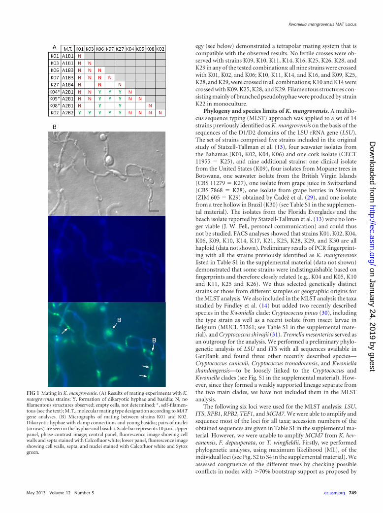
egy (see below) demonstrated a tetrapolar mating system that iscompatible with the observed results. No fertile crosses were ob-served with strains K09, K10, K11, K14, K16, K25, K26, K28, andK29 in any of the tested combinations: all nine strains were crossedwith K01, K02, and K06; K10, K11, K14, and K16, and K09, K25,K28, and K29, were crossed in all combinations; K10 and K14 werecrossed with K09, K25, K28, and K29. Filamentous structures con-sisting mainly of branched pseudohyphae were produced by strainK22 in monoculture.
Phylogeny and species limits of K. mangrovensis. A multilo-cus sequence typing (MLST) approach was applied to a set of 14strains previously identified as K. mangrovensis on the basis of thesequences of the D1/D2 domains of the LSU rRNA gene (LSU).The set of strains comprised five strains included in the originalstudy of Statzell-Tallman et al. (13), four seawater isolates fromthe Bahamas (K01, K02, K04, K06) and one cork isolate (CECT11955 � K25), and nine additional strains: one clinical isolatefrom the United States (K09), four isolates from Mopane trees inBotswana, one seawater isolate from the British Virgin Islands(CBS 11279 � K27), one isolate from grape juice in Switzerland(CBS 7868 � K28), one isolate from grape berries in Slovenia(ZIM 605 � K29) obtained by Cadež et al. (29), and one isolatefrom a tree hollow in Brazil (K30) (see Table S1 in the supplemen-tal material). The isolates from the Florida Everglades and thebeach isolate reported by Statzell-Tallman et al. (13) were no lon-ger viable (J. W. Fell, personal communication) and could thusnot be studied. FACS analyses showed that strains K01, K02, K04,K06, K09, K10, K14, K17, K21, K25, K28, K29, and K30 are allhaploid (data not shown). Preliminary results of PCR fingerprint-ing with all the strains previously identified as K. mangrovensislisted in Table S1 in the supplemental material (data not shown)demonstrated that some strains were indistinguishable based onfingerprints and therefore closely related (e.g., K04 and K05, K10and K11, K25 and K26). We thus selected genetically distinctstrains or those from different samples or geographic origins forthe MLST analysis. We also included in the MLST analysis the taxastudied by Findley et al. (14) but added two recently describedspecies in the Kwoniella clade: Cryptococcus pinus (30), includingthe type strain as well as a recent isolate from insect larvae inBelgium (MUCL 53261; see Table S1 in the supplemental mate-rial), and Cryptococcus shivajii (31). Tremella mesenterica served asan outgroup for the analysis. We performed a preliminary phylo-genetic analysis of LSU and ITS with all sequences available inGenBank and found three other recently described species—Cryptococcus cuniculi, Cryptococcus tronadorensis, and Kwoniellashandongensis—to be loosely linked to the Cryptococcus andKwoniella clades (see Fig. S1 in the supplemental material). How-ever, since they formed a weakly supported lineage separate fromthe two main clades, we have not included them in the MLSTanalysis.
The following six loci were used for the MLST analysis: LSU,ITS, RPB1, RPB2, TEF1, and MCM7. We were able to amplify andsequence most of the loci for all taxa; accession numbers of theobtained sequences are given in Table S1 in the supplemental ma-terial. However, we were unable to amplify MCM7 from K. hev-eanensis, F. depauperata, or T. wingfieldii. Firstly, we performedphylogenetic analyses, using maximum likelihood (ML), of theindividual loci (see Fig. S2 to S4 in the supplemental material). Weassessed congruence of the different trees by checking possibleconflicts in nodes with �70% bootstrap support as proposed by
FIG 1 Mating in K. mangrovensis. (A) Results of mating experiments with K.mangrovensis strains: Y, formation of dikaryotic hyphae and basidia; N, nofilamentous structures observed; empty cells, not determined; *, self-filamen-tous (see the text); M.T., molecular mating type designation according to MATgene analyses. (B) Micrographs of mating between strains K01 and K02.Dikaryotic hyphae with clamp connections and young basidia; pairs of nuclei(arrows) are seen in the hyphae and basidia. Scale bar represents 10 �m. Upperpanel, phase contrast image; central panel, fluorescence image showing cellwalls and septa stained with Calcofluor white; lower panel, fluorescence imageshowing cell walls, septa, and nuclei stained with Calcofluor white and Sytoxgreen.
Kwoniella mangrovensis MAT Locus
May 2013 Volume 12 Number 5 ec.asm.org 749
on January 24, 2019 by guesthttp://ec.asm
.org/D
ownloaded from
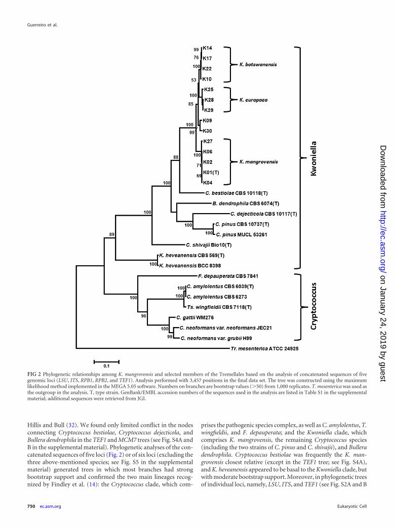
Hillis and Bull (32). We found only limited conflict in the nodesconnecting Cryptococcus bestiolae, Cryptococcus dejecticola, andBullera dendrophila in the TEF1 and MCM7 trees (see Fig. S4A andB in the supplemental material). Phylogenetic analyses of the con-catenated sequences of five loci (Fig. 2) or of six loci (excluding thethree above-mentioned species; see Fig. S5 in the supplementalmaterial) generated trees in which most branches had strongbootstrap support and confirmed the two main lineages recog-nized by Findley et al. (14): the Cryptococcus clade, which com-
prises the pathogenic species complex, as well as C. amylolentus, T.wingfieldii, and F. depauperata; and the Kwoniella clade, whichcomprises K. mangrovensis, the remaining Cryptococcus species(including the two strains of C. pinus and C. shivajii), and Bulleradendrophila. Cryptococcus bestiolae was frequently the K. man-grovensis closest relative (except in the TEF1 tree; see Fig. S4A),and K. heveanensis appeared to be basal to the Kwoniella clade, butwith moderate bootstrap support. Moreover, in phylogenetic treesof individual loci, namely, LSU, ITS, and TEF1 (see Fig. S2A and B
FIG 2 Phylogenetic relationships among K. mangrovensis and selected members of the Tremellales based on the analysis of concatenated sequences of fivegenomic loci (LSU, ITS, RPB1, RPB2, and TEF1). Analysis performed with 3,457 positions in the final data set. The tree was constructed using the maximumlikelihood method implemented in the MEGA 5.05 software. Numbers on branches are bootstrap values (�50) from 1,000 replicates. T. mesenterica was used asthe outgroup in the analysis. T, type strain. GenBank/EMBL accession numbers of the sequences used in the analysis are listed in Table S1 in the supplementalmaterial; additional sequences were retrieved from JGI.
Guerreiro et al.
750 ec.asm.org Eukaryotic Cell
on January 24, 2019 by guesthttp://ec.asm
.org/D
ownloaded from
and S4A), K. heveanensis was not or only weakly connected to theKwoniella clade.
The phylogenetic analysis of the combined MLST loci (Fig. 2)further revealed significant genetic heterogeneity among K. man-grovensis strains that clustered into three strongly supported andclosely related lineages: one includes the type strain (K01) as wellas K02, K04, K06, and K27; another includes K10, K14, K17, andK22; and the third includes K25, K28, and K29. The three lineagesare also apparent when the MCM7 locus was included in the anal-ysis (see Fig. S5 in the supplemental material). Moreover, thosethree lineages are genetically distinct in each of the six loci, exceptLSU (see Fig. S2A in the supplemental material), and the strains ofeach lineage clustered consistently on each tree (see Fig. S2 to S4),thus suggesting an absence of genetic exchange between the threelineages. We have applied the �w statistical test (27) to the 6-genedata set of the 12 K. mangrovensis strains mentioned above andfound no significant evidence for recombination. However, inclu-sion of strains K09 and K30 in the same test resulted in a signifi-cant probability for recombination. The latter two strains occu-pied inconsistent or intermediate positions on the trees (Fig. 2; seealso Fig. S2 to S4) and will be further discussed below. Nucleotidedifferences in the coding regions of the four protein-coding genesbetween strains of the three above-mentioned lineages were al-most always synonymous, and in RPB1 and TEF1, most of thenucleotide differences were located in the introns (data notshown). The phylogenetic trees depicted in Fig. 2 and Fig. S2 to S5suggest that K. mangrovensis should be confined to the stronglysupported clade represented by the five strains isolated from sea-water in tropical mangrove regions of the Atlantic, while the othertwo sister lineages, represented by the three European isolates andthe four Botswana isolates, respectively, correspond to two addi-tional sibling species, for which the names Kwoniella europaea andKwoniella botswanensis are proposed (Fig. 2). This proposal is sup-ported by the results of the mating experiments, because fertile
crosses were observed only among the strains of K. mangrovensisbut not between strains of K. mangrovensis and any of the twonovel taxa. Crosses among strains of K. europaea or K. botswanen-sis were, however, infertile, and thus we are unable to confirmtheir conspecificity based only on mating assays. In spite of theapparent absence of sexual reproduction in either K. europaea orK. botswanensis, their phylogenetic placement in the Kwoniellaclade (Fig. 2) argues for their inclusion in the genus Kwoniella inview of the recent implementation of the “one fungus, one name”rule in fungal nomenclature (33). Based on physiological test re-sults, the three species are similar (see Table S2). However, K.mangrovensis can be distinguished from K. europaea and K. bo-tswanensis by the inability of the former to grow at 35°C and toassimilate L-tartaric acid. K. europaea and K. botswanensis differonly in the assimilation of alpha-methyl-glucoside (fast in the lat-ter and delayed in the former) and L-tartaric acid.
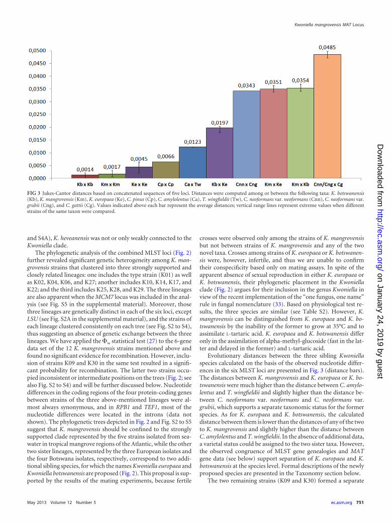
Evolutionary distances between the three sibling Kwoniellaspecies calculated on the basis of the observed nucleotide differ-ences in the six MLST loci are presented in Fig. 3 (distance bars).The distances between K. mangrovensis and K. europaea or K. bo-tswanensis were much higher than the distance between C. amylo-lentus and T. wingfieldii and slightly higher than the distance be-tween C. neoformans var. neoformans and C. neoformans var.grubii, which supports a separate taxonomic status for the formerspecies. As for K. europaea and K. botswanensis, the calculateddistance between them is lower than the distances of any of the twoto K. mangrovensis and slightly higher than the distance betweenC. amylolentus and T. wingfieldii. In the absence of additional data,a varietal status could be assigned to the two sister taxa. However,the observed congruence of MLST gene genealogies and MATgene data (see below) support separation of K. europaea and K.botswanensis at the species level. Formal descriptions of the newlyproposed species are presented in the Taxonomy section below.
The two remaining strains (K09 and K30) formed a separate
FIG 3 Jukes-Cantor distances based on concatenated sequences of five loci. Distances were computed among or between the following taxa: K. botswanensis(Kb), K. mangrovensis (Km), K. europaea (Ke), C. pinus (Cp), C. amylolentus (Ca), T. wingfieldii (Tw), C. neoformans var. neoformans (Cnn), C. neoformans var.grubii (Cng), and C. gattii (Cg). Values indicated above each bar represent the average distances; vertical range lines represent extreme values when differentstrains of the same taxon were compared.
Kwoniella mangrovensis MAT Locus
May 2013 Volume 12 Number 5 ec.asm.org 751
on January 24, 2019 by guesthttp://ec.asm
.org/D
ownloaded from
branch that is positioned next to K. europaea and K. botswanensisin the combined MLST trees (Fig. 2; see also Fig. S5 in the supple-mental material). Strain K30 had unique alleles for all MLST loci,except for RPB2, which had an identical sequence to that of K09;however, K09 shared the alleles of LSU, ITS, and MCM7 with K.europaea. FACS analyses demonstrated that both strains are hap-loid (data not shown). We have thus refrained from assigning adefinite taxonomic status to either of these two anomalous strainsuntil additional strains and respective genetic data are available toresolve if they are interspecies hybrids or novel taxa. As for the twostrains of C. pinus studied and in spite of their different origins(see Table S1 in the supplemental material), they had only a fewdifferences in the sequences of three of the six MLST loci and werethus considered conspecific (Fig. 2 and 3; see also Fig. S2 to S5 inthe supplemental material).
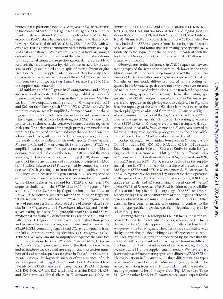
Identification of MAT genes in K. mangrovensis and siblingspecies. Our degenerate PCR-based strategy enabled us to amplifyfragments of genes with homologs in the C. neoformans MAT lo-cus from two compatible mating strains of K. mangrovensis, K01and K02, for the following loci: LPD1, RPO41, STE20, and SXI. Inthe latter case, we actually amplified a fragment containing the 5=regions of the SXI1 and SXI2 genes, as well as the intergenic spacer(this fragment will be henceforth designated SXI), because eachprimer was anchored in the conserved homeodomain region ofeach gene. The orientation of the single primer combination thatproduced the expected amplicon indicates that SXI1 and SXI2 areadjacent and divergently transcribed in K. mangrovensis, as foundpreviously in the tremellaceous tetrapolar species C. amylolentus,K. heveanensis, and T. mesenterica (8, 9). In the case of STE20, weamplified two fragments of the gene: one containing the kinasedomain, toward the 3= end of the gene (450 bp), and the otherspanning the Cdc42/Rac interactive binding (CRIB) domain, up-stream of the kinase domain and containing one intron (1,000bp). Possible linkage to MAT was verified by comparing the se-quences of each gene fragment from the two compatible strains ofK. mangrovensis, because only genes inside MAT are expected toexhibit marked mating-type-specific polymorphisms. Signifi-cantly different alleles were found for STE20 and SXI (e.g., 89%sequence similarity for the STE20 kinase 450-bp fragment; 74%similarity for the SXI2 673-bp fragment) but not for LPD1 orRPO41 (99% sequence similarity for the LPD1 388-bp fragment;99.7% sequence similarity for the RPO41 909-bp fragment). Inview of previous results on MAT structure of closely related spe-cies in the Cryptococcus and Kwoniella clades (12) and the ob-served mating-type-specific polymorphisms of STE20 and SXI, wepredict that the former is located in the P/R region of MAT and thelatter in the HD region. To confirm MAT specificity of those genesand to verify the mating system, we amplified and sequenced theSTE20 (CRIB-containing region) and SXI gene fragments fromthe full set of strains previously identified as K. mangrovensis (seeTable S1). We were also able to amplify and sequence the same locifor other species in the Kwoniella clade, B. dendrophila, C. bestio-lae, C. dejecticola, C. pinus, and C. shivajii (for the latter two speciesand B. dendrophila, we could not amplify SXI). Accession num-bers of the obtained sequences are given in Table S1 in the supple-mental material. Phylogenetic analyses of the sequences of eachlocus are presented in Fig. 4 (STE20) and 5 (SXI). We were able toidentify two STE20 alleles in K. mangrovensis (KmA1 in strainsK01, K03, K06, K07, and K27; and KmA2 in strains K02, K04, K05,and K08), two additional alleles in K. botswanensis (KbA1 in
strains K10, K11, and K22; and KbA2 in strains K14, K16, K17,K20, K23, and K24), and two more alleles in K. europaea (KeA1 instrains K25, K26, and K29; and KeA2 in strain K28) (see Table S1;Fig. 4). Strains K09 and K30 each had unique STE20 alleles. Wealso obtained an STE20 sequence from the mating type A2 strainof K. heveanensis and found that it is mating type specific (67%similarity to the sequence of the A1 allele), in contrast with thefindings of Metin et al. (9), who predicted that STE20 was notlocated within MAT.
Observed nucleotide differences in STE20 sequences betweenmating types of the same species were much lower in the threesibling Kwoniella species (ranging from 34 to 69) than in K. hev-eanensis (327) or the pathogenic Cryptococcus species (403 to 412).Nonetheless, nucleotide differences located in the coding se-quence in the Kwoniella species were not always synonymous andled to 5 to 7 amino acid substitutions in the translated sequencesbetween mating types (data not shown). The fact that mating typespecificity of STE20 is less pronounced in the three Kwoniella spe-cies is also apparent in the phylogenetic tree depicted in Fig. 4. Infact, the topology of the Kwoniella clade is more similar to thespecies phylogeny as obtained from the MLST analysis (Fig. 2),whereas among the species of the Cryptococcus clade, STE20 fol-lows a mating-type-specific phylogeny. Interestingly, whereasboth STE20 alleles of K. mangrovensis formed a single, well-sup-ported clade, those of K. botswanensis and K. europaea seemed tofollow a mating-type-specific phylogeny, with the KbA1 alleleclustering with the KeA1 allele and vice versa (Fig. 4).
For the SXI region, we identified four alleles in K. mangrovensis(KmB1 in strains K01, K03, K04, K05, and K08; KmB2 in strainK02; KmB3 in strains K06 and K07; and KmB4 in strain K27), asingle allele in K. botswanensis (KbB1), and three different allelesin K. europaea (KeB1 in strains K25 and K26; KeB2 in strain K28;and KeB3 in strain K29) (Fig. 5; see also Table S1 in the supple-mental material). The finding of distinct alleles of the two putativeMAT genes (STE20 and SXI) in K. mangrovensis, K. botswanensis,and K. europaea provides further key support for their separationat the species level. For the two anomalous strains, K30 had aunique SXI allele, and K09 surprisingly shared one of the SXIalleles (KeB1) of K. europaea (Fig. 5), which hints to the possibilityof this strain being a hybrid. The topology of the SXI tree (Fig. 5)reflects the high level of polymorphism of the 5= regions of the SXIgenes as observed in previous studies of related species (8, 9) thatclassified these genes as mating type unique, in contrast to themating-type-specific or species-specific phylogenies displayed byother MAT genes.
Assuming that STE20 belongs to the P/R locus, the latter ap-pears to be biallelic in each sibling species, whereas the HD locusdefined by the SXI alleles appears to be multiallelic, at least for K.mangrovensis and K. europaea. These results are compatible withthe hypothesis that the three sibling Kwoniella species are tetrapo-lar. This hypothesis is further corroborated by the fact that thealleles at both loci are not linked, as they are found in differentcombinations in the different strains of each species (Fig. 4 and 5;see also Table S1 in the supplemental material). We have in factidentified five different mating types with different STE20/SXI al-lele combinations in K. mangrovensis, three different mating typesin K. europaea, and two in K. botswanensis (see Table S1). Themolecular mating types are fully concordant with the results ofmating experiments for K. mangrovensis (Fig. 1A; see also TableS1). On the other hand, in K. europaea, we would expect fertile
Guerreiro et al.
752 ec.asm.org Eukaryotic Cell
on January 24, 2019 by guesthttp://ec.asm
.org/D
ownloaded from
matings between strains K25 and K28 or between K28 and K29.However, we did not observe any mating reactions with all four K.europaea strains. This result could be due to the fact that the testedstrains are sterile or to nonoptimal mating conditions, but we werenot able to confirm any of these hypotheses. Finally, in K. botswanen-sis, one likely explanation for the absence of observed sexual repro-duction is that all studied strains have the same SXI allele (KbB1).
We tried to design degenerate primers to amplify also the STE3and STE12 genes, which belong to the P/R locus in all heterothallicspecies of the Tremellales studied to date (12). This approachproved difficult due to the high sequence variability of those genesacross species. However, we observed that in most taxa for whichMAT structure has been elucidated, the two genes are almost al-ways adjacent, although their relative orientation varies across dif-ferent species. We designed one primer in the conserved domainsof each gene and ordered also the reverse complement of eachprimer so as to perform PCR amplifications using all four possible
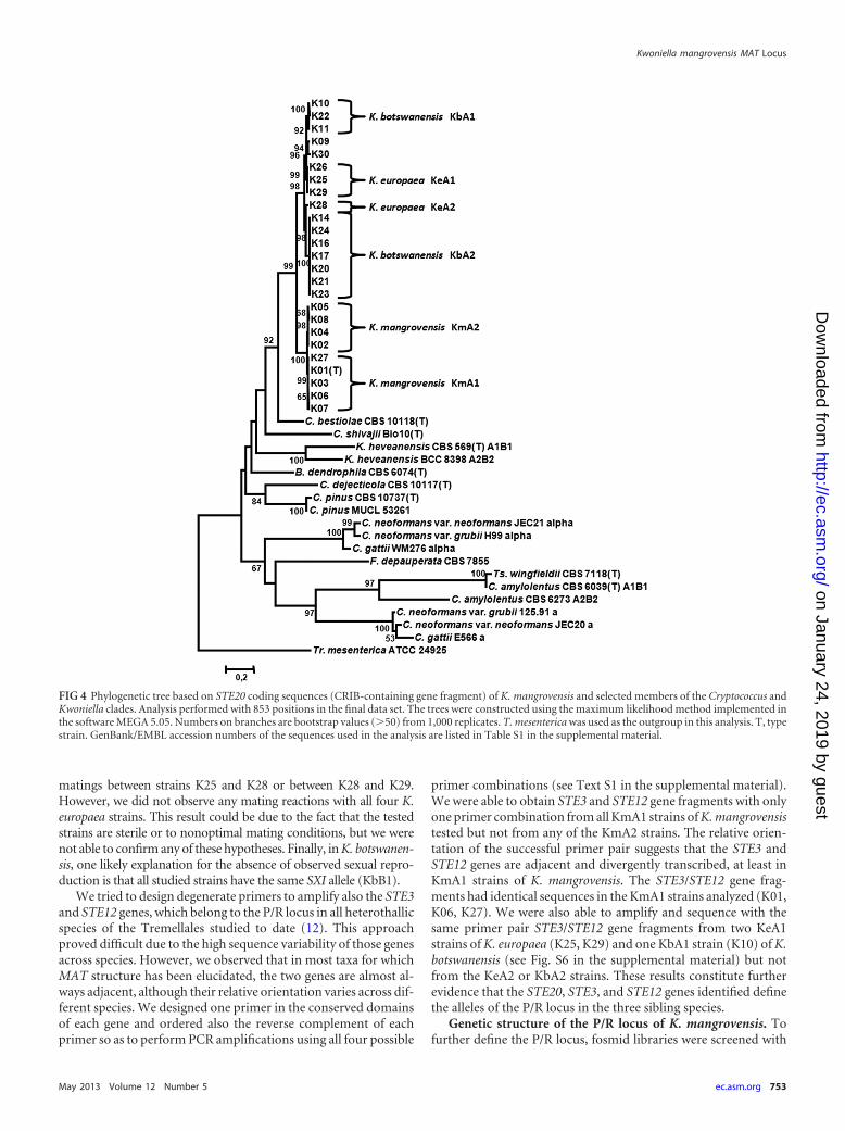
primer combinations (see Text S1 in the supplemental material).We were able to obtain STE3 and STE12 gene fragments with onlyone primer combination from all KmA1 strains of K. mangrovensistested but not from any of the KmA2 strains. The relative orien-tation of the successful primer pair suggests that the STE3 andSTE12 genes are adjacent and divergently transcribed, at least inKmA1 strains of K. mangrovensis. The STE3/STE12 gene frag-ments had identical sequences in the KmA1 strains analyzed (K01,K06, K27). We were also able to amplify and sequence with thesame primer pair STE3/STE12 gene fragments from two KeA1strains of K. europaea (K25, K29) and one KbA1 strain (K10) of K.botswanensis (see Fig. S6 in the supplemental material) but notfrom the KeA2 or KbA2 strains. These results constitute furtherevidence that the STE20, STE3, and STE12 genes identified definethe alleles of the P/R locus in the three sibling species.
Genetic structure of the P/R locus of K. mangrovensis. Tofurther define the P/R locus, fosmid libraries were screened with
FIG 4 Phylogenetic tree based on STE20 coding sequences (CRIB-containing gene fragment) of K. mangrovensis and selected members of the Cryptococcus andKwoniella clades. Analysis performed with 853 positions in the final data set. The trees were constructed using the maximum likelihood method implemented inthe software MEGA 5.05. Numbers on branches are bootstrap values (�50) from 1,000 replicates. T. mesenterica was used as the outgroup in this analysis. T, typestrain. GenBank/EMBL accession numbers of the sequences used in the analysis are listed in Table S1 in the supplemental material.
Kwoniella mangrovensis MAT Locus
May 2013 Volume 12 Number 5 ec.asm.org 753
on January 24, 2019 by guesthttp://ec.asm
.org/D
ownloaded from
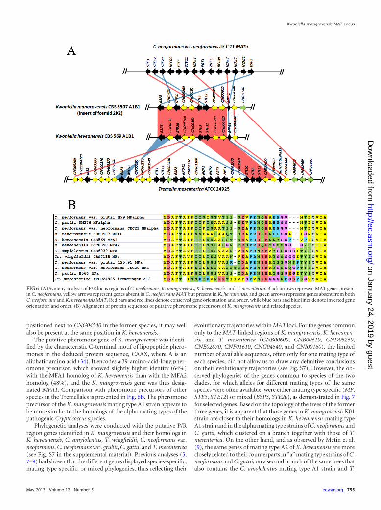
MAT gene probes produced based on the PCR amplicons of LPD1,RPO41, STE20 (kinase fragment), and SXI of strain K01 (matingtype A1B1) and on the STE20 kinase gene fragment of strain K02(A2B2). Positive fosmids in hybridization experiments were con-firmed by colony PCR using specific primers. We were able toconfirm positive fosmids of strain K01 containing LPD1 (two fos-mids), RPO41 (five fosmids), and STE20 (one fosmid: 2K2) genesbut not SXI. For strain K02, we were able to confirm only positivefosmids for LPD1 (one fosmid) and RPO41 (two fosmids).Because none of the fosmids were positive for more than oneprobe and the LPD1 and RPO41 genes appeared not to be matingtype specific, we focused on sequencing the 2K2 fosmid of K01.The fosmid insert was found to be 43 kb long, and annotation ofthe sequence obtained enabled identification of 12 genes with ho-mologs in the C. neoformans genome (Fig. 6A). Besides STE20,three other genes present in the P/R loci of other species in theTremellales, STE3, STE12, and a putative pheromone precursorgene (MFA1), were found in the vicinity, which suggests that infact the sequenced fosmid contains or is part of the P/R locus of K.mangrovensis. The annotation results also confirmed that theSTE3 and STE12 genes are adjacent and divergently transcribedand are in the close vicinity of the STE20 gene. Synteny analysis
with the homologous regions in C. neoformans, K. heveanensis,and T. mesenterica is presented in Fig. 6A. Some MAT genes arecommon to all species so far examined in the Tremellales (BSP3,MFA1, STE3, STE12, STE20; highlighted in black in Fig. 6A) andmay thus have been present in the ancestral MAT locus, whileothers are common only to K. heveanensis and/or T. mesenterica(CNB00600, CNB00610, CND05260, CNE02670, CNF01610,CNG04540, and CNI00160; highlighted in yellow in Fig. 6A) andcould thus have been more recently acquired into MAT in specificlineages. An almost complete synteny is observed between K.mangrovensis and the mating type A1B1 strain of K. heveanensis,the only exception being the pheromone precursor gene, which isinverted. This finding was unexpected because K. mangrovensisand K. heveanensis are not sister taxa (Fig. 2A) and previous com-parisons of MAT structure had shown low levels of synteny, withseveral rearrangements, translocations, and inversions, even be-tween closely related taxa (e.g., C. amylolentus and T. wingfieldii[8] or C. neoformans var. neoformans and C. neoformans var. grubii[5]). CNF01610 (chitin synthase gene) has a homolog in T. mes-enterica, not far from CNG04540 but apparently not in K. hev-eanensis (Fig. 6A); however, due to the observed synteny betweenK. mangrovensis and K. heveanensis and the fact that CNF01610 is
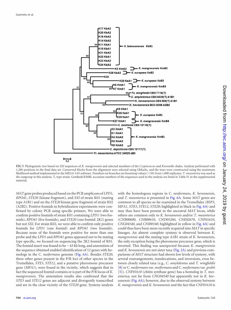
FIG 5 Phylogenetic tree based on SXI sequences of K. mangrovensis and selected members of the Cryptococcus and Kwoniella clades. Analysis performed with1,280 positions in the final data set. Conserved blocks from the alignment were selected using Gblocks, and the trees were constructed using the maximumlikelihood method implemented in the MEGA 5.05 software. Numbers on branches are bootstrap values (�50) from 1,000 replicates. T. mesenterica was used asthe outgroup in this analysis. T, type strain. GenBank/EMBL accession numbers of the sequences used in the analysis are listed in Table S1 in the supplementalmaterial.
Guerreiro et al.
754 ec.asm.org Eukaryotic Cell
on January 24, 2019 by guesthttp://ec.asm
.org/D
ownloaded from
positioned next to CNG04540 in the former species, it may wellalso be present at the same position in K. heveanensis.
The putative pheromone gene of K. mangrovensis was identi-fied by the characteristic C-terminal motif of lipopeptide phero-mones in the deduced protein sequence, CAAX, where A is analiphatic amino acid (34). It encodes a 39-amino-acid-long pher-omone precursor, which showed slightly higher identity (64%)with the MFA1 homolog of K. heveanensis than with the MFA2homolog (48%), and the K. mangrovensis gene was thus desig-nated MFA1. Comparison with pheromone precursors of otherspecies in the Tremellales is presented in Fig. 6B. The pheromoneprecursor of the K. mangrovensis mating type A1 strain appears tobe more similar to the homologs of the alpha mating types of thepathogenic Cryptococcus species.
Phylogenetic analyses were conducted with the putative P/Rregion genes identified in K. mangrovensis and their homologs inK. heveanensis, C. amylolentus, T. wingfieldii, C. neoformans var.neoformans, C. neoformans var. grubii, C. gattii. and T. mesenterica(see Fig. S7 in the supplemental material). Previous analyses (5,7–9) had shown that the different genes displayed species-specific,mating-type-specific, or mixed phylogenies, thus reflecting their
evolutionary trajectories within MAT loci. For the genes commononly to the MAT-linked regions of K. mangrovensis, K. heveanen-sis, and T. mesenterica (CNB00600, CNB00610, CND05260,CNE02670, CNF01610, CNG04540, and CNI00160), the limitednumber of available sequences, often only for one mating type ofeach species, did not allow us to draw any definitive conclusionson their evolutionary trajectories (see Fig. S7). However, the ob-served phylogenies of the genes common to species of the twoclades, for which alleles for different mating types of the samespecies were often available, were either mating type specific (MF,STE3, STE12) or mixed (BSP3, STE20), as demonstrated in Fig. 7for selected genes. Based on the topology of the trees of the formerthree genes, it is apparent that those genes in K. mangrovensis K01strain are closer to their homologs in K. heveanensis mating typeA1 strain and in the alpha mating type strains of C. neoformans andC. gattii, which clustered on a branch together with those of T.mesenterica. On the other hand, and as observed by Metin et al.(9), the same genes of mating type A2 of K. heveanensis are moreclosely related to their counterparts in “a” mating type strains of C.neoformans and C. gattii, on a second branch of the same trees thatalso contains the C. amylolentus mating type A1 strain and T.
FIG 6 (A) Synteny analysis of P/R locus regions of C. neoformans, K. mangrovensis, K. heveanensis, and T. mesenterica. Black arrows represent MAT genes presentin C. neoformans, yellow arrows represent genes absent in C. neoformans MAT but present in K. heveanensis, and green arrows represent genes absent from bothC. neoformans and K. heveanensis MAT. Red bars and red lines denote conserved gene orientation and order, while blue bars and blue lines denote inverted geneorientation and order. (B) Alignment of protein sequences of putative pheromone precursors of K. mangrovensis and related species.
Kwoniella mangrovensis MAT Locus
May 2013 Volume 12 Number 5 ec.asm.org 755
on January 24, 2019 by guesthttp://ec.asm
.org/D
ownloaded from
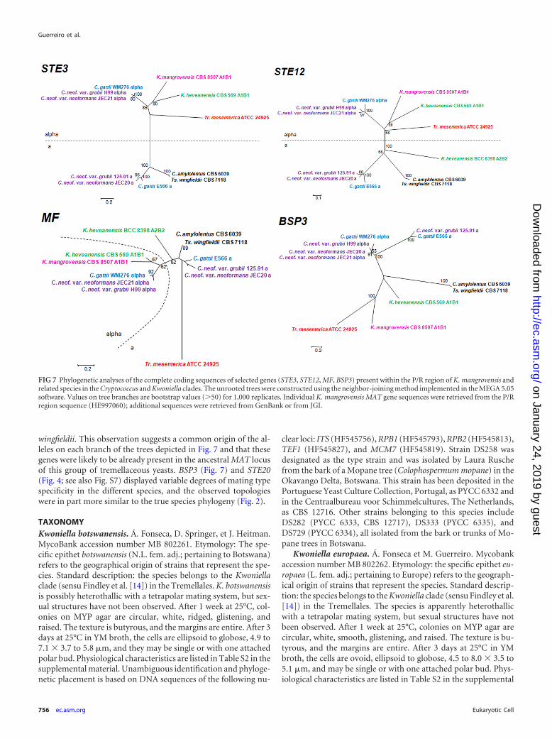
wingfieldii. This observation suggests a common origin of the al-leles on each branch of the trees depicted in Fig. 7 and that thesegenes were likely to be already present in the ancestral MAT locusof this group of tremellaceous yeasts. BSP3 (Fig. 7) and STE20(Fig. 4; see also Fig. S7) displayed variable degrees of mating typespecificity in the different species, and the observed topologieswere in part more similar to the true species phylogeny (Fig. 2).
TAXONOMYKwoniella botswanensis. Á. Fonseca, D. Springer, et J. Heitman.MycoBank accession number MB 802261. Etymology: The spe-cific epithet botswanensis (N.L. fem. adj.; pertaining to Botswana)refers to the geographical origin of strains that represent the spe-cies. Standard description: the species belongs to the Kwoniellaclade (sensu Findley et al. [14]) in the Tremellales. K. botswanensisis possibly heterothallic with a tetrapolar mating system, but sex-ual structures have not been observed. After 1 week at 25°C, col-onies on MYP agar are circular, white, ridged, glistening, andraised. The texture is butyrous, and the margins are entire. After 3days at 25°C in YM broth, the cells are ellipsoid to globose, 4.9 to7.1 � 3.7 to 5.8 �m, and they may be single or with one attachedpolar bud. Physiological characteristics are listed in Table S2 in thesupplemental material. Unambiguous identification and phyloge-netic placement is based on DNA sequences of the following nu-
clear loci: ITS (HF545756), RPB1 (HF545793), RPB2 (HF545813),TEF1 (HF545827), and MCM7 (HF545819). Strain DS258 wasdesignated as the type strain and was isolated by Laura Ruschefrom the bark of a Mopane tree (Colophospermum mopane) in theOkavango Delta, Botswana. This strain has been deposited in thePortuguese Yeast Culture Collection, Portugal, as PYCC 6332 andin the Centraalbureau voor Schimmelcultures, The Netherlands,as CBS 12716. Other strains belonging to this species includeDS282 (PYCC 6333, CBS 12717), DS333 (PYCC 6335), andDS729 (PYCC 6334), all isolated from the bark or trunks of Mo-pane trees in Botswana.
Kwoniella europaea. Á. Fonseca et M. Guerreiro. Mycobankaccession number MB 802262. Etymology: the specific epithet eu-ropaea (L. fem. adj.; pertaining to Europe) refers to the geograph-ical origin of strains that represent the species. Standard descrip-tion: the species belongs to the Kwoniella clade (sensu Findley et al.[14]) in the Tremellales. The species is apparently heterothallicwith a tetrapolar mating system, but sexual structures have notbeen observed. After 1 week at 25°C, colonies on MYP agar arecircular, white, smooth, glistening, and raised. The texture is bu-tyrous, and the margins are entire. After 3 days at 25°C in YMbroth, the cells are ovoid, ellipsoid to globose, 4.5 to 8.0 � 3.5 to5.1 �m, and may be single or with one attached polar bud. Phys-iological characteristics are listed in Table S2 in the supplemental
FIG 7 Phylogenetic analyses of the complete coding sequences of selected genes (STE3, STE12, MF, BSP3) present within the P/R region of K. mangrovensis andrelated species in the Cryptococcus and Kwoniella clades. The unrooted trees were constructed using the neighbor-joining method implemented in the MEGA 5.05software. Values on tree branches are bootstrap values (�50) for 1,000 replicates. Individual K. mangrovensis MAT gene sequences were retrieved from the P/Rregion sequence (HE997060); additional sequences were retrieved from GenBank or from JGI.
Guerreiro et al.
756 ec.asm.org Eukaryotic Cell
on January 24, 2019 by guesthttp://ec.asm
.org/D
ownloaded from
material. Unambiguous identification and phylogenetic place-ment are based on DNA sequences of the following nuclear loci:ITS (HE984339), RPB1 (HE997004), RPB2 (HE996978), TEF1(HE997051), and MCM7 (HE996990). Strain CECT 11955 wasdesignated as the type strain and was isolated by Carmela Belochfrom cork oak (Quercus suber) bark in Badajoz, Spain (35). Thisstrain has been deposited in the Portuguese Yeast Culture Collec-tion, Portugal, as PYCC 6162 and in the Centraalbureau voorSchimmelcultures, The Netherlands, as CBS 12714. Other strainsbelonging to this species include CBS 7868 from grape juice inSwitzerland and ZIM 605 (PYCC 6207) from grape berries in Slo-venia (29).
DISCUSSIONGenetic heterogeneity in K. mangrovensis and unveiling of twonovel sibling species. In their original description of K. man-grovensis, Statzell-Tallman et al. (13) studied 30 seawater isolates,mostly from mangrove habitats, 19 from the Bahamas and 11from the Florida Everglades, as well as two cork isolates fromSpain and one beach isolate from Florida. Crossing experimentsrevealed only seven fertile strains among the Bahamas isolates, onedesignated MAT A (strain K02) and the other six designated MATalpha (which included K01, K03, and K06). The authors proposeda bipolar mating system for K. mangrovensis and deemed the non-mating strains to be sterile. They also determined LSU and ITSsequences for representative strains and found little genetic vari-ability, except for one cork isolate and the beach isolate, which hadone nucleotide substitution in LSU and 6 to 7 substitutions in ITS,compared to the mangrove strains. These differences were notconsidered sufficient by Statzell-Tallman et al. (13) to warrantseparate taxonomic status for those strains without further matingand genetic studies.
Multilocus sequence typing using partial sequences of selectedhousekeeping genes has been used successfully for delimiting spe-cies in different yeasts (36). In the present study, we have comple-mented the MLST approach with the analysis of MAT genes due tothe central role played by MAT loci in regulating sexual reproduc-tion and thus in determining species boundaries (1, 3). The mul-tilocus approach involved five K. mangrovensis strains (four fromseawater and one from cork) from the original study (13), as wellas one additional isolate from the British Virgin Islands, one clin-ical isolate from the United States, four isolates from Mopane treesin Botswana, one isolate from a tree hollow in Brazil, and twoisolates from grapes in Europe (see Table S1 in the supplementalmaterial). The results showed that the tropical seawater isolates,which included the type strain of K. mangrovensis, formed amonophyletic lineage, while the four Botswana isolates and thethree European strains formed two additional sister lineages (Fig.2; see also Fig. S5 in the supplemental material). The consistentclustering of the strains on individual gene trees (see Fig. S2 to S4in the supplemental material), the apparent absence of recombi-nation between strains of each lineage, and the observed evolu-tionary distances between strains of the three sibling lineages (Fig.3) suggested a genetic separation between them that led us topropose two novel species represented by the Botswana and Eu-ropean isolates, K. botswanensis and K. europaea. This proposal isfurther supported by the sequence data of the two putative MATgene fragments, STE20 and SXI, obtained for the same strains,because distinct alleles of the two genes were found for strains ofthe three sibling species (Fig. 3 and 4). The geographic origin and
source of the strains belonging to K. mangrovensis (water samplesfrom mangrove habitats in the tropical western shores of theNorth Atlantic Ocean), K. botswanensis (Mopane trees in Bo-tswana), or K. europaea (plant-related substrates in Europe); theobserved physiological differences; as well as the results of matingexperiments are in line with the separation of the three sibling taxaat the species level. It is interesting to note that the novel Kwoniellaspecies appear to be associated with plant surfaces (albeit in verydifferent ecosystems), and although the strains of K. mangrovensiswere isolated from seawater, Statzell-Tallman et al. (13) specu-lated that they could have actually originated from the mangrovetrees of the surrounding coastal areas. The remaining two strains(K09, the only clinical isolate; and K30, isolated from a tree hollowin Brazil) were placed separately from the three Kwoniella specieson the MLST and MAT gene trees (Fig. 2, 4, and 5) and couldrepresent additional sibling taxa. However, due to the inconsistentclustering on individual gene trees (see Fig. S2 to S4) and thelimited number of strains, their taxonomic status is best consid-ered unresolved at present. Moreover, K09 shared some alleleswith K. europaea (LSU, ITS, MCM7, SXI) and could thus be aninterspecies hybrid.
Taxonomic status of taxa in the Cryptococcus and Kwoniellaclades. We included in our multilocus study several taxa in theCryptococcus and Kwoniella clades, and the phylogenetic analysesof the combined data set (Fig. 2; see also Fig. S5 in the supplemen-tal material) have confirmed that B. dendrophila, C. bestiolae, C.dejecticola, C. pinus, and C. shivajii all belong, together with K.mangrovensis, K. botswanensis, and K. europaea, to the Kwoniellaclade. In view of the recent implementation of the “one fungus,one name” rule to fungal nomenclature (33), the above-men-tioned Bullera species and four Cryptococcus species should all betransferred to the genus Kwoniella, but we refrain from formaliz-ing this proposal here since a taxonomic revision of the genus isbeyond the scope of the present paper. The phylogenetic positionof K. heveanensis was not consistent in the individual gene trees(see Fig. S2 to S4 in the supplemental material), but the five-geneML tree had this taxon at a basal position in the Kwoniella clade(Fig. 2) with 89% bootstrap support, thus justifying its inclusionin the genus, in accordance with the findings of Findley et al. (14).In the Cryptococcus clade, a single genus name should also be ad-opted, which should be Cryptococcus given that C. neoformans isthe type species of the genus and has precedence over Filobasi-diella. Tsuchiyaea wingfieldii was considered by Fonseca et al. (37)to be a synonym of C. amylolentus. However, the MAT gene anal-yses of Findley et al. (8) led the authors to propose that the twotaxa should be kept separate at the species level. Our multilocusand MAT gene analyses support the hypothesis of the latter au-thors even though the evolutionary distance between the two taxais less than half the value observed for the pair C. neoformans var.neoformans and C. neoformans var. grubii (Fig. 3). The ensuingtransfer of T. wingfieldii to the genus Cryptococcus as C. wingfieldiishould, however, await the study of more strains (only a singleisolate is currently known) to obtain crucial supporting evidence.
Mating system of K. mangrovensis and sibling species. Incontrast to the original assumption of a bipolar mating system(13), our results have shown that K. mangrovensis instead has atetrapolar system with two unlinked loci: a P/R locus defined by atleast the STE20 gene (see below) and an HD locus defined by twodivergently transcribed SXI genes (Fig. 3 and 4). Several lines ofevidence support this conclusion. First, we found two alleles for
Kwoniella mangrovensis MAT Locus
May 2013 Volume 12 Number 5 ec.asm.org 757
on January 24, 2019 by guesthttp://ec.asm
.org/D
ownloaded from
STE20 and four alleles for SXI among nine strains of K. man-grovensis. Moreover, the association of the different alleles at bothloci was found to vary in the different strains, defining five differ-ent mating types of eight possible combinations (Fig. 3 and 4; seealso Table S1 in the supplemental material). Based on the results ofprevious studies on the MAT loci of closely aligned taxa in theCryptococcus and Kwoniella clades for which tetrapolar systemshave been demonstrated (8, 9), K. mangrovensis STE20 defines abiallelic P/R locus and SXI defines a multiallelic HD locus. Addi-tionally, the adjacent position of the two divergently transcribedSXI1 and SXI2 homologs in K. mangrovensis, demonstrated by thefact that we amplified fragments of those two genes with a specificpair of primers anchored in the homeodomain regions of eachgene, has been found only in tetrapolar species (C. amylolentus, K.heveanensis, and T. mesenterica), whereas in the bipolar patho-genic Cryptococcus species, only one gene is present in the MATlocus of each mating type (5). The results of mating experiments(Fig. 1) were concordant with the molecular mating types deter-mined for the different strains and provide additional evidencesupporting our hypothesis. The same line of reasoning is applica-ble to K. botswanensis and K. europaea, and the results obtained forthe STE20 and SXI genes (Fig. 3 and 4) suggest that a tetrapolarmating system is also operating in this species. However, matingdata were not conclusive. The possibility that K. mangrovensiswould have a tetrapolar mating system had been suggested byMetin et al. (9) based on their study of the MAT locus of K. hev-eanensis, for which a tetrapolar system was demonstrated.
Evolution of MAT loci in the Tremellales. The order Tremel-lales comprises a large number of yeast producing taxa distributedinto several lineages, as deduced from phylogenetic analyses ofdifferent regions of the rDNA cistron (38, 39). The large majorityof species is asexual and classified in genera such as Bullera, Cryp-tococcus, Dioszegia, or Fellomyces, some of which are clearlypolyphyletic (38). However, it is possible and even likely thatmany of these species actually correspond to haploid states ofunrecognized heterothallic sexual taxa, as has been found for sev-eral species whose sexual states were uncovered upon mating ofcompatible strains of taxa previously regarded as purely asexual,e.g., C. amylolentus (8), C. neoformans (40), K. heveanensis (9), andK. mangrovensis (13). The order Tremellales contains in fact sev-eral sexual taxa classified in the genera Auriculibuller, Bulleromy-ces, Fibulobasidium, and Tremella, among others, but only limitedevidence is available on the respective mating systems (38). Somespecies are assumed to be bipolar (e.g., Auriculibuller fuscus, Bul-leromyces albus) and others tetrapolar (e.g., Tremella spp.) (38),but molecular evidence is lacking for most. In fact, information onthe genetic structure of MAT loci of members of the Tremellales isavailable only for some species in the Cryptococcus and Kwoniellaclades and for T. mesenterica (12).
In the present study, we have identified tetrapolar mating sys-tems in at least two of the three sibling taxa in the Kwoniella clade,K. mangrovensis and K. europaea. Our findings raise to five or sixthe number of taxa in the Tremellales with tetrapolar systems thathave been confirmed molecularly, thus lending further support tothe hypothesis that this mating system was present in the commonancestor of the Cryptococcus and Kwoniella clades and that thebipolar system of the pathogenic Cryptococcus species arose morerecently in the common ancestor of the latter species cluster (5, 8,9, 12). It will be of considerable interest to explore the matingsystems of members of other clades in the Tremellales, namely,
those that contain putative bipolar species, to ascertain if the lattermating system has arisen independently more than once in differ-ent lineages.
Annotation of the sequence of a fosmid insert containing theSTE20 gene from a mating type A1 strain of K. mangrovensis(strain K01) enabled us to determine the extended gene structureof the genomic region containing the putative P/R locus (Fig. 6A).Because we were not able to determine the structure of the corre-sponding region in a mating type A2 strain, we cannot as yet definewith certainty the boundaries of the P/R locus in K. mangrovensis.However, because we confirmed the mating type specificity ofSTE20 and the MF and STE3 genes are also likely mating typespecific, we predict that the P/R locus could span at least the 21-kbregion between STE20 and MF, comprising a total of eight genes,which include CNB00600, CNB00610, CND05260, CNI00160, andSTE12. To verify this hypothesis, we need to sequence the corre-sponding region in a mating type A2 strain. As mentioned above,the most remarkable feature of the characterized region in K.mangrovensis (mating type A1B1) was the almost perfect syntenywith the homologous region of K. heveanensis (mating typeA1B1). Previous studies on the MAT loci of different taxa in theTremellales had shown that these genomic regions are very plastic,with frequent gene inversions and translocations, not only be-tween closely related taxa but also between complementary mat-ing types of the same species (5, 8, 9). Our finding of conservedgene order and orientation (Fig. 6A) between two species that arenot even very close relatives (Fig. 2) is therefore unprecedented inthis group of yeasts. We predict that the P/R locus in K. heveanen-sis should span at least the same eight genes, because we confirmedthe mating type specificity of STE20 in this species. Metin et al. (9)were able to determine only a 5-gene fragment of the P/R locus ina complementary mating type A2 strain of K. heveanensis andfound that gene order is apparently not maintained between themating types. We therefore predict that the P/R loci of mating typeA1 strains and those of mating type A2 of K. mangrovensis and K.heveanensis form two diverged syntenic groups. A somewhat par-allel situation was observed in the distantly related red yeasts of theSporidiobolales (class Microbotryomycetes, subphylum Puccin-iomycotina) for which genomic regions around the P/R locus ex-hibited extensive rearrangements between complementary mat-ing types of each species, while synteny was well conserved acrossspecies when the homologous regions of the same mating typewere compared (41). A similar conclusion was reached by Kellneret al. (6) when comparing the triallelic P/R loci of different smutspecies in the Ustilaginomycetes. These findings suggest that theMAT gene structure of each allele of the P/R loci in different spe-cies was fixed long before speciation occurred. In the Tremellales,this is not apparently the case in the Cryptococcus clade, whereMAT restructuring appeared to be concomitant with speciation,but our limited evidence suggests synteny may have been con-served in the evolution of MAT in the Kwoniella clade. Determi-nation of the genetic structure of the P/R locus in a mating type A2strain of K. mangrovensis and in other species in the Kwoniellaclade will be necessary to test our hypothesis. Our phylogeneticanalyses of genes in the P/R locus (Fig. 7) further suggest that STE3and STE12 should be ancient MAT genes in the Tremellales due tothe apparent trans-specific polymorphisms deduced from the to-pology of the corresponding phylogenetic trees. However, theSTE20 gene may have been recruited more recently into the MATlocus in some members of the Kwoniella clade (namely in K. man-
Guerreiro et al.
758 ec.asm.org Eukaryotic Cell
on January 24, 2019 by guesthttp://ec.asm
.org/D
ownloaded from
grovensis) compared to the taxa in the Cryptococcus clade for whichthe association of STE20 with MAT appears to be more ancient inview of the observed mating-type-specific phylogeny (see Fig. 4).An alternative hypothesis is that STE20 is an ancient MAT gene inthe Tremellales, but its evolutionary trajectory may have been re-cently reset via gene conversion in some Kwoniella species.
Possible future strategies to gain further insight into the MATloci of K. mangrovensis include (i) the reprobing of the mating typeA2 strain fosmid library with additional MAT gene probes, (ii) aPCR-based approach to amplify and sequence MAT genes in theP/R locus of the mating type A2 strain based on the genes alreadyidentified in the homologous regions of the mating type A1 strainand of the complementary mating strains of K. heveanensis, or (iii)whole-genome sequence analysis of the mating type A1 and A2strains of K. mangrovensis.
The present study has demonstrated that exploration of theMAT loci structure in different saprobic taxa in the Tremellales isrevealing novel insights on the origin and evolution of these keygenomic regions in a very diverse but still lesser-known group ofbasidiomycetous yeasts. Ongoing and future genome sequencingprojects will contribute to provide additional insights on this cen-tral and exciting aspect of fungal biology.
ACKNOWLEDGMENTS
Work carried out at CREM was partly supported by Fundação para aCiência e a Tecnologia (Portugal) (FCT), project PTDC/BIA-MIC/113051/2009. Work at Duke was supported by NIH/NIAID R37 grantAI39115 and R01 grant AI50113 to J.H.
We are indebted to Sheng Sun (Duke University) and Michael Weiss(University of Tübingen) for discussions and advice, Anna Floyd (DukeUniversity) for technical assistance, and J. W. Fell (University of Miami),Neza Cadež (ZIM culture collection), Heide-Marie Daniel (MUCL cul-ture collection), Kennio Ferreira-Paim (Universidade Federal do Triân-gulo Mineiro, Brazil), and Shawn Lockhart (CDC) for providing some ofthe isolates used in this study.
REFERENCES1. Lee SC, Ni M, Li W, Shertz C, Heitman J. 2010. The evolution of sex: a
perspective from the fungal kingdom. Microbiol. Mol. Biol. Rev. 74:298 –340.
2. Fraser JA, Heitman J. 2005. Chromosomal sex-determining regions inanimals, plants and fungi. Curr. Opin. Genet. Dev. 15:645– 651.
3. Kües U, James TY, Heitman J. 2011. Mating type in Basidiomycetes:unipolar, bipolar, and tetrapolar patterns of sexuality, p 97–160. In Pög-geler S, Wöstemeyer J (ed), Evolution of fungi and fungal-like organisms,the Mycota, vol XIV. Springer Verlag, Berlin, Germany.
4. Bakkeren G, Kamper J, Schirawski J. 2008. Sex in smut fungi: structure,function and evolution of mating-type complexes. Fungal Genet. Biol.45(Suppl 1):S15–S21.
5. Fraser JA, Diezmann S, Subaran RL, Allen A, Lengeler KB, Dietrich FS,Heitman J. 2004. Convergent evolution of chromosomal sex-determiningregions in the animal and fungal kingdoms. PLoS Biol. 2:2243–2255. doi:10.1371/journal.pbio.0020384.
6. Kellner R, Vollmeister E, Feldbrügge M, Begerow D. 2011. Interspecificsex in grass smuts and the genetic diversity of their pheromone-receptorsystem. PLoS Genet. 7:e1002436. doi:10.1371/journal.pgen.1002436.
7. Findley K. 2010. Evolution of the mating-type locus and insights intosexual reproduction in the Cryptococcus species complex. Ph.D. thesis.Duke University, Durham, NC.
8. Findley K, Sun S, Fraser JA, Hsueh Averette Y-PAF, Li W, Dietrich FS,Heitman J. 2012. Discovery of a modified tetrapolar sexual cycle in Cryp-tococcus amylolentus and the evolution of MAT in the Cryptococcus speciescomplex. PLoS Genet. 8(2):e1002528. doi:10.1371/journal.pgen.1002528.
9. Metin B, Findley K, Heitman J. 2010. The mating type locus (MAT) andsexual reproduction of Cryptococcus heveanensis: insights into the evolu-
tion of sex and sex-determining chromosomal regions in fungi. PLoSGenet. 6(5):e1000961. doi:10.1371/journal.pgen.1000961.
10. Rodriguez-Carres M, Findley K, Sun S, Dietrich FS, Heitman J. 2010.Morphological and genomic characterization of Filobasidiella depaupe-rata: a homothallic sibling species of the pathogenic Cryptococcus speciescomplex. PLoS One 5:e9620. doi:10.1371/journal.pone.0009620.
11. Sun S, Metin B, Findley K, Fonseca Á, Heitman J. 2011. Validation ofKwoniella heveanensis, teleomorph of the basidiomycetous yeast Crypto-coccus heveanensis. Mycotaxon 116:227–229.
12. Hsueh Y-P, Metin B, Findley K, Rodriguez-Carres M, Heitman J. 2011.The mating-type locus of Cryptococcus: evolution of gene clusters govern-ing sex determination and sexual reproduction from the phylogenomicperspective, p 139 –149. In Heitman J, Kozel TR, Kwon-Chung KJ, PerfectJR, Casadevall A (ed), Cryptococcus: from human pathogen to model yeast,1st ed. ASM Press, Washington, DC.
13. Statzell-Tallman A, Belloch C, Fell JW. 2008. Kwoniella mangrovensisgen. nov., sp. nov. (Tremellales, Basidiomycota), a teleomorphic yeastfrom mangrove habitats in the Florida Everglades and Bahamas. FEMSYeast Res. 8:103–113.
14. Findley K, Rodriguez-Carres M, Metin B, Kroiss J, Fonseca Á, VilgalysR, Heitman J. 2009. Phylogeny and phenotypic characterization of patho-genic Cryptococcus species and closely related saprobic taxa in the Tremel-lales. Eukaryot. Cell 8:353–361.
15. Tanaka R, Taguchi H, Takeo K, Miyaji M, Nishimura K. 1996. Deter-mination of ploidy in Cryptococcus neoformans by flow cytometry. J. Med.Vet. Mycol. 34:299 –301.
16. Kurtzman CP, Fell JW, Boekhout T, Robert V. 2011. Methods forisolation, phenotypic characterization and maintenance of yeasts, p 87–110. In Kurtzman CP, Fell JW, Boekhout T (ed), The yeasts, a taxonomicstudy, 5th ed. Elsevier, Amsterdam, Netherlands.
17. Hall TA. 1999. BioEdit: a user-friendly biological sequence alignmenteditor and analysis program for Windows 95/98/NT. Nucleic Acids Symp.Ser. (Oxf.) 41:95–98.
18. Thompson J, Higgins D, Gibson T. 1994. CLUSTAL W: improving thesensitivity of progressive multiple sequence alignment through sequenceweighting, position-specific gap penalties and weight matrix choice. Nu-cleic Acids Res. 22:4673– 4680.
19. James TY, Kauff F, Schoch C, Matheny PB, Hofstetter V, Cox C, CelioG, Gueidan C, Fraker E, Miadlikowska J, Lumbsch HT, Rauhut A, ReebV, Arnold AE, Amtoft A, Stajich JE, Hosaka K, Sung GH, Johnson D,O’Rourke B, Crockett M, Binder M, Curtis JM, Slot JC, Wang Z,Wilson AW, Schüssler A, Longcore JE, O’Donnell K, Mozley-Standridge S, Porter D, Letcher PM, Powell MJ, Taylor JW, White MM,Griffith GW, Davies DR, Humber RA, Morton JB, Sugiyama J, Ross-man AY, Rogers JD, Pfister DH, Hewitt D, Hansen K, Hambleton S,Shoemaker RA, Kohlmeyer J, Volkmann-Kohlmeyer B, Spotts RA,Serdani M, Crous PW, Hughes KW, Matsuura K, Langer E, Langer G,Untereiner WA, Lücking R, Büdel B, Geiser DM, Aptroot A, DiederichP, Schmitt I, Schultz M, Yahr R, Hibbett D, Lutzoni F, McLaughlin D,Spatafora J, Vilgalys R. 2006. Reconstructing the early evolution of thefungi using a six gene phylogeny. Nature 443:818 – 822.
20. Schmitt I, Crespo A, Divakar PK, Fankhauser JD, Herman-Sackett E,Kalb K, Nelsen MP, Nelson NA, Rivas-Plata E, Shimp AD, Widhelm T,Lumbsch HT. 2009. New primers for promising single-copy genes infungal phylogenetics and systematics. Persoonia 23:35– 40.
21. White TJ, Bruns T, Lee S, Taylor J. 1990. Amplification and directsequencing of fungal ribosomal RNA genes for phylogenetics, p 315–322.In Innis MA, Gelfand DH, Sninsky JJ, White TJ (ed), PCR protocols: aguide to methods and applications. Academic Press, San Diego, CA.
22. O’Donnell K. 1993. Fusarium and its near relatives, p 225–233. In Reyn-olds DR, Taylor JW (ed), The fungal holomorph: mitotic, meiotic andpleomorphic speciation in fungal systematics. CAB International, Wall-ingford, United Kingdom.
23. Altschul SF, Madden TL, Schäffer AA, Zhang J, Zhang Z, Miller W,Lipman DJ. 1997. Gapped BLAST and PSI-BLAST: a new generation ofprotein database search programs. Nucleic Acids Res. 25:3389 –3402.
24. Edgar RC. 2004. MUSCLE: multiple sequence alignment with high accu-racy and high throughput. Nucleic Acids Res. 32:1792–1797.
25. Tamura K, Peterson D, Peterson N, Stecher G, Nei M, Kumar S. 2011.MEGA5: molecular evolutionary genetics analysis using maximum likeli-hood, evolutionary distance, and maximum parsimony methods. Mol.Biol. Evol. 28:2731–2739.
Kwoniella mangrovensis MAT Locus
May 2013 Volume 12 Number 5 ec.asm.org 759
on January 24, 2019 by guesthttp://ec.asm
.org/D
ownloaded from
26. Tavaré S. 1986. Some probabilistic and statistical problems in the analysisof DNA sequences. Lect. Math. Life Sci. 17:57– 86.
27. Bruen TC, Philippe H, Bryant D. 2006. A simple and robust statisticaltest for detecting the presence of recombination. Genetics 172:2665–2681.
28. Huson DH, Bryant D. 2006. Application of phylogenetic networks inevolutionary studies. Mol. Biol. Evol. 23:254 –267.
29. Cadež N, Zupan J, Raspor P. 2010. The effect of fungicides on yeastcommunities associated with grape berries. FEMS Yeast Res. 10:619 – 630.
30. Golubev WI, Pfeiffer L, Tomashevskaya MA. 2008. Cryptococcus pinussp. nov., an anamorphic basidiomycetous yeast isolated from pine litter.Int. J. Syst. Evol. Microbiol. 58:1968 –1971.
31. Ravella SR, James SA, Bond CJ, Roberts IN, Cross K, Retter A, HobbsPJ. 2010. Cryptococcus shivajii sp. nov.: a novel basidiomycetous yeastisolated from biogas reactor. Curr. Microbiol. 60:12–16.
32. Hillis DM, Bull JJ. 1993. An empirical test of bootstrapping as a methodfor assessing confidence in phylogenetic analysis. Syst. Biol. 42:182–192.
33. Hawksworth DL. 2011. A new dawn for the naming of fungi: impacts ofdecisions made in Melbourne in July 2011 on the future publication andregulation of fungal names. MycoKeys 1:7–20.
34. Caldwell GA, Naider F, Becker JM. 1995. Fungal lipopeptide matingpheromones: a model system for the study of protein prenylation. Micro-biol. Rev. 59:406 – 422.
35. Villa-Carvajal M, Coque JJR, Alvarez-Rodriguez ML, Uruburu F, Bel-
loch C. 2004. Polyphasic identification of yeasts isolated from bark of corkoak during the manufacturing process of cork stoppers. FEMS Yeast Res.4:745–750.
36. Bovers M, Hagen F, Kuramae EE, Boekhout T. 2008. Six monophyleticlineages identified within Cryptococcus neoformans and Cryptococcus gattiiby multi-locus sequence typing. Fungal Genet. Biol. 45:400 – 421.
37. Fonseca Á, Boekhout T, Fell JW. 2011. Cryptococcus Vuillemin, p 1661–1737. In Kurtzman CP, Fell JW, Boekhout T (ed), The yeasts, a taxonomicstudy, 5th ed, vol. 3. Elsevier, Amsterdam, Netherlands.
38. Boekhout T, Fonseca Á, Sampaio JP, Bandoni RJ, Fell JW, Kwon-Chung KJ. 2011. Discussion of teleomorphic and anamorphic basidiomy-cetous yeasts, p 1339 –1372. In Kurtzman CP, Fell JW, Boekhout T (ed),The yeasts, a taxonomic study, 5th ed, vol 3. Elsevier, Amsterdam, Neth-erlands.
39. Milanes AM, Diederich P, Ekman S, Wedin M. 2011. Phylogeny andcharacter evolution in the jelly fungi (Tremellomycetes, Basidiomycota,Fungi). Mol. Phylogenet. Evol. 61:12–28.
40. Kwon-Chung KJ. 1975. A new genus, Filobasidiella, the perfect state ofCryptococcus neoformans. Mycologia 67:1197–1200.
41. Coelho MA, Gonçalves P, Sampaio JP. 2011. Evidence for maintenanceof sex determinants but not of sexual stages in red yeasts, a group of earlydiverged basidiomycetes. BMC Evol. Biol. 11:249.
Guerreiro et al.
760 ec.asm.org Eukaryotic Cell
on January 24, 2019 by guesthttp://ec.asm
.org/D
ownloaded from