molecular and cellular pharmacology of the hypoxia ... · activated effector should be able to act...
TRANSCRIPT
1
Molecular and cellular pharmacology of the
hypoxia-activated prodrug TH-302 Fanying Meng1, James W. Evans1, Deepthi Bhupathi1, Monica Banica1, Leslie Lan1, Gustavo
Lorente1, Jian-Xin Duan1, Xiaohong Cai1, Alexandra M. Mowday2, Christopher P. Guise2, Andrej
Maroz2, Robert F. Anderson2, Adam V. Patterson2, Gregory C. Stachelek3, Peter M. Glazer3,
Mark D. Matteucci1, Charles P. Hart1
1 Threshold Pharmaceuticals, South San Francisco, CA USA
2 Auckland Cancer Society Research Centre, University of Auckland, Auckland, New Zealand 3 Department of Therapeutic Radiology, Yale University School of Medicine, New Haven, CT USA
Running title: TH-302, a hypoxia-activated prodrug
Key words: cancer, hypoxia, prodrug, DNA damage, DNA repair, in vitro cytotoxicity, spheroid,
multicellular layer
Abbreviation list: BEE, bystander effect efficiency; BER, base excision repair; Br-IPM, bromo-
isophosphoramide mustard; CHO, Chinese hamster ovary; DPI, diphenyliodonium chloride;
DSB, double-strand DNA break; FANCA, Fanconi anemia complementation group A; HAP,
hypoxia-activated prodrug; HCR, hypoxia cytotoxicity ratio; HDR, homology-dependent repair;
HIF-1, hypoxia-inducible factor-1; ICL, DNA interstrand cross-linking; MCL, multicellular layer;
NER, nucleotide excision repair; NHEJ, nonhomologous end-joining DNA repair; PE, plating
efficiency; POR, NADPH cytochrome P450 oxidoreductase; RT, room temperature; TBST, Tris-
buffered saline with Tween 20
Corresponding author: Fanying Meng, 170 Harbor Way, Suite 300, South San Francisco, CA
94080-6108; Phone (650) 474-8232; Fax (650) 474-2529; e-mail: [email protected]
Word count: 5000 Total number of figures: 5 Total number of tables: 1
on March 11, 2020. © 2011 American Association for Cancer Research. mct.aacrjournals.org Downloaded from
Author manuscripts have been peer reviewed and accepted for publication but have not yet been edited. Author Manuscript Published OnlineFirst on December 6, 2011; DOI: 10.1158/1535-7163.MCT-11-0634

2
Abstract
TH-302 is a 2-nitroimidazole triggered hypoxia-activated prodrug (HAP) of bromo
isophosphoramide mustard currently undergoing clinical evaluation. Here we describe TH-302
broad spectrum activity, hypoxia-selective activation, and mechanism of action. The
concentration x time (CT) dependence of TH-302 activation was examined as a function of
oxygen concentration, with reference to the prototypic HAP tirapazamine, and demonstrated
superior oxygen-inhibition of cytotoxicity and much improved dose-potency relative to
tirapazamine. Enhanced TH-302 cytotoxicity under hypoxia was observed across 32 human
cancer cell lines. One-electron reductive enzyme dependence was confirmed using cells
overexpressing human NADPH:cytochrome P450 oxidoreductase and radiolytic reduction
established the single-electron stoichiometry of TH-302 fragmentation (activation). Examining
downstream effects of TH-302 activity, we observed hypoxia-dependent induction of γH2AX
phosphorylation, DNA-cross-linking and cell cycle arrest. We utilized CHO cell-based DNA
repair mutant cell lines and established that lines deficient in homology-dependent repair, but not
lines deficient in base excision, nucleotide excision, or nonhomologous end-joining repair,
exhibited marked sensitivity to TH-302 under hypoxia. Consistent with this finding, enhanced
sensitivity to TH-302 was also observed in lines deficient in BRCA1, BRCA2 and FANCA.
Finally, we characterized TH302 activity in the three dimensional tumor spheroid and
multicellular layer (MCL) models. TH-302 showed much enhanced potency in H460 spheroids
compared to H460 monolayer cells under normoxia. MCLs comprised of mixtures of parental
HCT116 cells and HCT116 cells engineered to express an oxygen-insensitive bacterial
nitroreductase demonstrated that TH-302 exhibits a significant bystander effect.
on March 11, 2020. © 2011 American Association for Cancer Research. mct.aacrjournals.org Downloaded from
Author manuscripts have been peer reviewed and accepted for publication but have not yet been edited. Author Manuscript Published OnlineFirst on December 6, 2011; DOI: 10.1158/1535-7163.MCT-11-0634

3
Introduction
Regions of low oxygen concentration (hypoxia) within solid tumors are a prevalent
feature of the cancer phenotype. Both chronic (diffusion-limited) and acute (perfusion-limited)
tumor hypoxia have been described and both types of tumor hypoxia arise due to abnormal
aspects of tumor vasculature [1]. The extent and magnitude of tumor hypoxia has been shown in
multiple clinical studies to be a negative prognostic factor [2]. Multiple factors underlie the
correlation between tumor hypoxia and disease progression; in particular hypoxic tumor cells
resist conventional chemo- and radiation-therapies, and hypoxic tumors are more invasive and
metastatic. Hypoxia underlies treatment failure for multiple reasons [3]: (a) most anticancer
therapies target the proliferative aspect of tumor cell growth, and hypoxic cells tend to be
quiescent and non-hyperproliferative; (b) chronic hypoxic regions are relatively inaccessible and
require blood-borne chemotherapeutic agents to diffuse long distances to exert their effects; (c) in
the case of radiation therapy resistance, molecular oxygen is a critical requirement for radiation-
induced cytotoxicity; (d) hypoxic cells have lost sensitivity to apoptosis which can lessen the
activity of some anticancer agents; (e) hypoxia upregulates drug resistance proteins. Thus
hypoxic tumor cells are an attractive target for the discovery and development of novel cancer
therapies—both because of the central role of tumor hypoxia in treatment resistance and cancer
progression—and as hypoxia provides a basis for selective targeting and the sparing of normoxic
cells elsewhere in the body. There are a variety of therapeutic strategies being pursued for the
selective targeting of hypoxic tumor cells [4]. Hypoxia-activated prodrugs (HAPs) enable the
selective delivery of cytotoxic or cytostatic agents to hypoxic tumor cells. Teicher and Sartorelli
first described the use of nitroaromatic-based prodrugs that are selectively activated in hypoxic
tumor cells [5]. Other synthetic approaches have been described [6, 7] and reviewed [8]. Select
HAPs have progressed to clinical testing and include: tirapazamine, AQ4N, PR104, and most
recently, TH-302. Tirapazamine has progressed the furthest in clinical testing and has been the
on March 11, 2020. © 2011 American Association for Cancer Research. mct.aacrjournals.org Downloaded from
Author manuscripts have been peer reviewed and accepted for publication but have not yet been edited. Author Manuscript Published OnlineFirst on December 6, 2011; DOI: 10.1158/1535-7163.MCT-11-0634

4
subject of late-stage clinical trials [9]. No HAP to date has progressed to regulatory approval and
clinical use.
An ideal HAP requires the optimization of a number of distinct physical chemical,
biochemical, and pharmacologic properties to allow ultimate clinical success as an effective
anticancer agent [10]. These properties include: (a) appropriate tissue diffusion and
metabolism/binding properties that are harmonized and thus compatible with deep penetration
from the vascular bed into the hypoxic regions of tumors; (b) the oxygen concentration set-point
(k-value) for prodrug activation needs to be low enough to minimize activation in normal tissues,
but high enough to allow activation in regions characteristic of tumor hypoxia; (c) the released or
activated effector should be able to act on both actively dividing and quiescent tumor cells; (d)
the released effector ideally needs to exhibit moderate reactivity kinetics and an adequate
physicochemical profile to allow both cytotoxic action on the activating cells as well as allow
some diffusion to adjacent tumor cells to exert a local cytotoxic ‘bystander effect’. These select
properties are in addition to the typical pharmacokinetic and pharmacodynamic properties that
effective drugs require.
We have previously described the synthesis and hypoxia-selective cytotoxicity of TH-
302, a 2-nitroimidazole triggered bromo-isophosphoramide mustard (Br-IPM) [11]. Here we
describe its in vitro pharmacological profile in detail, including hypoxia-selective cytotoxicity
against 32 human cancer cell lines, oxygen dependence of cytotoxicity, drug exposure time, and
role of flavoprotein NADPH:cytochrome P450 reductase (POR) expression. The mechanism of
action was assessed with, γH2AX, DNA cross-linking, cell cycle analysis, and cell lines defective
in specific DNA repair pathways. Finally, multicellular tumor spheroid and multicellular layer
co-culture model systems were utilized to extend our findings on the ability of TH-302 to
penetrate multiple cell layers, selectively activate in hypoxic regions and exert a local bystander
effect.
on March 11, 2020. © 2011 American Association for Cancer Research. mct.aacrjournals.org Downloaded from
Author manuscripts have been peer reviewed and accepted for publication but have not yet been edited. Author Manuscript Published OnlineFirst on December 6, 2011; DOI: 10.1158/1535-7163.MCT-11-0634

5
Materials and Methods
Cell lines
All human cancer cell lines and NHEJ pair were from the ATCC. The POR
overexpressing cells line pairs have been described previously [12]. DNA repair mutant CHO cell
lines were provided by Martin Brown (Stanford University) [13]. The UWB1.289 pair was from
Elizabeth Swisher (University of Washington), the VC-8 pair was from Graeme Smith (KuDOS);
and the FANCA pair was from Sara Rockwell (Yale University). The cell lines have not been
recently tested and authenticated.
Reagents and antibodies
Pimonidazole and anti-pimonidazole antibody were from HPl, Inc. (Burlington, MA).
γH2AX MAb and cell cycle reagents were from Millipore (Billerica, MA). FITC-conjugated goat
anti-mouse secondary antibody and Alamar Blue were from Invitrogen (Carlsbad, CA). DPI and
puromycin were from Sigma (St. Louis, MO). Bioblend gas mixtures were from Praxair
(Houston, TX). Comet assay kit was from Trevigen (Gaithersburg, MD)
Radiation chemistry
Reduction of TH-302 in deaerated solutions was carried using a 60Co γ-ray irradiator
calibrated against the Fricke dosimeter. The resulting products were determined using HPLC/MS
combined with a diode array/API-ES. Pulse radiolysis studies were carried out as previously
described [14].
In vitro cytotoxicity assay
Exponentially growing cells were seeded 24 h prior to addition of test compounds. After
drug addition, the plates were incubated for 2 h, or longer if indicated, under defined oxygen
concentrations at 37oC in either an anaerobic chamber (Bactron II), a hypoxia chamber
on March 11, 2020. © 2011 American Association for Cancer Research. mct.aacrjournals.org Downloaded from
Author manuscripts have been peer reviewed and accepted for publication but have not yet been edited. Author Manuscript Published OnlineFirst on December 6, 2011; DOI: 10.1158/1535-7163.MCT-11-0634

6
(Hypoxystation), or a standard tissue-culture incubator. Using this method cells equilibrated
rapidly (<30 minutes) with the gas phase as demonstrated by linearity of time course of
cytotoxicity and use of dissolved oxygen probe (OxyLite, Oxford Optronics, Oxford, UK). Cells
were cultured 72h in complete medium after washing under normoxic conditions, and the viable
cells were quantified using AlamarBlue. For DPI experiments, cells were pretreated with 100
µM of DPI for 2h under air. Drug concentration resulting in growth inhibition of 50% (IC50)
relative to untreated control was calculated using Prism software (Irvine, CA).
In vitro clonogenic assay
The detailed procedure was described [11]. In brief, cells were seeded 24 h prior to
initiating treatment. Cells were then incubated with the drug for 2 h under controlled oxygen
concentrations. At the end of treatment, cells were plated and placed undisturbed in an incubator
for 10 days. Colonies were fixed and stained with crystal violet. Surviving fractions were
calculated by dividing the plating efficiencies (PEs) of treated cells by the PEs of untreated cells.
For VC-8, UWB1.289, and FANCA cell line pairs, cells were plated in triplicate in 6-well plastic
plates at 500-1000 cells/well and treated with TH-302 for 48 h under hypoxia, and then
continuously cultured for ~10 d in drug-free media. Cells were stained with crystal violet and
colonies of >50 cells were counted.
Detection of γH2AX
Cells were treated with vehicle or TH-302 for 2h under either normoxic (95% air/5%
CO2) or hypoxic (90% N2/5% CO2/5% H2) conditions, and then continuously incubated for the
indicated times in the absence of TH-302. H460 cells were permeabilized with 1% Triton X-100
and incubated with γH2AX monoclonal antibody for 2 h and goat anti-mouse-FITC for 1 h. Cells
were either imaged using a fluorescent microscope (Nikon T1-SM) or analyzed by flow
on March 11, 2020. © 2011 American Association for Cancer Research. mct.aacrjournals.org Downloaded from
Author manuscripts have been peer reviewed and accepted for publication but have not yet been edited. Author Manuscript Published OnlineFirst on December 6, 2011; DOI: 10.1158/1535-7163.MCT-11-0634

7
cytometry (Guava EasyCyte). γH2AX antibody binding were measured using flow cytometry
and analyzed using FCS Express software (De Novo Software, Los Angeles, CA). The average
γH2AX antibody staining relative to untreated controls was calculated based on geometric mean
fluorescence.
Comet assay
After seeding cells for 24h, TH-302 was added at the indicated concentrations and incubated for
24h either under air or 0.1% O2. Cells were washed twice to remove TH-302 and treated with 20
µM of bleomycin for 1h under air. After washing twice with PBS, comet assay was performed
with Trevigen’s single-cell electrophoresis system (Gaithersburg, MD). The data was analyzed
using Comet Assay IV software from Perceptive Instruments (Suffolk, UK).
Cell cycle analysis
Cells were treated with TH-302 for 2 h under either air or N2. Following wash, cells
were cultured for additional 24h. Cells were fixed in 75% ethanol and cell cycle distribution was
determined using Cell Cycle reagent (Millipore) and flow cytometry (Guava). The cell cycle data
were analyzed using Multicycle analysis software (Phoenix Flow Systems, San Diego, CA).
Spheroid culture and dissociation
Cells were seeded on 1% agarose-coated plate for 6 h, and then transferred into a T150
flask. Spheroids were continuously cultured and used at either day 8 or day 15. Trypsin was
used to dissociate spheroids into a single cell suspension. For detection of pimonidazole-positive
cells, monolayer cells or spheroids were labeled with pimonidazole (100 µM, 2 h) at the indicated
oxygen concentrations. After wash, cells were fixed and stained with FITC anti-pimonidazole
on March 11, 2020. © 2011 American Association for Cancer Research. mct.aacrjournals.org Downloaded from
Author manuscripts have been peer reviewed and accepted for publication but have not yet been edited. Author Manuscript Published OnlineFirst on December 6, 2011; DOI: 10.1158/1535-7163.MCT-11-0634
8
antibody (1 h, RT). Pimonidazole positive cells were detected using flow cytometry and analyzed
using FCS Express.
Multicellular layer culture bystander assay
The multicellular layer experiments were performed as previously described [15, 16].
The E. coli NfsA overexpressing HCT116 cell line has been described previously [17]. Cell lines
were mixed as necessary and seeded into collagen III-coated Millicell-CM membrane inserts
(Millipore) and incubated for 3 days at 37°C to form MCLs. Inserts were submerged in media and
flushed continuously with 5% CO2/95% O2. TH-302 was added to the media for 5 hours and then
MCLs were trypsinised, counted, serially diluted, and plated in both non-selective media (α-
MEM + 5% FBS) to determine total number of cells and selective media (α-MEM + 5% FBS + 1
µM puromycin) to determine activator cell number. On day 10 colonies containing >50 cells
were counted. Surviving fractions for each population were calculated as the PE for drug treated
MCLs/untreated controls. Bystander effect efficiency (BEE) was calculated as (Log IC90T-Log
IC90TC)/(Log IC90T-Log IC90AC).
on March 11, 2020. © 2011 American Association for Cancer Research. mct.aacrjournals.org Downloaded from
Author manuscripts have been peer reviewed and accepted for publication but have not yet been edited. Author Manuscript Published OnlineFirst on December 6, 2011; DOI: 10.1158/1535-7163.MCT-11-0634

9
Results
Radiolytic activation of TH-302
To explore the mechanism of TH-302 activation, pulse and steady-state radiolytic
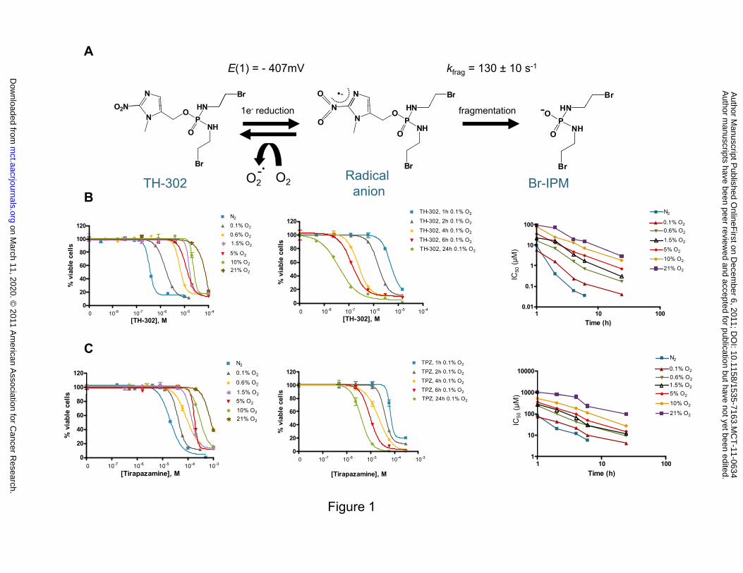
reduction methods were employed. Fragmentation occurred under anoxia via the TH-302 radical
anion and resulted in elimination of the active cytotoxin Br-IPM (Figure 1A). Fragmentation was
readily inhibited by molecular dioxygen. The measured one-electron reduction potential E(1) and
the first-order fragmentation rate constant (kfrag) following one-electron reduction were consistent
with the proposed mechanism (Figure 1A, Supplementary Figures S1-S4). This mechanism is in
contrast to the established activation mechanism for 2-nitroimidazoles such as misonidazole and
pimonidazole where the radical anion in the absence of oxygen undergoes further reduction to the
hydroxyl amine and elimination to a crosslinking nitrenium ion [18, 19].
Oxygen concentration- and time-dependence of TH-302 activation
To evaluate the stringency of hypoxia required to release the active Br-IPM, we
performed in vitro proliferation assay in H460 with different oxygen concentrations. As
illustrated in Figure 1B, TH-302 cytotoxic activity increased as the oxygen level decreased. In
addition, hypoxia-selective activation of TH-302 was time-dependent with longer drug exposures
providing greater TH-302 potency with a CT relationship that approached linearity
(Supplementary Table S1A). A similar profile for HAP tirapazamine was observed although with
much lower potency and inferior hypoxia cytotoxicity ratio (HCR) compared to TH-302 (Figure
1C and Supplementary Table S1B). This observation was also confirmed with clonogenic assays
(Supplementary Figure S5). In addition, TH-302 requires more severe hypoxia (~0.1% O2) for
high activity, while tirapazamine achieves near-maximal activity under conditions of moderate
hypoxia (0.6% -1.5% O2).
on March 11, 2020. © 2011 American Association for Cancer Research. mct.aacrjournals.org Downloaded from
Author manuscripts have been peer reviewed and accepted for publication but have not yet been edited. Author Manuscript Published OnlineFirst on December 6, 2011; DOI: 10.1158/1535-7163.MCT-11-0634

10
TH-302 exhibits hypoxia-selective cytotoxicity across a panel of human cancer cell lines
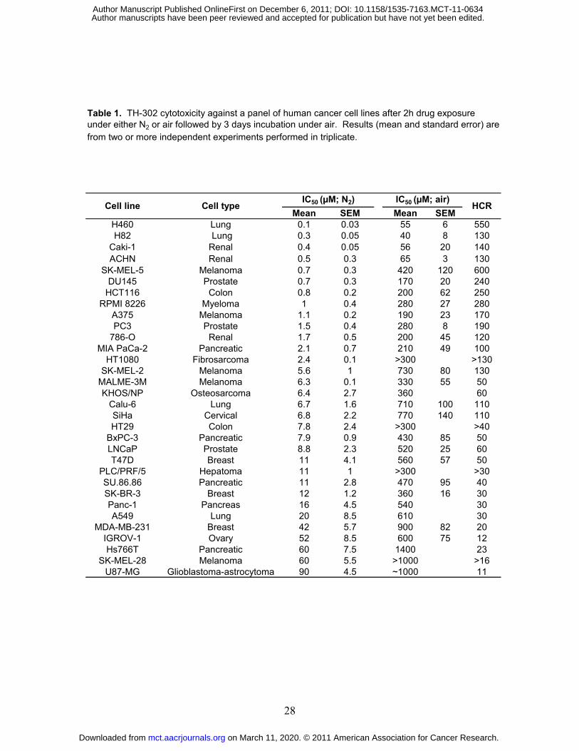
TH-302 cytotoxicity was evaluated across 32 human cancer cell lines. As shown in Table 1,
TH-302 cytotoxic potency was modest under normoxia with all IC50 values greater than 40 µM.
In contrast, substantially increased cytotoxicity was observed under hypoxic conditions in all 32
cell lines. Normoxic activity of TH-302 was also examined in the 60 cell line panel of the NCI
(the NCI-60). IC50 values ranged from 1 µM to ≥ 1000 µM after 48h continuous drug exposure
(Supplementary Table S1C).
Reductase-dependent TH-302 activity
We evaluated the relationship between the potency of TH-302 cytotoxicity and POR
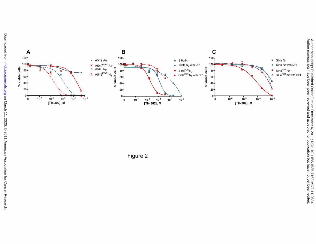
expression levels with two pairs of matched cell lines [12]. As demonstrated in Figure 2A and
Supplementary Table S2, A549 cells overexpressing POR exhibited a 7-fold increase in the
potency of TH-302 when tested under hypoxic conditions. A similar magnitude of enhancement
of TH-302 potency was also observed under aerobic conditions. TH-302 potency was likewise
increased in the SiHa cells overexpressing POR under both hypoxic and aerobic conditions
(Figure 2B and Supplementary Table S2). The effect of the mechanism-based flavoenzyme
inhibitor diphenyliodonium chloride (DPI) [20] on TH-302 mediated cytotoxicity was also
examined in SiHa and SiHaPOR. As shown in Figure 2B, TH-302 activity under hypoxia can be
inhibited by DPI in both SiHa and SiHaPOR. DPI inhibits TH-302 activity in SiHaPOR but not SiHa
under normoxia (Figure 2C).
TH-302 induces DNA damage
To evaluate whether TH-302 cross-links DNA, we examined the phosphorylation of the
histone variant H2AX (γH2AX) in H460 cells. There was an increase in γH2AX as a function of
time after TH-302 treatment (Supplementary Figure S6). Under hypoxia, γH2AX signal became
on March 11, 2020. © 2011 American Association for Cancer Research. mct.aacrjournals.org Downloaded from
Author manuscripts have been peer reviewed and accepted for publication but have not yet been edited. Author Manuscript Published OnlineFirst on December 6, 2011; DOI: 10.1158/1535-7163.MCT-11-0634

11
readily visible 8 h after exposure to TH-302 and continued to increase when examined at 24 h
post treatment. A similar profile was observed for TH-302 under normoxia although a 100-fold
higher concentration of TH-302 was required. The TH-302 dose-response for γH2AX induction
was also investigated using both flow cytometry and immunofluorescence microscopy. There
was an increase in γH2AX as a function of TH-302 concentration. Under hypoxia, TH-302 at
0.13 µM induced visible γH2AX binding after 24 h post-treatment (Figure 3B) while a >300-fold
concentration of TH-302 (45 µM) was required to induce similar γH2AX binding under normoxic
conditions (Figure 3A) and binding intensity was enhanced as TH-302 concentrations increased.
A direct biochemical assay for DNA cross-linking, the single-cell electrophoresis-based ‘comet
assay’, confirmed TH-302 dependent DNA-cross-linking (Figure 3C).
TH-302 causes cell cycle arrest in a concentration- and hypoxia-dependent manner
H460 cells treated with 0.05 µM of TH-302 under hypoxia led to the accumulation of
cells at the G2/M junction (Figure 3D and Supplementary Table S3). In contrast, cell cycle
distribution was not altered by 0.05 µM of TH-302 treatment under normoxia (Figure 3D). As
TH-302 concentration was increased cells arrested at all phases of the cell cycle. A similar
profile was observed with HT29 cells (Supplementary Figure S7 and Supplementary Table S3).
TH-302 enhances cytotoxicity in homology-dependent repair (HDR) mutant cell lines
To assess the relative importance of different DNA repair pathways involved in TH-302
activity, we first employed a panel of CHO cell-based cell lines with defects in different DNA
damage response and repair pathways. As shown in Figure 4A and B and Supplementary Table
S4A and S4B, little difference in sensitivity to TH-302 was observed with cells defective in
XRCC1 (EM9), ERCC5/XPG (UV135) and Ku80 (xrs-5). These results indicate that base
excision repair (BER), nucleotide excision repair (NER) and nonhomologous end-joining
on March 11, 2020. © 2011 American Association for Cancer Research. mct.aacrjournals.org Downloaded from
Author manuscripts have been peer reviewed and accepted for publication but have not yet been edited. Author Manuscript Published OnlineFirst on December 6, 2011; DOI: 10.1158/1535-7163.MCT-11-0634

12
(NHEJ), respectively, are not significantly involved in the response and repair of TH-302 adducts.
However irs1SF cells (defective in XRCC3) demonstrated either a 7-fold increase (Figure 4A and
Supplementary Table S4A) or 43-fold increase (Figure 4C and Supplementary Table S4C) in
sensitivity to TH-302. UV41 cells (defective in ERCC4/XPF) exhibited a 67-fold increase in TH-
302 sensitivity (Figure 4A). Cell line pairs comprised of one line deficient in BRCA1, BRCA2,
or FANCA, and a matched line complemented by the over-expression of the corresponding wild-
type proteins were also tested for their relative sensitivity to TH-302. As shown in Figure 4 (D-F)
and Supplementary Table S4D, all three of the deficient lines exhibited an enhanced sensitivity to
TH-302 compared to the wild-type protein complemented cognate lines. The BRCA2 mutant line
was particularly sensitive, exhibiting a sub-nanomolar IC90 (0.6 nM) for TH-302 when tested
under hypoxic conditions. Taken together, the results indicate that homology-dependent repair
(HDR) pathways are those primarily involved in TH-302 DNA damage repair.
Monoalkylating TH-302 analog activity
To investigate the importance of bis-functionality and cross-linking for TH-302 activity,
we compared the potency of TH-302 with TH-1197, an analog with only one reactive bromine
leaving group (Supplementary Figure S8). TH-302 exhibited 33-fold greater activity in AA8 wild
type cells under anoxia (IC50 2.6 µM) compared to TH-1197 (IC50 87 µM). In HDR defective
cells (irs1SF), an even greater enhancement (180-fold) of TH-302 potency (IC50 0.14 µM) was
observed compared to that of TH-1197 (IC50 25 µM) under anoxia.
TH-302 activity in 3D multicellular tumor spheroid model
Human tumor spheroids recapitulate aspects of the tumor microenvironment, including
multiple cell layers for drug penetration and oxygen concentration gradients. As shown in Figure
5A, 79% of H460 spheroid cells were pimonidazole-positive when incubated under 21% O2
conditions indicative of significant diffusion limited hypoxia. TH-302 showed 650-fold greater
on March 11, 2020. © 2011 American Association for Cancer Research. mct.aacrjournals.org Downloaded from
Author manuscripts have been peer reviewed and accepted for publication but have not yet been edited. Author Manuscript Published OnlineFirst on December 6, 2011; DOI: 10.1158/1535-7163.MCT-11-0634
13
activity in H460 spheroids compared with monolayer cells with IC50 values of 0.1 µM and 65
µM, respectively (Figure 5B and Supplementary Table S5A). In contrast, tirapazamine exhibited
only a 4 fold difference between monolayer and spheroids, which is consistent with previous
studies [21]. This observation was confirmed using clonogenic assays with d15 spheroids
(Supplementary Figure 9A) and d8 spheroids (Supplementary Figure 9B). The anthracycline
chemotherapeutic daunorubicin exhibited the converse pattern of greater potency against
monolayer cells which was expected given its poor tissue penetration properties. The results are
consistent with TH-302 exhibiting good penetrability, hypoxia activation, and bystander effect.
Enhanced activity of TH-302 in spheroids is due to microenvironmental factors
To determine the basis for the TH-302 sensitivity difference between monolayer cells
and cells from spheroids, we compared the cytotoxic activity of TH-302 between intact spheroids,
suspended cells from dissociated spheroids cells, monolayer cultures, and suspended cells from
monolayer cultures. Consistent with the proposed mechanism of activation, enhanced sensitivity
to TH-302 is only observed in intact H460 spheroids (IC50 0.2 µM) and not in cells from
dissociated spheroids (IC50 21 µM) (Figure 5C and Supplementary Table S5B). Cell sensitivity to
TH-302 was not altered by trypsinization. Hypoxic cell population was only detected in intact
spheroids but not suspended cells from dissociated spheroids cells (Supplementary Figure S9C).
Taken together, the results indicate that the dramatic TH-302 potency shift in spheroids is due
largely to the spheroid microenvironment and is not a consequence of cell autonomous changes
dependent on spheroid-based cell propagation.
Demonstration of bystander effect with multicellular layer cultures (MCLs)
To confirm the ability of TH-302 to exhibit a bystander effect, where warhead released in
hypoxic cells is able to diffuse locally to kill adjacent cells, we employed a mixture of TH-302
sensitive and resistant cell populations grown as three dimensional co-cultures [15, 16]. As
on March 11, 2020. © 2011 American Association for Cancer Research. mct.aacrjournals.org Downloaded from
Author manuscripts have been peer reviewed and accepted for publication but have not yet been edited. Author Manuscript Published OnlineFirst on December 6, 2011; DOI: 10.1158/1535-7163.MCT-11-0634
14
shown in Figure 5D and Supplementary Table S5C, parental HCT116 cells (target cells) were
relatively resistant to TH-302 exposure under hyperoxic (95% O2) conditions with IC90 of 19 µM.
Hyperoxic conditions serve to suppress the endogenous 1e- reductase mediated prodrug activation
by enhancing the O2 concentration mediated back-reaction from radical anion to prodrug (Figure
1A). HCT116 cells engineered to express the two-electron nitroreductase NfsA (activator cells)
displayed a marked sensitivity to TH-302 exposure under hyperoxia with IC90 of 0.09 µM. While
target cells alone were refractory, when grown in co-culture with 30% activators, they
experienced a marked 10-fold reduction in surviving fraction (IC90). The calculated bystander
effect efficiency (BEE) was 43% indicating the active metabolites generated by TH-302 reduction
are able to diffuse out of the cell of origin and act on surrounding neighboring target cells.
on March 11, 2020. © 2011 American Association for Cancer Research. mct.aacrjournals.org Downloaded from
Author manuscripts have been peer reviewed and accepted for publication but have not yet been edited. Author Manuscript Published OnlineFirst on December 6, 2011; DOI: 10.1158/1535-7163.MCT-11-0634

15
Discussion
The selective targeting of the hypoxic compartment of tumors provides an approach for
the development of novel anticancer therapies. Hypoxia-activated prodrugs (HAPs), based on
oxygen concentration sensitive nitroheterocyclic bioreductive triggers linked to cytotoxic or
cytostatic effectors, are an especially attractive design [10].
We established the one-electron reduction stoichiometry of TH-302 to fragment via the
radical anion and release the cytotoxic warhead Br-IPM. The rate of fragmentation (Kfrag 130 ± 10
s-1) is first-order, and sufficiently slow to be readily inhibited (back-oxidized) by molecular
dioxygen [14].
Having confirmed the oxygen-sensitive free-radical dependent mechanism of TH-302
activation we next employed multiple oxygen concentrations to profile TH-302 and compare its
relative oxygen concentration potency to the prototypical HAP tirapazamine [22, 23]. Different
lengths of time for drug exposure under the oxygen concentration atmospheres were employed.
We observed that tirapazamine achieved near-maximal activity against H460 cells under
conditions of moderate hypoxia (~1.5 – 0.6% O2) which is consistent with the reported oxygen
concentration required for half-maximal cytotoxicity in V79 [22] and SiHa cells [23]. Conversely,
for TH-302, most of its selectivity (~375-fold) is only obtained at 0.1% or less, with 0.6% O2
providing only moderate potency compared to 21%. This result is consistent with targeting the
more severe pathological hypoxia associated with tumor subregions while sparing moderate
hypoxic regions found in certain normal tissues.
We then tested the hypoxia-selective cytotoxicity profile of TH-302 across 32 human
cancer cell lines. All cell lines examined exhibited enhanced cytotoxicity when the TH-302
exposure was performed under hypoxic conditions. Of particular note was the finding of a wide
range of potencies that spanned more than two orders of magnitude among the individual cell
lines examined.
on March 11, 2020. © 2011 American Association for Cancer Research. mct.aacrjournals.org Downloaded from
Author manuscripts have been peer reviewed and accepted for publication but have not yet been edited. Author Manuscript Published OnlineFirst on December 6, 2011; DOI: 10.1158/1535-7163.MCT-11-0634
16
Given the variety of different mechanisms underlying the activation and mechanism of
action of TH-302, there are a number of differences between the cells, which either alone or in
combination could underlie the wide variation in potency observed. These factors could include
differences in the activity of one-electron reductases, differences in DNA repair mechanisms, and
differences in cell fate (cell cycle arrest, cell death) as a function of DNA damage. Overall,
aerobic potency was low and cytotoxicity was enhanced under hypoxic conditions. This result
can be contrasted to other HAPs that exhibit both hypoxia-dependent and hypoxia-independent
activation. For example, in addition to being activated by 1-electron reductases in a hypoxia-
dependent manner, the HAP PR104A is also activated in a hypoxia-independent manner by the 2-
electron oxidoreductase aldo-keto reductase 1C3 [24]. Cell lines in which this reductase is highly
expressed exhibit sensitivity to PR-104A in a hypoxia-independent manner [25]. Currently no
evidence exists to support a similar pathway of aerobic activation for TH-302 despite extensive
cell line studies.
The 2-nitroimidazole system has been partially characterized with regard to the spectrum
of enzymes capable of catalyzing this initial reduction [26] although uncertainties still remain
[27]. Walton and Workman first demonstrated the role of POR in the reduction of 2-
nitroimidazole [26]. Patterson and colleagues [28] demonstrated a correlation of POR activity
levels with the magnitude of HAP activation and cytotoxicity. We tested the ability of POR to
serve as an activating enzyme for TH-302. We observed a marked increase of potency of TH-302
in the lines over-expressing POR. The enhanced activity can be inhibited by the pan-
flavoenzyme inhibitor DPI. These results are consistent with the differential expression and
activity of POR potentially serving as one factor underlying the wide range of potencies observed
for TH-302 among different human cancer cell lines. These results also support the hypothesis
that the relative reductase levels in both tumor and normal tissue may contribute to the magnitude
of the clinical benefit and the therapeutic index in patients. Advances in biomarker analysis of
individual patients, whether based on tissue biopsies, serum or plasma samples, or circulating
on March 11, 2020. © 2011 American Association for Cancer Research. mct.aacrjournals.org Downloaded from
Author manuscripts have been peer reviewed and accepted for publication but have not yet been edited. Author Manuscript Published OnlineFirst on December 6, 2011; DOI: 10.1158/1535-7163.MCT-11-0634

17
tumor cells could be applied to patients and help guide decisions on TH-302 treatment based on
POR or other activating enzymes identified. Population level analysis of POR expression
suggests a high frequency of expression in malignancies of the ovary and liver [29].
We used a series of assays to explore the mechanism of action of TH-302. Given the bis-
alkylator nature of the warhead, our efforts were focused on DNA damage pathways. Histone
H2AX phosphorylation (γH2AX) has emerged as a robust and sensitive marker of the action of
DNA interstrand crosslinking (ICL) agents [30, 31]. We show here both a TH-302 concentration-
and time-dependent induction of γH2AX by both immunohistochemistry and flow cytometric
techniques. The concentrations of TH-302 necessary to induce γH2AX were the same magnitude
as those required for cell death and the observed time course was similar to that described
previously for other DNA cross-linking agents. Comet assay profiling [32] confirmed TH-302
induced DNA cross-linking. The observed cell cycle effects are consistent with the DNA targeted
mechanism of action for TH-302. Similar cell cycle effects have been described in multiple
myeloma cell lines treated with TH-302 [33].
To explore the specific nature of the DNA lesions that induced the γH2AX signaling and
subsequent cytotoxicity, we utilized a panel of matched cell line pairs that are defective in distinct
DNA damage repair enzymes [34-36]. The specific pathways we profiled included NER, BER,
NHEJ, and HDR, and mutants deficient in HDR proteins [37, 38] exhibited greatly enhanced
sensitivity to TH-302. BRCA and Fanconi proteins also play key roles in the response of cells to
DNA damage that results in HDR-based repair [39, 40]. Employing paired cell lines defective in
BRCA1, BRCA2, or FANCA together with their wild type protein expressing isogenic controls,
TH-302 exhibited an enhanced cytotoxic potency in the mutant lines. Taken together, these
results clearly implicate HDR as the pathway primarily responsible for the repair of TH-302
induced DNA damage. This HDR pathway has also been shown to be a principal pathway for the
repair of DNA damage induced by the HAPs tirapazamine [13] and PR104A [41]. Interestingly,
on March 11, 2020. © 2011 American Association for Cancer Research. mct.aacrjournals.org Downloaded from
Author manuscripts have been peer reviewed and accepted for publication but have not yet been edited. Author Manuscript Published OnlineFirst on December 6, 2011; DOI: 10.1158/1535-7163.MCT-11-0634

18
using one of the same pairs of lines (AA8 and irs1SF) Evans et al. observed a 4x enhanced
potency of tirapazamine in the irs1SF compared to the wild-type while we observed a 50x
enhancement with TH-302. This difference is consistent with TH-302’s activity being more
specific to the HDR mechanism of action as tirapazamine was shown to also yield lesion repair
by BER [13]. The findings that TH-302 activity is enhanced in the context of HDR repair
pathways is of particular note because of the recent finding that hypoxia results in decreased
expression of HR proteins, including XRCC-3, (the protein mutated in the irsSF1 line), BRCA1,
as well as other HDR-related genes [42-44]. This observation indicates that the in vivo sensitivity
to TH-302-mediated DNA damage may be enhanced in the same hypoxic compartment of the
tumor where the prodrug activation occurs. This could contribute to a favorable therapeutic index
if the cells responsible for prodrug activation are inherently more sensitive to the released
warhead than non-hypoxic cells. In addition, these mechanisms of action results have
implications for the clinical use of TH-302. Patient tumors with deficiencies in HDR (e.g.
through XRCC, FANC or BRCA mutations or other HDR defects) would be expected to be
hypersensitive to TH-302. These results provide the basis for the development of diagnostic tests
that could identify patients most likely to benefit from TH-302 based on their HDR status. For
example the potential prognostic value of the BRCA-like profile in ovarian carcinomas [45] may
be of interest, particularly if associated with the observations of elevated POR expression [29].
Likewise cancer drugs that that have been shown to downregulate HDR, such as the approved
agents gemcitabine, vorinostat, bortezomib, and imatinib [46-49] may combine with TH-302 in
additive or synergistic manners due to complementary mechanisms of action.
Three dimensional multicellular tumor spheroids recapitulate many aspects of the tumor
microenvironment, and are an especially relevant in vitro model system to profile the
multiparametric pharmacological activities of HAPs. Here we show that TH-302 kills a very high
fraction of spheroid cells, especially in comparison to the benchmark HAP tirapazamine.
on March 11, 2020. © 2011 American Association for Cancer Research. mct.aacrjournals.org Downloaded from
Author manuscripts have been peer reviewed and accepted for publication but have not yet been edited. Author Manuscript Published OnlineFirst on December 6, 2011; DOI: 10.1158/1535-7163.MCT-11-0634

19
Essentially all of the increase in TH-302 potency observed in the spheroid model can be ascribed
to the microenvironment of the spheroid system.
Multicellular layers comprised of mixtures of two metabolically distinct cell populations
are a useful method for assessing HAP bystander effect potential. The approach employs one cell
type which can efficiently activate TH-302 and is subject to its cytotoxic effects, and a second
cell type that cannot activate TH-302 but may be subject to the cytotoxic effect of the diffusible
cytotoxin if it is released from activator cells [15, 16]. By utilizing a two-electron bacterial
nitroreductase under hyperoxic conditions, clean metabolic boundaries are created at the cellular
level, a model that is difficult to reproduce using oxygen gradients [15, 17]. The TH-302 IC90
values for the two cell types were 19 and 0.09 µM, respectively. This ~200x ratio is broadly
aligned with cytotoxicity ratios observed with human cancer cell lines when profiled under oxic
and hypoxic conditions. The proportion of ‘activator’ cells in the MCLs ranged from 25% to
33%. This proportion of metabolically active hypoxic cells is typical of human tumour xenografts
as measured by pimonidazole binding. Thus the magnitude of the sensitizing effect and the
quantum of metabolically active cells in the MCL were considered broadly analogous to the
hypoxic cell compartment found in xenograft models. When the resistant ‘target’ cells were co-
cultured with this minority population ‘activator’ cells under hyperoxic conditions (95% O2/5%
CO2) that suppresses endogenous one-electron reductase mediated prodrug activation, TH-302
cytotoxicity was acquired via a process of cell-to-cell released warhead transfer. The calculated
bystander effect efficiency (BEE) was 43% which is indicative of a bystander effect of
‘intermediate’ efficiency. The TH-302 bystander effect can be compared to HAPs exhibiting a
large bystander effect, for example the 3,5-dinitrobenzamine-2-bromomustard SN27686, with
BEE values of >90% [16], and HAPs with no bystander effect, for example tirapazamine, with
BEE values estimated at 0% [15]. The intermediate nature of TH-302’s bystander effect provides
theoretical advantages whereby active metabolite concentrations are not excessively diluted as a
function of long-range diffusion distances, but rather provide a localized effect against adjacent
on March 11, 2020. © 2011 American Association for Cancer Research. mct.aacrjournals.org Downloaded from
Author manuscripts have been peer reviewed and accepted for publication but have not yet been edited. Author Manuscript Published OnlineFirst on December 6, 2011; DOI: 10.1158/1535-7163.MCT-11-0634
20
cells at intermediate oxygen tensions, considered to be a critically important subpopulation [50].
An intermediate diffusion-range also has the added advantage that the probability of systemic
recirculation of activated warhead is minimized.
In conclusion, the results described here highlight TH-302 as an optimized hypoxia-
activated prodrug. The wide range of human tumor cell lines in which it exhibits hypoxia-
selective cytotoxicity, coupled with no evidence consistent with hypoxia-independent mechanism
of cytotoxicity, supports the potential for its broad applicability in the context of established
chemotherapy and radiotherapy regimens. The described oxygen concentration profile for
activation is consistent with the selective targeting of the deep hypoxia found in solid tumors and
minor activation in tissues exhibiting nonpathological moderate hypoxia. The dependence of
cytotoxicity on POR expression highlights the potential to profile the expression of this and other
one-electron oxidoreductases. Identification of the pathways of DNA damage response and repair
involved with the activity of TH-302 point to mechanisms of sensitivity or resistance that could
have relevance to the clinical use of TH-302. TH-302 is currently in clinical trials for the
treatment of cancer.
on March 11, 2020. © 2011 American Association for Cancer Research. mct.aacrjournals.org Downloaded from
Author manuscripts have been peer reviewed and accepted for publication but have not yet been edited. Author Manuscript Published OnlineFirst on December 6, 2011; DOI: 10.1158/1535-7163.MCT-11-0634

21
References
1. Hockel M, Vaupel P. Tumor hypoxia: definitions and current clinical, biologic, and molecular
aspects. J Natl Cancer Inst 2001;93:266-76.
2. Vaupel P, Mayer A. Hypoxia in cancer: significance and impact on clinical outcome. Cancer
Metastasis Rev 2007;26:225-39.
3. Wilson WR, Hay MP. Targeting hypoxia in cancer therapy. Nat Rev Cancer 2011;11:393-410.
4. Brown JM, Wilson WR. Exploiting tumour hypoxia in cancer treatment. Nat Rev Cancer
2004;4:437-47.
5. Teicher BA, Sartorelli AC. Nitrobenzyl halides and carbamates as prototype bioreductive
alkylating agents. J Med Chem 1980;23:955-60.
6. Denny WA, Wilson WR. Considerations for the design of nitrophenyl mustards as agents with
selective toxicity for hypoxic tumor cells. J Med Chem 1986;29:879-87.
7. Firestone A, Mulcahy RT, Borch RF. Nitroheterocycle reduction as a paradigm for
intramolecular catalysis of drug delivery to hypoxic cells. J Med Chem 1991;34:2933-5.
8. Chen Y, Hu L. Design of anticancer prodrugs for reductive activation. Med Res Rev
2009;29:29-64.
9. Reddy SB, Williamson SK. Tirapazamine: a novel agent targeting hypoxic tumor cells. Expert
Opin Investig Drugs 2009;18:77-87.
10. Denny WA, Wilson WR. Bioreducible mustards: a paradigm for hypoxia-selective prodrugs
of diffusible cytotoxins (HPDCs). Cancer Metastasis Rev 1993;12:135-51.
on March 11, 2020. © 2011 American Association for Cancer Research. mct.aacrjournals.org Downloaded from
Author manuscripts have been peer reviewed and accepted for publication but have not yet been edited. Author Manuscript Published OnlineFirst on December 6, 2011; DOI: 10.1158/1535-7163.MCT-11-0634

22
11. Duan JX, Jiao H, Kaizerman J, Stanton T, Evans JW, Lan L, et al. Potent and highly selective
hypoxia-activated achiral phosphoramidate mustards as anticancer drugs. J Med Chem
2008;51:2412-20.
12. Guise CP, Wang AT, Theil A, Bridewell DJ, Wilson WR, Patterson AV. Identification of
human reductases that activate the dinitrobenzamide mustard prodrug PR-104A: a role for
NADPH:cytochrome P450 oxidoreductase under hypoxia. Biochem Pharmacol 2007;74:810-20.
13. Evans JW, Chernikova SB, Kachnic LA, Banath JP, Sordet O, Delahoussaye YM, et al.
Homologous recombination is the principal pathway for the repair of DNA damage induced by
tirapazamine in mammalian cells. Cancer Res 2008;68:257-65.
14. Anderson RF, Denny WA, Li W, Packer JE, Tercel M, Wilson WR. Pulse radiolysis
studies on the fragmentation of arylmethyl quaternary nitrogen mustards by one-electron
reduction in aqueous solution. J. Phys. Chem. A 1997;101:9704-9.
15. Wilson WR, Hicks KO, Pullen SM, Ferry DM, Helsby NA, Patterson AV. Bystander
effects of bioreductive drugs: potential for exploiting pathological tumor hypoxia with
dinitrobenzamide mustards. Radiat Res 2007;167:625-36.
16. Singleton DC, Li D, Bai SY, Syddall SP, Smaill JB, Shen Y, et al. The nitroreductase
prodrug SN 28343 enhances the potency of systemically administered armed oncolytic
adenovirus ONYX-411(NTR). Cancer Gene Ther 2007;14:953-67.
17. Prosser GA, Copp JN, Syddall SP, Williams EM, Smaill JB, Wilson WR, et al. Discovery
and evaluation of Escherichia coli nitroreductases that activate the anti-cancer prodrug CB1954.
Biochem Pharmacol 2010;79:678-87.
on March 11, 2020. © 2011 American Association for Cancer Research. mct.aacrjournals.org Downloaded from
Author manuscripts have been peer reviewed and accepted for publication but have not yet been edited. Author Manuscript Published OnlineFirst on December 6, 2011; DOI: 10.1158/1535-7163.MCT-11-0634

23
18. Wardman P, Dennis MF, Everett SA, Patel KB, Stratford MR, Tracy M. Radicals from
one-electron reduction of nitro compounds, aromatic N-oxides and quinones: the kinetic basis for
hypoxia-selective, bioreductive drugs. Biochem Soc Symp 1995;61:171-94.
19. Bolton JL, McClelland RA. Kinetics and mechanism of the decomposition in aqueous
solutions of 2-( Hydroxyamino)imidazoles. J. Am. Chem. Soc 1989;111:8172-81.
20. Tew DG. Inhibition of cytochrome P450 reductase by the diphenyliodonium cation. Kinetic
analysis and covalent modifications. Biochemistry 1993;32:10209-15.
21. Durand RE, Olive PL. Evaluation of bioreductive drugs in multicell spheroids. Int J Radiat
Oncol Biol Phys 1992;22:689-92.
22. Koch CJ. Unusual oxygen concentration dependence of toxicity of SR-4233, a hypoxic cell
toxin. Cancer Res 1993;53:3992-7.
23. Hicks KO, Siim BG, Pruijn FB, Wilson WR. Oxygen dependence of the metabolic
activation and cytotoxicity of tirapazamine: implications for extravascular transport and activity
in tumors. Radiat Res 2004;161:656-66.
24. Guise CP, Abbattista MR, Singleton RS, Holford SD, Connolly J, Dachs GU, et al. The
bioreductive prodrug PR-104A is activated under aerobic conditions by human aldo-keto
reductase 1C3. Cancer Res 2010;70:1573-84.
25. Singleton RS, Guise CP, Ferry DM, Pullen SM, Dorie MJ, Brown JM, et al. DNA cross-links
in human tumor cells exposed to the prodrug PR-104A: relationships to hypoxia, bioreductive
metabolism, and cytotoxicity. Cancer Res 2009;69:3884-91.
on March 11, 2020. © 2011 American Association for Cancer Research. mct.aacrjournals.org Downloaded from
Author manuscripts have been peer reviewed and accepted for publication but have not yet been edited. Author Manuscript Published OnlineFirst on December 6, 2011; DOI: 10.1158/1535-7163.MCT-11-0634

24
26. Walton MI, Workman P. Nitroimidazole bioreductive metabolism. Quantitation and
characterisation of mouse tissue benznidazole nitroreductases in vivo and in vitro. Biochem
Pharmacol 1987;36:887-96.
27. Joseph P, Jaiswal AK, Stobbe CC, Chapman JD. The role of specific reductases in the
intracellular activation and binding of 2-nitroimidazoles. Int J Radiat Oncol Biol Phys
1994;29:351-5.
28. Patterson AV, Saunders MP, Chinje EC, Talbot DC, Harris AL, Strafford IJ.
Overexpression of human NADPH:cytochrome c (P450) reductase confers enhanced sensitivity
to both tirapazamine (SR 4233) and RSU 1069. Br J Cancer 1997;76:1338-47.
29. Guise CP, Abbattista MR, Tipparaju SR, Lambie NK, Su J, Li D, et al. Diflavin
Oxidoreductases Activate the Bioreductive Prodrug PR104A Under Hypoxia. Mol Pharmacol
2012;81:1-10.
30. Olive PL, Banath JP. Kinetics of H2AX phosphorylation after exposure to cisplatin.
Cytometry B Clin Cytom 2009;76:79-90.
31. Clingen PH, Wu JY, Miller J, Mistry N, Chin F, Wynne P, et al. Histone H2AX
phosphorylation as a molecular pharmacological marker for DNA interstrand crosslink cancer
chemotherapy. Biochem Pharmacol 2008;76:19-27.
32. Ostling O, Johanson KJ. Microelectrophoretic study of radiation-induced DNA damages in
individual mammalian cells. Biochem Biophys Res Commun 1984;123:291-8.
33. Hu J, Handisides DR, Van Valckenborgh E, De Raeve H, Menu E, Vande Broek I, et al.
Targeting the multiple myeloma hypoxic niche with TH-302, a hypoxia-activated prodrug. Blood
2010;116:1524-7.
on March 11, 2020. © 2011 American Association for Cancer Research. mct.aacrjournals.org Downloaded from
Author manuscripts have been peer reviewed and accepted for publication but have not yet been edited. Author Manuscript Published OnlineFirst on December 6, 2011; DOI: 10.1158/1535-7163.MCT-11-0634

25
34. Jeggo PA, Kemp LM. X-ray-sensitive mutants of Chinese hamster ovary cell line. Isolation
and cross-sensitivity to other DNA-damaging agents. Mutat Res 1983;112:313-27.
35. Hoy CA, Thompson LH, Mooney CL, Salazar EP. Defective DNA cross-link removal in
Chinese hamster cell mutants hypersensitive to bifunctional alkylating agents. Cancer Res
1985;45:1737-43.
36. Fuller LF, Painter RB. A Chinese hamster ovary cell line hypersensitive to ionizing radiation
and deficient in repair replication. Mutat Res 1988;193:109-21.
37. Kuraoka I, Kobertz WR, Ariza RR, Biggerstaff M, Essigmann JM, Wood RD. Repair of an
interstrand DNA cross-link initiated by ERCC1-XPF repair/recombination nuclease. J Biol Chem
2000;275:26632-6.
38. Liu N, Lamerdin JE, Tebbs RS, Schild D, Tucker JD, Shen MR, et al. XRCC2 and XRCC3,
new human Rad51-family members, promote chromosome stability and protect against DNA
cross-links and other damages. Mol Cell 1998;1:783-93.
39. Powell SN, Kachnic LA. Roles of BRCA1 and BRCA2 in homologous recombination, DNA
replication fidelity and the cellular response to ionizing radiation. Oncogene 2003;22:5784-91.
40. Thompson LH, Hinz JM. Cellular and molecular consequences of defective Fanconi anemia
proteins in replication-coupled DNA repair: mechanistic insights. Mutat Res 2009;668:54-72.
41. Gu Y, Patterson AV, Atwell GJ, Chernikova SB, Brown JM, Thompson LH, et al. Roles of
DNA repair and reductase activity in the cytotoxicity of the hypoxia-activated dinitrobenzamide
mustard PR-104A. Mol Cancer Ther 2009;8:1714-23.
on March 11, 2020. © 2011 American Association for Cancer Research. mct.aacrjournals.org Downloaded from
Author manuscripts have been peer reviewed and accepted for publication but have not yet been edited. Author Manuscript Published OnlineFirst on December 6, 2011; DOI: 10.1158/1535-7163.MCT-11-0634

26
42. Bindra RS, Schaffer PJ, Meng A, Woo J, Maseide K, Roth ME, et al. Down-regulation of
Rad51 and decreased homologous recombination in hypoxic cancer cells. Mol Cell Biol
2004;24:8504-18.
43. Bindra RS, Gibson SL, Meng A, Westermark U, Jasin M, Pierce AJ, et al. Hypoxia-induced
down-regulation of BRCA1 expression by E2Fs. Cancer Res 2005;65:11597-604.
44. Chan N, Koritzinsky M, Zhao H, Bindra R, Glazer PM, Powell S, et al. Chronic hypoxia
decreases synthesis of homologous recombination proteins to offset chemoresistance and
radioresistance. Cancer Res 2008;68:605-14.
45. Konstantinopoulos PA, Spentzos D, Karlan BY, Taniguchi T, Fountzilas E, Francoeur N, et
al. Gene expression profile of BRCAness that correlates with responsiveness to chemotherapy
and with outcome in patients with epithelial ovarian cancer. J Clin Oncol 2010;28:3555-61.
46. Singh S, Le H, Shih SJ, Ho B, Vaughan AT. Suberoylanilide hydroxyamic acid
modification of chromatin architecture affects DNA break formation and repair. Int J Radiat
Oncol Biol Phys 2010;76:566-73.
47. Yarde DN, Oliveira V, Mathews L, Wang X, Villagra A, Boulware D, et al. Targeting the
Fanconi anemia/BRCA pathway circumvents drug resistance in multiple myeloma. Cancer Res
2009;69:9367-75.
48. Choudhury A, Zhao H, Jalali F, Al Rashid S, Ran J, Supiot S, et al. Targeting homologous
recombination using imatinib results in enhanced tumor cell chemosensitivity and
radiosensitivity. Mol Cancer Ther 2009;8:203-13.
on March 11, 2020. © 2011 American Association for Cancer Research. mct.aacrjournals.org Downloaded from
Author manuscripts have been peer reviewed and accepted for publication but have not yet been edited. Author Manuscript Published OnlineFirst on December 6, 2011; DOI: 10.1158/1535-7163.MCT-11-0634

27
49. Wachters FM, van Putten JW, Maring JG, Zdzienicka MZ, Groen HJ, Kampinga HH.
Selective targeting of homologous DNA recombination repair by gemcitabine. Int J Radiat Oncol
Biol Phys 2003;57:553-62.
50. Wouters BG, Brown JM. Cells at intermediate oxygen levels can be more important than the
"hypoxic fraction" in determining tumor response to fractionated radiotherapy. Radiat Res
1997;147:541-50.
on March 11, 2020. © 2011 American Association for Cancer Research. mct.aacrjournals.org Downloaded from
Author manuscripts have been peer reviewed and accepted for publication but have not yet been edited. Author Manuscript Published OnlineFirst on December 6, 2011; DOI: 10.1158/1535-7163.MCT-11-0634
28
Mean SEM Mean SEMH460 Lung 0.1 0.03 55 6 550H82 Lung 0.3 0.05 40 8 130
Caki-1 Renal 0.4 0.05 56 20 140ACHN Renal 0.5 0.3 65 3 130
SK-MEL-5 Melanoma 0.7 0.3 420 120 600DU145 Prostate 0.7 0.3 170 20 240
HCT116 Colon 0.8 0.2 200 62 250RPMI 8226 Myeloma 1 0.4 280 27 280
A375 Melanoma 1.1 0.2 190 23 170PC3 Prostate 1.5 0.4 280 8 190
786-O Renal 1.7 0.5 200 45 120MIA PaCa-2 Pancreatic 2.1 0.7 210 49 100
HT1080 Fibrosarcoma 2.4 0.1 >300 >130SK-MEL-2 Melanoma 5.6 1 730 80 130
MALME-3M Melanoma 6.3 0.1 330 55 50KHOS/NP Osteosarcoma 6.4 2.7 360 60
Calu-6 Lung 6.7 1.6 710 100 110SiHa Cervical 6.8 2.2 770 140 110HT29 Colon 7.8 2.4 >300 >40
BxPC-3 Pancreatic 7.9 0.9 430 85 50LNCaP Prostate 8.8 2.3 520 25 60T47D Breast 11 4.1 560 57 50
PLC/PRF/5 Hepatoma 11 1 >300 >30SU.86.86 Pancreatic 11 2.8 470 95 40SK-BR-3 Breast 12 1.2 360 16 30Panc-1 Pancreas 16 4.5 540 30A549 Lung 20 8.5 610 30
MDA-MB-231 Breast 42 5.7 900 82 20IGROV-1 Ovary 52 8.5 600 75 12Hs766T Pancreatic 60 7.5 1400 23
SK-MEL-28 Melanoma 60 5.5 >1000 >16U87-MG Glioblastoma-astrocytoma 90 4.5 ~1000 11
Table 1. TH-302 cytotoxicity against a panel of human cancer cell lines after 2h drug exposure under either N2 or air followed by 3 days incubation under air. Results (mean and standard error) are from two or more independent experiments performed in triplicate.
Cell typeCell line HCRIC50 (µM; N2) IC50 (µM; air)
on March 11, 2020. © 2011 American Association for Cancer Research. mct.aacrjournals.org Downloaded from
Author manuscripts have been peer reviewed and accepted for publication but have not yet been edited. Author Manuscript Published OnlineFirst on December 6, 2011; DOI: 10.1158/1535-7163.MCT-11-0634

29
Figure Legends
Figure 1. Schema of TH-302 reductive activation pathway and oxygen concentration- and time-
dependent cytotoxicity of TH-302. Direct fragmentation via the radical anion was confirmed by
pulse and steady-state reduction (A). Oxygen concentration- and time-dependent cytotoxicity of
TH-302. H460 monolayers were exposed to TH-302 at the indicated oxygen concentrations for
2h (B), or 0.1% oxygen for the indicated times in triplicate. Cells were then washed and cultured
for up to 72h under air in the absence of TH-302. The viable cells were quantified using Alamar
Blue and IC50s were calculated using Prism software. IC50 summary under different oxygen
concentrations as a function of time (D). Summary table is the mean of three independent
experiments (Supplementary Table S1A). H460 monolayers were exposed to tirapazamine at the
indicated oxygen concentrations for 2h (C), or 0.1% oxygen for the indicated times. Cells were
then washed and cultured for up to 72h under air in the absence of tirapazamine. The viable cells
were quantified using Alamar Blue and IC50s were calculated using Prism software. IC50 summary
under different oxygen concentrations as a function of time (Supplementary Table S1B).
Figure 2. NADPH-cytochrome P450-oxidoreductase (POR) dependent cytotoxicity of TH-302.
A549, A549POR (A) and SiHa, SiHaPOR (B, C) cell line pairs were treated with TH-302 under
either air or N2 for 2h. SiHa, SiHaPOR were pretreated with 100 µM of POR inhibitor DPI for 2h,
and then TH-302 for 2h either under N2 (B) or air (C) in triplicate. After wash, cells were
cultured for additional 70h under air in the absence of TH-302. The viable cells were quantified
using Alamar Blue and IC50s were calculated using Prism software. Similar results were obtained
in two independent experiments (Supplementary Table S2).
Figure 3. TH-302-mediated concentration-dependent induction of γH2AX, DNA cross-linking
and cell cycle arrest.. H460 cells were treated with TH-302 either under air (A ) or under N2 (B)
on March 11, 2020. © 2011 American Association for Cancer Research. mct.aacrjournals.org Downloaded from
Author manuscripts have been peer reviewed and accepted for publication but have not yet been edited. Author Manuscript Published OnlineFirst on December 6, 2011; DOI: 10.1158/1535-7163.MCT-11-0634

30
for 2h at the indicated concentrations, and then washed to remove TH-302. Cells were
continuously cultured in TH-302-free medium for additional 22 h. Cells were then harvested and
fixed. γH2AX binding was detected using either flow cytometry or fluorescence microscopy.
Similar results were obtained in two independent experiments. TH-302-mediated DNA cross-
linking was investigated using the comet assay. Cells were treated with TH-302 for 24h, and then
exposed to bleomycin for 1h. Tails were visulized using fluorescent microscope and the tail
moment box chart (Figure 3C) was generated using Comet IV software and the TH-302-mediated
cell G2/M and pan- cycle arrest was characterized using flow cytometry in H460 cells (Figure 3D
and Supplementary Table S3) and in HT29 cells (Supplementary Figure S7).
Figure 4. Enhanced TH-302 sensitivity in homology-dependent repair defective cell lines. (A)
Wild type CHO-based parental AA8 cells and isogenic DNA repair mutant cell lines, EM9,
UV41, UV135, and irs1SF; (B) Wild-type CHO-K1 and isogenic DNA repair mutant line xrs-5;
(C) AA8 and irs1SF; (D) BRCA1 wild type and mutant; (E) BRCA2 wild type and mutant; and
(F) FANCA wild type and mutant were treated with TH-302 in triplicate under either hypoxic or
normoxic conditions. Dose-dependent in vitro cytotoxicity by proliferation-based assays (A and
B) or clonogenic-based assay (C to F) were quantified (Supplementary Table S4A-S4D). Similar
results were obtained in at least two independent experiments
Figure 5. Enhanced TH-302 activity in H460 spheroids compared to H460 monolayers. Hypoxic
population (A) in monolayers under air, monolayers under hypoxia, spheroids under air, and
spheroids under hypoxia was determined by pimonidazole staining and flow cytometry. H460
spheroids and monolayers were treated with TH-302, tirapazamine, or daunorubicin at various
concentrations for 2h using either monolayer cells (solid line) or spheroids (dashed line), and
cytotoxicity was assessed using Alamar Blue (B). TH-302 activity was also examined in H460
monolayers, H460 single cell suspensions derived from monolayers, H460 spheroids, and single
on March 11, 2020. © 2011 American Association for Cancer Research. mct.aacrjournals.org Downloaded from
Author manuscripts have been peer reviewed and accepted for publication but have not yet been edited. Author Manuscript Published OnlineFirst on December 6, 2011; DOI: 10.1158/1535-7163.MCT-11-0634

31
cell suspensions derived from spheroids (C). Similar results were obtained in three independent
experiments (Supplementary Table S5A and S5B). TH-302 dose-response curves for hyperoxic
(5h, 95% O2) multicellular layers (MCLs) of HCT116 parental ‘target’ cells in the absence (T)
and presence (Tc) of NfsA nitroreductase-expressing HCT116 ‘activator’ cells (D). MCLs are
composed of ‘targets only’ (T) or as co-culture mixtures of 29% (± 4%) activators (A) and 71%
targets (Tc). Displacement of the Target cell survival curve to the left (T to Tc) is indicative of
the presence of a cytotoxic metabolite(s) able to diffusible out of the sensitive activator (D) cell
population. The calculated bystander effect efficiency is 43.4%. Results were obtained from two
independent experiments (Supplementary Table S5C).
on March 11, 2020. © 2011 American Association for Cancer Research. mct.aacrjournals.org Downloaded from
Author manuscripts have been peer reviewed and accepted for publication but have not yet been edited. Author Manuscript Published OnlineFirst on December 6, 2011; DOI: 10.1158/1535-7163.MCT-11-0634
A
HNN
O NBr
HNN
NBrO •-
HNBr
E(1) = - 407mV kfrag = 130 ± 10 s-1
PHN
O NH
ONO2N
Br
PHN
O NH
ONN
Br
O
302
PHN
O NH
HO
BrRadicalOO-•
1e- reduction fragmentation -
TH-302 Radical anion Br-IPMO2O2
80
100
120TH-302, 1h 0.1% O2
TH-302, 2h 0.1% O2
TH-302, 4h 0.1% O2
TH-302, 6h 0.1% O2
TH-302, 24h 0.1% O2ells
B
80
100
120N2
0.1% O2
0.6% O2
1.5% O2
lls ) 10
100
N2
0.1% O2
0.6% O2
5% O2
1.5% O2
0
20
40
60
80
0 10-8 10-7 10-6 10-5 10-4
TH 302, 24h 0.1% O2
[TH-302], M
% v
iabl
e ce
0
20
40
60
80
0 10-8 10-7 10-6 10-5 10-4
5% O2
10% O2
21% O2
[TH-302], M
% v
iabl
e ce
l
IC50
(µM
)
1 10 1000.01
0.1
1
5% O2
10% O2
21% O2
Time (h)[ ], Time (h)
C
100
120N2
0.1% O2
0.6% O2
1.5% O2s
100
120TPZ, 1h 0.1% O2
TPZ, 2h 0.1% O2
TPZ, 4h 0.1% O2
TPZ, 6h 0.1% O2ls 1000
10000
N2
0.1% O2
0.6% O2
5% O2
1.5% O2
IC50
(µM
)
0
20
40
60
80
0 10-7 10-6 10-5 10-4 10-3
5% O2
10% O2
21% O2
2
% v
iabl
e ce
lls
0
20
40
60
80
0 10-7 10-6 10-5 10-4 10-3
TPZ, 24h 0.1% O2
% v
iabl
e ce
l
1 10 1001
10
100
5% O2
10% O2
21% O2
Figure 1
0 10 10 10 10 10[Tirapazamine], M [Tirapazamine], M Time (h)
on March 11, 2020. ©
2011 Am
erican Association for C
ancer Research.
mct.aacrjournals.org
Dow
nloaded from
Author m
anuscripts have been peer reviewed and accepted for publication but have not yet been edited.
Author M
anuscript Published O
nlineFirst on D
ecember 6, 2011; D
OI: 10.1158/1535-7163.M
CT
-11-0634
60
80
100
120A549 Air
A549POR AirA549 N2
A549POR N2e ce
lls
60
80
100
120SiHa AirSiHa Air with DPI
SiHaPOR AirSiHaPOR Air with DPIe
cells80
100
120SiHa N2
SiHa N2 with DPI
SiHaPOR N2
SiHaPOR N2 with DPIe ce
lls
A B C
0
20
40
60 2
0 10-7 10-6 10-5 10-4 10-3
[TH-302], M
% v
iabl
e
0
20
40
60 SiHa Air with DPI
0 10-6 10-5 10-4 10-3
[TH-302], M
% v
iabl
e
0
20
40
60 SiHa N2 with DPI
0 10-7 10-6 10-5 10-4 10-3
[TH-302], M
% v
iabl
e
Figure 2
on March 11, 2020. ©
2011 Am
erican Association for C
ancer Research.
mct.aacrjournals.org
Dow
nloaded from
Author m
anuscripts have been peer reviewed and accepted for publication but have not yet been edited.
Author M
anuscript Published O
nlineFirst on D
ecember 6, 2011; D
OI: 10.1158/1535-7163.M
CT
-11-0634

AAir (µM) 0 1.6 5 15 45 133 400
BN2 (µM) 0 0.001 0.005 0.015 0.045 0.13 0.4
C 40
50
20 µM bleomycin (1h)
1 µM TH-302 air (24h) 1 µM TH-302 0.1% O2 (24h)
+- --
+
++
++ +
- --
--
-
--
0
10
20
30
Mom
ent
Group # 1 2 3 4 5 6
µ y ( )Group # 1 2 3 4 5 6
DNA content
Cou
nt
0 256 512 768 10240
13
25
38
50
DNA content
Cou
nt
0 256 512 768 10240
13
25
38
50
DNA content
Cou
nt
0 256 512 768 10240
13
25
38
50
DNA content
Cou
nt
0 256 512 768 10240
13
25
38
50
DNA content
Cou
nt
0 256 512 768 10240
13
25
38
50
DNA contentC
ount
0 256 512 768 10240
13
25
38
50
0 µM 0.0005µM 0.005µM 0.05µM 0.5µM 5 µMD
Air
Figure 3
DNA content
Cou
nt
0 256 512 768 10240
13
25
38
50
DNA content
Cou
nt
0 256 512 768 10240
13
25
38
50
DNA content
Cou
nt
0 256 512 768 10240
13
25
38
50
DNA content
Cou
nt
0 256 512 768 10240
13
25
38
50
DNA content
Cou
nt
0 256 512 768 10240
13
25
38
50
DNA content
Cou
nt
0 256 512 768 10240
13
25
38
50
N2
on March 11, 2020. ©
2011 Am
erican Association for C
ancer Research.
mct.aacrjournals.org
Dow
nloaded from
Author m
anuscripts have been peer reviewed and accepted for publication but have not yet been edited.
Author M
anuscript Published O
nlineFirst on D
ecember 6, 2011; D
OI: 10.1158/1535-7163.M
CT
-11-0634

120 1
A B C 120
20
40
60
80
100
UV41 N2EM9 N2
UV135 N2irs1SF N2
AA8 N2
AA8 Air% v
iabl
e ce
lls
0.00001
0.0001
0.001
0.01
0.1
1 AA8 AirAA8 N2irs1SF Airirs1SF N2
Surv
ivin
g Fr
actio
n
20
40
60
80
100
120CHO-K1; air
CHO-K1; N2
xrs-5; airxrs-5; N2
% v
iabl
e ce
lls
120120
00 10-10 10-9 10-8 10-7 10-6 10-5 10-4
[TH-302], M
0.0000010 10-10 10-9 10-8 10-7 10-6 10-5 10-4
[TH-302], (M)
D E F
00 10-10 10-9 10-8 10-7 10-6 10-5 10-4 10-3
[TH-302], M
20
40
60
80
100
120FANCA+/+ AirFANCA-/- AirFANCA+/+ N2FANCA-/- N2
% v
iabl
e ce
lls
20
40
60
80
100
120VC-8 BRAC2+/+ AirVC-8 BRAC2-/- AirVC-8 BRCA2+/+ N2VC-8 BRCA2-/- N2
% v
iabl
e ce
lls
20
40
60
80
100
120UWB1 BRCA1+/+ AirUWB1 BRCA1-/- AirUWB1 BRCA1+/+ N2UWB1 BRCA1-/- N2
% v
iabl
e ce
lls
00 10-10 10-9 10-8 10-7 10-6 10-5 10-4
[TH-302], M
00 10-10 10-9 10-8 10-7 10-6 10-5 10-4
[TH-302], M
00 10-10 10-9 10-8 10-7 10-6 10-5 10-4
[TH-302], M
Figure 4
on March 11, 2020. ©
2011 Am
erican Association for C
ancer Research.
mct.aacrjournals.org
Dow
nloaded from
Author m
anuscripts have been peer reviewed and accepted for publication but have not yet been edited.
Author M
anuscript Published O
nlineFirst on D
ecember 6, 2011; D
OI: 10.1158/1535-7163.M
CT
-11-0634

48
64
1%99% 48
64
0% 48
64
21% 48
64
8%
A
0
16
32
1%99%
0
16
32
0%100%
0
16
32
21%79%
0
16
3292%
8%
Pimonidazole100 101 102 103
Pimonidazole100 101 102 103
Pimonidazole100 101 102 103
Pimonidazole100 101 102 103
120 120120
M l Ai
B
20
40
60
80
100
% v
iabl
e ce
lls
20
40
60
80
100
% v
iabl
e ce
lls
20
40
60
80
100Monlayer AirSpheroid Air
% v
iabl
e ce
lls
00 10-8 10-7 10-6 10-5 10-4
[TH-302], M
00 10-8 10-7 10-6 10-5 10-4 10-3
[Tirapazamine], M
00 10-8 10-7 10-6
[Daunorubicin], M
120Attached monolayer
100
T (Target cells only)
C D
20
40
60
80
100y
Monolayer in suspensionSpheroidsDissociated spheroids
% v
iabl
e ce
lls
Surv
ivin
g Fr
actio
nBEE
10-2
10-1
TT
Tc (Target-Activator co-culture)Ac (Activator cells only)
00 10-8 10-7 10-6 10-5 10-4
[TH302], M
Figure 5
[TH-302], µM0 01 0 1 1 10 100
10-3
TC
AC
on March 11, 2020. ©
2011 Am
erican Association for C
ancer Research.
mct.aacrjournals.org
Dow
nloaded from
Author m
anuscripts have been peer reviewed and accepted for publication but have not yet been edited.
Author M
anuscript Published O
nlineFirst on D
ecember 6, 2011; D
OI: 10.1158/1535-7163.M
CT
-11-0634

Published OnlineFirst December 6, 2011.Mol Cancer Ther Fanying Meng, James W. Evans, Deepthi Bhupathi, et al. prodrug TH-302Molecular and cellular pharmacology of the hypoxia-activated
Updated version
10.1158/1535-7163.MCT-11-0634doi:
Access the most recent version of this article at:
Material
Supplementary
http://mct.aacrjournals.org/content/suppl/2011/12/06/1535-7163.MCT-11-0634.DC1
Access the most recent supplemental material at:
Manuscript
Authoredited. Author manuscripts have been peer reviewed and accepted for publication but have not yet been
E-mail alerts related to this article or journal.Sign up to receive free email-alerts
Subscriptions
Reprints and
To order reprints of this article or to subscribe to the journal, contact the AACR Publications
Permissions
Rightslink site. Click on "Request Permissions" which will take you to the Copyright Clearance Center's (CCC)
.http://mct.aacrjournals.org/content/early/2011/12/06/1535-7163.MCT-11-0634To request permission to re-use all or part of this article, use this link
on March 11, 2020. © 2011 American Association for Cancer Research. mct.aacrjournals.org Downloaded from
Author manuscripts have been peer reviewed and accepted for publication but have not yet been edited. Author Manuscript Published OnlineFirst on December 6, 2011; DOI: 10.1158/1535-7163.MCT-11-0634