molecular analysis of rfad gene, for heptose synthesis, and rfaf...
TRANSCRIPT
JOURNAL OF BACrERIOLOGY, Apr. 1994, p. 2379-23850021-9193/94/$04.00+0Copyright C) 1994, American Society for Microbiology
Molecular Analysis of the rfaD Gene, for Heptose Synthesis, and therfaF Gene, for Heptose Transfer, in Lipopolysaccharide
Synthesis in Salmonella typhimuriumDASSANAYAKE M. SIRISENA,t P. RONALD MAcLACHLAN,t SHU-LIN LIU,
ANDREW HESSEL, AND KENNETH E. SANDERSON*Salmonella Genetic Stock Centre, Department of Biological Sciences, University of Calgary,
Calgary, Alberta, Canada T2N 1N4
Received 15 November 1993/Accepted 15 February 1994
We report the analysis of three open reading frames of Salmonella typhimurium LT2 which we identified asrfaF, the structural gene for ADP-heptose:LPS heptosyltransferase II; rfaD, the structural gene for ADP-L-glycero-D-manno-heptose-6-epimerase; and part of kbl, the structural gene for 2-amino-3-ketobutyrate CoAligase. A plasmid carrying rfaF complements an rfaF mutant of S. typhimurium; rfaD and kbl are homologousto and in the same location as the equivalent genes in Escherichia coli K-12. The RfaF (heptosyl transferase II)protein shares regions of amino acid homology with RfaC (heptosyltransferase I), RfaQ (postulated to beheptosyltransferase III), and KdtA (ketodeoxyoctonate transferase), suggesting that these regions function inheptose binding. E. coli contains a block of DNA of about 1,200 bp between kbl and rfaD which is missing fromS. typhimurium. This DNA includes yibB, which is an open reading frame of unknown function, and twopromoters upstream of rfaD (P3, a heat-shock promoter, and P2). Both S. typhimurium and E. coli rfaD genesshare a normal consensus promoter (P1). We postulate that theyibB segment is an insertion into the line leadingto E. coli from the common ancestor of the two genera, though it could be a deletion from the line leading to S.typhimurium. The G+C content of the rfaLKZYJI genes of both S. typhimurium LT2 and E. coli K-12 is about 35%,much lower than the average for enteric bacteria; if this low G+C content is due to lateral transfer from a sourceof low G+C content, it must have occurred prior to evolutionary divergence of the two genera.
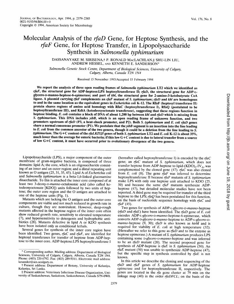
Lipopolysaccharide (LPS), a major component of the outermembrane of gram-negative bacteria, is composed of threedomains: lipid A; the core, which is an oligosaccharide consist-ing of an inner and an outer region; and a distal repeating unitknown as 0 antigen (25, 31, 35, 45). Lipid A of Escherichia coliand Salmonella typhimurium is a beta-1,6-linked glucosaminedisaccharide. To this is attached the inner core composed of atleast two 3-deoxy-D-manno-octulosonic acid (also called ke-todeoxyoctonate [KDO]) units followed by two units of hep-tose; the outer core region and the 0 antigen are attached toone of the heptose units (Fig. 1).
Mutants which are lacking the 0 antigen and the outer corecomponents are viable and not much reduced in growth rate inculture, though they are nonvirulent. However, deep-roughmutants affected in the heptose region of the inner core oftenshow reduced growth rate, sensitivity to elevated temperature(7), and hypersensitivity to detergents and hydrophobic anti-biotics (28). Mutants defective in lipid A or KDO synthesishave been isolated only as conditional lethals.
Several genes for synthesis of the inner core region havebeen identified. Two genes, rfaC and rfaF, are identified forheptosyl transferases for transfer of heptose from ADP-hep-tose to the inner core. ADP-heptose:LPS heptosyltransferase I
* Corresponding author. Mailing address: Department of BiologicalSciences, University of Calgary, Calgary, Alberta, Canada T2N 1N4.Phone: (403) 220-6792. Fax: (403) 289-9311. Electronic mail address:[email protected].
t Present address: Department of Botany, University of Kelaniya,Kelaniya, Sri Lanka.
t Present address: Veterinary Infectious Disease Organization, Uni-versity of Saskatchewan, Saskatoon, Saskatchewan, Canada S7N-OWO.
(hereafter called heptosyltransferase I) is encoded by the rfaCgene; an rfaC mutant of S. typhimurium, which does nottransfer heptose from ADP-heptose to lipid-(KDO)2-IVA, wascomplemented by the cloned gene (47). rfaC was also clonedfrom E. coli (8). The gene rfaF was inferred to determineheptosyltransferase II because rfaF mutants of S. typhimuriummake LPS with only one heptose unit attached to KDO (25,50) and because the same rfaF mutants synthesize ADP-heptose (47), but detailed molecular studies have not beenreported. A third gene may be required for transfer of the thirdheptose to the LPS; rfaQ has been postulated for this function,on the basis of nucleotide sequence homology with rfaC andrfaF (45).Two genes for synthesis of ADP-L-glycero-D-manno-heptose
(rfaD and rfaE) have been identified. The rfaD gene of E. coliencodes ADP-L-glycero-D-manno-heptose-6-epimerase, whichconverts ADP-D-glycero-D-manno-heptose to ADP-L-glycero-D-manno-heptose (9, 30); rfaD is also known as htrM and isrequired for viability of E. coli at high temperature (32).(Hereafter we refer to this gene as rfaD and to the enzyme asheptose epimerase.) A mutant of S. typhimurium produces LPScontaining some D-glycero-D-manno-heptose and was inferredto be an rfaD mutant (20). The second proposed gene forsynthesis of ADP-heptose is rfaE in S. typhimurium (50). AnrfaE mutant (50) was unable to synthesize ADP-heptose (47),but the specific step in synthesis controlled by rfaE is notknown.
In this article we describe the cloning and sequencing of therfaD and rfaF genes of S. typhimurium for ADP-heptoseepimerase and for heptosyltransferase II, respectively. Thegenes are located in the rfa gene cluster at 79 min on thelinkage map (40) in the order rfaDFCL; on the basis of the
2379
Vol. 176, No. 8
on June 19, 2018 by guesthttp://jb.asm
.org/D
ownloaded from
2380 SIRISENA ET AL.
FIG. 1. Proposed stereochemistry of the inner core of LPS of E. coli and S. typhimurium, adapted from Raetz (31). The alpha,1-5 bond betweenthe innermost heptose (residue I) and KDO is formed by heptosyltransferase I (rfaC protein); the alpha,1-3 bond between the second heptose(residue II) and the innermost heptose is formed by heptosyltransferase II (rfaF protein). Possible partial substitutions are indicated with dashedbonds. The outer core region is attached through a glucose residue to heptose (residue II).
orientation of the open reading frames (ORFs) they are
transcribed clockwise on the chromosome in the above order.In S. typhimurium the gene kbl, which determines glycineacetyltransferase, is adjacent to rfaD and independently tran-scribed counterclockwise. In E. coli K-12, gene organization issimilar except for a 1.2-kb insertion containing a 1-kb ORFcalled yibB or 26K between rfaD and kbl.
MATERIALS AND METHODS
Strains and bacteriological methods. The strains used in thisstudy are summarized in Table 1; all are S. typhimurium LT2unless indicated otherwise. The strains were maintained at- 70°C in 15% glycerol as described previously (43). Cells wereroutinely cultured in L broth (21), Luria-Bertani broth, or
2XYT medium (27). Antibiotics when required were added atthe following final concentrations (in micrograms per millili-ter): ampicillin, 50; tetracycline, 25; streptomycin, 200. Modi-fied minimal Davis medium was the defined medium (42). Forsolid media, Difco Bacto Agar was added to a final concentra-tion of 1.5%. Incubation was at 37°C unless stated otherwise.For screening the lambda library, cells were grown in lambdaagar (11). M9 medium (27) supplemented with 1% glucose,and 1% methionine assay medium (Difco) was used for proteinradiolabelling.
Genetic transformation. Transformation was by calciumchloride-heat shock methods of MacLachlan and Sanderson(24) or by electrotransformation methods of Dower et al. (12)as modified by Binotto et al. (2).The genomic library. The source of cloned genes was a
genomic library, consisting of S. typhimurium LT2 DNA par-tially digested with Sau3A and ligated into the BamHI site ofthe vector X1059. The library was obtained from R. Maurer(26) and is distributed from the Salmonella Genetic Stock
TABLE 1. Bacterial strains, plasmids, and bacteriophages
Strain, plasmid, or Genotype or description Referencebacteriophage or source
StrainsS. typhimuniumLB51010 metA22 metE551 trpC2 leu3121 5
ilv412 rpsL120 galE856 hsdL6hsdA29 hsdB121 xyl404 Hi-b H2e,n,x fla-66 nml (Fels 2)
SA3617 rfaF511/F'proA+B+ laclqlacZAM15 zzf::TnJO
SA3670 rfaF511 recAl srl-202::TnlOSA3770 rfa +SL3789 rfaF511
E. coli K-12JM109 recAI gyrA96 thi hsdR17 supE44
relA1 X A(lac-proAB)/F'proA+B+ lacl4 lacZAM15
DLT111 HfrC lambda srl::TnJO 49recA/pGP1-2
Plasmid pBluescriptKS+ ApR lacZ (3 kb)a Stratagene
BacteriophagesP22c2 Clear-plaque mutant of P22, 50
which adsorbs to S. typhimuriumwith 0 antigen
Felix 0 (FO) Virulent phage of S. typhimurium 50which requires a complete corefor adsorbtion
Ffm Lyses E. coli K-12 and rough 50mutants of S. typhimurium
a Also designated M13+.
J. BAc-rERIOL.
on June 19, 2018 by guesthttp://jb.asm
.org/D
ownloaded from
rfaD AND rfaF IN LPS SYNTHESIS IN S. TYPHIMURIUM 2381
5+i 110
Kb1,5
727Ev
R Ev _=-() c s Nlr ; B |/H H B E E H B
pT3 pT7 pKZ 106pT7 | p pT3 pKZ107pT3 _B pKZ108 (106)Nr_C pKZ1O9(106)H in: pKZ110(107)sacc pKZ113(107)Ev_B pKZ154(108)
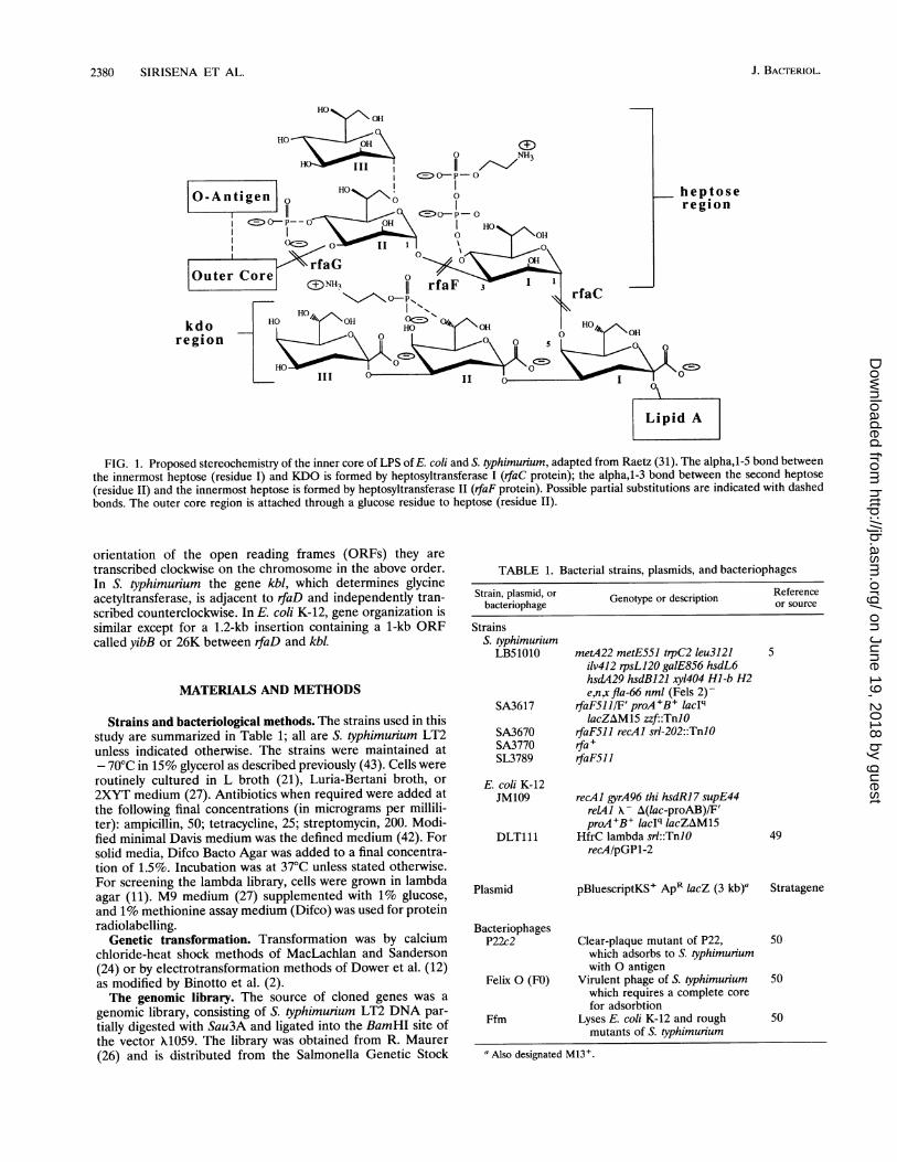
FIG. 2. Structure of the chromosome in the rfa region at 79 min on the linkage map of S. typhimunium and isolation of plasmids carrying genesin the rfaDF region. (A) The cysE genes are to the left (counterclockwise on the circular linkage map), and thepyrE genes are to the right (clockwiseon the linkage map) (40). The region is illustrated from the ClaI site at 0 kb to the BglII site at 25 kb. The left side of the region was determinedby probing digests of S. typhimunium genomic DNA with the EcoRV-BglII fragment from the left end of pKZ84 (47) (solid bar at 10 kb). The rightside was determined from plasmids pKZ84 (47); pKZ38 (23); and pKZ15, -26, and -27 (14). (B) Partial restriction map showing the positions ofcleavage sites above the line: C, ClaI; S, Sall; Nr, NruI; H, HindIII; Sac, Sacl; Ev, EcoRV; R, RsaI; B, BglII; E, EcoRI. Below the line the positionsof known rfa genes are shown. Genes in boxes (between C and I) are cloned (see plasmids in panel A) and previously sequenced (Figure 4). Genesin parentheses were cloned on the plasmids in panel A, but the nucleotide sequence is not known. The sequences of kbl, rfaD, and rfaF are fromthis study. (C) The DNA from a plaque from a library of S. typhimurium fragments cloned into X1059 (26) was cloned into pBluescriptKS+ in bothorientations in pKZ106 and -107, as illustrated by pT3 and pT7, the T3 and T7 promoters in pBluescript. These plasmids complement a mutationrfaF to restore normal LPS (+); -, failure to complement. Deletions of the plasmids were made to construct the plasmids from pKZ108 topKZ154. The restriction sites in the insert DNA are shown; the other restriction site is in the vector. The source of DNA for constructing theplasmid is indicated in parentheses after the name of the plasmid (e.g., pKZ108 was constructed from pKZ106 by digestion of the BglII site in theinsert and the BamHI site in the vector and religated).
Centre, University of Calgary. Phage carrying rfa genes weredetected as described earlier (47).
Gel electrophoresis of LPS. LPS isolation, proteinase Kdigestion, and silver staining of gels was as described byHitchcock and Brown (13). LPS was separated in 12% acryl-amide gels containing sodium dodecyl sulfate (SDS) (19) byusing Tris glycine buffer, pH 8.3.Plasmid isolation and cloning, DNA sequencing, and pro-
tein labelling. Plasmid isolation and cloning, DNA sequencing,and protein labelling methods were as described by Sirisena etal. (47). DNA sequence was determined by the dideoxy chaintermination method (44). The strains, constructed by trans-forming E. coli JM109 with plasmids (pBluescriptKS+ deriva-tives), were infected with helper phage R408 to prepare asingle-stranded template according to a protocol of StratageneInc. Sequenase and reagents from U.S. Biochemical Corp.were used for sequencing reactions. The DNA sequences wereanalyzed with the Genetics Computer Group software pack-age.T7 promoter-mediated gene expression. T7 promoter-medi-
ated gene expression was done by using plasmid pGP1-2 instrain DLT111 according to the methods of Tabor and Rich-ardson (49) as described earlier (47).
RESULTS
Cloning the rfaF and rfaD genes. The following plasmidscarry rfa genes of S. typhimurium at the clockwise or pyrE endof the rfa cluster: pKZ84 (rfaC) (47); pKZ38 (rfaLKZY) (23);and pKZ15, -26, and -27 (rfaJIBG) (14). A partial restrictionmap derived from analysis of these plasmids and the locationof the genes in this region is shown in the 10- to 25-kb regionin Fig. 2A and B. None of these plasmids complemented rfaF
or rfaD mutations. Transduction data (18, 41) indicated thatrfaD and rfaF are at the left end of the rfa genes (Fig. 2B),closest to cysE. The 0.5-kb EcoRV-BglII fragment from rfaF(solid bar in Fig. 2B) was used to probe digests of genomicDNA of S. typhimurium LT2 (data not shown), and thegenomic map in Fig. 2B from 0 to 10 kb was established. Alibrary of Sau3A fragments of S. typhimurium genomic DNAcloned into the BamHI site of X1059 (26) was probed with theabove rfaF fragment, and a 7.8 kb Clal fragment from apositive clone was ligated into the Clal site of pBS(KS)+ andtransformed into E. coli JM109. Two plasmids (pKZ106 and-107) which have the same 7.8-kb insert but in oppositeorientations were isolated (Fig. 2C). pKZ106 and -107 weretransformed into LB5010, a restriction-deficient strain of S.typhimunium, and DNA from these derivatives was used totransform other S. typhimurium strains. Both plasmids comple-mented the rfaF mutation in S. typhimurium, restoring thesmooth phenotype (data not shown). Deletions of these plas-mids were constructed by digestion with restriction enzymeswhich cut once in the multiple cloning site of the vector andone or more times in the insert; these plasmids are illustratedin Fig. 2C. These deletions show that rfaF complementationrequires the SacI-ClaI region at the right end. pKZ113 restoresnear wild-type LPS to SA3670 (rfaF511 recAl). The comple-mented strain has the same phage sensitivity as an rfa+ strain(P22S FOS Ffmr), is resistant to the hydrophobic agents sodiumdeoxycholate and nafcillin (Table 2), and has the wild-type LPSprofile on SDS-polyacrylamide gel electrophoresis (SDS-PAGE) (Fig. 3), but the strain is slightly more sensitive tonovobiocin than an rfa + strain (Table 2), indicating thatcomplementation is not quite complete.
Nucleotide sequence of the segment carrying rfiaF and rfiaD.The nucleotide sequence was derived from plasmids pKZ106,pKZ108, and pKZ154 as described in Materials and Methods.
cysE(A)
20 25 pyrE
(C) rfaF
|w7^ 7
VOL. 176, 1994
on June 19, 2018 by guesthttp://jb.asm
.org/D
ownloaded from

2382 SIRISENA ET AL.
TABLE 2. Complementation of a mutation in rfaF by hybrid plasmids carrying rfa genes
Sensitivity to hydrophobic agents"Sensitivity to phagec: MIC Inferred
Straina Partial genotype Inhibition zone (mm) pLg/ml) complementationby plasmid
P22 FO Ffm NaDoc Novo Naf Novo(400 p.g)' (30 p.g) (400 p.g)
SL3770 rfa + + + - 0 0 0 256SL3789 rfaF511 - - + S 5.5 NTf 1SA3670 rfaF511 recAl - - + 4 7 2.5 1SA3617 rfaF511 F'lacq - - + 2.5 7 3 1DS318 rfaF511JpKZ106 + + - R NT NT NT YesDS319 rfaF511 F'lacI4/pKZ107 + + - R NT NT NT YesSA3724 rfaF511 recAl/pKZ113 + + - 0 1 0 32 Yes
a See Table 1.b The complete genotype is in Table 1.c Determined by placing a drop of phage suspension (with about 5 x 10⁶ PFU of phage) on a lawn of sensitive cells on L agar. +, lysis; -, no lysis.dDetermined with disks containing the indicated amount of the compound. These disks were placed on the bacterial lawns prepared with cultures grown to mid-log
phase, incubated at 37'C, and the size of the inhibition zone was measured from the edge of the disk to the edge of the zone of inhibition. NaDoc, sodium deoxycholate;Novo, novobiocin; Naf, nafcillin. The MIC was the highest concentration of antibiotic in which the cells grew in Luria-Bertani broth.
S, inability to grow on L agar containing 0.1% sodium deoxycholate (sensitivity); R, ability to grow (resistance).f NT, not tested.
For S. typhimurium, the region from bp 1 to 2070, shown as ashaded box in Fig. 4, is previously unreported data from thegenes kbl, rfaD, and rfaF (described in'GenBank as STYRFADF, accession no. U06472). By linking previously reporteddata from the 3' end of rfaF to the right, as illustrated in Fig.4, a continuous meld of' ca. 10,162 bp was constructed asdescribed in the legend to Fig. 4. A meld of sequence over thesame' region of E. coli was assembled by Kenn Rudd at theNational Library of Medicine, National Institutes of Health(also shown in Fig. 4).The rfaF gene in S. typhimurium is 80% identical to the E.
coli rfaF at the nucleotide level and 95% similar at the aminoacid level (Fig. 4 and 5). Just upstream in E. coli is rfaD,previously identified by LPS and enzymatic studies (30); the S.typhimurium ORF in the same location has an identical size
FIG. 3. LPS of strains of S. typhimurium, separated by SDS-PAGE.LPS was extracted, separated, and silver stained as described inMaterials and Methods. Strains used and their partial genotypes:SA3724, rfaF511 recA1IpKZ113(rfaF+) (lane 1), SA3670, rfaF51recAl (lane 2), SL3770, rfa+ (lane 3).
(310 amino acids) and 86% identical and 97% homologous atthe nucleotide and amino acid levels, respectively.
Expression of the rfa genes in the T7 gene expressionsystem. Plasmids were transformed into strain DLT111 carry-ing plasmid pGP1-2, and protein expression in the presence ofrifampin, under the control of the T7 promoter, was measured.Plasmid pKZ113, which complements an rfaD mutant (Table2), carries the entire rfaF gene but only the 3' part of rfaD. Thisplasmid expresses a 36-kDa protein under control of the T7promoter; 'the size of the RfaF protein determined fromnucleotide sequence data is 39,000 Da (data not shown).
DISCUSSION
The following data indicate that the ORF upstream of rfaC(to the left in Fig. 6) is the rfaF gene, coding for heptosyltrans-ferase II. (i) Plasmid pKZ113, which carries the SacI-ClaIfragment cloned into pBluescriptKS+, has the complete rfaFORF but only the 3' part of rfaD (Fig. 2); this plasmid givesalmost complete complementation of an rfaF mutant (Table 2;Fig. 3). (ii) pKZ154, deleted for part of the ORF, did notcomplement rfaF. (iii) Plasmid pKZ113 produced a proteinwith gel mobility equivalent to 36 kDa, close to the sizeinferred from the nucleotide sequence. This gene shows 80%identity at the nucleotide level and 95% similarity at the aminoacid level (Fig. 4) to the equivalent E. coli sequence (45),indicating that they are homologous genes.The identity of rfaD as the structural gene for heptose-6-
epimerase is by sequence homology (86% at the nucleotidelevel) (Fig. 4) and location (upstream of rfaF) with rfaD of E.coli, which was shown earlier to complement an rfaD mutantand to be deficient in epimerase activity in cell extracts (30).Stein et al. (48) reported an rfaF-like mutant of Neisseriagonorrhoeae with properties resembling those of rfaF whichwas complemented by a cloned gene from N. gonorrhoeae; theyalso reported a mutant resembling rfaD.RfaC (heptosyltransferase I) and RfaF (heptosyltransferase
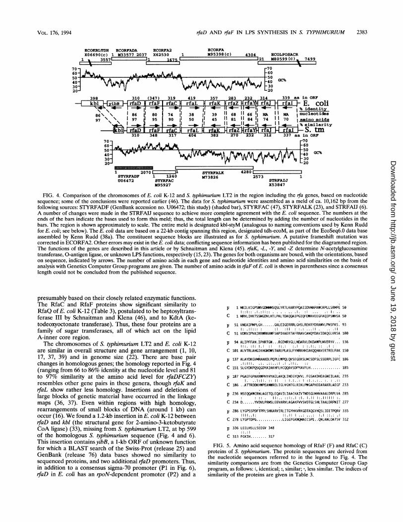
II) proteins show 90 and 95% similarity indices between S.typhimurium and E. coli (Table 3). In addition, the RfaC andRfaF proteins of S. typhimurium share regions of sequencehomology (Fig. 5), with a 49% similarity index (Table 3),
J. BACTERIOL.
on June 19, 2018 by guesthttp://jb.asm
.org/D
ownloaded from
rfaD AND rfaF IN LPS SYNTHESIS IN S. TYPHIMURIUM 2383
ECOKBLTDH ECORFADA ECORFA2X06690(c) 1 M33577 2037 X62530 I
1 WA 3557' 1675 C
70-
50-
30-
398 310 (347)
86\ \ I 86 I 8097 \ \ 97 d 95
310 38rfaF _310 348
319rfaC l
74 190 1
31770
50- -wo I4030120
ECORFA1 M95398(c)
419rfaL
3850
4a0404
357rfaK
3945
rfaK382
4304 ECOLPOSACR-121 M80599(C) 7499
-70F60
283 232 314 339 aa in ORFrfaZ rfa rf rfai E. coli114-I4--~ 4 i 4- 1% identity68 11 66 1 NA NA 1nucleotides
11 11 84 11 74 1 70 Iamino acids|. ! ! !~~1 !8,%simxilarity
hfaHrfarrfa-jE-HiruI!s S. tm270 232 312 337 aa in ORF
F70-60C_cn
4i GC%30
20
1 2070 L JLSTYRFADF 1 1240U06472 STYRFAC
M95927
STYRFALK 42850873826 2573 1
STRFAIJX53847
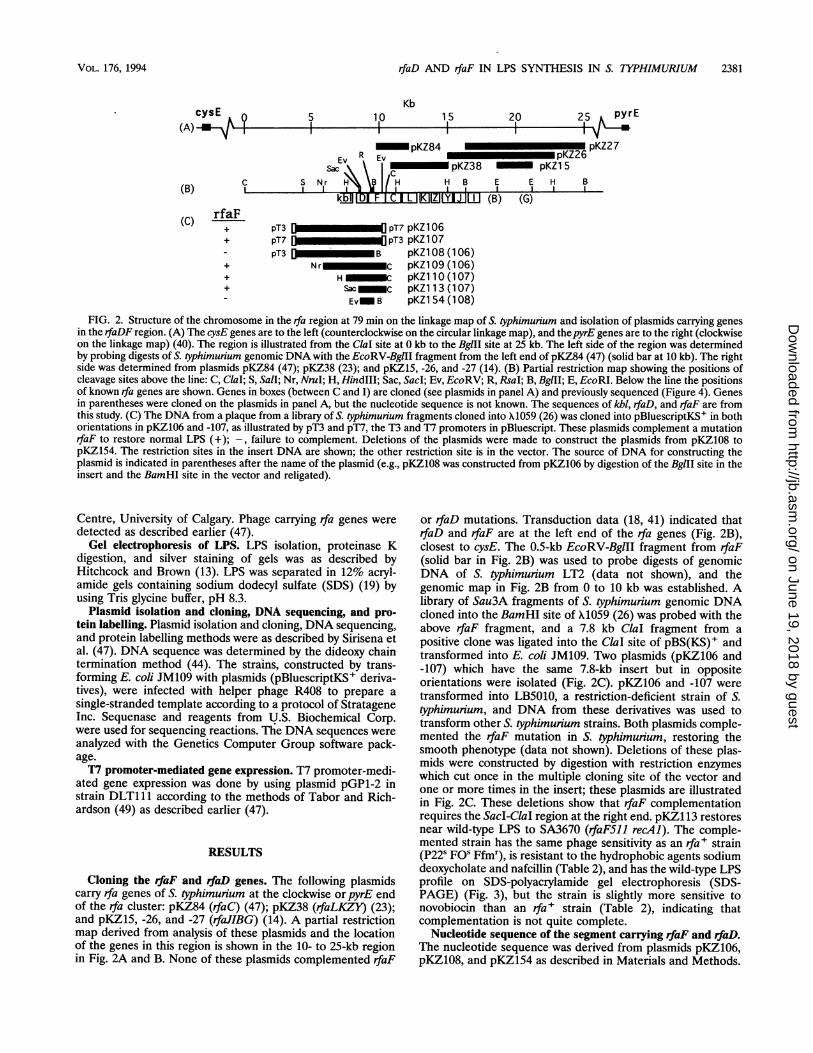
FIG. 4. Comparison of the chromosomes of E. coli K-12 and S. typhimurium LT2 in the region including the rfa genes, based on nucleotidesequence; some of the conclusions were reported earlier (46). The data for S. typhimurium were assembled as a meld of ca. 10,162 bp from thefollowing sources: STYRFADF (GenBank accession no. U06472; this study) (shaded bar), STYRFAC (47), STYRFALK (23), and STRFAIJ (6).A number of changes were made in the STRFAIJ sequence to achieve more complete agreement with the E. coli sequence. The numbers at theends of the bars indicate the bases used to form this meld; thus, the total length can be determined by adding the number of nucleotides in thebars. The region is shown approximately to scale. The entire meld is designated kbl-styM (analogous to naming conventions used by Kenn Ruddfor E. coli; see below). The E. coli data are based on a 22-kb contig spanning this region, designated tdh-ecoM, as part of the EcoSeq6.0 data baseassembled by Kenn Rudd (38a). The constituent sequence blocks are illustrated as for S. typhimurium. A putative frameshift mutation wascorrected in ECORFA2. Other errors may exist in the E. coli data; conflicting sequence information has been published for the diagrammed region.The functions of the genes are described in this article or by Schnaitman and Klena (45). rfaK, -L, -Y, and -Z determine N-acetylglucosaminetransferase, 0-antigen ligase, or unknown LPS functions, respectively (15, 23). The genes for both organisms are boxed, with the orientations, basedon sequence, indicated by arrows. The number of amino acids in each gene and nucleotide identities and amino acid similarities on the basis ofanalysis with Genetics Computer Group programs are given. The number of amino acids in rfaF of E. coli is shown in parentheses since a consensuslength could not be concluded from the published sequence.
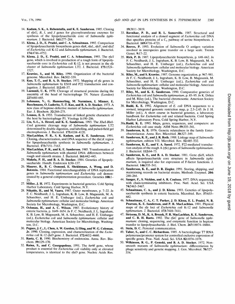
presumably based on their closely related enzymatic functions.The RfaC and RfaF proteins show significant similarity toRfaQ of E. coli K-12 (Table 3), postulated to be heptosyltrans-ferase III by Schnaitman and Klena (46), and to KdtA (ke-todeoxyoctonate transferase). Thus, these four proteins are afamily of sugar transferases, all of which act on the lipidA-inner core region.The chromosomes of S. typhimurium LT2 and E. coli K-12
are similar in overall structure and gene arrangement (1, 10,17, 37, 39) and in genome size (22). There are base pairchanges in homologous genes; the homology reported in Fig. 4(ranging from 66 to 86% identity at the nucleotide level and 81to 97% similarity at the amino acid level for rfaDFCZY)resembles other gene pairs in these genera, though rfaK andrfaL show rather less homology. Insertions and deletions oflarge blocks of genetic material have occurred in the linkagemaps (36, 37). Even within regions with high homology,rearrangements of small blocks of DNA (around 1 kb) canoccur (16). We found a 1.2-kb insertion in E. coli K-12 betweenrfaD and kbl (the structural gene for 2-amino-3-ketobutyrateCoA ligase) (33), missing from S. typhimurium LT2, at bp 599of the homologous S. typhimurium sequence (Fig. 4 and 6).This insertion contains yibB, a 1-kb ORF of unknown functionfor which a BLAST search of the Swiss-Prot (release 25) andGenBank (release 76) data bases showed no similarity tosequenced proteins, and two additional rfaD promoters. Thus,in addition to a consensus sigma-70 promoter (P1 in Fig. 6),rfaD in E. coli has an rpoN-dependent promoter (P2) and a
F 1 MKILVIGPSWVGDMMMSQSLYRTLKARYPQAIIDVMAPAWCRPLLSRMPE 50
C 1 MRVLIVKTSSMGDVLHTLPALTDAQQAIPGIQFDWAVEEGFAQIPSWHSA 50
F 51 VNEAIPMPLGH ...... GALEIGERRRLGHSLREKRYDRAWVLPNSFKS. 931: .11::: :1 :11 :.: . 11 .. .11
C 51 VDRVIPVAIRRWRKAWFSAPIKAERTAFRRAVCANQYDAVIDAQGLVKSA 100
F 94 ALIPFFAN.IPHRTGW... RGEMRYGLLNDARVLDKDAWPLMVERYV... 136I1:. :1: 1.1 :1 1:.: :.1 :.I:. :: .1:
C 101 ALVTRLAHGIKHGMDWSTAREPLASLFYNRKHHIAKQQHAVERTRELFAK 150
F 137 ALAYDKGVMRAAKDLPQPLLWPQLQVSEGEKSLMCSDFSLSSERPLIGFC 186.1:111..:.:.: .1.:. :
C 151 SLGYDKPQSQGDYAIAKHFLHCQQAVSDPYAVFLH............... 185
F 187 PGAEFGPAKRWPHYHYAELAKQLINEGYQVVL .FGSAKDHEAGNEILAAL 235
C 186 .ATTRDDKHWPEANWRELIGLVGNTGLRIKLPWGAPHEEARAKRLAEGF 233
F 236 NSEQQAWCRNLAGETQLEQAVILIAACKAIVTNDSGLMHVAAALDRPLVA 285
C 234 D.....YVDVLPRMSLEEVARVLAGAKFVVSVDTGLSHLTAALDRPNIT 277
F 286 LYGPSSPDFTPPLSHKARVIRLITGYHKVRKGDTAQGYHQSLIDITPQRV 3351111..1: 11.11 1 ..: ...: 1.1 ::.: .1
C 278 LYGPTDPG ........... LIGGYGKNQMACCSPE. .QNLANLDATSV 312
F 336 LEELHSLLSEEGV 348
C 313 FGKIH........ 317
FIG. 5. Amino acid sequence homology of RfaF (F) and RfaC (C)proteins of S. typhimurium. The protein sequences are derived fromthe nucleotide sequences referred to in the legend to Fig. 4. Thesimilarity comparisons are from the Genetics Computer Group Gapprogram, as follows: I, identical; :, similar; -, less similar. The indices ofsimilarity of the proteins are given in Table 3.
IGC%A~~vP~~~ L~3020
. - - 11
VOL. 176, 1994
on June 19, 2018 by guesthttp://jb.asm
.org/D
ownloaded from
2384 SIRISENA ET AL.
kbl rfaD479 5-589 599 600 35 610 620 10 640 0 650 660 670 SD 690 700S AAAATCCCCACGCATT6 .AATCACGCGTATCTCTC---------A CGGGTTATCAGTGAAT-GGATGAATAGTGTTATGATAAGAG-CATTCATGTCTGAGACAGTCTCTGACACCATAATTCAAAGGTTACAGTTATGATCATCGTTACCGGCGGS
I III// iI III Il lIIw AAATTCTCCACGLaTG .AATAAC GTGTAACTTGATGTCTCTCA -CTATGTAAAGGGCTGA-_-_AGGGATTC GGATG - -TGATGGTATGATACAGACATTC GTGTCTGAGATTGTCTC TGACTCCATAATTCGAAGGTTACAGTTATGATCATC GTTAC CGGCGG
3060 3170 3180 4410P 4420 4430 4440.1 0 4450 P0 4460 4470 4480 4490SD 4500 4510 4520kbl I-o.
rfaD
\\bBvCGCCATGAAGGACTAGCTAAAACC CAAACTATTGCAATTAGCATC4350 4360 4370 4380 4390 4400
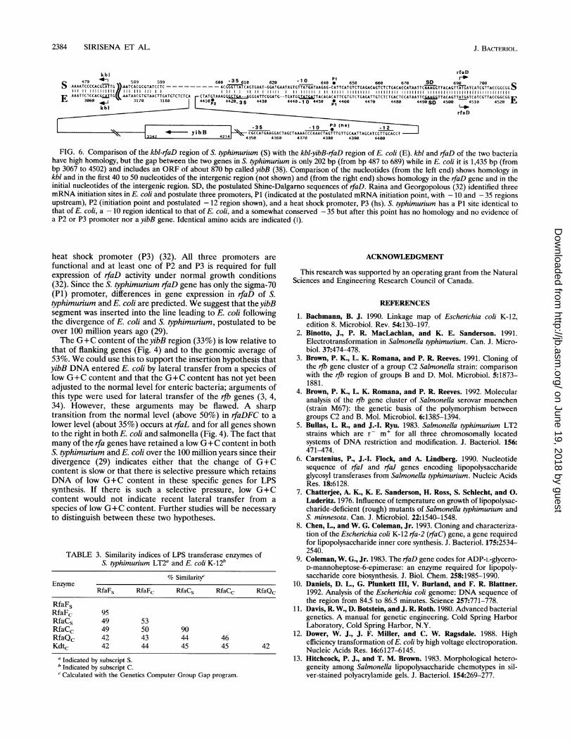
FIG. 6. Comparison of the kbl-rfaD region of S. typhimurium (S) with the kbl-yibB-rfaD region of E. coli (E). kbl and rfaD of the two bacteriahave high homology, but the gap between the two genes in S. typhimurium is only 202 bp (from bp 487 to 689) while in E. coli it is 1,435 bp (frombp 3067 to 4502) and includes an ORF of about 870 bp called yibB (38). Comparison of the nucleotides (from the left end) shows homology inkbl and in the first 40 to 50 nucleotides of the intergenic region (not shown) and (from the right end) shows homology in the rfaD gene and in theinitial nucleotides of the intergenic region. SD, the postulated Shine-Dalgarno sequences of rfaD. Raina and Georgopolous (32) identified threemRNA initiation sites in E. coli and postulate three promoters, P1 (indicated at the postulated mRNA initiation point, with - 10 and - 35 regionsupstream), P2 (initiation point and postulated - 12 region shown), and a heat shock promoter, P3 (hs). S. typhimurium has a P1 site identical tothat of E. coli, a - 10 region identical to that of E. coli, and a somewhat conserved - 35 but after this point has no homology and no evidence ofa P2 or P3 promoter nor a yibB gene. Identical amino acids are indicated (I).
heat shock promoter (P3) (32). All three promoters arefunctional and at least one of P2 and P3 is required for fullexpression of rfaD activity under normal growth conditions(32). Since the S. typhimurium rfaD gene has only the sigma-70(P1) promoter, differences in gene expression in rfaD of S.typhimurium and E. coli are predicted. We suggest that theyibBsegment was inserted into the line leading to E. coli followingthe divergence of E. coli and S. typhimurium, postulated to beover 100 million years ago (29).The G+C content of the yibB region (33%) is low relative to
that of flanking genes (Fig. 4) and to the genomic average of53%. We could use this to support the insertion hypothesis thatyibB DNA entered E. coli by lateral transfer from a species oflow G+C content and that the G+C content has not yet beenadjusted to the normal level for enteric bacteria; arguments ofthis type were used for lateral transfer of the rib genes (3, 4,34). However, these arguments may be flawed. A sharptransition from the normal level (above 50%) in rfaDFC to alower level (about 35%) occurs at rfaL and for all genes shownto the right in both E. coli and salmonella (Fig. 4). The fact thatmany of the rfa genes have retained a low G+C content in bothS. typhimurium and E. coli over the 100 million years since theirdivergence (29) indicates either that the change of G+Ccontent is slow or that there is selective pressure which retainsDNA of low G+C content in these specific genes for LPSsynthesis. If there is such a selective pressure, low G+Ccontent would not indicate recent lateral transfer from aspecies of low G+C content. Further studies will be necessaryto distinguish between these two hypotheses.
TABLE 3. Similarity indices of LPS transferase enzymes ofS. typhimurium LT2a and E. coli K-12b
% SimilaritycEnzyme
RfaFs RfaFc RfaCs RfaCc RfaQc
RfaFsRfaFc 95RfaCs 49 53RfaCc 49 50 90RfaQc 42 43 44 46Kdtc 42 44 45 45 42
aIndicated by subscript S.b Indicated by subscript C.c Calculated with the Genetics Computer Group Gap program.
ACKNOWLEDGMENT
This research was supported by an operating grant from the NaturalSciences and Engineering Research Council of Canada.
REFERENCES
1. Bachmann, B. J. 1990. Linkage map of Escherichia coli K-12,edition 8. Microbiol. Rev. 54:130-197.
2. Binotto, J., P. R. MacLachlan, and K. E. Sanderson. 1991.Electrotransformation in Salmonella typhimurium. Can. J. Micro-biol. 37:474-478.
3. Brown, P. K., L. K. Romana, and P. R. Reeves. 1991. Cloning ofthe rib gene cluster of a group C2 Salmonella strain: comparisonwith the rjb region of groups B and D. Mol. Microbiol. 5:1873-1881.
4. Brown, P. K., L. K. Romana, and P. R. Reeves. 1992. Molecularanalysis of the rib gene cluster of Salmonella serovar muenchen(strain M67): the genetic basis of the polymorphism betweengroups C2 and B. Mol. Microbiol. 6:1385-1394.
5. Bullas, L. R., and J.-I. Ryu. 1983. Salmonella typhimurium LT2strains which are r- m+ for all three chromosomally locatedsystems of DNA restriction and modification. J. Bacteriol. 156:471-474.
6. Carstenius, P., J.-I. Flock, and A. Lindberg. 1990. Nucleotidesequence of rfaI and rfaJ genes encoding lipopolysaccharideglycosyl transferases from Salmonella typhimurium. Nucleic AcidsRes. 18:6128.
7. Chatterjee, A. K., K. E. Sanderson, H. Ross, S. Schlecht, and 0.Luderitz. 1976. Influence of temperature on growth of lipopolysac-charide-deficient (rough) mutants of Salmonella typhimurium andS. minnesota. Can. J. Microbiol. 22:1540-1548.
8. Chen, L., and W. G. Coleman, Jr. 1993. Cloning and characteriza-tion of the Escherichia coli K-12 rfa-2 (rfaC) gene, a gene requiredfor lipopolysaccharide inner core synthesis. J. Bacteriol. 175:2534-2540.
9. Coleman, W. G., Jr. 1983. The rfaD gene codes for ADP-L-glycero-D-mannoheptose-6-epimerase: an enzyme required for lipopoly-saccharide core biosynthesis. J. Biol. Chem. 258:1985-1990.
10. Daniels, D. L., G. Plunkett III, V. Burland, and F. R. Blattner.1992. Analysis of the Escherichia coli genome: DNA sequence ofthe region from 84.5 to 86.5 minutes. Science 257:771-778.
11. Davis, R. W., D. Botstein, and J. R. Roth. 1980. Advanced bacterialgenetics. A manual for genetic engineering. Cold Spring HarborLaboratory, Cold Spring Harbor, N.Y.
12. Dower, W. J., J. F. Miller, and C. W. Ragsdale. 1988. Highefficiency transformation of E. coli by high voltage electroporation.Nucleic Acids Res. 16:6127-6145.
13. Hitchcock, P. J., and T. M. Brown. 1983. Morphological hetero-geneity among Salmonella lipopolysaccharide chemotypes in sil-ver-stained polyacrylamide gels. J. Bacteriol. 154:269-277.
J. BACTERIOL.
on June 19, 2018 by guesthttp://jb.asm
.org/D
ownloaded from
rfaD AND rfaF IN LPS SYNTHESIS IN S. TYPHIMURIUM 2385
14. Kadam, S. K., A. Rehemtulla, and K. E. Sanderson. 1985. Cloningof rfaG, B, I, and J genes for glycosyltransferase enzymes forsynthesis of the lipopolysaccharide core of Salmonella typhi-murium. J. Bacteriol. 161:277-284.
15. Klena, J. D., E. Pradel, and C. A. Schnaitman. 1992. Comparisonof lipopolysaccharide biosynthesis genes rfaK, rfaL, rfaY, and rfaZof Escherichia coli K-12 and Salmonella typhimurium. J. Bacteriol.174:4746-4752.
16. Klena, J. D., E. Pradel, and C. A. Schnaitman. 1993. The rfaSgene, which is involved in production of a rough form of lipopoly-saccharide core in Escherichia coli K-12, is not present in the rfacluster of Salmonella typhimurium LT2. J. Bacteriol. 175:1524-1527.
17. Krawiec, S., and M. Riley. 1990. Organization of the bacterialgenome. Microbiol. Rev. 54:502-539.
18. Kuo, T.-T., and B. A. D. Stocker. 1972. Mapping of rfa genes inSalmonella typhimurium by ES18 and P22 transduction and con-
jugation. J. Bacteriol. 112:48-57.19. Laemmli, U. K. 1970. Cleavage of structural proteins during the
assembly of the head of bacteriophage T4. Nature (London)227:680-685.
20. Lehmann, V., G. Hammerling, M. Nurminen, I. Minner, E.Ruschmann, 0. Luderitz, T.-T. Kuo, and B. A. D. Stocker. 1973. Anew class of heptose-defective mutant of Salmonella typhimurium.Eur. J. Biochem. 32:268-275.
21. Lennox, E. S. 1955. Transduction of linked genetic characters ofthe host by bacteriophage P1. Virology 1:190-206.
22. Liu, S.-L., A. Hessel, and K. E. Sanderson. 1993. The XbaI-BlnI-CeuI genomic cleavage map of Salmonella typhimurium LT2determined by double digestion, end labelling, and pulsed-field gelelectrophoresis. J. Bacteriol. 175:4104-4120.
23. MacLachlan, P. R., S. K. Kadam, and K. E. Sanderson. 1991.Cloning, characterization, and DNA sequence of the rfaLK regionfor lipopolysaccharide synthesis in Salmonella typhimurium. J.Bacteriol. 173:7151-7163.
24. MacLachian, P. R., and K. E. Sanderson. 1985. Transformation ofSalmonella typhimurium with plasmid DNA: differences betweenrough and smooth strains. J. Bacteriol. 161:442-445.
25. Makela, P. H., and B. A. D. Stocker. 1984. Genetics of lipopoly-saccharide. Handb. Endotoxin 1:50-137.
26. Maurer, R., B. C. Osmond, E. Shekhtman, A. Wong, and D.Botstein. 1984. Functional interchangeability of DNA replicationgenes in Salmonella typhimurium and Escherichia coli demon-strated by a general complementation procedure. Genetics 108:1-23.
27. Miller, J. H. 1972. Experiments in bacterial genetics. Cold SpringHarbor Laboratory, Cold Spring Harbor, N.Y.
28. Nikaido, H., and M. Vaara. 1987. Outer membranes, p. 7-22. InF. C. Neidhardt, J. L. Ingraham, K. B. Low, B. Magasanik, M. A.Schaechter, and H. E. Umbarger (ed.), Eschenichia coli andSalmonella typhimurium: cellular and molecular biology. AmericanSociety for Microbiology, Washington, D.C.
29. Ochman, H., and A. C. Wilson. 1987. Evolutionary history ofenteric bacteria, p. 1649-1654. In F. C. Neidhardt, J. L. Ingraham,K. B. Low, B. Magasanik, M. A. Schaechter, and H. E. Umbarger(ed.), Escherichia coli and Salmonella typhimurium: cellular andmolecular biology. American Society for Microbiology, Washing-ton, D.C.
30. Pegues, J. C., L. Chen, A. W. Gordon, Li Ding, and W. G. Coleman,Jr. 1990. Cloning, expression, and characterization of the Esche-richia coli K-12 rfaD gene. J. Bacteriol. 172:4652-4660.
31. Raetz, C. R. 1990. Biochemistry of endotoxins. Annu. Rev. Bio-chem. 59:129-170.
32. Raina, S., and C. Georgopolous. 1991. The htrM gene, whoseproduct is essential for Escherichia coli viability only at elevatedtemperatures, is identical to the rfaD gene. Nucleic Acids Res.
19:3811-3819.33. Ravnikar, P. D., and R. L. Somerville. 1987. Structural and
functional analysis of a cloned segment of Escherichia coli DNAthat specifies proteins of a C4 pathway of serine biosynthesis. J.Bacteriol. 169:4716-4721.
34. Reeves, P. 1993. Evolution of Salmonella 0 antigen variationinvolved in interspecies gene transfer on a large scale. TrendsGenet. 9:17-22.
35. Rick, P. D. 1987. Lipopolysaccharide biosynthesis, p. 648-662. InF. C. Neidhardt, J. L. Ingraham, K. B. Low, B. Magasanik, M. A.Schaechter, and H. E. Umbarger (ed.), Escherichia coli andSalmonella typhimurium: cellular and molecular biology. AmericanSociety for Microbiology, Washington, D.C.
36. Riley, M., and S. Krawiec. 1987. Genome organization, p. 967-981.In F. C. Neidhardt, J. L. Ingraham, K. B. Low, B. Magasanik, M.Schaechter, and H. E. Umbarger (ed.), Escherichia coli andSalmonella typhimurium: cellular and molecular biology. AmericanSociety for Microbiology, Washington, D.C.
37. Riley, M., and K. E. Sanderson. 1990. Comparative genetics ofEscherichia coli and Salmonella typhimurium, p. 85-95. In K. Drlicaand M. Riley (ed.), The bacterial chromosome. American Societyfor Microbiology, Washington, D.C.
38. Rudd, K. E. 1992. Alignment of E. coli DNA sequences to arevised, integrated genomic restriction map, p. 2.3-2.43. In J. H.Miller (ed.), A short course in bacterial genetics, a laboratoryhandbook for Escherichia coli and related bacteria. Cold SpringHarbor Laboratory Press, Cold Spring Harbor, N.Y.
38a.Rudd, K. E. 1993. Maps, genes, sequences, and computers: anEscherichia coli case study. ASM News 59:335-341.
39. Sanderson, K. E. 1976. Genetic relatedness in the family Enter-obacteriaceae. Annu. Rev. Microbiol. 30:327-349.
40. Sanderson, K. E., and J. R. Roth. 1988. Linkage map of Salmonellatyphimurium, edition VII. Microbiol. Rev. 52:485-532.
41. Sanderson, K. E., and Y. A. Saeed. 1972. P22-mediated transduc-tion analysis of the rough A (rfa) genes of Salmonella typhimurium.J. Bacteriol. 112:64-73.
42. Sanderson, K. E., and B. A. D. Stocker. 1981. Gene rfaH, whichaffects lipopolysaccharide core structure in Salmonella typhi-murium, is required also for expression of F-factor functions. J.Bacteriol. 146:535-541.
43. Sanderson, K. E., and D. R. Ziegler. 1991. Storing, shipping, andmaintaining records on bacterial strains. Methods Enzymol. 204:248-264.
44. Sanger, F., S. Nicklen, and A. R. Coulson. 1977. DNA sequencingwith chain-terminating inhibitors. Proc. Natl. Acad. Sci. USA74:5463-5467.
45. Schnaitman, C. A., and J. D. Klena. 1993. Genetics of lipopoly-saccharide synthesis in enteric bacteria. Microbiol. Rev. 57:655-682.
46. Schnaitman, C. A., C. T. Parker, J. D. Klena, E. L. Pradel, N. B.Pearson, K. E. Sanderson, and P. R. MacLachlan. 1991. Physicalmaps of the rfa loci of Escherichia coli K-12 and Salmonellatyphimurium. J. Bacteriol. 173:7410-7411.
47. Sirisena, D. M., K. A. Brozek, P. R. MacLachlan, K. E. Sanderson,and C. R. H. Raetz. 1992. The rfaC gene of Salmonella typhi-murium: cloning, sequencing, and enzymatic function in heptosetransfer to lipopolysaccharide. J. Biol. Chem. 267:18874-18884.
48. Stein, D. C. Personal communication.49. Tabor, S., and C. C. Richardson. 1985. A bacteriophage T7 RNA
polymerase/promoter system for controlled exclusive expression ofspecific genes. Proc. Natl. Acad. Sci. USA 82:1074-1078.
50. Wilkinson, R. G., P. Gemski, and B. A. D. Stocker. 1972. Non-smooth mutants of Salmonella typhimurium: differentiation byphage sensitivity and genetic mapping. J. Gen. Microbiol. 70:527-554.
VOL. 176, 1994
on June 19, 2018 by guesthttp://jb.asm
.org/D
ownloaded from