modulations in epidermal calcium regulate theexpression of differentiation-specific markers
TRANSCRIPT

ORIGINAL ARTICLE
Modulations in Epidermal Calcium Regulate theExpression of Di¡erentiation-Speci¢c Markers
Peter M. Elias,nw Sung K. Ahn,nz Mitsuhiro Denda,y Barbara E. Brown,n Debra Crumrine,n
Llewellyn K. Kimutai,n Laszlo K˛mˇves,n Seung H. Lee,z and Kenneth R. FeingoldnnDermatology and Medical Services,Veterans Administration Medical Center, and wDepartments of Dermatology and Medicine, University of CaliforniaSchool of Medicine, San Francisco, California; zDepartment of Dermatology,Yonsei University College of Medicine,Yongdong Severance Hospital, Seoul,Korea; and yShiseido Research Center,Yokohoma, Japan
Mammalian epidermis normally displays a distinctivecalcium gradient, with low levels in the basal/spinouslayers and high levels in the stratum granulosum.Although changes in stratum granulosum calcium reg-ulate the lamellar body secretory response to perme-ability barrier alterations, whether modulations incalcium also regulate the expression of di¡erentiation-speci¢c proteins in vivo remains unknown. As acute bar-rier perturbations reduce calcium levels in stratumgranulosum, we studied the regulation of murine epi-dermal di¡erentiation after loss of calcium accompany-ing acute barrier disruption and by exposure of suchacutely perturbed skin sites to either low (0.03 M) orhigh (1.8 M) calcium. Three hours after acute barrierdisruption, coincident with reduced calcium and ultra-structural evidence of accelerated lamellar body secre-tion, both northern analyses and in situ hybridizationrevealed decreased mRNA levels for loricrin, pro¢lag-grin, and involucrin in the outer epidermis, but protein
levels did not change signi¢cantly. Moreover, exposureof acutely disrupted skin sites to low calcium solutionssustained the reduction in mRNA levels, whereas expo-sure to high calcium solutions restored normal mRNAlevels (blocked by the L-type calcium channel inhibi-tor, nifedipine). Finally, with prolonged exposure to alow (o10% relative humidity) or high (480% relativehumidity) humidity, calcium levels increased and de-clined, respectively. Accordingly, mRNA and proteinlevels of the di¡erentiation-speci¢c markers increasedand decreased at low and high relative humidity, re-spectively. These results provide direct evidence thatacute and sustained £uctuations in epidermal calciumregulate expression of di¡erentiation-speci¢c proteinsin vivo, and demonstrate that modulations in epidermalcalcium coordinately regulate events late in epidermaldi¡erentiation that together form the barrier. Keywords:Ca channels/calcium/¢laggrin/involucrin/loricrin/permeabilitybarrier. J Invest Dermatol 119:1128 ^1136, 2002
Epidermal di¡erentiation is a vertically directed, vectorialprocess, with sequential expression of epidermis-speci¢c, structural proteins by suprabasal keratino-cytes, terminating in corni¢cation (Eckert et al, 1997).Involucrin, loricrin, and several other peptide precur-
sors are enzymatically crosslinked by transglutaminase 1 andother transglutaminases into a mechanically and chemically resis-tant sca¡old, the corni¢ed envelope (Polakowska and Goldsmith,1991; Reichert et al, 1993; Simon, 1994). Additional cytosolic pro-teins (e.g., ¢laggrin, keratins 1 and 10) also associate with the cor-ni¢ed envelope during terminal di¡erentiation (Steinert andMarekov, 1995). In cultured keratinocytes, most of these di¡eren-tiation-speci¢c proteins are regulated both at a transcriptionaland, in some cases, at a post-transcriptional level (e.g., transglu-taminase 1 and pro¢laggrin) by external calcium (Ca) levels(Hennings et al, 1980; Yuspa et al, 1989; Pillai et al, 1990; Resing
et al, 1993;Yamazaki et al, 1997). Under basal conditions, mammalianepidermis displays a Ca gradient, with low levels of Ca in thebasal and lower spinous layers, followed by an increase in extra-cellular and intracellular Ca that peaks in the stratum granulosum(SG) (Menon et al, 1985; Forslind et al, 1995; Mauro et al, 1998).Perturbations in permeability barrier function transiently alterthis epidermal Ca gradient, producing a steep decline in Ca levelsin the outer epidermis, followed by a restoration of Ca levels tonormal over 6^24 h in parallel with barrier recovery (Mao-Qianget al, 1997). Loss of Ca from the outer epidermis after acute barrierdisruption stimulates the secretion of a preformed pool of lamel-lar bodies (LBs) from the outermost SG cell, a response that facil-itates normal barrier recovery. Furthermore, using alternativemethods, we showed that changes in Ca in the outer epidermisdirectly regulate LB secretion, independent of barrier perturba-tion (Menon et al, 1994a; Lee et al, 1998), by a mechanism that in-volves Ca transport through L-type Ca2þ channels (Lee et al,1992).Whereas these studies show that modulations in epidermal Ca
regulate LB secretion, the increased levels of Ca that are presentnormally in the outer epidermis correlate spatially with the siteswhere epidermis generates late, di¡erentiation-speci¢c proteins,such as involucrin, ¢laggrin, and loricrin. Yet, whether modula-tions in epidermal Ca would dominate over a variety of other
Reprint requests to: Peter M. Elias, M.D., Dermatology Service (190),Veterans A¡airs Medical Centre, 4150 Clement Street, San Francisco, CA94121; Email: [email protected]: LB, lamellar body; RH, relative humidity; SG, stratum
granulosum;TEWL, transepidermal water loss.
Manuscript received March 28, 2002; accepted for publication June 13,2002; Manuscript accepted July 17, 2002.
0022-202X/02/$15.00 � Copyright r 2002 by The Society for Investigative Dermatology, Inc.
1128

endogenous factors that coregulate keratinocyte di¡erentiation(Eckert et al, 1997) is not known. Thus, despite repeated notationsto the likely relationship of these two phenomena, there is no di-rect evidence that links £uctuations in epidermal Ca levels withchanges in epidermal di¡erentiation.We provide here direct evi-dence that experimental alterations in epidermal Ca directly reg-ulate the expression of certain di¡erentiation-linked, keratinocytestructural proteins. Together with prior evidence for the stimula-tion of LB secretion by reductions in epidermal Ca, these studiesdemonstrate that barrier-induced £uctuations in epidermal Cacoordinately, but divergently, regulate the two major di¡erentia-tion programs that generate a normal stratum corneum: (i) thelipid-enriched extracellular matrix; and (ii) the corneocyte.
METHODS
Experimental perturbations The epidermal Ca gradient was disruptedin 6^8-wk-old, male hairless mouse skin (Charles River Laboratories,Philadelphia, PA) by either repeated cellophane tape stripping or acetonewipes until transepidermal water loss (TEWL) levels were 4 mg per cm2
per h, while animals were under general anesthesia (chloral hydrate,6^9 mg in normal saline, administered intraperitoneally, hourly). Saline-wiped or untreated sites on the contralateral £ank of the same animalsserved as controls. Our methods for acute barrier disruption and TEWLmeasurements have been described in detail elsewhere (Grubauer et al,1989; Menon et al, 1992b). In one model, samples were obtainedimmediately and 3 h after acute perturbations from treated and controlsites, and assessed for (i) status of the Ca gradient by ion capturecytochemistry, (ii) extent of LB secretion, and (iii) mRNA levels byin situ hybridization, northern blotting, and immunohistochemicalassessment of pro¢laggrin, involucrin, and loricrin levels (see below). In asecond model, the barrier was ¢rst disrupted as above, and then £anks ofsome animals were exposed to an external bath containing isotonic sucrosewith either 0.03 mM or 1.8 mM Ca (þ40 mM Kþ ) for 2.5 h (Lee et al,1992). In parallel bath experiments, the L-type Ca channel inhibitor,nifedipine (0.1 mM), was added to the 1.8 mM Ca solution (Lee et al, 1992).Samples were taken at the end of 2.5 h incubations for in situ hybridizationand northern blotting, as well as for assessment of LB secretion and the Cagradient (see below). In a third model, we subjected hairless mice toextremes in relative humidity (RH) for 2 wk. Animals were keptseparately in 7.2 l cages in which the RH was maintained at either 10%with a stream of dry air or 80% with humid air, as described previously(Denda et al, 1998). The temperature remained between 221C and 251C,and fresh air was circulated 100 times per hour, with diversion of the airstream away from the animals. Finally, the levels of ammonia were alwaysbelow 1 ppm. Prior studies have shown that barrier function changes inresponse to extremes in environmental humidity, with deterioration athigh and improvement at low humidities (Denda et al, 1998). After 2 wk,biopsies were obtained for ion capture cytochemistry, in situ hybridization,and immunohistochemistry, as below.
Assessment of epidermal calcium gradient Samples from treated andcontrol skin were removed and processed for ion capture cytochemistry, asdescribed previously (Menon et al, 1985). For Ca visualization by ioncapture precipitation, biopsies were taken from the £anks of each treatedand control group at 30 min (E15^25 min from the end of acetonetreatment), 60 min, 180 min, 360 min, and 24 h after barrier disruption, aswell as 2.5 h after bathing in iso-osmolar sucrose, with added 0.03 or1.8 mM Ca (Lee et al, 1992). Samples were minced ¢nely (o0.1 mm3),and immediately immersed in an ice-cold ¢xative, containing 2%glutaraldehyde, 2% formaldehyde, 90 mM potassium oxalate, and 1.4%sucrose, pH 7.4. After overnight ¢xation at 41C in the dark, samples werepost¢xed in 1% osmium tetroxide (OsO4), containing 2% potassiumpyroantimonate, at 41C in the dark for 2 h, rinsed in cold distilled water(adjusted to pH 10 with potassium hydroxide), and routinely processed andembedded in an Epon-epoxy resin mixture (see below). Ultrathin (60^80nm) sections were double-stained with uranyl acetate lead citrate, andexamined with a Zeiss electron microscope operating at 60 kV. Priorstudies have shown that this ultrastructural method demonstratesmodulations in Ca levels similarly to quantitative methods, such as ionanalysis by X-ray di¡raction, ion probe microanalysis, and electron probeX-ray emission (Forslind et al, 1985; Mauro et al, 1998). The number ofparticles per area was quanti¢ed from randomly obtained micrographs(415 each) from dry and humid-exposed animals (n¼ 4 each) usingcomputer software (NIH image).
Assessment of lamellar body secretion Biopsy samples were mincedto 1 mm3 pieces and ¢xed overnight (E16 h) at 41C in 2% glutaraldehydeand 2% paraformaldehyde with 0.06% calcium chloride in 0.1 M sodiumcacodylate bu¡er, pH 7.3. Specimens were then placed in 0.1 M sodiumcacodylate bu¡er prior to further processing. Tissue samples werepost¢xed in 1% OsO4 with potassium ferrocyanide (1.5%) in 0.1 Msodium cacodylate, pH 7.4, at room temperature in the dark for 1 h. Afterrinsing in bu¡er, tissue samples were dehydrated in a graded ethanol series,and embedded in a low-viscosity, epoxy resin mix of DER 736 and Epon812 (1:1). Thin sections were examined using a Zeiss 10 A electronmicroscope operating at 60 kV, after double staining with lead citrate anduranyl acetate.
In situ hybridization Digoxygenin (DIG)-labeled RNA probes todetect loricrin (30 noncoding region, 200 bases) (Mehrel et al, 1990) andpro¢laggrin (coding region 300 bases) (Yuspa et al, 1989) mRNAs weremade from linearized cDNA sequences (gifts from Dr. Stuart Yuspa,National Institute of Health), using reagents supplied by Boehringer-Mannheim (Indianapolis, IN). In situ hybridization was performed asdescribed previously (Komuves et al, 1998). Brie£y, the sections werehybridized at 401C and the hybridization of DIG-labeled probes to theendogenous mRNA was detected by anti-DIG-alkaline phosphatase(Boehringer-Mannheim). Alkaline phosphatase activity was revealed with5-bromo-4-chloro-3-indolyl phosphate/nitrotetrazolium blue substrate(Chemicon, Temecula, CA), containing 2 mM levamisole (Sigma, St.Louis, MO). Hybridization with DIG-labeled sense control probesresulted in no signal, indicating the speci¢city of hybridization with theantisense probe. Omitting the DIG-labeled antisense probes from thehybridization cocktail resulted in no signal, which demonstrated thatonly DIG-containing RNA hybrids were detected. Moreover, incubationwith the 5-bromo-4-chloro-3-indolyl phosphate/nitrotetrazolium bluesubstrate reagents alone resulted in no staining, showing that endogenousalkaline phosphatase activity did not contribute to the signal obtained.
Immunohistochemistry A⁄nity-puri¢ed rabbit antimouse antibodies(BabCo, Berkeley, CA), were used for immunohistochemical detection ofinvolucrin and loricrin, as described previously (K˛mˇves et al, 1998).A⁄nity-puri¢ed biotinylated goat antirabbit IgG, a⁄nity-puri¢edbiotinylated goat antimouse IgG, and avidin�biotin complex peroxidasewere purchased from Vector Laboratories (Burlingame, CA). Peroxidaseactivity was revealed with diaminobenzidine (QualTek Laboratories, SantaBarbara, CA), followed by counterstaining with methyl green. Omissionof the ¢rst antibodies, preabsorption of the antibodies with speci¢cproteins, or incubation with the substrate solution alone resulted in nosignal, showing that neither nonspeci¢c antibody binding, the avidin^biotin complex alone, exogenous peroxidase alone, nor endogenousperoxidase contributed to the observed signals.
Northern blotting Total RNA was isolated with TRIZOLs Reagent(Life Technologies, Grand Island, NY) following the manufacturer’sprotocol. Poly(A)þ mRNAwas isolated, and size-fractionated through a1% agarose gel containing 2.2 M formaldehyde, as described previously(Harris et al, 1998). The integrity of the mRNA was visualized followingacridine orange staining of the electrophoresed gel. The subfractionatedmRNA was transferred onto a nylon membrane ¢xed in place using aUV Stratalinker, model 2400 (Stratagene, La Jolla, CA), and hybridizedwith appropriate 32P-labeled probes overnight at 651C. Blots subsequentlywere washed twice with 0.1% sodium citrate/chloride bu¡er and 0.1%sodium dodecyl sulfate for 20 min at room temperature followed by 30min at 651C. Autoradiographs were incubated at �701C for appropriatetimes using Fuji RX X-ray ¢lm. Blots were probed with cyclophillin toassure equal loading, and to provide a denominator for quantitation byscanning densitometry. The cDNA probes for mouse ¢laggrin, ratinvolucrin, and mouse loricrin were gifts from Drs Robert Rice, U.C.Davis, and S.H. Yuspa, National Institute of Health.
Statistics Statistical comparisons were made using the Student’s t test.Where more than two groups were compared, a further ANOVA analysiswas performed.
RESULTS
Calcium depletion after acute barrier disruption results in atransient decrease in the expression of di¡erentiation-speci¢c proteins We initially assessed mRNA levels afteracute barrier disruption, which results in a loss of epidermal Cafor up to 6 h after barrier abrogation (Menon et al, 1992a). As
CALCIUM REGULATES EPIDERMAL DIFFERENTIATION 1129VOL. 119, NO. 5 NOVEMBER 2002
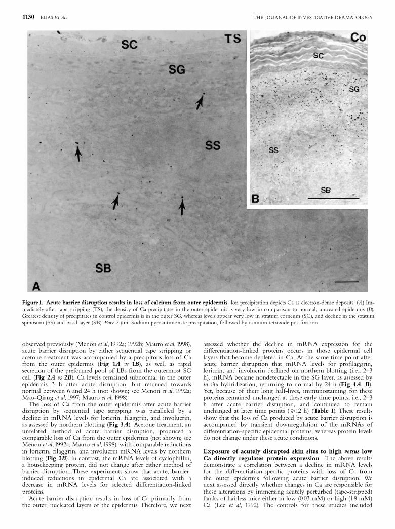
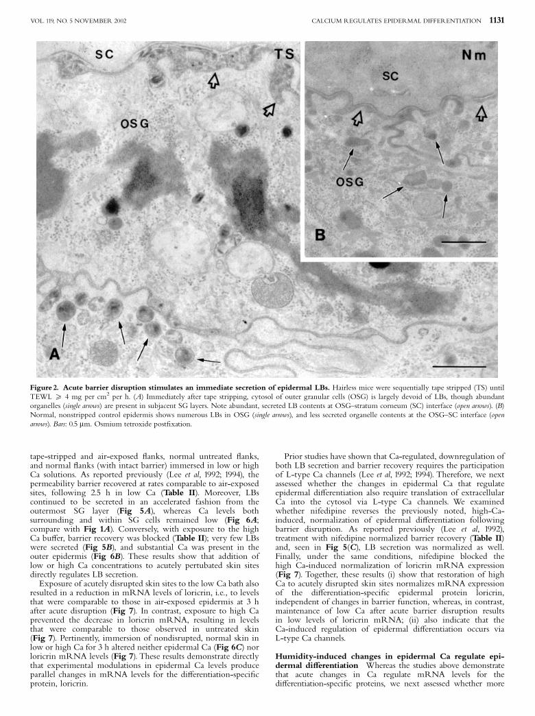
observed previously (Menon et al, 1992a; 1992b; Mauro et al, 1998),acute barrier disruption by either sequential tape stripping oracetone treatment was accompanied by a precipitous loss of Cafrom the outer epidermis (Fig 1A vs 1B), as well as rapidsecretion of the preformed pool of LBs from the outermost SGcell (Fig 2A vs 2B). Ca levels remained subnormal in the outerepidermis 3 h after acute disruption, but returned towardsnormal between 6 and 24 h (not shown; see Menon et al, 1992a;Mao-Qiang et al, 1997; Mauro et al, 1998).The loss of Ca from the outer epidermis after acute barrier
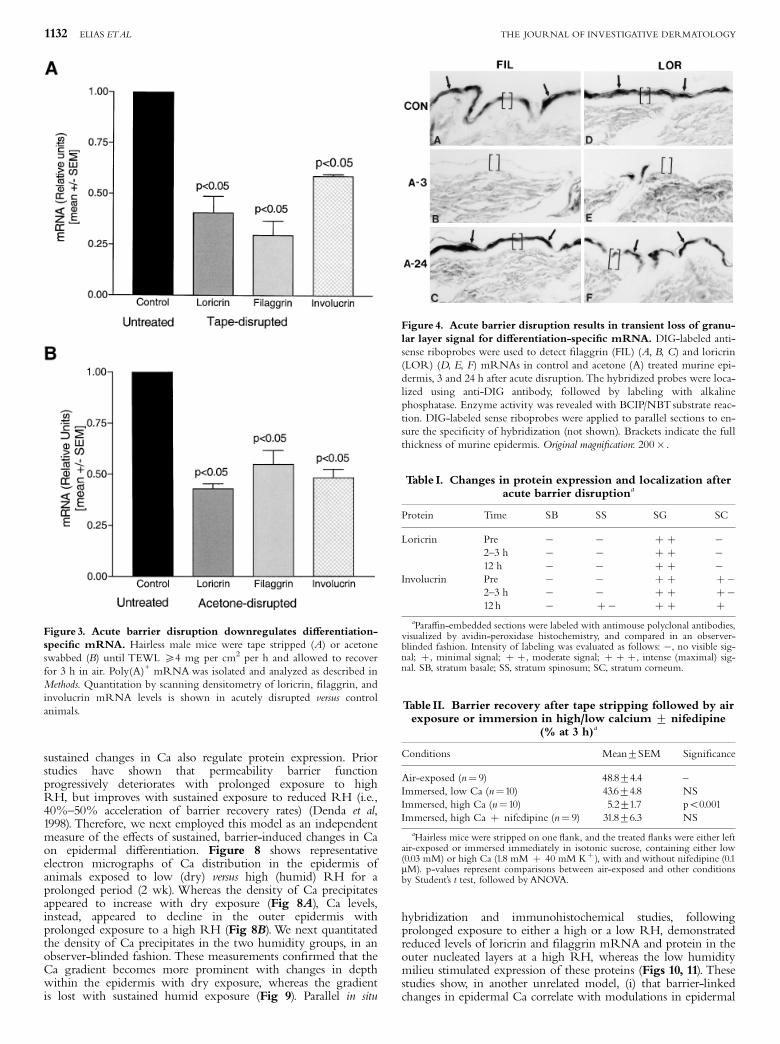
disruption by sequential tape stripping was paralleled by adecline in mRNA levels for loricrin, ¢laggrin, and involucrin,as assessed by northern blotting (Fig 3A). Acetone treatment, anunrelated method of acute barrier disruption, produced acomparable loss of Ca from the outer epidermis (not shown; seeMenon et al, 1992a; Mauro et al, 1998), with comparable reductionsin loricrin, ¢laggrin, and involucrin mRNA levels by northernblotting (Fig 3B). In contrast, the mRNA levels of cyclophillin,a housekeeping protein, did not change after either method ofbarrier disruption. These experiments show that acute, barrier-induced reductions in epidermal Ca are associated with adecrease in mRNA levels for selected di¡erentiation-linkedproteins.Acute barrier disruption results in loss of Ca primarily from
the outer, nucleated layers of the epidermis. Therefore, we next
assessed whether the decline in mRNA expression for thedi¡erentiation-linked proteins occurs in those epidermal celllayers that become depleted in Ca. At the same time point afteracute barrier disruption that mRNA levels for pro¢laggrin,loricrin, and involucrin declined on northern blotting (i.e., 2^3h), mRNA became nondetectable in the SG layer, as assessed byin situ hybridization, returning to normal by 24 h (Fig 4A, B).Yet, because of their long half-lives, immunostaining for theseproteins remained unchanged at these early time points; i.e., 2^3h after acute barrier disruption, and continued to remainunchanged at later time points (X12 h) (Table I). These resultsshow that the loss of Ca produced by acute barrier disruption isaccompanied by transient downregulation of the mRNAs ofdi¡erentiation-speci¢c epidermal proteins, whereas protein levelsdo not change under these acute conditions.
Exposure of acutely disrupted skin sites to high versus lowCa directly regulates protein expression The above resultsdemonstrate a correlation between a decline in mRNA levelsfor the di¡erentiation-speci¢c proteins with loss of Ca fromthe outer epidermis following acute barrier disruption. Wenext assessed directly whether changes in Ca are responsible forthese alterations by immersing acutely perturbed (tape-stripped)£anks of hairless mice either in low (0.03 mM) or high (1.8 mM)Ca (Lee et al, 1992). The controls for these studies included
Figure1. Acute barrier disruption results in loss of calcium from outer epidermis. Ion precipitation depicts Ca as electron-dense deposits. (A) Im-mediately after tape stripping (TS), the density of Ca precipitates in the outer epidermis is very low in comparison to normal, untreated epidermis (B).Greatest density of precipitates in control epidermis is in the outer SG, whereas levels appear very low in stratum corneum (SC), and decline in the stratumspinosum (SS) and basal layer (SB). Bars: 2 mm. Sodium pyroantimonate precipitation, followed by osmium tetroxide post¢xation.
1130 ELIAS ETAL THE JOURNAL OF INVESTIGATIVE DERMATOLOGY
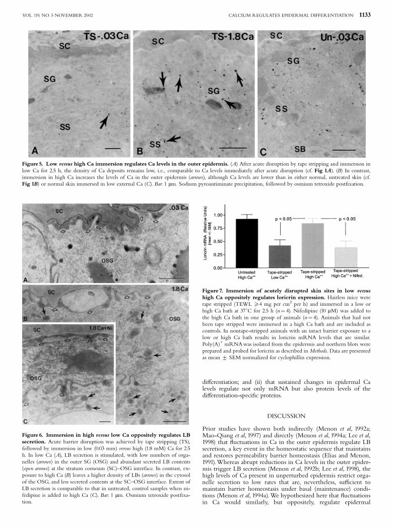
tape-stripped and air-exposed £anks, normal untreated £anks,and normal £anks (with intact barrier) immersed in low or highCa solutions. As reported previously (Lee et al, 1992; 1994), thepermeability barrier recovered at rates comparable to air-exposedsites, following 2.5 h in low Ca (Table II). Moreover, LBscontinued to be secreted in an accelerated fashion from theoutermost SG layer (Fig 5A), whereas Ca levels bothsurrounding and within SG cells remained low (Fig 6A;compare with Fig 1A). Conversely, with exposure to the highCa bu¡er, barrier recovery was blocked (Table II); very few LBswere secreted (Fig 5B), and substantial Ca was present in theouter epidermis (Fig 6B). These results show that addition oflow or high Ca concentrations to acutely pertubated skin sitesdirectly regulates LB secretion.Exposure of acutely disrupted skin sites to the low Ca bath also
resulted in a reduction in mRNA levels of loricrin, i.e., to levelsthat were comparable to those in air-exposed epidermis at 3 hafter acute disruption (Fig 7). In contrast, exposure to high Caprevented the decrease in loricrin mRNA, resulting in levelsthat were comparable to those observed in untreated skin(Fig 7). Pertinently, immersion of nondisrupted, normal skin inlow or high Ca for 3 h altered neither epidermal Ca (Fig 6C) norloricrin mRNA levels (Fig 7). These results demonstrate directlythat experimental modulations in epidermal Ca levels produceparallel changes in mRNA levels for the di¡erentiation-speci¢cprotein, loricrin.
Prior studies have shown that Ca-regulated, downregulation ofboth LB secretion and barrier recovery requires the participationof L-type Ca channels (Lee et al, 1992; 1994). Therefore, we nextassessed whether the changes in epidermal Ca that regulateepidermal di¡erentiation also require translation of extracellularCa into the cytosol via L-type Ca channels. We examinedwhether nifedipine reverses the previously noted, high-Ca-induced, normalization of epidermal di¡erentiation followingbarrier disruption. As reported previously (Lee et al, 1992),treatment with nifedipine normalized barrier recovery (Table II)and, seen in Fig 5(C), LB secretion was normalized as well.Finally, under the same conditions, nifedipine blocked thehigh Ca-induced normalization of loricrin mRNA expression(Fig 7). Together, these results (i) show that restoration of highCa to acutely disrupted skin sites normalizes mRNA expressionof the di¡erentiation-speci¢c epidermal protein loricrin,independent of changes in barrier function, whereas, in contrast,maintenance of low Ca after acute barrier disruption resultsin low levels of loricrin mRNA; (ii) also indicate that theCa-induced regulation of epidermal di¡erentiation occurs viaL-type Ca channels.
Humidity-induced changes in epidermal Ca regulate epi-dermal di¡erentiation Whereas the studies above demonstratethat acute changes in Ca regulate mRNA levels for thedi¡erentiation-speci¢c proteins, we next assessed whether more
Figure 2. Acute barrier disruption stimulates an immediate secretion of epidermal LBs. Hairless mice were sequentially tape stripped (TS) untilTEWL X 4 mg per cm2 per h. (A) Immediately after tape stripping, cytosol of outer granular cells (OSG) is largely devoid of LBs, though abundantorganelles (single arrows) are present in subjacent SG layers. Note abundant, secreted LB contents at OSG^stratum corneum (SC) interface (open arrows). (B)Normal, nonstripped control epidermis shows numerous LBs in OSG (single arrows), and less secreted organelle contents at the OSG^SC interface (openarrows). Bars: 0.5 mm. Osmium tetroxide post¢xation.
CALCIUM REGULATES EPIDERMAL DIFFERENTIATION 1131VOL. 119, NO. 5 NOVEMBER 2002
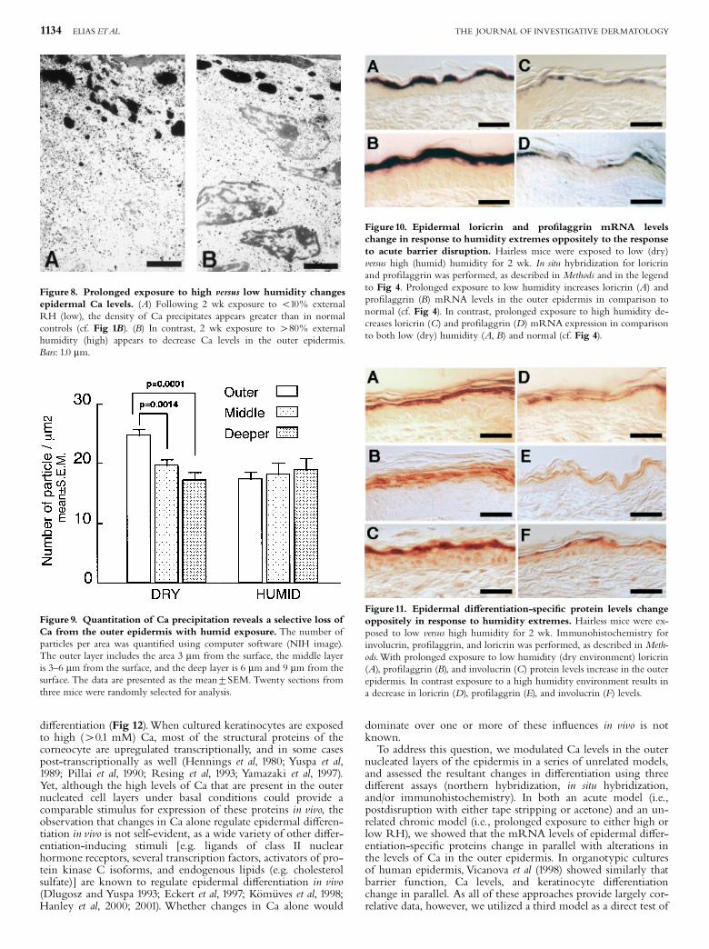
sustained changes in Ca also regulate protein expression. Priorstudies have shown that permeability barrier functionprogressively deteriorates with prolonged exposure to highRH, but improves with sustained exposure to reduced RH (i.e.,40%^50% acceleration of barrier recovery rates) (Denda et al,1998). Therefore, we next employed this model as an independentmeasure of the e¡ects of sustained, barrier-induced changes in Caon epidermal di¡erentiation. Figure 8 shows representativeelectron micrographs of Ca distribution in the epidermis ofanimals exposed to low (dry) versus high (humid) RH for aprolonged period (2 wk). Whereas the density of Ca precipitatesappeared to increase with dry exposure (Fig 8A), Ca levels,instead, appeared to decline in the outer epidermis withprolonged exposure to a high RH (Fig 8B).We next quantitatedthe density of Ca precipitates in the two humidity groups, in anobserver-blinded fashion. These measurements con¢rmed that theCa gradient becomes more prominent with changes in depthwithin the epidermis with dry exposure, whereas the gradientis lost with sustained humid exposure (Fig 9). Parallel in situ
hybridization and immunohistochemical studies, followingprolonged exposure to either a high or a low RH, demonstratedreduced levels of loricrin and ¢laggrin mRNA and protein in theouter nucleated layers at a high RH, whereas the low humiditymilieu stimulated expression of these proteins (Figs 10, 11). Thesestudies show, in another unrelated model, (i) that barrier-linkedchanges in epidermal Ca correlate with modulations in epidermal
Figure 3. Acute barrier disruption downregulates di¡erentiation-speci¢c mRNA. Hairless male mice were tape stripped (A) or acetoneswabbed (B) until TEWL X4 mg per cm2 per h and allowed to recoverfor 3 h in air. Poly(A)+ mRNAwas isolated and analyzed as described inMethods. Quantitation by scanning densitometry of loricrin, ¢laggrin, andinvolucrin mRNA levels is shown in acutely disrupted versus controlanimals.
Figure 4. Acute barrier disruption results in transient loss of granu-lar layer signal for di¡erentiation-speci¢c mRNA. DIG-labeled anti-sense riboprobes were used to detect ¢laggrin (FIL) (A, B, C) and loricrin(LOR) (D, E, F) mRNAs in control and acetone (A) treated murine epi-dermis, 3 and 24 h after acute disruption. The hybridized probes were loca-lized using anti-DIG antibody, followed by labeling with alkalinephosphatase. Enzyme activity was revealed with BCIP/NBTsubstrate reac-tion. DIG-labeled sense riboprobes were applied to parallel sections to en-sure the speci¢city of hybridization (not shown). Brackets indicate the fullthickness of murine epidermis. Original magni¢cation: 200� .
Table I. Changes in protein expression and localization afteracute barrier disruptiona
Protein Time SB SS SG SC
Loricrin Pre � � þ þ �2^3 h � � þ þ �12 h � � þ þ �
Involucrin Pre � � þ þ þ�2^3 h � � þ þ þ�12 h � þ� þ þ þ
aPara⁄n-embedded sections were labeled with antimouse polyclonal antibodies,visualized by avidin-peroxidase histochemistry, and compared in an observer-blinded fashion. Intensity of labeling was evaluated as follows: �, no visible sig-nal; þ, minimal signal; þ þ, moderate signal; þ þ þ, intense (maximal) sig-nal. SB, stratum basale; SS, stratum spinosum; SC, stratum corneum.
Table II. Barrier recovery after tape stripping followed by airexposure or immersion in high/low calcium 7 nifedipine
(% at 3 h)a
Conditions Mean7SEM Signi¢cance
Air-exposed (n¼ 9) 48.874.4 ^Immersed, low Ca (n¼10) 43.674.8 NSImmersed, high Ca (n¼10) 5.271.7 po0.001Immersed, high Ca þ nifedipine (n¼ 9) 31.876.3 NS
aHairless mice were stripped on one £ank, and the treated £anks were either leftair-exposed or immersed immediately in isotonic sucrose, containing either low(0.03 mM) or high Ca (1.8 mM þ 40 mM Kþ ), with and without nifedipine (0.1mM). p-values represent comparisons between air-exposed and other conditionsby Student’s t test, followed byANOVA.
1132 ELIAS ETAL THE JOURNAL OF INVESTIGATIVE DERMATOLOGY
di¡erentiation; and (ii) that sustained changes in epidermal Calevels regulate not only mRNA but also protein levels of thedi¡erentiation-speci¢c proteins.
DISCUSSION
Prior studies have shown both indirectly (Menon et al, 1992a;Mao-Qiang et al, 1997) and directly (Menon et al, 1994a; Lee et al,1998) that £uctuations in Ca in the outer epidermis regulate LBsecretion, a key event in the homeostatic sequence that maintainsand restores permeability barrier homeostasis (Elias and Menon,1991).Whereas abrupt reductions in Ca levels in the outer epider-mis trigger LB secretion (Menon et al, 1992b; Lee et al, 1998), thehigh levels of Ca present in unperturbed epidermis restrict orga-nelle secretion to low rates that are, nevertheless, su⁄cient tomaintain barrier homeostasis under basal (maintenance) condi-tions (Menon et al, 1994a).We hypothesized here that £uctuationsin Ca would similarly, but oppositely, regulate epidermal
Figure 5. Low versus high Ca immersion regulates Ca levels in the outer epidermis. (A) After acute disruption by tape stripping and immersion inlow Ca for 2.5 h, the density of Ca deposits remains low, i.e., comparable to Ca levels immediately after acute disruption (cf. Fig 1A). (B) In contrast,immersion in high Ca increases the levels of Ca in the outer epidermis (arrows), although Ca levels are lower than in either normal, untreated skin (cf.Fig 1B) or normal skin immersed in low external Ca (C). Bar: 1 mm. Sodium pyroantiminate precipitation, followed by osmium tetroxide post¢xation.
Figure 6. Immersion in high versus low Ca oppositely regulates LBsecretion. Acute barrier disruption was achieved by tape stripping (TS),followed by immersion in low (0.03 mm) versus high (1.8 mM) Ca for 2.5h. In low Ca (A), LB secretion is stimulated, with low numbers of orga-nelles (arrows) in the outer SG (OSG) and abundant secreted LB contents(open arrows) at the stratum corneum (SC)^OSG interface. In contrast, ex-posure to high Ca (B) leaves a higher density of LBs (arrows) in the cytosolof the OSG, and less secreted contents at the SC^OSG interface. Extent ofLB secretion is comparable to that in untreated, control samples when ni-fedipine is added to high Ca (C). Bar: 1 mm. Osmium tetroxide post¢xa-tion.
Figure 7. Immersion of acutely disrupted skin sites in low versushigh Ca oppositely regulates loricrin expression. Hairless mice weretape stripped (TEWL X4 mg per cm2 per h) and immersed in a low orhigh Ca bath at 371C for 2.5 h (n¼ 4). Nifedipine (10 mM) was added tothe high Ca bath in one group of animals (n¼ 4). Animals that had notbeen tape stripped were immersed in a high Ca bath and are included ascontrols. In nontape-stripped animals with an intact barrier exposure to alow or high Ca bath results in loricrin mRNA levels that are similar.Poly(A)+ mRNAwas isolated from the epidermis and northern blots wereprepared and probed for loricrin as described in Methods. Data are presentedas mean 7 SEM normalized for cyclophillin expression.
CALCIUM REGULATES EPIDERMAL DIFFERENTIATION 1133VOL. 119, NO. 5 NOVEMBER 2002
di¡erentiation (Fig 12).When cultured keratinocytes are exposedto high (40.1 mM) Ca, most of the structural proteins of thecorneocyte are upregulated transcriptionally, and in some casespost-transcriptionally as well (Hennings et al, 1980; Yuspa et al,1989; Pillai et al, 1990; Resing et al, 1993; Yamazaki et al, 1997).Yet, although the high levels of Ca that are present in the outernucleated cell layers under basal conditions could provide acomparable stimulus for expression of these proteins in vivo, theobservation that changes in Ca alone regulate epidermal di¡eren-tiation in vivo is not self-evident, as a wide variety of other di¡er-entiation-inducing stimuli [e.g. ligands of class II nuclearhormone receptors, several transcription factors, activators of pro-tein kinase C isoforms, and endogenous lipids (e.g. cholesterolsulfate)] are known to regulate epidermal di¡erentiation in vivo(Dlugosz and Yuspa 1993; Eckert et al, 1997; K˛mˇves et al, 1998;Hanley et al, 2000; 2001). Whether changes in Ca alone would
dominate over one or more of these in£uences in vivo is notknown.To address this question, we modulated Ca levels in the outer
nucleated layers of the epidermis in a series of unrelated models,and assessed the resultant changes in di¡erentiation using threedi¡erent assays (northern hybridization, in situ hybridization,and/or immunohistochemistry). In both an acute model (i.e.,postdisruption with either tape stripping or acetone) and an un-related chronic model (i.e., prolonged exposure to either high orlow RH), we showed that the mRNA levels of epidermal di¡er-entiation-speci¢c proteins change in parallel with alterations inthe levels of Ca in the outer epidermis. In organotypic culturesof human epidermis, Vicanova et al (1998) showed similarly thatbarrier function, Ca levels, and keratinocyte di¡erentiationchange in parallel. As all of these approaches provide largely cor-relative data, however, we utilized a third model as a direct test of
Figure 9. Quantitation of Ca precipitation reveals a selective loss ofCa from the outer epidermis with humid exposure. The number ofparticles per area was quanti¢ed using computer software (NIH image).The outer layer includes the area 3 mm from the surface, the middle layeris 3^6 mm from the surface, and the deep layer is 6 mm and 9 mm from thesurface. The data are presented as the mean7SEM. Twenty sections fromthree mice were randomly selected for analysis.
Figure 8. Prolonged exposure to high versus low humidity changesepidermal Ca levels. (A) Following 2 wk exposure to o10% externalRH (low), the density of Ca precipitates appears greater than in normalcontrols (cf. Fig 1B). (B) In contrast, 2 wk exposure to 480% externalhumidity (high) appears to decrease Ca levels in the outer epidermis.Bars: 1.0 mm.
Figure10. Epidermal loricrin and pro¢laggrin mRNA levelschange in response to humidity extremes oppositely to the responseto acute barrier disruption. Hairless mice were exposed to low (dry)versus high (humid) humidity for 2 wk. In situ hybridization for loricrinand pro¢laggrin was performed, as described in Methods and in the legendto Fig 4. Prolonged exposure to low humidity increases loricrin (A) andpro¢laggrin (B) mRNA levels in the outer epidermis in comparison tonormal (cf. Fig 4). In contrast, prolonged exposure to high humidity de-creases loricrin (C) and pro¢laggrin (D) mRNA expression in comparisonto both low (dry) humidity (A, B) and normal (cf. Fig 4).
Figure11. Epidermal di¡erentiation-speci¢c protein levels changeoppositely in response to humidity extremes. Hairless mice were ex-posed to low versus high humidity for 2 wk. Immunohistochemistry forinvolucrin, pro¢laggrin, and loricrin was performed, as described in Meth-ods.With prolonged exposure to low humidity (dry environment) loricrin(A), pro¢laggrin (B), and involucrin (C) protein levels increase in the outerepidermis. In contrast exposure to a high humidity environment results ina decrease in loricrin (D), pro¢laggrin (E), and involucrin (F) levels.
1134 ELIAS ETAL THE JOURNAL OF INVESTIGATIVE DERMATOLOGY

the hypothesis that changes in Ca regulate epidermal di¡erentia-tion (i.e., a bath model, with exposure of acutely disrupted skinsites to either low or high Ca) (Lee et al, 1992). These resultsshowed directly that changes in Ca regulate mRNA expressionof the di¡erentiation-speci¢c protein loricrin, i.e., exposure tolow versus high Ca suppressed and normalized mRNA levels, re-spectively. Thus, changes in mRNA levels occur when Ca levelschange both indirectly (i.e., with barrier disruption) or directly(i.e., with exposure to a low/high Ca bath, independent of barrierstatus). Finally, we showed that coexposure to high Ca plus theselective, dihydropyridine, L-type Ca channel inhibitor nifedi-pine blocks the normalization of mRNA levels for loricrin. Thisexperiment indicates further that Ca signaling of the di¡erentia-tion-speci¢c proteins involves Ca translocation across L-type Cachannels (Abernethy and Schwartz, 1999). Pertinently, a nifedi-pine-sensitive, L-type Ca channel likewise is required for inverseCa regulation of barrier recovery (Lee et al, 1992). Together, theseresults show unequivocally that both acute and sustained modu-lations in Ca levels in vivo regulate the expression of di¡erentia-tion-speci¢c epidermal proteins, and that such Ca regulationinvolves the participation of L-type Ca channels.The observation that mRNA but not protein levels are regu-
lated in parallel with acute changes in epidermal Ca levels can beexplained by the long half-lives of these proteins (Polakowskiand Goldsmith, 1991; Simon, 1994). In contrast, with more sus-tained, humidity-associated changes in Ca, we showed that notonly mRNA but also protein levels change. Several observationssuggest that the changes in protein levels that occur with sus-tained changes in epidermal Ca levels could have pathophysiolo-gic signi¢cance. Prior studies have shown that the sustainedabnormality in barrier function that occurs in essential fatty acidde¢ciency is paralleled by loss of the epidermal Ca gradient(Menon et al, 1994a). Accordingly, Ca levels are reduced in theouter epidermis in other, cutaneous pathologic entities that areaccompanied by a barrier abnormality, e.g., atopic dermatitis andpsoriasis (Menon et al, 1991; Pallon et al, 1996). The link betweendecreased barrier function and loss of epidermal Ca, in turn,could contribute to the decreased expression of di¡erentiation-linked proteins in these disorders (Detmar et al, 1990; Schroederet al, 1992; Ishida-Yamamoto and Iizuka, 1995; Ishida-Yamamotoet al, 1996; Fujimoto et al, 1997). Furthermore, because the dif-ferentiation-speci¢c proteins provide a critical sca¡old for
LB-derived, extracellular lamellar bilayers (Elias et al, in press),decreased di¡erentiation could, in turn, further aggravate thebarrier abnormality. Accordingly, a barrier abnormality (Elias et al,1981) accompanies the well-documented downregulation of dif-ferentiation-speci¢c proteins that occurs with administration ofsupraphysiologic doses of retinoids (Cline and Rice, 1983; Rubinand Rice, 1986; Floyd and Jetten, 1989; Hohl et al, 1991; Liew andYamanishi, 1992; Marvin et al, 1992; Monzon et al, 1996).Finally, our results suggest that the lipid and protein arms of
epidermal di¡erentiation are linked and coregulated in parallelby barrier-imposed changes in epidermal Ca levels (Fig 12). Un-der basal (maintenance) conditions, high levels of Ca in the outerepidermis promote epidermal protein expression, and simulta-neously restrict LB secretion. In contrast, after acute barrier per-turbations, epidermal Ca levels decrease in parallel with a steep,though transient, decline in mRNA levels for the di¡erentia-tion-speci¢c protein, whereas LB secretion simultaneously is sti-mulated. Thus, in all three models where the expression ofdi¡erentiation-speci¢c proteins is regulated by changes in Ca,parallel, but opposite, changes occur in LB secretion. LB secre-tion and enhanced lipid synthesis represent two key metabolicresponses that lead to barrier recovery after acute insults (Elias,1996). Whereas the transient downregulation in mRNA levelsthat occurs after barrier disruption could represent a speci¢cadaptation that directly facilitates barrier recovery, it could, in-stead, support barrier recovery indirectly by acting as a form ofmetabolic conservation.
Ms. Jerelyn Magnusson, LauraTeale, and Katie O’Donnell prepared this manuscript.These studies were supported byAR 39448 (PP), AR 19098, and AR 39739, as wellas the Medical Research Service, Department ofVeterans A¡airs.
REFERENCES
Abernethy DR, Schwartz JB: Calcium-antagonistic drugs. N Eng J Med 341:1447^1457, 1999
Cline PR, Rice RH: Modulation of involucrin and envelope competence in humankeratinocytes by hydrocortisone, retinol acetate, and growth arrest. Cancer Res43:3203^3207, 1983
Denda M, Sato J, MasudaY, et al: Exposure to a dry environment enhances epidermalpermeability barrier function. J Invest Dermatol 111:858^863, 1998
Figure12. Modulations in Ca levels coordinately regulate both lamellar body secretion and epidermal di¡erentiation.
CALCIUM REGULATES EPIDERMAL DIFFERENTIATION 1135VOL. 119, NO. 5 NOVEMBER 2002

Detmar M, Mayer-Da-Silva A, Stadler R, Orfanos CE: Initial hyperproliferationand incomplete terminal di¡erentiation of cultured human keratinocytes fromlesional and uninvolved psoriatic skin. Acta DermVenereol 70:295^299, 1990
Dlugosz A, Yuspa SH: Coordinate changes in gene expression which mark the spi-nous to granular cell transition in epidermis are regulated by protein kinase.J Cell Biol 120:217^225, 1993
Eckert RL, Crish JF, Robinson NA: The epidermal keratinocyte as a model for thestudy of gene regulation and cell di¡erentiation. Physiol Rev 77:397^424, 1997
Elias PM: Stratum corneum architecture, metabolic activity, and interactivity withsubjacent cell layers. Exp Dermatol 5:191^201, 1996
Elias PM, Menon GK: Structural and lipid biochemical correlates of the epidermalpermeability barrier. Adv Lipid Res 24:1^26, 1991
Elias PM, Fritsch P, Lampe M,Williams M, Brown B, Nemanic MK, Grayson S:Retinoid e¡ects on epidermal structure, di¡erentiation, and permeability. La-boratory Invest 44:531^540, 1981
Elias PM, Uchida Y, Rice RH, Schmuth M, Crumrine D, Komuves L, HolleranWM: Corni¢ed envelope (CE) and corneocyte-lipid envelope (CLE) forma-tion in patients with lamellar ichthyosis: pathogenesis of the permeability bar-rier abnormality. Exp Dermatol in press.
Floyd EE, Jetten AM: Regulation of type 1 (epidermal) transgllutaminase mRNAlevels during squamous di¡erentiation: down regulation by retinoid. Mol CellBiol 9:4846^4951, 1989
Forslind B, Kunst L, Malmqvist KG, Carlsson LE, Roomans GM: Quantitative cor-relative proton and electron microprobe analysis of biological specimens. His-tochemistry 82:425^427, 1985
Forslind B, Roomans GM, Carlsson LE, Malmquist KG, Akselsson KR: Elementalananlysis on freeze-dried sections of human skin: studies by electron microbeand particle induced X-ray emission analysis. Scan Electron Microsc 2:755^759,1984
FujimotoW, Naanishi G, Arata J, Jetten AM: Di¡erential expression of human cor-ni¢n alpha and beta in squamous di¡erentiating epithelial tissues and severalskin lesions. J Invest Dermatol 108:200^204, 1997
Grubauer G, Elias PM, Feingold KR:Transepidermal water loss: the signal for recov-ery of barrier structure and function. J Lipid Res 30:323^333, 1989
Hanley K, Ng DC, He SS, et al: Oxysteroids induce di¡erentiation in human kerati-nocytes and increase AP-1-dependent involucrin transcription. J Invest Dermatol114:545^553, 2000
Hanley K,Wood L, Ng DC et al: Involucrin transcription and expression of Fra-1,Fra-2, and Jun D are increased by cholesterol sulfate in human keratinocytes. JLipid Res 42:390^398, 2001
Harris IR, Farrell AM, Holleran WM, Jackson S, Grunfeld C, Elias PM, FeingoldKR: Parallel regulation of sterol regulatory element binding protein-2 andthe enzymes of cholesterol and fatty acid synthesis but not ceramide synthesisin cultured human keratinocytes and murine epidermis. J Lipid Res 39:412^422,1998
Hennings H, Michael D, Cheng C, Steinhert P, Holbrook K, Yuspa SH: Calciumregulation of growth and di¡erentiation of mouse epidermal cells in culture.Cell 19:245^254, 1980
Hohl D, Lichti U, Breitkreutz D, Steinert PM, Roop DR: Transcription of the hu-man loricrin gene in vitro is induced by calcium and cell density and suppressedby retinoic acid. J Invest Dermatol 96:414^418, 1991
Ishida-Yamamoto A, Iizuka H: Di¡erences in involucrin immunolabeling withincorni¢ed cell envelopes in normal and psoriatic epidermis. J Invest Dermatol104:391^395, 1995
Ishida-Yamamoto A, Eady R,Watt FM, Roop DR, Hohl D, Iizuka H: Immunoelec-tron microscopic analysis of corni¢ed cell envelope formation in normal andpsoriatic epidermis. J Histochem Cytochem 44:167^175, 1996
Komuves LG, Hanley K, JiangY, Elias PM,Williams ML, Feingold KR: Ligands andactivators of nuclear hormone receptors regulate epidermal di¡erentiation dur-ing fetal rat skin development. J Invest Dermatol 111:429^433, 1998
Lee SH, Elias PM, Proksch E, Menon GK, Mao-Qiang M, Feingold KR: Calciumand potassium are important regulators of barrier homeostasis in murine epi-dermis. J Clin Invest 89:530^538, 1992
Lee SH, Elias PM, Feingold KR, Mauro T: a role for ions in barrier recovery afteracute perturbation. J Invest Dermatol 102:976^979, 1994
Lee SH, Choi EH, Feingold KR, Jiang S, Ahn SK: Iontophoresis itself on hairlessmouse skin induces the loss of the epidermal calcium gradient without skinbarrier impairment. J Invest Dermatol 111:39^43, 1998
Liew FM,Yamanishi K: Regulation of transglutaminase 1 gene expression by 12-O-tetradecanoylphorbol-13-acetate, dexamethasone, and retinoic acid in culturedhuman keratinocytes. Exp Cell Res 202:310^315, 1992
Mao-Qiang M, MauroT, Bench G,Warren R, Elias PM, Feingold KR: Calcium andpotassium inhibit barrier recovery after disruption, independent of the type ofinsult in hairless mice. Exp Dermatol 6:36^40, 1997
Marvin KW, George MD, Fujimoto W, Saunders NA, Bernacki SH, Jetten AM:Corni¢n, a cross-linked envelope precursor in keratinocytes that is down-regulated by retinoids. Proc Natl Acad Sci USA 89:11026^11030, 1992
Mauro T, Bench G, Sidderas-Haddad E, Feingold K, Elias P, Cullander C: Acutebarrier perturbation abolishes the Ca2þ and Kþ gradients in murine epider-mis: quantitative measurement using PIXE. J Invest Dermatol 111:1198^1201, 1998
Mehrel T, Hohl D, Rothnagel JA, et al: Identi¢cation of a major keratinocyte cellenvelope protein, loricrin. Cell 61:1103^1112, 1990
Menon GK, Grayson S, Elias PM: Ionic calcium reservoirs in mammalian epidermis:ultrastructural localization by ion-capture cytochemistry. J Invest Dermatol84:508^512, 1985
Menon GK, Elias PM: Ultrastructural localization of calcium in psoriatic and nor-mal human epidermis. Arch Dermatol 127:57^63, 1991
Menon GK, Elias PM, Lee SH, Feingold KR: Localization of calcium in murineepidermis following disruption and repair of the permeability barrier. CellTissue Res 270:503^512, 1992a
Menon GK, Feingold KR, Elias PM: Lamellar body secretory response to barrierdisruption. J Invest Dermatol 98:279^289, 1992b
Menon GK, Elias PM, Feingold KR: Integrity of the permeability barrier is crucialfor maintenance of the epidermal calcium gradient. Br J Dermatol 130:139^147,1994a
Monzon RI, LaPRes JJ, Hudson LG: Regulation of involucrin gene expression byretinoic acid and glucocorticoids. Cell Growth Di¡er 7:1751^1759, 1996
Pallon J, Malmquist KG,Werner-LindeY, Forslind B: PIXE analysis of pathologicalskin with special reference to psoriasis and atopic dry skin. Cell Mol Biol 42:111^118, 1996
Pillai S, Bickle DD, Mancianti ML, Cline D, Hinlenbergs M: Calcium regulation ofgrowth and di¡erentiation of normal human Keratinocytes: Modulationof di¡erentiation competence by stages of growth and extracellular calcium.J Cell Physiol 143:294^302, 1990
Polakowska RR, Goldsmith LA: The cell envelope and transglutaminase. In Gold-smith LA, ed. Biochemistry, Physiology and Molecular Biology of the Skin. Oxford:Oxford University Press, 1991: pp 168^201
Reichert U, Michel S, Schmidt R: The corni¢ed envelope: a key structure of term-inally di¡erentiating keratinocytes.. In: Darmon M, Blumenberg M, eds. Mo-lecular Biology of the Skin. The Keratinocyte. San Diego: Academic Press, 1993:pp 107^150
Resing KA, Al-Alawi N, Blomquist C, Fleckman P, Dale BA: Independent regula-tion of two cytoplasmic processing stages of the intermediate ¢lament-asso-ciated protein ¢laggrin and role of Ca2þ in the second stage. J Biol Chem268:25139^25145, 1993
Rubin AL, Rice RH: Di¡erential regulation by retinoic acid and calcium of trans-glutaminases in cultured neoplastic and normal human keratinocytes. CancerRes 46:2356^2361, 1986
Schroeder WT, Thacher SM, Stewart-Galetka S: Type 1 keratinocyte transglutami-nase: expression in human skin and psoriasis. J Invest Dermatol 99:27^34, 1992
Simon M: The epidermal corni¢ed envelope and its precursors. In: Leigh IM, LaneEB,Watt FM, eds.The Keratinocyte Handbook. Cambridge: Cambridge Univer-sity Press, 1994: pp: 107^160
Steinert PM, Marekov LN: The proteins ela¢n, ¢laggrin, keratin intermediate ¢la-ments, loricrin, and small proline-rich proteins 1 and 2 are isodipeptide cross-linked components of the human epidermal corni¢ed cell envelope. J BiolChem 270:17702^17711, 1995
Vicanova J, Boelsma E, Mommaas AM, et al: Normalization of epidermal calciumdistribution pro¢le in reconstructed human epidermis is related to improve-ment of terminal di¡erentiation and stratum corneum barrier formation. J In-vest Dermatol 111:97^106, 1998
Yamazaki M, Ishidoh K,Yasushi S, et al: Cytoplasmic processing of human pro¢lag-grin by active u-calpain. Biochem Biophys Res Com 235:653^656, 1997
Yuspa SH, Kilkenny A, Steinert PM, Roop DR: Expression of murine epidermaldi¡erentiation markers is tightly regulated by restricted extracellular calciumconcentrations in vitro. J Cell Biol 109:1207^1217, 1989
1136 ELIAS ETAL THE JOURNAL OF INVESTIGATIVE DERMATOLOGY