modiflcations of joint mobility range induced …
TRANSCRIPT
MODIFlCATIONS OF JOINT MOBILITY RANGE INDUCED BY ECCENTRIC CONTRACTION
R. Saggini1, L. Dragani2, M. A. Giamberardin02
ISports Medicine Department 2Institute of Medical Pathophysiology
University of Chieti Italy
INTRODUCTION It is a well-known experience that a heavy or unaccustomed effort is followed by
a kind of pain and discomfort, arising some hours after the end of the exercise and persisiting for several days (Hill, 1951; Hough, 1902). Asmussen (1956) first noted that this form of pain is due to movements in wich the active muscle is stretched. This type of movement is commonly defined as "eccentric contraction," or "negative work," or "eccentric exercise". This clinical condition is also defined as "Delayed Onset Muscle Soreness" (DOMS) and is characterized by, other than the delayed-onset muscle pain (824 hours) and a lowering in muscle pain threshold (24-48 hours), a swelling of the exerted limb (72 hours) (Armstrong, 1984; Newham et al., 1983).
Ultrastructural evaluations showed that dynamic eccentric contraction causes a muscle damage, consisting of the following alterations: a) myofibrils disorganization; b) Z-lines disruption; c) cell membrane lesions (Newham et al., 1983). This damage is indirectly confirmed and evidenced by a massive, delayed (fourth-fifth day after the effort) release of creatine-kinase (CK) in the serum (Newham et al., 1983). Stiffness and restricted range of movement in the joints related to the eccentrically-exerted muscles are also described but the phenomena has been investigated less.
The aim of this study is to evaluate the range of the knee joint mobility before and after a standardized eccentric effort. Pain thresholds and perceived pain intensity are also evaluated in order to find possible correlation with the modification of the joint range.
METHODOLCXJY Twelve healthy subjects, aged 24-35 years, were used in this experiment. They
performed a step-test (20 minute duration, 15 cycles/min) to exercise the quadriceps femoris muscle of one side eccentrically (Mills et al., 1981). The day before the test, all the subjects underwent evaluation of Pressure Pain Threshold (P'TT) of the exerted muscle and Passive joint Mobility (PjM) of the knee. These evaluations were repeated immediately after the test and on the following four days. On the same days, the perceived pain was also measuted by means of Visual Analog Scale (VAS).
PPT was assessed by means of a force-transducer equipped with a 2.5 cm diameter rounded probe and a digital analyser (unit: kg/£) applied on 12 pre-established points on the quadriceps muscle surface. The mean of the 12 recordings was taken as the final threshold value. PjM was measured by means of a manual goniometer. The exer· cised lower limb was moved from the rest position to the maximal pain-free flexion. The goniometer was placed with its fulcrum on the external surtace of the knee. The degree at which the subject felt the minimum discomfort was recorded. The data were statistically analyzed using a Student's t-test for paired data.
186
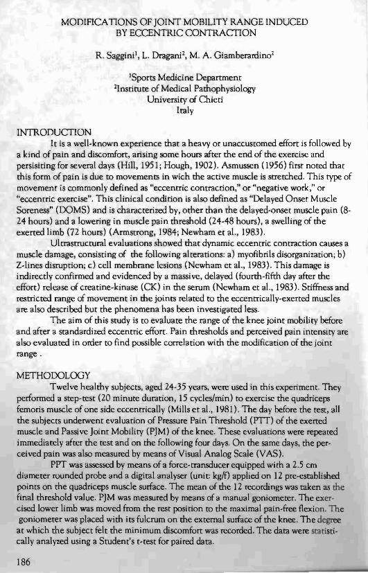
RESULTS PIT values were the following: 6.85 ± 1.22 kg/f before the test, 6.89 ± 1.09 kg/f
immediately after and 5.02 ± 1.55 kg/f, 439 ± 1.38 kg/f, 5.54 ± I..59 kg/f and 6.74 ± 1.04 kg/f respectively on days 1,2,3 and 4 after the effort (Figure 1). Statistical analysis showed a significant decrease after 24 hours (p<O.OOl), 48 hours (p<O.OOl) and 72 hours (p<O.Ol) in the exerted muscle.
4-0
35
30 -~~.'..""'"
,......... '. 25 / ""
! .V>
~ 20 :' ". / "'.
'5 / i \,
..,.." .•.•. 10
i5 :'
./O.L.-----+------+----~-----_:_---<
before test 1st 2nd 3rd 4th Days after eccentric effort
Figure 1. Pressure pain threshold as a function of days after eccentric effort.
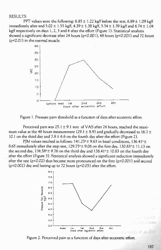
Perceived pain was 25.1 ± 9.1 mm of VAS after 24 hours, reached the maximum value at the 48 hours measurement (29.1 ± 8.9) and gradually decreased to 16.1 ± 10.1 on the third day and 7.8 ± 6.6 on the fourth day after the effort (Figure 2).
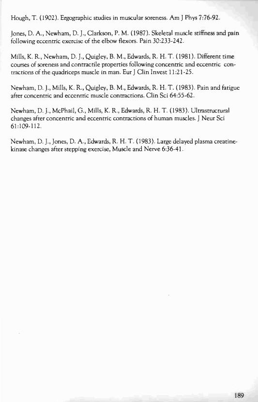
P]M values resulted as follows: 141.25°± 9.63 in basal conditions, 136.41°± 8.65 immediately after the step-test, 129.75o ± 9.06 on the first day, 130.83°± 11.13 on the second day, 134.58°± 9.76 on the third day and 138.41°± 10.83 on the fourth day after the effort (Figure 3). Statistical analysis showed a significant reduction immediately after the test (p<0.02) that became more pronounced on the first (p<O.OOl) an-d second (p<0.(102) day and lasting up to 72 hours (p<0.05) after the effort.
8.0
7.0
I" ~.O
] 4.5
4.0
3.5
3.0
7.5
~ 6.5
~ 6.0
'6~ ~.~ 0- ><
bos& 1.0. 1.t 2nd 3rd 4th Coy. oft.,. .<t,centrle .ft~rt
Figure 2. Perceived pain as a function of days after eccentric etfort.
187

150
145
140 '"
135
130
125 base i.a. 1 2 .3 4
Days
Figure 3. Passive joint mobility as a function of days after eccentric effort.
DISCUSSION DOMS is a complex of phenomena whose explanation is still unclear; particu
larly, the temporal dissociation between symptoms and signs of muscle damage. The reduction of PJM has been attributed to a shonening of non-contractile elements as well as to an edema due to a damage of the connective tissue induced by the eccentric contraction stress. However, the maximal thickness of the eccentrically exerted limb is usually found at 72 hours while the reduction of PJM starts immediately after the effort (Jones et al., 1987).
The results of the present study show that the temporal pattern of PJM reduction is fairly similar to that of pain and hyperalgesia while muscle damage indices (CK release in the serum and byoptic abnormalities in the muscle) become evident only in the following days (Newham et al., 1983; Newham et al., 1983). This could suggest as a possible alternative explanation a reflex origin of the phenomenon. Painful symptoms, by means of a reflex arc, could be able to induce an increase of tonic activation of agonist muscles producing an active limitation of joint excursion of anti nociceptive meaning. Jones' s study excluded this hypothesis but his experiment was carried out on the biceps brachii muscle. That muscle has different functional properties from quadriceps femoris muscle such as: 1) no postural properties; and 2) flexion and not extension function. However, further work is needed for more complete knowledge of the phenomenon, this work being quite preliminary.
REFERENCES Asmussen, E. (I 956). Obsevations on experimental muscle soreness. Acta Phys Scan 28:364-382.
Armstrong, R. B. (I984). Mechanisms of exercise-induced delayed-onset muscle soreness: a brief review. Med Sci Spons Exer 6:529-538.
HiLl, A. V. (I 951). Mechanics of voluntary muscle. Lancet2:947-951.
vv C;v
a
188
Hough, T. (1902). Ergographic studies in muscular soreness. Am] Phys 7:76-92.
]ones, D. A., Newham, D. ]., Clarkson, P. M. (1987). Skeletal muscle stiffness and pain following eccentric exercise of me elbow flexors. Pain 30:233-242.
Mills, K. R., Newham, D. ]., Quigley, B. M., Edwards, R. H. T. (1981). Different time courses of soreness and contractile properties following concentric and eccentric contractions of me quadriceps muscle in man. Eur] Clin Invest 11 :21-25.
Newham, D. J., Mills, K. R., Quigley, B. M., Edwards, R. H. T. (1983). Pain and fatigue after concentric and eccentric muscle contractions. Clin Sci 64:55-62.
Newham, D. J., McPhail, G., Mills, K. R., Edwards, R. H. T. (1983). Ultrastructural changes after concentric and eccentric contractions of human muscles. JNeur Sci 61:109-112.
Newham, D.]., ]ones, D. A., Edwards, R. H. T. (1983). Large delayed plasma creatinekinase changes after stepping exercise, Muscle and Nerve 6:36-41.
189