modifications of midpalatal sutural density induced by rapid maxillary expansion: a low-dose...
TRANSCRIPT
ORIGINAL ARTICLE
Modifications of midpalatal sutural densityinduced by rapid maxillary expansion: Alow-dose computed-tomography evaluation
Lorenzo Franchi,a Tiziano Baccetti,a Roberta Lione,b Ezio Fanucci,c and Paola Cozzad
Florence and Rome, Italy
Introduction: The aim of this study was to evaluate the density of the midpalatal suture as assessed by low-dose computed tomography (CT) before rapid maxillary expansion (RME) (T0), at the end of active expansion(T1), and after a retention period of 6 months (T2). Methods: The study sample comprised 17 prepubertal sub-jects (mean age, 11.2 years) with constricted maxillary arches and unilateral or bilateral posterior crossbite.The total amount of expansion was 7 mm in all subjects. Multi-slice low-dose CT scans were taken at T0,T1, and T2. On axial CT scanned images, 4 regions of interest (ROIs) were placed along the midpalatal suture(anterior [AS ROI] and posterior [PS ROI]) and in 2 regions of palatal bone (anterior and posterior). Density wasmeasured in Hounsfield units. The Mann-Whitney U test and Friedman analysis of variance (ANOVA) with post-hoc test were used (P\0.05). Results: The densities in the AS and PS ROIs were significantly smaller than thereference bone densities before RME therapy. Both AS and PS ROIs showed significant decreases in densityfrom T0 to T1, significant increases from T1 to T2, and no significant differences from T0 to T2. Conclusions:The effective opening of the midpalatal suture by RME in prepubertal subjects was associated with a significantdecrease in sutural density. The sutural density after 6 months of retention post-RME indicated reorganizationof the midpalatal suture, since it showed values similar to the pretreatment ones. (Am J Orthod DentofacialOrthop 2010;137:486-8)
Rapid maxillary expansion (RME) is an effectiveorthopedic procedure commonly used to treatmaxillary transverse deficiencies in growing pa-
tients.1 Transverse dentoskeletal changes in the maxillain response to RME are produced mainly through dis-ruption of the midpalatal suture.2 Histologic studieson both humans and animals supported the opening ofthe midpalatal suture after RME therapy.3,4 In a studyof 6 monkeys, Cleall et al3 reported that, at the momentof suture opening, the area was filled with unorganizedfibrous conjunctive tissue; 6 months after expansion, the
aAssistant professor, Department of Orthodontics, University of Florence,
Florence, Italy; Thomas M. Graber Visiting Scholar, Department of Orthodon-
tics and Pediatric Dentistry, School of Dentistry, University of Michigan, Ann
Arbor.bResearch fellow, Department of Orthodontics, University of Rome ‘‘Tor
Vergata,’’ Rome, Italy.cProfessor, Department of Radiology, University of Rome ‘‘Tor Vergata,’’ Rome,
Italy.dProfessor and chair, Department of Orthodontics, University of Rome ‘‘Tor
Vergata,’’ Rome, Italy.
The authors report no commercial, proprietary, or financial interest in the
products or companies described in this article.
Reprint requests to: Tiziano Baccetti, Department of Orthodontics, Univer-
sity of Florence, Via del Ponte di Mezzo, 46-48, Florence, Italy 50127;
e-mail, [email protected].
Submitted, July 2009; revised and accepted, October 2009.
0889-5406/$36.00
Copyright � 2010 by the American Association of Orthodontists.
doi:10.1016/j.ajodo.2009.10.028
486
suture was well organized and appeared histologicallynormal.
Dimensional changes in the midpalatal suture pro-duced by RME in growing subjects have been investi-gated by conventional radiographic techniques such asposteroanterior cephalograms2,5 and occlusal radio-graphs.6 Recently, the opening of the midpalatal sutureafter RME was evaluated with computed tomography(CT), which provides accurate 3-dimensional assess-ment of skeletal changes.7-9 The use of CT has been pro-posed also for the quantitative evaluation of bone densitywith the Hounsfield index in selected regions of interest(ROI).10 The same index has been used to assess bonedensity for implant placement in the maxillary bones.11
The aim of this study was to evaluate treatment andposttreatment changes produced by RME in the densityof the midpalatal suture with a low-dose CT protocol.
MATERIAL AND METHODS
The prospective study sample comprised 17 whitesubjects (7 boys, 10 girls) with a mean age of 11.2 years(range, 8-14 years) who sought orthodontic treatment atthe Department of Orthodontics of the University ofRome ‘‘Tor Vergata.’’ Criteria for enrollment in thestudy were as follows: constricted maxillary archeswith or without unilateral or bilateral posterior
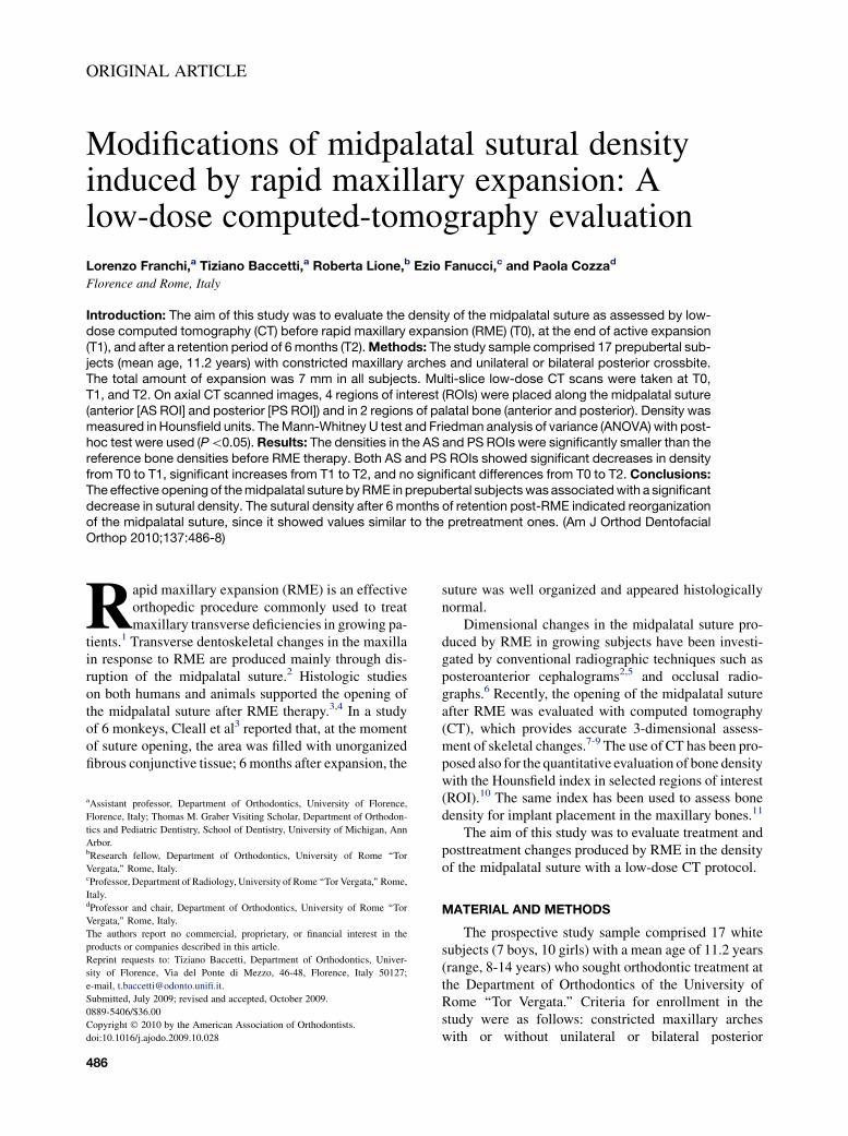
Fig. Axial scans taken A, before RME, B, at the end of the active expansion phase, and C, after re-tention of 6 months, with the 4 ROIs where density was recorded. The numbers indicate where theROIs were located at the 3 observation points.
American Journal of Orthodontics and Dentofacial Orthopedics Franchi et al 487Volume 137, Number 4
crossbite, and prepubertal stages of cervical vertebralmaturation (CS1-CS3) as assessed on lateral cephalo-grams.12 This project was approved by the ethical com-mittee, and informed consent was obtained from thepatients’parents.
Each patient underwent a standardized protocol ofRME with the butterfly palatal expander that followedthe basic design of Haas.13 The expansion screw was acti-vated twice per day (0.25 mm per turn) for 14 days, for a to-tal amount of screw expansion of 7 mm in all subjects.Then the screw was tied off with a ligature wire, and the ex-pander was kept in place as a passive retainer for 6 months.
Multi-slice low-dose CT scans were taken beforerapid palatal expansion (T0), at the end of the active ex-pansion phase (T1), and after the retention period of 6months (T2). The low-dose CT scan protocol was de-scribed previously.14 Standardized axial CT images par-allel to the palatal plane and passing through trifurcationof the maxillary right first molar were acquired and en-larged by a 3-times magnification factor with specificsoftware (Light-Speed 16, General Electric MedicalSystem, Milwaukee, Wis). On the enlarged images, 4ROIs that extended to an area of 1 mm2 were placedby 1 trained operator (R.L.) for the calculation of valuesof density in Hounsfield units (HU) (Fig). The operatorwas blinded to the patient being measured. The 4 ROIsin the palatal region were defined as follows.
1. Anterior sutural ROI (AS ROI): density valuesmeasured in the ROI located along the midpalatalsuture 5 mm in front of the center of the nasopala-tine duct at T0, T1, and T2.
2. Posterior sutural ROI (PS ROI): density valuesmeasured in the ROI located along midpalatal
suture 5 mm posterior to the center of the nasopala-tine duct at T0, T1, and T2.
3. Anterior bony ROI (AB ROI): density values mea-sured in the ROI located on the palatal bone 3 mmlaterally (on the right side) to the AS ROI at T0.
4. Posterior bony ROI (PB ROI): density values mea-sured in the ROI located on the palatal bone 3 mmlaterally (on the right side) to the PS ROI at T0.
The last 2 values were used as references to comparethe density of the midpalatal suture with that of the bonypalate.
Statistical analysis
One operator (R.L.) performed all measurements atthe same scanner console and repeated all measure-ments 1 month later. Systematic and random errors ofthe 4 measurements of the 17 subjects at all observa-tions periods were calculated with paired t tests andDahlberg’s formula,15 respectively. The Mann-WhitneyU test was used to compare the density between the su-tural ROIs and the bony ROIs at T0, and between the an-terior and posterior sutural ROIs at the 3 time points.The changes in density from T0 through T2 both inthe anterior and posterior sutural ROIs were contrastedwith the Friedman repeated measures ANOVA on ranksfollowed by the Tukey post-hoc test (SigmaStat 3.5, Sy-stat Software, Point Richmond, Calif). The level of sig-nificance was set at P \0.05.
RESULTS
No systematic errors for any of the 4 density mea-sures at the different observation periods were found

488 Franchi et al American Journal of Orthodontics and Dentofacial Orthopedics
April 2010
(P .0.05). Random errors for density ranged from 0.17at the PS ROI at T1 to 0.40 at the PS ROI at T0. Thevalues of bone density in the AS ROI and the PS ROIat T0 (563.3 6 183.2 HU and 741.7 6 167.1 HU, re-spectively) were significantly smaller than those in theAB ROI and the PB ROI at T0 (1057.5 6 129.4 HUand 1102.8 6 160.9 HU, respectively). The density inthe AS ROI was significantly smaller than in the PSROI at T0, but no significant differences were recordedbetween the 2 sutural ROIs at T1 and at T2. Both AS andPS ROIs showed significant decreases in density fromT0 to T1, significant increases from T1 to T2, and nostatistically significant differences from T0 to T2.
DISCUSSION
We applied low-dose CT to the evaluation of thechanges in density produced by RME in the midpalatalsuture after active expansion and after 6 months of re-tention. CT has been used in oral implantology to assessbone density for a proposed implant site.10,11 However,no previous studies with CT scans to evaluate the den-sity of craniofacial sutures in growing subjects werefound.
The gray level values recorded in the AS ROI beforeRME were significantly smaller than the values mea-sured in the PS ROI. This difference in density explainsthe greater opening in the anterior vs the posterior su-tural regions after the active phase of expansion, asdemonstrated in a previous study.9 Lione et al9 reportedthat the amount of transverse increase at the middle su-tural width (corresponding to PS ROI in our study) wasabout 70% of that observed in the anterior sutural width(corresponding to AS ROI in our study).
The gray level values of the midpalatal suture beforeRME were significantly smaller than those in the palatalbone, both anteriorly and posteriorly. Sutural densitiesin the AS ROI and the PS ROI were about 50% and30% smaller, respectively, than the corresponding pala-tal bone ROIs. This finding indicates that, at a prepuberalstage of skeletal development, the midpalatal suture hasnot completed its ossification. RME produced a suturalopening in all patients of our sample that was confirmedby a reduction of density after active expansion. Thevalue of density at T1 was 70% smaller than the valueat T0. After the 6-month retention period, the midpalatalsuture appeared to be reorganized with density similarto the value at T0. This confirms previous histologic ob-servations indicating that a retention period of 6 monthswith the RME in place allows for reorganization of thesuture.3 In future investigations, it would be interestingto test whether the density of the midpalatal suture tendsto increase in postpubertal subjects and whether
a different density correlates with a different amountof suture opening. Low-dose CT could then provideinformation about individual responsiveness of themidpalatal suture to RME therapy.
CONCLUSIONS
1. In prepubertal subjects, the density of the midpala-tal suture is less than the density measured in themaxillary bone.
2. The reduced sutural density after expansion indi-cates that RME therapy before puberty produceseffective opening of the midpalatal suture.
3. Six months of retention after RME allows reorgani-zation of the midpalatal suture with density valuessimilar to the pretreatment ones.
REFERENCES
1. McNamara JA. Maxillary transverse deficiency. Am J Orthod
Dentofacial Orthop 2000;117:567-70.
2. Wertz RA. Skeletal and dental changes accompanying rapid mid-
palatal suture opening. Am J Orthod 1970;58:41-66.
3. Cleall JF, Bayne DI, Posen JM, Subtelny JD. Expansion of the
midpalatal suture in the monkey. Angle Orthod 1965;35:23-35.
4. Melsen B. Palatal growth studied on human autopsy material. A
histologic microradiographic study. Am J Orthod 1975;68:42-54.
5. Cameron CG, Franchi L, Baccetti T, McNamara JA Jr. Long-term
effects of rapid maxillary expansion: a posteroanterior cephalomet-
ric evaluation. Am J Orthod Dentofacial Orthop 2002;121:129-35.
6. Inoue H. Radiographic observation of rapid expansion of human
maxilla. Bull Tokyo Med Dent Univ 1970;17:219-29.
7. Garib DG, Henriques JFC, Janson G, De Freitas MR, Coelho RA.
Rapid maxillary expansion—tooth tissue-borne versus tooth-
borne expanders: a computed tomography evaluation of dentoske-
letal effects. Angle Orthod 2005;75:548-57.
8. da Silva Filho OG, Lara TS, da Silva HC, Bertoz FA. Post expansion
evaluation of the midpalatal suture in children submitted to rapid
palatal expansion: a CT study. J Clin Pediatr Dent 2006;31:142-8.
9. Lione R, Ballanti F, Franchi L, Baccetti T, Cozza P. Treatment and
posttreatment skeletal effects of rapid maxillary expansion stud-
ied with low-dose computed tomography in growing subjects.
Am J Orthod Dentofacial Orthop 2008;134:389-92.
10. Shapurian T, Damoulis PD, Reiser GM, Griffin TJ, Rand WM.
Quantitative evaluation of bone density using the Hounsfield
index. Int J Oral Maxillofac Implants 2006;21:290-7.
11. Martinez H, Davarpanah M, Missika P, Celletti R, Lazzara R.
Optimal implant stabilization in low density bone. Clin Oral
Implants Res 2001;12:423-32.
12. Baccetti T, Franchi L, McNamara JA. The cervical vertebrae mat-
uration (CVM) method for the assessment of optimal treatment
timing in dentofacial orthopedics. Semin Orthod 2005;11:119-29.
13. Cozza P, Giancotti A, Petrosino A. Rapid palatal expansion in
mixed dentition using a modified expander: a cephalometric
investigation. J Clin Orthod 2001;28:129-34.
14. Ballanti F, Lione R, Fanucci E, Franchi L, Baccetti T, Cozza P. Im-
mediate and post-retention effects of rapid maxillary expansion
investigated by computed tomography in growing patients. Angle
Orthod 2009;79:24-9.
15. Dahlberg G. Statistical methods for medical and biological
students. New York: Interscience Publications; 1940.