modern physics updated
TRANSCRIPT

1
MODERN PHYSICS
(LEVEL III)
Radioactivity and its applications
Concepts to be learnt:
Introduction:
Antoine Henry Becquerel, was familiar with the work of Wilhelm Conrad Roentgen.Roentgen
discovered “mysterious" rays, called as X-rays. They werecapable of producing an image of
bones on a photographic plate. This excited scientists of his day, including Becquerel. Becquerel
chose to study the related phenomena of fluorescence and phosphorescence. In March of 1896,
quite by accident, he made a remarkable discovery.
Becquerel put his wrapped photographic plates away in a darkened drawer, along with some
crystals containing uranium. Much to his surprise, the plates were exposed during storage by
invisible rays or radiations coming from the uranium. This was the discovery of Radioactivity.
Becquerel did not pursue his discovery of radioactivitymuch but others did. Working in the
Becquerel laboratory, Marie Curie and her husband, Pierre, began what became a lifelong study
of radioactivity.
Becquerel had already noted that uranium radiations could turn air near the sample into
conductor of electricity. Pierre and Marie Curie measured the conductivity inducedby various
elements. They tested an ore of uranium, pitchblende, for its ability to turn air into a conductor of
electricity. The Curies found that the pitchblende (UO2) produced a current 300 times stronger
than that produced by pure uranium. After much grueling work, Curies were able to extract
enough polonium and another radioactive element, radium.
Radioactivity:
Becquerel, Pierre and Marie Curie worked with uranium salts. He kept the concentration of
uranium salt constant and measured the intensity of radiations by changing physical and
chemical conditions. He changed temperature, strength of electric and magnetic field, pressure
etc. The intensity of radiations remained the same. This clearly showed that the phenomenon
could not be due to orbital electrons. Therefore he concluded that the phenomenon was
connected to nucleus.
α, β & γ radiations, Properties of α, β & γ radiations, Mass Defect, Energy mass
relationship, Binding Energy. Fission, Radioactive transformation, Chain reaction,
Law of radioactive decay, half life time, Nuclear Fusion, Radioisotopes and their
uses.
Natural radioactivity is a spontaneous phenomenon. Certain heavy unstable elements emit
radiations from their nuclei and become stable. This property of the some substances is
known as radioactivity and the substances or elements which possess this property are called
as radioactive substances.
2
Types of radiations: (α, β& γ radiations):
Rutherford (1897) first found that the radiations emitted by radioactive substances are more than
one type. He found some are more penetrating and some rays are less penetrating. He called the
less penetrating rays as α-rays and more penetrating rays as β rays. In 1900, Sir Villard found
third type of rays which were more penetrating than others. He gave the name γ rays.
Rutherford’s Experiment:
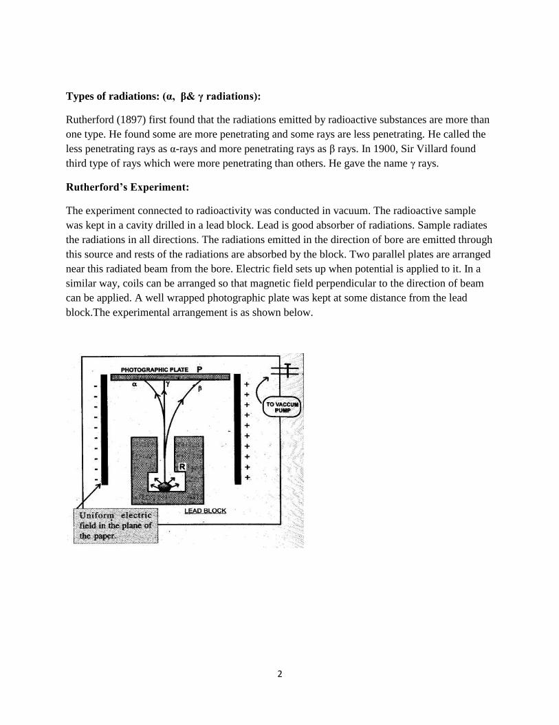
The experiment connected to radioactivity was conducted in vacuum. The radioactive sample
was kept in a cavity drilled in a lead block. Lead is good absorber of radiations. Sample radiates
the radiations in all directions. The radiations emitted in the direction of bore are emitted through
this source and rests of the radiations are absorbed by the block. Two parallel plates are arranged
near this radiated beam from the bore. Electric field sets up when potential is applied to it. In a
similar way, coils can be arranged so that magnetic field perpendicular to the direction of beam
can be applied. A well wrapped photographic plate was kept at some distance from the lead
block.The experimental arrangement is as shown below.

3
Observations:
There was a single dark spot on the photographic plate when electric or magnetic field was not
applied. This indicated that the radiations travel in straight line in absence of electric or magnetic
field (force).
When electric field was applied then there observed three spots on the photographic plate. It
clearly indicated that the radiated beam splits in to three in presence of electrostatic force. The
inferences drawn are as follows:
i) The part of the beam that deflected towards the negative plate must be consisted of
the positively charged particles. These are known as αrays.
ii) The part of the beam that deflected towards the positive plate must be consisted the
negatively charged particles. These are known as β rays.
iii) The rays that went in straight lineeven in presence of electrostatic field do not contain
any charged particles. Further it was proved that these are electromagnetic waves.
These are known as the γ rays.
iv) β particles deflect more than α particles. It indicated that β particles are lighter than
αparticles.
These radiations were studied systematically further. It was found that α particles are
helium nuclei.β particles are electrons and gammas are electromagnetic radiations.
There properties are tabulated as follows.

4
Sr.
No
properties α particles β particles γ rays
1 Nature Positively charged
helium nuclei, 2He4.
Negatively charged
particles called as
electrons.
Electromagnetic
waves of short
wavelengths
2 Charge Twice that of charge
on electron = 3.2 X
10-19
C.
= 1.6 X 10⁻¹⁹
C. No charge.
3 Mass = 6.62 X 10⁻²⁷
Kg. = 9.1 X 10⁻³¹
Kg. No mass.
4 Velocity Ranges from 106 m/s
to 107 m/s
Ranges from 1.08
X 108 m/s to 2.94 X
108 m/s
Velocity of light
i.e. 3 X 10⁸ m/s
5 Effect on
photographic
plate
Affects the
photographic plate.
Affects the
photographic plate.
Affects the
photographic
plate.
6 Ionization
power
Very strong, roughly
10000 times that of γ
particles and 100
times that of β
particles.
Moderately strong,
roughly 100 times
that of γ particles.
Very small
compared to α
and β particles.
7 Penetrating
power
Penetrating power is
very small, 1/10000
times that of γ rays.
Penetrating power
is moderate, 1/100
times that of γ rays.
Very high.
8 Effect of
Electric and
Magnetic field.
Deflected by electric
and magnetic field.
Deflected by
electric and
magnetic field.
No deflection
by electric or
magnetic field.
9 Fluorescence Produces
fluorescence in
fluorescent material
(ZnS).
Produces
fluorescence in
fluorescent material
(ZnS).
Produces
fluorescence in
fluorescent
material (ZnS).
10 Effect on Body Destroy living cells,
tissues and cause
biological damage.
As their penetration
power is large, they
cause greater
biological damage.
Penetration
power being
highest, cause
immense
biological
damage.
11 Applications They have large
momentum,
therefore large K. E.
They are used to
bombard atomic
nuclei to obtain new
nuclei.
They produce
x-rays when
stopped in tungsten.
They are very
useful in
treatment of
cancer.

5
Mass Defect:
Scientists have accurately found the masses of the proton and the neutron. They calculated the
masses of various nuclei. When sum of the masses of individual protons and individual neutrons
is made for a particular nucleus, it was found that this ‘sum’ mass was always greater than actual
mass of the nucleus. This difference was termed as the mass defect. It was denoted by ∆m.
In simple words, the definition of mass defect can be stated as follows:
Definition: The difference between the calculated mass of unbound system and experimentally
measured mass of nucleus is called mass defect. It is denoted by Δm. It can be calculated as
follows:
Mass defect = (calculated mass of unbound system) - (measured mass of nucleus)
i.e, Δm= (sum of masses of protons and neutrons) - (measured mass of nucleus)
Classically a bound system is at a lower energy level than its unbound constituents. The energy
must have liberated in the form of γ rays and heat.
Energy mass relationship:
Mass–energy equivalence is the concept that the mass of a body is a measure of its energy
content. In this concept the total internal energy E of a body at rest is equal to the product of its
rest mass m and a suitable conversion factor to transform from units of mass to units of energy.
Albert Einstein proposed mass–energy equivalence in 1905. The equivalence is described by the
famous equation
whereE is energy, m is mass, and c is the speed of light in a vacuum. The formula is
dimensionally consistent and does not depend on any specific system of measurement units.
E = mc2 has sometimes been used as an explanation for the origin of energy in nuclear processes,
but mass–energy equivalence does not explain the origin of such energies. The origin of the
nuclear energy must be in the work done against ‘the strong nuclear force’. Instead, this
relationship merely indicates that the large amounts of energy released in nuclear reactions can
be understood through mass loss. Mass-loss may give released energy from the system.
Binding Energy:
Binding Energy is the energy required to disassemble a whole system into separate constituents.
A bound system typically has a lower potential energy than its constituent parts. This keeps the

6
system together- often this means that energy is released upon the creation of a bound state. The
usual convention is that this corresponds to a positive binding energy.
In general, binding energy represents the mechanical work which must be done against the forces
which hold an object together. At the nuclear level, binding energy is also equivalent to the
energy liberated when a nucleus is created from other nucleons. This nuclear binding energy is
derived from the strong nuclear force and is the energy required to disassemble a nucleus into the
same number of free unbound neutrons and protons from which the nucleus is composed.
When the nucleons are grouped together to form a nucleus, they lose a small amount of mass,
i.e., there is mass defect. This mass defect is released as (often radiant) energy according to the
relation E = mc2; thus
binding energy = mass defect × c2.
Nuclear Fission:
We are aware that protons and neutrons are nuclear particles. Each proton has positive charge
and its magnitude is exactly equal to the magnitude of negative charge on electron. We know
that like charges repel each other. Neutrons are electrically neutral. Protons remain in small
region of nucleus because of strong nuclear force. This force is short range force; it is attractive
and approximately thousand times larger than electrostatic force. The attraction due to nuclear
strong force supersedes the electrostatic repulsion. This is one of the explanations of protons in
nucleus forming a stable system along with neutrons.
The mass of each nucleus was expected to be sum of the mass of all protons plus the mass of all
neutrons. This was relevant to law of conservation of mass. Actually the mass of each nucleus
was detected less than that calculated by sum formula. We have already seen ‘the mass energy
equivalence’. The protons and neutrons are bound by strong forces and some energy is removed
during the nucleus forming process. The mass corresponding to this energy (m = E/c2) appears as
less mass than expected. This is known as the ‘mass defect’.
As the atomic number of the nucleus goes on increasing the number of protons and the number
of neutrons goes on increasing. However the rate of growth of neutrons is more than rate of
growth of protons. Therefore the number of neutrons is more than protons in nucleus. As the
nucleus becomes very large in size like ,the nuclear strong force starts becoming
lesseffective. The nucleus becomes unstable and becomes susceptible to fission. It spontaneously
disintegrates and emits one or more of α, β& γ radiations. It also emits energy along with
radiations and comes to stable state.

7
Figure 1 :Uranium Sample
If such nuclei are bombarded by slow neutrons then first the neutron is absorbed. The nucleus
becomes more unstable and breaks in to fragments. This is called as the induced fission. Otto
Hahn and Strassman first discovered that breaks it to [
] and [ ] if it bombarded
by slow neutrons. Barium and Krypton atoms produced are called as the daughter nuclei. They
travel in different directions with large speed. Large amount of heat energy is also evolved in this
reaction. The reaction can be stated as:
+
= [ ] + [
] + 3 + Energy. [1]
Nuclear Fission: The splitting of the heavy nucleus into two lighter nuclei together with the
large amount of energy is called as the nuclear fission.
The amount of energy released in each fission event is about 200 MeV.
There are three neutrons released in the reaction.These three neutrons can trigger another three
atoms and produce nine more neutrons. This multiplication can continue and self
generating fission may occur. This process of self generating fission is called as the ‘chain
reaction’.
The chain reaction of is represented by following figure.

8
Nuclear fission as a source of energy:
The energy released can be calculated from the masses of fissionable atomss and the masses of
fragments. We have the following data
1 amu= 1.66 x 10⁻²⁷
kg.
Mass of = 235.043915amu.
Mass of = 1.008665 amu.
Mass of = 140.913900 amu.
Mass of = 91.897300 amu.
From this data mass of constituents of left hand side of equation [1] = 236.052580 amu and that
of all product atoms and particles on right hand side = 235.837195 amu. Therefore it is clear that
the mass of the products is less than the mass of the constituents.
Mass difference = 236.052580 – 235.837195
= 0.215385 amu.
Any mass can be converted in to equivalent energy using Einstein’s equation E = mc2. The
energy equivalent of 1a.m.u. is 931MeV.
Therefore the energy released in above fission reaction E is
E = mass difference x 931 MeV
= 0.215384 x 931 MeV
= 200.5 MeV.
This is the amount of energy released when one atom of undergoes fission.
This energy is released in the form of heat.
The average life time of slow neutrons is 10-4
seconds and that of faster ones is 10-7
sec. It means
in one second, 104 to 10
7 times uranium atoms will undergo fission. The number of breaking
atoms (fragmentations) will increase in geometric progression. If we assume minimum number

9
offragmentations 104 per sec, then [10
4]3
atoms will undergo fragmentation in one sec. It means
1012
atoms willfragment to produce approximately 1012
x 200 MeV = 2.00 x 10¹⁸
eV. = 0.32 J of
heat energy in one second. In the next second another 1036
fragmentations will produce 0.32 x
1036
J of heat energy. In third second 10108
fragmentations will take place producing 0.32 x 10108
J of energy. This is essentially the uncontrolled chain reaction which leads to the nuclear
explosion.
This reaction can be controlled by controlling the number of fissionable atoms in the sample. It is
also controlled by using the moderators like heavy water and cadmium rods. The system in
which the nuclear reaction is controlled and executed is called as the ‘the nuclear reactor’. The
evolved energy is used to produce electricity. The energy released in atomic reactor is used to
heat the water and make steam. Further the steam is superheated to run the steam turbine and
generate electricity. In India we have such atomic power plants at Trombay, Tarapur,
Kalpakkam, Kota etc. Still India produces only about 1.5% of its power generation by atomic
energy.
Radioactive transformation:
A radioactive nucleus emits α and β radiations and becomes the nucleus of entirely new element.
This is also called as the ‘nuclear disintegration’. The laws of radioactive decay are stated as
follows
I) When α particle is emitted, the mass number of radioactive atom decreases by 4 and
atomic number decreases by 2.
→
+
II) When β particle is emitted, the mass number of radioactive atom remains the same and
atomic number increases by 1.
→
+
III) When γ rays are emitted there is no change in the mass number and atomic number of
the element. The nucleus of the exited atom comes to lower state of energy by emitting the γ
radiations and becomes more stable.
Test your understanding:
We have seen that the fission of one atom of U-235 generates 202.5MeV. You are aware that
1eV = 1.6 × 10⁻¹⁹
J. This energy is liberated in the form of heat.Suppose you have 1 kg of
material. Each atom undergoes alpha decay once only. All energy liberated in the form of
heat is used to heat the water for taking bath. Calculate how many people can take bath by hot
water using this liberated nuclear energy. Assume that, a normal person consumes 10 liters of hot

10
water which is 300 C above the room temperature for his bath. (One mole contains Avegadro
number of atoms and 1 cal = 4.18 J.)
[Answer:
One atom emits 202.5 MeV = 202.5 X 106 X 1.6 X
-19 = 3.24 X 10
-11 J
Therefore in one mole will emit = 6.023 X 1023
X 3.24 X 10-11
J = 19.5 X10⁻¹²
J
Energy liberated in Alpha decay by 1 kg of U235
= Energy liberated per mole X number of moles
Energy liberated in Alpha decay by 1 kg of U235
= 19.54 (1000 /235) X 1012
= 83.15X 10¹²
J.
= (83.15 / 4.18) X 1012
= 19.90 X10¹²
cal.
A normal person consumes 10 lt.of hot water for taking bath, which is normally at 600C. If
water is heated from 300C to 60
0C, then
Amount of heat required for heating =10000 X 30 = 300000 = 3 X 10⁵ cal.
Therefore with the above heat (19.90 X 10¹²
/ 3 X 10⁵) = 6.63 X 10
7persons.
This means with this much amount of energy emitted only in Alpha decay, a huge population of
6.63 Cr can take the bath by hot water.
This gives better quantitative understanding about hugeness of amount of energy. ]
Laws of radioactive decay, half life time:
Rutherford and Soddy discovered that the rate at which radioactive substance disintegrates and
emits the radiations is independent of physical conditions like temperature, pressure etc. They
also proved that rate of radiation is independent of chemical condition. The rate of emission
remains same for that radioactive element when it is present in any compound. They also
detected that the substance reduces exponentially in its quantity. The law is stated as follows.
Law of radioactive decay: The rate at which number of atoms radiates at any instant is directly
proportional to the number of atoms present at that instant.
It clearly means the number of radioactive substance goes on reducing with time. The amount of
radioactive substance goes on reducing with time. The substance becomes half of its quantity
after some time.
11
Half life time (T1/2): The half life time of any radioactive substance is defined as the time
required reducing that substance to half of its original quantity. It is characteristics of that
substance and it is denoted by λ.
Nuclear Fusion:
We have seen that heavy atoms are unstable. They disintegrate and emit radiations. They break
to from stable product nuclei. In another natural phenomenon very light atoms like hydrogen and
its isotopes combine to form more stable elements. This natural phenomenon is called as the
fusion.
Fusion: The process of combining lighter atoms to form stable nuclear atom by emitting energy
is called as the nuclear fusion.
We all know that the life on earth is due to energy that we receive from Sun. Sun is the huge ball
constituted of about 71% of hydrogen, 27.1% of helium. There are other elements found and they
are in rest of about 2%. These elements are oxygen, carbon, nitrogen, silicon, magnesium,
neon,ironand sulphur. We note here that the compositions given in reference books and on
internet are different and list of elements also is different. We will go with Wikipedia as the most
authentic source of information.
The spectroscopic measurements state that the surface temperature of the Sun is nearly 5800 K
while the estimated temperature of the core of the sun is about millions of Kelvin. The reaction
on the surface of the Sun is the fusion reaction of hydrogen to helium.
6C14
–Isotope and carbon dating:
Cosmic rays essentially contain showers of high energy charged particles as well as high energy
neutrons. Most of the charged particles are absorbed in the ionosphere and they increase the
thickness and density of the ionosphere. This is due to earth’s magnetic field. Few charged
particles reach the atmosphere penetrating the entire atmosphere.
There are high energy neutrons also in the cosmic rays. These neutrons are absorbed in the upper
layers of atmosphere. These neutrons combine with atmospheric nitrogen and form new isotope
of carbon. The reaction is given by
0n1 + 7N
14→ 1H
1(proton) + 6C
14
6C14
isotope of carbon is radioactive. It is reacts with the atmospheric oxygen to form carbon
dioxide molecules. The percentage of radioactive carbon dioxide in atmosphere remains almost
constant. This is because radioactive carbon dioxide decays by emitting β particles to form again
nitrogen. The reaction is stated as
6C14
→ 7N14
+ 0e-1
(beta particle)

12
Green plants consume carbon dioxide during photosynthesis. Hence plants absorb this
radioactive carbon along with normal non radioactive carbon dioxide. All living animals eat
plants and they grow. Hence each living thing possesses a very small percentage of radioactive
carbon in them. Radioactive carbon decays to convert itself in nitrogen in living animals also.
Hence the percentage of radioactive carbon is constant till the animal is living. The amount of
radioactive goes on reducing when the animal dies. Hence by measuring the rate of beta particle
emission, date at which animal died, can be calculated.This process is known as the carbon
dating.
Half life period of 6C14
carbon is extremely large, 5730 years. Hence carbon dating is extremely
useful in archeology, in deciding the age of the dugout samples. So also it is useful in deciding
the age of bones and teeth of the buried human and animalbodies.
Uses of Radioactivity:
1 )Uses in medicine:
The affected part of the patient’s body is exposed to radioactive source. Generally γ radiations
are used. The deceased tissue dies and helps to control the decease. This therapy is called as
‘radiation therapy’. Radiation therapy is used in cancer, tumor, leukemia etc.
Some radioactive salts like radioiodine, radioactive NaCl etc are used is detecting decease
affected areas.
2) Scientific uses:
To study age of rocks, planets and other ancient objects.
To study growth of plants.
To study nature of nuclear forces.
Concept:
Thermionic emission: We have seen that there are free electrons inside the metal when we
learnt the current electricity. These electrons are mobile within the metal as a result of ‘metallic
bond’ which is characteristic of metals. It is customarily said that they form an electron gas
inside the metal. Therefore each electron is said to possess certain average kinetic energy. This
kinetic energy is a function of absolute temperature (T) of the metal. It is assumed that the
Thermionic emission, Cathode ray tube, Cathode rays and their properties, Deflection
of cathode rays by electric and magnetic field, X-rays, their properties and uses,
Radiation hazards.
hazards.

13
electrons do not come outside the metal on its own at room temperature. This is because metal as
a whole is neutral and it tries to remain neutral. It will remain with one ‘e’ positive charge if an
electron leaves the piece of metal. This provides binding on the electron to remain within the
metal. In terms of kinetic energy it is said that the average kinetic energy of an electron is
insufficient to cross the surface of the metal.
Truly speaking, each atom gives one or more electrons to form a metallic bond and it becomes an
ion. The average positions of these ions remain fixed. They are said to vibrate about their mean
positions. They possess some average kinetic energy. The electrons which are free also possess
the average kinetic energy. As the temperature of the metal goes on increasing the average
kinetic energy of ions as well as electrons goes on increasing. When theaverage kinetic energy of
an electron increases beyond certain limit, the electron comes out of the metal surface. As the
electron comes out due to thermal energy, it is known as the ‘thermion’.
Thermionic emission: The phenomenon of emission of electrons from the surface of a metal due
to increase in temperature is called as the ‘thermionic emission’.
Work function: The minimum energy required for an electron to ooze out from the metal
surface is known as the work function of that metal. It is conventionally denoted by ‘Ф’.
The work function is the characteristics of each metal. Therefore the work function is different
for different metals.
The work function is normally measured in ‘eV’, which is the unit of energy.
One eV:The energy acquired by the electron when it accelerates through one volt is called as one
eV energy.
Rate of emission of thermions:
It is experimentally found that the rate of emission of thermions depends on following three
factors:
I) Nature of the metal surface: The rate of thermions is inversely proportional to work
function of the metal. i.e. If the work function of the metal is low then the rate of
emission is high and if the work function of the metal is high then the rate of
emission is low.
II) Temperature of the metal: The rate of emission is directly proportional to the
temperature of the metal. The rate of emission of thermions increases with increase
in temperature.
III) The surface area of the metal: The rate of emission of thermions is directly
proportional to surface area of the metal, i.e. larger the surface area greater is the
rate of emission.
Characteristics of good electron emitter:

14
I) A metal is a good emitter if its melting point is high. The metal does not melt quickly
on heating.
II) A metal becomes good emitter when the work function is low.
The melting point of tungsten (W) is 3655 K. Tungsten is normally used for making filaments.
The work function of tungsten is 4.6 eV. Thermions start emitting from 2500 K.
Thoriated tungsten filament is the tungsten filament coated with Thorium dioxide. The work
function of such filament is 2.6 eV. The filament starts emitting electrons from 2000 K onwards.
If tungsten is coated with alkali metal oxides then electron emission is at fairly low temperatures.
The work functions of such compositions are in the range 1 eV to 1.5 eV and the electron
emission starts at about 1000 K.
Application: Thoriated tungsten filaments are used for obtaining electron beam in Cathode ray
tubes. CRTs are extensively used in Cathode ray oscilloscopes, TVs, computer monitors and
monitors in medical application.
Thotiated tungsten filaments were used in incandescent lamps. The use of such lamps is reduced
as the their efficiency is low. Still they are used in automobile lamps.
Cathode ray tube, Cathode rays and properties, Deflection of cathode rays electric and
magnetic field:
The modern atomic theory is also nearly 100 years old. J.J. Thompson, an English chemist
discovered a particle 1836 times lighter than the Hydrogen atom. It was named as the ‘Electron.’
He developed an instrument called as ‘Cathode Ray Tube’ (CRT). The schematic diagram of the
CR tube is as shown below:
15
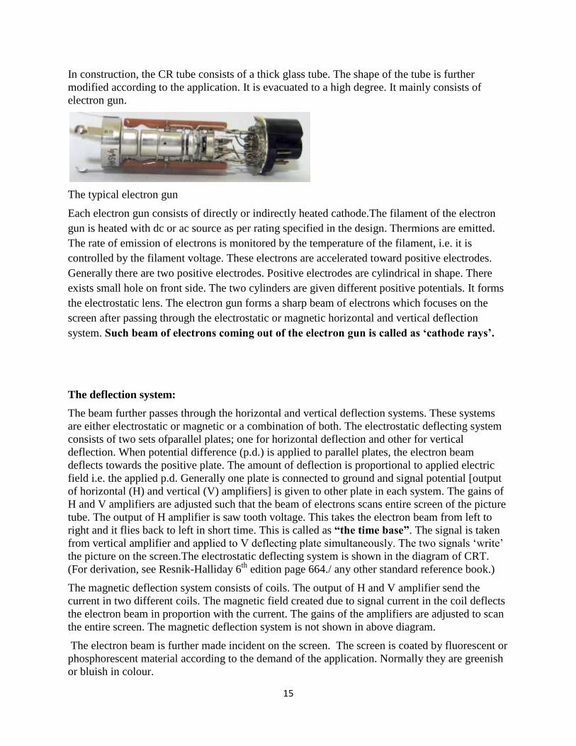
In construction, the CR tube consists of a thick glass tube. The shape of the tube is further
modified according to the application. It is evacuated to a high degree. It mainly consists of
electron gun.
The typical electron gun
Each electron gun consists of directly or indirectly heated cathode.The filament of the electron
gun is heated with dc or ac source as per rating specified in the design. Thermions are emitted.
The rate of emission of electrons is monitored by the temperature of the filament, i.e. it is
controlled by the filament voltage. These electrons are accelerated toward positive electrodes.
Generally there are two positive electrodes. Positive electrodes are cylindrical in shape. There
exists small hole on front side. The two cylinders are given different positive potentials. It forms
the electrostatic lens. The electron gun forms a sharp beam of electrons which focuses on the
screen after passing through the electrostatic or magnetic horizontal and vertical deflection
system. Such beam of electrons coming out of the electron gun is called as ‘cathode rays’.
The deflection system:
The beam further passes through the horizontal and vertical deflection systems. These systems
are either electrostatic or magnetic or a combination of both. The electrostatic deflecting system
consists of two sets ofparallel plates; one for horizontal deflection and other for vertical
deflection. When potential difference (p.d.) is applied to parallel plates, the electron beam
deflects towards the positive plate. The amount of deflection is proportional to applied electric
field i.e. the applied p.d. Generally one plate is connected to ground and signal potential [output
of horizontal (H) and vertical (V) amplifiers] is given to other plate in each system. The gains of
H and V amplifiers are adjusted such that the beam of electrons scans entire screen of the picture
tube. The output of H amplifier is saw tooth voltage. This takes the electron beam from left to
right and it flies back to left in short time. This is called as “the time base”. The signal is taken
from vertical amplifier and applied to V deflecting plate simultaneously. The two signals ‘write’
the picture on the screen.The electrostatic deflecting system is shown in the diagram of CRT.
(For derivation, see Resnik-Halliday 6th
edition page 664./ any other standard reference book.)
The magnetic deflection system consists of coils. The output of H and V amplifier send the
current in two different coils. The magnetic field created due to signal current in the coil deflects
the electron beam in proportion with the current. The gains of the amplifiers are adjusted to scan
the entire screen. The magnetic deflection system is not shown in above diagram.
The electron beam is further made incident on the screen. The screen is coated by fluorescent or
phosphorescent material according to the demand of the application. Normally they are greenish
or bluish in colour.

16
Monitor of your computer, Picture tube of T.V. in your home, various monitors in speciality
hospitals, CRO- cathode ray oscilloscopes in modern laboratories essentially contain the CR
tubes. They are extensively used in today’s modern lifestyle.
Properties of the Cathode Rays:
1. Cathode rays are negatively charged particles. They are the stream of electrons.
2. The energy of the electron is decided by the positive potential on the anode.
3. Cathode rays produce heat when they are retarded in metallic anode.
4. They are charged particles, therefore they can be deflected by electric and magnetic field.
5. They are energetic particles therefore they produce fluorescence and phosphorescence
when they strike certain materials like ZnS, Al2S3, soda glass, quartz, etc.
6. Cathode rays produce X-rays when they are stopped in metals of high density, e.g.
Tungston, Copper etc.
7. Cathode rays emerge normally from the cathode-surface and are not affected by the
position of the anode.Cathode rays travel in straight line and form shadows.
8. Cathode rays are particles having mass; they carry momentum along with them.Cathode
rays exert mechanical pressure.

17
9. Cathode rays can produce physical and chemical changes: The cathode rays affect the
photographic plate and turn the colour of lithium chloride into violet.
10. Cathode rays can ionize gases : Cathode rays, on colliding with the atoms of gases, eject
electrons from them.
11. Cathode rays can penetrate through metal-foils : Cathode rays penetrate through light
metals. If an aluminium foil is placed normally in the path of cathode rays, the rays are
found on the other side of the foil.
Background Radiations:
There are some sources of radiations which continuously emit radiations. Further it is not
possible to control them. These radiations are fairly small in amount therefore generally we do
not take precautions also. Whenever we count the radiations from a source or from certain
affected body / object, these radiations also exist. We subtract the ‘background count’ in
precision measurements. Otherwise these radiations are ignored. These radiations are called as
the background radiations.
We ignore the hazards of background radiations because they are too small in amount and it is
impracticable to take corrective measures all the time.
The background radiations are due to
i) Cosmic rays reaching the earth constantly.
ii) Radioactive substances present around like radium, carbon-14, potassium-40,
polonium etc.
iii) Radioactive rocks present in the Earth’s crust.

18
X-Rays:
On December 22 1895, Roentgen "photographed" his wife's hand,
revealing the unmistakable image of her skeleton, complete with
wedding ring. Roentgen's wife had placed her hand in the path of
X-rays. Roentgen's discovery of these "mysterious" rays, capable of
producing an image on a photographic plate excited scientists of his
days.
Production of X-rays:
Roentgen discovered X-rays while working with crookes tube.
Later on Coolige tubes were discovered for the production of X-
rays. It was known to Roentgen that when high energetic electrons
are stooped in dense target like tungsten or copper, X-rays are produced. It also produces large
amount of heat, hence tubes with water cooling systems for target were developed. A typical
tube is as shown below.
X-ray tube:
C is the filament, generally made up of tungsten. Uh is the filament supply. ‘A’ is thick copper or
tungsten target. It works as the anode. Ua is the high voltage dc source ranging up to 100 kV. Win
and Wout are the inlet and outlet for the water to cool the target. Water removes heat from the
target and increases the life of the target i.e. the life of the X-ray tube. The whole assembly is
fitted in a highly evacuated thick glass tube.
When filament heats, it emits electrons.Anode is positive and it is given high voltage. The
electrons emitted from the cathode are subjected to high electric field. They are accelerated to
high value. They hit the target and lose their K. E. to the target. As they are continuously slowed
down, they emit continuous X-rays. They knock out electrons from innermost K or M shell of

19
target atoms. When electrons from outer shells take their position, they emit characteristic X-rays
of the target atoms. Rest of the energy of incident electron is converted in to heat energy. Heat is
taken away by water else the target melts or ‘burns’ out.
X-rays is a form of electromagnetic radiation. X-rays have a wavelength in the range of 0.01 to
10 nanometers, corresponding to frequencies in the range from 3 × 10¹⁶
Hz to 3 × 10¹⁹
Hz and
energies in the range 120 eV to 120 keV. They are shorter in wavelength than UV rays and
longer than gamma rays.
Properties of X-rays:
i) X-rays are e. m. waves therefore they can be reflected, refracted, diffracted,
interferred and polarized.
ii) Their wavelengths are in the range of 0.01 nm to 10 nm and frequencies are in the
range of 3 × 10¹⁶
Hz to 3 × 10¹⁹
Hz.
iii) They are e. m. waves they travel with the speed of light.
iv) They are high energy waves, therefore they affect photographic plate, and they
produce fluorescence and phosphorescence.
v) They penetrate through materials, papers, tissues etc.
vi) They are stopped by the metals, bones and teeth. Hence shadows of bones and teeth
can be taken by X-rays.
Uses of X-rays:
Medical uses:i) to photographs the fractured bones ii) tooth photography iii) tomography
of tissues iv) radiotherapy and there are many other medical uses of X-rays. X-ray crystallography in which the pattern produced by the diffractionof X-rays through the
closely spaced lattice of atoms in a crystal is recorded and then analysed to reveal the nature of that lattice. A related technique, fiber diffraction, was used by Rosalind Franklin to discover the double helical structure of DNA.[46]
X-ray astronomy, which is an observational branch of astronomy, which deals with the study of X-ray emission from celestial objects.
X-ray microscopic analysis, which uses electromagnetic radiation in the soft X-ray band to produce images of very small objects.
X-ray fluorescence, a technique in which X-rays are generated within a specimen and detected. The outgoing energy of the X-ray can be used to identify the composition of the sample.
Industrial radiography uses X-rays for inspection of industrial parts, particularly welds. Airport security luggage scanners use X-rays for inspecting the interior of luggage for security
threats before loading on aircraft. Border security truck scanners use X-rays for inspecting the interior of trucks for at country
borders. X-ray fine art photography X-ray photoelectron spectroscopy is a chemical analysis technique relying on the photoelectric
effect, usually employed in surface science.

20
Radiation Hazards:
Nuclear radiations are energetic particles. They impart energy when they are incident on the
body. Especially ‘γ rays’ possess high energy. They may knock out electrons from organic
molecules. If a certain person remains exposed to energetic radiations for long time then
molecules may break up or their normal behavior will be hampered. Sometimes the death of that
organism is also possible.
There are two main types of damages.
1. Pathological damages:If certain organ is exposed to high dose of radiations then that
organ stops its normal functioning and it becomes dead. Such damage is called as the
pathological damage.
2. Genetic damage: The high energy radiations may affect the amino acids at the gene
sites. Such damage will not be seen immediately. It will show its effects in next
generations. Such damages are called as the genetic damages.
It clearly gives the message that we must take proper precautions while handling the
radioactive materials and working in system having radioactive materials.
Safety precautions:
1. Nuclear reactors should be embedded in thick concrete walls. This is to prevent from the
radioactive gases and neutrons.
2. Nuclear material must be kept in thick lead containers with proper lead covered windows.
3. Nuclear material should be handled with mechanical tongs.
4. Workers in nuclear establishments must wear lead lined aprons, gloves and they should
wear special lead glasses for protection.
5. Workers must wear special film badges. These badges are also exposed to the radiations
along with the persons. These badges indicate the amount of dose received so that proper
care can be taken.
6. Periodic and compulsory medical checkup is essential to the persons working in the
nuclear establishments.
References:
1) http://www.accessexcellence.org/AE/AEC/CC/radioactivity.php
2) Wikipedia