modeling physical changes in hsv …d-scholarship.pitt.edu/9515/1/saraannjackson2005.pdf ·...
TRANSCRIPT

MODELING PHYSICAL CHANGES IN HSV GENOMES THAT OCCUR DURING
LYTIC AND LATENT INFECTIONS: THE ROLE OF ICP0.
by
Sara Ann Jackson
BS, Allegheny College, 2000
Submitted to the Graduate Faculty of
University of Pittsburgh in partial fulfillment
of the requirements for the degree of
Doctor of Philosophy
University of Pittsburgh
2005
ii
UNIVERSITY OF PITTSBURGH
FACULTY OF ARTS AND SCIENCES
This dissertation " Modeling physical changes in HSV genomes that occur during lytic and latent
infections: the role of ICP0" was presented
by
Sara Ann Jackson
It was defended on
October 19, 2005
and approved by
Dr. Neal DeLuca, Ph.D. Dr. Stephen Phillips, Ph.D.
Dissertation Director/Major Advisor Department of Molecular Genetics
Department of Molecular Genetics and Biochemistry
and Biochemistry
Dr. Donald DeFranco, Ph.D. Dr. Richard Wood, Ph.D.
Department of Pharmacology Department of Pharmacology
Dr. Saleem Khan, Ph.D.
Department of Molecular Genetics
and Biochemistry

iii
A modified version of data presented in Chapter 3 appeared in " Relationship of Herpes Simplex
Virus genome configuration to productive and persistent infections." Sara A. Jackson & Neal A.
DeLuca, Proceedings from the National Academy of Sciences, 2003, volume 100(13), pages
7871-7876, copyright © 2003, National Academy of Sciences of the United States of America.
All rights reserved.

iv
MODELING PHYSICAL CHANGES IN HSV GENOMES THAT OCCUR
DURING LYTIC AND LATENT INFECTIONS: THE ROLE OF ICP0
Sara Ann Jackson, Ph.D.
University of Pittsburgh, 2005
Herpes Simplex Virus (HSV) is a pervasive human pathogen that can establish both
symptomatic productive infections and asymptomatic latent infections. During infection,
the HSV genome undergoes physical changes that are regulated by cellular and viral
proteins. These changes lead to either a template for genome replication during the
productive cycle or a persistent stable genome configuration during latency. Changes in
viral genomes and events leading to these changes during a particular life cycle are not
clearly understood. Using both Gardella gel analysis for circular HSV genomes and
restriction enzyme analysis for end-to-end linkages of HSV genomes, we show that HSV
genomes circularize only in the absence of the HSV immediate early gene ICP0. In the
presence of ICP0 genome circularization is inhibited. Although HSV replication has been
previously thought to initiate by a theta mechanism from a circular genome template, these
results suggest that HSV replication initiates from a linear genome template due to the
presence of ICP0 during lytic infection. We also show that circular genomes are the stable
form during long-term persistent infections that model latency. Because HSV genomes
begin as linear, double-stranded DNA during infection, host cells may treat the ends of
incoming genomes as DNA double strand breaks (DSB) and subsequently repair these ends
by circularization of genomes. However, it is unclear if/how these DSB repair pathways
contribute to the manipulation of HSV genome configurations during infection and how
viral proteins, in particular ICP0, may alter/counteract this repair response to form a
template for replication during the productive cycle. Here we show that the cellular DSB
repair mechanism, non-homologous end-joining (NHEJ), is the major mechanism by which
HSV genomes are circularized. ICP0 not only inhibits HSV genome circularization but
also affects the abundance of proteins involved in NHEJ and the distribution of other
repair proteins during infection. The study presented here begins to uncover how the
interplay between host cell repair responses and the virus' reply to these responses
contributes to forming either a genome template for replication during the productive cycle
or a persistent stable configuration during latency.

v
TABLE OF CONTENTS
1. INTRODUCTION ..............................................................................................................1
1.1. Overview.....................................................................................................................1
1.2. Herpesviridae ..............................................................................................................3
1.3. HSV pathogenesis .......................................................................................................4
1.4. HSV genome ...............................................................................................................6
1.5. HSV productive infection ............................................................................................8
1.5.1. Cascade of viral gene expression..........................................................................9
1.5.2. Mechanism of replication...................................................................................11
1.5.3. Host cell death ...................................................................................................13
1.6. HSV latency ..............................................................................................................14
1.7. The role of ICP0 during HSV infection......................................................................15
1.7.1. Structure and function........................................................................................16
1.7.2. Protein stability and turnover .............................................................................16
1.7.3. ND10.................................................................................................................18
1.8. Host cell DNA repair pathways during HSV infection ...............................................19
1.8.1. Non-homologous end-joining (NHEJ)................................................................20
1.8.2. Homologous recombination (HR) ......................................................................21
1.8.3. Mre11/Rad50/NBS1 repair response ..................................................................22
2. RATIONALE....................................................................................................................24
3. RELATIONSHIP OF HERPES SIMPLEX VIRUS GENOME CONFIGURATION TO
PRODUCTIVE AND PERSISTENT INFECTIONS.................................................................30
3.1. Abstract.....................................................................................................................30
3.2. Introduction...............................................................................................................30
3.3. Materials and Methods ..............................................................................................32
3.3.1. Cells and Viruses. ..............................................................................................32
3.3.2. Gardella gels......................................................................................................33
3.3.3. Electroelution. ...................................................................................................34
3.4. Results.......................................................................................................................34
3.4.1. Accumulation of low-mobility, non-linear HSV genomes in the absence but not
presence of ICP0. ..............................................................................................................34
3.4.2. Low-mobility HSV genomes represent circularized genomes.............................36
3.4.3. Multiplicity dependence and kinetics of genome circularization during infection38
3.4.4. Low-levels of ICP0 inhibit genome circularization.............................................39
3.4.5. ICP0 inhibits genome circularization during productive infections. ....................41
3.4.6. Circular genomes are the persistent form during long-term quiescent infections
that model latency. ............................................................................................................43
3.5. Discussion .................................................................................................................45
4. ICP0 INHIBITS CIRCULARIZATION OF HSV GENOMES DURING PRODUCTIVE
INFECTIONS: COMPARISON OF GARDELLA GEL ANLAYSIS FOR CIRCLES AND
RESTRICTION ENZYME ANALYSIS FOR END-TO-END LINKAGES OF GENOMES.....49
4.1. Abstract.....................................................................................................................49

vi
4.2. Note: Introduction/Results/Discussion ......................................................................49
5. Inhibition of DNA repair and genome circularization by HSV ICP0..................................64
5.1. Abstract.....................................................................................................................64
5.2. Introduction...............................................................................................................65
5.3. Methods ....................................................................................................................68
5.3.1. Viruses. .............................................................................................................68
5.3.2. Cells. .................................................................................................................68
5.3.3. Immunofluorescence..........................................................................................69
5.3.4. Western Blots. ...................................................................................................70
5.3.5. Gardella gel analysis. .........................................................................................71
5.4. Results.......................................................................................................................71
5.4.1. Formation of M/R/N complexes and H2AX foci as a function of ICP0.............71
5.4.2. Levels of DNA repair proteins as a function of ICP0..........................................76
5.4.3. NHEJ is involved in circularization of HSV-1 genomes .....................................79
5.5. Discussion .................................................................................................................83
6. SUMMARY AND GENERAL DISCUSSION..................................................................88
6.1. Summary of results....................................................................................................88
6.2. New models of HSV replication ................................................................................91
6.3. Pivotal role of ICP0 in promoting productive cycle....................................................94
6.4. Circularization, latency, and reactivation ...................................................................96
BIBLIOGRAPHY.....................................................................................................................99

vii
LIST OF FIGURES
Figure 1. The HSV genome........................................................................................................7
Figure 2. Gardella gel analysis of d109- and d106-infected cells. ..............................................35
Figure 3. Characterization of the low-mobility form of the d109 genome. ................................37
Figure 4. ICP0 expression from adenovirus inhibits circularization of d109 in trans. ................40
Figure 5. Circularization of the HSV genome during productive infection in the presence and
absence of ICP0. ...............................................................................................................42
Figure 6. Stability of circular and linear d109 genomes during long-term persistent infection...44
Figure 7. Analysis of d99/I2.3 genomes. ..................................................................................51
Figure 8. Western blot analysis of d99/I2.3 virus......................................................................53
Figure 9. ICP0 inhibits HSV genome circularization during infection. .....................................55
Figure 10. ICP0 inhibits formation end-to-end genome linkages during infection.....................57
Figure 11. Formation of M/R/N foci as a function of ICP0 during abortive infection.................72
Figure 12. Rad50 and NBS1 foci accumulate and persist during abortive infection in the
presence of ICP0. ..............................................................................................................74
Figure 13. Formation of Mre11 and NBS1 foci as a function of ICP0 during productive infection
in which replication is inhibited.........................................................................................75
Figure 14. Colocalization of Mre11 and H2AX foci in the absence but not presence of ICP0
during abortive infection. ..................................................................................................77
Figure 15. Global level of DNA repair proteins as a function of ICP0. .....................................78
Figure 16. Circularization of HSV genomes in cells deficient in NHEJ. ...................................80
Figure 17. NBS1 does not affect HSV genome circularization..................................................82

1
1. INTRODUCTION
1.1. Overview
The primary goal of viruses is not to kill, but to reproduce resulting in progeny that will spread
and again initiate the infectious cycle. Viruses lack the machinery to replicate independently,
and subsequently require a host to provide many of the necessities required for replication.
Viruses have evolved mechanisms to manipulate hosts into facilitating this replication during
infection. However, many of the normal physiological processes of the host are compromised by
the virus, and consequently the host attempts to inhibit many of the processes required for virus
production. Viruses have adapted mechanisms to evade this host anti-viral response. One such
escape mechanism is the ability of viral genomes to persist in cells in a non-toxic, quiescent state
called latency until an appropriate environment for virus production is stimulated.
Latency is a hallmark of herpesvirus infections and is analogous to the well-studied
lambda bacteriophage lysogenic state, during which most genes encoded by the integrated
bacteriophage genome are silent and only few bacteriophage genes that maintain lysogeny are
expressed (142). As with lysogenic bacteriophage infections, during latent herpesvirus
infections only a limited group of viral genes are expressed, but the productive cycle can be
readily stimulated. The cycling between productive and latent infection provides a continuous
mechanism for herpesvirus production. During the productive, or lytic, cycle all herpesviruses
share a general progressive cascade of gene expression that is executed through a combination of
virus and host cell transcription and translation machinery. The earliest genes expressed in this
cascade prepare the cell for further viral gene expression, which in turn promotes virus

2
replication and production of progeny virions. In the case of herpes simplex virus (HSV), this
cascade of gene expression is initiated by a combination of host cellular factors and viral proteins
provided by the infecting virion.
During HSV-1 productive infection, the tegument protein VP16 and host factors bind
motifs present in HSV promoters and thereby initiate transcription of the first class of genes
within the progressive cascade. Of the genes activated by this complex (ICP0, ICP4, ICP22,
ICP27, and ICP47), four of them (ICP0, ICP4, ICP27, and ICP22) promote further viral gene
expression. The ability of these genes to facilitate and in some cases, activate, viral gene
expression largely depends on the cellular proteins/mechanism with which each protein interacts.
Of these four genes, only ICP0 leads to indiscriminant transactivation of both cellular and viral
genes. ICP0 is a major regulator of the balance between productive and latent infections, with its
presence facilitating the productive cycle both during primary infections and reactivation from
latency. ICP0 is the only HSV protein that induces gene expression from otherwise repressed
HSV genomes during latency. One relatively well characterized activity of ICP0 results in
degradation of cellular proteins. It follows that ICP0 is thought to create conditions permissive
for viral gene expression and replication through inhibiting repressors of the productive cycle.
During infection, the HSV genome undergoes physical alterations that are regulated by
both cellular and viral proteins. These changes ultimately lead to a template for genome
replication during the productive cycle or a persistent stable genome configuration during
latency. During the productive cycle HSV genomes are very dynamic: they actively express
genes, replicate, and are packaged into progeny virions. During latency viral genomes are
somewhat static, persisting in a stable transcriptionally repressed state. Changes in HSV
genomes and events leading to these changes during a particular life cycle are not clearly
3
understood. This is largely due to the fact that little is known about how the host cell responds to
incoming HSV genomes and how the virus manipulates this response. Because HSV genomes
begin as linear, double-stranded DNA during infection, host cells may treat the ends of incoming
genomes as DNA double-strand breaks (DSB). However, it is unclear if/how these DSB repair
pathways contribute to manipulation of HSV genome configurations during infection and how
viral proteins may alter/counteract this repair response. The study presented here begins to
uncover how the interplay between host cell repair responses and the virus' reply to these
responses contributes to forming either a genome template for replication during the productive
cycle or a stable configuration during latency. It highlights the role of ICP0 as both a major
facilitator to productive infection and a regulator of genome configuration.
1.2. Herpesviridae
Herpesviruses consist of a large family of viruses that infect all groups of vertebrates, including
humans. There are eight known human herpesviruses: herpes simplex virus type-1 and type-2
(HSV1 and HSV2), human cytomegalovirus (HCMV), varicella-zoster virus (VZV), Epstein-
Barr virus (EBV), and human herpes virus 6, 7, 8 (HHV6, HHV7, and HHV8). These viruses
cause a variety of significant diseases in humans. For example, HSV1 and HSV2 cause severe
facial and genital lesions, respectively; VZV causes chicken pox and then shingles during
reactivation from latency; and EBV causes mononucleosis and B cell lymphomas. In
immunocompromised patients, these infections can be more severe, with otherwise mild HCMV
and HHV8 infections developing into retinitis and Kaposi's sarcoma, respectively. (162)
All members of the Herpesvirus family share structurally similar virions separated into
layers containing a linear double-stranded DNA genome ranging from 120-230 KB at the core
(162). The viral DNA is packaged into an icosohedral structure composed of 162 capsomer

4
proteins (162). Surrounding the capsid is an amorphous tegument region that can contain viral
proteins responsible for initiation of the productive cycle. Surrounding the tegument is a lipid
envelope from which glycoprotein spikes protrude to the outer surface (156). These
glycoproteins facilitate interactions with host cells and many times designate the tropism of each
virus for a particular tissue or cell (156).
Although all share the ability to form productive infections and persist in a latent state
from which the productive cycle can be stimulated, herpesviruses can be divided into three
groups: alpha, beta, and gamma (162). Classification into these three groups is based upon tissue
tropism, pathogenicity, and properties during infection of cultured cells (162).
Alphaherpesviruses can infect a broad range of cells and hosts, in which they replicate vary
rapidly and can reside during latency in neurological tissues. Of the human herpesviruses,
HSV1, HSV2, and VZV are classified as alphaherpesviruses. Betaherpesviruses and
gammaherpesviruses infect a much more restricted host range, in which they replicate slowly in
cells of glandular and/or lymphatic nature. HCMV, HHV6, and HHV7 are classified as
betaherpesviruses, while EBV and HHV8 are classified as gammaherpesviruses. Understanding
each herpesvirus within the context of its sub-group can be useful in understanding the functions
of homologous viral genes during various herpesviral infections, albeit in different environments.
1.3. HSV pathogenesis
As with all herpesviruses, HSV can establish either a symptomatic productive (lytic) infection or
an asymptomatic latent infection. HSV enters the host by infection of epithelial cells, causing
painful vesicular lesions primarily on the face and genitals, which are the typical sites of human-
to-human transmission (162). Primary infection with HSV can lead to life-long, latent infection
of sensory neurons. Although latent infections are asymptomatic, periodic reactivation from

5
latency produces recurrent lytic infections, resulting in recurrent lesions. Common types of
stresses, such as ultra-violet light, menses, malnutrition, excessive fatigue, and anxiety can
induce reactivation (196). Recurrent lytic infections are highly prevalent in
immunocompromised patients and patients receiving immunosuppressive drug therapy. These
individuals are more vulnerable to the more serious consequences of HSV disease including
blindness due to keratitis, organ failure due to visceral infections, and potentially even death due
to encephalitis (196).
HSV disease begins with primary infections that result in productive replication in cells
of the epidermis and dermis (162). At this point, progeny viruses may spread to nerves
innervating the primary site of infection. HSV undergoes retrograde transport from nerve
termini to the cell body where the viral genome can establish a latent state within the nuclei (32).
During latency the HSV genome remains genetically intact and persists in a stable
extrachromosomal state (126) that is associated with nucleosomes in a chromatin structure (40)
from which gene expression is largely repressed (154, 165, 196) and the latency-associated
transcript (LAT) is transcribed (196). Dynamic interactions between HSV, the host neuron, and
the local immune system may contribute to maintaining latency, with CD8+ T cells providing
constant surveillance of infected neurons harboring HSV genomes that may express low-levels
of productive cycle genes (97). These CD8+
T cells may prevent the productive cycle and virion
formation through IFN- production, which have inhibitory antiviral effects at various stages of
virus replication (168), and through other yet undefined mechanisms (98). In response to
environmental and in turn physiological stresses, repressed latent states can be reactivated by de-
repression of the viral genome through unknown mechanisms that may be facilitated by ICP0
and involve the stress-induced loss of CD8+ T cell surveillance and repression of HSV gene

6
expression (98). Progeny HSV then travels by anterograde transport down the axon of the nerve,
spreading the infection to the primary site in which the innervating nerve was initially infected
(32). Cycling between the productive and latent phases allows for a continual reservoir of virus
that can cause recurrent lesions, resulting in life-long HSV disease.
1.4. HSV genome
Within virions, the HSV genome exists as a large (~152kb), linear double-stranded DNA
molecule that contains single-strand nicks or gaps (161). The genome is divided into two regions
of unique long (UL) and unique short (US) sequences (Figure 1), which comprise approximately
82% and 18% of the total genome, respectively (99, 112, 122, 123). The UL and US regions are
each bracketed by inverted repeats (112). The terminal repeats of UL and US occur in a direct
orientation, while the joint repeats connecting the UL and US regions occur in an inverted
orientation with respect to their direct terminal repeats (112) (Figure 1). Although the repeat
sequences are interrupted by some short unique sequences, for sake of simplicity, the repeats
bracketing the UL region have been designated ab and b'a', while those bracketing the US region
have been designated ac and c'a' (161) (Figure 1). The a repeat is approximately 250-500bp in
length, depending on the HSV strain, and is located at the extreme termini of both UL and US
components and within the UL/US junction (161). The a repeat occurs in only one copy at the US
termini but in multiple copies at the UL termini and within the joint repeats connecting the UL
and US regions (112).
Through recombination of the terminal direct repeats with the internal inverted repeats,
the UL and US regions may invert with respect to one another (36, 77). This recombination event
may be initiated by multiple random double-strand breaks that arise during DNA replication
across the inverted repeats of the genome or during cleavage events at the internal inverted "a"

7
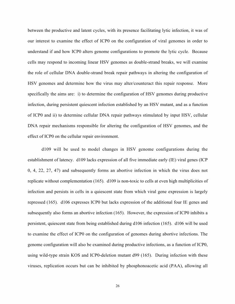
Figure 1. The HSV genome.
The unique-long (UL) and unique-short (US) regions are bracketed by direct terminal repeats
(TRL and TRS). Direct repeats are designated (alanb,) and (cas). These repeats are inverted
within the internal repeats (IRL and IRS), which are designated (b', an', c') within the region
connecting the UL and US sequences. There are multiple 'a' repeats where indicated (an). The
one base overhang on the 3' strand of terminal a repeats is depicted by an incomplete rectangle
(90). The position and direction of immediate early genes ICP 0, 4, 22, 27, and 47 are indicated,
as are the positions of the origins of replication in each long and short region (oriL and oriS).
al an b b' an' c' c as
0 27 0 4 22 47 4
oriL oriS oriS
TRL UL IRL IRS US TRS

8
repeat sequences (114, 171, 179); viruses lacking inverted repeats do not invert (150).
Subsequently, HSV genomes exist in four isomeric arrangements, in which the UL region inverts,
the US region inverts, both the UL and the US regions invert, or neither the UL nor the US region
inverts. During infection, one isomer produces all four isomers in equimolar concentrations,
suggesting a direct correlation between isomerization of viral genomes and replication (161).
The HSV genome contains approximately 80 genes, which are grouped into three classes
based on their temporally regulated expression (161). As their names suggest, immediate early
(IE), early (E), and late (L) genes are progressively expressed during HSV productive infection.
The five IE genes, ICP 0, 4, 22, 27, and 47, cooperatively initiate the HSV productive cycle.
These IE genes are largely located in the repeated sequences of the genome, while E and L genes
are largely located in the UL and US components (Figure 1). The genome contains two copies of
the ICP0 and ICP4 genes within the repeats bracketing the UL and US regions, respectively. In
addition, the HSV genome contains three origins from which replication initiates. The UL
component contains one origin of replication and each c repeat region of the US component
contains one origin of replication (Figure 1).
1.5. HSV productive infection
HSV recognizes potential host cells through interactions of glycoproteins that protrude from the
lipid envelope of virions with glycoproteins on the cell surface, herparin sulfate proteoglycans,
and cell surface receptors called herpesviral entry mediators (HVEM), which include HveA,
HveB, and HveC (156). Following attachment, glycoproteins mediate the fusion of the virion
lipid envelope with the cell plasma membrane, allowing the viral capsid to enter the cytoplasm of
the cell. After entry, the capsid is transported to nuclear pores, where viral DNA is injected into
the nucleus and may begin to express genes and replicate.

9
1.5.1. Cascade of viral gene expression
Viral gene expression progresses from immediate early genes (IE or alpha), to early genes (E or
beta), and then to late genes (L or gamma), using host cell RNA polymerase II (RNA Pol II) and
the host cell transcription machinery (79, 84, 120, 146). By definition, IE genes are expressed in
the absence of de novo viral protein synthesis. Together, a tri-partite complex of VP16 supplied
in the tegument of the infecting virion, the cellular octamer DNA-binding protein (oct-1), and the
host cellular factor (HCF) protein initiate IE gene expression by binding to promoter elements
(TAATGARAT) present in all HSV IE promoters (4, 17, 152). IE gene expression has been
observed as early as 30 minutes postinfection in cell culture systems and peaks between 2 and 4
hours postinfection (83, 84). IE genes are the major regulators of viral gene expression and as
such, have a variety of activities that all function in promoting the productive cycle. There are
five IE genes expressed, which include infected cell polypeptides (ICP) 0, 4, 22, 27, and 47. Of
the five IE genes, four are activators of viral gene expression. These gene products direct the
expression of subsequent E and L viral genes
ICP4 is the main regulator of viral gene expression, activating both E and L viral genes
(38, 52, 68, 69, 138, 155) through its interactions with the transcription machinery of the host (5,
20, 33, 71, 88, 180) and binding sites that recognize E and L promoters (5, 41). In the absence of
ICP4, E and L genes are not transcribed and only the remaining IE genes are expressed (38).
ICP4 also possesses the ability to repress viral gene expression (15, 38, 128, 138), which leads to
over-expression of the remaining IE genes when ICP4 is absent (38). Like ICP4, ICP27 is
required for the productive cycle and activates E and L gene expression (121, 166, 191). It also
modulates mRNA processing on both the viral and cellular level, through regulating post-
transcriptional modifications to cellular and viral RNA and the export of certain mRNA's from

10
the nucleus (74, 75, 122, 124, 125, 169). These modulations may contribute to ICP27's ability to
shut-off off host protein synthesis. ICP22 facilitates L gene expression and is subsequently not
necessary for virus replication. It affects the stability and processing of ICP0 transcripts (21) and
the phosphorylated state of the RNA Pol II C-terminal domain (158). The phosphosphorylated
state of RNA Pol II in turn affects the association and activity of RNA Pol II with viral
promoters and thus transcription of these genes (158). ICP0 is an indiscriminant transactivator of
gene expression and as such, is the only viral protein that activates all three classes of viral genes
(24, 120). It affects proteins associated with a variety of cellular processes including those
associated with ND10 structures (55, 116), cell cycle regulation (53, 94), transcription (95),
translation (94), proteolysis (53, 55, 59), and DNA repair (107, 143). How these effects may
underlie the ability of ICP0 to promote the productive cycle will be covered in detail in a section
below, as it is a major focus of this report. Of the five IE genes, ICP47 is the only protein that
does not induce cytotoxicity during infection and the only protein that does not direct
transcriptional activation of other viral genes. Rather, ICP47 inhibits antigen presentation and
subsequent recognition of infected cells by the immune system, in particular CD8+ lymphocytes,
through interactions with the transporter of antigen processing (TAP) molecule that disables
transport of the major histocompatibility complex (MHC) class I and its associated antigens to
the cell surface (2, 64, 80, 104, 190).
Aside from ICP47, IE gene products direct the expression of subsequent E and L viral
genes. E gene expression peaks between 5 and 7 hours postinfection (83, 84), and these gene
products mainly contribute to viral replication, encoding the viral DNA polymerase and other
proteins necessary for replication, including the single-stranded DNA binding protein (ICP8), the
DNA helicase-primase, the origin-binding protein (UL9), and enzymes involved in nucleotide

11
metabolism (10). After E protein synthesis begins, viral replication can occur as early as 3 hours
postinfection and proceed up to 15 hours post infection (83, 84). Soon after replication initiates,
L genes are expressed. L gene expression peaks at 8 hours postinfection, and the products of
these genes mainly consist of structural proteins required for reassembly of mature progeny
virions, which are released approximately 18 hours postinfection (83, 84).
1.5.2. Mechanism of replication
After IE and E gene expression, replication can proceed from three origins on the HSV genome,
although none of these origins are uniquely required for replication (87, 113, 151, 181). A
variety of DNA electrophoresis techniques have been used to examine the configuration of HSV
genomes prior to and during the onset of replication, creating a model by which HSV replication
initiates from a circular genome template. The first study to provide evidence for this template
of genome replication was done by Poffenberger and Roizman (149). These investigators
developed a non-inverting HSV genome by deleting internal inverted repeat sequences from the
genome, subsequently creating a virus I358 that is held in one isomeric arrangement (149, 150).
The authors have shown generation of end-to-end linkages of these viral genomes early during
infection and in the presence of inhibitors to gene expression (149). They interpret the formation
of these linkages to be indicative of intra-molecular joining of genome termini by circularization
but admit that the formation of end-to-end linkages could also be indicative of inter-molecular
joining of genome termini through concatemerization (149). Analysis for the progressive loss of
genome termini has also suggested that HSV genomes circularize early during productive wild-
type infection of CV-1 cells and both neuron-differentiated and undifferentiated rat-derived
PC12 cells (186), but again, these results do not differentiate between concatemerization,
circularization, and in these cases degradation of genome termini. Pulse field gel electrophoresis

12
(PFGE) has been used to differentiate between concatemerized and circularized HSV genomes.
Early during productive infection, regardless of viral gene expression, HSV DNA is retained
within PFGE wells, and this DNA is diminished for genome termini (65). Creating on average
one double-strand nick in well-retained HSV DNA allows the HSV DNA to migrate a similar
distance as one unit-length linear HSV genome, suggesting well-retained genomes are circular
(65). However this study only demonstrated these unit-length circles in the absence of viral gene
expression, not during the productive cycle when templates of replication are formed.
Together analyses for end-to-end linkages of genomes and PFGE studies have provided
foundations for our current model of HSV replication, which initiates from a circular genome
template via a theta mechanism and then proceeds into a rolling circle mechanism (162). This
model is supported by the fact that only one viral genome is necessary for replication and the fact
that the ends of viral genomes, which contain homologous direct a repeats, are suitable substrates
for ligation or recombination of genome termini into circular genomes (162). Theta replication
also eliminates loss of genetic material due to incomplete lagging-strand synthesis that would
otherwise arise from successive cycles of semi-discontinuous replication of DNA genomes from
linear templates. However, although the aforementioned studies have never clearly distinguished
between circular and concatemeric forms during productive infection, they provide the only
direct evidence for the initial theta mode of replication. This ambiguous evidence for the initial
theta mode of replication has, in part, lead to a theory for the later rolling circle stage of
replication, in which an initial circular template is necessary. Although there is evidence that
genomes exist as highly branched concatemers during later stages of replication (7, 174, 175)
and a rolling circle mechanism is predicted to produce concatemers of genomes (162), a more

13
thorough examination of the initial genome template for replication is required in order to
understand the mechanism of replication at later stages.
In the present study, we challenge the previous model of HSV replication by providing for
the first time evidence that replication is initiated from a linear genome template, while circular
genomes are the stable configuration during latency (89). However, a very recent paper (185)
has presented a rebuttal to our results. Using HSV viruses that lack 'a' repeat sequences but
contain parts of the HSV packaging signal at each termini, these authors suggest that end-to-end
linkages of genomes indicative of circular genomes are present both early during infection in the
presence of ICP0 and in the absence of HSV gene expression (185). However, there are many
problems with the author's interpretation of their data, which arise from inherent complexities
present within the technique these authors employed. These problems/complications will be
addressed in Chapter 4, in which we provide further evidence that during the productive cycle
ICP0 inhibits circularization of HSV genomes, providing a linear genome template from which
HSV replication initiates.
1.5.3. Host cell death
After assembly and egress of mature progeny virions, HSV infected cells die. Cells accumulate
damage to their cytoskeleton (42, 78) and chromatin (26, 73, 113, 139, 140, 182). They often
fuse to form huge multi-nucleated cells and eventually round up. Cell death is in part due to the
cytotoxic effects imposed by the activities and downstream effects of IE proteins. As described
above, in order to carry out the productive cycle, IE proteins disrupt many of the normal
processes of host cell metabolism, including those involved in transcription, translation, DNA
repair, proteolysis, and cell cycle regulation. Although some of these disruptions are present to
inhibit host anti-viral responses, such as antigen presentation, others are due to the virus'
14
requirement of cellular proteins/machinery. Viral factors coordinate the redistribution of
necessary cellular proteins to sites of HSV replication and also shut-off host protein synthesis,
freeing the translation machinery for viral protein synthesis. ICP27 has been shown to alter the
export of host mRNA (74, 169, 170). In addition, the HSV virion host shut-off protein (vhs) that
is provided in the tegument of the infecting virion affects mRNA stability by degrading ribosome
associated mRNA (46, 101-103, 141). Together, these resulting changes in host cell metabolism
are significant and likely to contribute to host cell death.
1.6. HSV latency
Latent HSV infections are established in sensory neurons that innervate the primary site of
productive infection. During HSV latency, the productive cycle of viral gene expression is
repressed (154, 165, 187) and only LAT's, whose function remains obscure, are transcribed
(196). From a latent state, the productive cycle can be readily stimulated by various
physiological stresses and also ICP0 (15, 16, 29, 196). In fact, ICP0 is the only HSV protein that
can stimulate gene expression from repressed genomes. During latency HSV genomes persist in
an extrachromosomal state (126) that is bound by nucleosomes in a chromatin arrangement (40),
which may contribute to the stability and transcriptional repression of the genome. The ability of
HSV genomes to adopt a repressed, stable state is necessary for its non-toxic persistence during
these life-long infections. However, this process must be reversible for the productive cycle to
occur during reactivation.
In order to understand how the physical characteristics of HSV genomes during latency
may contribute to its stability and persistence, many studies have examined the configuration of
HSV genomes during persistent infections in vitro and during latent infections in vivo. DNA
isolated from the trigeminal ganglia of both latently infected mice and humans is depleted for

15
genome ends (44), suggesting that during latency the genome exists in an endless, potentially
circular state. Neuronal-differentiated PC12 cells have provided an in vitro model system for
latent HSV infections (9). During long-term infections of these cells with wild-type virus, HSV
genomes become quiescent and do not produce progeny virus; the genomes persist in these cells
in an endless state, suggesting that HSV genomes are circular during infections that model
latency (186). However, terminal HSV genome fragments have been detected in some latently
infected human trigeminal ganglia (44), and the termini of a replication-defective virus are
relatively stable during long-term infections in differentiated PC12 cells (12). These studies
have subsequently left some ambiguity with respect to HSV genome configurations during
latency. Importantly, these studies have not differentiated between concatemerized and
circularized genomes. In the present study, we provide unambiguous evidence that HSV
genomes persist in a stable circular configuration during long-term quiescent infections that
model latency.
1.7. The role of ICP0 during HSV infection
ICP0 is one of the five IE viral genes and is expressed within the first hour of infection. It is an
indiscriminate transactivator of both viral and cellular gene expression, however it does not bind
to promoters (92). Many of the properties of ICP0 have been deduced from phenotypic analyses
of a series of ICP0-mutated viruses. ICP0 facilitates, but is not necessary for productive
infection, as ICP0-deleted viruses can replicate, although inefficiently unless high doses of input
viral particles are used (164, 183). ICP0 is necessary for derepression of gene expression during
normally persistent, quiescent HSV infections that model latency (76, 165) and is important for
efficient reactivation of latent infections in mice (15, 29, 109, 153). It follows that ICP0 is
thought to indiscriminantly facilitate viral gene expression by inhibiting repression of viral gene

16
expression. This inhibition of repression could be how ICP0 facilitates gene expression both
during initial infections and during reactivation from a latent state. Since these phenotypic
analyses, ICP0 has been shown to interact with and/or affect a variety of cellular proteins
involved in cell cycle regulation (53, 94), transcription (95), translation (94), proteolysis (53, 55,
59), and ND10 bodies (55, 116). The functions of these interactions have been largely
interpreted in the context of ICP0's ability to inhibit productive cycle repressors. Although it is
not completely understood how, these interactions underlie the ability of ICP0 to facilitate gene
expression and the productive cycle.
1.7.1. Structure and function
Within the HSV genome, the ICP0 gene occurs in two copies, one located within each set of
repeats flanking the UL region (Figure 1). The ICP0 transcript is spliced into a 2.7KB mRNA
that contains three exons (146, 147). Translation of this transcript results in a 775 amino acid
protein with a molecular weight of 110 kDa. Both amino acid sequence and
mutational/biochemical studies have revealed many of the molecular characteristics of ICP0 that
may contribute to its function. ICP0 contains various potential sites for post-translational
modifications, including several N-terminal casein kinase II sites, two central proline rich
regions, a central serine-threonine tract, and a C-terminal serine-alanine rich sequence (147).
ICP0 also contains several protein interaction domains, including a nuclear localization signal, an
acidic N-terminal domain, a C-terminal dimerization domain, a cyclin D3 binding domain, a
herpes-associated ubiquitin specific protease (HAUSP) binding domain, and a C3HC4 RING
finger domain (47, 49, 59, 96, 135, 147). The RING finger is the only domain that is conserved
in ICP0 homologues of other herpesviruses (27, 132, 133, 189, 201).
1.7.2. Protein stability and turnover

17
ICP0 has been shown to induce the degradation of: PML and Sp100 (23, 55, 116), which are
constituents of ND10 bodies; the DNA-dependent Protein Kinase Catalytic Subunit (DNA PKcs)
(107, 143), which is critical for VDJ recombination and DNA double-strand break repair via
non-homologous end-joining (NHEJ) (30); and CENPA and CENPC (53), which are histone3-
like centromere proteins. The ability of ICP0 to induce the degradation of cellular proteins may
underlie the ability of ICP0 to promote the productive cycle. These proteins have been proposed
to somehow be involved in cellular repression mechanisms that target the parental HSV genome.
ICP0-induced protein degradation has been linked to a cellular mechanism for protein
turnover involving the ubiqutin-targeted degradation of proteins via the 26S proteasome, as
inhibitors (MG132) of this pathway inhibit ICP0's effect (51). Ubiquitination is one of the
various post-translational modifications that can result in protein instability (28). Ubiquitin is a
76kDa protein that associates with lysine residues of target proteins. Ubiquitin conjugating
enzymes (E2 enzymes) catalyze the targeted transfer of ubiquitin onto target proteins, until
polyubiquitin chains accumulate and bind to the 26S proteasome, leading to rapid degradation of
the poly-ubiquitinated protein by proteasomal enzymes. Ubquitination is a reversible process, as
ubiquitin may also be removed from protein substrates by ubiquitin specific proteases (USP).
Both ICP0's RING finger motif and the proteosome are required for ICP0-induced
degradation of the aforementioned proteins (60). RING finger motifs are common to E3
ubiquitin ligases, which stimulate and probably regulate the activity of E2 ubiquitin conjugating
enzymes, which transfer ubiquitin onto target proteins (63). Indeed, ICP0 has been shown to
contain E3 ubiquitin ligase activity (13, 192). Hence, ICP0 is thought to direct the conjugation
of ubiquitin onto specific proteins, targeting them for degradation via the proteasome. During
HSV infection, ICP0 interacts with the herpes-associated ubiquitin specific protease termed

18
HAUSP, further associating ICP0 with ubiquitin-related protein stability (58, 59). Although
HAUSP functions to remove ubiquitin from targeted proteins, ICP0 may use this interaction to
free ubiquitin molecules for use in ubiquitination of other specific proteins (51). Although ICP0
induces degradation of proteins through the 26S proteasome and can induce conjugation of
ubiquitin chains (13, 192), it has never been shown to specifically conjugate ubiquitin onto
proteins, thereby inducing their degradation, either in vitro or in vivo. Nonetheless, ICP0 clearly
induces the proteasome-dependent degradation of cellular proteins, which have since been
thought to be somehow inhibitory to the productive cycle of the virus.
1.7.3. ND10
Early during infection ICP0 localizes to and then disrupts ND10 structures, which are
conglomerates of nuclear proteins that form on average ten nuclear domains (ND10) or foci
within dividing cells (115, 116). ND10 structures are composed of various proteins including the
promyelocytic leukemia protein (PML) and SP100, which are both modified by a small
ubiquitin-like molecule (SUMO-1) (118). ND10 bodies and their associated proteins have been
shown to play roles in translation, transcription, DNA repair, DNA replication, RNA stability,
and RNA transport (12). Although their precise functions are not known, ND10 bodies are
modified during the cell cycle (56), disturbed by heat shock (119), disrupted by infections with
various viruses (50).
During infection with HSV and many other DNA viruses, parental genomes localize to
the periphery of ND10 structures (117). HSV may replicate at these sites after disruption of
ND10 by ICP0, which is accomplished in part through the degradation of PML and SP100 (117).
Both the RING finger domain and C-terminal domain of ICP0 are required for efficient
disruption of and localization to ND10 (57, 115). These regions of ICP0 are critical for ICP0's

19
ability to facilitate viral gene expression and the productive cycle (47, 48, 57, 115).
Accumulations of the ICP0-interacting protein HAUSP can also be found in or close to ND10
bodies (59). The heterochromatin protein (HP1) can be found at both ND10 structures and
centromeres (54, 108, 173), which are also disrupted by ICP0 through degradation of the
centromere proteins CENP-A and CENP-C, leading to disruption of centromere structure and
mitotic failure (53). Although exactly how these proteins affect virus production and the lytic
cycle is not understood, they are thought to be inhibitory to the productive cycle due to the
ability of ICP0 to induce their degradation and subsequently disrupt the processes with which
they are associated. Furthermore, the RING finger motif of ICP0 is both required for ICP0 to
facilitate the productive cycle and necessary for the degradation of these proteins and subsequent
disruption of these structures (60).
1.8. Host cell DNA repair pathways during HSV infection
Cells have developed a variety of mechanisms to repair breaks in DNA, because these breaks
present potential loss and manipulation of vital genetic material. During infection, the HSV
genome is presented as a foreign, double-stranded DNA molecule with unprotected DNA ends.
These ends may be detected as DNA double strand breaks (DSB's) by host cells, which
subsequently attempt to repair these breaks through DNA DSB repair pathways. The two
primary pathways by which DSB's are repaired in mammalian cells are non-homologous end-
joining (NHEJ) and homologous recombination (HR). Although these mechanisms are distinct,
some repair proteins may play overlapping roles in both mechanisms. Repair of ends by HR
requires exchange of homologous DNA sequences, while NHEJ involves direct ligation of DNA
sequences with little sequence homology required.

20
Studies concerning HSV genome configurations during productive infection and during
latency have suggested that at some point during the HSV life cycles viral genomes circularize
(44, 65, 89, 149, 159, 185, 186). In the current study we present evidence that HSV genomes
circularize in the absence of ICP0 and circular genomes serve as a stable configuration during
persistent quiescent states that model latency (89). Circularization of HSV genomes could be a
result of host cell DNA repair responses to unprotected genome ends present on linear double
stranded HSV genomes. Through these repair responses, genome ends may be joined, creating
circular genomes. Circularization is consistent with the structure of the genome, which contains
direct a repeat sequences located at genome termini (Figure 1) (161). These repeats render the
HSV genome a suitable substrate for circularization by either HR or NHEJ repair mechanisms.
1.8.1. Non-homologous end-joining (NHEJ)
Interestingly, NHEJ may be the dominant repair pathway of DSB's in non-dividing neurons (31,
66, 163), where latent HSV infections are established and where genomes may persist in an
endless state, most likely as unit length circles (44, 159). DNA PKcs, Ku70, Ku80, XRCC4, and
DNA Ligase IV are cellular proteins critical for NHEJ in vivo (30). Ku70 and Ku80 subunits
dimerize, forming the regulatory subunits of the DNA PK holoenzyme (70). The Ku dimer
binds DNA ends (8, 130), recruiting the catalytic subunit of DNA PK(cs) to sites of DNA DSB's
(70). Once associated with DNA-bound Ku subunits, DNA PKcs catalyzes the end-joining
reaction through a combination of phosphorylation events (127) that leads to recruitment of the
XRCC4/DNA Ligase IV tetramer, which enzymatically ligates DNA ends together (198).
There is evidence that HSV manipulates NHEJ repair pathways during infections,
potentially creating an environment that promotes the productive cycle. ICP0 induces the
degradation of DNA PKcs during infection in a cell-type specific manner, inducing nearly

21
complete degradation in Hela S3 cells (107, 143), inducing degradation by less than 50% in Hep-
2 cells (188), and not inducing degradation to observable levels in Vero cells (200). In Hep-2
cells, although DNA PKcs is somewhat degraded, complexes of DNA PKcs, Ku80, Ku70, and
XRCC4 co-precipitate with the HSV single-stranded DNA binding protein, ICP8, at times in
which ICP8 is located at HSV prereplicative sites and replication compartments, suggesting a
direct and/or indirect interaction between these NHEJ repair proteins and viral genomes (188).
In fact, Ku70 has been shown to bind some late HSV promoter sequences (148). During
infection DNA PKcs and Ku80 are redistributed from their normally disperse nuclear
localization to HSV replication compartments (188, 200). In mouse embryonic fibroblasts
(MEFs) lacking Ku70 (188) and human glioblastoma cells lacking DNA PKcs (143), yields of
replicated wild-type virus is increased up to approximately 50 and 100 fold respectively,
suggesting that proteins involved in NHEJ are inhibitory to HSV replication. Although NHEJ
proteins are clearly present at sites of HSV genomes, appear to affect HSV replication, and may
be affected by ICP0, the exact role of NHEJ proteins and why/how the virus manipulates this
repair mechanism during HSV infection remains unclear. NHEJ subsequently remains a likely
mechanism by which genomes are circularized during infection.
1.8.2. Homologous recombination (HR)
Because the termini of HSV genomes contain direct a repeats, HR is another potential
mechanism by which the cell may repair genome ends and subsequently circularize HSV
genomes. Like NHEJ proteins, HR proteins localize to sites of HSV replication. WRN, BRCA1,
BLM, and RPA, all of which may be involved in HR repair, associate with ICP8 (188) at
prereplicative and replication compartments and redistribute from their normal nuclear
localization to these sites during infection (188, 200). However, whereas virus growth is

22
increased in cells deficient in NHEJ, in cells deficient in WRN both virus growth and
recombination is decreased, suggesting a non-essential but facultative role for WRN in virus
replication (188). Rad51, which is the only protein absolutely required for HR, redistributes to
HSV prereplicative sites and replication compartments during infection (200).
Like NHEJ proteins, HR proteins have the potential to participate in the repair of genome
termini by circularization. However, unlike DNA PKcs degradation observed during infection,
the steady state levels of examined HR repair proteins are not altered during HSV infection
(200). In fact, HR activity increases during replication, a time in which genome isomerization
occurs (161). HR proteins may be involved in isomerization and repair of damaged genomes
during replication. There have been in vitro studies done aimed at determining if NHEJ or HR
mechanisms circularize DNA during HSV infection (204, 205). However, these studies used
linearized plasmids as substrates for repair in transfected cells that have been either infected with
HSV or transfected with HSV expression vectors (204, 205). Although these studies have shown
that both NHEJ and HR mechanisms can circularize linearized plasmid DNA during HSV
infection, they suggest that HR is the major mechanism responsible for this circularization.
Because the substrate for circularization was artificial in these investigations, additional studies
examining the mechanism by which HSV genomes circularize using viral genomes as substrates
for repair are required to differentiate between NHEJ and HR mechanisms.
1.8.3. Mre11/Rad50/NBS1 repair response
Additional proteins that may be involved in both NHEJ and HR repair are altered during HSV
infection. A complex of proteins, Mre11/Rad50/NBS1 (M/R/N), has been implicated in cellular
DNA double-strand break (DSB) repair processes by both NHEJ and HR mechanisms through its
ability to recognize and in part process DSB's. During repair of DSB's, M/R/N complexes
23
localize to foci of the phosphorylated form (serine 139) of histone variant H2AX ( -H2AX) (19,
144), which has been shown to co-localize with and act in the repair of DSB's (157, 160, 172).
During HSV infection, both Mre11 and Rad50 either directly and/or indirectly associate with the
HSV single-stranded DNA binding proteins, ICP8, which is localized to HSV replication
compartments (188). In fact, M/R/N complexes relocate to replication compartments during
infection (111, 188, 200). In addition, NBS1 is modified by phosphorylation during productive
wild-type HSV infections (111, 178, 200). NBS1 phosphorylation occurs during DNA damage
responses that are signaled through the ATM pathway (202).
M/R/N complexes act as sensors of DNA DSB's and may facilitate in ATM recruitment
to DSB's and subsequent activation of ATM by inducing the dissociation of inactive ATM
multimers into active ATM monomers, which in turn have downstream effects on DNA repair,
cell cycle arrest, and apoptosis (1, 105). During ATM activation and recruitment to DSB's, many
proteins including NBS1 are phosphorylated. Indeed the ATM pathway is activated during
productive HSV infections (111, 178), with this repair response possibly recognizing replicating
HSV genomes as abnormal structures. M/R/N complexes and ATM signaling may be beneficial
for the productive cycle of the virus, because in some differentiated neurons, where a latent state
can be established and this ATM repair response is compromised, there is a significant defect in
HSV replication and replication compartment formation (111). However the exact role of
M/R/N complexes and the ATM pathway during HSV infection remains largely uncharacterized,
in part, due to the fact that it is not known whether this pathway is stimulated by events leading
to and during replication or in response to incoming parental HSV genomes. The present study
addresses these issues, suggesting that early during infection incoming HSV induces M/R/N
complex formation independent of ATM activation.

24
2. RATIONALE
In host cells, the HSV genome undergoes physical alterations that are regulated by both cellular
and viral proteins. During the lytic cycle HSV genomes are very dynamic, progressively
changing throughout infection; viral genomes actively express genes, replicate, and are
packaged, ultimately producing mature viral progeny. By contrast, during latency viral genomes
persist in a transcriptionally repressed chromatin state in the nuclei of sensory nerves (40, 154,
187). Although many physical differences in HSV-1 genomes during the lytic and latent cycle
have been characterized, little is known about how these characteristics develop during infection
and furthermore, how these developing characteristics contribute to productive or latent life
cycles.
Research that has examined the configuration of viral DNA during the various stages of
lytic life cycle along with the known structural characteristics of the viral genome has led to a
model of the progressive changes in HSV genomes during lytic infection, in which replication is
initiated by a theta mechanism (12, 65, 149, 185). The template for theta replication is a circular
HSV genome. Research that has examined the configuration of viral DNA during latency has
led to a model in which HSV genomes persist in a stable circular configuration (12, 44).
However, changes in viral genomes and events leading to these changes during a particular life
cycle are not clear because it is not understood how the host cell responds to incoming HSV
genomes and how the virus manipulates this response, ultimately leading either to a template for
replication during productive lytic infections or to a stable configuration during persistent latent
infections.
Because HSV genomes begin as linear, double-stranded DNA during infection, host cells
may treat the ends of incoming genomes as DNA double strand breaks (161). The host cell may

25
subsequently attempt to repair these double-strand breaks via DNA double strand break repair
pathways, resulting in intramolecular joining of genome termini through genome circularization.
Some DNA repair proteins, including those involved in homologous recombination (HR), those
involved in non-homologous end joining (NHEJ), and those that may be involved in both
mechanisms are present at sites of HSV genomes during infection (111, 148, 178, 188, 200).
However, it is not known how these repair proteins contribute to the manipulation of HSV
genome configurations during infection and how viral proteins may alter or counteract this repair
response. Furthermore, it is not known how the interplay between repair responses and the virus'
reaction to these responses contributes to forming either a genome template for replication
during the lytic cycle or a stable configuration during latency.
ICP0 promotes the lytic cycle both during primary infections and during reactivation
from latency (15, 16, 29, 76, 92). ICP0 interacts with and/or affects cellular proteins associated
with ND10 structures (55, 116), transcription (95), translation (94), cell cycle regulation (53, 94),
DNA repair (107, 143), and proteolysis (53, 55, 59). These interactions are thought to underlie
the process by which ICP0 promotes lytic infection. As a ubiquitin ligase, ICP0 may act by
targeting proteins, most likely those that inhibit the productive cycle, for degradation via the
proteosome (13, 192). However, the mechanism(s) by which ICP0 promotes the lytic cycle are
far from being entirely understood.
In order to more clearly understand changes in the configuration of viral genomes during
both productive and latent life cycles and events leading to these changes, we have used a variety
of HSV mutant viruses to examine the configuration of viral genomes as persistent and
productive infections are established; these events model changes leading to and during the latent
and lytic life cycles, respectively. Because ICP0 is such a pivotal regulator in the balance
26
between the productive and latent cycles, with its presence facilitating lytic infection, it was of
our interest to examine the effect of ICP0 on the configuration of viral genomes in order to
understand if and how ICP0 alters genome configurations to promote the lytic cycle. Because
cells may respond to incoming linear HSV genomes as double-strand breaks, we will examine
the role of cellular DNA double-strand break repair pathways in altering the configuration of
HSV genomes and determine how the virus may alter/counteract this repair response. More
specifically the aims are: i) to determine the configuration of HSV genomes during productive
infection, during persistent quiescent infection established by an HSV mutant, and as a function
of ICP0 and ii) to determine cellular DNA repair pathways stimulated by input HSV, cellular
DNA repair mechanisms responsible for altering the configuration of HSV genomes, and the
effect of ICP0 on the cellular repair environment.
d109 will be used to model changes in HSV genome configurations during the
establishment of latency. d109 lacks expression of all five immediate early (IE) viral genes (ICP
0, 4, 22, 27, 47) and subsequently forms an abortive infection in which the virus does not
replicate without complementation (165). d109 is non-toxic to cells at even high multiplicities of
infection and persists in cells in a quiescent state from which viral gene expression is largely
repressed (165). d106 expresses ICP0 but lacks expression of the additional four IE genes and
subsequently also forms an abortive infection (165). However, the expression of ICP0 inhibits a
persistent, quiescent state from being established during d106 infection (165). d106 will be used
to examine the effect of ICP0 on the configuration of genomes during abortive infections. The
genome configuration will also be examined during productive infections, as a function of ICP0,
using wild-type strain KOS and ICP0-deletion mutant d99 (165). During infection with these
viruses, replication occurs but can be inhibited by phosphonoacetic acid (PAA), allowing all

27
events taking place prior to replication to evolve but inhibiting replication itself (85). In theory,
PAA addition should allow the template for genome replication to be observed. The
configuration of the aforementioned viruses will be examined using Gardella gel analysis.
Gardella gels resolve large circular forms from their linear counterparts of the same molecular
weight as discrete species within an agarose gel, as was first demonstrated in a study
distinguishing persisting circular Epstein Barr Virus (EBV) episomes in Raji cells from their
linear counterparts in virions (67).
Gardella gel analysis for circular genomes will be used in conjunction with an additional
analysis for circularization, which has been previously demonstrated by Poffenberger and
Roizman (149). Both analyses will be used to compare the configuration of a non-inverting HSV
mutant I358 (149) to an ICP0 deleted version of I358 (d99/I2.3) that we will construct. Both
I358 and d99/I2.3 are deleted for internal repeat sequences within their genomes and
subsequently do not isomerize. Although a virus lacking the internal repeat sequences is not
required for Gardella gel analysis, it is required for restriction enzyme analysis due to sequence
homology within the internal repeats of the viral genome with the terminal repeats of the
genome. During restriction enzyme analysis, DNA from infected cells is digested and then
probed for a terminal genome fragment, which will also detect a novel fragment that includes
both termini, if viral genomes are linked end-to-end, as they are when circularized. Novel
restriction fragments containing end-to-end linkages of viral genomes can only be distinguished
in the absence of homologous internal repeat sequences. Both Gardella gel analysis and
restriction enzyme analysis will be used to assess HSV genome configurations during productive
infections (I358), as a function of ICP0 (d99/I2.3).

28
Host cell DNA repair responses to incoming HSV genomes may alter the configuration
of genomes, ultimately leading to either a template for genome replication during the productive
cycle or a stable genome configuration during latency. We will examine both the effect of ICP0
on cellular DNA DSB repair pathways and the effect of these pathways on the configuration of
HSV genomes. Although it is known that repair proteins localize to HSV replication
compartments (111, 178, 188, 200), it is not known if they are recruited as a result of parental
HSV genomes or due to events that occur during HSV replication. The localization and levels of
repair proteins in response to incoming HSV will be examined during abortive and productive
infections and as a function of ICP0, using the aforementioned viruses with immunofluorescence
and Western blotting techniques. The effect of cellular DNA repair pathways on changes in
HSV genome configurations will be assessed by Gardella gel analysis of HSV genome
configurations in cells deleted for specific DNA repair proteins.
Using these approaches we will model changes that occur to viral genomes as both lytic
and latent infections are established and will also begin to clarify how these changes evolve. For
the first time, we will provide evidence for diverging physical changes in HSV genomes during
the establishment of each life cycle of the virus and importantly show that the interplay between
cellular responses to HSV genomes and ICP0 regulates these changes. In particular, we will
provide evidence that host cell DNA repair pathways manipulate the configuration of HSV
genomes and that ICP0 may alter these pathways, potentially promoting a template for
replication during the lytic cycle. This ability of ICP0 to control the configuration of viral
genomes is a newly identified mechanism by which ICP0 can promote the lytic cycle of the
virus. Along with suggesting a revised model of HSV replication during productive infection,

29
we will also provide a mechanism by which HSV genomes can persist long-term in a stable
configuration during latency.

30
3. RELATIONSHIP OF HERPES SIMPLEX VIRUS GENOME CONFIGURATION
TO PRODUCTIVE AND PERSISTENT INFECTIONS.
3.1. Abstract
Infection of susceptible cells by herpes simplex virus can lead to productive infection, or latency,
where the genomes persist in the nuclei of peripheral neurons in a quiescent state. Using the
HSV strain d109, which does not express any viral genes and thus establishes a quiescent state in
most cells, we observed that a fraction of genomes circularized upon infection. The expression
of ICP0, which is known to be involved in reactivation from latency and the promotion of
productive infection, inhibited the formation of circular genomes. Circular genomes were not
observed upon infection of fully permissive cells by wild type virus, either in the presence or
absence of viral DNA replication. However, productive infection in the absence of ICP0 resulted
in the accumulation of a subpopulation of circular genomes. The proportion of circular genomes
formed during infection with an ICP0 mutant was greater at low multiplicity of infection, a
condition where ICP0 mutants replicate poorly. In the complete absence of viral gene
expression, it was found that only circular genomes persisted in cells. These results suggest that
circularization of the HSV genome may not occur early in the productive phase of wt HSV
infection, but rather during establishment of a quiescent state or latency, providing a possible
strategy for long-term persistence. Additionally, the circularization and possible fate of HSV
genomes is regulated by an activity of ICP0.
3.2. Introduction
Herpes Simplex Virus Type 1 (HSV-1), can establish both productive and latent infections.
Productive infection with HSV-1 results in epithelial lesions, such as cold sores, and can lead to
latent infection in the trigeminal ganglia of neurons that innervate the primary site of infection
(162). During latency the viral genome persists in a largely repressed, chromatin-associated state

31
(40) that can be derepressed, or reactivated, to produce recurrent productive infections. The
HSV-1 immediate early (IE) protein ICP0 is crucial for reactivation (15, 29, 109).
The HSV genome is a linear double stranded DNA molecule existing as 4 isomers that
differ with respect to the orientation of a long and a short region about a joint (36, 77). Both the
long and short regions are bracketed by inverted repeats, and a 250-500 bp sequence (termed the
“a” sequence) is directly repeated at the genomic termini (112, 176, 195, 197). Inverted copies
of the “a” sequence are also present at the joint. During both productive and latent infection the
genome assumes a configuration indicative of end-joining since genomic joint sequences
increase in abundance relative to their homologous counterparts at the genomic termini (44, 65,
149, 159). End-joining can occur in the presence of inhibitors of viral protein synthesis, which
has been interpreted as the circularization of the HSV genome as a function of cellular
mechanisms (65, 149). Consequently, it is thought that circularization of viral genomes occurs
prior to the onset of productive or latent infections, implying that this process occurs independent
of the type of infection established. In addition, it provides the foundation for the current model
that HSV-1 genomes replicate initially by a theta mechanism (161). However, an increase in
joint regions can also result from the formation of concatemers, complicated branched structures
that result during replication, or possibly from intermolecular ligation or recombination.
Therefore, circularization of HSV-1 genomes has not been clearly demonstrated.
In the present study we differentiate between circular, linear and higher order genomic
structures to test the hypothesis that the HSV genome circularizes in the absence of viral gene
expression. The abundance and persistence of circular genomes during productive and quiescent
infections was also determined. Quiescent infection was used as a model for latent infection and
was achieved using an HSV-1 mutant, d109, which does not express the five HSV IE proteins,

32
and contains an EGFP transgene under control of the HCMV immediate early promoter (165).
In cultured cells, d109 establishes a persistent and quiescent state, in which EGFP expression is
largely repressed but can be derepressed by providing ICP0, in trans (82, 165). These
characteristics are similar to events that occur during latency and reactivation from latency.
ICP0 is an E3 ubiquitin ligase (13, 192) and has been proposed to promote lytic
infections by specifically destabilizing cellular proteins that inhibit the lytic viral life cycle.
ICP0 is known to induce the degradation of proteins associated with ND10 bodies (116), which
are discrete nuclear foci where HSV-1 genomes may localize early during infection (117).
Interestingly, recent studies have suggested these foci to be sites of DNA double strand break
repair (18). It is possible that the ends of linear HSV-1 genomes may be treated as double strand
DNA breaks and subsequently repaired by circularizing genomes early during infection (161).
Therefore, we also addressed whether ICP0 affects the circularization of the HSV genome,
possibly providing an additional basis for its role as an effector of lytic and latent infections.
3.3. Materials and Methods
3.3.1. Cells and Viruses.
Vero, F06 (167), E11 (166) cells were cultured as previously described. Raji cells were similarly
maintained in RPMI-1640 medium. The viruses, d109, d106, d99 (165), and n212 (15)
(provided by Priscilla Schaffer, Harvard Medical School, Boston MA) are HSV-1 mutants
derived from the wild type strain KOS. Each mutant was grown and assayed for infectivity
(plaque forming units per milliliter (PFU/ml)) in the appropriate complementing cell lines (F06
and E11) as previously described (165). AdS.11E4(ICP0), an adenovirus vector that expresses
ICP0, and AdS.11D, the empty adenoviral vector, were provided by Douglas Brough at GenVec,
Inc., Gaithersburg, Md., and have previously been described (82).

33
Confluent monolayers of Vero cells were infected with viruses at the multiplicity of
infection (moi) indicated. Where indicated, 400 ug/mL phosphonoacetic acid (PAA; Lancaster)
was added to the cultures during viral infections. For each time course, viral infections were
staggered such that all time points were harvested simultaneously. 0 hour time points were
performed by incubating the virus with cells on ice for 1 hr prior to harvesting. In order to
determine the configuration of d109 genomes during long-term persistent infections, d109
infected Vero cells were cultured in media with 2% FBS and incubated at 34º.
3.3.2. Gardella gels.
Gardella gels were performed essentially as described (67). Briefly, an approximately 25cm x
15cm horizontal agarose gel was poured such that the upper portion (~3cm), where the wells
were molded, contained 2% SDS, 1mg/mL proteinase K, and 0.8% Type IIA: medium EEO
agarose (Sigma) in TBE (0.09 M Tris-base, 0.09 M boric acid, 2 mM EDTA pH 8.0). The lower
portion of the gel (~22cm) contained 0.75% agarose in TBE. Infected cells were washed with
TBS (0.14 M NaCl, 2.7 mM KCl, 25 mM Tris-base, pH 7.4), scraped into TBS, and then pelleted
by low-speed centrifugation. Nuclei were isolated as previously described (165). For each
Gardella gel, either (5x10⁶) infected cells or nuclei were resuspended in 50μL loading dye (15%
Ficoll, 0.01% bromophenol blue) and then loaded into each well. Gels were run for 3 hr at 45V
to efficiently lyse the cells, and then 38 hr at 120V to separate circular from linear viral genomes.
Gels were analyzed by Southern blot as previously described (165). The hybridized filters were
then exposed to x-ray film. For quantitative analysis, the filters were analyzed by a Storm 840
phosphorimager (Molecular Dynamics).
The probe used to detect d106 and d109 on Southern blots consisted of a 1.6-kb Pac I
fragment of pucGFP (165), containing the EGFP gene under control of the HCMV IE promoter.

34
The probe used to detect KOS, d99 and n212 genomes was the 1.8-kb BamHI-Y fragment of
pKBY, which contains a portion of the ICP4 gene of HSV-1. The plasmid 1040 (provided by
David Rowe, University of Pittsburgh), which contains the internal repeats of the EBV genome,
was used to detect EBV genomes.
3.3.3. Electroelution.
Gel slices from 4 lanes of circular or 4 lanes of linear d109 DNA, separated from 5x10⁶ Vero
cells per lane, were excised from Gardella gels. The locations of the bands in the gel were
determined by conducting a Southern blot as described above on lanes from the same gel, and
using the resulting autoradiogram as a guide to excise the bands from the remainder of the gel.
DNA was eluted from each gel slice in 0.5x TBE in dialysis bags for 6 hr at 120V. The eluted
DNA was digested with both BamHI and SacI (New England Biolabs) according to
manufacturers instructions. The digested DNA was then analyzed by Southern blot using the
BamHI Y fragment as probe, as described above.
3.4. Results
3.4.1. Accumulation of low-mobility, non-linear HSV genomes in the absence but not
presence of ICP0.
Previous studies suggesting that the viral genome circularizes prior to viral DNA replication and
during latency (65, 149, 159) did not rule out that the observed end-joining could be due to the
formation of concatemers, or more complicated branched structures. Gardella gels resolve large
circular forms from their linear counterparts as discrete species within the gel, as was first
demonstrated in a study distinguishing persisting circular Epstein Barr Virus (EBV) episomes in
Raji cells from their linear counterparts in virions (67). This analysis has also been used to
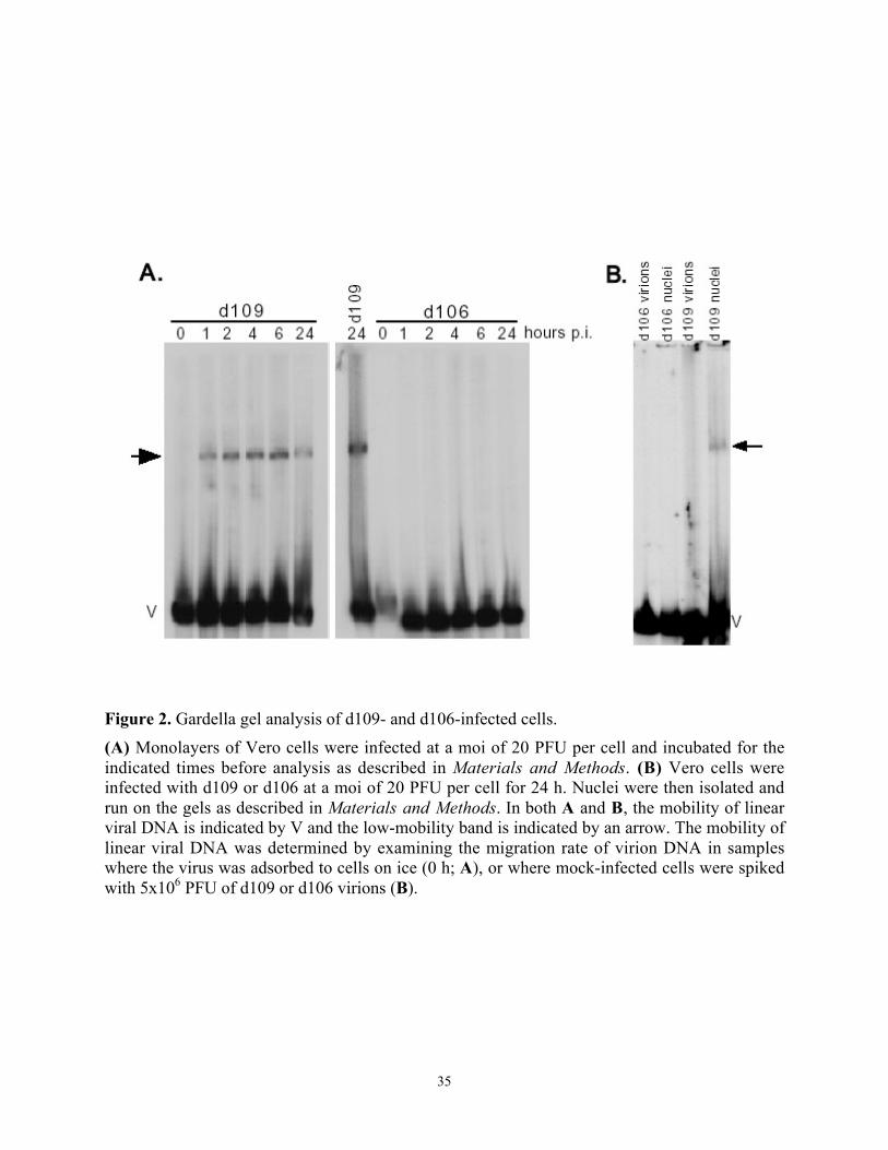
35
Figure 2. Gardella gel analysis of d109- and d106-infected cells.
(A) Monolayers of Vero cells were infected at a moi of 20 PFU per cell and incubated for the
indicated times before analysis as described in Materials and Methods. (B) Vero cells were
infected with d109 or d106 at a moi of 20 PFU per cell for 24 h. Nuclei were then isolated and
run on the gels as described in Materials and Methods. In both A and B, the mobility of linear
viral DNA is indicated by V and the low-mobility band is indicated by an arrow. The mobility of
linear viral DNA was determined by examining the migration rate of virion DNA in samples
where the virus was adsorbed to cells on ice (0 h; A), or where mock-infected cells were spiked
with 5x10⁶ PFU of d109 or d106 virions (B).

36
establish that Varicella-Zoster Virus (VZV) circularizes shortly after infection (100). To
examine changes in the HSV genome subsequent to infection and in the absence of viral gene
expression, d109 infected cells were applied to a Gardella gel. Within 1h post infection a novel
band of lower mobility than the linear virion DNA was observed, which increased in intensity up
to 6 hours (Fig.2A). About 7-10% of input genomes were converted to this form. Similar results
were obtained when nuclei were analyzed (Fig. 2B) implying that many genomes associated with
nuclei did not circularize at this moi. In contrast, the lower mobility species was not observed
with d106 infected cell or nuclei samples (Fig.2). d106 genetically differs from d109 in that the
ICP0 gene is intact, and thus ICP0 is expressed in d106 infected cells (165). Early and late viral
genes are poorly expressed if at all since d106, like d109, is deleted for both copies of ICP4 (38),
and subsequently do not replicate. Material hybridizing to the probe was not observed in the
wells of any of the samples.
3.4.2. Low-mobility HSV genomes represent circularized genomes.
A series of experiments were conducted to further characterize the novel low-mobility form of
the d109 genome. Gardella gels are very sensitive for resolving large linear forms from their
circular counterparts. They are not as sensitive for resolving linear or circular molecules of
similar sizes (67). Therefore, a circular genome of similar size to HSV would have a similar
mobility to the novel-low mobility form of the d109 genome, if this form was circular. The 178
kb EBV genome in Raji cells is circular and readily resolved from linear EBV genomes on
Gardella gels (67). A comparison of the mobility of the low-mobility form of the d109 genome
and the EBV genome in Raji cells revealed that the circular EBV genome had a slightly reduced
mobility relative to the novel form of the d109 (~130 kb) genome (Fig. 3A). This suggests that

37
Figure 3. Characterization of the low-mobility form of the d109 genome.
(A) d109 virions (lane 1), d109-infected cells (lane 2), and Raji cells (lane 3) were analyzed on a
Gardella gel and probed for HSV and EBV genomes. (B) Template for eluting regions a, b, c,
and d from a Gardella gel of d109 virions (lane 4) and d109-infected cells (lane 5). (C) The DNA
in samples a–d in B were analyzed as described in Materials and Methods to observe the
abundance of joint (J) and terminal (T) fragments. Different amounts of d were loaded on the gel.
(D) Vero cells were infected at the indicated moi of d109 for 24 h, run on a Gardella gel,
subjected to Southern blot analysis, and quantified. (E) The kinetics of circularization of d109
genomes in Vero cells was determined as described in the text.
D. E.
38
the novel form of the d109 genome is circular. To further test the hypothesis that the low-
mobility form of the d109 genome was circular we eluted this material from a Gardella gel (Fig.
3B) and performed restriction enzyme digestions and Southern blots probing for genomic joints
and termini (Fig. 3C). Circular genomes should have only genomic joints when blots are
hybridized with a probe homologous to both termini and joints. The ratio of joint to terminal
fragments was considerably greater for the low mobility form (c) relative to ratio observed for
eluted linear DNA (b and d) (Fig. 3C). The small amount of terminal fragments detected in the
material eluted from the low mobility band was most probably due to contaminating linear DNA
in the region of the low mobility band since terminal fragments were detected when the same
region was analyzed from the gel track containing virion DNA (a). Therefore based on the
mobility relative to circular EBV, and the enrichment for joint regions relative to linear DNA, we
conclude that the low mobility form of the d109 genome is circular.
3.4.3. Multiplicity dependence and kinetics of genome circularization during infection.
At high moi, only a relatively small proportion of input d109 genome circularized (Fig. 2A).
Therefore, we next determined the fraction of d109 genomes that circularized as a function of
moi. 45% of adsorbed d109 genomes circularized at a moi of 1 PFU/cell, and that this proportion
decreased as a function of increasing moi (Fig. 3D). The degree of circularization at moi of 0.3
PFU/cell was also relatively high. The greater degree of circularization at a lower moi was also
evident in the infections with the ICP0 mutant, d99 (Fig. 5). This result suggests that in Vero
cells there is a limited capacity for the cell to support circularization. Similar results were
observed in human embryonic lung cells (data not shown). However, given that circularization
occurs in the absence of viral gene expression, it is likely that the capacity of a given cell type to
support circularization of the HSV genome will vary.

39
Fig. 2A also suggests that circularization occurs within 6h post infection. In order to obtain
an accurate view of the kinetics of circularization, 3 PFU/cell of d109 were adsorbed to a
monolayer of Vero cells at 4º for 1 h, and then incubated at 37º for different times. Following
adsorption, there was a 1-hour lag, after which time genomes began to circularize (Fig. 3E). At
this moi about 15% of the genomes were circular after 24 h (Fig. 3D), so that the amount of
circularization seen at 6 h post adsorption represents maximum circularization. These kinetics
follow those for the previously published kinetics of HSV entry (37), however they are relatively
slow considering that at 6 h post adsorption, progeny virions are beginning to be produced in a
productive infection. Additionally, at 1 h post adsorption, physiologically significant levels of
ICP0 are expressed in a productive infection (82).
3.4.4. Low-levels of ICP0 inhibit genome circularization.
The expression of ICP0 from the d106 genome precluded the formation of circular HSV
genomes (Fig 2A). ICP0 is over expressed from the d106 genome relative to wt HSV, leading to
aberrant alterations of host cell metabolism (81, 82). However, when driven from the E4
promoter in an adenovirus vector deleted for the E1, E3, and E4 regions, the expression of ICP0
is >1000 fold less than from d106, and is similar to that in the first hour of wt HSV infection
(82). When cells were coinfected with d109 and the adenovirus expressing ICP0, the HCMV IE
promoter-EGFP transgene on the d109 genome was quantitatively activated (Fig. 4A), as
previously demonstrated (82). In addition, the d109 genomes did not circularize in cells infected
with the adenovirus expressing ICP0 (Fig 4B), again demonstrating that the expression of ICP0
inhibits the circularization of the HSV genome. The empty adenovirus vector had no effect on
EGFP expression from d109, or on the circularization of the d109 genome. The probe used in
this experiment contains the HCMV IE promoter, allowing the simultaneous detection of both

40
Figure 4. ICP0 expression from adenovirus inhibits circularization of d109 in trans.
(A) Phase-contrast and EGFP fluorescence images of Vero cells infected with d109. Cells
infected with d109 were superinfected with AdS.11D (Ad.) or AdS.11E4(ICP0) (Ad.ICP0) at a
moi of '50 PFU per cell directly postinfection with d109 where indicated. The infected cultures
were analyzed 24h postinfection. (B) d109-infected cells were superinfected with the
adenoviruses as described above and analyzed on Gardella gels.

41
d109 and the adenoviruses. Similar quantities of linear adenovirus genomes can be seen in the
lanes containing samples from both adenovirus infections. The mobility of the linear adenovirus
genome relative to the linear HSV genome is consistent with previous results (67). These results
demonstrate that the very low levels of ICP0 are sufficient to inhibit circularization.
3.4.5. ICP0 inhibits genome circularization during productive infections.
Since ICP0 is abundantly synthesized prior to HSV DNA replication, the observation that the
levels of ICP0 synthesized very early in infection can inhibit circularization of the genome
presents a paradox in that previous studies examining end-joining have put forth the hypothesis
that wt HSV genomes circularize, and that this circular form may serve as the template for
genome replication (161). To address this paradox, samples of wt (KOS)- and ICP0 mutant
(d99)- virus infected cells were analyzed on Gardella gels (Fig. 5). d99 is deleted for both copies
of ICP0 (165). ICP0-defective mutants are replication competent, however they replicate slowly
at low multiplicities of infection, and not every infectious center initiated by a single virion
results in a plaque (164, 183). ICP0 is thought to function very early in infection, and increases
the probability that an infection will proceed through the entire lytic cycle. The growth defect of
ICP0 mutants is diminished at high multiplicities of infection (164, 183). Accordingly, cells
infected at a moi of 20 (Fig. 5A) and 1 (Fig. 5B) PFU/cell were used for the analysis. In
addition, samples of cells infected with d109 at the corresponding moi where included on each
gel along with samples of cells infected with the test virus at the corresponding moi, but
incubated in the presence of PAA to inhibit viral DNA replication.
At a moi of 20 PFU/cell hybridizing material began to accumulate in the wells beginning at
2h for KOS and 4h for d99 (Fig 5A). This is consistent with replicating DNA since this material
was not observed in the presence of PAA. Moreover, there was an increase in the abundance of

42
Figure 5. Circularization of the HSV genome during productive infection in the presence and
absence of ICP0.
Vero cells were infected with wt virus (strain KOS) or d99 and incubated in the presence or
absence of PAA for the indicated times before gel analysis. D109-infected cells were also
included on all of the gels. (A) moi of 20 PFU per cell. (B) moi of 1 PFU per cell. (C)
Overexposure of the d99 gel in A. (D) Vero cells were infected at moi of 3 PFU per cell of the
indicated virus and incubated for 6 h in the presence of PAA before gel analysis.

43
linear forms beginning at 4h for KOS, and 6h for d99. These are inferred to be processed
progeny genomes. However, in the case of KOS there was no evidence of the accumulation of
material that comigrated with circular d109 genomes, in the presence or absence of PAA (Fig
5A). However, there was a low but detectable level of circular genomes in d99 infected cells,
both in the presence and absence of PAA. An overexposure of the d99 (moi =20 PFU/cell) gel is
shown in fig. 5C to clearly document the presence of circular genomes. Overexposure of the
KOS gel failed to reveal the presence of circular genomes (data not shown). The difference
between KOS and d99 was more pronounced at low moi (Fig. 5B). In KOS infected cells,
replicative DNA (DNA in the wells) became evident by 4h post infection, however, there was no
evidence of circular genomes, in the presence or absence of PAA. In contrast to KOS, a
significant amount of circular genomes were seen in d99-infected cells (Fig. 5B). Circular
genomes became evident very shortly after infection and increased in abundance up to 6h post
infection. The proportion of circular relative to linear genomes accumulating in the presence of
PAA in d99-infected cells was similar to that in d109-infected cells. Lastly, since d99 is entirely
deleted for both copies of ICP0 (165), we tested another ICP0 allele, n212, to minimize the
possibility that unknown genetic elements in the ICP0 region might be responsible for the
observed effect. n212 exhibits no ICP0 activity by virtue of stop codons at residue 212 in both
copies of ICP0 (15). Figure 5D demonstrates that the circularization of the n212 genome was
similar to that of the d99 genome. Therefore, the accumulation of circles occurs in a productive
infection in the absence of ICP0, and is more pronounced at low moi, a condition in which
genome replication and virus production are limited.
3.4.6. Circular genomes are the persistent form during long-term quiescent infections
that model latency.

44
Figure 6. Stability of circular and linear d109 genomes during long-term persistent infection.
Vero cells were infected with d109 at 20 PFU per cell and incubated for the indicated time
before analysis, as described in Materials and Methods.

45
At low multiplicities of infection, the abundance of linear KOS and d99 genomes remaining after
6h incubation in the presence of PAA was reduced relative to the amounts of input genomes and
genomes present at early times post infection (Fig. 5B). This raises the possibility that linear
non-replicating genomes may be somewhat unstable. d109, and other viruses limited in the
expression of HSV IE proteins, are not toxic to cells, and their genomes have been demonstrated
to persist in cultured cells in a quiescent state (154, 165). Therefore, to determine the relative
stability of circular versus linear genomes, the fate of persisting d109 genomes in Vero cells was
determined with respect to the abundance of linear and circular forms (Fig 6). Shortly after
infection with d109 about 7-10% of the genomes circularized. The steady state level of circular
d109 genomes decreased somewhat over the course of 27 days. However, the abundance of
linear genomes decreased dramatically such that by 27 days post infection, only circular forms
where detectable. Therefore, circular genomes are more stable, and are the predominant form of
persisting HSV genomes in quiescent infections.
3.5. Discussion
Using Gardella gels we have demonstrated that a fraction of input HSV genomes circularized
following infection in the absence of viral gene expression from the d109 genome (Fig. 2). This
result is consistent with previous observations of end-joining (149) and “well DNA” in pulse
field gels (65) of wt virus genomes in the presence of inhibitors of protein synthesis. However,
the previously observed end-joining and “well DNA” in pulse field gels during productive
infection with wt virus is not consistent with our observations, if it is interpreted to be due to
circularization of the genome. We failed to observe circular wt (KOS) genomes during
productive infection (Fig. 5). Therefore, the previously observed end-joining and “well DNA” in
pulse field gels may represent replicative intermediates and/or recombination products. The

46
failure to observe circular genomes in wt HSV infected cells prior to the onset of DNA synthesis
suggests that circular genomes are not the natural initial replication template.
Another observation that argues against the circularization of the genome prior to DNA
replication is the lack of circularization during infections aborted prior to DNA replication.
d106, which is similar to d109 except that its ICP0 genes are intact, did not circularize (Fig. 2).
Circular genomes were also not observed during wt infection in the presence of PAA (Fig 5).
PAA inhibits the activity of the HSV DNA polymerase (85). The failure to observe circular
genomes in the presence of PAA is significant since all the viral proteins required for DNA
replication are present, and all the events that occur up until DNA replication have occurred.
Previous studies also failed to observe end-joining in the presence of PAA (149). These authors,
like us, also concluded that the linear genomes present during wt virus infection in the presence
of PAA were less stable. In contrast, it was recently reported that the ratio of genomic termini to
joint regions decreases upon infection of CV-1 cells by wt virus in the presence of PAA (186).
However, it is possible that this decrease may be due to exonucleolytic degradation of linear
genomes, which would tend to decrease the termini to joint fragment ratio, and be consistent
with the decreased stability of linear genomes observed by others (149) and us. Also consistent
with our observation, was that the “well hybridization” to HSV sequences in pulse field gels of
HSV infected cells was sensitive to PAA (65). These authors subsequently stated that they had
not ruled out the possibility that DNA molecules other than circles might serve as initial
templates for viral replication (65). In our studies, the temporal pattern of hybridization on
Gardella gels proceeds from linear genomes, to DNA in the wells, and then to an increase in the
abundance of linear genomes, without proceeding through a unit length circular intermediate
(Fig. 5). Genome replication may proceed from the three origins on linear genomes producing
47
complicated branched structures that are modified and/or resolved by homologous recombination
and by the viral cleavage/packaging machinery. Therefore, additional models to describe the
initiation of HSV DNA replication should be tested.
ICP0 inhibited the formation of circular genomes (Figs. 2, 4, and 5). In particular, the
level of ICP0 expressed from the adenovirus construct used in this study is sufficient for the
activation of quiescent HSV genomes, the complementation of HSV ICP0 mutants, the inhibition
of a virion-induced cellular antiviral response, and the disruption of PML (45, 82), all processes
potentially relevant to the equilibrium between latency and productive infection. We propose
that the inhibition of circularization by ICP0 is an additional mechanism for how ICP0 may
regulate the balance between the productive and latent modes of the HSV life cycle. HSV
genomes may circularize as a consequence of cellular mechanisms following infection.
However, the expression of ICP0 very early in infection inhibits circularization. Interestingly,
circles have been detected in Gardella gels of VZV infected cells (100), and by electron
microscopy in pseudorabies virus (PRV) infection (90). However, the VZV Orf61 and PRV
EP0, which both possess structural and function similarities to HSV ICP0 (132, 133), differ from
HSV ICP0 in that they are expressed later in infection with these viruses. A fraction of VZV and
PRV genomes may circularize prior to the synthesis of the ICP0 homologs. In the case of HSV,
it may be that linear molecules serve as better templates for viral DNA replication. If this is
case, the circular genomes accumulating during low moi productive infections with ICP0
mutants (Fig.5) may in part explain why ICP0 mutants are significantly impaired for replication
at low moi. The relative efficiency with which circular and linear genomes serve as templates
for replication and transcription will need to be examined to test this hypothesis.

48
The mechanism by which ICP0 inhibits circularization may involve the targeted
degradation via the ubiquitin pathway of cellular proteins involved in end-joining, which may be
due to either homologous recombination between the direct repeats at the genomic termini, or
nonhomologous end-joining. The later process requires the DNA-dependant protein kinase (34),
which has been shown by two groups to be targeted for degradation by ICP0 (107, 143).
However since proteins involved in both processes (18), as well as ICP0 (57), can be found at
ND10 structures, a number of possible mechanisms for the inhibition of circularization by ICP0
will need to be tested.
The circular form of the genome produced in the absence of ICP0 was more stable in
cells relative to the linear structures, providing a mechanism for long-term persistence of
genomes in cells. In d109-infected cells, the circular form was the only form persisting one
month after infection (Fig. 6). Therefore, in an environment such as neurons, where the
abundance and/or activity of ICP0 is regulated or reduced (25, 86), the genomes in some infected
cells may circularize leading to persistence and latency, and in others remain linear, resulting in
an abortive or lytic infection depending on the abundance of other viral regulatory and
replication proteins.

49
4. ICP0 INHIBITS CIRCULARIZATION OF HSV GENOMES DURING
PRODUCTIVE INFECTIONS: COMPARISON OF GARDELLA GEL ANLAYSIS
FOR CIRCLES AND RESTRICTION ENZYME ANALYSIS FOR END-TO-END
LINKAGES OF GENOMES.
4.1. Abstract
We have previously suggested that in the absence of the HSV immediate early protein ICP0
HSV-1 genomes circularize and that genome circularization is inhibited by ICP0 during
productive infections. Here we provide further evidence that ICP0 inhibits genome
circularization during productive infection with a non-inverting HSV-1 mutant, I358, by creating
an ICP0 deleted version of I358. Both Gardella gel analysis for circular genomes and restriction
enzyme analysis for end-to-end linkages of viral genomes consistently show that in the absence
of ICP0 I358 genomes circularize, while in the presence of ICP0 I358 genomes are retained in a
linear configuration, which may serve as a template for genome replication.
4.2. Note: Introduction/Results/Discussion
During infection, the HSV genome undergoes physical alterations in the nuclei of infected
cells. These alterations are regulated by both cellular and viral proteins and ultimately lead to
either a genome template for replication during the productive, lytic life cycle or to a stable
genome configuration during the persistent, latent life cycle of the virus. Research that has
examined the configuration of viral DNA during various stages of the virus life cycle along with
the known structural characteristics of the viral genome has lead to a model of HSV DNA
replication that is initiated by a theta mechanism (65, 149, 162, 185, 186). The template for theta
mode replication is a circular viral genome. We have recently challenged this mechanism of
replication by showing that the HSV immediate early protein, ICP0, inhibits genome
circularization early during infection, thereby potentially preventing a theta mode of replication
from occurring (89). In the absence of ICP0, HSV genomes circularize. In this note, we attempt

50
to clarify discrepancies between our results that suggest HSV genomes circularize only in the
absence of ICP0 activity (89) and literature that suggests genomes circularize early during
infection both in the presence and absence of viral gene expression, including ICP0 (65, 149,
185, 186). The ultimate goal of this work is to determine whether or not circular genomes are
present early during productive infection and can subsequently serve as the initial template for
genome replication.
We examined the configuration of a non-inverting HSV genome, I358 (courtesy of Bernard
Roizman), early during infection, prior to the onset of replication. I358 is a non-inverting (non-
isomerizing) HSV genome due to deletion of internal inverted a' and b' repeat sequences and
truncation of internal inverted c' repeat sequences of the genome that subsequently retains the
genome in one, prototypic, isomeric arrangement (149, 150). I358 forms novel end-to-end
genome linkages during infection, as a result of either intramolecular linkages of single genomes
(circularization) and/or intermolecular linkages of multiple genomes (concatemerization) (149).
We examined circularization of I358 using both restriction enzyme analysis for end-to-end
linkages and Gardella gel analysis in which large linear DNA molecules are separated from large
circular DNA molecules of the same molecular weight through horizontal gel electrophoresis of
whole cells with subsequent Southern blotting/hybridization to detect viral DNA (67, 89). In
both analyses, unpurified virus stocks were prepared from the supernatants of cells (U20S or
Vero) infected at low multiplicities and harvested when cytopathic effects were observed.
Purified virus stocks were obtained by ultracentrifugation of unpurified stocks through a dextran
T-10 gradient (37). Purified and unpurified stocks were used where indicated.
Using both analyses, we examined circularization of the non-inverting I358 virus in both the
presence and absence of ICP0 by creating an ICP0 deleted version of the I358 virus. This was

51
Virus Mutations ICP0 ICP27 ICP4
d99 ICP0 deletion - 16.9, 13.7 16.9, 13.7, 5.6
I358 a', b' deletion and truncated c' 12.6 10 6
d99/I2.3 ICP0, a', b' deletion and truncated c' - 10 ~5.6
Figure 7. Analysis of d99/I2.3
genomes.
A. Wild type HSV (WT) genome
showing UL and US regions that are
bracketed by direct repeats (ab, and ca,
respectively). Direct repeats are
inverted (b', a', c') within the region
connecting the UL and US sequences.
There are multiple 'a' repeats where
indicated (an). The position and
direction of immediate early genes ICP
0, 4, 22, 27, and 47 are indicated, as are
relevant EcoRI (E) sites. Below WT
genome are representative relevant
EcoRI maps of d99 isomer genomes
and also I358 and d99/I2.3 genomes
that are held in one isomeric arrangement. EcoRI fragments expected to hybridize to ICP 0, 4, or 27 sequences are
in bold and indicated by fragment size in kilobases (KB). B. Table summarizing mutations of each virus, along
with the expected sizes (KB) of EcoRI fragments after hybridization to each respective probe (ICP 0, 4, 27). C.
DNA was isolated from d99, I358 (I), and d99/I2.3 (2.3) purified virions and then digested with EcoRI and resolved
on a 0.8% agarose gel. After Southern blotting, DNA was hybridized to probes containing sequences for ICP0
(EcoRI/PstI fragment of pW3DHS8) (39), ICP4 (BamHI Y fragment of pKBY) (166), or ICP27 (pKHX-BH) (11).
B.
E E E E
8.1 16.9 5.6
8.1 10 5.6
11.4 13.7 5.6
12.6 10 6
WT
UL US
an b b' an' c' c a
> > < < > < >
0 27 0 4 22 47 4
d99
isomers
I358
A.
d99/I2.3
C.

52
done in U2OS cells through plaque purification of recombinants between I358 and another
mutant derived in our laboratory, d99 (165), from which both copies of ICP0 have been deleted.
Although ICP0 deficient viruses have severe growth defects (164, 183), they grow to normal
levels in U20S cells, due to inherent activities within the cells that complement many activities
of ICP0 (203). The subsequent virus d99/I2.3 contains the UL terminus of d99, which is deleted
for ICP0, and the UL/US joint of I358, which is deleted for the other copy of ICP0 within the
deleted UL internal inverted repeats. Like I358, d99/I2.3 lacks internal inverted repeat sequences
within the UL/US junction of the genome and therefore does not isomerize. Figure 7A and 7B
lists the expected sizes of EcoRI genome fragments generated from DNA isolated from purified
virions of each respective virus. Figure 7C shows the subsequent Southern hybridization,
performed as previously described (165), to ICP0 viral DNA sequences, confirming that I358
contains ICP0 sequences (12.6KB) while both d99 and d99/I2.3 lack ICP0. A similar EcoRI
analysis with hybridization to ICP27 sequences confirms that d99/I2.3 contains the UL/US joint
sequence (10KB) of I358, not of d99 (13.7 or 16.9 KB) (Figure 7C). EcoRI analysis for ICP4
sequences indicates that like I358, d99/I2.3 lacks the internal ICP4 sequences that d99 contains
within the US internal inverted repeats (13.7 or 16.9 KB); both I358 and d99/I2.3 only contain
the US terminal copy of ICP4 (Figure 7C). However, the US terminus of d99/2.3 is similar in
size to that of d99 (5.6KB) rather than I358 (6KB). The 'a' repeats of d99 are approximately
250bp (165) while those of I358 are approximately 450bp (150). Recombination and/or cleavage
and packaging events during generation of the d99/I358 recombinant, which occurs at the 'a'
repeats (161, 193), appears to have altered the size of the terminal US 'a' repeat sequence, making
the US terminal fragment of d99/I2.3 (~5.6KB) more similar to d99 (5.6KB) than I358 (6KB).
Note, in DNA isolated from each respective purified virion prep, a small fraction of EcoRI

53
Figure 8. Western blot analysis of d99/I2.3 virus.
Western blot analysis for ICP0 (#1112; 1:5000; Goodwin Institute for Cancer Research, Inc.,
Plantation, Fla.), ICP4 (N15; 1:5000) (177), and Ku70 (1:5000; Santa Cruz Biotechnology) in
mock (M), KOS (K), I358 (I), d99, and d99/I2.3 (2.3) infected Vero whole cell (moi 1) extracts
at 24 hours post infection. Staining for Ku70 was used as a loading control.

54
fragments that are consistent with the expected size of UL and US terminal junction fragments for
both I358 (18.6KB) and d99/I2.3 (~13.7-14.1KB) hybridized to ICP4 probes (Figure 7C). This
fraction of UL and US junction fragments was not completely eliminated but greatly reduced by
virion purification. Consistent with Southern analysis, Western blots (165) of whole cell
extracts isolated from KOS (wild type), d99, I358, and d99/I2.3 infected Vero cells indicates that
d99/I2.3 expresses ICP4 to similar levels of the other examined viruses but lacks expression of
ICP0 even after 24 hours post infection, similar to d99 (Figure 8).
Using Gardella gel analysis, we examined circularization of I358 and d99/I2.3 during
infection of Vero cells in the presence and absence of phosphonoacetic acid (PAA; Lancaster) to
inhibit virus DNA replication (85). d109 has been used as a control for both the presence and
migration rate of circular genomes in Vero cells during Gardella gel analysis (89). All infections
were preformed on ice in TBS (+/- 400ug PAA) and then placed in fresh media (DMEM +/-
400ug PAA) at 370C, as previously described (89). Cells were harvested at the indicated times
post absorption on ice; 0 hour time points correspond to cells harvested after 1 hour absorption
on ice. During I358 infection of Vero cells, we observed a pattern of hybridization that
proceeded from linear viral DNA to replicating well DNA after 6 hours post absorption on ice
(Figure 9A). However, unlike during infection with d109 in which circular genomes were
detected, during I358 infection circular genomes were not detected, importantly, in either the
presence or absence of PAA. In fact, the pattern of hybridization observed during I358 infection
was similar to that which we have previously observed for wild type strain KOS using this
analysis, albeit with slower kinetics (89). These results suggest that I358 genomes do not
circularize early during infection or during events leading up to replication.

55
Figure 9. ICP0 inhibits HSV genome circularization during infection.
Gardella gel analysis of I358 (A) and d99/I2.3 (B) infected (moi 5) Vero cells, using unpurified
virus. Cells were harvested at the indicated times post absorption and loaded into the wells of
Gardella gels. After electrophoresis, gels were Southern blotted, hybridizing to the HSV BamHI
Y fragment of pKBY. Infections were done in the presence of 400ug/mL PAA where indicated.
d109 infected Vero cells were loaded as a control for detection of circular (O) and linear (l)
genomes. Note that d109 infection in (B) was only done at moi 0.5.
A. B.
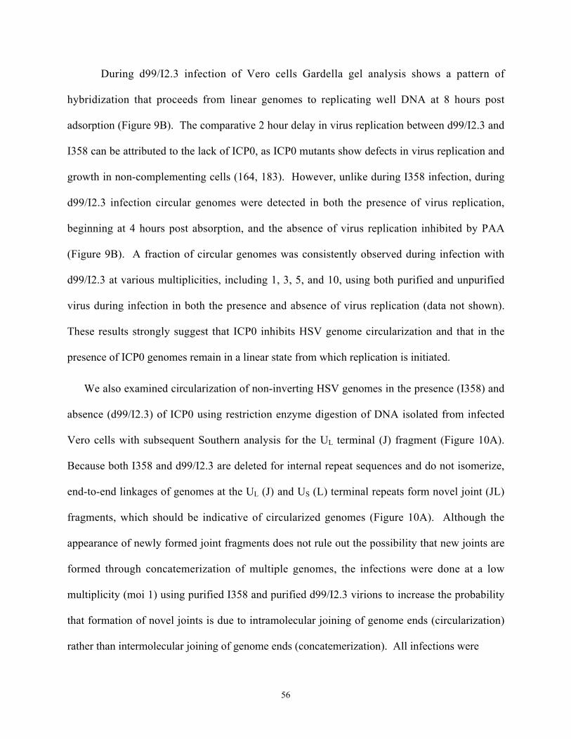
56
During d99/I2.3 infection of Vero cells Gardella gel analysis shows a pattern of
hybridization that proceeds from linear genomes to replicating well DNA at 8 hours post
adsorption (Figure 9B). The comparative 2 hour delay in virus replication between d99/I2.3 and
I358 can be attributed to the lack of ICP0, as ICP0 mutants show defects in virus replication and
growth in non-complementing cells (164, 183). However, unlike during I358 infection, during
d99/I2.3 infection circular genomes were detected in both the presence of virus replication,
beginning at 4 hours post absorption, and the absence of virus replication inhibited by PAA
(Figure 9B). A fraction of circular genomes was consistently observed during infection with
d99/I2.3 at various multiplicities, including 1, 3, 5, and 10, using both purified and unpurified
virus during infection in both the presence and absence of virus replication (data not shown).
These results strongly suggest that ICP0 inhibits HSV genome circularization and that in the
presence of ICP0 genomes remain in a linear state from which replication is initiated.
We also examined circularization of non-inverting HSV genomes in the presence (I358) and
absence (d99/I2.3) of ICP0 using restriction enzyme digestion of DNA isolated from infected
Vero cells with subsequent Southern analysis for the UL terminal (J) fragment (Figure 10A).
Because both I358 and d99/I2.3 are deleted for internal repeat sequences and do not isomerize,
end-to-end linkages of genomes at the UL (J) and US (L) terminal repeats form novel joint (JL)
fragments, which should be indicative of circularized genomes (Figure 10A). Although the
appearance of newly formed joint fragments does not rule out the possibility that new joints are
formed through concatemerization of multiple genomes, the infections were done at a low
multiplicity (moi 1) using purified I358 and purified d99/I2.3 virions to increase the probability
that formation of novel joints is due to intramolecular joining of genome ends (circularization)
rather than intermolecular joining of genome ends (concatemerization). All infections were

57
Figure 10. ICP0 inhibits formation end-to-end genome linkages during infection.
A. The I358 genome, showing deletion of internal inverted repeats an' and b' and truncation of c'
(c'*). The relevant BglII sites are indicated and are similar in d99/I2.3. Vero cells were infected
(moi 1) with purified I358 (B) or purified d99/I2.3 (C) virus stocks and harvested at the indicated
times post absorption. 400ug/mL PAA was used to inhibit replication where indicated. DNA
was isolated from cells, digested with BglII, and then resolved on a 0.8% agarose gel, which was
Southern blotted and probed for HSV sequences that hybridize to the UL (J) terminus and,
subsequently the novel joint (JL) sequence. Defective genomes described by others are noted
(**) (149). Note lane 2 in C was slightly underloaded, as observed on the ethidium bromide
staining of gel prior to blotting (not shown).
A.
BglII BglII
------ J -----
UL US
an b c'* c a
---L----
I358
B.
C.

58
preformed on ice in TBS (+/- 400ug PAA), placed in fresh media (+/- 400ug PAA) at 370C, and
then harvested at the indicated times post absorption on ice; 0 hour time points correspond to
cells harvested after 1 hour absorption on ice. However, only purified virus stocks were used to
infect cells during restriction enzyme analysis. At the indicated times post absorption, DNA was
isolated from infected cells, digested with BglII, and then resolved on an agarose gel. DNA was
transferred to a membrane, which was then, hybridized to HSV-1 XbaI/BamHI sequences,
spanning 10635-11819bp of the genome, that was isolated from a cloned EcoRI JK fragment.
This 1.18kb XbaI/BamHI fragment was used to detect UL termini (J) and subsequently joint (JL)
viral DNA fragments during Southern analysis. These joint (JL) fragments should only be
present in end-to-end linked viral genomes.
We first examined formation of novel joint fragments (JL) during I358 infection of Vero
cells in both the presence and absence of replication inhibited by PAA (Figure 10B). Joint (JL)
fragments were only observed at 6 hours post absorption of I358. In addition, at 6 hours post
absorption for both I358 and d99/I2.3, we also observed a small fraction of bands that arise from
'defective genomes' above the joint (JL) fragment; these bands and 'defective genomes' have
been previously observed and described by others (149). Importantly, joint fragments were not
observed at 0 hours post absorption, from 1-4 hours post absorption, or moreover, at 6 hours post
absorption when virus replication was inhibited by PAA (Figure 10B). Treatment of cells with
PAA during infection allows all events leading up to replication to occur but inhibits replication
itself, in theory, holding the viral genome in a configuration that will serve as a template for
replication. Novel joint fragments that accumulate by 6 hours post absorption in the absence of
PAA (JL; Figure 10B) correspond to replicative well DNA during Gardella gel analysis of I358
at 6 hours post absorption (Figure 9A). We have previously speculated that replicative well

59
DNA within Gardella gels is highly branched and/or concatemerized genomes that are
byproducts of ongoing replication (89); the abundance of joint fragments during times in which
well DNA is observed supports this theory. Consistent with the instability of linear genomes that
we have previously observed during Gardella gel analysis of various HSV viruses (d109, KOS,
and d99) (89) and others have noted (149, 185), the UL terminus (J fragment) was unstable
throughout infection with I358 both in the presence and absence of PAA (J; Figure 10B). This
instability is more pronounced in restriction enzyme analysis than in Gardella gel analysis, most
likely because a lower multiplicity of infection was used when end-to-end genome junctions
were examined.
By contrast, upon BglII digestion and analysis for d99/I2.3 end-to-end genome junctions, we
observed a small, but detectable, fraction of joint (JL) fragments beginning at 4 hours post
absorption of Vero cells that became highly abundant by 12 hours post absorption (JL; Figure
10C). The highly abundant joint fragments at 12 hours post absorption correspond to replicative
well DNA observed as early as 8 hours post absorption during Gardella gel analysis of d99/I2.3
(Figure 9B). Again, replication of d99/I2.3 is somewhat delayed with respect to I358 replication
due to the lack of ICP0 and the UL terminus (J fragment) was unstable throughout infection both
in the presence and absence of PAA (Figure 10B, 10C). The appearance of joint fragments at 4
hours (Figure 10C) corresponds to the appearance of circular genomes that we observed during
Gardella gel analysis at 4 hours post absorption (Figure 9B). Importantly, at later times (6 hr, 12
hr) post absorption, when replication was inhibited by PAA, a small fraction of joints was
observed (PAA; Figure 10C), consistent with the small fraction of circular genomes that we have
observed in the presence of PAA during Gardella gel analysis of d99/I2.3 (PAA, Figure 9B).
However, the proportion of d99/I2.3 joint (JL) fragments, that appear early during infection (4hr)
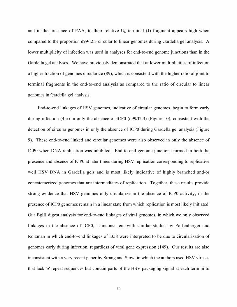
60
and in the presence of PAA, to their relative UL terminal (J) fragment appears high when
compared to the proportion d99/I2.3 circular to linear genomes during Gardella gel analysis. A
lower multiplicity of infection was used in analyses for end-to-end genome junctions than in the
Gardella gel analyses. We have previously demonstrated that at lower multiplicities of infection
a higher fraction of genomes circularize (89), which is consistent with the higher ratio of joint to
terminal fragments in the end-to-end analysis as compared to the ratio of circular to linear
genomes in Gardella gel analysis.
End-to-end linkages of HSV genomes, indicative of circular genomes, begin to form early
during infection (4hr) in only the absence of ICP0 (d99/I2.3) (Figure 10), consistent with the
detection of circular genomes in only the absence of ICP0 during Gardella gel analysis (Figure
9). These end-to-end linked and circular genomes were also observed in only the absence of
ICP0 when DNA replication was inhibited. End-to-end genome junctions formed in both the
presence and absence of ICP0 at later times during HSV replication corresponding to replicative
well HSV DNA in Gardella gels and is most likely indicative of highly branched and/or
concatemerized genomes that are intermediates of replication. Together, these results provide
strong evidence that HSV genomes only circularize in the absence of ICP0 activity; in the
presence of ICP0 genomes remain in a linear state from which replication is most likely initiated.
Our BglII digest analysis for end-to-end linkages of viral genomes, in which we only observed
linkages in the absence of ICP0, is inconsistent with similar studies by Poffenberger and
Roizman in which end-to-end linkages of I358 were interpreted to be due to circularization of
genomes early during infection, regardless of viral gene expression (149). Our results are also
inconsistent with a very recent paper by Strang and Stow, in which the authors used HSV viruses
that lack 'a' repeat sequences but contain parts of the HSV packaging signal at each termini to

61
suggest that end-to-end linkages of genomes, indicative of circular genomes not concatemers, are
present early during infection in the presence of ICP0 and in the absence of HSV gene
expression (185). However, there are many problems with the interpretation of data in both of
these papers, arising from inherent complexities present within the technique used by both
authors to analyze end-to-end genome junctions. We also initially experienced many of these
issues during our analysis of end-to-end linkages of genomes.
Because U L and U S terminal junction fragments are highly abundant during virus DNA
replication, as we (Figure 10) and others (149, 185) have shown, these junctions are present in
virus stocks grown in cell culture due to lysis of cells, where replicating viral DNA is present.
Using unpurified virus in our restriction enzyme analysis for end-to-end linkages of genomes led
to an abundance of UL and US junction fragments within our unpurified virus stocks, even at 0
hours post absorption on ice (data not shown), during which the intensity of junctions increased
with increasing multiplicity of infection. The end-to-end linkages of I358 noted by Poffenberger
and Roizman may represent replicative intermediates and/or recombination products containing
UL and US terminal junctions derived from unpurified virus inoculum due to the high multiplicity
of infection (40 PFU/cell) used (149). Unfortunately, 0 hour time points were not provided
during their analysis to determine otherwise. During our analysis using unpurified I358,
although this junction fragment was present at 0 hours, the intensity did not change throughout
infection until 6 hours post absorption during replication (data not shown). In fact, even after
virions were purified we observed a very small, but notable, fraction of UL and US junction
fragments within our purified virus stocks for both I358 and d99/I2.3, as we noted above (ICP4;
Figure 7C). Importantly, when purified virus was used, this fraction was not present at 0 hours
post absorption during analysis for end-to-end linkages of either I358 or d99/I2.3; UL and US

62
junctions only formed early during d99/I2.3, not I358, infection and at later times in the presence
of PAA during d99/I2.3, not I358, infection (Figure 10). In Strang and Stow's analysis, end-to-
end linked genome junctions, in their case UL(r) and UL(l) junctions, are present in unpurified
virion preps used to spike mock infected lysed cells and at 0 hours post absorption with
unpurified virus (185). Furthermore, in Figure 5 of their paper, the fraction of UL(r) and UL(l)
junctions detected in the presence of PAA throughout infection and in the absence of PAA early
during infection was not much greater than that observed at 0 hours post absorption in each
respective experiment. At 1 hour post absorption (+PAA), which shows the most dramatic
increase in UL(r) and UL(l) junction fragments, the lane was over loaded, which is demonstrated
by the relative increased stability of T1 and T2 fragments in the presence of PAA at this time as
compared to the relative decreased stability of T1 and T2 fragments in the absence of PAA at
this time. Also, the lane corresponding to 2 hours postabsorption (+PAA) was underloaded.
Differences in the observed presence of end-to-end genome linkages are most likely due to
contaminating genome junctions, and we suggest these differences are resolved by using purified
virus stocks. In our hands, both Gardella analysis and BglII analysis for end-to-end genome
linkages, when purified virus is used, consistently suggest that HSV genomes circularize only in
the absence of ICP0 and that ICP0 inhibits genome circularization. Moreover, these results
strongly suggest that in the presence of ICP0 genomes remain in a linear state from which
replication is initiated. Subsequently, an initial theta mechanism of HSV replication does not
seem likely. Genome replication may proceed from the three origins of replication on linear
genomes producing highly branched structures that are modified and/or resolved by
recombination and by the viral cleavage/packaging machinery. Lagging strand synthesis of
linear HSV genomes may be completed through mechanisms such as recombination-dependent

63
replication (RDR), a mechanism by which T4 phage lagging strand synthesis is accomplished
(136, 137).

64
5. INHIBITION OF DNA REPAIR AND GENOME CIRCULARIZATION BY HSV
ICP0
5.1. Abstract
We have recently shown that in the absence of immediate early protein ICP0 HSV-1 genomes
circularize and that genome circularization is inhibited by ICP0 during productive infections.
From these conclusions, we have hypothesized that the ends of input HSV genomes are treated
as double strand breaks (DSB) that are repaired early during infection via circularization of viral
genomes by cellular DNA DSB repair pathways and that ICP0 inhibits these pathways. We
examined the effect of ICP0 on proteins involved in DNA repair events in order to study how
ICP0 alters the cellular DNA repair environment, leading to inhibition of genome circularization.
During DNA DSB repair, a complex of Mre11/Rad50/NBS1 (M/R/N) proteins form discrete
nuclear foci that co-localize with phosphorylated H2AX ( H2AX) foci, which accumulate at
DSB's. Early during abortive HSV infection (1hr) in the absence of IE gene expression, M/R/N
and H2AX foci were observed and colocalized but dissipated at later times (6 hr). In contrast,
when ICP0 was expressed, M/R/N and H2AX foci accumulated and persisted throughout
abortive infection but did not colocalize. For the first time, these results suggest that incoming
HSV stimulates cellular DNA repair processes that dissipate or are completed later during
infection in the absence of ICP0. In the presence of ICP0 this repair response persists and is
altered, as M/R/N and H2AX foci do not co-localize. ICP0 does not affect the global level of
M/R/N proteins during infection, and M/R/N complexes do not affect circularization of HSV
genomes. However, ICP0 does induce the degradation of the nonhomologous end-joining
(NHEJ) protein DNA PKcs in a cell type specific manner. We examined the effect of NHEJ
proteins on circularization of HSV-1 genomes. In cells mutant for NHEJ proteins, genome
circularization was greatly delayed and decreased. These results suggest a major role for NHEJ

65
in HSV genome circularization. Together, the results provide evidence for cellular DNA repair
responses to incoming HSV genomes and alterations in these repair responses as a result of an
activity of ICP0.
5.2. Introduction
Lytic infection with HSV-1 begins with the expression of five immediate early (IE) viral genes,
designated infected cell polypeptide (ICP) 0, 4, 22, 27, and 47. IE proteins initiate a cascade of
viral gene expression that leads to virus genome replication and production of progeny viruses.
Latent HSV-1 infection occurs when the viral genome persists in a nucleosome-associated state
(40) in which viral gene expression is largely repressed, thereby precluding virus replication.
However, latent infections can reactivate to produce recurrent lytic infections.
ICP0 plays a pivotal role in balancing the lytic and latent life cycles by greatly enhancing
the probability of the lytic cycle both during primary infection and reactivation events. Early
during lytic infection, ICP0 localizes to and disrupts ND10 bodies (116), which are discrete
nuclear foci where HSV-1 replication centers may be located (117). ND10 bodies play a role in
a variety of cellular pathways including cellular stress responses and chromosomal DNA repair
pathways (118). ICP0 has E3 ubiquitin ligase activity and subsequently induces the in vivo
proteasome-dependent degradation of PML and Sp100 (23), which are constituents of ND10
bodies, of the DNA-dependent Protein Kinase Catalytic Subunit (DNA PKcs) (107) (143), which
is critical for VDJ recombination and DNA double-strand break repair via non-homologous end-
joining (NHEJ) (30), and of CENPA and CENPC (53), which are histone-3 like centromere
proteins. Exactly how ICP0 activity promotes the lytic cycle remains unclear, although it
ultimately leads to indiscriminate transactivation of viral gene expression.

66
We have recently shown that ICP0 also inhibits circularization of HSV-1 genomes (89).
In the absence of ICP0 the HSV-1 genome circularizes. Viral genomes are thought to persist as
circles during latency, where viral gene expression is largely repressed (44, 159). Therefore
ICP0 activity may control the balance between the lytic and latent life cycles, in part, by
regulating the configuration of viral genomes. We hypothesize that in the absence of ICP0
activity the ends of linear HSV-1 genomes are treated as double-strand DNA breaks that are
repaired via circularization of viral genomes by cellular DNA double-strand break repair
pathways. ICP0 could therefore inhibit circularization of HSV-1 genomes by inhibiting DNA
repair pathways.
In mammalian cells, homologous recombination (HR) and non-homologous end-joining
(NHEJ) are the two primary pathways by which double-strand breaks in DNA are repaired.
Distinct proteins play a role in each NHEJ and HR repair, however some proteins, such as the
Mre11/Rad50/NBS1 (M/R/N) complex, may have overlapping roles in both mechanisms (35).
M/R/N complexes are sensors of DNA double-strand breaks and activate the ATM-mediated
repair pathway (1, 105). Many proteins involved in HR have redundant functions and are
subsequently somewhat disposable to the mechanism. However, one protein Rad51 appears to
be necessary for HR (134). Ku70, Ku80, DNA PKcs, XRCC4, and DNA Ligase IV are critical
for NHEJ in vivo (30). Ku70 and Ku80 subunits dimerize, forming the regulatory subunits of the
DNA PK holoenzyme (70). The Ku dimer binds DNA ends (8, 130), recruiting the catalytic
subunit of DNA PK (DNA PKcs) to sites of DNA double strand breaks (70). Once associated
with DNA-bound Ku subunits, DNA PKcs catalyzes the end-joining reaction through a
combination of phosphorylation events (127) that leads to recruitment of the XRCC4/DNA
Ligase IV tetramer, which enzymatically ligates the DNA ends together (198). Little sequence
67
homology is required for NHEJ repair, while HR involves the exchange of homologous DNA
sequences.
The HSV genome is a linear double stranded DNA molecule that is divided into unique
long and unique short regions that are connected by a joint sequence (195) (197). Both long and
short regions are bracketed by homologous inverted repeats that contain a 250-500 bp sequence,
termed the 'a' sequence, in a directly repeating orientation at each termini but in an inverted
orientation at the UL/US junction (197) (161). Thus, the genome is a suitable substrate for either
HR or NHEJ repair mechanisms.
HSV infection induces activation and signaling to proteins involved in DNA damage
responses. In particular, during HSV infection ATM and Mre11 are necessary for activation and
signaling that results in modification of repair proteins (ATM, Chk2, 53BP1, and NBS1) and
many times redistribution of these repair proteins to HSV replication compartments (111, 178).
Additional studies have also shown redistribution of other repair proteins (DNA PKcs, Ku80,
Rad50, Rad51, BRCA1, WRN, BLM, MSH2, and RPA) involved in NHEJ and HR mechanisms
to viral replication compartments (188, 200). However, whether these repair proteins are
signaled as a result of input linear viral genomes, are recruited to repair the highly nicked HSV
genome during replication, or play a role in inter- and intra- molecular recombination events that
occur during viral replication has not been determined.
Our focus is to examine how the host cell responds to incoming HSV genomes and how
the virus, in particular ICP0, may counteract or manipulate this response, ultimately leading to
inhibition of genome circularization. Using replication defective mutant viruses, we examined
the distribution of DNA repair proteins in response to input HSV and the effect of ICP0 on this
distribution. We also examined the ability of DNA repair pathways to circularize HSV genomes

68
during infection. Together, the results provide evidence for cellular DNA repair responses to
incoming HSV and an ICP0-induced effect on these responses. They also strongly suggest a
major role for NHEJ in circularization of HSV-1 genomes.
5.3. Methods
5.3.1. Viruses.
The mutant viruses: d109, d105, d106, and d99 were derived from wild type strain KOS and
used at the indicated multiplicities of infection (moi) (165). Where indicated, 400ug/mL
phosphonoacetic acid (PAA; Lancaster Synthesis) was added to Tris-buffered saline (TBS;
0.14M NaCl, 2.7 mM KCl, 25mM Tris-base, pH 7.4) or the appropriate cell culture media during
and after viral absorption to inhibit virus replication. Virus was allowed to absorb to HEL and
Vero cells for one hour on ice in TBS in order to synchronize infections. Virus was allowed to
absorb to other cells lines for one hour at room temperature in TBS. After all infections, cells
were washed 1x with TBS and the appropriate media was placed on cells until indicated
harvesting times post-infection.
5.3.2. Cells.
Human Embryonic lung (HEL) cells (ATCC) were used for all immunofluorescence
experiments and for Western Blot analyses. HEL cells were maintained in DMEM
supplemented with 10% FBS and grown in 5% CO2 at 370C. Cells were used at low passage
(<20). Similarly, Vero (ATCC) cells used in Western Blot analyses were maintained in DMEM
supplemented with 5% FBS and grown in 5% CO2 at 370C.
Cells mutated in genes involved in DNA doubles strand break repair used in Gardella gel
analyses were cultured as follows. Primary fibroblasts from an apparently healthy human
(NBS1+; Coriell Cell Repository) and primary fibroblasts from an individual homozygous for a

69
deletion of 5 nucleotides in exon 6 of the NBS1 gene (NBS-; Coriell Cell Repository), resulting
in a truncated protein from a frameshift and subsequent premature termination at codon 218,
were cultured in MEM Eagle-Earle BSS 2x concentration of essential and non-essential amino
acids and vitamins with 2mM L-glutamine and 15% FBS under 5% CO2 at 370C. MO59J, which
lack DNA PKcs activity, and MO59K (both ATCC), which contain normal levels of DNA PKcs
activity, cell lines isolated from the same human glioblastoma tumor were cultured in a 1:1
mixture of DMEM and Ham's F12 medium with 2.5 mM L-glutamine and supplemented with
0.5mM sodium pyruvate, 0.05mM non-essential amino acids and 10% FBS under 5% CO2 at
370C. SV40 T-antigen transformed mouse embryonic fibroblasts (MEFs) lacking (-/-) or
expressing (+/+) Ku80 were provided by Dr. Andre Nussenzweig (NIH/NCI). Both cell lines
were cultured in DMEM with 10% FBS under 5% CO2 at 370C. SV40 T-antigen transformed
MEFs lacking both p53 and DNA ligase4 (p53-/-lig4-/-) and MEFs expressing DNA ligase 4 but
lacking p53 (p53-/-) were provided by Dr. Frederick Alt (CBR Institute for Biomedical
Research, Boston) and were cultured in DMEM supplemented with 10% FBS and grown under
5% CO2 at 370C.
5.3.3. Immunofluorescence.
HEL cells were plated one day prior to infection on 18mm circular glass coverslips (VWR) at a
densitly of 1.5x10⁶ cells per 60mm dish in media. Prior to infection, confluent HELs in 60mm
dishes (4 coverslips per dish) were chilled on ice and rinsed with 40C TBS. Infections were done
on ice for 1 hour with rocking, after which warm media (with 400ug/mL PAA for KOS and d99)
was added. Cells were incubated immediately at 370C until the desired times post-infection. At
desired times post-infection, coverslips were rinsed with warm TBS and rapidly submerged in -

70
200C methanol for exactly 5 minutes to fix the cells. Cells were allowed to dry thoroughly prior
to beginning the indirect immunofluorescence protocol.
Cells were stained for various proteins using standard indirect immunofluorescence
techniques. Antibody dilutions are as follows: rabbit anti-NBS1 (1:200; NOVUS Biologicals),
mouse anti-Mre11 (1:100; GeneTex), mouse anti-PML (1:30; Santa Cruz Biotechnology), mouse
anti-Rad50 (1:100; GeneTex), and rabbit anti- H2AX (1:200; Upstate Biologicals). Primary
antibodies were followed by secondary anti-mouse IgG-conjugated Alexa Fluor488 or anti-rabbit
IgG-conjugated Alexa Fluor594 (both 1:500; Molecular Probes). Controls using only secondary
antibodies were performed to ensure no non-specific staining or bleed-through was present (data
not shown). Coverslips were mounted in gelvatol on glass slides and imaged at 100x power with
a Nikon Diaphot 300 epifluorescent microscope. Images were collected using the Spot program
and analyzed with Adobe Photoshop software.
5.3.4. Western Blots.
Standard Western Blot protocols were used to examine the levels of DNA repair proteins in
whole cell extracts of HEL and Vero cells infected at the indicated multiplicities. Briefly,
proteins were resolved on denaturing (SDS) Tris-HCl polyacrylamide gels and then transferred at
250mAmps (40C) onto Hybond-P membranes (Amersham Biosciences). Non-specific binding
was blocked with 5% milk in PBS-0.5%Tween20. Membranes were washed with PBS-
0.5%Tween20 3x10 minutes before and after primary antibody staining and 3x10 minutes after
secondary antibody staining. To detect proteins, membranes were incubated for 1 hour with the
following primary antibodies at the indicated dilutions in PBS-0.5%Tween20 with 3% milk:
DNA PKcs (1:1000; Upstate Biotechnology), mouse monoclonal Ku80 (1:5000; Upstate
Biotechnology), rabbit polyclonal Ku70-H308 (1:5000; Santa Cruz Biotechnology), mouse

71
monoclonal Rad51-3C10 (1:2000; Upstate Biotechnology), mouse monoclonal ICP0-1112
(1:5000; Goodwin Institute for Cancer Research, Inc., Plantation, Fla.), mouse polyclonal
XRCC4 (1:1000; GeneTex), mouse monoclonal Rad50-13B3 (1:500; GeneTex), mouse
monoclonal Mre11-12D7 (1:400; GeneTex), and rabbit polyclonal NBS1/p95/nibrin (1:10,000;
Novus Biologicals). Primary antibodies were detected with secondary anti-mouse (1:4x10⁴) or
anti-rabbit (1:1.5x10⁴) IgG HRP conjugated antibodies (Promega) diluted in PBS-0.5%Tween20
with 3% milk and were visualized by ECL plus Western Blotting Detection System (Amersham
Biosciences) upon exposure of membrane to film.
5.3.5. Gardella gel analysis.
Gardella gels were assembled and preformed as previously described (89). After infection of
5x10⁶ cells at the indicated multiplicity of infection, cells were harvested at the indicated time
points by washing with TBS, scraping into TBS, and then pelleting by low-speed centrifugation.
Cell pellets were resuspended in 50uL loading dye (15% Ficoll, 0.01% bromophenol blue) and
loaded into the wells of the Gardella gel. Gels were run at 45V for 3 hours to lyse cells and then
120V for 38 hours to separate circular from linear genomes of the same molecular weight.
Southern blots were used to visualize viral DNA. The 1.8kb BamHI-Y fragment of pKBY (166),
which contains a portion of the ICP4 gene of HSV-1, was used as the probe to detect viral
genomes during all Southern blot analyses.
5.4. Results
5.4.1. Formation of M/R/N complexes and H2AX foci as a function of ICP0.
Foci of the phosphorylated (serine 139) form of histone variant H2AX ( H2AX) accumulate at
sites of DNA double-strand breaks (DSB) (157, 160, 172). M/R/N foci colocalize with H2AX
foci during repair of DNA DSBs that occur as a result of ionizing radiation (IR) (19) (144).

72
Figure 11. Formation of M/R/N foci as a function of ICP0 during abortive infection.
HEL cells were infected with d109 (d-l) or d105 (m-u) at an moi of 20 prior to fixation and
indirect immunofluorescence (IF) staining at 1 hour (d-f; m-o), 2 hours (g-i; p-r), or 6 hours (j-l;
s-u) post infection. A. Rad50 (green) and NBS1 (red) B. Mre11 (green) and NBS1 (red). For
both (A) and (B), colocalization is shown in yellow and mock infected cells are shown (a-c).
Highly resolute, colocalizing M/R/N foci can be seen in the presence (d105; inset u) but not
absence (d109) of ICP0.
A B

73
Using HSV-1 mutant strains d109 and d105, we examined M/R/N and H2AX foci formation
during viral infection of HEL cells as a function of ICP0. d109 is an HSV-1 mutant that lacks
expression of all five IE viral genes and subsequently establishes a non-toxic persistent quiescent
state from which gene expression can be de-repressed by ICP0 (165). d105 is an HSV-1 mutant
that expresses ICP0 but lacks expression of the additional four IE viral genes (165). Both d109
and d105 form abortive infections in which neither mutant replicates due to lack of ICP4
expression.
We examined formation of M/R/N foci during abortive HSV infections as a function of
ICP0. In the presence of ICP0, discrete and highly resolute foci of colocalizing Rad50 and
NBS1 (d105, m-u; Figure 11A) proteins and colocalizing Mre11 and NBS1 (d105, m-u; Figure
11B) proteins were observed throughout infection, indicating that M/R/N complexes form in the
presence of ICP0 during abortive infections. However the numerous, highly resolute M/R/N foci
were not as evident in the absence of ICP0 (d109, d-l; Figure 11A and 11B) or during mock
infections (mock, a-c; Figure 11A and 11B). These complexes accumulated and persisted up to
24 hours post infection with d105 (a-c and d-f; Figure 12). Co-localizing Mre11 and NBS1 foci
were also observed during wild-type KOS (g-i; Figure 13) but not ICP0-mutant d99 (d-f; Figure
13) infections carried out in the presence of phosphonoacetic acid to inhibit virus replication but
allow for IE gene expression (85). d99 lacks expression of ICP0, but expresses the additional
four IE genes and thus replicates in the absence of PAA.
We next examined the formation of H2AX foci and its colocalization with M/R/N foci
during abortive infection as a function of ICP0. With closer inspection, at one hour post
infection with d109, we observed some, yet less resolute, Mre11 foci (g; Figure 14). These
Mre11 foci clearly co-localized with H2AX foci (g-i; Figure 14). In the majority of cells, both

74
Figure 12. Rad50 and NBS1 foci accumulate and persist during abortive infection in the
presence of ICP0.
HEL cells were infected with d105 at an moi of 20 and fixed at 12 (a-c) or 24 (d-f) hours post
infection. Indirect Rad50 and NBS1 IF staining is shown in green and red, respectively.
Colocalization is indicated in yellow.

75
Figure 13. Formation of Mre11 and NBS1 foci as a function of ICP0 during productive
infection in which replication is inhibited.
HEL cells were infected with KOS (g-i) or ICP0-mutant d99 (d-f) at moi of 10 and fixed 6 hours
post infection at which time cells were stained for either Mre11 (green) or NBS1 (red) by
indirect IF. Cells were infected in the presence of PAA to inhibit virus replication. Staining of
mock infected cells (a-c) is shown, and colocalization of Mre11 and NBS1 is shown in yellow.

76
Mre11 and H2AX foci dissipated by 6 hours post infection (j-l; Figure 14), with Mre11/ H2AX
staining resembling the field of mock-infected cells (a-c; Figure 14). By contrast, when ICP0
was expressed, both Mre11 (m, p; Figure 14) and H2AX (n, q; Figure 14) foci were observed
throughout d105 infection, with many foci being strongly detected at 6 hours post infection (p-q;
Figure 14), as in IR treated cells (d-e; Figure 14). Interestingly, in the presence of ICP0, M/R/N
complexes do not co-localize with H2AX foci (o, r; Figure 14) as they do in IR treated cells (f;
Figure 14). These results suggest that infection with d109 induces a cellular DNA repair
response early during infection that dissipates or is completed later in infection, as evidenced by
the formation and then loss of co-localizing M/R/N and H2AX foci. ICP0 appears to alter this
repair response to incoming HSV in that M/R/N foci accumulate and persist throughout infection
and do not colocalize with H2AX foci.
5.4.2. Levels of DNA repair proteins as a function of ICP0.
We next examined the levels of DNA repair proteins as a function of ICP0 expression (Figure
15). For both infected HEL and Vero whole cell extracts, we compared the level of DNA repair
proteins in the presence of ICP0 (d106, KOS) and in the absence of ICP0 (d109, d99) at 6 hours
post abortive (d106, d109) and productive (KOS, d99) infection. d106, like d105, expresses
ICP0 but lacks expression of the additional four IE genes (165). Levels of proteins involved in
NHEJ (DNA PKcs, Ku70, Ku80, XRCC4) and HR (Rad51) were examined, as well as levels of
proteins that constitute the M/R/N complex (Mre11, Rad50, NBS1). Of the proteins examined,
only the level of DNA PKcs was dramatically reduced in the presence of ICP0 during both d106
and KOS infection of HEL cells, relative to that observed during mock, d109, or d99 infection of
HEL cells. Consistently, it has been previously shown that DNA PKcs undergoes proteasome-

77
Figure 14. Colocalization of Mre11 and H2AX foci in the absence but not presence of ICP0
during abortive infection.
HEL cells were infected with d109 (g-l) or d105 (m-r) at an moi of 30 and fixed 1 hr (g-i; m-o)
or 6 hr (j-l; p-r) post infection. Indirect IF was used to detect Mre11 (green) and H2AX (red);
colocalization is shown in yellow. Cells in a-c were mock infected, with approximately only 5%
of cells containing Mre11/ H2AX foci in nuclei (inset c). Cells in panels d-f were treated with 5
Gy ionizing radiation (IR) and allowed to recover for 30 minutes prior to fixation and staining.
Colocalization of Mre11 and H2AX can be seen in IR treated cells (inset f) and early during
d109 infection (inset i) but not during d105 infection (inset r).

78
A. B.
Figure 15. Global level of DNA repair proteins as a function of ICP0.
Western blot analysis of HEL (A) and Vero (B) whole cell extracts at 6hr post-infection with the
indicated virus (moi = 10). The DNA repair proteins analyzed are indicated.

79
dependent degradation as a result of ICP0 expression in human-derived Hela (107) (143) but not
monkey-derived Vero cell lines (200).
In addition, during KOS infection and to a lesser extent during ICP0-mutant d99 infection
of both HEL and Vero cells, the mobility of NBS1 was slightly decreased (NBS1; Figure 15),
which we project to be due to phosphorylation. Phosphorylation of NBS1 indicates induction of
cellular ATM DNA repair processes (202) and has been observed by others during HSV
infection (111, 200) (178). However, our study indicates that NBS1 modification does not occur
as a function of ICP0 expression but rather occurs in response to additional factors that are
present during both wild-type KOS and ICP0-mutant d99 infections.
5.4.3. NHEJ is involved in circularization of HSV-1 genomes
We hypothesized that DNA repair pathways play a role in circularizing HSV-1 genomes in the
absence of ICP0 expression and that ICP0 inhibits circularization by inhibiting DNA repair
pathways. Due to ICP0-induced degradation of DNA PKcs, which plays a major role in NHEJ,
we examined circularization of d109 genomes in cells lacking NHEJ proteins using Gardella gel
analysis (Figure 16). M059J cells are a human glioblastoma derived cell line that lack DNA
PKcs activity (106). M059K cells are their complementing cell line that contain normal levels of
DNA PKcs activity and were derived from the same human glioblastoma tumor (106). In
M059K cells we detected circles in Gardella gels between 1 and 2 hours post infection, which is
consistent with our previously determined kinetics of d109 circularization in Vero cells (Figure
16A). However, in M059J cells the kinetics of d109 circularization were severely delayed
(Figure 16A). A similar delay in the kinetics of d109 circularization was also observed in mouse
embryonic fibroblasts (MEF) lacking Ku80 expression relative to a complementing MEF cell
line expressing Ku80 (Figure 16B) (194). In addition, the fraction of d109 genomes that

80
Figure 16. Circularization of HSV genomes in cells deficient in NHEJ.
A. MO59J (DNA PKcs -) or M059K (DNA PKcs +) human gliablastoma cells were infected
with d109 (moi 20) for the indicated times and analyzed by Gardella gels. Arrow indicates
position of circular genomes. B. Ku80+/+ or Ku80-/- MEFs were infected with d109 (moi 20)
for the indicated times and analyzed by Gardella gels. C. Vero, ligase4-/-p53-/-(a) MEFs,
ligase4-/-p53-/-(b) MEFs, and p53-/- MEFs were infected for 6 hours with d109 or d106 (moi
20) and then analyzed by a Gardella gel. Circular (O) and linear (l) genomes are indicated.
A. B. C.
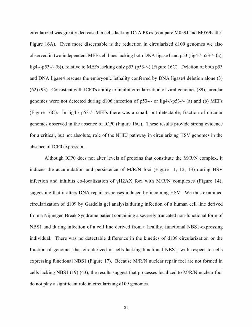
81
circularized was greatly decreased in cells lacking DNA PKcs (compare M059J and M059K 4hr;
Figure 16A). Even more discernable is the reduction in circularized d109 genomes we also
observed in two independent MEF cell lines lacking both DNA ligase4 and p53 (lig4-/-p53-/- (a),
lig4-/-p53-/- (b)), relative to MEFs lacking only p53 (p53-/-) (Figure 16C). Deletion of both p53
and DNA ligase4 rescues the embryonic lethality conferred by DNA ligase4 deletion alone (3)
(62) (93). Consistent with ICP0's ability to inhibit circularization of viral genomes (89), circular
genomes were not detected during d106 infection of p53-/- or lig4-/-p53-/- (a) and (b) MEFs
(Figure 16C). In lig4-/-p53-/- MEFs there was a small, but detectable, fraction of circular
genomes observed in the absence of ICP0 (Figure 16C). These results provide strong evidence
for a critical, but not absolute, role of the NHEJ pathway in circularizing HSV genomes in the
absence of ICP0 expression.
Although ICP0 does not alter levels of proteins that constitute the M/R/N complex, it
induces the accumulation and persistence of M/R/N foci (Figure 11, 12, 13) during HSV
infection and inhibits co-localization of H2AX foci with M/R/N complexes (Figure 14),
suggesting that it alters DNA repair responses induced by incoming HSV. We thus examined
circularization of d109 by Gardella gel analysis during infection of a human cell line derived
from a Nijmegen Break Syndrome patient containing a severely truncated non-functional form of
NBS1 and during infection of a cell line derived from a healthy, functional NBS1-expressing
individual. There was no detectable difference in the kinetics of d109 circularization or the
fraction of genomes that circularized in cells lacking functional NBS1, with respect to cells
expressing functional NBS1 (Figure 17). Because M/R/N nuclear repair foci are not formed in
cells lacking NBS1 (19) (43), the results suggest that processes localized to M/R/N nuclear foci
do not play a significant role in circularizing d109 genomes.

82
Figure 17. NBS1 does not affect HSV genome circularization.
Human cells lacking functional NBS1 (-/-) or expressing functional NBS1 (+/+) were infected
with d109 at an moi of 10, harvested at the indicated times post infection, and analyzed by
Gardella gels. Circular (O) and linear (l) genomes are indicated.

83
5.5. Discussion
We have previously shown that ICP0 inhibits HSV genome circularization (89) and have
subsequently hypothesized that in the absence of ICP0 activity the ends of HSV genomes are
treated as double-strand DNA breaks that are repaired via circularization of viral genomes by
cellular DNA double strand break repair pathways. ICP0 could therefore inhibit circularization
of HSV genomes by inhibiting DNA repair pathways. In this study we have examined both the
effect of ICP0 on the cellular DNA DSB repair environment and the effect of repair pathways on
circularization of HSV genomes. Incoming HSV appears to stimulate DNA repair events, and
DNA repair pathways in particular NHEJ play a major role in HSV genome circularization.
ICP0 both affects DNA repair events and inhibits HSV genome circularization.
M/R/N complexes are essential for DSB repair and genomic stability. These foci
accumulate at sites of ongoing DNA repair (19, 144). Others have shown these foci accumulate
at sites of HSV replication (111, 188, 200) and suggest that these repair proteins facilitate the
HSV productive cycle (111). Here we demonstrate that in the presence of ICP0, M/R/N
accumulate and persist both during abortive infection (d106) (Figure 11, 12) and, consistent with
the results of others (200), during productive infection (KOS+PAA) when replication is inhibited
by PAA (Figure 13). H2AX foci, which are markers for ongoing DNA DSB repair (157, 160,
172), also accumulate during abortive infections in the presence of ICP0, as we have previously
observed (72). However, unlike during DSB repair events, M/R/N and H2AX foci do not
colocalize and do persist when ICP0 is present (Figure 14). Although not as apparent in Figure
11, both M/R/N and H2AX foci also form in the absence of ICP0 (d109) early (1hr) during
abortive HSV infection (Figure 14). Importantly, H2AX foci colocalize with M/R/N foci
formed early during d109 infection, but at later times (6hr) both foci dissipate, which is similar to

84
the formation and then dissipation of these foci during DSB repair events. These results suggest
that input HSV stimulates DNA repair events that dissipate or are completed at later times during
infection in the absence of ICP0. Importantly, these repair events are affected by the presence of
ICP0, as both H2AX and M/R/N foci accumulate and persist and do not colocalize.
These results are the first to show that during abortive infections, both H2AX foci and
M/R/N complexes form in response to input HSV (Figure 11, 14) and that ICP0 promotes the
accumulation and persistence of these complexes (Figure 12) but disables their association with
each other (Figure 14). However, whether or not M/R/N complexes or H2AX foci form at sites
of input HSV genomes remains undetermined. We did not observe correlations between the
number of H2AX foci formed and the number of input HSV genomes during infections at
various multiplicities (data not shown), which suggest that repair foci form at sites of cellular
DNA. Early events during HSV-1 infection induce damage to host cell chromatin, including
chromosome uncoiling, fragmentation, and margination (26, 139, 140, 145, 182). These events
require HSV entry and IE gene expression (61, 140) and subsequently could be caused by ICP0
during abortive infections, thereby inducing the accumulation and persistence of M/R/N and
H2AX foci. Consistently, ICP0 induces the loss of kinetochore proteins CENPC and CENPA,
which may lead to chromosome instability (53). However, M/R/N foci do not colocalize with
H2AX during infection, as would be expected during DSB repair events, suggesting that the
repair process is somehow disrupted in the presence of ICP0.
The levels of M/R/N protein constituents are not affected in either the presence or
absence of ICP0 during both productive and abortive infections (Figure 15). However, during
productive infections, NBS1 is most likely modified by phosphorylation, as has been observed
by others (200) (111) (178). M/R/N complexes are sensors to DNA DSB's and recruit/activate

85
ATM (1, 105). NBS1 is phosphorylated by ATM during activation of the ATM repair pathway
(202). We have shown that this modification is not dependent on ICP0 but does require
additional viral proteins other than ICP0; others have suggested it to be dependent on the
presence of the HSV DNA polymerase HP66 (200). During abortive infections, incoming HSV
appears to stimulate DNA repair events that induce M/R/N and H2AX formation, but these
events occur in the absence of detectable ATM activation, as NBS1 phosphorylation is not
present during abortive infections (d109, d106; Figure 15). Others have also observed the
absence of detectable ATM activation during abortive infections with replication incompetent
virus (HSV amplicon vector) (178). Regardless of ICP0, the ATM repair pathway does not
appear to be activated by input HSV. During HSV infection, additional factors must play a role
in the activation of ATM. Lack of HP66 inhibits recruitment of repair proteins, including NBS1,
to prereplicative sites and inhibits activation of ATM, as evidenced by the absence of NBS1
phosphorylation (200). Perhaps recruitment of NBS1 to prereplicative sites, which are not
present during abortive infections, is required for ATM activation.
We have previously established that ICP0 inhibits HSV genome circularization (89) and
have shown here that ICP0 alters DNA repair events involving M/R/N complexes that occur
independent of ATM activation in response to incoming HSV. Although ICP0 appears to alter
these DNA repair events, M/R/N complexes and the ATM pathway do not play a role in
circularization of HSV genomes, as HSV circularization remains unaffected during abortive
infections of cells lacking functional NBS1, in which M/R/N complexes do not form (Figure 17)
(19, 43). Others have shown that M/R/N complexes and their associated processes may facilitate
HSV replication (111). ICP0 may somehow direct this facilitation by promoting the

86
accumulation and persistence of M/R/N complexes and/or by potentially altering their nuclear
distribution.
Of the proteins we examined, only the level of DNA PKcs was decreased in HEL cells
but not Vero cells as a function of ICP0 (Figure 15). The levels of other examined proteins were
not affected in either the presence or absence of ICP0 during both productive and abortive
infections. Consistently, it has been previously shown that DNA PKcs undergoes proteasome-
dependent degradation as a result of ICP0 expression in human-derived Hela (107) (143) but not
monkey-derived Vero cell lines (200). Because of the cell type specific degradation of the global
level of DNA PKcs, we suggest that ICP0 may have a localized effect, possibly only occurring
where ICP0, viral genomes, and DNA repair proteins colocalize early during infection.
Consistently, in Hep-2 cells DNA PKcs is somewhat degraded and complexes of DNA PKcs,
Ku80, Ku70, and XRCC4 co-precipitate with the HSV single stranded DNA binding protein,
ICP8, at times in which ICP8 is localized to HSV genomes, suggesting a direct and/or indirect
interaction between these NHEJ repair proteins with viral genomes (188). Furthermore, only
autophosphorylated forms of DNA PKcs, which are a very small fraction of the very abundant
global level of DNA PKcs, localize to sites of DNA DSB's during repair events (22).
In cells deficient in NHEJ repair, HSV genome circularization is reduced (Figure 16).
DNA PKcs and Ku80 play a critical but not absolute role in recognition and repair of DSB via
NHEJ (30), which is consistent with the reduction and delay but not abrogation of circularization
in cells lacking these proteins (Figure 16A, 16B). DNA Ligase4 is the most essential protein in
the NHEJ mechanism due to its role in the enzymatic ligation of DNA ends (62, 198).
Consistently, circularization is almost completely inhibited in cells lacking DNA Ligase4 (Figure

87
16B). These results provide strong evidence for a role of NHEJ in circularization of HSV
genomes.
Infected cells appear to treat ends of linear double-stranded HSV genomes as breaks in
DNA, and NHEJ serves as a mechanism to repair these breaks, subsequently circularizing HSV
genomes. Linear un-integrated cDNA copies of retroviral genomes, including HIV, also undergo
circularization (14, 91, 110) during infection and are substrates for the host cell NHEJ DNA
repair pathway (91, 110). Some NHEJ proteins may be inhibitory to HSV replication (143, 188).
HSV may evade host NHEJ pathways by expressing ICP0 to inhibit this pathway. In some cells
types, ICP0 induces the global degradation of the NHEJ protein, DNA PKcs; in other cell types
the effect of ICP0 may be more localized than global. This process would mirror that found
during adenovirus (AV) infection, in which Mre11 promotes concatemerization of AV genomes,
while AV E4 products inhibit concatemerization by promoting degradation of M/R/N proteins
(184). Circularization of HSV genomes by NHEJ could result in a stable configuration during
latency (44, 89, 159). In neurons, where HSV establishes latency, the abundance and/or activity
of ICP0 may be reduced (25) (86) and NHEJ may be the predominant pathway by which DSB's
in DNA are repaired (31, 66, 163). Competition between NHEJ mechanisms, which attempt to
circularize HSV genomes, and the ability of ICP0 to inhibit circularization by altering/inhibiting
the DNA repair environment, may in part affect if and how latency is established. When ICP0
prevails the balance may be swayed in favor of productive infections, when NHEJ processes
prevail the balance may be swayed in favor of latency.
88
6. SUMMARY AND GENERAL DISCUSSION
6.1. Summary of results
The study presented here models changes in HSV genomes as they occur during the
establishment of productive and latent infections. HSV mutant viruses deleted for specific
subsets of viral genes were used to elucidate how host cells respond to incoming linear, double-
stranded HSV genomes and how the virus manipulates this response to promote the productive
cycle. The results of this study have important implications in understanding the mechanisms by
which HSV genomes replicate during the productive cycle, the mechanisms by which stable
genome configurations are established during latency, and the pivotal role that ICP0 plays in the
balance between the productive and latent life cycles.
In Chapter 3, we have shown by Gardella gel analysis (67) that during infection HSV
genomes circularize in the absence of ICP0 and that ICP0 inhibits genome circularization (89).
d109 is an HSV mutant that lacks expression of all five IE viral genes and can subsequently
persist in a non-toxic state in nuclei of infected cells (165). D109 forms an abortive infection in
which the virus does not replicate and was used to model events leading to and during latency
(165). During long-term infections that model latency, d109 genomes persist in a stable circular
configuration. However, early during abortive infections, expression of ICP0 in cis or in trans
can inhibit genome circularization. ICP0 also inhibits genome circularization early during
productive infections in which HSV replicates and when productive replication is inhibited by
phosphonoacetic acid (PAA). Circular genomes are only present during productive infections
with ICP0 mutants. Because ICP0 is available to inhibit genome circularization within the first
hour of wild-type infection and genome circularization occurs between 1 and 2 hours post
infection in the absence of ICP0, circular genomes are not likely to be present during the lytic
cycle. Furthermore, if circular genomes were required for replication during productive

89
infections, then all genomes that replicate would have to initially circularize. During infection,
we detected as little as 2% of input genomes in a circular configuration by Gardella gel analysis,
while at least 10% of these input genomes can replicate. If circularization were required for
replication, then at least 10% of input genomes would have to circularize to serve as templates
for replication and this fraction of circular genomes would have been detected by Gardella gel
analysis, which is sensitive enough to detect as little as 2% of genomes in a circular state.
Contrary to previous literature that suggests HSV replication is initiated from a circular genome
template via a theta mechanism (65, 149, 185, 186), we suggest that HSV genomes replicate
from a linear template, because ICP0 inhibits genome circularization and hence theta mode
replication which requires circular genomes.
Because in Chapter 3 we have challenged the widely accepted concept that circular genomes
serve as the initial template for HSV genome replication, we have employed/compared two
different analyses to examine HSV genome configurations in Chapter 4. By comparing our
method of Gardella gel analysis (67, 89) for circular genomes to other's methods of restriction
enzyme analysis for end-to-end linkages of viral genomes (149, 185), we have clarified
discrepancies between our results that suggest HSV genomes circularize only in the absence of
ICP0 activity (89) and literature that suggests genomes circularize early during infection in both
the presence and absence of viral gene expression, including ICP0 (65, 149, 184, 186). The
ultimate goal of Chapter 4 has been to determine whether or not circular genomes are present
early during productive infection and can subsequently serve as the initial template for genome
replication.
In Chapter 4, we have examined circularization during productive infections with a non-
inverting HSV mutant I358 (149, 150) and an ICP0-deleted version of I358 termed d99/I2.3.

90
Using these viruses, Gardella gel analysis has shown that circular genomes are present in the
absence but not presence of ICP0 during productive infections. Consistently, using restriction
enzyme analysis, we have also found that end-to-end linkages of viral genomes, indicative of
circularized genomes, are only present early during infection, prior to replication, in the absence
of ICP0 (d99/I2.3). End-to-end linkages of genomes formed at later times during replication in
both the presence and absence of ICP0 correspond to replicative well DNA found in Gardella
gels in both the presence and absence of ICP0 and are likely to be indicative of highly branched
and/or concatemerized genomes generated during replication. Importantly, novel end-to-end
linkages of viral genomes were only formed when purified virus stocks were used; when
unpurified stocks were used end-to-end linkages were detected prior to viral entry, during
infection, and in unpurified stocks. The use of unpurified virus stocks in this analysis for end-to-
end linked genome junctions may result in the presence of contaminating end-to-end linked
genome fragments, which could be misinterpreted as HSV genome circularization and account in
part for inconsistencies between the results of other's (149, 185) with ours (89). Together both
our Gardella gel analysis for circular genomes and our restriction enzyme analysis for end-to-end
linkages of genomes using purified HSV stocks provide strong evidence that during productive
infections HSV genomes only circularize in the absence of ICP0 and that ICP0 inhibits genome
circularization. It follows that circular genomes are not likely the initial template for HSV
replication, rather, HSV genomes may replicate from a linear template. In addition, the ability of
ICP0 to inhibit genome circularization, thereby providing a linear template for HSV replication,
is a newly defined mechanism by which ICP0 may promote the lytic cycle.
We have hypothesized that ends of linear, double-stranded HSV genomes are recognized as
double-strand breaks (DSB) in DNA by host cells. In the absence of ICP0 activity, DNA DSB

91
repair mechanisms in host cells may repair the ends of HSV genomes by circularization. ICP0
may inhibit HSV genome circularization by inhibiting these repair pathways. In Chapter 5 we
examined the effect of ICP0 on the cellular DSB repair environment and the effect of cellular
DSB repair pathways on circularization of HSV genomes. Using mutant HSV viruses, we show
that Mre11/Rad50/NBS1 (M/R/N) DSB repair complexes form in response to incoming HSV
during abortive infections and that ICP0 promotes the accumulation and persistence of these
complexes but disables their association with a marker for DSB's in DNA ( H2AX). This
suggests that ICP0 somehow alters the DNA repair environment during infection. Although
M/R/N complexes are sensors of DSB's and recruit/activate the ATM repair response (1, 105),
the ATM pathway is not activated during abortive infections; it is activated independent of ICP0
during productive infections. We show that M/R/N complexes and the ATM pathway do not
affect genome circularization; others have shown that they may facilitate HSV genome
replication (111). Rather, non-homologous end-joining (NHEJ) mechanisms may circularize
HSV genomes and may be inhibited in certain cell types by ICP0 (107, 143, 188). These results
suggest that host cells respond to incoming parental HSV genomes as foreign DNA with DSB's.
ICP0 may promote the productive cycle of the virus by inhibiting these DSB repair mechanisms,
providing a linear genome template from which replication can initiate.
6.2. New models of HSV replication
After IE and E gene expression, replication can proceed from three origins of replication on the
HSV genome. A model for HSV replication, in which only one HSV genome is required, has
been deduced from both experimental evidence and the known structural characteristics of HSV
genomes (10, 21). Many studies have provided evidence that circular genomes are present early
during HSV infection, in both the presence and absence of viral gene expression (65, 149, 185,
92
186). All of these studies deduced circularization of genomes by showing either end-to-end
linkages or loss of genome termini during infection. Because end-to-end linkages of viral
genomes have been detected early during infection regardless of gene expression (65, 149, 185,
186), circles have been considered the default configuration of HSV genomes during infection.
These circular genomes have been proposed to serve as templates for replication during the
productive cycle (149, 185) and to serve as stable configurations during latency (44, 89, 159,
186).
HSV replication has been thought to proceed from this circular genome template by an
initial theta and subsequent rolling circle mechanism (10, 162), producing genome concatemers
during the replication process (6). This model is supported by the fact that only one viral
genome is necessary for replication and the fact that the ends of viral genomes, which contain
homologous direct a repeats, are suitable substrates for ligation or recombination of genome
termini into circular genomes (162). The evidence for the initial theta mode of replication has, in
part, led to theory for the later rolling circle stage of replication, which requires an initial circular
template. There is evidence that genomes exist as highly branched concatemers during later
stages of replication (6, 174, 175), and a rolling circle mechanism is predicted to produce
concatemers of genomes (162), although not nearly as highly branched as those observed (174,
175).
However, in the present study we challenge the previous model of HSV replication by
providing evidence that replication is initiated from a linear genome template, while circular
genomes are the stable configuration during latency. Within Chapters 3 and 4 we clarify in
detail how/why our results deviate from those interpreted by others. Many inconsistencies of our
interpretations with others can be explained by technique (Chapter 4). Using multiple

93
techniques, including Gardella gel analysis for circular genomes and examining the formation of
end-to-end linkages of genomes, we consistently show that HSV genomes circularize in the
absence of ICP0 and that ICP0 inhibits genome circularization early during infection, providing a
linear genome template from which replication is most likely initiated. For these reasons, a new
model of replication in which replication is initiated from a linear not circular genome is
required.
In light of our published results from Chapter 3 (89), studies have begun to consider
alternative mechanisms by which HSV replicates from a linear genome template (136, 137).
Replication of HSV may begin on a single, linear genome template from origins of replication
that require HSV proteins to initiate and proceed into semi-discontinuous replication. However,
replication of a linear HSV genome would encounter problems when the replication fork reaches
genome termini, as removal of the RNA primer from the lagging-strand termini would create a
gap that would progressively eliminate genetic material upon successive cycles of replication.
One speculated mechanism by which HSV can circumvent this problem is through
recombination-dependent replication (RDR), a mechanism by which T4 phage lagging strand
synthesis is accomplished (129). In T4, the end of one linear genome invades into a homologous
region of another linear genome, utilizing it as a template to complete lagging-strand synthesis
(129). However, only one HSV genome is required for replication and thus itself must be a
sufficient template from which replication proceeds. The homologous inverted repeats
bracketing the UL and US regions of the genome provide a way in which RDR can complete
replication of a single linear HSV genome. HSV genome termini may use the homologous
internal inverted repeats as templates for lagging-strand synthesis. During further rounds of
replication, the end of one linear genome may use the homologous regions of another linear

94
genome or its own internal inverted repeats as templates for lagging-strand synthesis. RDR
would generate highly branched replication intermediates that are commonly seen in HSV
infected cells (174, 175) and we have observed in wells of Gardella gels during HSV replication
(89). Furthermore, studies have reconstituted recombination-dependent DNA synthesis using
HSV-1 enzymes (136, 137). These authors have also proposed that in addition to a means of
replication, RDR may repair double-strand breaks that may occur during replication at collapsed
replication forks and/or during isomerization events (136). RDR fits both empirical and
theoretical observations thus far made for HSV genome configurations prior to and during
replication and subsequently remains a likely mechanism by which HSV genomes replicate.
6.3. Pivotal role of ICP0 in promoting productive cycle.
ICP0 facilitates HSV production during both lytic infections and during reactivation from a
repressed latent state. Although ICP0 is known to induce the degradation of cellular proteins
(53, 107, 116, 143) and indiscriminantly transactivate gene expression, the mechanisms by which
ICP0 promotes productive infections remains unclear. Because ICP0 is the only HSV protein
capable of inducing gene expression from otherwise quiescent HSV genomes, ICP0 is likely to
promote the lytic cycle by inhibiting repressors of virus gene expression and production. In the
present study we provide evidence for a new mechanism by which ICP0 may promote the lytic
cycle.
In Chapters 3 and 4, we show that ICP0 inhibits HSV genome circularization, providing
a linear genome template from which replication may be initiated. HSV genomes may
circularize as a consequence of cellular mechanisms following infection, but the expression of
ICP0 very early in infection inhibits circularization. Although circular herpesvirus genomes
have been detected in Gardella gels of VZV infected cells (100) and by electron microscopy in

95
pseudorabies virus (PRV) infection (90), this circularization may take place prior to the synthesis
of their respective ICP0 homologs, as VZV Orf61 and PRV EP0 proteins, which both possess
structural and function similarities to HSV ICP0 (132, 133), differ from HSV ICP0 in that they
are expressed later during infection with these viruses. In the case of HSV, linear molecules may
serve as more efficient templates for genome replication. This may in part explain why ICP0
mutants are significantly impaired for replication at low multiplicities of infection (164, 183), a
condition in which a higher fraction of circular genomes accumulate (Chapter 3).
Exactly how ICP0 inhibits circularization remains unclear, however, it is likely that ICP0
inhibits DNA DSB repair pathways responsible for genome circularization. As a known inducer
of protein degradation and with its activity as a ubiquitin ligase (13, 192), ICP0 may inhibit
circularization by the targeted degradation of repair proteins via the proteasome pathway. In this
study, we show that NHEJ repair pathways circularize HSV genomes; others have shown that
proteins involved in NHEJ are inhibitory to the productive cycle of HSV (143, 188). NHEJ
requires the DNA-dependant protein kinase (34), which has been shown by others and us to be
targeted for proteasome-mediated degradation by ICP0 (107, 143, 188). The ability of NHEJ
repair mechanisms to circularize HSV genomes and of ICP0 to inhibit this repair pathway would
conceptually mirror that found during adenovirus (AV) infection, in which the M/R/N repair
complex promotes concatemerization of AV genomes, while AV E4 gene products inhibit
concatemerization by promoting degradation of M/R/N proteins (184).
Although ICP0 alters the DNA repair response of host cells to incoming HSV by altering
the nuclear distribution of M/R/N complexes and their association with a marker for DNA
DSB's, it does not induce the degradation of M/R/N constituents. M/R/N complexes can be
found at ND10 bodies (18) and at sites of HSV replication (111, 178, 188, 200), which is likely

96
to initiate at the periphery of ND10 bodies (117). Studies have shown that M/R/N complexes
may facilitate HSV replication (111). Early during infection, ICP0 localizes to and disrupts
ND10 bodies by inducing the degradation of their major constituents PML and Sp100 (116, 117).
Disruption of ND10 may release or make available proteins such as M/R/N complexes that may
facilitate the lytic cycle. ICP0 also localizes to and disrupts centromeres by inducing the
degradation of the centromere proteins CENPA and CENPC (53). The heterochromatin protein
HP1 can be found at both centromeres and ND10 bodies during HSV infection (51, 53) and has
been associated with transcriptional silencing (131). Proteins such as HP1 could lead to
silencing of HSV genomes. In this case, disruption of ND10 and centromere structures may
disable repressors of the HSV productive cycle. Through complex interactions with proteins at
specific subnuclear sites and by targeting specific proteins for degradation, ICP0 may both
inhibit repressors and facilitate activators. Much more research is required to delineate the exact
role that each cellular protein affected by ICP0 plays in either promoting or inhibiting the
productive cycle. Those that repress the productive cycle are likely to be inhibited by ICP0,
while those that promote the productive cycle are likely to be facilitated by ICP0.
6.4. Circularization, latency, and reactivation
Latent HSV infections are established in sensory neurons that innervate the primary site of
productive infection. During HSV latency, the productive cycle of viral gene expression is
repressed and HSV genomes persist in an extrachromosomal state (126) that is bound by
nucleosomes in a chromatin arrangement (40). The ability of HSV genomes to adopt a
repressed, stable state is necessary for its non-toxic persistence during these life-long infections.
Many studies have examined the configuration of HSV genomes during persistent infections in
vitro and during latent infections in vivo. DNA isolated from the trigeminal ganglia of both

97
latently infected mice and humans lacks genome ends (44, 159). During long-term infection of
neuronal-differentiated PC12 cells in which genomes become quiescent and do not produce
progeny virus, HSV genomes persist in an endless state (186). In this study we have used
Gardella gel analysis to show that HSV genomes persist as stable circles during persistent and
quiescent infections that model latency (89). The presence of endless genomes during latency
and the presence of circular genomes observed by us and endless genomes observed by others
during models of latency provide strong evidence that HSV genomes persist as circles during
life-long latent infections.
Linear un-integrated cDNA copies of retroviral genomes, including HIV, undergo
circularization (14, 91, 110) during infection and are substrates for the host cell NHEJ DNA
repair pathway (91, 110). During infection host cell DNA repair pathways also induce
adenovirus concatemerizaion, which is inhibited by adenovirus proteins (111, 199). We have
similarly hypothesized that host cells respond to incoming linear, double-stranded HSV genomes
as DNA double-strand breaks (DSB) that are subsequently repaired by circularization via host
cell DNA repair pathways. ICP0 may inhibit circularization by inhibiting these repair pathways.
Consistently, in Chapter 5 we show that NHEJ circularizes HSV genomes and that ICP0 may be
somewhat inhibitory to NHEJ. Interestingly, NHEJ may be the major mechanism of DSB repair
in neurons (31, 66, 163), where HSV latency is established and the abundance and/or activity of
ICP0 may be regulated or reduced (25, 86) during HSV infection. When productive infections
spread to innervating neurons, these neurons may attempt to repair genome termini by NHEJ
mechanisms. In this environment, genomes may circularize in some infected cells, where the
level/activity of ICP0 is low, leading to persistence and latency, and in other infected cells,

98
where the level/activity of ICP0 is high, genomes may remain linear, resulting in an abortive or
lytic infection depending on the abundance of other viral regulatory and replication proteins.
From a latent state, the productive cycle can be reactivated by various physiological
stresses and also ICP0 (15, 29, 109). However, in the current study, we have provided evidence
that HSV genome configurations diverge during diverging life cycles of HSV. During the
productive cycle, we suggest that genomes replicate from a linear template, while during latency
we suggest genomes persist as stable circles. During reactivation, stable circular genomes must
either serve as genome templates for replication or must become linear to serve as templates for
replication. How the configuration of genomes change during reactivation remains unclear.
During reactivation, physiological stresses are thought to somewhat de-repress viral gene
expression, allowing for expression of ICP0, which is thought to further de-repress viral gene
expression by inhibiting cellular repressors (51). We have shown that ICP0 inhibits genome
circularization, providing a linear template for genome replication during productive infections.
It follows that ICP0 may also play a role in providing a genome template for replication during
reactivation. Further experiments are required to determine whether or not circular or linear
genomes serve as templates for replication during reactivation and if ICP0 plays a role in
regulating this configuration during reactivation, potentially creating yet another mechanism by
which ICP0 promotes the productive cycle.

99
BIBLIOGRAPHY
1. Abraham, R. T., and R. S. Tibbetts. 2005. Cell biology. Guiding ATM to broken DNA.
Science 308:510-1.
2. Ahn, K., T. H. Meyer, S. Uebel, P. Sempe, H. Djaballah, Y. Yang, P. A. Peterson, K.
Fruh, and R. Tampe. 1996. Molecular mechanism and species specificity of TAP
inhibition by herpes simplex virus ICP47. Embo J 15:3247-55.
3. Barnes, D. E., G. Stamp, I. Rosewell, A. Denzel, and T. Lindahl. 1998. Targeted
disruption of the gene encoding DNA ligase IV leads to lethality in embryonic mice. Curr
Biol 8:1395-8.
4. Batterson, W., and B. Roizman. 1983. Characterization of the herpes simplex virion-
associated factor responsible for the induction of alpha genes. J Virol 46:371-7.
5. Beard, P., S. Faber, K. W. Wilcox, and L. I. Pizer. 1986. Herpes simplex virus
immediate early infected-cell polypeptide 4 binds to DNA and promotes transcription.
Proc Natl Acad Sci U S A 83:4016-20.
6. Becker, Y., Y. Asher, E. Weinberg-Zahlering, S. Rabkin, A. Friedmann, and E.
Kessler. 1978. Defective herpes simplex virus DNA: circular and circular-linear
molecules resembling rolling circles. J Gen Virol 40:319-35.
7. Ben-Porat, T., and S. A. Tokazewski. 1977. Replication of herpesvirus DNA. II.
Sedimentation characteristics of newly synthesized DNA. Virology 79:292-301.
8. Blier, P. R., A. J. Griffith, J. Craft, and J. A. Hardin. 1993. Binding of Ku protein to
DNA. Measurement of affinity for ends and demonstration of binding to nicks. J Biol
Chem 268:7594-601.
9. Block, T., S. Barney, J. Masonis, J. Maggioncalda, T. Valyi-Nagy, and N. W. Fraser.
1994. Long term herpes simplex virus type 1 infection of nerve growth factor-treated
PC12 cells. J Gen Virol 75 ( Pt 9):2481-7.
10. Boehmer, P. E., and I. R. Lehman. 1997. Herpes simplex virus DNA replication. Annu
Rev Biochem 66:347-84.
11. Bond, V. C., and S. Person. 1984. Fine structure physical map locations of alterations
that affect cell fusion in herpes simplex virus type 1. Virology 132:368-76.
12. Borden, K. L. 2002. Pondering the promyelocytic leukemia protein (PML) puzzle:
possible functions for PML nuclear bodies. Mol Cell Biol 22:5259-69.
13. Boutell, C., S. Sadis, and R. D. Everett. 2002. Herpes simplex virus type 1 immediate-
early protein ICP0 and is isolated RING finger domain act as ubiquitin E3 ligases in
vitro. J Virol 76:841-50.

100
14. Butler, S. L., E. P. Johnson, and F. D. Bushman. 2002. Human immunodeficiency
virus cDNA metabolism: notable stability of two-long terminal repeat circles. J Virol
76:3739-47.
15. Cai, W., T. L. Astor, L. M. Liptak, C. Cho, D. M. Coen, and P. A. Schaffer. 1993.
The herpes simplex virus type 1 regulatory protein ICP0 enhances virus replication
during acute infection and reactivation from latency. J Virol 67:7501-12.
16. Cai, W. Z., and P. A. Schaffer. 1989. Herpes simplex virus type 1 ICP0 plays a critical
role in the de novo synthesis of infectious virus following transfection of viral DNA. J
Virol 63:4579-89.
17. Campbell, M. E., J. W. Palfreyman, and C. M. Preston. 1984. Identification of herpes
simplex virus DNA sequences which encode a trans-acting polypeptide responsible for
stimulation of immediate early transcription. J Mol Biol 180:1-19.
18. Carbone, R., M. Pearson, S. Minucci, and P. G. Pelicci. 2002. PML NBs associate
with the hMre11 complex and p53 at sites of irradiation induced DNA damage.
Oncogene 21:1633-40.
19. Carney, J. P., R. S. Maser, H. Olivares, E. M. Davis, M. Le Beau, J. R. Yates, 3rd, L.
Hays, W. F. Morgan, and J. H. Petrini. 1998. The hMre11/hRad50 protein complex
and Nijmegen breakage syndrome: linkage of double-strand break repair to the cellular
DNA damage response. Cell 93:477-86.
20. Carrozza, M. J., and N. A. DeLuca. 1996. Interaction of the viral activator protein ICP4
with TFIID through TAF250. Mol Cell Biol 16:3085-93.
21. Carter, K. L., and B. Roizman. 1996. Alternatively spliced mRNAs predicted to yield
frame-shift proteins and stable intron 1 RNAs of the herpes simplex virus 1 regulatory
gene alpha 0 accumulate in the cytoplasm of infected cells. Proc Natl Acad Sci U S A
93:12535-40.
22. Chan, D. W., B. P. Chen, S. Prithivirajsingh, A. Kurimasa, M. D. Story, J. Qin, and
D. J. Chen. 2002. Autophosphorylation of the DNA-dependent protein kinase catalytic
subunit is required for rejoining of DNA double-strand breaks. Genes Dev 16:2333-8.
23. Chelbi-Alix, M. K., and H. de The. 1999. Herpes virus induced proteasome-dependent
degradation of the nuclear bodies-associated PML and Sp100 proteins. Oncogene
18:935-41.
24. Chen, J., and S. Silverstein. 1992. Herpes simplex viruses with mutations in the gene
encoding ICP0 are defective in gene expression. J Virol 66:2916-27.
25. Chen, X., J. Li, M. Mata, J. Goss, D. Wolfe, J. C. Glorioso, and D. J. Fink. 2000.
Herpes simplex virus type 1 ICP0 protein does not accumulate in the nucleus of primary
neurons in culture. J Virol 74:10132-41.
26. Chenet-Monte, C., F. Mohammad, C. M. Celluzzi, P. A. Schaffer, and F. E. Farber.
1986. Herpes simplex virus gene products involved in the induction of chromosomal
aberrations. Virus Res 6:245-60.
27. Cheung, A. K. 1989. DNA nucleotide sequence analysis of the immediate-early gene of
pseudorabies virus. Nucleic Acids Res 17:4637-46.

101
28. Ciechanover, A. 1994. The ubiquitin-proteasome proteolytic pathway. Cell 79:13-21.
29. Clements, G. B., and N. D. Stow. 1989. A herpes simplex virus type 1 mutant
containing a deletion within immediate early gene 1 is latency-competent in mice. J Gen
Virol 70 ( Pt 9):2501-6.
30. Collis, S. J., T. L. DeWeese, P. A. Jeggo, and A. R. Parker. 2005. The life and death of
DNA-PK. Oncogene 24:949-61.
31. Convery, E., E. K. Shin, Q. Ding, W. Wang, P. Douglas, L. S. Davis, J. A. Nickoloff,
S. P. Lees-Miller, and K. Meek. 2005. Inhibition of homologous recombination by
variants of the catalytic subunit of the DNA-dependent protein kinase (DNA-PKcs). Proc
Natl Acad Sci U S A 102:1345-50.
32. Cook, M. L., and J. G. Stevens. 1973. Pathogenesis of herpetic neuritis and ganglionitis
in mice: evidence for intra-axonal transport of infection. Infect Immun 7:272-88.
33. Cook, W. J., B. Gu, N. A. DeLuca, E. B. Moynihan, and D. M. Coen. 1995. Induction
of transcription by a viral regulatory protein depends on the relative strengths of
functional TATA boxes. Mol Cell Biol 15:4998-5006.
34. Critchlow, S. E., and S. P. Jackson. 1998. DNA end-joining: from yeast to man. Trends
Biochem Sci 23:394-8.
35. D'Amours, D., and S. P. Jackson. 2002. The Mre11 complex: at the crossroads of dna
repair and checkpoint signalling. Nat Rev Mol Cell Biol 3:317-27.
36. Delius, H., and J. B. Clements. 1976. A partial denaturation map of herpes simplex
virus type 1 DNA: evidence for inversions of the unique DNA regions. J Gen Virol
33:125-33.
37. DeLuca, N., D. Bzik, S. Person, and W. Snipes. 1981. Early events in herpes simplex
virus type 1 infection: photosensitivity of fluorescein isothiocyanate-treated virions. Proc
Natl Acad Sci U S A 78:912-6.
38. DeLuca, N. A., A. M. McCarthy, and P. A. Schaffer. 1985. Isolation and
characterization of deletion mutants of herpes simplex virus type 1 in the gene encoding
immediate-early regulatory protein ICP4. J Virol 56:558-70.
39. DeLuca, N. A., and P. A. Schaffer. 1987. Activities of herpes simplex virus type 1
(HSV-1) ICP4 genes specifying nonsense peptides. Nucleic Acids Res 15:4491-511.
40. Deshmane, S. L., and N. W. Fraser. 1989. During latency, herpes simplex virus type 1
DNA is associated with nucleosomes in a chromatin structure. J Virol 63:943-7.
41. DiDonato, J. A., J. R. Spitzner, and M. T. Muller. 1991. A predictive model for DNA
recognition by the herpes simplex virus protein ICP4. J Mol Biol 219:451-70.
42. Dienes, H. P., A. Knoblich, and D. Falke. 1985. Loss of surface fibronectin after
infection of cultured cells by HSV-1 and 2. Arch Virol 86:223-37.
43. Dong, Z., Q. Zhong, and P. L. Chen. 1999. The Nijmegen breakage syndrome protein is
essential for Mre11 phosphorylation upon DNA damage. J Biol Chem 274:19513-6.

102
44. Efstathiou, S., A. C. Minson, H. J. Field, J. R. Anderson, and P. Wildy. 1986.
Detection of herpes simplex virus-specific DNA sequences in latently infected mice and
in humans. J Virol 57:446-55.
45. Eidson, K. M., W. E. Hobbs, B. J. Manning, P. Carlson, and N. A. DeLuca. 2002.
Expression of herpes simplex virus ICP0 inhibits the induction of interferon-stimulated
genes by viral infection. J Virol 76:2180-91.
46. Elgadi, M. M., C. E. Hayes, and J. R. Smiley. 1999. The herpes simplex virus vhs
protein induces endoribonucleolytic cleavage of target RNAs in cell extracts. J Virol
73:7153-64.
47. Everett, R. D. 1988. Analysis of the functional domains of herpes simplex virus type 1
immediate-early polypeptide Vmw110. J Mol Biol 202:87-96.
48. Everett, R. D. 1989. Construction and characterization of herpes simplex virus type 1
mutants with defined lesions in immediate early gene 1. J Gen Virol 70 ( Pt 5):1185-202.
49. Everett, R. D. 1987. A detailed mutational analysis of Vmw110, a trans-acting
transcriptional activator encoded by herpes simplex virus type 1. Embo J 6:2069-76.
50. Everett, R. D. 2001. DNA viruses and viral proteins that interact with PML nuclear
bodies. Oncogene 20:7266-73.
51. Everett, R. D. 2000. ICP0, a regulator of herpes simplex virus during lytic and latent
infection. Bioessays 22:761-70.
52. Everett, R. D. 1984. Trans activation of transcription by herpes virus products:
requirement for two HSV-1 immediate-early polypeptides for maximum activity. Embo J
3:3135-41.
53. Everett, R. D., W. C. Earnshaw, J. Findlay, and P. Lomonte. 1999. Specific
destruction of kinetochore protein CENP-C and disruption of cell division by herpes
simplex virus immediate-early protein Vmw110. Embo J 18:1526-38.
54. Everett, R. D., W. C. Earnshaw, A. F. Pluta, T. Sternsdorf, A. M. Ainsztein, M.
Carmena, S. Ruchaud, W. L. Hsu, and A. Orr. 1999. A dynamic connection between
centromeres and ND10 proteins. J Cell Sci 112 ( Pt 20):3443-54.
55. Everett, R. D., P. Freemont, H. Saitoh, M. Dasso, A. Orr, M. Kathoria, and J.
Parkinson. 1998. The disruption of ND10 during herpes simplex virus infection
correlates with the Vmw110- and proteasome-dependent loss of several PML isoforms. J
Virol 72:6581-91.
56. Everett, R. D., P. Lomonte, T. Sternsdorf, R. van Driel, and A. Orr. 1999. Cell cycle
regulation of PML modification and ND10 composition. J Cell Sci 112 ( Pt 24):4581-8.
57. Everett, R. D., and G. G. Maul. 1994. HSV-1 IE protein Vmw110 causes redistribution
of PML. Embo J 13:5062-9.
58. Everett, R. D., M. Meredith, and A. Orr. 1999. The ability of herpes simplex virus type
1 immediate-early protein Vmw110 to bind to a ubiquitin-specific protease contributes to
its roles in the activation of gene expression and stimulation of virus replication. J Virol
73:417-26.
103
59. Everett, R. D., M. Meredith, A. Orr, A. Cross, M. Kathoria, and J. Parkinson. 1997.
A novel ubiquitin-specific protease is dynamically associated with the PML nuclear
domain and binds to a herpesvirus regulatory protein. Embo J 16:1519-30.
60. Everett, R. D., A. Orr, and C. M. Preston. 1998. A viral activator of gene expression
functions via the ubiquitin-proteasome pathway. Embo J 17:7161-9.
61. Fortunato, E. A., and D. H. Spector. 2003. Viral induction of site-specific chromosome
damage. Rev Med Virol 13:21-37.
62. Frank, K. M., N. E. Sharpless, Y. Gao, J. M. Sekiguchi, D. O. Ferguson, C. Zhu, J.
P. Manis, J. Horner, R. A. DePinho, and F. W. Alt. 2000. DNA ligase IV deficiency in
mice leads to defective neurogenesis and embryonic lethality via the p53 pathway. Mol
Cell 5:993-1002.
63. Freemont, P. S. 2000. RING for destruction? Curr Biol 10:R84-7.
64. Galocha, B., A. Hill, B. C. Barnett, A. Dolan, A. Raimondi, R. F. Cook, J. Brunner,
D. J. McGeoch, and H. L. Ploegh. 1997. The active site of ICP47, a herpes simplex
virus-encoded inhibitor of the major histocompatibility complex (MHC)-encoded peptide
transporter associated with antigen processing (TAP), maps to the NH2-terminal 35
residues. J Exp Med 185:1565-72.
65. Garber, D. A., S. M. Beverley, and D. M. Coen. 1993. Demonstration of circularization
of herpes simplex virus DNA following infection using pulsed field gel electrophoresis.
Virology 197:459-62.
66. Garber, P. M., G. M. Vidanes, and D. P. Toczyski. 2005. Damage in transition. Trends
Biochem Sci 30:63-6.
67. Gardella, T., P. Medveczky, T. Sairenji, and C. Mulder. 1984. Detection of circular
and linear herpesvirus DNA molecules in mammalian cells by gel electrophoresis. J Virol
50:248-54.
68. Gelman, I. H., and S. Silverstein. 1985. Identification of immediate early genes from
herpes simplex virus that transactivate the virus thymidine kinase gene. Proc Natl Acad
Sci U S A 82:5265-9.
69. Godowski, P. J., and D. M. Knipe. 1986. Transcriptional control of herpesvirus gene
expression: gene functions required for positive and negative regulation. Proc Natl Acad
Sci U S A 83:256-60.
70. Gottlieb, T. M., and S. P. Jackson. 1993. The DNA-dependent protein kinase:
requirement for DNA ends and association with Ku antigen. Cell 72:131-42.
71. Gu, B., and N. DeLuca. 1994. Requirements for activation of the herpes simplex virus
glycoprotein C promoter in vitro by the viral regulatory protein ICP4. J Virol 68:7953-
65.
72. Hadjipanayis, C. G., and N. A. DeLuca. 2005. Inhibition of DNA repair by a herpes
simplex virus vector enhances the radiosensitivity of human glioblastoma cells. Cancer
Res 65:5310-6.
104
73. Hampar, B., and S. A. Ellison. 1961. Chromosomal aberrations induced by an animal
virus. Nature 192:145-7.
74. Hardwicke, M. A., and R. M. Sandri-Goldin. 1994. The herpes simplex virus
regulatory protein ICP27 contributes to the decrease in cellular mRNA levels during
infection. J Virol 68:4797-810.
75. Hardy, W. R., and R. M. Sandri-Goldin. 1994. Herpes simplex virus inhibits host cell
splicing, and regulatory protein ICP27 is required for this effect. J Virol 68:7790-9.
76. Harris, R. A., R. D. Everett, X. X. Zhu, S. Silverstein, and C. M. Preston. 1989.
Herpes simplex virus type 1 immediate-early protein Vmw110 reactivates latent herpes
simplex virus type 2 in an in vitro latency system. J Virol 63:3513-5.
77. Hayward, G. S., R. J. Jacob, S. C. Wadsworth, and B. Roizman. 1975. Anatomy of
herpes simplex virus DNA: evidence for four populations of molecules that differ in the
relative orientations of their long and short components. Proc Natl Acad Sci U S A
72:4243-7.
78. Heeg, U., H. P. Dienes, S. Muller, and D. Falke. 1986. Involvement of actin-containing
microfilaments in HSV-induced cytopathology and the influence of inhibitors of
glycosylation. Arch Virol 91:257-70.
79. Heine, J. W., R. W. Honess, E. Cassai, and B. Roizman. 1974. Proteins specified by
herpes simplex virus. XII. The virion polypeptides of type 1 strains. J Virol 14:640-51.
80. Hill, A., P. Jugovic, I. York, G. Russ, J. Bennink, J. Yewdell, H. Ploegh, and D.
Johnson. 1995. Herpes simplex virus turns off the TAP to evade host immunity. Nature
375:411-5.
81. Hobbs, W. E., 2nd, and N. A. DeLuca. 1999. Perturbation of cell cycle progression and
cellular gene expression as a function of herpes simplex virus ICP0. J Virol 73:8245-55.
82. Hobbs, W. E., D. E. Brough, I. Kovesdi, and N. A. DeLuca. 2001. Efficient activation
of viral genomes by levels of herpes simplex virus ICP0 insufficient to affect cellular
gene expression or cell survival. J Virol 75:3391-403.
83. Honess, R. W., and B. Roizman. 1974. Regulation of herpesvirus macromolecular
synthesis. I. Cascade regulation of the synthesis of three groups of viral proteins. J Virol
14:8-19.
84. Honess, R. W., and B. Roizman. 1975. Regulation of herpesvirus macromolecular
synthesis: sequential transition of polypeptide synthesis requires functional viral
polypeptides. Proc Natl Acad Sci U S A 72:1276-80.
85. Honess, R. W., and D. H. Watson. 1977. Herpes simplex virus resistance and sensitivity
to phosphonoacetic acid. J Virol 21:584-600.
86. Hsu, W. L., and R. D. Everett. 2001. Human neuron-committed teratocarcinoma NT2
cell line has abnormal ND10 structures and is poorly infected by herpes simplex virus
type 1. J Virol 75:3819-31.

105
87. Igarashi, K., R. Fawl, R. J. Roller, and B. Roizman. 1993. Construction and properties
of a recombinant herpes simplex virus 1 lacking both S-component origins of DNA
synthesis. J Virol 67:2123-32.
88. Imbalzano, A. N., and N. A. DeLuca. 1992. Substitution of a TATA box from a herpes
simplex virus late gene in the viral thymidine kinase promoter alters ICP4 inducibility but
not temporal expression. J Virol 66:5453-63.
89. Jackson, S. A., and N. A. DeLuca. 2003. Relationship of herpes simplex virus genome
configuration to productive and persistent infections. Proc Natl Acad Sci U S A
100:7871-6.
90. Jean, J. H., and T. Ben-Porat. 1976. Appearance in vivo of single-stranded
complementary ends on parental herpesvirus DNA. Proc Natl Acad Sci U S A 73:2674-8.
91. Jeanson, L., F. Subra, S. Vaganay, M. Hervy, E. Marangoni, J. Bourhis, and J. F.
Mouscadet. 2002. Effect of Ku80 depletion on the preintegrative steps of HIV-1
replication in human cells. Virology 300:100-8.
92. Jordan, R., and P. A. Schaffer. 1997. Activation of gene expression by herpes simplex
virus type 1 ICP0 occurs at the level of mRNA synthesis. J Virol 71:6850-62.
93. Karanjawala, Z. E., N. Adachi, R. A. Irvine, E. K. Oh, D. Shibata, K. Schwarz, C. L.
Hsieh, and M. R. Lieber. 2002. The embryonic lethality in DNA ligase IV-deficient
mice is rescued by deletion of Ku: implications for unifying the heterogeneous
phenotypes of NHEJ mutants. DNA Repair (Amst) 1:1017-26.
94. Kawaguchi, Y., R. Bruni, and B. Roizman. 1997. Interaction of herpes simplex virus 1
alpha regulatory protein ICP0 with elongation factor 1delta: ICP0 affects translational
machinery. J Virol 71:1019-24.
95. Kawaguchi, Y., M. Tanaka, A. Yokoymama, G. Matsuda, K. Kato, H. Kagawa, K.
Hirai, and B. Roizman. 2001. Herpes simplex virus 1 alpha regulatory protein ICP0
functionally interacts with cellular transcription factor BMAL1. Proc Natl Acad Sci U S
A 98:1877-82.
96. Kawaguchi, Y., C. Van Sant, and B. Roizman. 1997. Herpes simplex virus 1 alpha
regulatory protein ICP0 interacts with and stabilizes the cell cycle regulator cyclin D3. J
Virol 71:7328-36.
97. Khanna, K. M., R. H. Bonneau, P. R. Kinchington, and R. L. Hendricks. 2003.
Herpes simplex virus-specific memory CD8+ T cells are selectively activated and
retained in latently infected sensory ganglia. Immunity 18:593-603.
98. Khanna, K. M., A. J. Lepisto, V. Decman, and R. L. Hendricks. 2004. Immune
control of herpes simplex virus during latency. Curr Opin Immunol 16:463-9.
99. Kieff, E. D., S. L. Bachenheimer, and B. Roizman. 1971. Size, composition, and
structure of the deoxyribonucleic acid of herpes simplex virus subtypes 1 and 2. J Virol
8:125-32.
100. Kinchington, P. R., W. C. Reinhold, T. A. Casey, S. E. Straus, J. Hay, and W. T.
Ruyechan. 1985. Inversion and circularization of the varicella-zoster virus genome. J
Virol 56:194-200.

106
101. Kwong, A. D., and N. Frenkel. 1989. The herpes simplex virus virion host shutoff
function. J Virol 63:4834-9.
102. Kwong, A. D., and N. Frenkel. 1987. Herpes simplex virus-infected cells contain a
function(s) that destabilizes both host and viral mRNAs. Proc Natl Acad Sci U S A
84:1926-30.
103. Kwong, A. D., J. A. Kruper, and N. Frenkel. 1988. Herpes simplex virus virion host
shutoff function. J Virol 62:912-21.
104. Lacaille, V. G., and M. J. Androlewicz. 1998. Herpes simplex virus inhibitor ICP47
destabilizes the transporter associated with antigen processing (TAP) heterodimer. J Biol
Chem 273:17386-90.
105. Lee, J. H., and T. T. Paull. 2005. ATM activation by DNA double-strand breaks
through the Mre11-Rad50-Nbs1 complex. Science 308:551-4.
106. Lees-Miller, S. P., R. Godbout, D. W. Chan, M. Weinfeld, R. S. Day, 3rd, G. M.
Barron, and J. Allalunis-Turner. 1995. Absence of p350 subunit of DNA-activated
protein kinase from a radiosensitive human cell line. Science 267:1183-5.
107. Lees-Miller, S. P., M. C. Long, M. A. Kilvert, V. Lam, S. A. Rice, and C. A. Spencer.
1996. Attenuation of DNA-dependent protein kinase activity and its catalytic subunit by
the herpes simplex virus type 1 transactivator ICP0. J Virol 70:7471-7.
108. Lehming, N., A. Le Saux, J. Schuller, and M. Ptashne. 1998. Chromatin components
as part of a putative transcriptional repressing complex. Proc Natl Acad Sci U S A
95:7322-6.
109. Leib, D. A., D. M. Coen, C. L. Bogard, K. A. Hicks, D. R. Yager, D. M. Knipe, K. L.
Tyler, and P. A. Schaffer. 1989. Immediate-early regulatory gene mutants define
different stages in the establishment and reactivation of herpes simplex virus latency. J
Virol 63:759-68.
110. Li, L., J. M. Olvera, K. E. Yoder, R. S. Mitchell, S. L. Butler, M. Lieber, S. L.
Martin, and F. D. Bushman. 2001. Role of the non-homologous DNA end joining
pathway in the early steps of retroviral infection. Embo J 20:3272-81.
111. Lilley, C. E., C. T. Carson, A. R. Muotri, F. H. Gage, and M. D. Weitzman. 2005.
DNA repair proteins affect the lifecycle of herpes simplex virus 1. Proc Natl Acad Sci U
S A 102:5844-9.
112. Locker, H., and N. Frenkel. 1979. BamI, KpnI, and SalI restriction enzyme maps of the
DNAs of herpes simplex virus strains Justin and F: occurrence of heterogeneities in
defined regions of the viral DNA. J Virol 32:429-41.
113. Longnecker, R., and B. Roizman. 1986. Generation of an inverting herpes simplex
virus 1 mutant lacking the L-S junction a sequences, an origin of DNA synthesis, and
several genes including those specifying glycoprotein E and the alpha 47 gene. J Virol
58:583-91.
114. Martin, D. W., and P. C. Weber. 1996. The a sequence is dispensable for isomerization
of the herpes simplex virus type 1 genome. J Virol 70:8801-12.

107
115. Maul, G. G., and R. D. Everett. 1994. The nuclear location of PML, a cellular member
of the C3HC4 zinc-binding domain protein family, is rearranged during herpes simplex
virus infection by the C3HC4 viral protein ICP0. J Gen Virol 75 ( Pt 6):1223-33.
116. Maul, G. G., H. H. Guldner, and J. G. Spivack. 1993. Modification of discrete nuclear
domains induced by herpes simplex virus type 1 immediate early gene 1 product (ICP0).
J Gen Virol 74 ( Pt 12):2679-90.
117. Maul, G. G., A. M. Ishov, and R. D. Everett. 1996. Nuclear domain 10 as preexisting
potential replication start sites of herpes simplex virus type-1. Virology 217:67-75.
118. Maul, G. G., D. Negorev, P. Bell, and A. M. Ishov. 2000. Review: properties and
assembly mechanisms of ND10, PML bodies, or PODs. J Struct Biol 129:278-87.
119. Maul, G. G., E. Yu, A. M. Ishov, and A. L. Epstein. 1995. Nuclear domain 10 (ND10)
associated proteins are also present in nuclear bodies and redistribute to hundreds of
nuclear sites after stress. J Cell Biochem 59:498-513.
120. Mavromara-Nazos, P., S. Silver, J. Hubenthal-Voss, J. L. McKnight, and B.
Roizman. 1986. Regulation of herpes simplex virus 1 genes: alpha gene sequence
requirements for transient induction of indicator genes regulated by beta or late (gamma
2) promoters. Virology 149:152-64.
121. McCarthy, A. M., L. McMahan, and P. A. Schaffer. 1989. Herpes simplex virus type 1
ICP27 deletion mutants exhibit altered patterns of transcription and are DNA deficient. J
Virol 63:18-27.
122. McGeoch, D. J., M. A. Dalrymple, A. J. Davison, A. Dolan, M. C. Frame, D. McNab,
L. J. Perry, J. E. Scott, and P. Taylor. 1988. The complete DNA sequence of the long
unique region in the genome of herpes simplex virus type 1. J Gen Virol 69 ( Pt 7):1531-
74.
123. McGeoch, D. J., A. Dolan, S. Donald, and D. H. Brauer. 1986. Complete DNA
sequence of the short repeat region in the genome of herpes simplex virus type 1. Nucleic
Acids Res 14:1727-45.
124. McLauchlan, J., A. Phelan, C. Loney, R. M. Sandri-Goldin, and J. B. Clements.
1992. Herpes simplex virus IE63 acts at the posttranscriptional level to stimulate viral
mRNA 3' processing. J Virol 66:6939-45.
125. McMahan, L., and P. A. Schaffer. 1990. The repressing and enhancing functions of the
herpes simplex virus regulatory protein ICP27 map to C-terminal regions and are
required to modulate viral gene expression very early in infection. J Virol 64:3471-85.
126. Mellerick, D. M., and N. W. Fraser. 1987. Physical state of the latent herpes simplex
virus genome in a mouse model system: evidence suggesting an episomal state. Virology
158:265-75.
127. Merkle, D., P. Douglas, G. B. Moorhead, Z. Leonenko, Y. Yu, D. Cramb, D. P.
Bazett-Jones, and S. P. Lees-Miller. 2002. The DNA-dependent protein kinase interacts
with DNA to form a protein-DNA complex that is disrupted by phosphorylation.
Biochemistry 41:12706-14.

108
128. Michael, N., and B. Roizman. 1993. Repression of the herpes simplex virus 1 alpha 4
gene by its gene product occurs within the context of the viral genome and is associated
with all three identified cognate sites. Proc Natl Acad Sci U S A 90:2286-90.
129. Miller, E. S., E. Kutter, G. Mosig, F. Arisaka, T. Kunisawa, and W. Ruger. 2003.
Bacteriophage T4 genome. Microbiol Mol Biol Rev 67:86-156, table of contents.
130. Mimori, T., and J. A. Hardin. 1986. Mechanism of interaction between Ku protein and
DNA. J Biol Chem 261:10375-9.
131. Moazed, D. 2001. Common themes in mechanisms of gene silencing. Mol Cell 8:489-98.
132. Moriuchi, H., M. Moriuchi, H. Dean, A. K. Cheung, and J. I. Cohen. 1995.
Pseudorabies virus EPO is functionally homologous to varicella-zoster virus ORF61
protein and herpes simplex virus type 1 ICPO. Virology 209:281-3.
133. Moriuchi, H., M. Moriuchi, H. A. Smith, S. E. Straus, and J. I. Cohen. 1992.
Varicella-zoster virus open reading frame 61 protein is functionally homologous to
herpes simplex virus type 1 ICP0. J Virol 66:7303-8.
134. Morrison, C., and S. Takeda. 2000. Genetic analysis of homologous DNA
recombination in vertebrate somatic cells. Int J Biochem Cell Biol 32:817-31.
135. Mullen, M. A., D. M. Ciufo, and G. S. Hayward. 1994. Mapping of intracellular
localization domains and evidence for colocalization interactions between the IE110 and
IE175 nuclear transactivator proteins of herpes simplex virus. J Virol 68:3250-66.
136. Nimonkar, A. V., and P. E. Boehmer. 2003. Reconstitution of recombination-dependent
DNA synthesis in herpes simplex virus 1. Proc Natl Acad Sci U S A 100:10201-6.
137. Nimonkar, A. V., and P. E. Boehmer. 2004. Role of protein-protein interactions during
herpes simplex virus type 1 recombination-dependent replication. J Biol Chem
279:21957-65.
138. O'Hare, P., and G. S. Hayward. 1985. Evidence for a direct role for both the 175,000-
and 110,000-molecular-weight immediate-early proteins of herpes simplex virus in the
transactivation of delayed-early promoters. J Virol 53:751-60.
139. O'Neill, F. J., and C. P. Miles. 1969. Chromosome changes in human cells induced by
herpes simplex, types 1 and 2. Nature 223:851-2.
140. O'Neill, F. J., and F. Rapp. 1971. Early events required for induction of chromosome
abnormalities in human cells by herpes simplex virus. Virology 44:544-53.
141. Oroskar, A. A., and G. S. Read. 1989. Control of mRNA stability by the virion host
shutoff function of herpes simplex virus. J Virol 63:1897-906.
142. Panet, A., E. Braun, A. Honigman, and I. Steiner. 2005. Involvement of cellular death
signals in the reactivation of herpes simplex virus type 1 and lambda bacteriophage from
a latent state. J Theor Biol 236:88-94.
143. Parkinson, J., S. P. Lees-Miller, and R. D. Everett. 1999. Herpes simplex virus type 1
immediate-early protein vmw110 induces the proteasome-dependent degradation of the
catalytic subunit of DNA-dependent protein kinase. J Virol 73:650-7.

109
144. Paull, T. T., E. P. Rogakou, V. Yamazaki, C. U. Kirchgessner, M. Gellert, and W.
M. Bonner. 2000. A critical role for histone H2AX in recruitment of repair factors to
nuclear foci after DNA damage. Curr Biol 10:886-95.
145. Peat, D. S., and M. A. Stanley. 1986. Chromosome damage induced by herpes simplex
virus type 1 in early infection. J Gen Virol 67 ( Pt 10):2273-7.
146. Pereira, L., M. H. Wolff, M. Fenwick, and B. Roizman. 1977. Regulation of
herpesvirus macromolecular synthesis. V. Properties of alpha polypeptides made in HSV-
1 and HSV-2 infected cells. Virology 77:733-49.
147. Perry, L. J., F. J. Rixon, R. D. Everett, M. C. Frame, and D. J. McGeoch. 1986.
Characterization of the IE110 gene of herpes simplex virus type 1. J Gen Virol 67 ( Pt
11):2365-80.
148. Petroski, M. D., and E. K. Wagner. 1998. Purification and characterization of a cellular
protein that binds to the downstream activation sequence of the strict late UL38 promoter
of herpes simplex virus type 1. J Virol 72:8181-90.
149. Poffenberger, K. L., and B. Roizman. 1985. A noninverting genome of a viable herpes
simplex virus 1: presence of head-to-tail linkages in packaged genomes and requirements
for circularization after infection. J Virol 53:587-95.
150. Poffenberger, K. L., E. Tabares, and B. Roizman. 1983. Characterization of a viable,
noninverting herpes simplex virus 1 genome derived by insertion and deletion of
sequences at the junction of components L and S. Proc Natl Acad Sci U S A 80:2690-4.
151. Polvino-Bodnar, M., P. K. Orberg, and P. A. Schaffer. 1987. Herpes simplex virus
type 1 oriL is not required for virus replication or for the establishment and reactivation
of latent infection in mice. J Virol 61:3528-35.
152. Post, L. E., S. Mackem, and B. Roizman. 1981. Regulation of alpha genes of herpes
simplex virus: expression of chimeric genes produced by fusion of thymidine kinase with
alpha gene promoters. Cell 24:555-65.
153. Preston, C. M. 2000. Repression of viral transcription during herpes simplex virus
latency. J Gen Virol 81:1-19.
154. Preston, C. M., and M. J. Nicholl. 1997. Repression of gene expression upon infection
of cells with herpes simplex virus type 1 mutants impaired for immediate-early protein
synthesis. J Virol 71:7807-13.
155. Quinlan, M. P., and D. M. Knipe. 1985. Stimulation of expression of a herpes simplex
virus DNA-binding protein by two viral functions. Mol Cell Biol 5:957-63.
156. Rajcani, J., and A. Vojvodova. 1998. The role of herpes simplex virus glycoproteins in
the virus replication cycle. Acta Virol 42:103-18.
157. Redon, C., D. Pilch, E. Rogakou, O. Sedelnikova, K. Newrock, and W. Bonner. 2002.
Histone H2A variants H2AX and H2AZ. Curr Opin Genet Dev 12:162-9.
158. Rice, S. A., M. C. Long, V. Lam, P. A. Schaffer, and C. A. Spencer. 1995. Herpes
simplex virus immediate-early protein ICP22 is required for viral modification of host

110
RNA polymerase II and establishment of the normal viral transcription program. J Virol
69:5550-9.
159. Rock, D. L., and N. W. Fraser. 1983. Detection of HSV-1 genome in central nervous
system of latently infected mice. Nature 302:523-5.
160. Rogakou, E. P., D. R. Pilch, A. H. Orr, V. S. Ivanova, and W. M. Bonner. 1998.
DNA double-stranded breaks induce histone H2AX phosphorylation on serine 139. J Biol
Chem 273:5858-68.
161. Roizman, B. 1979. The structure and isomerization of herpes simplex virus genomes.
Cell 16:481-94.
162. Roizman, B., and Sears, A.E. 1996. Herpes Simplex viruses and their replication, p.
2231-2295. In B. N. Fields, Knipe, D.M. & Howely, P.M. (ed.), Fields Virology, vol. 3.
Lippincott-Raven, Philadelphia, PA.
163. Rothkamm, K., I. Kruger, L. H. Thompson, and M. Lobrich. 2003. Pathways of
DNA double-strand break repair during the mammalian cell cycle. Mol Cell Biol
23:5706-15.
164. Sacks, W. R., and P. A. Schaffer. 1987. Deletion mutants in the gene encoding the
herpes simplex virus type 1 immediate-early protein ICP0 exhibit impaired growth in cell
culture. J Virol 61:829-39.
165. Samaniego, L. A., L. Neiderhiser, and N. A. DeLuca. 1998. Persistence and expression
of the herpes simplex virus genome in the absence of immediate-early proteins. J Virol
72:3307-20.
166. Samaniego, L. A., A. L. Webb, and N. A. DeLuca. 1995. Functional interactions
between herpes simplex virus immediate-early proteins during infection: gene expression
as a consequence of ICP27 and different domains of ICP4. J Virol 69:5705-15.
167. Samaniego, L. A., N. Wu, and N. A. DeLuca. 1997. The herpes simplex virus
immediate-early protein ICP0 affects transcription from the viral genome and infected-
cell survival in the absence of ICP4 and ICP27. J Virol 71:4614-25.
168. Samuel, C. E. 1991. Antiviral actions of interferon. Interferon-regulated cellular proteins
and their surprisingly selective antiviral activities. Virology 183:1-11.
169. Sandri-Goldin, R. M., M. K. Hibbard, and M. A. Hardwicke. 1995. The C-terminal
repressor region of herpes simplex virus type 1 ICP27 is required for the redistribution of
small nuclear ribonucleoprotein particles and splicing factor SC35; however, these
alterations are not sufficient to inhibit host cell splicing. J Virol 69:6063-76.
170. Sandri-Goldin, R. M., and G. E. Mendoza. 1992. A herpesvirus regulatory protein
appears to act post-transcriptionally by affecting mRNA processing. Genes Dev 6:848-
63.
171. Sarisky, R. T., and P. C. Weber. 1994. Requirement for double-strand breaks but not
for specific DNA sequences in herpes simplex virus type 1 genome isomerization events.
J Virol 68:34-47.

111
172. Sedelnikova, O. A., E. P. Rogakou, I. G. Panyutin, and W. M. Bonner. 2002.
Quantitative detection of (125)IdU-induced DNA double-strand breaks with gamma-
H2AX antibody. Radiat Res 158:486-92.
173. Seeler, J. S., A. Marchio, D. Sitterlin, C. Transy, and A. Dejean. 1998. Interaction of
SP100 with HP1 proteins: a link between the promyelocytic leukemia-associated nuclear
bodies and the chromatin compartment. Proc Natl Acad Sci U S A 95:7316-21.
174. Severini, A., A. R. Morgan, D. R. Tovell, and D. L. Tyrrell. 1994. Study of the
structure of replicative intermediates of HSV-1 DNA by pulsed-field gel electrophoresis.
Virology 200:428-35.
175. Severini, A., D. G. Scraba, and D. L. Tyrrell. 1996. Branched structures in the
intracellular DNA of herpes simplex virus type 1. J Virol 70:3169-75.
176. Sheldrick, P., and N. Berthelot. 1975. Inverted repetitions in the chromosome of herpes
simplex virus. Cold Spring Harb Symp Quant Biol 39 Pt 2:667-78.
177. Shepard, A. A., and N. A. DeLuca. 1991. Activities of heterodimers composed of
DNA-binding- and transactivation-deficient subunits of the herpes simplex virus
regulatory protein ICP4. J Virol 65:299-307.
178. Shirata, N., A. Kudoh, T. Daikoku, Y. Tatsumi, M. Fujita, T. Kiyono, Y. Sugaya, H.
Isomura, K. Ishizaki, and T. Tsurumi. 2005. Activation of ataxia telangiectasia-
mutated DNA damage checkpoint signal transduction elicited by herpes simplex virus
infection. J Biol Chem 280:30336-41.
179. Smiley, J. R., C. Lavery, and M. Howes. 1992. The herpes simplex virus type 1 (HSV-
1) a sequence serves as a cleavage/packaging signal but does not drive recombinational
genome isomerization when it is inserted into the HSV-2 genome. J Virol 66:7505-10.
180. Smith, C. A., P. Bates, R. Rivera-Gonzalez, B. Gu, and N. A. DeLuca. 1993. ICP4, the
major transcriptional regulatory protein of herpes simplex virus type 1, forms a tripartite
complex with TATA-binding protein and TFIIB. J Virol 67:4676-87.
181. Smith, C. A., M. E. Marchetti, P. Edmonson, and P. A. Schaffer. 1989. Herpes
simplex virus type 2 mutants with deletions in the intergenic region between ICP4 and
ICP22/47: identification of nonessential cis-acting elements in the context of the viral
genome. J Virol 63:2036-47.
182. Stich, H. F., T. C. Hsu, and F. Rapp. 1964. Viruses and Mammalian Chromosomes. I.
Localization of Chromosome Aberrations after Infection with Herpes Simplex Virus.
Virology 22:439-45.
183. Stow, N. D., and E. C. Stow. 1986. Isolation and characterization of a herpes simplex
virus type 1 mutant containing a deletion within the gene encoding the immediate early
polypeptide Vmw110. J Gen Virol 67 ( Pt 12):2571-85.
184. Stracker, T. H., C. T. Carson, and M. D. Weitzman. 2002. Adenovirus oncoproteins
inactivate the Mre11-Rad50-NBS1 DNA repair complex. Nature 418:348-52.
185. Strang, B. L., and N. D. Stow. 2005. Circularization of the Herpes Simplex Virus Type
1 Genome upon Lytic Infection. J Virol 79:12487-94.

112
186. Su, Y. H., M. J. Moxley, A. K. Ng, J. Lin, R. Jordan, N. W. Fraser, and T. M. Block.
2002. Stability and circularization of herpes simplex virus type 1 genomes in quiescently
infected PC12 cultures. J Gen Virol 83:2943-50.
187. Taylor, T. J., M. A. Brockman, E. E. McNamee, and D. M. Knipe. 2002. Herpes
simplex virus. Front Biosci 7:d752-64.
188. Taylor, T. J., and D. M. Knipe. 2004. Proteomics of herpes simplex virus replication
compartments: association of cellular DNA replication, repair, recombination, and
chromatin remodeling proteins with ICP8. J Virol 78:5856-66.
189. Telford, E. A., M. S. Watson, K. McBride, and A. J. Davison. 1992. The DNA
sequence of equine herpesvirus-1. Virology 189:304-16.
190. Tomazin, R., A. B. Hill, P. Jugovic, I. York, P. van Endert, H. L. Ploegh, D. W.
Andrews, and D. C. Johnson. 1996. Stable binding of the herpes simplex virus ICP47
protein to the peptide binding site of TAP. Embo J 15:3256-66.
191. Uprichard, S. L., and D. M. Knipe. 1996. Herpes simplex ICP27 mutant viruses exhibit
reduced expression of specific DNA replication genes. J Virol 70:1969-80.
192. Van Sant, C., R. Hagglund, P. Lopez, and B. Roizman. 2001. The infected cell protein
0 of herpes simplex virus 1 dynamically interacts with proteasomes, binds and activates
the cdc34 E2 ubiquitin-conjugating enzyme, and possesses in vitro E3 ubiquitin ligase
activity. Proc Natl Acad Sci U S A 98:8815-20.
193. Varmuza, S. L., and J. R. Smiley. 1985. Signals for site-specific cleavage of HSV
DNA: maturation involves two separate cleavage events at sites distal to the recognition
sequences. Cell 41:793-802.
194. Wachsberger, P. R., W. H. Li, M. Guo, D. Chen, N. Cheong, C. C. Ling, G. Li, and
G. Iliakis. 1999. Rejoining of DNA double-strand breaks in Ku80-deficient mouse
fibroblasts. Radiat Res 151:398-407.
195. Wadsworth, S., R. J. Jacob, and B. Roizman. 1975. Anatomy of herpes simplex virus
DNA. II. Size, composition, and arrangement of inverted terminal repetitions. J Virol
15:1487-97.
196. Wagner, E. K., and D. C. Bloom. 1997. Experimental investigation of herpes simplex
virus latency. Clin Microbiol Rev 10:419-43.
197. Wagner, M. J., and W. C. Summers. 1978. Structure of the joint region and the termini
of the DNA of herpes simplex virus type 1. J Virol 27:374-87.
198. Wang, H., Z. C. Zeng, A. R. Perrault, X. Cheng, W. Qin, and G. Iliakis. 2001.
Genetic evidence for the involvement of DNA ligase IV in the DNA-PK-dependent
pathway of non-homologous end joining in mammalian cells. Nucleic Acids Res
29:1653-60.
199. Weiden, M. D., and H. S. Ginsberg. 1994. Deletion of the E4 region of the genome
produces adenovirus DNA concatemers. Proc Natl Acad Sci U S A 91:153-7.
200. Wilkinson, D. E., and S. K. Weller. 2004. Recruitment of cellular recombination and
repair proteins to sites of herpes simplex virus type 1 DNA replication is dependent on

113
the composition of viral proteins within prereplicative sites and correlates with the
induction of the DNA damage response. J Virol 78:4783-96.
201. Wirth, U. V., C. Fraefel, B. Vogt, C. Vlcek, V. Paces, and M. Schwyzer. 1992.
Immediate-early RNA 2.9 and early RNA 2.6 of bovine herpesvirus 1 are 3' coterminal
and encode a putative zinc finger transactivator protein. J Virol 66:2763-72.
202. Wu, X., V. Ranganathan, D. S. Weisman, W. F. Heine, D. N. Ciccone, T. B. O'Neill,
K. E. Crick, K. A. Pierce, W. S. Lane, G. Rathbun, D. M. Livingston, and D. T.
Weaver. 2000. ATM phosphorylation of Nijmegen breakage syndrome protein is
required in a DNA damage response. Nature 405:477-82.
203. Yao, F., and P. A. Schaffer. 1995. An activity specified by the osteosarcoma line U2OS
can substitute functionally for ICP0, a major regulatory protein of herpes simplex virus
type 1. J Virol 69:6249-58.
204. Yao, X. D., and P. Elias. 2001. Recombination during early herpes simplex virus type 1
infection is mediated by cellular proteins. J Biol Chem 276:2905-13.
205. Yao, X. D., M. Matecic, and P. Elias. 1997. Direct repeats of the herpes simplex virus a
sequence promote nonconservative homologous recombination that is not dependent on
XPF/ERCC4. J Virol 71:6842-9.