mobility shift detection of phosphorylated proteins · - 1 - mobility shift detection of...
TRANSCRIPT

- 1 -
Mobility Shift Detection of Phosphorylated Proteins
- Phosphate Affinity SDS-PAGE using Acrylamide-pendant Phos-tagTM - Ver. 8 (2010/8)
1. Introduction Phosphorylation is a fundamental covalent post-translational modification that regulates the function, localization, and binding specificity of target proteins. Methods for determining the phosphorylation status of proteins (i.e., phosphoproteomics) are thus very important with respect to the evaluation of diverse biological and pathological processes. In 2002, Prof. Koike's group (Hiroshima University) reported that a dinuclear metal complex (i.e., 1,3-bis[bis(pyridin-2-ylmethyl)amino]propan-2-olato dizinc(II) complex) acts as a selective phosphate-binding tag molecule, Phos-tagTM in an aqueous solution at a neutral pH (e.g., Kd = 25 nM for phenyl phosphate dianion, Ph-OPO3
2-). Since then, various methods for phosphoproteome research have been developed using Phos-tagTM derivatives. Here, we demonstrate a novel application for detection of phosphorylated proteins in SDS-PAGE using an analogous Phos-tagTM complex with two manganese(II) ions, Mn2+–Phos-tagTM (a dinuclear manganese complex of acrylamide-pendant Phos-tagTM ligand).
2. Description of Acrylamide-pendant Phos-tagTM
The acrylamide-pendant Phos-tagTM ligand (Phos-tagTM AAL-107) provides a phosphate affinity SDS-PAGE for mobility shift detection of phosphorylated proteins. This method requires only a general mini-slab PAGE system. The product is supplied as light yellow viscous oil (each at 10 mg in an airtight plastic tube), which has no irritant effect on the skin. Below 4˚C, the product is stable for at least 1 year.
3. Warning and Limitations
Phos-tagTM AAL-107 is not for use in human diagnostic and the therapeutic procedures. Do not use internally or externally in human or animals. It's used only for research. Care should be taken to avoid contact with skin or eyes. In the case of contact with skin or eyes wash immediately with water.
4. Advantages of Phos-tagTM SDS-PAGE
# Radioactive and chemical labels are avoided. # Phosphoprotein isotypes can be detected as multiple migration bands in the same lane. # The procedure is almost the same as that for the general SDS-PAGE. # The binding specificity of Phos-tagTM is independent on amino acid and sequence context. # Downstream procedures such as Western blot analysis and MS analysis are applicable. # Phos-tagTM AAL-107 dissolved in distilled water is stable for at least 3 months. # The time-course ratio of phosphorylated and non-phosphorylated proteins can be determined. # Separation of phosphoprotein isotypes having the same number of phosphate groups is possible.
N
OH
N
N N
NN
NH
O
HN O
Phos-tagTM AAL-107
Mol. Wt.: 595

- 2 -
5. Principle of Mn2+–Phos-tagTM SDS-PAGE
6. Solutions for Phos-tagTM SDS-PAGE
Sol. A: 30% w/v acrylamide solution (100 mL, stored at ca. 4˚C in the dark) # acrylamide 29.0 g # N,N'-methylene-bisacrylamide 1.0 g # distilled water for preparation of the 100 mL solution a proper quantity Sol. B: 1.5 mol/L Tris-HCl buffered solution (0.4% w/v SDS, 100 mL) # Tris 18.2 g # SDS 0.40 g # distilled water 80 mL # 6 mol/L aqueous HCl for pH adjustment at pH 8.8 a proper quantity # distilled water for preparation of the 100 mL solution a proper quantity Sol. C: 0.50 mol/L Tris-HCl buffered solution (0.4% w/v SDS, 100 mL) # Tris 6.1 g # SDS 0.40 g # distilled water 90 mL # 6 mol/L aqueous HCl for pH adjustment at pH 6.8 a proper quantity # distilled water for preparation of the 100 mL solution a proper quantity
Sol. D: 5.0 mmol/L Phos-tagTM AAL Solution containing 3% (v/v) MeOH # Phos-tagTM AAL-107 10 mg # methanol 0.10 mL # distilled water 3.2 mL The oily product, Phos-tagTM AAL-107 (10 mg) placed in a small plastic tube is completely dissolved in 0.10 mL methanol. The solution is diluted with 3.2 mL distilled water by pipetting. If a trace amount of insoluble material appeared as white fine powder (impurity) in the solution, it can be separated by centrifuging (2000xg, 10 min) using two 2-mL microtubes. Store the solution in the 2-mL microtubes at 4˚C in the dark. From the supernatant solution, 45 mini-slab gels (50 µmol/L Phos-tagTM, 1-mm-thick, 9-cm-wide, 9-cm-long) can be prepared.
Sol. E: 10 mmol/L MnCl2 solution (50 mL, stored at room temperature) # MnCl2 (H2O)4 (FW. 197.9) 0.10 g # distilled water 50 mL The MnCl2 solution is stable for at least 6 months. Do not use the other anion salt, such as Mn(NO3)2.
- 3 -
Sol. F: 10 % w/v diammonium peroxydisulfate solution (0.30 mL) # (NH4)2S2O8 (FW. 228.2) 30 mg # distilled water 0.30 mL The solution should be freshly prepared just before the acrylamide polymerization. Sol. G: Electrode buffer (0.5 L, pH is near 8.4, stored at room temperature)
# Tris (25 mmol/L) 1.50 g # SDS 0.50 g # glycine (192 mmol/L) 7.2 g # distilled water 0.50 L
Sol. H: Sample buffer 3x (10 mL, stored at –20˚C) # bromophenol blue (BPB, a tracking dye) 1.5 mg # SDS 0.60 g # glycerol 3.0 mL # Sol. C 3.9 mL # 2-mercaptoethanol 1.5 mL # distilled water for preparation of the 10 mL solution a proper quantity
Sol. I: Acidic solution for fixation of proteins (1 L) # acetic acid 0.10 L # methanol 0.40 L # distilled water 0.50 L
Sol. J: CBB staining solution (0.5 L) # Coomassie Brilliant Blue (CBB) 1.25 g # methanol 0.20 L # acetic acid 50 mL # distilled water 0.25 L
After dissolving CBB in methanol, acetic acid and water are added into the solution.
Sol. K: Washing and destaining solution (1 L) # methanol 0.25 L # acetic acid 0.10 L # distilled water 0.65 L
Resolving gel solution (7 mL: e.g., 100 µmol/L Phos-tagTM & 10% w/v acrylamide) # Sol. A 2.33 mL # Sol. B 1.75 mL # Sol. D 0.14 mL # Sol. E (the same volume of Sol. D) 0.14 mL # distilled water 2.52 mL # TEMED (tetramethylethylenediamine) 20 µL # Sol. F 0.10 mL

- 4 -
Stacking gel solution (2 mL: e.g., 4.5% w/v acrylamide) # Sol. A 0.30 mL # Sol. C 0.50 mL # distilled water 1.08 mL # TEMED (tetramethylethylenediamine) 20 µL # Sol. F 0.10 mL 7. Casting Gels
1) Set up the casting apparatus (e.g., ATTO AE-6500 mini-slab gel system).
2) Prepare the resolving gel solution by mixing the solutions (see above, except for the catalysts Sol. F
and TEMED). Degas the mixed solution less than 40 mmHg (0.05 atm) under stirring for 15 min.
« See Troubleshooting 1 »
3) Add appropriate volume of Sol. F and TEMED (e.g., 50 µL and 10 µL, respectively) into the degassed solution and mix gently. The volumes must be adjusted for the Phos-tag electrophoresis.
4) Transfer the resolving gel solution (6.3 mL) between the plates, pore distilled water (1 mL) on top of the resolving gel solution, and then allow the gel to polymerize for ca. 30 min.
5) Stacking gel solution (see above) is prepared by a similar manner for the resolving gel. 6) Rinse the top of the resolving gel with distilled water and dry off the residual liquid with a paper towel. 7) Pore the stacking gel solution (1.8 mL) on top of the resolving gel and then insert the comb. 8) Allow the gel to polymerize for 1 h.
8. Sample Preparation
1) Mix sample with 3 µL Sol. H (+ a proper amount of distilled water) in a microcentrifuge tube and heat at 95 ˚C for 5 min (total volume of 9 µL).
2) Allow the solution to cool to room temperature. 3) Load the sample solution (e.g., 1.5 µL/well) using a micropipette.
Phosphorylated proteins: # 1.8 mg/mL phosphorylated protein (e.g., α-casein, β-casein, or ovalbumin) 1.0 µL # Sample buffer 3x (= Sol. H) 3.0 µL # distilled water 5.0 µL Dephosphorylated proteins: # 0.3 mg/mL dephosphorylated protein (e.g., α-casein, β-casein, or ovalbumin) (alkaline phosphatase-treatmented proteins) 6.0 µL # Sample buffer 3x (= Sol. H) 3.0 µL Reagents for the dephosphorylation at 37 ˚C: # 10 mg/mL phosphorylated protein solution 50 µL # 0.50 M Tris-HCl buffer (pH 9.0) containing 0.10 M MgCl2 10 µL # Sterilized water 39 µL # Alkaline phosphatase (Sigma-Aldrich) 0.33 unit # Reaction stop solution (= Sol. H) 3.0 µL/6.0 µL sample

- 5 -
9. Electrophoresis 1) Assemble the electrophoresis equipments (e.g., ATTO AE-6500 mini-slab gel system) and fill the
electrode chambers with the electrode buffer (= Sol. G). 2) Gently remove the comb from the stacking gel and load the samples into the wells using a
micropipette. 3) Attach the leads to power supply (e.g., ATTO AE-8750 Power Station 1000XP). Run the gels under a
constant current condition (30 mA/gel) until the BPB reaches the bottom of the resolving gel.
« See Troubleshooting 2 »
10. CBB Staining
1) Fix the proteins in the gel by soaking in Sol. I (50 mL) for ca. 10 min with gentle agitation. 2) Stain the gel by soaking in the staining solution (50 mL Sol. J) for ca. 2 h with gentle agitation. 3) Wash the gel in the destaining solution (e.g., 50 mL x 3 Sol. K) to remove excess stain until the
background is sufficiently clear. Take a photograph of the gel. # The more sensitive staining methods (e.g., silver staining and SYPRO Ruby staining) and other
detection methods (e.g., immunoblotting) would be available. 11. Hints for Western Blotting
Elimination of the manganese ion from the gel is necessary before electroblotting. Just after the electrophoresis, the gel is soaked in a general transfer buffer containing 1 mmol/L EDTA for 10 min with gentle agitation. Next, the gel is soaked in a general transfer buffer without EDTA for 10 min with gentle agitation. These handling increase the transfer efficiency of the phosphorylated and dephosphorylated proteins onto a PVDF membrane. A wet-tank method is strongly recommended for the effective protein transfer from the Mn2+–Phos-tagTM acrylamide gel to the PVDF membrane. The blotting conditions, such as time and temperature, must be optimized for the target phosphoprotein in the Phos-tag gel.
ATTO
ATTO
+_
- 6 -
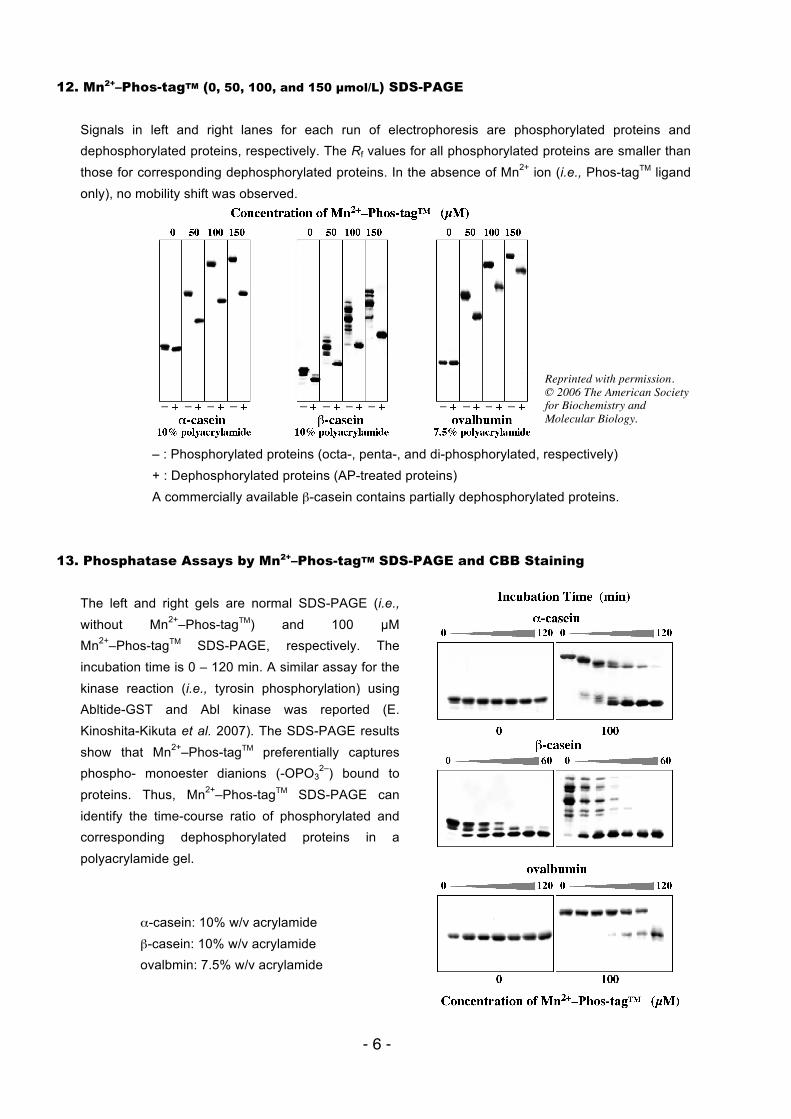
12. Mn2+–Phos-tagTM (0, 50, 100, and 150 µmol/L) SDS-PAGE Signals in left and right lanes for each run of electrophoresis are phosphorylated proteins and dephosphorylated proteins, respectively. The Rf values for all phosphorylated proteins are smaller than those for corresponding dephosphorylated proteins. In the absence of Mn2+ ion (i.e., Phos-tagTM ligand only), no mobility shift was observed.
– : Phosphorylated proteins (octa-, penta-, and di-phosphorylated, respectively) + : Dephosphorylated proteins (AP-treated proteins) A commercially available β-casein contains partially dephosphorylated proteins.
13. Phosphatase Assays by Mn2+–Phos-tagTM SDS-PAGE and CBB Staining The left and right gels are normal SDS-PAGE (i.e., without Mn2+–Phos-tagTM) and 100 µM Mn2+–Phos-tagTM SDS-PAGE, respectively. The incubation time is 0 – 120 min. A similar assay for the kinase reaction (i.e., tyrosin phosphorylation) using Abltide-GST and Abl kinase was reported (E. Kinoshita-Kikuta et al. 2007). The SDS-PAGE results show that Mn2+–Phos-tagTM preferentially captures phospho- monoester dianions (-OPO3
2–) bound to proteins. Thus, Mn2+–Phos-tagTM SDS-PAGE can identify the time-course ratio of phosphorylated and corresponding dephosphorylated proteins in a polyacrylamide gel.
α-casein: 10% w/v acrylamide β-casein: 10% w/v acrylamide ovalbmin: 7.5% w/v acrylamide
Reprinted with permission. © 2006 The American Society for Biochemistry and Molecular Biology.
- 7 -
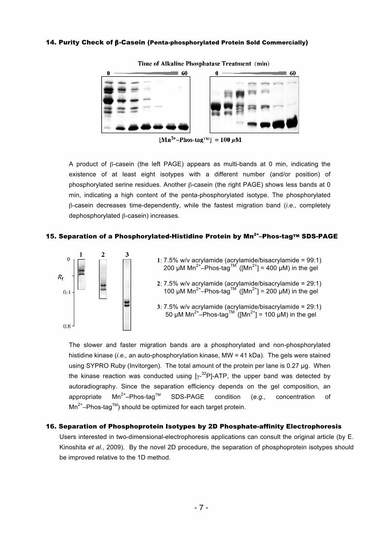
14. Purity Check of β-Casein (Penta-phosphorylated Protein Sold Commercially)
A product of β-casein (the left PAGE) appears as multi-bands at 0 min, indicating the existence of at least eight isotypes with a different number (and/or position) of phosphorylated serine residues. Another β-casein (the right PAGE) shows less bands at 0 min, indicating a high content of the penta-phosphorylated isotype. The phosphorylated β-casein decreases time-dependently, while the fastest migration band (i.e., completely dephosphorylated β-casein) increases.
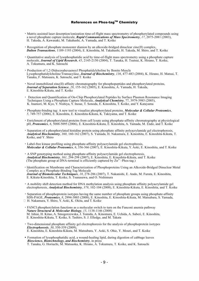
15. Separation of a Phosphorylated-Histidine Protein by Mn2+–Phos-tagTM SDS-PAGE
1: 7.5% w/v acrylamide (acrylamide/bisacrylamide = 99:1) 200 µM Mn2+–Phos-tagTM ([Mn2+] = 400 µM) in the gel 2: 7.5% w/v acrylamide (acrylamide/bisacrylamide = 29:1) 100 µM Mn2+–Phos-tagTM ([Mn2+] = 200 µM) in the gel 3: 7.5% w/v acrylamide (acrylamide/bisacrylamide = 29:1) 50 µM Mn2+–Phos-tagTM ([Mn2+] = 100 µM) in the gel
The slower and faster migration bands are a phosphorylated and non-phosphorylated histidine kinase (i.e., an auto-phosphorylation kinase, MW = 41 kDa). The gels were stained using SYPRO Ruby (Invitorgen). The total amount of the protein per lane is 0.27 µg. When the kinase reaction was conducted using [γ-32P]-ATP, the upper band was detected by autoradiography. Since the separation efficiency depends on the gel composition, an appropriate Mn2+–Phos-tagTM SDS-PAGE condition (e.g., concentration of Mn2+–Phos-tagTM) should be optimized for each target protein.
16. Separation of Phosphoprotein Isotypes by 2D Phosphate-affinity Electrophoresis
Users interested in two-dimensional-electrophoresis applications can consult the original article (by E. Kinoshita et al., 2009). By the novel 2D procedure, the separation of phosphoprotein isotypes should be improved relative to the 1D method.

- 8 -
« Troubleshooting 1 » Generally, the Rf values of all proteins (i.e., both phosphorylated and dephosphorylated proteins) in Mn2+–Phos-tagTM SDS-PAGE are smaller than those in normal SDS-PAGE. Please determine the best electrophoresis conditions, such as percentage of acrylamide gel and concentration of Mn2+–Phos-tagTM
(e.g., 100 µM), for the sufficient separation between phosphorylated and dephosphorylated proteins. In particular, a dilute Mn2+–Phos-tagTM (5 ~ 25 µM) polyacrylamide gel should be used for a complex sample such as cell lysate containing various phosphorylated and non-phosphorylated proteins. A phosphorylated protein, ovalbumin (45 kDa) in a molecular-weight protein marker can be used for the gel-shift check of Mn2+–Phos-tagTM SDS-PAGE. In this gel check procedure, prestained protein markers should be avoided. Some stained proteins interact with the Phos-tag gel resulting in broad and/or distorted bands.
« Troubleshooting 2 » Various contaminants (e.g., EDTA, inorganic salts, surfactant) in the sample proteins solution often disorder the electrophoresis bands (i.e., waving and/or tailing). In order to minimize the disorder, the desalting of the sample is recommended before the sample loading. For example, a dialysis filtration is used to decrease the amount of the low molecules in the sample. Before the pH measurement for the buffer solutions, the pH-electrode system should be calibrated using the two pH buffer solutions (e.g., pH 4 and 7). The pH of the electrophoresis buffers is one of the most important factors for the separation of phosphoprotein isotypes.
- 9 -
References on Phos-tagTM Chemistry
・Matrix-assisted laser desorption/ionization time-of-flight mass spectrometry of phosphorylated compounds using a novel phosphate capture molecule, Rapid Communications of Mass Spectrometry, 17, 2075-2081 (2003), H. Takeda, A. Kawasaki, M. Takahashi, A. Yamada, and T. Koike ・Recognition of phosphate monoester dianion by an alkoxide-bridged dinuclear zinc(II) complex, Dalton Transactions, 1189-1193 (2004), E. Kinoshita, M. Takahashi, H. Takeda, M. Shiro, and T. Koike ・Quantitative analysis of lysophosphatidic acid by time-of-flight mass spectrometry using a phosphate capture
molecule, Journal of Lipid Research, 45, 2145-2150 (2004), T. Tanaka, H. Tsutsui, K. Hirano, T. Koike, A. Tokumura, and K. Satouchi ・Production of 1,2-Didocosahexaenoyl Phosphatidylcholine by Bonito Muscle
Lysophosphatidylcholine/Transacylase, Journal of Biochemistry, 136, 477-483 (2004), K. Hirano, H. Matsui, T. Tanaka, F. Matsuura, K. Satouchi, and T. Koike
・Novel immobilized zinc(II) affinity chromatography for phosphopeptides and phosphorylated proteins, Journal of Separation Science, 28, 155-162 (2005), E. Kinoshita, A. Yamada, H. Takeda, E. Kinoshita-Kikuta, and T. Koike ・ Detection and Quantification of On-Chip Phosphorylated Peptides by Surface Plasmon Resonance Imaging Techniques Using a Phosphate Capture Molecule, Analytical Chemistry, 77, 3979-3985 (2005), K. Inamori, M. Kyo, Y. Nishiya, Y. Inoue, T. Sonoda, E. Kinoshita, T. Koike, and Y. Katayama ・Phosphate-binding tag: A new tool to visualize phosphorylated proteins, Molecular & Cellular Proteomics, 5, 749-757 (2006), E. Kinoshita, E. Kinoshita-Kikuta, K. Takiyama, and T. Koike ・Enrichment of phosphorylated proteins from cell lysate using phosphate-affinity chromatography at physiological pH, Proteomics, 6, 5088-5095 (2006), E. Kinoshita-Kikuta, E. Kinoshita, A. Yamada, M. Endo, and T. Koike ・Separation of a phosphorylated histidine protein using phosphate affinity polyacrylamide gel electrophoresis, Analytical Biochemistry, 360, 160-162 (2007), S. Yamada, H. Nakamura, E. Kinoshita, E. Kinoshita-Kikuta, T.
Koike, and Y. Shiro ・Label-free kinase profiling using phosphate-affinity polyacrylamide gel electrophresis, Molecular & Cellular Proteomics, 6, 356-366 (2007), E. Kinoshita-Kikuta, Y. Aoki, E. Kinoshita, and T. Koike ・A SNP genotyping method using phosphate-affinity polyacrylamide gel electrophoresis, Analytical Biochemistry, 361, 294-298 (2007), E. Kinoshita, E. Kinoshita-Kikuta, and T. Koike (The phosphate group at DNA-terminal is efficiently captured by Zn2+–Phos-tag.) ・Identification on Membrane and Characterization of Phosphoproteins Using an Alkoxide-Bridged Dinuclear Metal
Complex as a Phosphate-Binding Tag Molecule Journal of Biomolecular Techniques, 18, 278-286 (2007), T. Nakanishi, E. Ando, M. Furuta, E. Kinoshita, E. Kikuta-Kinoshita, T. Koike, S. Tsunasawa, and O. Nishimura ・A mobility shift detection method for DNA methylation analysis using phosphate affinity polyacrylamide gel
electrophoresis, Analytical Biochemistry, 378, 102-104 (2008), E. Kinoshita-Kikuta, E. Kinoshita, and T. Koike ・Separation of phosphoprotein isotypes having the same number of phosphate groups using phosphate-affinity
SDS-PAGE, Proteomics, 8, 2994-3003 (2008), E. Kinoshita, E. Kinoshita-Kikuta, M. Matsubara, S. Yamada, H. Nakamura, Y. Shiro, Y. Aoki, K. Okita, and T. Koike ・FANCI phosphorylation functions as a molecular switch to turn on the Fanconi anemia pathway Nature Structural & Molecular Biology, 15, 1138-1146 (2008) M. Ishiai, H. Kitao, A. Smogorzewska, J. Tomida, A. Kinomura, E. Uchida, A. Saberi, E. Kinoshita, E. Kinoshita-Kikuta, T. Koike, S. Tashiro, S. J. Elledge, and M. Takata ・Two-dimensional phosphate affinity gel electrophoresis for the analysis of phosphoprotein isotypes Electrophoresis, 30, 550-559 (2009), E. Kinoshita, E. Kinoshita-Kikuta, M. Matsubara, Y. Aoki, S. Ohie, Y. Mouri, and T. Koike ・Formation of lysophosphatidic acid, a wound-healing lipid, during digestion of cabbage leaves Bioscience, Biotechnology, and Biochemistry, in press T. Tanaka, G. Horiuchi, M. Matsuoka, K. Hirano, A. Tokumura, T. Koike, and K. Satouchi
- 10 -
・A Phos-tag-based fluorescence resonance energy transfer system for the analysis of the dephosphorylation of phosphopeptides, Analytical Biochemistry, 388, 235-241, (2009) K. Takiyama, E. Kinoshita, E. Kinoshita-Kikuta, Y. Fujioka, Y. Kubo, and T. Koike ・Phos-tag beads as an immunoblotting enhancer for selective detection of phosphoproteins in cell lysates Analytical Biochemistry, 389, 83-85, (2009) E. Kinoshita-Kikuta, E. Kinoshita, and T. Koike ・Mobility shift detection of phosphorylation on large proteins using a Phos-tag SDS-PAGE gel strengthened with agarose, Proteomics, 9, 4098- 4101 (2009) E. Kinoshita, E. Kinoshita-Kikuta, H. Ujihara, and T. Koike ・Separation and detection of large phosphoproteins using Phos-tag SDS-PAGE, Nature Protocols, 4, 1513-1521 (2009), E. Kinoshita, E. Kinoshita-Kikuta, and T. Koike ・A clean-up technology for the simultaneous determination of lysophosphatidic acid and sphingosine-1-phosphate by matrix-assisted laser desorption/ionization time-of-flight mass spectrometry using a phosphate-capture molecule, Phos-tag, Rapid Communications in Mass Spectrometry, 24, 1075-1084 (2010) J. Morishige, M. Urikura, H. Takagi, K. Hirano, T. Koike, T. Tanaka, and K. Satouchi ・Genotyping and mapping assay of single-nucleotide polymorphisms in CYP3A5 using DNA-binding zinc(II) complexes, Clinical Biochemistry, 43, 302-306 (2010), E. Kinoshita, E. Kinoshita-Kikuta, H. Nakashima, and T. Koike ・The DNA-binding activity of mouse DNA methyltransferase 1 is ragulated phosphorylation with casein kinase 1σ/ε, Biochemical Journal, 427, 489-497 (2010) Y. Sugiyama, N. Hatano, N. Sueyoshi, I. Suetake, S. Tajima, E. Kinoshita, E. Kinoshita-Kikuta, T. Koike, and I. Kameshita
***************** Phosphoproteome Analyses using Phos-tagTM PAGE ***************** ・Analysis of the CK2-dependent phosphorylation of serine 13 in Cdc37 using a phospho-specific antibody and
phospho-affinity gel electrophoresis, FEBS Journal, 274, 5690-5703 (2007), Y. Miyata and E. Nishida ・Spatial regulation of Fus3 MAP kinase activity through a reaction-diffusion mechanism in yeast pheromone signalling, Nat. Cell Biol., 9 1319-1326 (2007), C. I. Maeder, M. A. Hink, A. Kinkhabwala, R. Mayr, P. I. H. Bastiaens and M. Knop ・Identification of Ubiquitin Ligase Activity of RBCK1 and Its Inhibition by Splice Variant RBCK2 and Protein Kinase Cβ, J. Biol. Chem., 283, 11575-11585 (2008), K. Tatematsu1, N. Yoshimoto, T. Okajima, K. Tanizawa, and S. Kuroda ・Protein phosphatase Dusp26 associates with KIF3 motor andpromotes N-cadherin-mediated cell–cell adhesion, Oncogene, 28, 752-761 (2009), N. Tanuma, M. Nomura, M. Ikeda, I. Kasugai, Y. Tsubaki, K. Takagaki, T. Kawamura, Y. Yamashita, I. Sato, M. Sato, R. Katakura, K. Kikuchi, and H. Shima ・A highly sensitive technique to measure myosin regulatory light chain phosphorylation: the first quantification in renal arterioles, Inovative Methodology, 294, F1487-F1492 (2008), K. Takeya, K. Loutzenhiser, M. Shiraishi, R. Loutzenhiser, and M. P. Walsh ・Regulation of PKD by the MAPK p38d in Insulin Secretion and Glucose Homeostasis, Cell, 136, 235-248 (2009), G. Sumara, I. Formentini, S. Collins, I. Sumara, R. Windak, B. Bodenmiller, R. Ramracheya, D. Caille, H. Jiang, K. A. Platt, P. Meda, R. Aebersold, P. Rorsman, and R. Ricci1 ・The wheat germ cell-free based screening of protein substrates of calcium/calmodulin-dependent protein kinase II delta, FEBS Lett., 582, 1795-1801 (2008), T. Masaoka, M. Nishi, A. Ryo, Y. Endo, and T. Sawasaki ・Virus Infection Triggers SUMOylation of IRF3 and IRF7, Leading to the Negative Regulation of Type I Interferon Gene Expression, J. Biol. Chem., 283, 25660-25670 (2008), T. Kubota, M. Matsuoka, T. Chang, P. Tailor, T. Sasaki, M. Tashiro, A. Kato, and K. Ozato ・A Rac GTPase-Activating Protein, MgcRacGAP, Is a Nuclear Localizing Signal-Containing Nuclear Chaperone in the Activation of STAT Transcription Factors, Mol. Cell. Biol., 29, 1796-1813 (2009), T. Kawashima, Y. C. Bao, Y. Minoshima, Y. Nomura, T. Hatori, T. Hori, T. Fukagawa, T. Fukada, N. Takahashi, T. Nosaka, M. Inoue, T. Sato, M. Kukimoto-Niino, M. Shirouzu, S. Yokoyama, and T. Kitamura ・Elucidation of the Heme Binding Site of Heme-regulated Eukaryotic Initiation Factor 2α Kinase and the Role of the Regulatory Motif in Heme Sensing by Spectroscopic and Catalytic Studies of Mutant Proteins, J. Biol. Chem., 283, 18782-18791 (2008), J. Igarashi, M. Murase, A. Iizuka, F. Pichierri, M. Martinkova, and T. Shimizu
- 11 -
・Inhibition of rat aortic smooth muscle contraction by 2-methoxyestradiol, Am. J. Physiol. Heart Circ. Physiol., 295, H1935–H1942 (2008), Y. Gui, X. Zheng, J. Zheng, and M. P. Walsh ・Phosphorylation of the Budding Yeast 9-1-1 Complex Is Required for Dpb11 Function in the Full Activation of the UV-Induced DNA Damage Checkpoint, Mol. Cell. Biol., 28, 4782–4793 (2008), F. Puddu, M. Granata, L. D. Nola, A. Balestrini, G. Piergiovanni, F. Lazzaro, M. Giannattasio, P. Plevani, and M. Muzi-Falconi ・Interplay between S-Cyclin-dependent Kinase and Dbf4-dependent Kinase in Controlling DNA Replication through Phosphorylation of Yeast Mcm4 N-Terminal Domain, Mol. Biol. Cell, 19, 2267-2277 (2008), A. Devault, E. Gueydon, and E. Schwob ・Dbf4-Dependent Cdc7 Kinase Links DNA Replication to the Segregation of Homologous Chromosomes in Meiosis I, Cell, 135, 662-678 (2008), J. Matos, J. J. Lipp, A. Bogdanova, S. Guillot, E. Okaz, M. Junqueira, A. Shevchenko, and W. Zachariae ・Universally applicable methods for monitoring response regulator aspartate phosphorylation both in vitro and in vivo using Phos-tag-based reagents, Anal. Biochem., 376, 73-82 (2008), C. M. Barbieri and A. M. Stock ・In vivo regulation of Yorkie phosphorylation and localization, Development, 135, 1081-1088 (2008), H. Oh and K. D. Irvine ・Coimmunopurification of Phosphorylated Bacterial- and Plant-Type Phosphoenolpyruvate Carboxylases with the Plastidial Pyruvate Dehydrogenase Complex from Developing Castor Oil Seeds, Plant Physiology, 146, 1346-1357 (2008), R. G. Uhrig, B. O’Leary, H. E. Spang, J. A. MacDonald, Y. She, and W. C. Plaxton ・Alternative suppression of transcription from two desaturase genes is the key for species-specific sex pheromone biosynthesis in two Ostrinia moths, Insect Biochem. Mol. Biol., 39, 62-67 (2009), R. Sakai, M. Fukuzawa, R. Nakano, S. Tatsuki, and Y. Ishikawa ・hnRNP-U is a specific DNA-dependent protein kinase substrate phosphorylated in response to DNA double-strand breaks, Biochem. Biophy. Res. Commun, 381, 59-64 (2009), F. M. Berglund and P. R. Clarke ・Diphosphorylation of regulatory light chain of myosin IIA is responsible for proper cell spreading, Biochem. Biophys. Res. Commun., 381, 682–687 (2009) N. Hirata, M. Takahashi, and M. Yazawa ・Involvement of SIRT7 in resumption of rDNA transcription at the exit from mitosis, J. Cell Sci., 122, 489-498 (2009), A. Grob, P. Roussel, J. E. Wright, B. McStay, D. Hernandez-Verdun, and V. Sirri ・Antagonistic regulation of Fus2p nuclear localization by pheromone signaling and the cell cycle, J. Cell Biol., 184, 409-422 (2009), C. A. Ydenberg and M. D. Rose ・Dysbindin-1, a Schizophrenia-Related Protein, Functionally Interacts with the DNA- Dependent Protein Kinase Complex in an Isoform-Dependent Manner, PLoS ONE, 4, e4199 (2009), S. Oyama, H.Yamakawa, N. Sasagawa, Y. Hosoi, E. Futai, and S. Ishiura ・Kinome Profiling in Pediatric Brain Tumors as a New Approach for Target Discovery, Cancer Res., 69, 5987–5995 (2009) A. H. Sikkema, S. H. Diks, W. F.A. den Dunnen, A. ter Elst, F. J.G. Scherpen, E. W. Hoving, R. Ruijtenbeek, P. J. Boender, R. de Wijn, W. A. Kamps, M. P. Peppelenbosch, and E. S.J.M. de Bont ・The output of Hedgehog signaling is controlled by the dynamic association between Suppresor of Fused and the Gil proteins, Genes & Development., 24, 670–682 (2010) E. W. Humke, K. V. Dorn, L. Milenkovic, M. P. Scott, and R. Rohatigi ・Quantitative Measurement of in Vivo Phosphorylation States of Cdk5 Activator p35 by Phos-tag SDS-PAGE, Molecular & Cellular Proteomics, 9, 1133-1143 (2010), T. Hosokawa, T. Saito, A. Asada, K. Fukunaga, and S. Hisanaga
Edited by E. Kinoshita-Kikuta, E. Kinoshita, and T. Koike