mls 602 : general & medical microbiology lecture 4b
TRANSCRIPT

COLLEGE OF MEDICINE, NURSING & HEALTH SCIENCES
MLS 602 : General & Medical Microbiology
LECTURE 4B : Mycobacterium and Microbiological Specimen
Ms. Edwina Razak

Learning outcomes:After this lecture, the student would be able to:
Discuss the general characteristics of Mycobacterium.
Describe the cell wall structure of Mycobacterium
Demonstrate identification of different specimen collection bottles, storage and transportation.
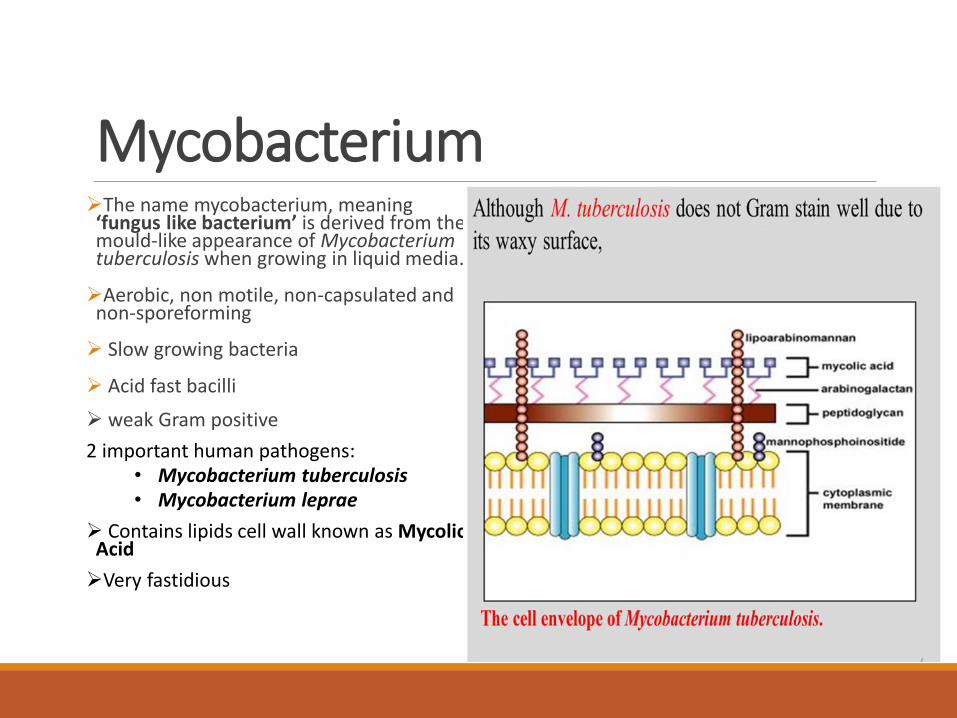
MycobacteriumThe name mycobacterium, meaning ‘fungus like bacterium’ is derived from the mould-like appearance of Mycobacterium tuberculosis when growing in liquid media.
Aerobic, non motile, non-capsulated and non-sporeforming
Slow growing bacteria
Acid fast bacilli
weak Gram positive
2 important human pathogens:• Mycobacterium tuberculosis• Mycobacterium leprae
Contains lipids cell wall known as Mycolic Acid
Very fastidious

The cell StructureThe cell wall consists of lipids, proteins and polysaccharides.
The lipid content accounts for 60% of the cell wall weight.
The cell wall is made up of fourdistinct layers:
Peptidoglycan (murein) layer
Arabinogalactan layer
Mycolic acid layer
Mycosides (peptidoglycolipids or phenolic glycolipids)
Peptidoglycan layer: It is the innermost layer which maintains the shape and rigidity of the cell.
ii. Arabinogalactan layer: It lies external to the peptidoglycan layer.Serves to connect peptidoglycan with the outer mycolic acid layer
iii. Mycolic acid layer: Lipid rich and has limited permeability of their cell walls and the general insusceptibility of bacteria to toxic agents
iv.Mycosides (peptidoglycolipids or phenolic glycolipids): These form the outermost layer which stimulates the formation of an agglutinin in blood serum.

Acid Fast Bacilli (AFB)


Mycobacterium tuberculosis (MTB)Epidemiology:
Worldwide. About 1/3 of the population are infected by this organism.
Population at the greatest risk are immunocompromised patients (HIV), drug/alcohol abusers, homeless, and individuals exposed to disease patients
Humans are the only natural reservoir
Person to person transmission through infectious aerosols

Typical Progression of Pulmonary TuberculosisTarget cells - macrophages
Pneumonia
Granuloma formation with fibrosis
Caseous necrosis ◦ Tissue becomes dry & amorphous
(resembling cheese)◦ Mixture of protein & fat (assimilated
very slowly)
Calcification◦ Ca++ salts deposited
Cavity formation◦ Center liquefies & empties into
bronchi


Mycobacterium leprae

Mycobacterium leprae
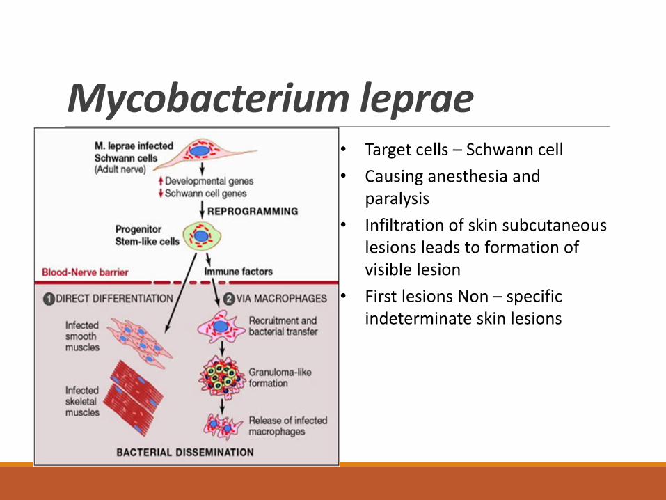
Mycobacterium leprae• Target cells – Schwann cell
• Causing anesthesia and paralysis
• Infiltration of skin subcutaneous lesions leads to formation of visible lesion
• First lesions Non – specific indeterminate skin lesions

Deformities
Mycobacterium lepraepre and post treatment

Laboratory diagnosis of Mycobacterium
Tuberculin skin test (also called a Mantoux tuberculin test)
Acid Fast Bacilli (Ziehl Neelsenstain)
Culture - Lowenstein-Jensen
Molecular methods e.gGeneXpert

Ziehl-Neelsen staining

General Overview of Virulence factors of bacteria.What are Virulence Factors?
Virulence factors help bacteria to (1) invade the host, (2) cause disease, and (3) evade host defenses.
Examples of Virulence Factors
Adherence Factors: Many pathogenic bacteria colonize mucosal sites by using pili (fimbriae) to adhere to cells
Invasion Factors: Surface components that allow the bacterium to invade host cells can be encoded on plasmids, but more often are on the chromosome.
Capsules: Many bacteria are surrounded by capsules that protect them from opsonization and phagocytosis.
Endotoxins: The lipopolysaccharide endotoxins on Gram-negative bacteria cause fever, changes in blood pressure, inflammation, lethal shock, and many other toxic events.

More Examples of VFsExotoxins: Exotoxins include several types of protein toxins and enzymes produced and/or secreted from pathogenic bacteria. Major categories include cytotoxins, neurotoxins, and enterotoxins.
Siderophores: Siderophores are iron-binding factors that allow some bacteria to compete with the host for iron, which is bound to hemoglobin, transferrin, and lactoferrin.
Enzymes – secreted by some bacteria to allow invasion and spread in host
Neuraminidase - blockage of cellular respiration, destruction of a cell, violation of a function of organs and tissues (central and peripheric nervous system, cardiovascular system, kidneys)
Hyaluronidase - destruction of a stroma of a connecting tissue (rising of permeability of vessels, edema of tissues)
Haemolysin – lysis of RBC, hemorrhagic set of symptoms
Note: Each group of bacteria has its own virulence factors that enables it to survive in the host cell.
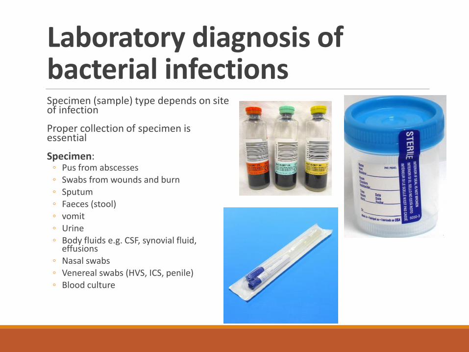
Laboratory diagnosis of bacterial infectionsSpecimen (sample) type depends on site of infection
Proper collection of specimen is essential
Specimen:◦ Pus from abscesses◦ Swabs from wounds and burn◦ Sputum◦ Faeces (stool)◦ vomit◦ Urine◦ Body fluids e.g. CSF, synovial fluid,
effusions◦ Nasal swabs◦ Venereal swabs (HVS, ICS, penile)◦ Blood culture


Summary: What have we learned so far……… Staphylococcus
Streptococcus
Gram Positive spore forming Rods
Gram Positive Non- spore forming Rods
Gram Variable bacteria
Branching and filamentous form of bacteria
Mycobacterium group
Microbiological specimen
Tutorial1. Draw and clearly label all the parts of a bacterial cell
2. List all the different parts of the bacterial cell and describe their different functions
3. Draw and label the structure of a gram positive cell wall
4. Differentiate between the wall of a gram positive bacteria and an acid fast bacteria
5. List virulence factors and its function for the following bacteria:
o Staphylococcus
oStreptococcus Group A and B
oStreptococcus pneumoniae