mixed chimerism and tolerance without whole body...
TRANSCRIPT
IntroductionThere are numerous clinical situations for which bonemarrow transplantation (BMT) might be the treatmentof choice. However, standard preparative regimens forBMT generally include whole body irradiation (WBI) tocreate space (1) or sufficient immunosuppression (2) forbone marrow engraftment. Such regimens are so toxicon their own that few patients are offered this optionuntil they are late in the course of their disease. Thesesituations include patients with hemoglobinopathies,such as sickle cell disease and thalassemia (3–5), as wellas patients with end-stage organ failure, who could betreated by allogeneic organ transplantation without thecomplications of long-term immunosuppression, ifthey could be made tolerant by BMT (6, 7). For this rea-son, recent studies in this laboratory have been directedtoward developing means of establishing mixedhematopoietic chimerism using nonmyeloablative andrelatively nontoxic conditioning regimens.
In mice, long-term hematopoietic engraftment hasbeen achieved in unconditioned recipients by injection
of high doses of syngeneic bone marrow (8, 9). Morerecently, this approach has been extended to theachievement of hematopoietic engraftment and donor-specific tolerance across a full MHC barrier using sub-lethal irradiation (10) or without myelosuppressive hostconditioning, by using depleting mAb’s, local thymicirradiation, and very high doses of MHC-mismatchedbone marrow cells (11).
These results are extremely promising for our clini-cal goals. However, for a variety of reasons, engraft-ment may be easier to achieve in mice than in largeanimals and humans, and many regimens that havebeen successful in mice have not been effective clini-cally (12–15). Testing in a large animal model is there-fore critical to extending this nonmyelosuppressiveapproach to the establishment of hematopoieticchimerism for human transplantation. Stable mixedchimerism after BMT has been demonstrated in dogs,using dog leukocyte antigen–identical (DLA-identi-cal) littermates (2, 16). Recently we have also been suc-cessful in establishing stable mixed chimerism across
The Journal of Clinical Investigation | June 2000 | Volume 105 | Number 12 1779
Mixed chimerism and tolerance without whole body irradiation in a large animal model
Yasushi Fuchimoto,1 Christene A. Huang,1 Kazuhiko Yamada,1 Akira Shimizu,2
Hiroshi Kitamura,2 R.B. Colvin,2 Vincent Ferrara,1 Michael C. Murphy,1 Megan Sykes,1
Mary White-Scharf,3 David M. Neville, Jr.,4 and David H. Sachs1
1Transplantation Biology Research Center, and2Department of Pathology, Massachusetts General Hospital/Harvard Medical School, Boston, Massachusetts, USA3BioTransplant Inc., Boston, Massachusetts, USA4Laboratory of Molecular Biology, National Institute of Mental Health, Bethesda, Maryland, USA
Address correspondence to: David H. Sachs, Transplantation Biology Research Center, Massachusetts General Hospital, MGH-East, Building 149-9019, 13th Street, Boston, Massachusetts 02129, USA. Phone: (617) 726-4065; Fax: (617) 726-4067; E-mail: [email protected].
Yasushi Fuchimoto’s present address is: Tokyo Metropolitan Kiyose Children’s Hospital, Tokyo, Japan.Akira Shimizu’s present address is: Department of Pathology, Nippon Medical School, Tokyo, Japan.Yasushi Fuchimoto and Christene A. Huang contributed equally to this work.
Received for publication October 18, 1999, and accepted in revised form April 26, 2000.
Mixed hematopoietic chimerism may provide a treatment for patients with nonmalignant hemato-logic diseases, and may tolerize patients to organ allografts without requiring chronic immunosup-pression. However, the toxicity of the usual conditioning regimens has limited the clinical applica-bility of this approach. These regimens generally include some level of whole body irradiation (WBI),which is thought to facilitate engraftment either by making room for donor hematopoietic stem cellsor by providing sufficient host immunosuppression to enable donor cells to engraft. Here, we haveestablished mixed chimerism across both minor and major histocompatibility barriers in swine, byusing high doses of peripheral blood stem cells in the absence of WBI. After mixed chimerism wasestablished, swine leukocyte antigen–matched (SLA-matched) donor skin grafts were tolerated andmaintained for a prolonged period, whereas third-party SLA-matched skin was rejected promptly.Donor-matched kidney allografts were also accepted without additional immunosuppression.Because of its low toxicity, this approach has potential for a wide range of clinical applications. Ourdata may indicate that niches for engrafting stem cells are filled by mass action and that WBI, whichserves to empty some of these niches, can be omitted if the donor inoculum is sufficiently large andif adequate host T-cell depletion is achieved before transplant.
J. Clin. Invest. 105:1779–1789 (2000).

swine leukocyte antigen–matched (SLA-matched) bar-riers in sublethally irradiated miniature swine (17). Todate, mixed chimerism has not been achieved acrossfull MHC barriers in large animal models without sig-nificant myelosuppression.
In the present study, we have extended our nonmye-loablative protocol, making it nonmyelosuppressive, byeliminating WBI from the conditioning regimen usedto allow hematopoietic cell transplantation. Trans-plants were performed across full major histocompat-ibility antigen mismatches, as well as across minor his-tocompatibility antigen mismatches, using high dosesof peripheral blood stem cell (PBSC) as a source ofhematopoietic cells. Tolerance was confirmed in theseanimals by skin grafting or transplantation of donor-matched kidney allografts.
MethodsAnimals. Transplant donors and recipients wereselected from our herd of MGH partially inbred,MHC-defined miniature swine. PBSC donors were4–8 months old and weighed 30–50 kg. Recipientswere 8–12 weeks old and weighed 5–15 kg. Theimmunogenetic characteristics of this herd and intra-MHC recombinant haplotypes have been describedpreviously (18, 19).
PBSC collection and infusion. PBSC collection has beendescribed previously (20, 21). A stem cell–mobilizingregimen consisting of recombinant porcine stem cellfactor (pSCF; 100 µg/kg) in combination with recom-binant porcine–IL-3 (pIL-3; 100 µg/kg) (both fromBiotransplant, Boston, Massachusetts, USA), with orwithout recombinant human G-CSF (rhu G-CSF;Amgen Inc., Thousand Oaks, California, USA), wasadministered subcutaneously. Collection of PBSCswas achieved by leukapheresis (COBE BCT Inc., Lake-wood, Colorado, USA) beginning on day 5 of cytokinetherapy and continuing daily for 3 or 6 days. PBSC,either fresh or frozen and quickly thawed, were adjust-ed to a concentration of 2.0 × 10⁸/mL, and the appro-priate volume was infused through the intravenousline over a 15- to 20-minute period. PBSCs wereadministered on day 0.
Thymic irradiation. Thymic irradiation (TI) wasadministered on day –2. Irradiation was adminis-tered to a single field encompassing the thymus,defined by an x-axis including the width of the jawand a y-axis including the distance from the top ofthe sternum to the temporomandibular joint. Theaverage radiation field size was 10 × 4 cm with asource separation distance of 78 cm. The definedfield was irradiated with 700 cGy or 1,000 cGy froma cobalt irradiator source at a rate of 175 cGy/minand to a tissue depth of 2 cm.
Peripheral blood T-cell depletion. T-cell depletion wasachieved using the reagent pCD3-CRM9, a new mutantDiphtheria toxin–anti-swineCD3 conjugate (22). Onday –2, 0.05 mg/kg pCD3-CRM9 was administeredintravenously to recipient animals.
CyA treatment. Recipients received CyA through agastric tube (Neoral; Novartis Pharmaceuticals, EastHanover, New Jersey, USA) at 15–30 mg/kg/d in divid-ed doses from day –1 to day 30 to maintain a bloodlevel of 300–800 ng/mL.
Antibodies and flow cytometry. The following swine-spe-cific antibodies were used to monitor depletion andrecovery of cell populations in the pig by flow cytometryafter pCD3-CRM9 administration and PBSC infusion:anti-CD1 mouse mAb 76-7-4 (mouse IgG2aK) (23); anti-CD3 mouse mAb 898H2-6-15 (mouse IgG2aK) (24);anti-CD3a mAb BB23-8E6 IgG1, and anti-CD5 mAbBB6-9G12 IgG1 provided by M. Pescovitz (Indiana Uni-versity, Indianapolis, Indiana, USA; ref. 25). Flow cytom-etry was performed using a Becton Dickinson FACScan(San Jose, California, USA). Unseparated heparinizedperipheral blood (PB) was distributed into staining tubes(Falcon 2054) at 100 µL/tube and washed twice using 2mL flow cytometry buffer (HBSS containing Ca2+ andMg2+/0.1% BSA/0.1% NaN3). Cells were stained withoptimal concentrations of the primary mouse anti-pigmAb (either unconjugated or FITC conjugated) for 30minutes at room temperature. For indirect staining, goatanti-mouse Ig (H&L) FITC (GAMF) (Sigma Immuno-chemicals, St. Louis, Missouri, USA) was added for anadditional 30 minutes at room temperature. For detec-tion of immunotoxin-coated cells, second step reagentalone was added. To determine the percent of donor cellsamong peripheral blood cell populations, PB was incu-bated with the pig monocyte/granulocyte-specific FITC-conjugated mAb SWC3a 74-22-15 (Balb/c, IgG1K) (23)together with either the donor-specific biotin-conjugat-ed mAb 1038H-10-9 (B10.PD1, IgMK) specific for pigallelic antigen (PAA) (26) or an IgM isotype-matchednegative control mAb, followed by phycoerythrin strep-tavidin (PESA; PharMingen, San Diego, California,USA). Red blood cells were lysed, and the white cells werefixed using FACS lysing solution (Becton Dickinson)before acquisition. Data were analyzed using Winlist listmode analysis software (Verity Software House, Top-sham, Maine, USA). For flow cytometry of thymus cellsuspensions, cells were distributed into tubes at 1 × 10⁶
cells per tube. Staining was identical to that alreadydescribed here except that no FACS lysing solution wasadded. Propidium iodide was added immediately beforeacquisition in order to exclude dead cells.
Histology. Thymic tissues were obtained by sequentialbiopsies. Formalin-fixed tissue sections were stainedwith hematoxylin and eosin (H&E) and examinedmicroscopically. For immunohistochemical study ofthymus in animals receiving single haplotype–mis-matched PBSC, frozen tissues were used, and sectionswere stained with the mAb16-7-D4 (27), specific forSLAc class I (donor type), using indirect immunoper-oxidase technique. To identify the lineage of SLAc classI–positive cells (donor cells), frozen tissues werestained using sequential immunoperoxidase andimmunoalkaline phosphatase techniques to detectkeratin (DAKO A/S, Glastrup, Denmark) or ISCR 3,
1780 The Journal of Clinical Investigation | June 2000 | Volume 105 | Number 12

specific for SLA class II DR. Control stains were per-formed by substitution of the primary antibody withan irrelevant murine mAb.
Skin grafts. Skin grafts were performed by techniquespublished previously (28). Briefly, split-thickness skinwas harvested from the donor and placed on a deepsplit-thickness bed on the recipient’s dorsal thorax.Grafts were examined daily until rejection occurred.Rejection was determined macroscopically and definedas diffuse cyanosis and induration of the graft.
Kidney transplantation. The surgical procedures usedfor kidney transplants have been described in detailpreviously (29, 30). A semipermanent indwelling Hick-man silastic central venous catheter was placed surgi-cally into the external jugular vein. The catheter facili-tated CyA administration and frequent blood samplingfor monitoring of renal function, and in vitro assays.Rejection was monitored by plasma creatinine tests andhistological examination of biopsy tissue.
Preparation of PBMCs. For separation of PBMCs, fresh-ly heparinized whole blood was diluted 1:2 with HBSS(GIBCO BRL, Gaithersburg, Maryland, USA), andmononuclear cells were obtained by gradient centrifu-gation using lymphocyte separation medium(Organon Teknika, Durham, North Carolina, USA).The mononuclear cells were washed once with HBSS,and contaminating red cells were lysed with ammoni-um chloride potassium buffer (B&B Research Labora-tory, Fiskeville, Rhode Island, USA). Cells were thenwashed with HBSS and resuspended in tissue culturemedium. All cell suspensions were kept at 4°C untilused in cellular assays.
Cell-mediated lympholysis. Media for cell-mediated lym-pholysis (CML) cultures consisted of RPMI 1640(GIBCO BRL) supplemented with 6% FCS (SigmaChemical Co., St. Louis, Missouri, USA), 100 U/mLpenicillin, 135 µg/mL streptomycin (GIBCO BRL), 50µg/mL gentamicin (GIBCO BRL), 10 mM HEPES(Fisher Scientific, Pittsburgh, Pennsylvania, USA), 2mM L-glutamine (GIBCO BRL), 1 mM sodium pyru-vate (BioWhittaker Inc., Walkersville, Maryland, USA),and 5 × 10⁻⁵ M β-2 mercaptoethanol (Sigma ChemicalCo.). Media used for the effector phase of CML assaysconsisted of Basal Medium Eagle (GIBCO BRL) sup-plemented with 6% serum replacement medium(Sigma Chemical Co.). CML assays were performed asdescribed previously (29, 30). Briefly, lymphocyte cul-tures containing 4 × 10⁶ responder and 4 × 10⁶ irradi-ated (25 Gy) stimulator PBMCs in 2 mL of mediumwere incubated for 6 days at 37°C in 7% CO2 and 100%humidity. Bulk cultures were harvested, and effectorcells were tested on 51Cr-labeled PHA blasts. The testswere run at serially diluted effector/target ratios(100:1, 50:1, 25:1, 12:1). After 5.5 hours of effector cellincubation with the 5 × 10³ targets, the supernatantswere harvested and 51Cr release was determined on agamma counter. Baseline levels were measured as therate of spontaneous release of 51Cr from 5 × 10³ tar-gets. The results were expressed as percentage specificlysis: % specific lysis = [experimental release (cpm) –spontaneous release (cpm) / maximum release (cpm)– spontaneous release (cpm)] × 100.
ResultsConditioning regimen. Animals were conditioned with anontoxic protocol consisting of TI and in vivo T-celldepletion before donor cell infusion. The dose of TIand donor PBSCs were increased during the course ofthese experiments to achieve consistent engraftmentacross full mismatch barriers. Figure 1 is a schematicdiagram of the preparative regimen for hematopoieticcell transplantation that was used in all subsequentlytransplanted animals. A summary of animals receivingPBSC transplantation (PBSCT) under this protocol isshown in Table 1.
PBSC mobilization and collection. We have recentlydemonstrated that a combination of porcine pSCF,pIL-3, and human G-CSF could be used to mobilized
The Journal of Clinical Investigation | June 2000 | Volume 105 | Number 12 1781
Figure 1Diagram of preparative regimen used for PBSCT. AAn additional3 days of leukapheresis were necessary to yield 200 × 108/kg foranimal no. 13101.
Table 1Summary of animals treated with nonmyelosuppressive regimen plus PBSCT
Animal no. SLA Thymic irradiation PBSC dose Engraftment GvHD Clinical(cGy) (× 108/kg) Thymic chimerism outcome
12757 ac→ac 700 110A Yes No Healthy12963 cc→cc 700 100A Yes No Healthy12885 ac→ad 700 100 No No Healthy13476 cd→ad 700 150 Yes No Healthy13100 ac→ad 1000 100 Yes Yes Sacrificed day 7313101 ac→ad 1000 200 Yes Yes Healthy
Miniature swine received thymic irradiation and 0.05 mg/kg pCD3-CRM9 followed by PBSC transplantation with a 30-day course of CyA. ADonor PBSCs werefrozen and thawed prior to infusion. All other transplants were done using fresh donor cells.
stem cell progenitors into the peripheral blood ofminiature swine and that stem cell progenitors mobi-lized under these conditions were capable of reconsti-tuting a lethally irradiated animal (20). After cytokinetreatments, white blood cell counts in one representa-tive donor animal rapidly rose to a peak of approxi-mately 80 × 103/mm3. PBSCs were collected by leuka-pheresis on days 5–7, and 18 × 1010 mononuclear cellswere collected. This mobilization procedure thus pro-vides more than 10 times the number of cells achievedby bone marrow harvest from vertebral bodies andlong bones (31) and more than 50 times the numberof cells achievable by harvests from living donors (31).
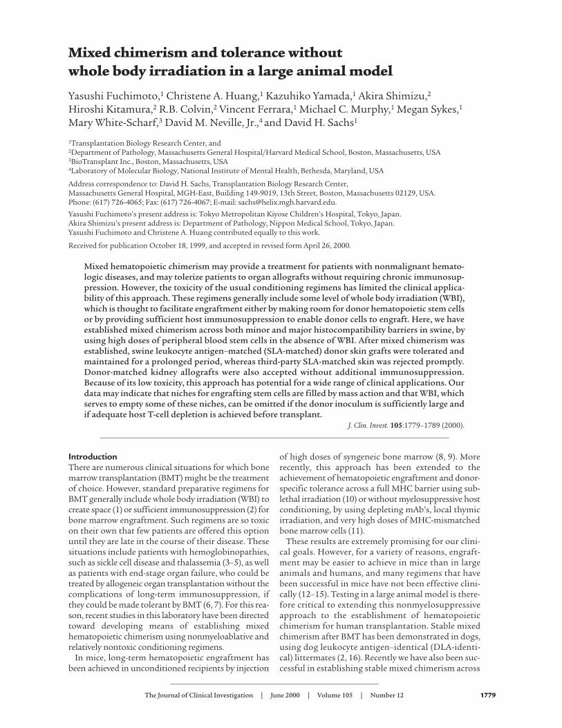
Peripheral blood T-cell depletion in conditioned recipients.The establishment of mixed chimerism depends oneffective in vivo T-cell depletion of the recipient (22).Figure 2 shows the course of peripheral blood T-celldepletion from pretreatment to the time of donor cellinfusion. More than 90% peripheral blood T-cell deple-tion was achieved by day 0 in animals that received 700cGy TI and anti-CD3 immunotoxin, and more than98% T-cell depletion was achieved by day 0 in animalsthat received 1,000 cGy TI and immunotoxin. In addi-tion, the few remaining peripheral T cells were almostentirely coated with antibody by day 0. It should benoted that the vascular leak syndrome associated withimmunotoxin treatment (32, 33) has never beenobserved in this protocol.
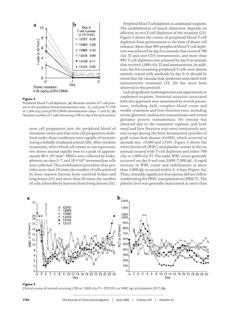
Lack of significant myelosuppression and organ toxicity inconditioned recipients. Potential toxicities associatedwith this approach were monitored by several param-eters, including daily complete blood count andweekly creatinine and liver function tests, includingserum glutamic oxaloacetic transaminase and serumglutamic pyruvic transaminase. No toxicity wasobserved due to the treatment regimen, and bothrenal and liver function tests were consistently nor-mal except during the brief documented episodes ofgraft versus host disease (GvHD), which occurred inanimals nos. 13100 and 13101. Figure 3 shows thewhite blood cell (WBC) and platelet counts in the sixanimals treated with T-cell depletion and either 700cGy or 1,000 cGy TI. The nadir WBC count generallyoccurred on day 0 and was 2,000–7,000/µL. A rapidincrease in WBC count and stabilization at morethan 5,000/µL occurred within 2–4 days (Figure 3a).Thus, clinically significant leucopenia did not followconditioning for PBSC transplantation (PBSCT). Theplatelet level was generally maintained at more than
1782 The Journal of Clinical Investigation | June 2000 | Volume 105 | Number 12
Figure 2Peripheral blood T-cell depletion. (a) Absolute number of T cells pres-ent in the peripheral blood pretreatment (day –2), and post TI (700or 1,000 cGy) and pCD3-CRM9 administration (days –1 and 0). (b)Absolute number of T cells remaining in PB on day 0 for each animal.
Figure 3Clinical course of animals receiving (700 or 1,000 cGy TI + DTCD3) on WBC (a) and platelets (PLT) (b).

100,000/µL, and platelet infusions were not required(Figure 3b). The general condition of the animalsreceiving this treatment regimen was very stable, andinfection was never observed.
Analysis of engraftment and tolerance in animalsreceiving high-dose SLA-identical PBSC infusion
Multilineage peripheral blood chimerism. Chimerism wasmeasured by flow cytometry using our PAA mAb,which recognizes a pig allelic antigen present on allswine leukocytes (26). Donor and recipient animalswere selected that were PAA+ and PAA–, respectively,to facilitate chimerism detection. Animals nos.12757 and 12963 were treated with 700 cGy TI,pCD3-CRM9 0.05 mg/kg, 30 days of CyA adminis-tration, and 100 × 108/kg SLA-identical PBSC infu-sion. In both animals, lymphocyte chimerism wasachieved and rose rapidly after day 20, peaking atapproximately 40% (Figure 4a). One animal showeda transient decrease in lymphocyte chimerism aftercessation of CyA treatment. In both animals,chimerism gradually decreased at days 40–60, reach-ing a stable level of 15–20%, which was maintainedfor more than 300 days. Chimerism in the myeloid(monocyte and granulocyte) lineages was present atthe 5–10% level and was also maintained for morethan 300 days.
Thymic chimerism. Thymic biopsies taken in the first2 weeks revealed an atrophic thymus in both animalsafter TI (data not shown). By 20 days after transplant,the thymus began to grow, as assessed by visualiza-tion at biopsy. Thymic chimerism increased rapidly atthis time, peaking on day 45. Thereafter, thymicchimerism decreased, stabilizing at 5–10% by day 60and remaining at that level for more than 300 days inboth animals (Figure 4b).
Skin grafts. Skin grafts were performed on day 60 totest for the establishment of donor-specific toler-ance. One animal accepted the donor skin graftindefinitely (> 250 days), and the other animalshowed prolonged donor skin graft survival, butrejected on day 45. Both animals rejected MHC-matched third-party skin grafts completely by 10days (Table 2). As shown in Figure 4, both animalsachieved successful lymphohematopoietic engraft-ment, and stable levels of thymic and peripheralchimerism were maintained in these animals evenafter skin grafting. The significance of these skingraft survival times is discussed later here.
Engraftment in animals receiving high-dose singlehaplotype–mismatched PBSC infusion
700 cGy TI. The same protocol (700 cGy TI, T-cell deple-tion, and 100 or 150 × 108/kg donor PBSCs) wasapplied to two single haplotype–mismatched recipi-ents. One of these animals (no. 12885) showed a levelof 5–10% lymphocyte chimerism on day 30. However,after cessation of the CyA, this level graduallydecreased, and, despite an additional infusion of 20 ×108/kg mobilized PBSCs from the same donor,chimerism could not be detected after day 50.
The Journal of Clinical Investigation | June 2000 | Volume 105 | Number 12 1783
Table 2Survival of skin grafts of SLA-identical mixed chimeric pigs
Animal no. Donor skin 3P MHC-matched skin
12757 > 300 912963 45 9, 9A
AAnimal no. 12963 received two different MHC-matched third-party (3P)skin grafts.
Figure 4Chimerism among animals receiving SLA-matched PBSCT. (a)Peripheral blood; (b) thymus.

Chimerism in the myeloid lineage was barely detectablefrom the beginning (Figure 5a). In addition, thymicchimerism in this animal was never detectable, eventhough the thymus appeared healthy and growing ondays 38 and 45 as assessed by visualization at biopsy.
In the second animal (no. 13476), engraftment wasachieved with a high level of lymphoid chimerism(50–60%) from the beginning. Myeloid chimerism sta-bilized to approximately 25–30% by day 20, but thenfell after day 50 to become almost undetectable.Thymic chimerism was detected in biopsies performedon day 28 (15%), rose by day 40 (40%), but fell again onday 60 (14%) and was absent by day 98. Chimerism inlymphoid lineages persisted in this animal after cessa-tion of CyA on day 30, but appeared to be graduallydecreasing by day 300 (Figure 5a). No clinical signs ofGvHD have been observed.
TI (1,000 cGy). The results described here suggestedthat the preparative regimen was sufficient to permitengraftment of MHC-matched PBSCs, but was proba-bly at the threshold for permitting engraftment acrossan MHC barrier. We have previously studied lympho-hematopoietic engraftment in other species condi-tioned with a regimen of sublethal WBI plus TI. In bothmice (1) and monkeys (34), we found that successfulengraftment could be achieved across MHC barriersusing 300 cGy WBI and 700 cGy TI. In these regimens,the total irradiation to the thymus is 1,000 cGy. We rea-soned that the failure of engraftment in animal no.12885 may have been due to insufficient total TI.
Therefore, the level of TI in this “no WBI” regimen wasincreased to 1,000 cGy for subsequent single haplotypeSLA–mismatched animals.
Two animals (nos. 13100 and 13101) treated with1,000 cGy TI, T-cell depletion, 30 days of CyA, and 100× 108 to 200 × 108/kg donor PBSCs both engrafted anddeveloped mixed chimerism (Figure 5b). Animal no.13100 showed 60–70% chimerism for the first 3 weeks,but this level decreased dramatically after cessation ofCyA on day 30. Shortly thereafter, the animal devel-oped a high fever, a high WBC count, and became lessactive, presumably reflecting the onset of GvHD (seelater here). This animal recovered spontaneously by day36. Concurrent with recovery, donor lymphocytechimerism rapidly increased, and 70–80% lymphocytechimerism was achieved and maintained from day 40to the time of sacrifice on day 73. The level ofchimerism in the myeloid lineages was lower than inthe lymphocyte lineage (10–20%) at all periods (Figure5b). Thymic chimerism in this animal reached 4–7% byday 35, at which time the animal showed thymic atro-phy, probably induced by GvHD.
Animal no. 13101 achieved 80% donor lymphocytechimerism by the second week and maintained thislevel for more than 120 days. Donor chimerism in themyeloid lineage increased by the second week and wassubsequently maintained at 40–50% until day 40. Atthis time, the animal showed a transient increase(80%) in myeloid chimerism through day 60, afterwhich granulocyte chimerism increased and mono-
1784 The Journal of Clinical Investigation | June 2000 | Volume 105 | Number 12
Figure 5Peripheral chimerism among animals receiving one-haplotype–mismatched PBSCT with (a) 700 cGy TI; (b) 1,000 cGy TI.

cyte chimerism decreased (Figure 5b). Thymicchimerism in this animal also reached 4–7% by day 35,at which time the animal also showed thymic atrophy.However, all signs of GvHD resolved by day 90, andthe thymus began to grow, achieving normal size and20% thymic chimerism by day 158 (Figure 6, a–e). Fig-ure 6a shows that donor PAA+ thymocytes deter-mined by FACS were present among the immatureCD1-positive thymocytes and among the matureCD3-high thymocytes. The animal remained a mixedchimera for more than 400 days after PBSCT, with60% donor lymphocytes, 10% donor monocytes, and5% donor granulocytes.
Histology. Figure 6 (b–e) shows the immunohisto-chemical staining of a thymic biopsy from animal no.13101 on day 158 after transplantation. H&E stainingrevealed normal thymic structure with well-developedcortex and medulla (Figure 6b). Using a donor-specificclass I mAb, donor cells were clearly seen in themedullary region (brown cells in Figure 6c). Doublestaining with anti-donor class I (blue) and cytokeratin(brown) in Figure 6d demonstrated that the donor cellsin the medulla were not epithelial cells, whereas doublestaining for donor class I (blue) and pan-pig class II(brown) in Figure 6e demonstrated that the donor cellswere class II positive, and their morphology was con-sistent with a dendritic cell type (Figure 6e).
In both of these animals, clinical signs of GvHD,including grade 1–2 skin rash and diarrhea, wereobserved immediately after the cessation of CyAadministration. The skin rash disappeared sponta-neously in both animals. However, animal no. 13100had to be sacrificed on day 73 owing to persistentdiarrhea. The pathological examination confirmedthat this animal suffered the complications of chron-ic GvHD, and not of toxicity due to the preparativeregimen. In animal no. 13101, the diarrhea disap-peared spontaneously, and the animal appearedhealthy by day 90 and has remained healthy for morethan 400 days.
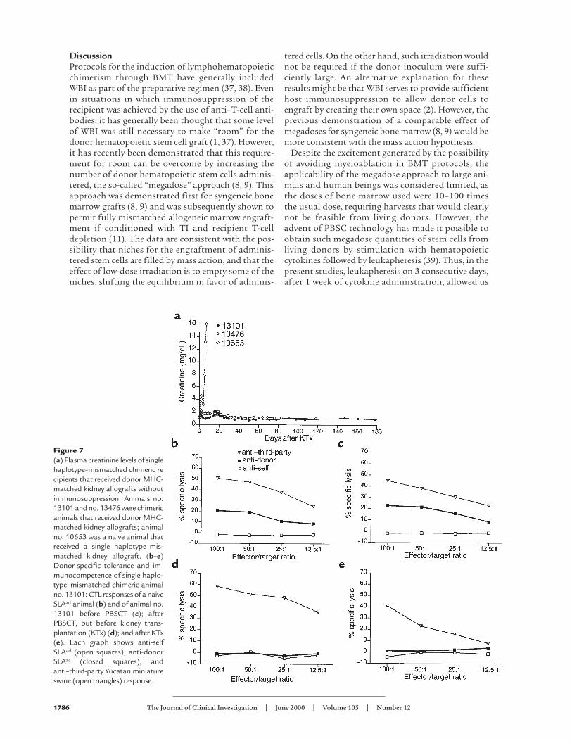
Induction of tolerance as assessed by renal allografts. Intwo of these long-term mixed chimerism animals(nos. 13101 and 13476), we tested induction of toler-ance by transplantation of an MHC donor-matchedrenal allograft. Animal nos. 13101 and 13476 receiveda donor-matched kidney allograft, on post-PBSCTdays 190 and 98, respectively. In both cases, the PBSCdonor and the kidney allograft donor were differentanimals, presumably differing for multiple minorantigen disparities. In both cases, the kidneys wereaccepted long term without the use of immunosup-pressive medications after kidney transplantation. Asshown in Figure 7a, both animals demonstrated atransient rise in plasma creatinine on postoperativeday (POD) 17, as has been reported previously in thismodel for SLA-matched renal allografts differing forminor antigen disparities (35). Thereafter the crea-tinines stabilized, and the animals continued to enjoynormal renal function with plasma creatinine levels at
less than 1.5 mg/dL for more than 120 days. In bothanimals, kidney biopsies demonstrated minimalinterstitial infiltration with mononuclear cells onPODs 30, 60, and 100. These findings are in contrastto previously reported data for animals receiving one-haplotype–mismatched kidney allografts withoutimmunosuppression, all of which rejected kidneyallografts by day 12 (36). A representative control ani-mal (no. 10653) is shown in Figure 7a.
In vitro evidence for tolerance and demonstration ofimmunocompetence. Figure 7 (b–e) shows the CMLresults for animal no. 13101(SLAad), in comparison tothe responses of a naive host type animal (SLAad) runas a control in each assay (Figure 7b). As also seen inFigure 7, animal no. 13101 demonstrated significantanti-donor SLAac response (> 20%) before receivingPBSCs (Figure 7c). However, after PBSC administra-tion, both before (Figure 7d) and after (Figure 7e) thekidney allograft, this animal demonstrated donor-specific unresponsiveness in CML. Immunocompe-tence was demonstrated in all cases by a highanti–third-party response to Yucatan miniature swine(Charles River Breeding Lab, Windham, Maine, USA).The mixed lymphocyte reaction results likewisedemonstrated significant anti-donor responses beforeadministration of PBSCs, and donor-specific unre-sponsiveness and immunocompetence after adminis-tration of PBSCs (data not shown).
The Journal of Clinical Investigation | June 2000 | Volume 105 | Number 12 1785
Figure 6Chimerism detected by flow cytometry and immunohistochem-istry in thymic biopsy tissue from animal no. 13101 on day 158.(a) Double staining of thymus by FACS. (b–e) Histology (immuno-histochemistry).
DiscussionProtocols for the induction of lymphohematopoieticchimerism through BMT have generally includedWBI as part of the preparative regimen (37, 38). Evenin situations in which immunosuppression of therecipient was achieved by the use of anti–T-cell anti-bodies, it has generally been thought that some levelof WBI was still necessary to make “room” for thedonor hematopoietic stem cell graft (1, 37). However,it has recently been demonstrated that this require-ment for room can be overcome by increasing thenumber of donor hematopoietic stem cells adminis-tered, the so-called “megadose” approach (8, 9). Thisapproach was demonstrated first for syngeneic bonemarrow grafts (8, 9) and was subsequently shown topermit fully mismatched allogeneic marrow engraft-ment if conditioned with TI and recipient T-celldepletion (11). The data are consistent with the pos-sibility that niches for the engraftment of adminis-tered stem cells are filled by mass action, and that theeffect of low-dose irradiation is to empty some of theniches, shifting the equilibrium in favor of adminis-
tered cells. On the other hand, such irradiation wouldnot be required if the donor inoculum were suffi-ciently large. An alternative explanation for theseresults might be that WBI serves to provide sufficienthost immunosuppression to allow donor cells toengraft by creating their own space (2). However, theprevious demonstration of a comparable effect ofmegadoses for syngeneic bone marrow (8, 9) would bemore consistent with the mass action hypothesis.
Despite the excitement generated by the possibilityof avoiding myeloablation in BMT protocols, theapplicability of the megadose approach to large ani-mals and human beings was considered limited, asthe doses of bone marrow used were 10–100 timesthe usual dose, requiring harvests that would clearlynot be feasible from living donors. However, theadvent of PBSC technology has made it possible toobtain such megadose quantities of stem cells fromliving donors by stimulation with hematopoieticcytokines followed by leukapheresis (39). Thus, in thepresent studies, leukapheresis on 3 consecutive days,after 1 week of cytokine administration, allowed us
1786 The Journal of Clinical Investigation | June 2000 | Volume 105 | Number 12
Figure 7(a) Plasma creatinine levels of singlehaplotype–mismatched chimeric recipients that received donor MHC-matched kidney allografts withoutimmunosuppression: Animals no.13101 and no. 13476 were chimericanimals that received donor MHC-matched kidney allografts; animalno. 10653 was a naive animal thatreceived a single haplotype–mis-matched kidney allograft. (b–e)Donor-specific tolerance and im-munocompetence of single haplo-type–mismatched chimeric animalno. 13101: CTL responses of a naiveSLAad animal (b) and of animal no.13101 before PBSCT (c); afterPBSCT, but before kidney trans-plantation (KTx) (d); and after KTx(e). Each graph shows anti-selfSLAad (open squares), anti-donorSLAac (closed squares), andanti–third-party Yucatan miniatureswine (open triangles) response.

to achieve a collection of 1.8 × 10¹¹ mononuclearcells, more than 50 times the number usually har-vested from the bone marrow of comparable livingdonor animals. As demonstrated by the histology inFigure 6 (b–e), tolerance induced by these cells mostlikely involves central deletion by donor dendriticcells in the thymus. As demonstrated here, tolerancehas been confirmed in these animals by acceptance ofeither skin grafts or vascularized organ allograftswithout immunosuppression.
Another important facet of the preparative regimenrequired to achieve engraftment without WBI, is effec-tive depletion of mature T cells from the recipient.Although such depletion has been possible with antis-era and with mAb’s in mice (40), similar depletion inlarge animals and humans has been difficult to achieve(41, 42). Previous studies in miniature swine haveshown coating of peripheral T cells by such antibodies,without effective depletion (42). However, the use of atoxin-conjugated anti-CD3 mAb (pCD3-CRM9) hasnow enabled depletion to an extent compatible withengraftment (22). Use of a similar immunotoxin hasbeen demonstrated to be effective in nonhuman pri-mates (43–45). Clinical protocols may eventually relyon such immunotoxins, or may be possible with newmAb’s that have shown markedly effective T-cell deple-tion in humans (46, 47) but which unfortunately can-not be tested in nonhuman primates, as they arespecies-specific. Although CD3 immunotoxins havebeen reported to cause vascular leak syndrome in someprotocols (32, 33), no vascular leak syndrome has beenobserved in miniature swine treated with pCD3-CRM9in this nonmyelosuppressive regimen without WBI.
Depletion of mature recipient and donor T cells isachieved in mice by the use of high doses of unconju-gated mAb’s. These antibodies remain active in therecipient plasma for 2 weeks after administration (48)and are thought to be responsible for subsequentdepletion of donor T cells in the bone marrow inocu-lum, thus preventing GvHD. In contrast, T-cell deple-tion with pCD3-CRM9 involves treatment with a lowdose of conjugated antibody, which is no longer activein the plasma by the time that donor PBSCs are admin-istered (data not shown). We have therefore utilized a30-day course of CyA in the present protocol in anattempt to prevent the induction of GvHD by donor Tcells. This treatment was effective in preventing GvHDin recipients of SLA-matched transplants, but was onlypartially effective for SLA-mismatched transplants. Wehave therefore subsequently increased CyA treatmentby tapering over an additional 30 days in the case ofmismatched transplantation, and our preliminary dataindicate that this additional treatment is effective inpreventing this complication (Y. Fuchimoto et al., man-uscript in preparation). In addition to its role in pre-venting GvHD, CyA may also help to facilitate engraft-ment by preventing residual mature T cells among hostthymocytes from causing intrathymic rejection ofdonor cells (see later here).
Our data are consistent with previous murine studiesand recent dog studies indicating that bone marrowspace is not an absolute requirement for lymphohe-matopoietic engraftment (2, 8, 9, 11). However, theimportance of TI as part of the preparative regimen sug-gests that thymic space or elimination of thymic allore-activity is an absolute requirement for the achievementof engraftment of MHC-mismatched marrow, a findingalso supported by previous data in mice (1, 9, 48, 49).Thus, in one of the two animals treated with only 700cGy of TI, initial engraftment was lost shortly aftertreatment with CyA that was discontinued on day 30,and in the other animal, engraftment was more persist-ent but nevertheless not as stable as in animals treatedwith 1,000 cGy of TI. We reasoned that for MHC-mis-matched reconstitution, additional TI might be neces-sary, as it is known that antibody-mediated depletion isless effective in the thymus than peripherally (1, 22).When TI was increased to 1,000 cGy, the same totalthymic dose received in nonmyeloablative protocolsincorporating 300 cGy WBI plus 700 cGy TI wasachieved (1). In this setting, both recipients of one hap-lotype MHC-mismatched marrow engrafted. Immuno-logical sites in addition to the thymus may be affectedby the TI as administered. These sites would undoubt-edly include some marrow tissue in the nearby ribs, ver-tebrae, and sternum, as well as the blood recirculatingthrough the radiotherapy field during the course of irra-diation. This additional irradiation could potentiallyaffect engraftment by creating some marrow space or bysuppressing the immune system to some degree. How-ever these effects, if present, are clearly much less myelo-suppressive and immunosuppressive than those of WBI.Thus, unlike recipients of sublethal WBI (17), none ofthe animals receiving TI without WBI required anyblood support or developed any signs of immunodefi-ciency after transplantation. Recent data in mice showthat costimulatory blockade with CTLA4Ig and/or anti-CD40L antibodies is capable of enabling lasting engraft-ment without TI, probably because costimulatoryblockade obviates the need for complete T-cell depletion(50). Therefore, we may be able to eliminate all irradia-tion from the preparative regimen by incorporating cos-timulatory blockade into our no-WBI regimen, andstudies along these lines are currently in progress.
One animal in this study demonstrated prolongeddonor skin graft survival after an MHC-matched PBSCinfusion, with eventual rejection of the skin graft by day45. However, this animal nevertheless maintainedperipheral and thymic mixed chimerism and accepted aheart transplant from the same donor (51). These datasuggest that despite the induction of central toleranceby donor PBSCs in this animal, skin-specific antigens towhich the recipient was not tolerized by PBSCs werepresent on the donor skin graft. The other animal test-ed in the same way showed permanent donor skin sur-vival. These data are consistent with what is knownabout skin-specific antigens in mice (52–54). Because itis likely that only a few alleles for such skin-specific anti-
The Journal of Clinical Investigation | June 2000 | Volume 105 | Number 12 1787

gens exist within our swine herd, some animals could beexpected to share common alleles by chance, and there-fore a certain percentage of chimeric animals might beexpected to accept skin grafts, whereas others wouldreject. The subject of skin-specific antigens in miniatureswine will be discussed in detail elsewhere (Y. Fuchimo-to, manuscript in preparation).
The ability to achieve hematopoietic stem cellengraftment without WBI has many advantages forclinical applications. Previous studies in miniatureswine, using WBI as part of the preparative regimen,have produced pancytopenia over the first 2 weeks afterbone marrow administration, necessitating bloodproduct support (55). Similar blood product supportis required in most large animal models of BMT, as wellas in humans (31, 56, 57). However, as seen in the pres-ent studies, when engraftment and mixed chimerismare obtained without WBI, the recipient animals do notdevelop sufficient pancytopenia to require blood prod-uct support. Although the dangers of pancytopenia areacceptable in the treatment of hematologic malignan-cies, they pose a major impediment to the use of BMTin the treatment of less immediately life-threateningdiseases, such as hemoglobinopathies. Likewise, if tol-erance induction by BMT were possible without sig-nificant myelosuppression, our data indicate that thisprotocol might be applicable to the treatment of end-stage organ failure by organ transplantation. We there-fore believe that the extension of the “no WBI”approach to the establishment of mixed chimerismacross MHC barriers may have important implicationsfor the treatment of these non–life-threatening diseasesthrough hematopoietic reconstitution.
AcknowledgmentsThe authors thank T. Kawai and B. Nikolic for their crit-ical review of the manuscript; A. Griesemer, A. Hansen,and K. Allison for expert animal care; J. Sachs for edito-rial assistance; and L.A. Bernardo for help in manuscriptpreparation. The authors also acknowledge McGaw,Inc. for generously providing FreAmine III 10%, LloydPharmaceuticals for providing Xylazine, Abbott Labo-ratories for providing Ancef and Liposyn III 20%, Ortho-McNeil Pharmaceuticals for providing Ofloxin, andNovartis Pharmaceuticals for providing Neoral OralSoln. This work was supported in part by NationalInstitutes of Health (grants 5RO1 AI-39755 and 2PO1HL-18646) and by the Yates Charitable Foundation.
1. Sharabi, Y., and Sachs, D.H. 1989. Mixed chimerism and permanent spe-cific transplantation tolerance induced by a nonlethal preparative regi-men. J. Exp. Med. 169:493–502.
2. Storb, R., et al. 1999. Stable mixed hematopoietic chimerism in dogleukocyte antigen-identical littermate dogs given lymph node irradia-tion before and pharmacologic immunosuppression after marrow trans-plantation. Blood. 94:1131–1136.
3. Walters, M.C., et al. 1996. Bone marrow transplantation for sickle celldisease. N. Engl. J. Med. 335:369–376.
4. Walters, M.C., et al. 1996. Barriers to bone marrow transplantation forsickle cell anemia. Biol. Blood Marrow Transplant. 2:100–104.
5. Storb, R., et al. 1998. Current and future preparative regimens for bonemarrow transplantation in thalassemia. Ann. NY Acad. Sci. 850:276–287.
6. Ildstad, S.T., and Sachs, D.H. 1984. Reconstitution with syngeneic plusallogeneic or xenogeneic bone marrow leads to specific acceptance ofallografts or xenografts. Nature. 307:168–170.
7. Sykes, M., and Sachs, D.H. 1988. Mixed allogeneic chimerism as anapproach to transplantation tolerance. Immunol. Today. 9:23–27.
8. Stewart, F.M., Crittenden, R.B., Lowry, P., Pearson-White, S., and Que-senberry, P.J. 1993. Long-term engraftment of normal and post-5-fluo-rouracil murine marrow into normal nonmyeloablated mice. Blood.81:2566–2571.
9. Sykes, M., Szot, G.L., Swenson, K., Pearson, D.A., and Wekerle, T. 1998.Separate regulation of peripheral hematopoietic and thymic engraft-ment. Exp. Hematol. 26:457–465.
10. Bachar-Lustig, E., Rachamim, N., Li, H.-W., Lan, F., and Reisner, Y. 1995.Megadose of T cell-depleted bone marrow overcomes MHC barriers insublethally irradiated mice. Nat. Med. 1:1268–1273.
11. Sykes, M., Szot, G.L., Swenson, K.A., and Pearson, D.A. 1997. Inductionof high levels of allogeneic hematopoietic reconstitution and donor-spe-cific tolerance without myelosuppressive conditioning. Nat. Med.3:783–787.
12. Soderling, C.C.B., Song, C.W., Blazar, B.R., and Vallera, D.A. 1985. A cor-relation between conditioning and engraftment in recipients of MHC-mismatched T cell-depleted murine bone marrow transplants. J.Immunol. 135:941–946.
13. Gutstein, N.L., and Wofsy, D. 1986. Administration of F(ab′)2 fragmentsof monoclonal antibody to L3T4 inhibits humoral immunity in micewithout depleting L3T4+ cells. J. Immunol. 137:3414–3419.
14. Vallera, D.A., Soderling, C., Carson, G., and Kersey, J. 1981. Bone marrowtransplantation across major histocompatibility barriers in mice: theeffect of elimination of T cells from donor grafts by treatment with mon-oclonal Thy1.2 plus complement or antibody alone. Transplantation.31:218–222.
15. Kernan, N.A., Flomenberg, N., Dupont, B., and O’Reilly, R.J. 1987. Graftrejection in recipients of T-cell-depleted HLA-nonidentical marrowtransplants for leukemia. Transplantation. 43:842–847.
16. Storb, R., et al. 1997. Stable mixed hematopoietic chimerism in DLA-identical littermate dogs given sublethal total body irradiation beforeand pharmacological immunosuppression after marrow transplanta-tion. Blood. 89:3048–3054.
17. Huang, C.A., et al. 2000. Stable mixed chimerism and tolerance using anon-myeloablative preparative regimen in a large animal model. J. Clin.Invest. 105:173–181.
18. Sachs, D.H., et al. 1976. Transplantation in miniature swine. I. Fixationof the major histocompatibility complex. Transplantation. 22:559–567.
19. Pennington, L.R., Lunney, J.K., and Sachs, D.H. 1981. Transplantationin miniature swine. VIII. Recombination within the major histocom-patibility complex of miniature swine. Transplantation. 31:66–71.
20. Colby, C., et al. 2000. Cytokine mobilized peripheral blood progenitorcells for allogeneic reconstitution of miniature swine. Transplantation.69:135–140.
21. Kozlowski, T., et al. 1999. Effect of pig-specific cytokines on mobiliza-tion of hematopoietic progenitor cells in pigs and on pig bone marrowengraftment in baboons. Xenotransplantation. 6:17–27.
22. Huang, C.A., et al. 1999. In vivo T cell depletion in miniature swine usingthe swine CD3 immunotoxin, pCD3-CRM9. Transplantation. 68:855–860.
23. Pescovitz, M.D., Lunney, J.K., and Sachs, D.H. 1984. Preparation andcharacterization of monoclonal antibodies reactive with porcine PBL. J.Immunol. 133:368–375.
24. Huang, C.A., et al. 1999. Characterization of a monoclonal anti-porcineCD3 antibody. Xenotransplantation. 5:201–212.
25. Saalmuller, A., et al. 1996. The second international swine CD workshop.Vet. Immunol. Immunopathol. 54:155–158.
26. Fuchimoto, Y., et al. 1999. An allelic non-histocompatibility antigen withwide tissue distribution as a marker for chimerism in pigs. Tissue Anti-gens. 54:43–52.
27. Ivansoka, D., Sun, D.C., and Lunney, J.K. 1991. Production of mono-clonal antibodies reactive with polymorphic and monomorphic deter-minants of SLA class I gene products. Immunogenetics. 33:220–223.
28. Leight, G.S., et al. 1978. Transplantation in miniature swine. III. Effectsof MSLA and A-O blood group matching on skin allograft survival. Tis-sue Antigens. 12:65–74.
29. Kirkman, R.L., Colvin, R.B., Flye, M.W., Williams, G.M., and Sachs, D.H.1979. Transplantation in miniature swine. VII. Evidence for cellularimmune mechanisms in hyperacute rejection of renal alografts. Trans-plantation. 28:24–30.
30. Kortz, E.O., et al. 1990. Mechanism of tolerance following class I dis-parate renal allografts in miniature swine: cellular responses of tolerantanimals. Transplantation. 49:1142–1149.
31. Pennington, L.R., et al. 1988. Bone marrow transplantation in miniatureswine. I. Development of the model. Transplantation. 45:21–26.
32. Thrush, G.R., Lark, L.R., Clinchy, B.C., and Vitetta, E.S. 1996. Immuno-toxins: an update. Annu. Rev. Immunol. 14:49–71.
1788 The Journal of Clinical Investigation | June 2000 | Volume 105 | Number 12

33. Contreras, J.L., et al. 1998. Peritransplant tolerance induction with anti-CD3-immunotoxin: a matter of proinflammatory cytokine control.Transplantation. 65:1159–1169.
34. Kawai, T., et al. 1995. Mixed allogeneic chimerism and renal allograft tol-erance in cynomolgus monkeys. Transplantation. 59:256–262.
35. Rosengard, B.R., Ojikutu, C.A., Fishbein, J., Kortz, E.O., and Sachs, D.H.1992. Selective breeding of miniature swine leads to an increased rate ofacceptance of MHC-identical, but not of class-I disparate, renal allo-grafts. J. Immunol. 149:1099–1103.
36. Gianello, P.R., et al. 1995. Induction of tolerance to renal allograftsacross single-haplotype MHC disparities in miniature swine. Transplan-tation. 59:884–890.
37. Tomita, Y., Sachs, D.H., and Sykes, M. 1994. Myelosuppressive condi-tioning is required to achieve engraftment of pluripotent stem cells con-tained in moderate doses of syngeneic bone marrow. Blood. 83:939–948.
38. Popitz-Bergez, F.A., et al. 1988. Bone marrow transplantation in minia-ture swine. II. Effect of selective genetic differences on marrow engraft-ment and recipient survival. Transplantation. 45:27–31.
39. Aversa, F., et al. 1994. Successful engraftment of T-cell-depleted hap-loidentical “three-loci” incompatible transplants in leukemia patientsby addition of recombinant human granulocyte colony-stimulating fac-tor-mobilized peripheral blood progenitor cells to bone marrow inocu-lum. Blood. 84:3948–3955.
40. Cobbold, S.P., Martin, G., Qin, S., and Waldmann, H. 1986. Monoclon-al antibodies to promote marrow engraftment and tissue graft tolerance.Nature. 323:164–166.
41. Kawai, T., Wong, J., MacLean, J., Cosimi, A.B., and Wee, S.L. 1994. Char-acterization of a monoclonal antibody (6G12) recognizing the cynomol-gus monkey CD3 antigen. Transplant. Proc. 26:1845–1846.
42. Suzuki, T., Sundt, T.M., Mixon, A., and Sachs, D.H. 1990. In vivo treat-ment with anti-porcine T-cell antibodies. Transplantation. 50:76–81.
43. Neville, D.M.J., et al. 1996. A new reagent for the induction of T-celldepletion, anti-CD3-CRM9. J. Immunother. Emphasis Tumor Immunol.19:85–92.
44. Thomas, J.M., et al. 1997. Preclinical studies of allograft tolerance in rhe-sus monkeys: a novel anti-CD3-immunotoxin given peritransplant withdonor bone marrow induces operational tolerance to kidney allografts.Transplantation. 64:124–135.
45. Knechtle, S.J., et al. 1998. Primate renal transplants using immunotox-in. Surgery. 124:438–446.
46. Przepiorka, D., et al. 1998. A phase II study of BTI-322, a monoclonalanti-CD2 antibody, for treatment of steroid-resistant acute graft-versus-host disease. Blood. 92:4066–4071.
47. Dumont, C., et al. 1998. Potent apoptotic signaling and subsequentunresponsiveness induced by a single CD2 mAb (BTI-322) in activatedhuman peripheral T cells. J. Immunol. 160:3797–3804.
48. Tomita, Y., Khan, A., and Sykes, M. 1996. Mechanism by which addi-tional monoclonal antibody (mAB) injections overcome the requirementfor thymic irradiation to achieve mixed chimerism in mice receivingbone marrow transplantation after conditioning with anti-T cell mABsand 3-Gy whole body irradiation. Transplantation. 61:477–485.
49. Tomita, Y., Sachs, D.H., Khan, A., and Sykes, M. 1996. Additional mon-oclonal antibody (mAB) injections can replace thymic irradiation toallow induction of mixed chimerism and tolerance in mice receivingbone marrow transplantation after conditioning with anti-T cell mABsand 3-Gy whole body irradiation. Transplantation. 61:469–477.
50. Wekerle, T., and Sykes, M. 1999. Mixed chimerism as an approach for theinduction of transplantation tolerance. Transplantation. 68:459–467.
51. Schwarze, M.L., et al. 2000. Mixed hematopoietic chimerism induceslong term tolerance to cardiac allografts in miniature swine. Ann. Thorac.Surg. In press.
52. Boyse, E.A., Lance, E.M., Carswell, E.A., Cooper, S., and Old, L.J. 1970.Rejection of skin allografts by radiation chimaeras: selective gene actionin the specification of cell surface structure. Nature. 227:901–903.
53. Boyse, E.A., Carswell, E.A., Scheid, M.P., and Old, L.J. 1973. Tolerance ofSk-incompatible skin grafts. Nature. 244:441–442.
54. Steinmuller, D., and Lofgreen, J.S. 1974. Differential survival of skin andheart allografts in radiation chimaeras provides further evidence for Skhistocompatibility antigen. Nature. 248:796–797.
55. Sachs, D.H. 1992. MHC homozygous miniature swine. In Swine as mod-els in biomedical research. M.M. Swindle, D.C. Moody, and L.D. Phillips,editors. Iowa State University Press. Ames, Iowa, USA. 3–15.
56. Ladiges, W.C., Storb, R., and Thomas, E.D. 1990. Canine models of bonemarrow transplantation. Lab. Anim. Sci. 40:11–15.
57. Atkins, K. 1990. Reconstruction of the haemopoietic and immune sys-tems after marrow transplantation. Bone Marrow Transplant. 5:209–226.
The Journal of Clinical Investigation | June 2000 | Volume 105 | Number 12 1789