mir-328 functions as an rna decoy to modulate...
TRANSCRIPT

miR-328 Functions as an RNA Decoyto Modulate hnRNP E2 Regulationof mRNA Translation in Leukemic BlastsAnna M. Eiring,1 Jason G. Harb,1 Paolo Neviani,1 Christopher Garton,1 Joshua J. Oaks,1 Riccardo Spizzo,6 Shujun Liu,2
Sebastian Schwind,2,3 Ramasamy Santhanam,1 Christopher J. Hickey,2,3 Heiko Becker,2,3 Jason C. Chandler,2
Raul Andino,5 Jorge Cortes,6 Peter Hokland,7 Claudia S. Huettner,8 Ravi Bhatia,9 Denis C. Roy,10 Stephen A. Liebhaber,11
Michael A. Caligiuri,1,2,3 Guido Marcucci,1,2,3 Ramiro Garzon,1,2,3 Carlo M. Croce,1,3 George A. Calin,6
and Danilo Perrotti1,3,4,*1Human Cancer Genetics Program, Department of Molecular Virology, Immunology, and Medical Genetics2Department of Internal Medicine3Comprehensive Cancer Center4Center for RNA BiologyThe Ohio State University, Columbus, OH 43210, USA5Department of Microbiology & Immunology, University of California, San Francisco, CA 94143, USA6Department of Leukemia and Department of Experimental Therapeutics, Center for RNA Interference and Non-Coding RNAs,M.D. Anderson Cancer Center, Houston, TX 77030, USA7Department of Hematology, Aarhus University, 8000 Aarhus C, Denmark8Dana Farber Cancer Institute, Harvard Medical School, Boston, MA 02115, USA9Department of Hematopoietic Stem Cell and Leukemia Research, City of Hope National Medical Center, Duarte, CA 91010, USA10Department of Hematology-Oncology, Maisonneuve-Rosemont Hospital and University of Montreal, Montreal, Quebec H3T 1J4, Canada11Department of Genetics and Medicine, University of Pennsylvania School of Medicine, Philadelphia, PA 19104, USA*Correspondence: [email protected] 10.1016/j.cell.2010.01.007
SUMMARY
MicroRNAs and heterogeneous ribonucleoproteins(hnRNPs) are posttranscriptional gene regulatorsthat bind mRNA in a sequence-specific manner.Here, we report that loss of miR-328 occurs in blastcrisis chronic myelogenous leukemia (CML-BC) ina BCR/ABL dose- and kinase-dependent mannerthrough the MAPK-hnRNP E2 pathway. Restorationof miR-328 expression rescues differentiation andimpairs survival of leukemic blasts by simultaneouslyinteracting with the translational regulator poly(rC)-binding protein hnRNP E2 and with the mRNAencoding the survival factor PIM1, respectively. Theinteraction with hnRNP E2 is independent of themicroRNA’s seed sequence and it leads to releaseof CEBPA mRNA from hnRNP E2-mediated transla-tional inhibition. Altogether, these data reveal thedual ability of a microRNA to control cell fate boththrough base pairing with mRNA targets and througha decoy activity that interferes with the function ofregulatory proteins.
INTRODUCTION
Dysfunctional posttranscriptional gene regulation by sequence-specific RNA-binding proteins (RBPs) plays a critical role in the
pathogenesis and evolution of human diseases (Carpenteret al., 2006; Glisovic et al., 2008; Keene, 2007), including theblastic phase of chronic myelogenous leukemia (Melo andBarnes, 2007; Perrotti and Neviani, 2007). In this disease stage,increased activity of the BCR/ABL oncoprotein alters process-ing, export, and/or translation of mRNAs encoding tumorsuppressors, oncoproteins, and critical regulators of differentia-tion by aberrantly modulating the expression and function ofRBPs with sequence-specific mRNA-binding activity (Eiringet al., 2008; Perrotti and Neviani, 2007). Differentiation arrest inmyeloid blast crisis chronic myelogenous leukemia (CML-BC)depends on the BCR/ABL-MAPK-induced activity of hnRNPE2 (Chang et al., 2007; Perrotti et al., 2002), a poly(rC)-bindingprotein that controls translation of specific mRNAs (Ostareck-Lederer and Ostareck, 2004). In CML-BC, but not chronic phase(CML-CP) CD34+ bone marrow (BM) progenitors, hnRNP E2 ishighly expressed and, upon interaction with the C-rich elementin the 50 untranslated region (UTR) ofCEBPAmRNA, suppressestranslation (Chang et al., 2007; Perrotti et al., 2002) of this masterregulator of myeloid differentiation (Tenen, 2003).hnRNP E2 RNA-binding activity does not require posttransla-
tional modifications, and its expression is transiently induced bycytokines (e.g., IL-3) in myeloid precursors (Chang et al., 2007).Nonetheless, the early stages of myelopoiesis are characterizedby cytokine-dependent proliferation and C/EBPa-mediateddifferentiation, suggesting that the hnRNP E2:CEBPA interactionmight be prevented by a hnRNP E2-interacting protein or RNA,or by another RBP interacting with the CEBPA 50UTR. Becausesmall noncoding RNAs are regulators of critical cell functions
652 Cell 140, 652–665, March 5, 2010 ª2010 Elsevier Inc.

including normal and leukemic hematopoiesis (Chen et al., 2004;Garzon et al., 2006), we hypothesized that differentiation arrestof myeloid CML-BC blasts results from impaired expression ofa microRNA (miRNA) that, in addition to exerting its genesilencing activity through canonical binding to mRNA regulatoryregions (Bartel, 2009; Friedman et al., 2009), also strongly com-petes with CEBPA mRNA for binding to hnRNP E2. Here, wereport the existence of miRNA-dependent posttranslationalcontrol of biological processes through both base pairing withcomplementary mRNAs and sequence-dependent interferencewith the mRNA-regulatory function of RNA-binding proteins(decoy activity).
RESULTS
BCR/ABL-Dependent Downregulation of miR-328ExpressionComparative miRNA arrays revealed that a discrete number ofmiRNAs were more than 3-fold modulated in a BCR/ABL dose-and kinase-dependent manner in cell line models and patient-derived myeloid CML-BCCD34+ versus CML-CPCD34+ BM pro-genitors (not shown). Among the miRNAs downregulated inCML-BCCD34+, we focused on miR-328 because its matureform harbors a C-rich element that resembles the negative regu-latory hnRNP E2-binding site contained in the CEBPA intercis-tronic mRNA region (Figure 1A).Northern blot and qRT-PCR analyses confirmed that miR-328
downregulation is markedly induced by BCR/ABL expressionin 32Dcl3 myeloid precursors (32D-BCR/ABL) and primarylineage-negative (Lin!) mouse BM cells (Figure 1B). Reductionof miR-328 expression depends on BCR/ABL kinase activityas imatinib treatment (2 mM; 24 hr) significantly increasedmiR-328 expression in 32D-BCR/ABL andK562 cells (Figure 1B),as well as in Lin! BM cells from leukemic SCLtTA-BCR/ABLmice (n = 3/group) (Figure 1C). As expected, imatinib did notrescue miR-328 levels in imatinib-resistant BCR/ABL(T315I)cells (not shown). Finally, miR-328 expression was also signifi-
cantly reduced in myeloid CML-BCCD34+ (n = 6) compared toCML-CPCD34+ (n = 6) BM patient cells (Figure 1C), suggestingits possible involvement in blastic transformation.
miR-328 Competes with CEBPA mRNA for Bindingto hnRNP E2To determine whether the C-rich element present in maturemiR-328 is a bona fide hnRNP E2-binding site, RNA electropho-retic mobility shift assays (REMSA), UV crosslinking, and RNAimmunoprecipitation (RIP) assays were performed using malt-ose-binding protein (MBP)-tagged hnRNP E2 (MBP-hnRNPE2), 32D-BCR/ABL cytoplasmic cell lysates expressing highlevels of hnRNP E2 (Perrotti et al., 2002), and/or lysates of32Dcl3 and 32D-BCR/ABL cells expressing a Flag-taggedhnRNP E2. REMSA (Figure 2A and Figure S1A available online)and UV crosslinking (Figure 2B) revealed that a 32P-labeledmiR-328 oligoribonucleotide efficiently formed a complex withrecombinant, endogenous, and ectopic hnRNP E2 proteins butnot with MBP alone or lysates of 32Dcl3 cells. Accordingly, themobility of hnRNP E2:miR-328 complexes was identical to thatformed using the uORF/spacer region of CEBPAmRNA (CEBPAuORF) oligoribonucleotide (Figures 2A and 2B), and miR-328binding to hnRNP E2was four times stronger than that ofCEBPAuORF (Figure 2A). By contrast, the BCR/ABL-regulatedmiR-330,which is not C-rich, neither interacted with hnRNP E2 nordisturbed hnRNP E2:miR-328 complex formation (Figures 2Aand 2B). miR-328 and CEBPA uORF binding to endogenoushnRNP E2 was also impaired by imatinib (Figure 2A) that, report-edly, inhibits hnRNP E2 expression in BCR/ABL+ cells (Changet al., 2007).To determine whether hnRNP E2:miR-328 interaction requires
integrity of the miRNA seed sequence (Figure 2B, underlinednucleotides) that, as known, is essential for canonical miRNA:mRNA target interaction, UVcrosslinking assayswere performedusing 32D-BCR/ABL and 32D-Flag-E2 lysates, as well as amiR-328oligoribonucleotideharboringamutatedseedsequencethat retains the wild-type C-rich character (miR-328-Mut)
A B C
Figure 1. miR-328 Is Downregulated in CML-BC(A) hnRNP E2 consensus binding sites (red) in the precursor and mature (gray box) miR-328 and in the CEBPA uORF/spacer region.
(B) Left:Northern blot andRT-PCR showmiR-328 levels andwestern blot showsBCR/ABL levels in 32Dcl3 and untreated or imatinib-treated 32D-BCR/ABL cells;
right: northern blot and RT-PCR show miR-328 levels in vector- and BCR/ABL-infected lineage-negative (Lin!) mouse BM cells.
(C) qRT-PCR shows miR-328 levels in untreated (n = 3) and imatinib-treated (n = 3) Lin! BM cells from leukemic SCLtTA-BCR/ABL mice (left) and in CD34+ BM
progenitors from CML-CP (n = 6) and CML-BC (n = 6) patients (right). snRNA U6 and GRB2 levels were used as loading controls (mean ± standard error of the
mean [SEM]).
Cell 140, 652–665, March 5, 2010 ª2010 Elsevier Inc. 653

A
C
D
E
B
Figure 2. miR-328 Competes with CEBPA mRNA for Binding to hnRNP E2(A) REMSA shows binding of miR-328, CEBPA uORF, and miR-330 RNA probes to MBP-tagged hnRNP E2 (lanes 2, 4, 6, 7–12), MBP (lanes 1, 3, 5), and cyto-
plasmic lysates of 32Dcl3 (lanes 14, 19) and untreated or imatinib-treated 32D-BCR/ABL cells (lanes 13, 15–18, 20–22). Cold competitor RNAs were used as
indicated (lanes 8–9, 11–12, 17–18, 22).
(B) Top: UV crosslinking shows binding of miR-328 (lanes 1–3 and 10),CEBPA uORF (lanes 4–6), the seed sequence-mutatedmiR-328 (miR-328-Mut) (lanes 7–9),
and miR-330 (lane 11) to hnRNP E2 in 32Dcl3, 32D-BCR/ABL, and 32D-Flag-E2 cell lysates (lanes 1–6). Bottom: Western Blots show levels of hnRNP E2 and
GRB2 in the lysates used in UV crosslinking. Sequence of thewild-type and seed sequence-mutatedmiR-328with substituted nucleotides indicated by asterisks.
(C) RNA Immunoprecipitation (RIP) assay for miR-328 performed on anti-hnRNP E2 immunoprecipitates (IPs) from lysates of untreated (lane 3), imatinib-treated
(lane 5), and pSR-miR-328-transduced (lane 7) 32D-BCR/ABL (6.15 clone) cells. RIP with a nonrelated IgG served as controls (lane 8). IN: input RNA; MWM:
molecular weight marker (lane 1); NTC: nontemplate control; -RT: no reverse transcribed PCR reaction. RIP was also observed with ectopically expressed
Flag-hnRNP E2 proteins (see Figure S1).
(D) Left: RIP assays forCEBPAmRNA (top) andmiR-328 (bottom) performed on anti-hnRNPE2 (lanes 5 and 9) and nonrelated IgG (lanes 3 and 7) IPs fromparental
(lanes 2–5) and miR-328-expressing (lanes 6–9) 32D-BCR/ABL cells. IN: RNA input; MWM: molecular weight marker (lane 1); NTC: nontemplate PCR control.
hnRNP E2 RIP assays for a nonrelated mRNA (i.e., SET) are reported in Figure S1. Inset top right: Northern blot shows levels of ectopic miR-328 in vector- (lanes
1 and 3) andmiR-328-transduced (lanes 2 and 4) 32Dcl3 and 32D-BCR/ABL cells. snRNAU6was analyzed for normalization. Right: Densitometric analyses of the
654 Cell 140, 652–665, March 5, 2010 ª2010 Elsevier Inc.
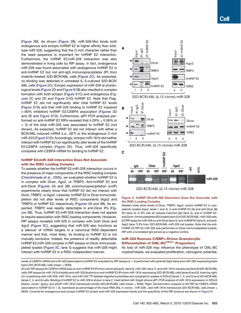
(Figure 2B). As shown (Figure 2B), miR-328-Mut binds bothendogenous and ectopic hnRNP E2 at higher affinity than wild-type miR-328, suggesting that the C-rich character rather thanthe seed sequence is important for hnRNP E2 interaction.Furthermore, the hnRNP E2:miR-328 interaction was alsodemonstrated in living cells by RIP assay. In fact, endogenousmiR-328 was found associated with endogenous hnRNP E2 inanti-hnRNP E2 but not anti-IgG immunoprecipitates (IP) fromimatinib-treated 32D-BCR/ABL cells (Figure 2C). As expected,no binding was detected in untreated IL-3-cultured 32D-BCR/ABL cells (Figure 2C). Ectopic expression of miR-328 at physio-logical levels (Figure 2D and Figure S1B) also resulted in complexformation with both ectopic (Figure S1C) and endogenous (Fig-ures 2C and 2D and Figure S1D) hnRNP E2. Note that Flag-hnRNP E2 did not significantly alter total hnRNP E2 levels(Figure S1A) and that miR-328 binding to hnRNP E2 impaired("80% inhibition) hnRNP E2:CEBPA association (Figures 2Dand 2E and Figure S1D). Furthermore, qRT-PCR analyses per-formed on anti-hnRNP E2 RIPs revealed that 4.26% ± 0.56% (n= 3) of the total miR-328 was associated to hnRNP E2 (notshown). As expected, hnRNP E2 did not interact with either aBCR/ABL-induced mRNA (i.e., SET) or the endogenous C-richmiR-223 (Figure S1D). Accordingly, ectopicmiR-181bdid neitherinteract with hnRNP E2 nor significantly alter levels of the hnRNPE2:CEBPA complex (Figure 2E). Thus, miR-328 specificallycompetes with CEBPA mRNA for binding to hnRNP E2.
hnRNP E2:miR-328 Interaction Does Not Associatewith the RISC Loading ComplexTo assess whether the hnRNP E2:miR-328 interaction occurs inthe presence of major components of the RISC loading complex(Chendrimada et al., 2005), we evaluated whether hnRNP E2 isin complex with Dicer, Ago2, or TRBP2. Anti-hnRNP E2 andanti-Dicer (Figures 3A and 3B) coimmunoprecipitation (coIP)experiments clearly show that hnRNP E2 did not interact withDicer, TRBP2, or Ago2. Likewise, hnRNP E2 or Dicer immunode-pletion did not alter levels of RISC components (Ago2 andTRBP2) or hnRNP E2, respectively (Figures 3A and 3B). As ex-pected, TRBP2 was readily detectable in anti-Dicer IPs (Fig-ure 3B). Thus, hnRNP E2:miR-328 interaction does not appearto require association with RISC loading components. However,RIP assays revealed miR-328 in complex with both Dicer andAgo2 (Figure 3C), suggesting that miR-328 also functions asa silencer of mRNA targets in a canonical RISC-dependentmanner and that, most likely, its binding to hnRNP E2 is notmutually exclusive. Indeed, the presence of readily detectablehnRNP E2:miR-328 complex in RIP assays on Dicer-immunode-pleted lysates (Figure 3C, lane 5) suggests that miR-328 mightinteract with hnRNP E2 in a RISC-independent manner.
miR-328 Rescues C/EBPa-Driven GranulocyticDifferentiation of CML-BCCD34+ ProgenitorsAs loss of miR-328 may influence the phenotype of CML-BCmyeloid blasts, we evaluated proliferation, clonogenic potential,
levels of CEBPAmRNA andmiR-328 associated to hnRNP E2 evaluated by RIP assays (n = 4) performed with parental (light bars) and miR-328-expressing (dark
bars) 32D-BCR/ABL cells (mean ± SEM).
(E) Left: RIPassays forCEBPAmRNA (top) on anti-hnRNPE2 IPs fromvector (pSuper)- (lane5),miR-328- (lane7), andmiR-181b-transduced (lane9) 32D-BCR/ABL
cells; RIP assays formiR-181b (middle) andmiR-328 (bottom) on anti-hnRNPE2 IPs frommiR-181b-expressing 32D-BCR/ABL cells (lanes 8 and 9). Inset top right:
UV crosslinking with miR-328, miR-181b, and miR-330 32P-labeled oligoribonucleotides and cytoplasmic lysates of 32Dcl3 (lanes 1, 3, and 5) and 32D-BCR/ABL
(lanes 2, 4, and 6) cells. Binding of hnRNP E2 to miR-328 is shown in lane 2. Inset bottom left: Graph shows qRT-PCR analysis of miR-181b expression in 32Dcl3
(black), vector- (gray), and pSUP-miR-181b-transduced (white) 32D-BCR/ABL cells (mean ± SEM). Right: Densitometric analysis of the RIP’ed CEBPA mRNA
associated to hnRNP E2 (n = 3), expressed as percentage of the input RNA (IN), in vector-, miR-328-, and miR-181b-transduced 32D-BCR/ABL cells (mean ±
SEM). Controls for endogenous and ectopic hnRNP E2 protein and miR-328 expression levels and the specificity of the RIP protocol are shown in Figure S1.
A
B
C
Figure 3. hnRNP E2:miR-328 Interaction Does Not Associate withthe RISC Loading ComplexWestern blots show levels of Dicer, TRBP2, Ago2, and/or hnRNP E2 in cyto-
plasmic lysates (input; lanes 1 and 4), in anti-hnRNP E2 (A) and anti-Dicer (B)
IPs (lane 3), in IPs with an isotype-matched IgG (lane 2), and in hnRNP E2-
andDicer- immunodepleted (ID) lysates (lane5)of 32D-BCR/ABL-miR-328cells.
(C)RIPassay formiR-328onanti-Dicer (lane4), anti-hnRNPE2 (lane5), andanti-
Ago2 (lane 7) IPs from 32D-BCR/ABL-miR-328 cell lysates. Note that the anti-
hnRNP E2 RIP for miR-328 was performed on Dicer-immunodepleted lysates.
RIP with a nonrelated IgG served as a negative control.
Cell 140, 652–665, March 5, 2010 ª2010 Elsevier Inc. 655

A
B
C
E F
D
Figure 4. miR-328 Rescues Granulocytic Differentiation through Restoration of C/EBPa Expression(A) miR-328 levels in (left) 32Dcl3 cells undergoing G-CSF-induced differentiation; (middle) Lin!/Sca+/Kit+ HSC, CMP/GMP/MEP committed progenitors and
mature neutrophil BM subpopulations from wild-type C57BL/6 mice (mean ± SEM); and (right) CD34+ human BM cells undifferentiated (white) and induced to
differentiate for the indicated time toward the erythroid (light gray), megakaryocytic (dark gray), granulocytic (red), or monocytic (black) lineages (mean ± SEM).
(B) Wright-Giemsa-stained cytospins of G-CSF-treated (0–7 days) pSuper-, miR-328-, miR-328-Mut-, miR-223-, and/or miR-181b-infected 32Dcl3 and/or 32D-
BCR/ABLcells (mean±SEM).LevelsofmiR-223 inBCR/ABL+cell linesandprimary cells andeffectof ectopicmiR-223oncell proliferationare reported in FigureS2.
656 Cell 140, 652–665, March 5, 2010 ª2010 Elsevier Inc.
and G-CSF-driven differentiation in miR-328-transduced GFP+
BCR/ABL+ cell lines, BCR/ABL+ Lin! mouse BM, and/or CML-BCCD34+ cells. As controls, we assessed the effects of ectopicmiR-328-Mut, miR-181b, and the myeloid differentiation-related(Chen et al., 2004) miR-223 in BCR/ABL+ cells. Of note, ectopicmiR-328 or miR-223 levels in BCR/ABL+ cells were similar tothose in nontransformed cells (Figure 2D and Figure S2A), thusexcluding off-target effects due to overexpression.Although endogenous miR-328 (Figure 1) and, to a lower
extent, miR-223 (Figure S2B) were downregulated in 32D-BCR/ABL and K562 cells, their ectopic expression did nothave a significant effect on IL-3-dependent and/or -independentgrowth (Figure S2B). Likewise, ectopic miR-328 did not accel-erate the kinetics of 32Dcl3 neutrophil maturation (Figure 4B),consistent with the barely detectable levels of hnRNP E2 in32Dcl3 cells (Perrotti et al., 2002) and the increased expressionof endogenous miR-328 in 32Dcl3, human CD34+ (n = 2) andmouse Lin!/Sca+/Kit+ (n = 3) BM (NBM) progenitors undergoinggranulocytic differentiation (Figure 4A). Conversely, miR-328levels were not significantly different in normal CD34+ BM cellsbefore and after differentiation toward other hematopoietic line-ages (Figure 4A). As expected (Fazi et al., 2005), miR-223enhanced 32Dcl3 differentiation (not shown).In agreement with the potential role of miR-328 as an antago-
nist of hnRNP E2 differentiation inhibitory activity, forced expres-sionofmiR-328 at physiological levels (Figure 2DandFigureS2A)efficiently rescued granulocytic differentiation of newly estab-lished (15 days after BCR/ABL infection) 32D-BCR/ABL cells(Figure 4B). In fact, the majority (82.1% ± 3.9%) of miR-328-expressing 32D-BCR/ABL cells were postmitotic metamyelo-cytes, bands, and segmented neutrophils after 7 days of G-CSF-supplemented culture (Figure 4B). As expected, G-CSF-treatedvector- and miR-181b-transduced BCR/ABL+ cells remainedblasts (5.1% ± 0.5% and 11.0% ± 3.2% postmitotic cells)(Figure 4B). By contrast, miR-328-Mut expression efficientlyinduced 32D-BCR/ABL differentiation (88.6% ± 3.1% postmi-totic cells) (Figure 4B).Although terminal differentiation was also a characteristic
of miR-223-overexpressing 32D-BCR/ABL cells, only ectopicmiR-328 but not miR-223 expression (Figure S2C) restoredG-CSF-driven maturation of GFP+ CD34+ BM progenitors frommyeloid CML-BC patients (n = 6) (Figure 4C). In fact, both miR-328- and miR-328-Mut-expressing CML-BCCD34+ BM culturesbecame bands and segmented neutrophils (88.8% ± 2.4% and85.2% ± 4.2% postmitotic cells) after 10 days in rhG-CSF(25 ng/ml). By contrast, morphology of miR-223-expressingCML-BCCD34+ (n = 6) progenitors remained similar to untrans-duced cells (n = 6), appearing arrested at the myeloblast stageafter 7–10 days in G-CSF-containing medium (16.9% ± 1.7%
postmitotic cells) (Figure 4C) with unchanged levels of the gran-ulocyte/macrophage markers CD11b or CD14 (not shown).Consistent with the essential role of C/EBPa in neutrophil
maturation of BCR/ABL+ blasts (Ferrari-Amorotti et al., 2006;Perrotti et al., 2002; Wagner et al., 2006), C/EBPa expressionwas readily detectable in miR-223- and miR-328-transduced32D-BCR/ABL myeloid precursors, and in miR-328- but notmiR-223-expressing CML-BCCD34+ BM progenitors and miR-181b-expressing 32D-BCR/ABL cells (Figure 4E). This wasdependent neither on hnRNP E2 downregulation (Figure 4E)nor on increased CEBPA mRNA levels (Figure 4F and Fig-ure S2C). In agreement with its ability to bind hnRNP E2, miR-328-Mut also restored C/EBPa expression (Figure 4E), indicatingthat the differentiation-promoting effects of miR-328 do notresult from the seed sequence-dependent silencing of miR-328mRNA targets. Furthermore, myeloperoxidase (MPO), a markerof granulocyte/macrophage commitment and a direct transcrip-tional target of C/EBPa (Rosmarin et al., 1989;Wang et al., 2001),was also significantly increased in G-CSF-cultured miR-328-(18.7% ± 0.2% [uninfected] versus 53.3% ± 7.3% [miR-328];p < 0.004) but not miR-223- (18.7% ± 0.2% [uninfected] versus22.2% ± 3.6% [miR-223]; p = 0.28) expressing CML-BCCD34+
cells (n = 3) (Figure 4D). Finally, the dissimilar response of miR-223-transduced 32D-BCR/ABL versus CML-BCCD34+ progeni-tors to G-CSF might depend on differences in the mechanism(s)controlling expression of NFI-A (Figure S2D), a miR-223-nega-tive regulator (Fazi et al., 2005).
miR-328 Restores CEBPA mRNA TranslationBoth In Vitro and In VivoAs hnRNP E2:miR-328 binding in vitro is more efficient than thatofCEBPA uORF (Figure 2), andmiR-328 expression antagonizeshnRNP E2:CEBPA interaction (Figures 2D and 2E) most likely bycompeting for binding to hnRNP E2, it is plausible that miR-328releases CEBPA from the translation inhibitory effects of hnRNPE2. Thus, we assessed the effect of miR-328 on CEBPA transla-tion in rabbit reticulocyte lysate and in an in vivo mouse model ofmyeloid CML-BC. In the latter, BCR/ABL+ cell differentiation isdriven solely by ectopic C/EBPa expression, which is underthe control of its uORF/spacer mRNA element (Chang et al.,2007). As reported (Perrotti et al., 2002), translation of CEBPAmRNA was markedly impaired ("80% inhibition) in in vitro trans-lation reactions programmedwith aCEBPA construct containingthe uORF/spacer intercistronic region (pcDNA3-WT-uORF-C/EBPa) and the recombinant fusion protein MBP-hnRNP E2(Figure 5A) but not when performed in the absence of exogenoushnRNP E2 (Figure 5A). Addition of 1000-fold excess of maturemiR-328 but not miR-330 resulted in an almost 100% increaseof newly synthesized 35S-C/EBPa protein (CEBPA+hnRNP E2
(C) Wright-Giemsa-stained cytospins of primary G-CSF-treated (0–10 days) uninfected and miR-328-, miR-328-Mut-, and miR-223-infected CML-BCCD34+ BM
progenitors (mean ± SEM). For levels of ectopic miR-328 and miR-223 expression in primary CML-BC cells, see Figure S2.
(D)Myeloperoxidase (MPO) immunostaining of G-CSF-treated uninfected andmiR-328- andmiR-223-transducedCML-BCCD34+ BMcells. Data are representative
of three independent experiments (mean ± SEM).
(E) Western blot shows C/EBPa, hnRNP E2, and GRB2 levels in G-CSF-treated 32Dcl3 and empty vector-, miR-328-, miR-223-, miR-328-Mut-, and/or miR-181b-
infected 32D-BCR/ABL and CML-BCCD34+ BM cells (right).
(F) qRT- PCR shows levels of CEBPA in empty vector-, miR-223-, or miR-328-infected CML-BC cells (mean ± SEM). qRT- PCR showing CEBPA mRNA levels in
empty vector-, miR-223-, or miR-328-transduced 32D-BCR/ABL cells (mean ± SEM) is reported in Figure S2.
Cell 140, 652–665, March 5, 2010 ª2010 Elsevier Inc. 657

A E
B
C
D
658 Cell 140, 652–665, March 5, 2010 ª2010 Elsevier Inc.

versusCEBPA+hnRNP E2+miR-328; p < 0.005) (Figure 5A). Notethat addition of miR-328 in the absence of MBP-hnRNP E2 didnot significantly affect CEBPA mRNA translation (Figure 5A).Moreover, the large amount of MBP-hnRNP E2 (Figure 5A) mightjustify the incomplete rescue of CEBPA translation.To assess whether forced miR-328 expression rescues
neutrophilic maturation of differentiation-arrested BCR/ABL+
blasts through restoration of CEBPA mRNA translation, weused the aberrant 32D-BCR/ABL long-term cultured 6.15 cellclone that exhibits extremely high levels of BCR/ABL and hnRNPE2 but is unable to undergo G-CSF-driven differentiation due totranscriptional suppression of CEBPa expression (Figure 5C).Indeed, 6.15 cells completely rely on translation of ectopicCEBPA mRNA for differentiation. Thus, parental and 6.15 cellsexpressing a GFP-WT-uORF/spacer-C/EBPa (6.15-WT-uORF),which contains the hnRNP E2 translation inhibitory element,were retrovirally transduced with the pSUPERIOR-retro-puro-miR-328 (6.15-WT-uORF-miR-328) or with the empty vector(6.15-WT-uORF-pSUP). Differentiation assays confirmed thatthe ability of miR-328 to induce neutrophil maturation is depen-dent on and mediated by the presence of CEBPA mRNA, asexpression of miR-328 in parental 6.15 cells failed to rescuedifferentiation, whereas 91.7% ± 6.4% of 6.15-WT-uORF-miR-328 cells were postmitotic after 7 days of culture in G-CSF(Figure 5B). Furthermore, forced miR-328 expression neitherdecreased hnRNPE2 protein nor increasedCEBPAmRNA levels(Figure 5C), suggesting that the restoration of C/EBPa proteinexpression in 6.15-WT-uORF-miR-328 cells (Figure 5C) resultsfrom the ability of miR-328 to interfere with hnRNP E2 translationinhibitory activity. Accordingly, anti-hnRNP E2 RIP assays per-formed with 6.15-WT-uORF-pSUP and 6.15-WT-uORF-miR-328 lysates revealed that miR-328 expression and, therefore,formation of the hnRNP E2:miR-328 complex (Figure 5D) mark-edly decreased levels of the hnRNP E2-bound CEBPA mRNA(Figure 5D).To determine whether miR-328 influences the CML-BC-like
disease process induced by transplantation of BCR/ABL-expressing cells, SCID mice (n = 13 per group) were intrave-nously injected with 6.15-WT-uORF-miR-328 or 6.15-WT-uORF-pSUP cells (5 3 105 GFP+-puromycin-selected cells/mouse), and engraftment was assessed 1 week later by nestedRT-PCR-mediated BCR/ABL detection in peripheral blood (notshown). After 3 weeks, three mice/group were sacrificed for
visual and histopathologic examination of hematopoietic organsand for flow cytometric quantification of GFP+/GR1+ differenti-ated BM cells. Consistent with the almost complete hnRNPE2-dependent translational inhibition of C/EBPa expression in6.15-WT-uORF cells (Chang et al., 2007), hematoxylin/eosin-stained sections of BM (Figure 5E), spleen, and liver (not shown)from 6.15-WT-uORF-pSUP-injected mice showed spleno-megaly and massive infiltration of myeloid blasts with a lowdegree of differentiation. A few myeloid cells undergoing termi-nal neutrophil differentiation were occasionally observed in BMfrom 6.15-WT-uORF-pSUP-injected mice (mean fluorescenceintensity [MFI]: 14.98 ± 1.84 GFP+/GR1+ BCR/ABL+ cells). Bycontrast, spleens from 6.15-WT-uORF-miR-328-injected miceappeared normal in weight or slightly hyperplastic, and histo-pathologic analysis of BM (Figure 5E), spleen, and liver (notshown) showed marked infiltration by mature neutrophils andmyeloid precursors at postmitotic stages of differentiation (MFI:63.84 ± 5.40 GFP+/GR1+ BCR/ABL+ cells) when compared toage-matched controls (Figure 5E), suggesting that miR-328also negatively regulates survival pathways in CML-BC althoughits major effect appears to be on differentiation. In fact, althoughno significant difference in survival time was noted, the remain-ing 6.15-WT-uORF-pSUP-injected mice died of a CML-BC-likeleukemia, whereas 6.15-WT-uORF-miR-328-injected animalssuccumbed from an aggressive CML-CP-like myeloproliferativedisorder (not shown). Altogether, these in vitro and in vivo resultsindicate that rescue of granulocytic maturation of differentiation-arrested BCR/ABL+ cells by miR-328 is likely due to its directbinding to hnRNP E2 that, in turn, prevents translational inhibi-tion of CEBPA mRNA.
A BCR/ABL-MAPK-hnRNP E2 Pathway SuppressesmiR-328 Transcription through Inhibition of C/EBPaWe recently reported that high levels of BCR/ABL expression/kinase activity, as observed in CML-BC (Jamieson et al., 2004;Schultheis et al., 2005), impair C/EBPa expression through theMAPK(ERK1/2)-dependent regulation of hnRNP E2 expression/activity (Changetal., 2007). TodeterminewhetherBCR/ABLusesthe same signaling pathway to suppress miR-328 expressionin CML-BC, miR-328 levels were evaluated in G-CSF-cultured(24–48 hr) parental and newly established 32D-BCR/ABL cellstreated with imatinib (2 mM) or MEK1 inhibitors U0126 (25 mM)and CI-1040 (10 mM) and after overexpression (MSCV-Flag-E2)
Figure 5. In Vitro and In Vivo Interference of miR-328 with hnRNP E2 Translation Inhibition of C/EBPa Expression(A) Levels of newly synthesized 35S-C/EBPa protein in RRL translation reactions programmed with CEBPA mRNA (derived from pcDNA3-WT-uORF-C/EBPa)
(black), CEBPA mRNA and mature miR-328 RNA oligonucleotides (dark gray), CEBPA mRNA and recombinant MBP-hnRNP E2 protein either alone (light
gray) or in the presence of mature miR-328 (red), or miR-330 (white; negative control) RNA oligonucleotides. Data are expressed as percentage of the mean ±
SEM and are representative of three different experiments performed in duplicate. Inset: Western blot shows levels of both endogenous RRL hnRNP E2 and
recombinant MBP-hnRNP E2.
(B) Wright-Giemsa-stained cytospins of G-CSF-treated (0–7 days) 6.15-pSUP, 6.15-miR-328, and 6.15-WT-uORF-miR-328 cells (mean ± SEM).
(C) Left: Levels of hnRNP E2, endogenous and HA-tagged C/EBPa, and GRB2 proteins and miR-328 and snRNA U6 in parental 32Dcl3, 6.15-pSUP-transduced,
and miR-328-transduced 6.15-WT-uORF cells; right: RT-PCR and qRT-PCR show levels of CEBPA mRNA in 32Dcl3, 6.15, 6.15-miR-328, and 6.15-WT-uORF-
HA-CEBPA cells either uninfected or infected with pSUP or miR-328 constructs. GAPDH levels were measured for normalization (mean ± SEM).
(D) RIP assays for CEBPA mRNA (top) and miR-328 (bottom) on anti-hnRNP E2 (lanes 5 and 9) and nonrelated IgG (lanes 3 and 7) IPs from 6.15-uORF-pSUP
(lanes 2–5) and 6.15-uORF-miR-328 cells (lanes 6–9). IN: input RNA.
(E) Top: H&E-staining of BM shows maturation of BCR/ABL+ cells in mice injected with p-SUPERIOR vector- (middle) and miR-328-transduced (right) 6.15-WT-
uORF cells. Age-matched mice (left) served as a control. FACS analysis shows mean fluorescence intensity (MFI; mean ± SEM) of differentiated GFP+Gr1+BCR/
ABL+ cells at 3 weeks post-transplant from BM of 3 mice/group. Bottom: Visual analysis and weight of spleens from the same groups of mice (mean ± SEM).
Cell 140, 652–665, March 5, 2010 ª2010 Elsevier Inc. 659

or shRNA-mediated downregulation (pSR-hnRNP E2 shRNA) ofhnRNP E2. Inhibition of BCR/ABL or MEK1 kinases stronglyenhanced miR-328 expression (Figure 6A), suggesting thatBCR/ABL-mediated suppression of miR-328 requires MAP-K(ERK) activity. Ectopic hnRNP E2 impairedmiR-328 expressionin 32Dcl3 cells, with no noticeable effect in 32D-BCR/ABLcells (Figure 6B) that already express high levels of hnRNP E2(Perrotti et al., 2002). By contrast, shRNA-mediated downregula-tion of hnRNP E2 efficiently rescued miR-328 expression (Fig-ure 6B). Thus, the BCR/ABL-MAPK-induced hnRNP E2 maydirectly regulate miR-328 nuclear export, processing, and/orstability or indirectly influence miR-328 transcription. Of interest,modulation of hnRNP E2 levels did not alter miR-223 expressionin 32D-BCR/ABL cells, whereas hnRNP E2 overexpressioninhibited miR-223 in 32Dcl3 cells (Figure S2E), consistent withthe notion that hnRNP E2 reduces C/EBPa expression (Perrottiet al., 2002), thereby avertingC/EBPa-dependentmiR-223 trans-activation (Fazi et al., 2005).
Transcription Element Search System-mediated (http://www.cbil.upenn.edu/cgi-bin/tess) sequence analysis revealed fourputative C/EBPa-binding sites scattered within 1500 bpupstream of the mouse pre-miR-328 (Figure 6C). Thus, chro-matin immunoprecipitation (ChIP) assays were performedusing nuclear extracts from GFP-sorted HA-C/EBPa-expressing32Dcl3 cells (32D-HA-C/EBPa) and four sets of primers, each
encompassing one of the potential C/EBPa-binding sites. In vivophysical interaction between HA-C/EBPa and the miR-328 pro-moter region was detected in ChIP assays performed on anti-HAbut not anti-Flag (negative control) immunoprecipitates withprimer sets #1 and #3 containing the human/mouse-conservedC/EBPa-binding sites located at nucleotides !1119 to !1112and !565 to !557, respectively (Figure 6C). Accordingly,ectopic C/EBPa expression markedly induced miR-328 levelsin myeloid precursors (Figure 6C), altogether suggesting thata BCR/ABL-MAPK-hnRNP E2 pathway downregulates miR-328 expression through inhibition of C/EBPa, thus impedingenhancement of miR-328 transcription (Figure 6D).
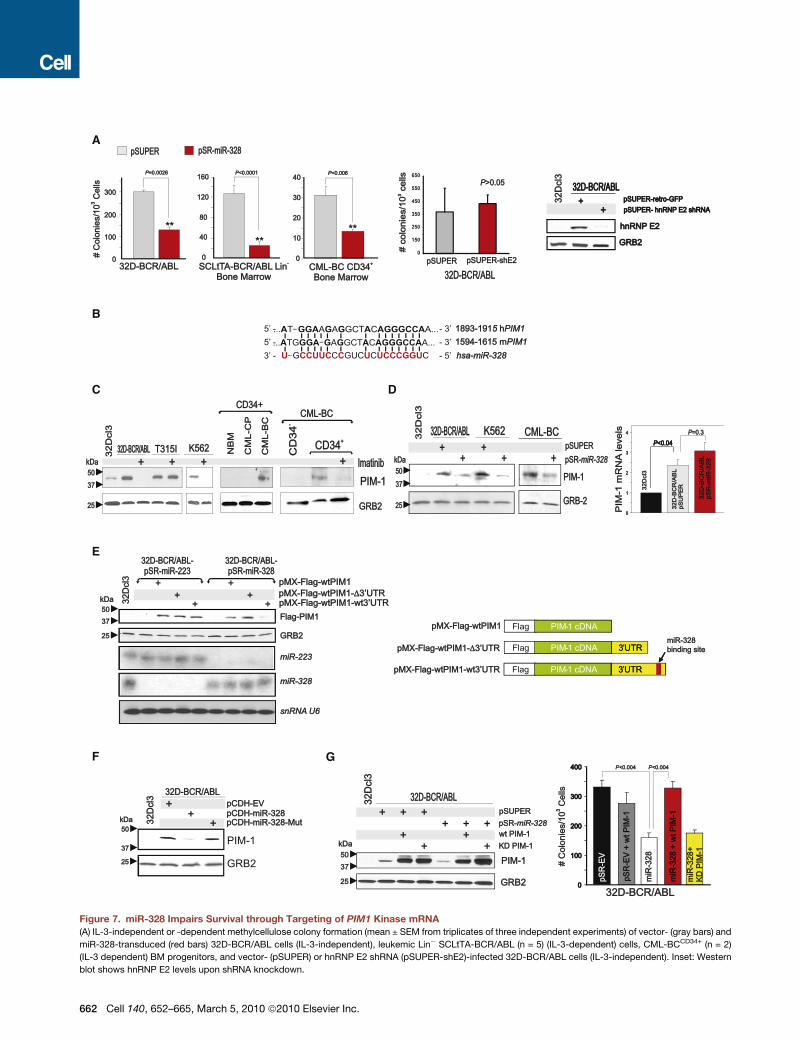
miR-328 Impairs CML-BCCD34+ Clonogenic Potentialand Canonically Suppresses PIM1 ExpressionEctopic miR-328 expression reduced colony formation in 32D-BCR/ABL, Lin! SCLtTA-BCR/ABL (n = 5), and CML-BCCD34+
(n = 2) BM cells by 75%, 83%, and 75%, respectively (Figure 7A).The effect of miR-328 on clonogenicity of BCR/ABL+ cells isindependent from hnRNP E2:miR-328 interaction, as shRNA-mediated hnRNP E2 downregulation did not affect BCR/ABL-driven colony formation (Figure 7A). Thus, miRanda, PicTar, andTargetScan bioinformatics algorithms were utilized to identifymiR-328 targets regulating CML-BC progenitor cell survival.Among the predicted miR-328 human/mouse mRNA targets,
A B
C D
Figure 6. Pathways Regulating miR-328 ExpressionmiR-328 levels in 32Dcl3 and/or 32D-BCR/ABL cells (A) treated with imatinib or the MAPK inhibitors U0126 and CI-1040 or (B) expressing a Flag-hnRNP E2 (left)
or a shRNA-targeting hnRNP E2 (right).
(C) Top: Representation of the C/EBPa-binding sites within the miR-328 promoter; bottom: Chromatin immunoprecipitation (ChIP) with anti-HA antibody shows
binding of HA-C/EBPa to miR-328 promoter sequences in 32D-HA-C/EBPa cells (left: ChIP blot; middle: densitometric analysis). Anti-Flag immunoprecipitates
served as negative controls. Bars indicate the mean ± SEM from three independent experiments; northern and western blots (right) show levels of miR-328 and
HA-C/EBPa, respectively, in parental and 32D-HA-C/EBPa cells. U6 snRNA and GRB2 protein levels were used as controls.
(D) Model of the molecular network regulating miR-328 expression in CML-BC and miR-328 decoy activity in BCR/ABL+ myeloid cell differentiation by direct
interference with hnRNP E2 translation inhibition of C/EBPa expression.
660 Cell 140, 652–665, March 5, 2010 ª2010 Elsevier Inc.

PIM1 (Figure 7B) kinase is important for survival of BCR/ABL+
cell lines (Nieborowska-Skorska et al., 2002). PIM1 protein wasstrongly upregulated in a BCR/ABL kinase-dependent mannerin cell lines and CD34+ CML-BC (n = 2) versus CML-CP andNBM BM cells (Figure 7C).Ectopic miR-328 expression in 32D-BCR/ABL, K562, and
CML-BCCD34+ cells decreased PIM1 protein without significantlyaffecting its mRNA levels (Figure 7D), suggesting that miR-328might impair mRNA translation upon interaction with the PIM130UTR. To formally demonstrate that miR-328 silences PIM1expression through its interaction with the miR-328-bindingsite, we cloned the wild-type (pMX-Flag-WTPIM1-WT30UTR)and miR-328-binding site-deleted (pMX-Flag-WTPIM1-D30UTR)PIM1 30UTR (Figure 7E) into a pMX-Flag-WTPIM1 plasmidand transduced these constructs into 32D-BCR/ABL-miR-328or, as a negative control, 32D-BCR/ABL-miR-223 cells. Asexpected, ectopic Flag-PIM1 expression was lower in pMX-Flag-WTPIM1-WT30UTR-transduced 32D-BCR/ABL-miR-328cells but not in cells transduced with pMX-Flag-WTPIM1-D30UTRorwithPIM1 cDNAonly (pMX-Flag-WTPIM1) (Figure 7E).Accordingly, Flag-PIM1 expression derived from pMX-Flag-WTPIM1-WT30UTR was barely detectable in miR-328- but notin miR-223-transduced cells, in which its expression was similarto that of Flag-WTPIM1 and Flag-WTPIM1-D30UTR (Figure 7E),indicating that decreased ectopic PIM1 levels are not due toloss of other miRNA-binding sites within the 196 bp-deleted30UTR. Expression of the seed sequence-mutated mir-328(miR-328-Mut) impaired the ability of miR-328 to canonicallysuppress PIM1 expression (Figure 7F). Thus, miR-328 specifi-cally silences PIM1 expression through interaction with thePIM1 30UTR. In agreement with the importance of PIM1 forsurvival of BCR/ABL+ cells, expression of a wild-type PIM1cDNA lacking the 30UTR (WT PIM1) but not of a kinase-deficient(KD PIM1) PIM1 cDNA into 32D-BCR/ABL-miR-328 cells(Figure 7G) completely restored IL-3-independent colony forma-tion (Figure 7G), suggesting that miR-328-dependent inhibitionof BCR/ABL-driven clonogenic potential results from directPIM1 downregulation. Note that WT PIM1 alone did not affect32D-BCR/ABL-pSR-EV clonogenic potential (Figure 7G).
DISCUSSION
Altered miRNA expression has been tightly associated withcancer development and progression (Friedman et al., 2009;Garzon et al., 2006). Among the miRNAs differentially expressedin CML, we focused on miR-328 and provided a series ofevidence highlighting two important concepts. The first repre-sents a paradigm shift to the notion that miRNAs act primarilyas negative posttranscriptional regulators of gene expressionand proposes for miRNAs a function termed decoy activity.The second identifies miR-328 as a molecular relay, the loss ofwhich is important for the differentiation arrest of progressingCML-BC blasts.
miR-328 Decoy ActivityAs miRNAs base pair with mRNA 30UTRs in a sequence-specificmanner (Bartel, 2009), it is conceivable that miRNAs could inter-fere with the activity of RNA-binding proteins (e.g., hnRNPs),
either indirectly by pairing with RBP-binding sites contained inspecific mRNAs (George and Tenenbaum, 2006) or directlythrough binding the RBP itself and impeding RBP:mRNA interac-tion. Herein we demonstrated that miRNAs can act as directinhibitors of RBP activity. In fact, miR-328 specifically interactsin a seed sequence-independent manner and, most likely,through its C-rich clusters, with the translational inhibitor poly(rC)-binding protein hnRNP E2. This, in turn, prevents and/ordisplaces CEBPA mRNA binding to hnRNP E2 and rescuesCEBPA mRNA translation both in vitro and in vivo. In supportof the notion that miR-328 and, most likely, other miRNAs mayact as ‘‘decoy’’ molecules for RBPs, which upon binding couldcontrol synthesis, processing, export, stability, and/or transla-tion of specific mRNA subsets, a proteomics-based study inepithelial A431 cells reported that forced miR-328 expressionnot only decreased levels of different genes but also upregulateda subset of proteins (Wang et al., 2008). Interestingly, 37% ofthese upregulated proteins have mRNAs with complex 50UTRs(e.g., uORF or multiple ATGs) containing C-rich elements repre-senting potential hnRNP E2-binding sites. Thus, it is reasonableto speculate that upregulation of some of these proteins mightresult from interference with hnRNP E2 activity. Furthermore,there is evidence that miRNAs, other components of the RISCcomplex (Parker and Sheth, 2007) and RBPs (e.g., hnRNP E2)are present in processing bodies (P bodies) (Fujimura et al.,2008), dynamic subcellular structures where mRNAs are com-plexed with RBPs and/or miRNAs for translational suppressionor decay (Parker and Sheth, 2007). In this scenario, miR-328may compete with CEBPA mRNA for binding to hnRNP E2that, in turn, releases CEBPA and allows its loading onto poly-somes for translation. It is also possible that hnRNP E2 notonly prevents C/EBPa-dependent induction of pri-miR-328 tran-scription but also directly promotes miR-328 decay. Accord-ingly, an inverse correlation exists between hnRNP E2 andmiR-328 expression in CML-BC, and hnRNP E2 more efficientlybinds tomiR-328 than toCEBPA. However, as hnRNP E2 contin-uously shuttles between nucleus and cytoplasm as well as in andout of P bodies (Fujimura et al., 2008; Makeyev and Liebhaber,2002), knowledge of the subcellular location where initial bindingof hnRNP E2 to miR-328 or CEBPA occurs remains elusive,although a plausible assumption is the cytoplasm as their asso-ciation was detected using cytoplasmic extracts.MicroRNA interaction with sequence-specific RBPs is not
unprecedented; however, none of the reported mechanismsmatch the case of hnRNP E2 and miR-328. For example, hnRNPA1, another RBP upregulated in CML-BC (Perrotti and Neviani,2007), binds the primary miR-17-92 transcript to allow process-ing of pre-miR-18a (Guil and Caceres, 2007). Interestingly,expression of the miR-17-92 cluster is upregulated in CML andis important for proliferation and reduced susceptibility toapoptosis of K562 cells (Venturini et al., 2007). However hnRNPE2:miR-328 interaction does not seem to affect miR-328 biogen-esis, as no accumulation of primary or precursor miR-328 wasdetected in BCR/ABL+ cells (not shown). It was also shownthat miR-369-3 interacts with the AU-rich region (ARE) of TNF-amRNA, which recruits the AGO2-FXR1 RBP complex to the AREelement itself and upregulates or represses translation underserum-starved or proliferating conditions, respectively
Cell 140, 652–665, March 5, 2010 ª2010 Elsevier Inc. 661
A
B
C D
E
F G
Figure 7. miR-328 Impairs Survival through Targeting of PIM1 Kinase mRNA(A) IL-3-independent or -dependent methylcellulose colony formation (mean ± SEM from triplicates of three independent experiments) of vector- (gray bars) and
miR-328-transduced (red bars) 32D-BCR/ABL cells (IL-3-independent), leukemic Lin! SCLtTA-BCR/ABL (n = 5) (IL-3-dependent) cells, CML-BCCD34+ (n = 2)
(IL-3 dependent) BM progenitors, and vector- (pSUPER) or hnRNP E2 shRNA (pSUPER-shE2)-infected 32D-BCR/ABL cells (IL-3-independent). Inset: Western
blot shows hnRNP E2 levels upon shRNA knockdown.
662 Cell 140, 652–665, March 5, 2010 ª2010 Elsevier Inc.

(Vasudevan et al., 2007). However, it is unlikely that hnRNP E2 isrecruited to CEBPA mRNA by miR-328, as no miR-328 targetsites are present in the 50 and 30UTRs of human and mouseCEBPA mRNA. Because our data indicate that miR-328 bindshnRNPE2 in a seed sequence-independentmanner and stronglyprevents its interaction with the C-rich intercistronic element ofCEBPA mRNA, it is safe to conclude that hnRNP E2 binding tomiR-328 and CEBPA mRNA are mutually exclusive. Interest-ingly, as binding to hnRNP E2 is mediated by the C-rich nucleo-tide clusters present in miR-328, it is possible that miR-328 alsoalters the function of other hnRNPs (PCBP1 and hnRNP K) that,like hnRNP E2, bind C-rich elements within the 50 and 30UTRs(Ostareck-Lederer and Ostareck, 2004). Indeed, preliminarydata indicate that miR-328 interacts with hnRNP K, albeit witha lower affinity than the interaction with hnRNP E2 (not shown).Because hnRNP K expression/activity is also enhanced inCML-BCCD34+ cells in a BCR/ABL-MAPK-dependent manner(Notari et al., 2006), and both hnRNP K and hnRNP E2 are essen-tial for IRES-mediated induction of MYC mRNA translation(Evans et al., 2003), we can speculate that loss of miR-328 inCML might favor disease progression by also promoting hnRNPK/E2-dependent MYC translation activation. Seemingly, we canalso speculate that the mRNA stabilizing and destabilizingactivity of the AU-rich element (ARE) RNA-binding proteinsAUF1 (hnRNP D) and HuR (Lal et al., 2004), respectively, canbe influenced by the putative decoy activity of miRNAs bearingthe ARE element in their nucleotide sequence (e.g., miR-95;uucaacggguAUUUAuugagca). Indeed, TargetScan-based anal-ysis revealed that several mature miRNAs contain the AUUUAelement in their sequence. Thus, it is conceivable that thesemiRNAs can antagonize the ability of AUF1 and HuR to regulateexpression of mRNAs encoding cytokine receptors (e.g.,GM-CSF, TNF-a, and interleukins), oncogenes (e.g., c-Mycand FOS), tumor suppressors (e.g., p53), and cell-cycle regula-tors (e.g., p21 and cyclin D1) (Hinman and Lou, 2008; Khabar,2005; Lal et al., 2004). Likewise, the function of the CGG-repeatbinding FMRP RBP, which is the cause of the Fragile X mentalretardation syndrome and normally regulates translation ofmRNAs controlling synaptic function (Brown et al., 2001; Darnellet al., 2001; Zalfa et al., 2003), might be influenced by the alteredexpression of CGG-rich miRNAs (e.g., miR-572 and miR-638).Thus, it is likely that the decoy activity of miRNAs is not limitedto miR-328 but can be extended to other miRNAs containing
nucleotide sequences resembling the consensus RNA-bindingsites for RBPs that are involved in different normal cell functionsand in neoplastic as well as non-cancer-related diseases.
miR-328: AMolecular Relay in CMLDisease ProgressionEven though a few miRNAs are aberrantly regulated in CML(Agirre et al., 2008; Bueno et al., 2008; Venturini et al., 2007),evidence of their involvement in disease progression is still lack-ing. The failure of myeloid CML-BC progenitors to undergomaturation depends on increased BCR/ABL activity which, inaddition to enhancing survival, proliferation, genomic instability,and self-renewal (Melo and Barnes, 2007), allows the hnRNP E2inhibitory effect on C/EBPa that, per se, is sufficient to reinstatedifferentiation of Ph(+) blasts (Ferrari-Amorotti et al., 2006; Per-rotti et al., 2002). We demonstrated that loss of miR-328 occursin CML-BCCD34+ but not CML-CPCD34+ myeloid progenitors, andthat forced miR-328 expression at physiological levels rescuesC/EBPa-driven granulocytic maturation and impairs survival ofCML-BC blasts. However, the proapoptotic effect of miR-328does not seem to depend on its decoy activity and, therefore,on C/EBPa-induced differentiation and growth arrest (Keeshanet al., 2003), as shRNA-mediated hnRNP E2 downregulationdoes not influence BCR/ABL-driven clonogenic potential.Rather, we showed that impaired colony formation is the conse-quence of miR-328 canonical activity that targets PIM1 mRNA,thus repressing PIM1 expression and survival-promoting activity(Hoover et al., 2001; Nieborowska-Skorska et al., 2002). Indeed,forced expression of a wild-type, but not a kinase-deficient,PIM1 cDNA lacking the 30UTR into miR-328-expressing cellsfully rescued BCR/ABL clonogenicity. However, the mainin vivo effect of miR-328 seems to be on differentiation ratherthan survival, as forced miR-328 expression did not delay leuke-mogenesis but reverted the blast crisis-like phenotype to a dis-ease that resembles a myeloproliferative-like disorder, althoughthe absence ofmarked splenomegalymay suggest that a portionof miR-328+/BCR/ABL+ cells underwent apoptosis. AlthoughmiR-223 was also described as a positive regulator of neutrophilmaturation in APL cells (Fazi et al., 2005), our data in primaryleukemic samples and work in miR-223 knockout animals (John-nidis et al., 2008) argue against a general role for miR-223 as aninducer of myeloid differentiation.Mechanistically, we showed that BCR/ABL uses the same
MAPK(ERK1/2)-hnRNP E2 signaling pathway (see model in
(B) miR-328-binding site (red) within the 30UTR of mouse and human PIM1 mRNA.
(C) PIM1 protein levels in 32Dcl3, untreated or imatinib-treated 32D-BCR/ABL (wild-type and T315I) and K562 cells (left); in CD34+ BM cells from healthy donors
(NBM), CML-CP, and CML-BC patients (middle); and in the CD34!, untreated and imatinib-treated CD34+ BM fractions from a CML-BC patient.
(D) Left: Effect of miR-328 expression on PIM1 protein levels in 32D-BCR/ABL, K562, and CML-BCCD34+ BM cells. 32Dcl3 cells were used as a negative control;
right: PIM1mRNA expression by qRT-PCR in 32Dcl3, vector-transduced, andmiR-328-transduced 32D-BCR/ABL. Representative of triplicates from three inde-
pendent experiments (mean ± SEM).
(E) Left: Levels of ectopic Flag-PIM1 proteins from constructs lacking (pMX-Flag-WTPIM1) and harboring the wild-type (pMX-Flag-WTPIM1-WT30UTR) or 196
base pair end-terminal-deleted (pMX-Flag-WTPIM1-D30UTR) PIM1 30UTR in miR-223- or miR-328-expressing 32D-BCR/ABL. Northern blot shows levels of
miR-223, miR-328, and snRNA U6. Right: Schematic representation of the Flag-PIM1 constructs.
(F) Effect of seed sequence-mutated (miR-328-Mut) on endogenous PIM1 expression in parental 32Dcl3 and empty vector (pCDH)-, miR-328-, andmiR-328-Mut-
infected 32D-BCR/ABL cells.
(G) Left: Endogenous and ectopic (wild-type [WT PIM-1] and kinase-deficient [KD PIM-1]) PIM1 protein levels in 32Dcl3, pSUPER- and pSR-miR-328-infected
32D-BCR/ABL cells; right: graph shows rescue of IL-3-independent clonogenic activity of miR-328-expressing (white) 32D-BCR/ABL cells to normal levels
(black) by ectopic wild-type (red) but not kinase-deficient (yellow) PIM1 construct lacking the 30UTR. Effect of PIM1 forced expression on vector-transduced
clonogenicity (gray). Bars represent the mean ± SEM of colony numbers from three independent experiments.
Cell 140, 652–665, March 5, 2010 ª2010 Elsevier Inc. 663

Figure 6D) to suppress C/EBPa (Chang et al., 2007) andmiR-328expression. Notably, constitutive MAPK activation by BCR/ABLoccurs in CML-BC but not CML-CP (Notari et al., 2006). More-over, similar to the positive feedback loop described for miR-223 and C/EBPa (Fazi et al., 2005), we showed that C/EBPaalso interacts with the miR-328 promoter, thus enhancing itstranscription.
In conclusion, the discovery of dual activities for miR-328 thatprofoundly affect myeloid cell differentiation and survival not onlyadd a new layer to the complexity of mechanisms regulating thephenotype of CML-BC progenitors but, more importantly, high-light the ability of miRNAs to alter mRNA metabolism by actingalso as molecular decoys for RNA-binding proteins.
EXPERIMENTAL PROCEDURES
Additional details on all the methods are available online in the Extended
Experimental Procedures.
Clonogenic and Viability AnalysisMethylcellulose clonogenic assays were carried out by plating 103 32Dcl3 and
derivative cell lines, 104 CML-BCCD34+, or 104 Lin! SCLtTA-BCR/ABL BM
progenitors in 0.9% MethoCult (Stem Cell Technologies) in the presence or
absence of rIL-3 (100 ng/ml). Colonies (>100 mm) from cell lines and primary
cells were scored 7 and 15 days later, respectively. Human and mouse cell
lines and primary cell source and culture conditions as well as plasmids and
retro/lentiviral vectors are reported in the Extended Experimental Procedures.
In Vitro and In Vivo Differentiation AssaysIn vitro granulocytic differentiation was induced for 7–10 days with 25 ng/ml
rG-CSF.Morphologic differentiation was assessed byWright/Giemsa staining.
For in vivo differentiation, 10-week-old ICR-SCID mice (n = 13 per group) were
intravenously (i.v.) injected (53 105 cells/mouse) with pSUPERIOR.retro.puro-
or pSUP-miR-328-transduced 6.15-WT-uORF-CEBPA(GFP+) cells.
RNA Extraction, Northern Blot, and Real-Time PCRTotal RNA was used in northern blot, RT-PCR, and/or qRT-PCR for the anal-
ysis of miRNA and mRNA expression. U6 snRNA and GAPDH levels were
analyzed for normalization of miRNA and mRNA PCRs, respectively.
REMSA, UV Crosslinking, and RNA ImmunoprecipitationRecombinant MBP-hnRNP E2 (Chang et al., 2007) and 32Dcl3 or 32D-BCR/
ABL cytoplasmic extracts were used in REMSA and UV crosslinking as
described (Perrotti et al., 2002). RIP and miR-328 RT-PCR were performed
as described (Keene et al., 2006; Wang et al., 2008).
In Vitro Translation AssayIn vitro translation assays using the transcription/translation-coupled rabbit
reticulocyte lysate system (Promega) were performed with pcDNA3-WT-
uORF-C/EBPa in the presence or absence of 1 mg recombinant MBP-hnRNP
E2 (Perrotti et al., 2002) either with or without 10003 mature miR-328 or
miR-330 oligoribonucleotides.
Western Blotting, Coimmunoprecipitation, and ChIP AssaysFor western blot, 1 3 107 cells were lysed (0#C; 30 min) in 50–100 ml RIPA
buffer, clarified, and subjected to SDS-PAGE. For C/EBPa detection, 106 cells
were directly lysed in 20 ml Laemmli buffer and denatured prior to SDS-PAGE
and transfer to nitrocellulose. For coIP, cells were lysed on ice with immuno-
precipitation buffer and 1.0 mg of protein was used in coimmunoprecipitation
assays. Chromatin immunoprecipitation (ChIP) assays were performed using
an EZ-Chip kit (Millipore).
StatisticsData were analyzed as follows: (1) two-tailed paired Student’s t test for assays
with identical cell lines, untreated and imatinib-treated SCLtTA-BCR/ABL
cells, RIP assay densitometric and qRT-PCR, and in vitro translation assays;
(2) two-tailed independent Student’s t test for clonogenic assays with unpaired
miRNA-infected Lin! BM cells; and (3) the Mann-Whitney rank sums test for
assays with unpaired CML patient samples. A p value of less than 0.05 was
considered statistically significant.
SUPPLEMENTAL INFORMATION
Supplemental Information includes Extended Experimental Procedures and
two figures and can be found with this article online at doi:10.1016/j.cell.
2010.01.007.
ACKNOWLEDGMENTS
This work was supported in part by grants from the National Cancer Institute
CA095512 (D.P.), CA16058 (OSU-CCC), NIH, Bethesda, MD, USA; the US
Army, CML Research Program, W81XWH-07-1-0270 (D.P.); the American-
Italian Cancer Foundation (P.N.); Fonds de la Recherche en Sante du Quebec
(D.C.R.); and AGGRS from The OSU Graduate School (A.M.E.). D.P. is
a Scholar of The Leukemia and Lymphoma Society. G.A.C. is a Fellow of the
M.D. Anderson Research Trust and Scholar of the University of Texas System
Regents. We thank J.-S. Chang, M. Odeh, and A. Martin for technical assis-
tance and S. Lee for editorial assistance; T. Skorski (Temple University, Phila-
delphia, PA, USA) for providing PIM1 cDNAs; F. Racke (OSU, Columbus OH,
USA) for histopathology analysis; B. Calabretta (TJU, Philadelphia, PA,
USA), R. Arlinghaus (MDACC, Houston, TX, USA), and K. Huebner (OSU) for
critical reading of the manuscript.
Received: March 12, 2009
Revised: September 25, 2009
Accepted: January 5, 2010
Published: March 4, 2010
REFERENCES
Agirre, X., Jimenez-Velasco, A., San Jose-Eneriz, E., Garate, L., Bandres, E.,
Cordeu, L., Aparicio, O., Saez, B., Navarro, G., Vilas-Zornoza, A., et al.
(2008). Down-regulation of hsa-miR-10a in chronic myeloid leukemia CD34+
cells increases USF2-mediated cell growth. Mol. Cancer Res. 6, 1830–1840.
Bartel, D.P. (2009). MicroRNAs: Target recognition and regulatory functions.
Cell 136, 215–233.
Brown, V., Jin, P., Ceman, S., Darnell, J.C., O’Donnell, W.T., Tenenbaum, S.A.,
Jin, X., Feng, Y., Wilkinson, K.D., Keene, J.D., et al. (2001). Microarray identi-
fication of FMRP-associated brain mRNAs and altered mRNA translational
profiles in fragile X syndrome. Cell 107, 477–487.
Bueno,M.J., Perez deCastro, I., Gomez deCedron,M., Santos, J., Calin, G.A.,
Cigudosa, J.C., Croce, C.M., Fernandez-Piqueras, J., and Malumbres, M.
(2008). Genetic and epigenetic silencing of microRNA-203 enhances ABL1
and BCR-ABL1 oncogene expression. Cancer Cell 13, 496–506.
Carpenter, B., MacKay, C., Alnabulsi, A., MacKay, M., Telfer, C., Melvin, W.T.,
and Murray, G.I. (2006). The roles of heterogeneous nuclear ribonucleopro-
teins in tumour development and progression. Biochim. Biophys. Acta 1765,
85–100.
Chang, J.S., Santhanam, R., Trotta, R., Neviani, P., Eiring, A.M., Briercheck, E.,
Ronchetti, M., Roy, D.C., Calabretta, B., Caligiuri, M.A., and Perrotti, D. (2007).
High levels of the BCR/ABL oncoprotein are required for the MAPK-hnRNP E2
dependent suppression of C/EBPalpha-driven myeloid differentiation. Blood
110, 994–1003.
Chen, C.Z., Li, L., Lodish, H.F., and Bartel, D.P. (2004). MicroRNAs modulate
hematopoietic lineage differentiation. Science 303, 83–86.
664 Cell 140, 652–665, March 5, 2010 ª2010 Elsevier Inc.
Chendrimada, T.P., Gregory, R.I., Kumaraswamy, E., Norman, J., Cooch, N.,
Nishikura, K., and Shiekhattar, R. (2005). TRBP recruits the Dicer complex to
Ago2 for microRNA processing and gene silencing. Nature 436, 740–744.
Darnell, J.C., Jensen, K.B., Jin, P., Brown, V., Warren, S.T., and Darnell, R.B.
(2001). Fragile Xmental retardation protein targets G quartet mRNAs important
for neuronal function. Cell 107, 489–499.
Eiring, A.M., Neviani, P., Santhanam, R., Oaks, J.J., Chang, J.S., Notari, M.,
Willis, W., Gambacorti-Passerini, C., Volinia, S., Marcucci, G., et al. (2008).
Identification of novel posttranscriptional targets of the BCR/ABL oncoprotein
by ribonomics: requirement of E2F3 for BCR/ABL leukemogenesis. Blood 111,
816–828.
Evans, J.R., Mitchell, S.A., Spriggs, K.A., Ostrowski, J., Bomsztyk, K., Ostarek,
D., andWillis, A.E. (2003). Members of the poly (rC) binding protein family stim-
ulate the activity of the c-myc internal ribosome entry segment in vitro and
in vivo. Oncogene 22, 8012–8020.
Fazi, F., Rosa, A., Fatica, A., Gelmetti, V., DeMarchis, M.L., Nervi, C., and Boz-
zoni, I. (2005). A minicircuitry comprised of microRNA-223 and transcription
factors NFI-A and C/EBPalpha regulates human granulopoiesis. Cell 123,
819–831.
Ferrari-Amorotti, G., Keeshan, K., Zattoni, M., Guerzoni, C., Iotti, G., Cattelani,
S., Donato, N.J., and Calabretta, B. (2006). Leukemogenesis induced by wild-
type and STI571-resistant BCR/ABL is potently suppressed by C/EBPalpha.
Blood 108, 1353–1362.
Friedman, R.C., Farh, K.K., Burge, C.B., andBartel, D.P. (2009). Mostmamma-
lian mRNAs are conserved targets of microRNAs. Genome Res. 19, 92–105.
Fujimura, K., Kano, F., and Murata, M. (2008). Identification of PCBP2, a facil-
itator of IRES-mediated translation, as a novel constituent of stress granules
and processing bodies. RNA 14, 425–431.
Garzon, R., Fabbri, M., Cimmino, A., Calin, G.A., and Croce, C.M. (2006).
MicroRNA expression and function in cancer. Trends Mol. Med. 12, 580–587.
George, A.D., and Tenenbaum, S.A. (2006). MicroRNA modulation of RNA-
binding protein regulatory elements. RNA Biol. 3, 57–59.
Glisovic, T., Bachorik, J.L., Yong, J., and Dreyfuss, G. (2008). RNA-binding
proteins and post-transcriptional gene regulation. FEBS Lett. 582, 1977–1986.
Guil, S., and Caceres, J.F. (2007). The multifunctional RNA-binding protein
hnRNP A1 is required for processing of miR-18a. Nat. Struct. Mol. Biol. 14,
591–596.
Hinman, M.N., and Lou, H. (2008). Diverse molecular functions of Hu proteins.
Cell. Mol. Life Sci. 65, 3168–3181.
Hoover, R.R., Gerlach, M.J., Koh, E.Y., and Daley, G.Q. (2001). Cooperative
and redundant effects of STAT5 and Ras signaling in BCR/ABL transformed
hematopoietic cells. Oncogene 20, 5826–5835.
Jamieson, C.H., Ailles, L.E., Dylla, S.J., Muijtjens, M., Jones, C., Zehnder, J.L.,
Gotlib, J., Li, K., Manz, M.G., Keating, A., et al. (2004). Granulocyte-macro-
phage progenitors as candidate leukemic stem cells in blast-crisis CML. N.
Engl. J. Med. 351, 657–667.
Johnnidis, J.B., Harris, M.H., Wheeler, R.T., Stehling-Sun, S., Lam, M.H.,
Kirak, O., Brummelkamp, T.R., Fleming, M.D., and Camargo, F.D. (2008).
Regulation of progenitor cell proliferation and granulocyte function by micro-
RNA-223. Nature 451, 1125–1129.
Keene, J.D. (2007). RNA regulons: coordination of post-transcriptional events.
Nat. Rev. Genet. 8, 533–543.
Keene, J.D., Komisarow, J.M., and Friedersdorf, M.B. (2006). RIP-Chip: the
isolation and identification of mRNAs, microRNAs and protein components
of ribonucleoprotein complexes from cell extracts. Nat. Protoc. 1, 302–307.
Keeshan, K., Santilli, G., Corradini, F., Perrotti, D., and Calabretta, B. (2003).
Transcription activation function of C/EBPalpha is required for induction of
granulocytic differentiation. Blood 102, 1267–1275.
Khabar, K.S. (2005). The AU-rich transcriptome: more than interferons and
cytokines, and its role in disease. J. Interferon Cytokine Res. 25, 1–10.
Lal, A., Mazan-Mamczarz, K., Kawai, T., Yang, X., Martindale, J.L., and Gor-
ospe, M. (2004). Concurrent versus individual binding of HuR and AUF1 to
common labile target mRNAs. EMBO J. 23, 3092–3102.
Makeyev, A.V., and Liebhaber, S.A. (2002). The poly(C)-binding proteins:
a multiplicity of functions and a search for mechanisms. RNA 8, 265–278.
Melo, J.V., and Barnes, D.J. (2007). Chronic myeloid leukaemia as a model of
disease evolution in human cancer. Nat. Rev. Cancer 7, 441–453.
Nieborowska-Skorska, M., Hoser, G., Kossev, P., Wasik, M.A., and Skorski, T.
(2002). Complementary functions of the antiapoptotic protein A1 and serine/
threonine kinase pim-1 in the BCR/ABL-mediated leukemogenesis. Blood
99, 4531–4539.
Notari, M., Neviani, P., Santhanam, R., Blaser, B.W., Chang, J.S., Galietta, A.,
Willis, A.E., Roy, D.C., Caligiuri, M.A., Marcucci, G., and Perrotti, D. (2006).
A MAPK/HNRPK pathway controls BCR/ABL oncogenic potential by regu-
lating MYC mRNA translation. Blood 107, 2507–2516.
Ostareck-Lederer, A., and Ostareck, D.H. (2004). Control of mRNA translation
and stability in haematopoietic cells: the function of hnRNPs K and E1/E2. Biol.
Cell 96, 407–411.
Parker, R., and Sheth, U. (2007). P bodies and the control of mRNA translation
and degradation. Mol. Cell 25, 635–646.
Perrotti, D., Cesi, V., Trotta, R., Guerzoni, C., Santilli, G., Campbell, K., Iervo-
lino, A., Condorelli, F., Gambacorti-Passerini, C., Caligiuri, M.A., and Cala-
bretta, B. (2002). BCR-ABL suppresses C/EBPalpha expression through inhib-
itory action of hnRNP E2. Nat. Genet. 30, 48–58.
Perrotti, D., and Neviani, P. (2007). FrommRNAmetabolism to cancer therapy:
chronic myelogenous leukemia shows the way. Clin. Cancer Res. 13, 1638–
1642.
Rosmarin, A.G., Weil, S.C., Rosner, G.L., Griffin, J.D., Arnaout, M.A., and
Tenen, D.G. (1989). Differential expression of CD11b/CD18 (Mo1) and myelo-
peroxidase genes during myeloid differentiation. Blood 73, 131–136.
Schultheis, B., Szydlo, R., Mahon, F.X., Apperley, J.F., and Melo, J.V. (2005).
Analysis of total phosphotyrosine levels in CD34+ cells from CML patients to
predict the response to imatinib mesylate treatment. Blood 105, 4893–4894.
Tenen, D.G. (2003). Disruption of differentiation in human cancer: AML shows
the way. Nat. Rev. Cancer 3, 89–101.
Vasudevan, S., Tong, Y., and Steitz, J.A. (2007). Switching from repression to
activation: microRNAs can up-regulate translation. Science 318, 1931–1934.
Venturini, L., Battmer, K., Castoldi, M., Schultheis, B., Hochhaus, A., Muck-
enthaler, M.U., Ganser, A., Eder, M., and Scherr, M. (2007). Expression of
the miR-17-92 polycistron in chronic myeloid leukemia (CML) CD34+ cells.
Blood 109, 4399–4405.
Wagner, K., Zhang, P., Rosenbauer, F., Drescher, B., Kobayashi, S., Radom-
ska, H.S., Kutok, J.L., Gilliland, D.G., Krauter, J., and Tenen, D.G. (2006).
Absence of the transcription factor CCAAT enhancer binding protein alpha
results in loss of myeloid identity in bcr/abl-induced malignancy. Proc. Natl.
Acad. Sci. USA 103, 6338–6343.
Wang, C.H., Lee, D.Y., Deng, Z., Jeyapalan, Z., Lee, S.C., Kahai, S., Lu, W.Y.,
Zhang, Y., and Yang, B.B. (2008). MicroRNA miR-328 regulates zonation
morphogenesis by targeting CD44 expression. PLoS ONE 3, e2420.
Wang, W., Wang, X., Ward, A.C., Touw, I.P., and Friedman, A.D. (2001).
C/EBPalpha andG-CSF receptor signals cooperate to induce themyeloperox-
idase and neutrophil elastase genes. Leukemia 15, 779–786.
Zalfa, F., Giorgi, M., Primerano, B., Moro, A., Di Penta, A., Reis, S., Oostra, B.,
and Bagni, C. (2003). The fragile X syndrome protein FMRP associates with
BC1 RNA and regulates the translation of specific mRNAs at synapses. Cell
112, 317–327.
Cell 140, 652–665, March 5, 2010 ª2010 Elsevier Inc. 665

Supplemental Information
EXTENDED EXPERIMENTAL PROCEDURES
Cell Cultures and Primary Cells32Dcl3 murine myeloid precursor cells, Ph1(+) erythroleukemia K562 cells, and derivative lines were maintained in IMDM plus 10%FBS and 2 mM L-glutamine (GIBCO). 10%WEHI-conditioned medium was used as the source of mIL-3. 293T cells were maintainedin culture in DMEM, 10% FBS and 2 mM L-glutamine. 32D-BCR/ABL, 32D-BCR/ABL(T315I), and miRNA-expressing cells weregenerated by retroviral infection followed by antibiotics-mediated selection or FACS-mediated sorting of GFP+ cells (Perrottiet al., 2002). Newly established 32D-BCR/ABL cells (which express CEBPA mRNA but not C/EBPa protein) (Perrotti et al., 2002)were used in differentiation assays. Murine BM cells from femurs of C57BL/6 or leukemic SCLtTA-BCR/ABL mice (Koschmiederet al., 2005) underwent Lin- magnetic-activated cell sorting (MACS, Miltenyi Biotech) and were grown for 2 days in IMDM mediumcontaining murine IL-3 (2 ng/ml), IL-6 (2 ng/ml), SCF (10 ng/ml), Flt3-ligand (5 ng/ml), and GM-CSF (5 ng/ml) (R&D Systems) priorto infection with MigR1, MigR1-p210BCR/ABL (W. Pear, UPENN, Philadelphia, PA), or miRNA retroviral vectors. All animal studieswere performed with approval of The OSU Institutional Animal Care and Use Committee. For patient specimens, frozenmononuclearBM cells of unidentified CML-CP (91%–100% Ph1(+) by FISH) and myeloid CML-BC patients were Ficoll separated, cultured o/n inIMDM plus 30% FBS, 2mM L-glutamine supplemented with IL-3 (20 ng/ml), IL-6 (20 ng/ml), Flt-3 ligand (100 ng/ml), andKL (100 ng/ml) (Stem Cell Technologies), and the CD34+ fraction was isolated using the CD34 MultiSort kit (Miltenyi Biotec). Frozensamples of CD34+ BM cells from healthy donors were purchased from Cincinnati Children’s Hospital, Cincinnati, OH. Patient spec-imens were obtained from the OSU Leukemia Tissue Bank, Columbus OH; Maisonneuve-Rosemont Hospital, Montreal, Quebec,Canada; City of Hope National Medical Center, Duarte, CA; MD Anderson Cancer Center, Houston TX; and Aarhus University,Denmark. All the performed experiments were approved by The OSU Institutional Review Board. Where indicated, cells were treatedwith the following kinase inhibitors: 1–2 mM imatinib mesylate (Novartis Oncology), 25 mM U0126 (Promega), or 10 mM CI-1040(Pfizer).
In Vitro and In Vivo Differentiation AssaysIn vitro granulocytic differentiation was induced by exposing cells to 25 ng/ml rhG-CSF for 7–10 days. Morphologic differentiationwas assessed byWright/Giemsa staining of cytospins. miR-328 expression in different lineages was extrapolated frommiRNA arrayanalysis of hematopoietic precursors obtained by culturing for 14 days human (non-mobilized) CD34+ BM cells (n = 2) in EPO/SCF/IL-3 (erythroid), G-CSF/GM-CSF/SCF/IL-6/IL-3 (granulocytic), TPO/SCF/IL-3 (megakaryocytic) and M-CSF/GM-CSF/IL-6/IL-3/SCF(monocytic). Morphology and lineage-specific antibody staining was used to assess differentiation. Total RNA from lineage-committed cells at different days of culture was hybridized in duplicate to OSU v3.0 miRNA chip. After quantiles normalization, differ-entially expressed miRNAs were identified by using the univariate t test within the BRB array tools. For in vivo differentiation,10-week-old ICR-SCID mice (n = 13 per group) were i.v. injected (5 3 105 cells/mouse) with pSUPERIOR.retro.puro- or pSUP-miR-328-transduced 6.15-WT-uORF-CEBPA(GFP+) cells. Engraftment was assessed 1 week after cell injection by nested RT-PCR-mediated detection of BCR/ABL transcripts in circulating peripheral blood cells (Eiring et al., 2008). After 3 weeks, micewere sacrificed and BM, spleen, and liver were subjected to visual and histological (H&E staining) analyses and flow-cytometricdetection of Gr1+GFP+ BM cells using PE-conjugated GR1mAb (PharMingen). Light microscopy was performed on a Zeiss Axioskop2 Plus microscope equipped with a Plan-Neo 403/0.75NA objective and a Canon Powershot A70 camera. Images were capturedusing Canon Remote Capture software and Adobe Photoshop CS.
PlasmidsThe pSRaMSVtkneo-BCR/ABL, pMSCVpuro-Flag-hnRNP E2, MigRI-HA-CEBPA, pcDNA3-WTuORF-C/EBPa constructs have beendescribed (Perrotti et al., 2002).
pSUPER-shE2The human hnRNP E2 sequence targeting human and mouse mRNAs was subcloned into the pSUPER.retro.neo.GFP vector aspreviously described (Eiring et al., 2008).
pSR- and pCDH-miR-223, pSR- and pCDH-miR-328, pCDH-miR-328-Mut, pSUP-miR-328, and pSUP-miR-181bPre-miR-328, pre-miR-223, and pre-miR-181b were PCR amplified from 32Dcl3 genomic DNA (see below for primer sequences) andcloned into either the retroviral pSuper.retro.neo.GFP or pSuperior.retro.puro vectors (OligoEngine), or the lentiviral pCDH-CMV-MCS-EF1-copGFP vector (System Biosciences). The pCDH-miR-328-Mut vector was mutated in the seed sequence with the Quik-Change Site-Directed Mutagenesis kit (Stratagene) according to the manufacturer’s instructions.
pMX-Flag-WTPIM1-WT30UTR and pMX-Flag-WTPIM1-D30UTR (Deleted of the miR-328-Binding Site)The wild-type (658 base pairs) or 30-deleted (462 base pairs) PIM1 30UTRs were RT-PCR amplified from 32D-BCR/ABL mRNA (seebelow for primer sequences) and cloned into the pMX-Flag-WTPIM1 plasmid. pMX-Flag-WTPIM1 and pMX-Flag-KD-PIM1 havebeen described (Nieborowska-Skorska et al., 2002).
Cell 140, 652–665, March 5, 2010 ª2010 Elsevier Inc. S1
Cloning Primers and SequencespSUPER.retro.neo.GFPmiR-223 Forward: 50-CATAGATCTTCCAGTTGCACATCTTCCAGC-30
miR-223 Reverse: 50-CATAAGCTTAAAAAGAGAGCTTCATGTTTCATAAGC-30
miR-328 Forward: 50-CATAGATCTAAGAGCTCATGGAAACTGTGG-30
miR-328 Reverse: 50-CATAAGCTTAAAAAACAGCGTTGCTGTGTGAGCT-30
pSUPERIOR.retro.puromiR-328 Forward: 50-CATAGATCTAAGAGCTCATGGAAACTGTGG-30
miR-328 Reverse: 50-CATAAGCTTAAAAAACAGCGTTGCTGTGTGAGCT-30
miR-181b Forward: 50-CATAGATCTGGCTGGTTACTAAGGGAGAA-30
miR-181b Reverse: 50-CATAAGCTTAAAAAGTAGCAGCTCCCACTCACA-30
pCDH-CMV-MCS-EF1-copGFPmiR-223 Forward: 50-CATGAATTCTCCAGTTGCACATCTTCCAGC-30
miR-223 Reverse: 50-CATGGATCCAAAAAGAGAGCTTCATGTTTCATAAGC-30
miR-328 Forward: 50-CATGAATTCAAGAGCTCATGGAAACTGTGG-30
miR-328 Reverse: 50-CATGGATCCAAAAAACAGCGTTGCTGTGTGAGCT-30
miR-328-Mut Primer #1: 50-GAAAGTATCTACAGCCCCATTCCCGCTCTGCCCTTCCGTCC-30
miR-328-Mut Primer #2: 50-GGACGGAAGGGCAGAGCGGGAATGGGGCTGTAGATACTTTC-30
pMX-Flag-WTPIM1WT30UTR Forward: 50-CATGCGGCCGCCAGCCTTTCTGCTGCTGTC-30
WT30UTR Reverse: 50-CATGCGGCCGCTTGTGCGTTCTGTGTGAGGT-30
D30UTR Forward: 50-CATGCGGCCGCCAGCCTTTCTGCTGCTGTC-30
D30UTR Reverse: 50-CATGCGGCCGCCCAGGCAGAGTTTGAGAAGC-30
RNA Extraction, Northern Blot, and Real-Time PCRTotal RNAwas used in northern blot, RT-PCR, and/or qRT-PCR for the analysis of miRNA andmRNA expression levels. qRT-PCR fordetection of CEBPA mRNA levels was performed using the PCR primers indicated below. Total RNA was isolated using Trizol (Invi-trogen) and analyzed for miRNA expression by northern blot and/or qRT-PCR. For northern blot, RNA (1–20 mg) was fractionated ona 15% denaturing polyacrylamide-urea gel (Bio-Rad) and subject to hybridization (18 hr; 43!C) with 32P-labeled miR-223, miR-328,miR-328-Mut, or U6 snRNA probes (see below for probe sequences).
Northern Hybridization Probes and SequencesmiR-328: 50-ACGGAAGGGCAGAGAGGGCCAG-30
miR-328-Mut: 50-GGACGGAAGGGCAGAGCGGGAATG-30
snRNA U6: 50-GCAGGGGCCATGCTAATCTTCTCTGTATCG-30
miR-223: 50-GGGGTATTTGACAAACTGACA-30
For qRT-PCR, mature miR-328, miR-223, and miR-181b, as well as U6 snRNA looped primers were used according to the manu-facturer’s instructions (Applied Biosystems). Where indicated, resulting PCR products were fractionated on a 15% denaturing poly-acrylamide-urea gel (Bio-Rad) and subject to staining with ethidium bromide. qRT-PCR for detection of PIM1 and SETmRNA levelswas performed as described (Harb et al., 2008; Neviani et al., 2005). qRT-PCR for detection of CEBPAmRNA levels was performedusing the PCR primers indicated below. U6 snRNA and GAPDH levels were analyzed for normalization of miRNA and mRNA PCRs,respectively.
REMSA, UV Crosslinking, and RNA ImmunoprecipitationRecombinant MBP-hnRNP E2 (Chang et al., 2007) and 32Dcl3 or 32D-BCR/ABL cytoplasmic extracts were used in REMSA and UVcrosslinking as described (Perrotti et al., 2002). Briefly, reactions performed with 1 mg MBP-hnRNP E2 or 10 mg cytoplasmic extractswere incubated (30 min, RT) with a 32P-labeled miR-328, miR-328-Mut, miR-330, miR-181b, or CEBPA uORF (Perrotti et al., 2002)oligoribonucleotide and resolved in 5% native-PAGE/0.5X TBE for REMSA or in 4%–15% SDS-PAGE for UV crosslinking analysis(see below for RNA oligonucleotide probe sequences). For competition assays, 500- to 2000-fold molar excess of single-strandedoligoribonucleotides was added to the reaction. RNA immunoprecipitation (RIP) was performed as described (Keene et al., 2006).Briefly, 32D-BCR/ABL, 32D-Flag-E2, 32D-BCR/ABL-Flag-E2, and 6.15-WTuORF cells were transduced with either pSR-miR-328,pSUP-miR-328, pSUP-miR-181b, or the empty vector, lysed (5 min, 0!C) in 100 mM KCl, 5 mM MgCl2, 10 mM HEPES [pH 7.0],0.5% NP40, 1 mM DTT, 100 units/ml RNase OUT (Invitrogen), 400 mM vanadyl-ribonucleoside complex and protease inhibitors(Roche), clarified and stored o/n at "80!C. Ribonucleoprotein particle-enriched lysates were incubated with either protein G-(anti-Flag or anti-HA), sepharose A- (anti-Dicer, anti-hnRNP E2, and rIgG), or sepharose A/agarose G-coupled beads (anti-Ago2and rIgG) (4!C; 2 h). Beads were subsequently washed four times with 50 mM TRIS/HCl, pH 7.0, 150 mM NaCl, 1 mM MgCl2, and0.05%NP-40, and twice after addition of 1MUrea. IPswere digestedwith proteinase K (55!C; 300) and hnRNPE2-associatedmRNAs
S2 Cell 140, 652–665, March 5, 2010 ª2010 Elsevier Inc.
and miRNAs were isolated as described above. RT-PCR for CEBPA was performed with the PCR primers indicated below. RT-PCRfor miR-328 was performed as described (Wang et al., 2008).
RNA Oligonucleotides and SequencesmiR-328: 50-CUGGCCCUCUCUGCCCUUCCGU-30
miR-328-Mut: 50-CAUUCCCGCUCUGCCCUUCCGU-30
miR-330: 50-GCAAAGCACACGGCCUGCAGAGA-30
miR-181b: 50-AACAUUCAUUGCUGUCGGUGGGU-30
CEBPA uORF: 50-CUGGCCAUGCCGGGAGAACUCUAACUCCCCCAUGGAG-30
Western Blotting and CoimmunoprecipitationForWestern blot, 13 107 cells were lysed (0!C; 30min) in 50–100 ml RIPA buffer (150mMNaCl, 1%NP40, 0.1%SDS, 50mMTris [pH8.0]) containing 1 mM PMSF, 25 mg/ml aprotinin, 10 mg/ml leupeptin, 100 mg/ml pepstatin A, 5 mM benzamidine, 1 mM Na3VO4, 50mMNaF, 10mM b-glycerol-phosphate, clarified (12,0003 g; 4!C, 300), and subjected to SDS-PAGE. For C/EBPa detection, 106 cellswere directly lysed in 20 ml Laemmli buffer and denatured prior to SDS-PAGE and transfer to nitrocellulose. For coimmunoprecipi-tation, cells were lysed on ice with immunoprecipitation buffer (10 mM HEPES, pH 7.4, 150 mM NaCl, 1% NP-40, and 0.1% SDS)supplemented with protease (Complete EDTA free, Roche) and phosphatase (PhosStop, Roche) inhibitors. 1.0 mg of protein in200 ml of buffer was incubated with 25 ml of sepharose A beads previously coated with 4 mg of antibody (2 hr, 4!C). Half of the immu-noprecipitate was then separated by SDS-PAGE and transferred to nitrocellulose. Antibodies used were: rabbit polyclonal anti-hnRNP E2 (Chkheidze et al., 1999; Gamarnik and Andino, 1997; Waggoner and Liebhaber, 2003); anti-PIM1, anti-C/EBPa, anti-NFI-A, anti-Dicer, clone H-212, and anti-TRBP2, clone S-11 (Santa Cruz Biotechnology); anti-phosphotyrosine, clone 4G10(Upstate); anti-GRB2 (BD Transduction Laboratories); anti-HA.11 (Covance); anti-Flag, clone M2 (Sigma); and anti-Ago2, cloneC34C6 (Cell Signaling). Rabbit IgG (rIgG) antibody (Santa Cruz Biotechnology, Inc.) served as an isotype control.
Chromatin ImmunoprecipitationChIP assays were performed using an EZ-Chip kit (Millipore). Briefly, MigR1-HA-C/EBPa-transduced (Perrotti et al., 2002) 32Dcl3cells were formaldehyde-crosslinked, lysed and incubated with protein G-coupled anti-HA.11 and anti-Flag (negative control) anti-bodies for immunoprecipitation (IP) of ectopic HA-C/EBPa. C/EBPa-bound DNAwas PCR amplified with primers for the four putativeC/EBPa-binding sites located upstream of the mouse pre-miR-328 gene (see below for PCR primer sequences).
PCR Primers and SequencesC/EBPa ChIP: miR-328 PrimersCEBP1 Forward: 50-CCACAGGTAGAACTAAGGATGGAC-30
CEBP1 Reverse: 50-CTTTCTCCATCAGCTATGACCAC-30
CEBP2 Forward: 50-AGGTATGTGGCCTATAGGGAGAG-30
CEBP2 Reverse, 50-AAGCTATGGTTTTGCTGTTTATCC-30
CEBP3 Forward: 50-GAGTGATGAGAGGGCTCTGG-30
CEBP3 Reverse: 50-CTGGTTAACGACTCTCAATCGTC-30
CEBP4 Forward: 50-ATAGGTGAGGGCATTCACTTTG-30
CEBP4 Reverse: 50-CTGAATAAGACCTGGAAGGAGATG-30
CEBPA RT-PCR PrimersCEBPA Forward: 50-GCGAGCACGAGACGTCTATAGA-30
CEBPA Reverse: 50-GCCAGGAACTCGTCGTTGAA-30
SUPPLEMENTAL REFERENCES
Chkheidze, A.N., Lyakhov, D.L., Makeyev, A.V., Morales, J., Kong, J., and Liebhaber, S.A. (1999). Assembly of the alpha-globin mRNA stability complex reflects
binary interaction between the pyrimidine-rich 30 untranslated region determinant and poly(C) binding protein alphaCP. Mol. Cell. Biol. 19, 4572–4581.
Gamarnik, A.V., and Andino, R. (1997). Two functional complexes formed by KH domain containing proteins with the 50 noncoding region of poliovirus RNA. RNA
3, 882–892.
Harb, J.G., Chyla, B.I., and Huettner, C.S. (2008). Loss of Bcl-x in Ph+ B-ALL increases cellular proliferation and does not inhibit leukemogenesis. Blood 111,
3760–3769.
Koschmieder, S., Gottgens, B., Zhang, P., Iwasaki-Arai, J., Akashi, K., Kutok, J.L., Dayaram, T., Geary, K., Green, A.R., Tenen, D.G., and Huettner, C.S. (2005).
Inducible chronic phase of myeloid leukemia with expansion of hematopoietic stem cells in a transgenic model of BCR-ABL leukemogenesis. Blood 105, 324–
334.
Neviani, P., Santhanam, R., Trotta, R., Notari, M., Blaser, B.W., Liu, S., Mao, H., Chang, J.S., Galietta, A., Uttam, A., et al. (2005). The tumor suppressor PP2A is
functionally inactivated in blast crisis CML through the inhibitory activity of the BCR/ABL-regulated SET protein. Cancer Cell 8, 355–368.
Waggoner, S.A., and Liebhaber, S.A. (2003). Identification of mRNAs associated with alphaCP2-containing RNP complexes. Mol. Cell. Biol. 23, 7055–7067.
Cell 140, 652–665, March 5, 2010 ª2010 Elsevier Inc. S3

A B C
D
Figure S1. miR-328 Competes with CEBPA mRNA for Binding to hnRNP E2, Related to Figure 2(A) Western blot shows levels of Flag-tagged and endogenous hnRNP E2 in parental and Flag-hnRNP E2-expressing 32D-BCR/ABL cells. GRB2 levels were
analyzed for controls.
(B) Northern blot shows levels of miR-328 in 32Dcl3 (lane 1), in vector- (lane 2) and in pSR-miR328-transduced (lane 3) 32D-BCR/ABL-Flag-E2 cells. snRNA U6
levels served as a loading control.
(C) RIP assay shows association of mature miR-328 to ectopic hnRNP E2 in anti-Flag RIPs from miR-328-expressing 32D-Flag-E2 and 32D-BCR/ABL-Flag-E2
cells. Anti-HA RIPs were used as negative controls.
(D) RIP assay shows association of miR-328 and CEBPA mRNA, but not miR-223 or SET mRNA, to endogenous hnRNP E2 in anti-hnRNP E2 RIPs from empty
vector- and miR-328-expressing 6.15 and 6.15-uORF cells.
S4 Cell 140, 652–665, March 5, 2010 ª2010 Elsevier Inc.

A
B
C
D
E
Figure S2. Regulation of miR-223 Expression in BCR/ABL+ Cells, Related to Figure 4(A) Levels of ectopic miR-223 or miR-328 (left) and their effect on proliferation of (right) 32Dcl3, 32D-BCR/ABL, and K562 cells.
(B) Left: Northern blot and qRT-PCR analyses show expression of miR-223 in 32Dcl3, untreated and imatinib-treated BCR/ABL+ cell lines. Right: qRT-PCR anal-
yses show expression of miR-223 in CD34+ bone marrow progenitors from CML-CP (n = 6) and CML-BC (n = 6) patients (mean ± SEM). U6 snRNA was used for
normalization. Notably, qRT-PCR revealed no significant changes (p = 0.35) in miR-223 levels in CML-BCCD34+ (n = 6) versus CML-CPCD34+ (n = 6) BM cells
(mean ± SEM).
(C) Left: qRT-PCR shows levels of miR-223 and miR-328 in vector (pCDH)-, miR-223-, and miR-328-infected CML-BC progenitors (mean ± SEM). Right: qRT-
PCR shows levels of CEBPA in empty vector-, miR-223-, or miR-328-infected 32D-BCR/ABL cells (mean ± SEM).
(D) Left: Western blot shows that NFI-A expression is induced in a BCR/ABL kinase-dependent manner. Middle: Ectopic miR-223 could efficiently reduce NFI-A
levels in 32D-BCR/ABL and K562 cells. Right: By contrast, NFI-A was not influenced by ectopic miR-223 expression in CML-BCCD34+ progenitors. While NFI-A
was slightly induced in CML-CPCD34+ compared to CD34+ progenitors from normal bone marrow (NBMCD34+), its expression was not different in CML-CPCD34+
versus CML-BCCD34+ progenitors (n = 2), and was inhibited rather than induced by imatinib treatment (n = 3).
(E) Left: Northern blot analysis shows miR-223 levels in parental and Flag-hnRNP E2-expressing 32Dcl3 and 32D-BCR/ABL cells. Right: Effect of shRNA-medi-
ated hnRNP E2 downregulation on miR-223 levels. U6 snRNA and GRB2 protein levels were used as controls.
Cell 140, 652–665, March 5, 2010 ª2010 Elsevier Inc. S5