mir-20a regulates proliferation, differentiation and...
TRANSCRIPT

RESEARCH ARTICLE
miR-20a regulates proliferation, differentiation and apoptosis inP19 cell model of cardiac differentiation by targeting SmoothenedFeng Ai, Yanwei Zhang and Bangtian Peng*
ABSTRACTMicroRNA (miR)-20a, a member of the miR-17-92 cluster related tocardiac development, was obviously downregulated in myocardiallydifferentiated P19 cells compared with normal P19 cells.Smoothened (SMO) is a member of the Hh pathway. Hh signalinginduces cardiac differentiation in P19 cells, and SMO mediates theHh pathway during embryonic development. Using bioinformaticprediction software Targetscan (http://www.targetscan.org/), PicTar(http://pictar.bio.nyu.edu), and miRBase (http://microrna.sanger.ac.uk/), miR-20a and the 3′-untranslated region (3′-UTR) of SMOmRNA were predicted to have complementary binding regions.Accordingly, we inferred that miR-20a might act as a regulator ofSMO, and regulate proliferation, differentiation and apoptosis in P19cells. We determined the expression of miR-20a, SMO and markerproteins of cardiomyocytes (cTnT, GATA4 and desmin) byquantitative real-time PCR (qRT-PCR) and western blot assays,and found that P19 cells had differentiated into cardiomyocytessuccessfully at differentiation day 10, and downregulation ofmiR-20aand upregulation of SMO existed in myocardially differentiated P19cells. Cell proliferation, differentiation and apoptosis detectionshowed that miR-20a upregulation inhibited proliferation anddifferentiation and enhanced apoptosis in P19 cells. Moreover, weverified that miR-20a directly targeted SMO and knockdown ofSMO and miR-20a overexpression had similar effects on P19 cellproliferation, differentiation and apoptosis, which verified thespeculation that miR-20a inhibits proliferation and differentiationand enhances apoptosis in P19 cells by directly targeting SMO. Ourresults suggest that miR-20a may be a potential target againstcongenital heart diseases.
KEY WORDS: miR-20a, P19 cell, Proliferation, Differentiation,Apoptosis, Smoothened
INTRODUCTIONThe vertebrate heart, derived from the mesodermal cells, is the firstfunctional organ forming in vertebrate development (Zaffran andFrasch, 2002). The formation of a mature normal heart is acomplicated course which depends on the sequential expression ofmultiple genes and various pathways, including the Hedgehog(Hh), the BMP, the Notch, and the Wnt/β-Catenin pathway
(Bajolle et al., 2009; Miyazono et al., 2010). Many studies havereported that any deletions or mutations in the above proceduresare likely to cause cardiac malformation (Qin et al., 2013; Zhaoet al., 2007; Zhu et al., 2015). Congenital heart defect (CHD) isthe most common disease of the newborn and accounts for about40% of perinatal deaths and over 25% of deaths within the firstmonth of life (Trojnarska et al., 2009). The impaired process ofembryonic stem cell differentiation into cardiomyocytes caused bygene deletions or mutations is the primary cause of CHD(Hoffman, 1995; Hoffman and Kaplan, 2002), thus, betterunderstanding of the molecular mechanism of embryonic stemcell differentiation into cardiomyocytes will contribute to findingeffective approaches for CHD treatment.
MicroRNAs (miRNAs, miRs) are a class of single-stranded,non-coding, small RNA molecules (19∼23 ribonucleotides)which regulate gene expression by targeting the 3′-untranslatedregion (3′-UTR) of mRNAs for translational repression and/ordegradation (Wienholds and Plasterk, 2005). miRs are recentlyfound to be activated in CHDs (Kehler et al., 2015; Van Rooijand Olson, 2007; Xu et al., 2009), and many studies haveconfirmed a relationship between miRs and cardiogenesis(Catalucci et al., 2008; Thum et al., 2008).
P19 cells are pluripotent stem cells and have an ability todifferentiate into multiple cell types. Low concentrations ofdimethyl sulfoxide induce P19 cells to differentiate intocardiomyocytes. Therefore, P19 cells are used widely as amyocardial cell model in myocardial differentiation to elucidatethe molecular mechanism that controls their proliferation anddifferentiation, which may give a new insight into the underlyingmechanisms of heart development.
Matured miR-20a-5p as a member of the miR-17-92 clusterassociated with cardiac development was obviouslydownregulated in cardiomyocytes compared with normal P19cells (Hu et al., 2011); however, how miR-20a plays a role in thedifferentiation of P19 cells into cardiomyocytes is unknown. TheHh pathway is critical in the process of embryonic development.Gianakopoulos et al. found that the Hh pathway inducescardiomyogenesis in P19 cells (Gianakopoulos and Skerjanc,2005). Smoothened (SMO), a member of the Hh pathway,mediates Hh pathway during embryonic development (Yanget al., 2010) and with the application of bioinformatic softwareTargetscan (http://www.targetscan.org/), PicTar (http://pictar.bio.nyu.edu), and miRBase (http://microrna.sanger.ac.uk/) miR-20aand SMO mRNA 3′-UTR were found to have possible bindingsites. Therefore we deduced that miR-20a might reversely regulatethe expression of SMO by binding to the 3′-UTR of SMO mRNA,thereby regulating proliferation, differentiation and apoptosis inP19 cells. In the present study, we aimed to display the role andregulatory mechanism of miR-20a in heart development, whichwill also provide new insights into finding novel and effectivetherapeutic approaches for CHD treatment.Received 21 April 2016; Accepted 11 July 2016
Department of Cardiovascular Surgery, Henan Provincial People’s Hospital andPeople’s Hospital of Zhengzhou University, Zhengzhou 450000, People’s republicof China.
*Author for correspondence ([email protected])
B.P., 0000-0003-0110-607X
This is an Open Access article distributed under the terms of the Creative Commons AttributionLicense (http://creativecommons.org/licenses/by/3.0), which permits unrestricted use,distribution and reproduction in any medium provided that the original work is properly attributed.
1260
© 2016. Published by The Company of Biologists Ltd | Biology Open (2016) 5, 1260-1265 doi:10.1242/bio.019182
BiologyOpen
by guest on July 28, 2018http://bio.biologists.org/Downloaded from
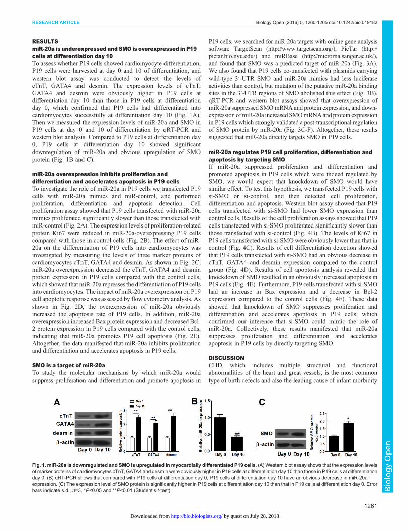
RESULTSmiR-20a is underexpressed and SMO is overexpressed in P19cells at differentiation day 10To assess whether P19 cells showed cardiomyocyte differentiation,P19 cells were harvested at day 0 and 10 of differentiation, andwestern blot assay was conducted to detect the levels ofcTnT, GATA4 and desmin. The expression levels of cTnT,GATA4 and desmin were obviously higher in P19 cells atdifferentiation day 10 than those in P19 cells at differentiationday 0, which confirmed that P19 cells had differentiated intocardiomyocytes successfully at differentiation day 10 (Fig. 1A).Then we measured the expression levels of miR-20a and SMO inP19 cells at day 0 and 10 of differentiation by qRT-PCR andwestern blot analysis. Compared to P19 cells at differentiation day0, P19 cells at differentiation day 10 showed significantdownregulation of miR-20a and obvious upregulation of SMOprotein (Fig. 1B and C).
miR-20a overexpression inhibits proliferation anddifferentiation and accelerates apoptosis in P19 cellsTo investigate the role of miR-20a in P19 cells we transfected P19cells with miR-20a mimics and miR-control, and performedproliferation, differentiation and apoptosis detection. Cellproliferation assay showed that P19 cells transfected with miR-20amimics proliferated significantly slower than those transfected withmiR-control (Fig. 2A). The expression levels of proliferation-relatedprotein Ki67 were reduced in miR-20a-overexpressing P19 cellscompared with those in control cells (Fig. 2B). The effect of miR-20a on the differentiation of P19 cells into cardiomyocytes wasinvestigated by measuring the levels of three marker proteins ofcardiomyocytes cTnT, GATA4 and desmin. As shown in Fig. 2C,miR-20a overexpression decreased the cTnT, GATA4 and desminprotein expression in P19 cells compared with the control cells,which showed that miR-20a represses the differentiation of P19 cellsinto cardiomyocytes. The impact of miR-20a overexpression on P19cell apoptotic response was assessed by flow cytometry analysis. Asshown in Fig. 2D, the overexpression of miR-20a obviouslyincreased the apoptosis rate of P19 cells. In addition, miR-20aoverexpression increased Bax protein expression and decreased Bcl-2 protein expression in P19 cells compared with the control cells,indicating that miR-20a promotes P19 cell apoptosis (Fig. 2E).Altogether, the data manifested that miR-20a inhibits proliferationand differentiation and accelerates apoptosis in P19 cells.
SMO is a target of miR-20aTo study the molecular mechanisms by which miR-20a wouldsuppress proliferation and differentiation and promote apoptosis in
P19 cells, we searched for miR-20a targets with online gene analysissoftware TargetScan (http://www.targetscan.org/), PicTar (http://pictar.bio.nyu.edu/) and miRBase (http://microrna.sanger.ac.uk/),and found that SMO was a predicted target of miR-20a (Fig. 3A).We also found that P19 cells co-transfected with plasmids carryingwild-type 3′-UTR SMO and miR-20a mimics had less luciferaseactivities than control, but mutation of the putative miR-20a bindingsites in the 3′-UTR regions of SMO abolished this effect (Fig. 3B).qRT-PCR and western blot assays showed that overexpression ofmiR-20a suppressed SMOmRNA and protein expression, and down-expression ofmiR-20a increased SMOmRNAand protein expressionin P19 cells which strongly validated a post-transcriptional regulationof SMO protein by miR-20a (Fig. 3C-F). Altogether, these resultssuggested that miR-20a directly targets SMO in P19 cells.
miR-20a regulates P19 cell proliferation, differentiation andapoptosis by targeting SMOIf miR-20a suppressed proliferation and differentiation andpromoted apoptosis in P19 cells which were indeed regulated bySMO, we would expect that knockdown of SMO would havesimilar effect. To test this hypothesis, we transfected P19 cells withsi-SMO or si-control, and then detected cell proliferation,differentiation and apoptosis. Western blot assay showed that P19cells transfected with si-SMO had lower SMO expression thancontrol cells. Results of the cell proliferation assays showed that P19cells transfected with si-SMO proliferated significantly slower thanthose transfected with si-control (Fig. 4B). The levels of Ki67 inP19 cells transfected with si-SMOwere obviously lower than that incontrol (Fig. 4C). Results of cell differentiation detection showedthat P19 cells transfected with si-SMO had an obvious decrease incTnT, GATA4 and desmin expression compared to the controlgroup (Fig. 4D). Results of cell apoptosis analysis revealed thatknockdown of SMO resulted in an obviously increased apoptosis inP19 cells (Fig. 4E). Furthermore, P19 cells transfected with si-SMOhad an increase in Bax expression and a decrease in Bcl-2expression compared to the control cells (Fig. 4F). These datashowed that knockdown of SMO suppresses proliferation anddifferentiation and accelerates apoptosis in P19 cells, whichconfirmed our inference that si-SMO could mimic the role ofmiR-20a. Collectively, these results manifested that miR-20asuppresses proliferation and differentiation and acceleratesapoptosis in P19 cells by directly targeting SMO.
DISCUSSIONCHD, which includes multiple structural and functionalabnormalities of the heart and great vessels, is the most commontype of birth defects and also the leading cause of infant morbidity
Fig. 1. miR-20a is downregulated and SMO is upregulated in myocardially differentiated P19 cells. (A) Western blot assay shows that the expression levelsof marker proteins of cardiomyocytes cTnT, GATA4 and desmin were obviously higher in P19 cells at differentiation day 10 than those in P19 cells at differentiationday 0. (B) qRT-PCR shows that compared with P19 cells at differentiation day 0, P19 cells at differentiation day 10 have an obvious decrease in miR-20aexpression. (C) The expression level of SMO protein is significantly higher in P19 cells at differentiation day 10 than that in P19 cells at differentiation day 0. Errorbars indicate s.d., n=3. *P<0.05 and **P<0.01 (Student’s t-test).
1261
RESEARCH ARTICLE Biology Open (2016) 5, 1260-1265 doi:10.1242/bio.019182
BiologyOpen
by guest on July 28, 2018http://bio.biologists.org/Downloaded from

and mortality (Capozzi et al., 2008; Eleftheriades et al., 2012).miRNAs, widely distributed in animals and land plants, post-transcriptionally modulate gene expression and participate invarious biological processes, including development, cellsurvival, proliferation, and differentiation (Axtell and Bowman,
2008; Bartel, 2004). Computational predictions indicate that 60% ofprotein-encoding genes are modulated by miRNAs in humans(Friedman et al., 2009). Of these miRNAs, many have been found tobe associated to cardiogenesis and act as new biomarkers andpotential targets for CHD. For instance, miR-19b overexpression
Fig. 3. SMO is a direct target of miR-20a.(A) Bioinformatics-based target prediction analysis showsthat SMO is a potential target of miR-20a and thebinding site is on the 3′-UTR of SMO. Black lines indicatethe predicted binding sites. (B) P19 cells co-transfectedwith plasmid containing 3′-UTR-WT regions of SMO andmiR-20a mimics have significantly decreased luciferaseactivity compared with control, while mutation of theputative miR-20a binding sites in the 3′-UTR regions ofSMO abolishes this effect. (C,D) Overexpression ofmiR-20a inhibits SMO mRNA and protein expression inP19 cells. (E,F) Downregulation of miR-20a enhancesSMOmRNA and protein expression in P19 cells. *P<0.05,Error bars indicate s.d., n=3. **P<0.01 and ***P<0.001(Student’s t-test).
Fig. 2. miR-20a overexpression suppresses proliferation and differentiation and promotes apoptosis in P19 cells. (A) P19 cells transfected with miR-20amimics proliferate significantly slower than those transfected with miR-control. Error bars indicate s.e.m. (n=3). (B) The expression level of Ki67 protein isobviously decreased in miR-20a-overexpressing P19 cells compared with that in control cells. Error bars indicate s.d. (n=3). (C) P19 cells transfected withmiR-20a mimics have obvious decreases in cTnT, GATA4 and desmin protein expression compared with the control cells. Error bars indicate s.d. (n=3). (D) P19cells transfected with miR-20a mimic have significantly higher apoptosis rate than the control cells. Error bars indicate s.d. (n=3). (E) P19 cells transfectedwith miR-20a mimics have a marked increase in Bax protein expression and an obvious decrease in Bcl-2 protein expression compared with the control cells.Error bars indicate s.d., n=3. *P<0.05, **P<0.01 and ***P<0.001 (Student’s t-test).
1262
RESEARCH ARTICLE Biology Open (2016) 5, 1260-1265 doi:10.1242/bio.019182
BiologyOpen
by guest on July 28, 2018http://bio.biologists.org/Downloaded from

promotes proliferation and differentiation but suppresses apoptosisin P19 cells by inactivating the Wnt/β-catenin pathway, andmiR-20b overexpression enhances apoptosis and promotesdifferentiation in P19 cells through activating the BMP pathway(Qin, 2013; Zhu, 2015). These findings suggest that miR-19b andmiR-20b may represent potential targets for CHD treatment andprovide novel insights into the molecular mechanisms underlyingheart diseases. In order to find new therapeutic targets for CHDtreatment and obtain a better understanding of the mechanismsunderlying cardiac diseases, more studies are needed to assess theroles of miRNAs in cardiogenesis.Some reports have showed that themiR-17-92 cluster, containing 6
mature miRNAs (miR-17, miR-18a, miR-19a, miR-19b-1, miR-20a,and miR-92-1), plays important roles in the development of lung,heart, blood and vessel. For example, (Lu et al., 2007) reported thatupregulation of miR-17-92 enhances proliferation and suppressesdifferentiation of lung epithelial progenitor cells. Ventura et al.documented that the mice lacking miR-17-92 have severelyventricular septal defects in the hearts, thereby leading to smallerembryos and postnatal death, which suggests an important function ofthis cluster during cardiac development (Ventura et al., 2008).Moreover, miR-18 and miR-19 modulate CTGF and TSP-1expression and the two proteins are important to healthy cardiacageing, which suggests that thesemiRNAs are involved in age-relatedcardiac remodelling (van Almen et al., 2011). miR-20a, as a memberof the miR-17-92 cluster, was reported to have been dysregulated inheart failure patients treated with cardiac resynchronization therapyand inhibit stress-induced cardiomyocyte apoptosis by targetingEgln3/PHD3 (Marfella et al., 2013; Frank et al., 2012). Recently,miR-20a is obviously downregulated in myocardially differentiatedP19 cells compared with normal P19 cells and may be functionally
related to cardiogenic processes, though its precise role remainselusive.
In the present study, we used P19 cells as a myocardial cellmodel to establish a miR-20a overexpression cell line andinvestigated the roles of miR-20a in cardiac differentiation. Wefound that miR-20a upregulation suppresses proliferation anddifferentiation and promotes apoptosis in P19 cells, and studiedthe molecular mechanisms by which miR-20a overexpressionhas the same effects. With the application of bioinformaticpredictions, we identified several potential binding targets of miR-20a, and chose SMO for further study. SMO is a transmembraneprotein that can activate the downstream Hh pathway. Severalrecent studies have documented that some small syntheticinhibitors suppress the Hh signalling pathway by specificallybinding to SMO or antagonizing the activity of SMO (Chen et al.,2002a,b; Frank-Kamenetsky et al., 2002). The Hh signallingpathway is a primary embryonic signalling cascade that modulatesstem cell and progenitor cell differentiation in a great manydevelopmental processes, and can induce cardiomyogenesis inP19 cells (Gianakopoulos and Skerjanc, 2005; Ingham andPlaczek, 2006). Dual-luciferase assay verified that miR-20atargets SMO directly and the expression levels of SMO mRNAand protein were modulated by miR-20a. Although some targetsof miRNA-20a are enriched in cell cycle regulating genes,knockdown of SMO suppressed proliferation and differentiationand enhanced apoptosis in P19 cells, which could mimic theeffect of miR-20a overexpression. All the data suggested that miR-20a inhibits proliferation and differentiation and promotesapoptosis in P19 cells at least partly by directly targeting SMO.
In summary, downregulation of miR-20a and upregulation ofSMO exist in myocardially differentiated P19 cells. We provided
Fig. 4. Knockdown of SMO suppresses proliferation and differentiation and promotes apoptosis in P19 cells. (A) P19 cells transfected with si-SMOhad lower SMOprotein expression than control cells. Error bars indicate s.d. (n=3). (B) P19 cells transfected with si-SMO proliferate significantly slower than thosetransfected with si-control. Error bars indicate s.e.m. (n=3). (C) The expression level of Ki67 in P19 cells transfected with si-SMO is obviously lower than that incontrol. Error bars indicate s.d. (n=3). (D) P19 cells transfected with si-SMO have obvious decreases in cTnT, GATA4 and desmin protein expressioncompared with the control cells. Error bars indicate s.d. (n=3). (E) Knockdown of SMO results in an obviously increased apoptosis in P19 cells. Error bars indicates.d. (n=3). (F) P19 cells transfected with si-SMO have a marked increase in Bax expression and an obvious decrease in Bcl-2 expression compared with thecontrol cells. Error bars indicate s.d., n=3. *P<0.05, **P<0.01 and ***P<0.001 (Student’s t-test).
1263
RESEARCH ARTICLE Biology Open (2016) 5, 1260-1265 doi:10.1242/bio.019182
BiologyOpen
by guest on July 28, 2018http://bio.biologists.org/Downloaded from

evidence to verify that SMO is a binding target of miR-20a, andmiR-20a modulates proliferation, differentiation and apoptosis ofP19 cells by targeting SMO. These data may provide novel insightsinto the regulatory mechanisms underlying heart diseases.Moreover, miR-20a may be a possible target for the treatment ofCHD. Consequently, the next step is to study whether dysregulationof miR-20a contributes to CHD in vivo.
MATERIALS AND METHODSCell culture and induction of differentiationP19 cells were obtained from the American type culture collection(ATCC; Manassas, VA, USA). Cells were cultured in α-minimal essentialmedium (α-MEM; Invitrogen, Melbourne, VIC, Australia) supplementedwith 10% fetal bovine serum (FBS; Gibco BRL, Grand Island, NY, USA),100 µg/ml streptomycin, and 100 U/ml penicillin in a humidifiedincubator containing 5% CO2 at 37°C. To induce cardiac differentiation,cells were cultivated as aggregates for 4 days in 1% α-MEM containing10% FBS and 1% dimethyl sulfoxide (Sigma-Aldrich, St. Louis, MO,USA) on bacteriologic dishes in a humidified incubator containing 5%CO2 at 37°C. The medium was replaced every 2 days thereafter. On day 4,the cell aggregates were transferred to cell culture flasks, and on day 10 ofdifferentiation, cells were harvested. The levels of marker proteins ofcardiomyocytes, such as cTnT, GATA4 and desmin were detected bywestern blot assay.
Quantitative real-time polymerase chain reaction (qRT-PCR)Total RNA samples were extracted from cultured cells using Trizol reagent(Invitrogen, Carlsbad, CA, USA) according to the manufacturer’s protocol,and reverse transcribed into cDNA by using the AMV reverse transcriptasekit (Promega A3500; Madison, WI, USA). miR-20a expression wasdetermined by TaqMan miRNA assays (Ambion, Foster City, CA, USA),and U6 (Applied Biosystems, Foster City, CA, USA) was used as an internalcontrol. The expression of SMOmRNAwas determined by PrimeScript RT-PCR kits (Takara Biochemicals, Kyoto, Japan), and β-actin was used as aninternal control.
Western blot assayAnti-cTnT, anti-β-actin and anti-Ki67 antibodies were purchased from SantaCruz Biotechnology (Santa Cruz, CA, USA). Anti-GATA4 and anti-desminwere bought from Cell Signaling Technology (Danvers, MA, USA). Anti-SMO, anti-Bax and anti-Bcl-2 antibodies were purchased from Abcam(Cambridge, MA, USA). Cells were added into tubes containing lysis buffer(50 mmol/l Tris-HCl, 0.2% sodium deoxycholate, 0.2% SDS, 1% TritonX-100, and 1 mmol/l EDTA at pH 7.4). The lysate supernatant was collectedafter centrifugation at 14,000 g for 30 min at 4°C. Subsequently, proteinconcentrations were determined using a BCA protein detection kit (KeygenBiotech. Co. Ltd., Nanjing, China) according to the manufacturer’sinstructions. Western blot assay was performed as previously described(Shen et al., 2012).
Cell transfectionmiR-20a mimics, miR-20a inhibitors, small-interfering RNAs (siRNA)targeting SMO (si-SMO) and their respective controls were obtained fromGenePharma (Shanghai GenePharma Co. Ltd., China). P19 cells inexponential growth were plated at a density of 3×10⁵ cells/plate andincubated for 24 h, and then transfected with miR-20a mimics, miR-control,si-SMO or si-control at a 100 nM concentration by using Lipofectamine-2000 (Invitrogen) according to the manufacturer’s instructions.
CCK-8 assayCells with 3×10⁴ cells per well were seeded in 96-well plates and cultured inα-MEM containing 10% FBS for 24 h until they were adherent. Cell growthwas monitored every day for a period of 5 days, and the proliferation ratewasassessed by cell counting Kit-8 (CCK-8; Peninsula Labs, Belmont, CA,USA). Briefly, 5 µl of CCK-8 solution was added to eachwell, and the plateswere further incubated for 2 h. Then the absorbance at 450 and 650 nm was
measured by using an ELISA reader, and the differences between theabsorbance values were recorded as the optical density.
Flow cytometry analysisCells were harvested using trypsin/EDTA, washed in PBS, resuspended in1×binding buffer at a concentration of 1×10⁶ cells/ml, and stained with 5 µlannexin V-FITC and 5 µl PI at room temperature for 15 min (BDPharmingen, San Diego, CA, USA). The FITC and PI fluorescent signalswere measured by flow cytometry (BD Biosciences, San Jose, CA, USA).
Target prediction analysisBioinformatics-based target prediction analysis was performed usingavailable bioinformatics logarithms on-line TargetScan 7.0 (http://www.targetscan.org/), PicTar (http://pictar.bio.nyu.edu), and miRBase (http://microrna.sanger.ac.uk/).
Luciferase reporter assayThe fragment of wild-type SMO 3′-UTR (3′-UTR-WT) containing putativemiR-20a binding sites was amplified by PCR, and mutant SMO 3′-UTR(3′-UTR-MUT) was generated by mutating the conserved miR-20a bindingsites using an overlap-extension PCR method. The fragment containing3′-UTR-WT or 3′-UTR-MUT regions of SMO was inserted intopsiCHECK-2 vectors (GenePharma, Shanghai, China) containing bothrenilla and firefly luciferase reporter genes. Subsequently, the psiCHECK-2vectors which contained wild-type or mutant 3′-UTR sequences of SMOwere transfected into miR-20a-overexpressing cells. After 24 h, theluciferase activities of firefly and renilla were assessed using dualluciferase reporter assay system (Promega) in accordance with themanufacturer’s protocol.
Statistical analysisThe GraphPad Prism 5 software (GraphPad Software, San Diego, CA,USA; http://www.graphpad.com) was used for all statistical analyses. Datawere expressed as mean±standard error (s.e.m.) or standard deviation(s.d.). The difference between groups was examined by Student’s t-test.Values of *P<0.05, **P<0.01 and ***P<0.001 were consideredstatistically significant.
Competing interestsThe authors declare no competing or financial interests.
Author contributionsB. P. designed the experiments, wrote the main manuscript text, and reviewed therevised manuscript. F. A. and Y. Z. revised the manuscript. F. A. analyzed the data.All authors conducted the experiments and read the manuscript.
FundingThis research received no specific grant from any funding agency in the public,commercial or not-for-profit sectors.
ReferencesAxtell, M. J. and Bowman, J. L. (2008). Evolution of plant microRNAs and their
targets. Trends Plant Sci. 13, 343-349.Bajolle, F., Zaffran, S. and Bonnet, D. (2009). Genetics and embryological
mechanisms of congenital heart diseases. Arch. Cardiovasc. Dis. 102, 59-63.Bartel, D. P. (2004). MicroRNAs: genomics, biogenesis, mechanism, and function.
Cell 116, 281-297.Capozzi, G., Caputo, S., Pizzuti, R., Martina, L., Santoro, M., Santoro, G.,
Sarubbi, B., Iacono, C., D’Alto, M. and Bigazzi, M. C. (2008). Congenital heartdisease in live-born children: incidence, distribution, and yearly changes in theCampania Region. J. Cardiovasc. Med. 9, 368-374.
Catalucci, D., Latronico, M. V. G. and Condorelli, G. (2008). MicroRNAs controlgene expression. Ann. N. Y. Acad. Sci. 1123, 20-29.
Chen, J. K., Taipale, J., Cooper, M. K. and Beachy, P. A. (2002a). Inhibition ofHedgehog signaling by direct binding of cyclopamine to Smoothened. GenesDev. 16, 2743-2748.
Chen, J. K., Taipale, J., Young, K. E., Maiti, T. and Beachy, P. A. (2002b). Smallmolecule modulation of Smoothened activity. Proc. Natl. Acad. Sci. USA 99,14071-14076.
Eleftheriades, M., Tsapakis, E., Sotiriadis, A., Manolakos, E., Hassiakos, D. andBotsis, D. (2012). Detection of congenital heart defects throughout pregnancy;
1264
RESEARCH ARTICLE Biology Open (2016) 5, 1260-1265 doi:10.1242/bio.019182
BiologyOpen
by guest on July 28, 2018http://bio.biologists.org/Downloaded from

impact of first trimester ultrasound screening for cardiac abnormalities. J. Matern.Fetal Neonatal Med. 25, 2546-2550.
Frank, D., Gantenberg, J., Boomgaarden, I., Kuhn, C., Will, R., Jarr, K.-U., Eden,M., Kramer, K., Luedde, M., Mairbaurl, H. et al. (2012). MicroRNA-20a inhibitsstress-induced cardiomyocyte apoptosis involving its novel target Egln3/PHD3.J. Mol. Cell Cardiol. 52, 711-717.
Frank-Kamenetsky, M., Zhang, X. M., Bottega, S., Guicherit, O., Wichterle, H.,Dudek, H., Bumcrot, D., Wang, F. Y., Jones, S. and Shulok, J. (2002). Small-molecule modulators of Hedgehog signaling: identification and characterization ofSmoothened agonists and antagonists. J. Biol. 1, 10.
Friedman, R. C., Farh, K. K.-H., Burge, C. B. and Bartel, D. P. (2009). Mostmammalian mRNAs are conserved targets of microRNAs. Genome Res. 19,92-105.
Gianakopoulos, P. J. and Skerjanc, I. S. (2005). Hedgehog signaling inducescardiomyogenesis in P19 cells. J. Biol. Chem. 280, 21022-21028.
Hoffman, J. (1995). Incidence of congenital heart disease: II. Prenatal incidence.Pediatr. Cardiol. 16, 155-165.
Hoffman, J. I. E. and Kaplan, S. (2002). The incidence of congenital heart disease.J. Am. Coll. Cardiol. 39, 1890-1900.
Hu, D. L., Liu, Y. Q., Chen, F. K., Sheng, Y. H., Yang, R., Kong, X. Q., Cao, K. J.,Zhang, J. S. and Qian, L. M. (2011). Differential expression of microRNAs incardiac myocytes compared to undifferentiated P19 cells. Int. J. Mol. Med. 28,59-64.
Ingham, P. W. and Placzek, M. (2006). Orchestrating ontogenesis: variations on atheme by sonic hedgehog. Nat. Rev. Genet. 7, 841-850.
Kehler, L., Biro, O., Lazar, L., Rigo, J. and Nagy, B. (2015). Elevated hsa-miR-99alevels in maternal plasma may indicate congenital heart defects. Biomed. Rep. 3,869-873.
Lu, Y., Thomson, J. M., Wong, H. Y. F., Hammond, S. M. and Hogan, B. L. M.(2007). Transgenic over-expression of the microRNAmiR-17-92 cluster promotesproliferation and inhibits differentiation of lung epithelial progenitor cells.Dev. Biol.310, 442-453.
Marfella, R, Di Filippo, C., Potenza, N., Sardu, C., Rizzo, M. R., Siniscalchi, M.,Musacchio, E., Barbieri, M., Mauro, C., Mosca, N. et al. (2013). CirculatingmicroRNA changes in heart failure patients treated with cardiac resynchronizationtherapy: responders vs. non-responders. Eur. J. Heart Fail. 15, 1277-1288.
Miyazono, K., Kamiya, Y. and Morikawa, M. (2010). Bone morphogenetic proteinreceptors and signal transduction. J. Biochem. 147, 35-51.
Qin, D.-N., Qian, L., Hu, D.-L., Yu, Z.-B., Han, S.-P., Zhu, C., Wang, X. and Hu, X.(2013). Effects of miR-19b overexpression on proliferation, differentiation,
apoptosis and Wnt/β-catenin signaling pathway in P19 cell model of cardiacdifferentiation in vitro. Cell Biochem. Biophys. 66, 709-722.
Shen, Y.-H., Song, G.-X., Liu, Y.-Q., Sun, W., Zhou, L.-J., Liu, H.-L., Yang, R.,Sheng, Y.-H., Qian, L.-M. and Kong, X.-Q. (2012). Silencing of FABP3 promotesapoptosis and induces mitochondrion impairment in embryonic carcinoma cells.J. Bioenerg. Biomemb. 44, 317-323.
Thum, T., Catalucci, D. and Bauersachs, J. (2008). MicroRNAs: novel regulatorsin cardiac development and disease. Cardiovasc. Res. 79, 562-570.
Trojnarska, O., Grajek, S., Katarzynski, S. and Kramer, L. (2009). Predictors ofmortality in adult patients with congenital heart disease. Cardiol. J. 16, 341-347.
van Almen, G. C., Verhesen, W., van Leeuwen, R. E. W., van de Vrie, M.,Eurlings, C., Schellings, M. W. M., Swinnen, M., Cleutjens, J. P. M., vanZandvoort, M. A. M. J. and Heymans, S. (2011). MicroRNA-18 and microRNA-19 regulate CTGF and TSP-1 expression in age-related heart failure. Aging Cell10, 769-779.
VanRooij, E. andOlson, E. N. (2007). MicroRNAs: powerful new regulators of heartdisease and provocative therapeutic targets. J. Clin. Investig. 117, 2369-2376.
Ventura, A., Young, A. G., Winslow, M. M., Lintault, L., Meissner, A., Erkeland,S. J., Newman, J., Bronson, R. T., Crowley, D. and Stone, J. R. (2008).Targeted deletion reveals essential and overlapping functions of the miR-17∼92family of miRNA clusters. Cell 132, 875-886.
Wienholds, E. and Plasterk, R. H. (2005). MicroRNA function in animaldevelopment. FEBS Lett. 579, 5911-5922.
Xu, J., Hu, Z., Xu, Z., Gu, H., Yi, L., Cao, H., Chen, J., Tian, T., Liang, J. and Lin, Y.(2009). Functional variant in microRNA-196a2 contributes to the susceptibility ofcongenital heart disease in a Chinese population. Hum. Mut. 30, 1231-1236.
Yang, B., Sun, H.-Y., Chen, W.-H., Wen, J.-L., Shi, X.-T. and Wang, Y.-M. (2010).Lentivirus-mediated SMO RNA interference inhibits SMO expression and cellproliferation, and affects the cell cycle in LNCaP and PC3 cancer cell lines. AsianJ. Androl. 12, 196.
Zaffran, S. and Frasch, M. (2002). Early signals in cardiac development. Circul.Res. 91, 457-469.
Zhao, Y., Ransom, J. F., Li, A., Vedantham, V., von Drehle, M., Muth, A. N.,Tsuchihashi, T., McManus, M. T., Schwartz, R. J. and Srivastava, D. (2007).Dysregulation of cardiogenesis, cardiac conduction, and cell cycle in mice lackingmiRNA-1-2. Cell 129, 303-317.
Zhu, S., Hu, X., Yu, Z., Peng, Y., Zhu, J., Liu, X., Li, M., Han, S. and Zhu, C. (2015).Effect of miR-20b on apoptosis, differentiation, the BMP signaling pathway andmitochondrial function in the P19 cell model of cardiac differentiation in vitro.PLoSONE 10, e0123519.
1265
RESEARCH ARTICLE Biology Open (2016) 5, 1260-1265 doi:10.1242/bio.019182
BiologyOpen
by guest on July 28, 2018http://bio.biologists.org/Downloaded from