minimal residual disease evaluation in hematologic ... residual disease evaluation in hematologic...
TRANSCRIPT

Minimal Residual Disease Evaluation in Hematologic
Malignancies
Anne Tierens, MD, PhD
University Health Network

My Disclosures
• None
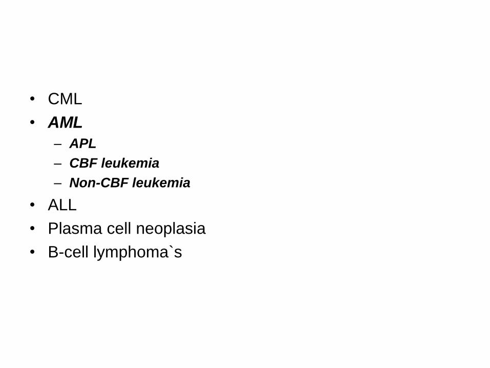
• CML
• AML
– APL
– CBF leukemia
– Non-CBF leukemia
• ALL
• Plasma cell neoplasia
• B-cell lymphoma`s

• Background
• Methodologies
• Clinical significance
• Conclusions and perspectives
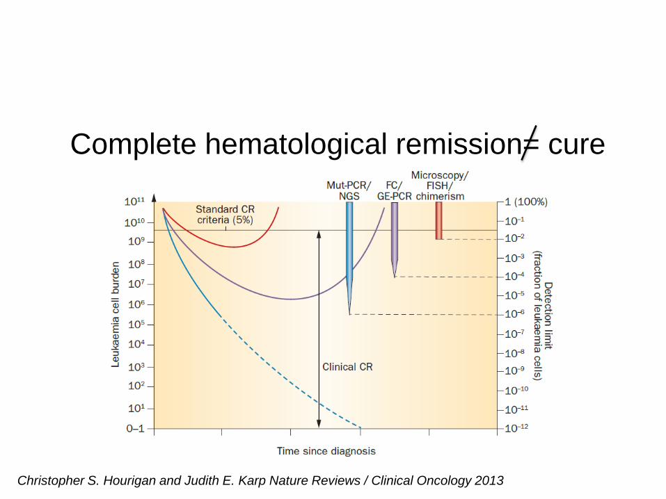
Complete hematological remission= cure
Christopher S. Hourigan and Judith E. Karp Nature Reviews / Clinical Oncology 2013
• Multiparameter Flow Cytometry (MFC)
• PCR
– Fusion transcripts
– Gene mutations
– Gene overexpression
CS Hourigan and JE Karp Nature reviews, Clinical Oncology 2013 (review)
D Grimwade and S Freeman. Blood 2014 prepublished July, 21 (review)

Proportion of AML patients informative for MRD detection
by RT-qPCR for leukemia-specific targets according to age
D Grimwade and S Freeman. Blood 2014 (prepublished July, 21)

Multiparameter Flow cytometry
• Leukemia-associated immunophenotypes
(LAPs)
• Different from normal
• Leukemia stem cells
MF Greaves. Br J Cancer 1975; LW Terstappen et al. Leukemia 1992; JF San Miguel. Blood 1997;
MA Macedo et al. Ann Hematol 1995; A Al-Mawali. Am J Clin Pathol 2008;
C Langebrake et al. Cytometry 2005; EL Sievers et al. Blood 2003; MR. Loken Blood 2012
Van Rhenen et al Leukemia 2008; JM Gerber et al Blood 2012
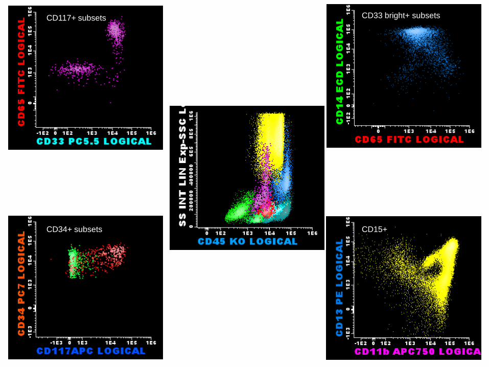
CD117+ subsets
CD34+ subsets
CD33 bright+ subsets
CD15+

Identification of aberrant marker expression
on AML blasts
Core markers
CD34+ cells
CD117+/CD34- cells
IP changes:
1. CD13 bright expression
2. CD7 coexpression
NPM1+ AML case 1: Diagnostic sample

CD34+ cells CD117+/CD34- cells
NPM1 AML case 1: post induction sample
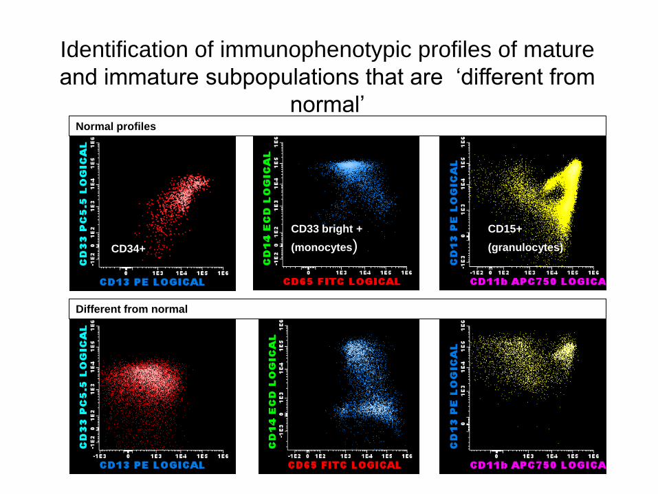
Identification of immunophenotypic profiles of mature
and immature subpopulations that are ‘different from
normal’
CD34+
CD33 bright +
(monocytes)) CD15+
(granulocytes))
Normal profiles
Different from normal
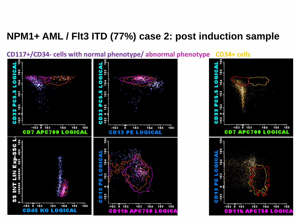
CD117+/CD34- cells with normal phenotype/ abnormal phenotype CD34+ cells
NPM1+ AML / Flt3 ITD (77%) case 2: post induction sample

Leukemia stem cells (CD34+/CD38-)
Leukemic stem cells:
12.5 % of leukemic blasts
3.5% of total nucleated cells
Aberrant phenotype:
HLA-DR negative
CD7 positive
CD123 positive
Remaining issues
• Evolving antibody panels
• Quantitation of MFC-MRD
• Definition of MRD positivity / negativity
D Grimwade and S Freeman. Blood 2014 prepublished July, 21 (review)
JM Jaso, SA Wang, JL Jorgensen and P Lin. Bone Marrow Transplantation 2014 (review)
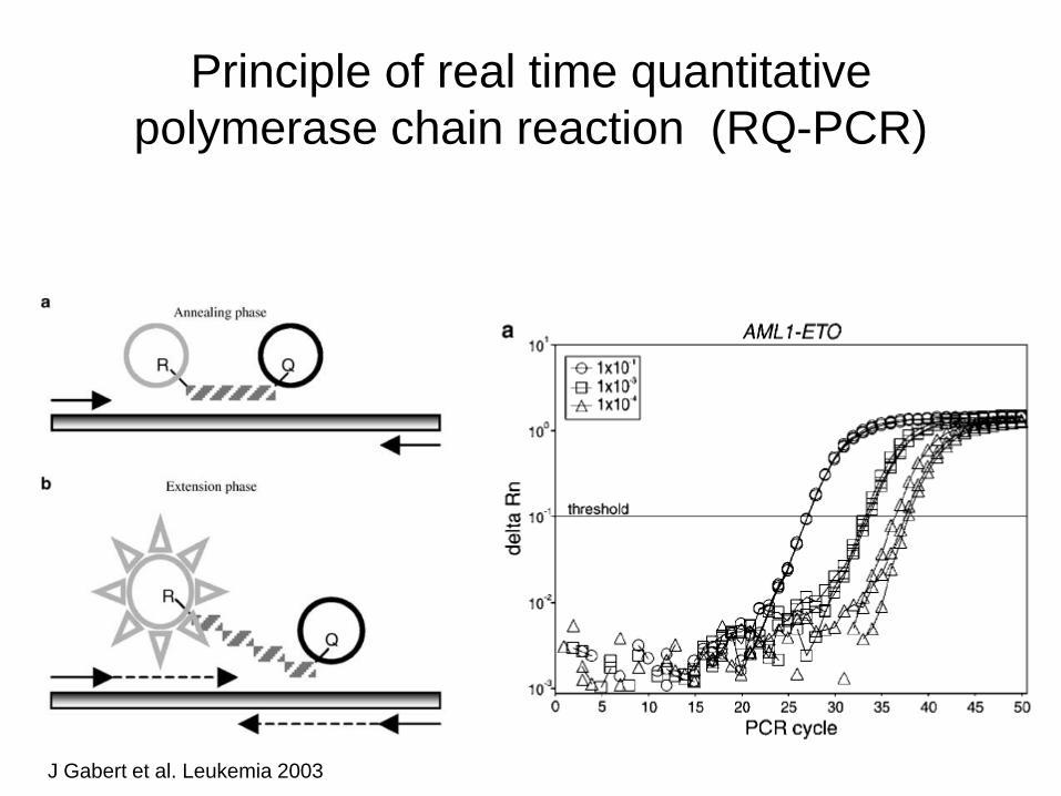
Principle of real time quantitative
polymerase chain reaction (RQ-PCR)
J Gabert et al. Leukemia 2003
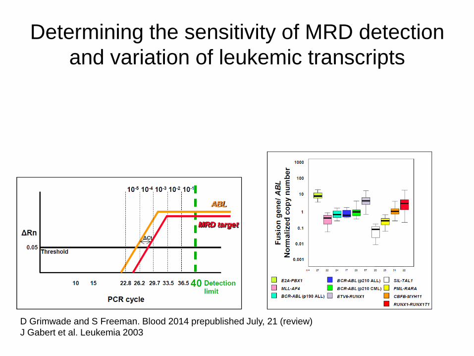
Determining the sensitivity of MRD detection
and variation of leukemic transcripts
D Grimwade and S Freeman. Blood 2014 prepublished July, 21 (review)
J Gabert et al. Leukemia 2003

Monitoring PML-RARA fusion transcripts:
standard of care in APL
* Bone marrow is recommended sample
* Required assay sensitivity: 10-4
* Low-risk disease (WBC < 10 X 10e9/L): MRD monitoring until end of treatment
* High-risk disease (WBC > 10 X 10e9/L and slow kinetics): 3 monthly MRD monitoring
* MRD monitoring in clinical trials to assess safety and efficacy of de-intensified treatment
(ongoing study: I-BFM group / childhood APL)
D Grimwade et al. J Clin Oncol 2009
* MRD is the most powerful predictor of disease relapse
(p < 00001; 406 patients included in MRC AML 15 trial)
* Initiation of pre-emptive therapy on reappearance of
PCR positivity

Minimal residual disease can identify CBF
leukemia patients at risk for relapse No of
patients
Time points
Cut- off levels
Transcripts
MRD response and Clinical relevance
J Liu Yin et al. Blood 2012
(MRC AML-15 trial)
163 pts with t(8;21)
115 pts with inv(16)
After induction course 1
After therapy
Serial monitoring
RUNX1-RUNX1T1: > 3-log
reduction (BM samples)
CBF beta-MYH11: < 10 copies
(PB samples)
RUNX1-RUNX1T1: > 500
copies (BM) and 100 copies
(PBL)
CBFbeta-MYH11: > 50 copies
(BM) and > 10 copies (PBL)
47% of pts: CIR of 4%
51% of pts: CIR of 21%
Predicts Relapse
Predicts Relapse
Median time from PCR positivity to hematological relapse:
4.9 months (BM) and 4.5 months (PBL) for t(8;21) AML pts
3 months (BM and PBL) for inv(16) AML pts
E Jourdan et al. Blood 2013
(The French AML intergroup)
96 pts with t(8;21)
102 pts with inv(16)
After 2nd course (MRD2)
> 3-log reduction of
RUNX1-RUNX1T1 and
CBFbeta-MYH11 transcripts
(BM samples)
71/91pts (78%) with t(8;21) and 53/85 pts (62%) with inv(16)
Associated with lower hazard of relapse (SHR=0.27), longer
RFS (HR=0.34) and trend for longer OS (HR= 0.46)
MRD is independent risk factor by multivariate analysis
(not c-Kit and/ or Flt3 ITD mutation)
HH Zhu et al. Blood 2013
(AML05 multicenter trial)
116 MRD eligible pts
with t(8;21)
After 2nd consolidation
> 3-log reduction of RUNX1-
RUNX1T transcripts = major
molecular response (MMR)
(BM samples)
Non- MMR (HR)= 69/116 pts (59%)
MMR (LR)= 47/116 pts (40%)
Non-MMR: 40 pts received HSCT and 29 pts CT: :CIR of
22% versus 78.9%
MRD, treatment strategy and c-KIT mutation status:
Independent risk factors by multivariate analysis
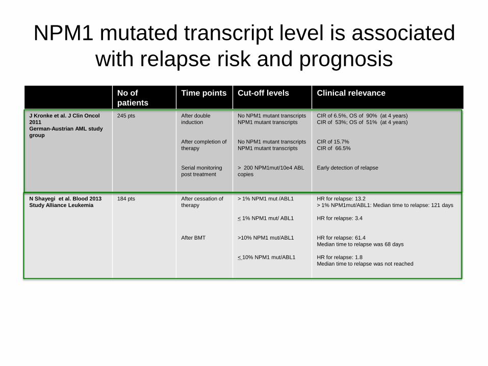
NPM1 mutated transcript level is associated
with relapse risk and prognosis
No of
patients
Time points
Cut-off levels
Clinical relevance
J Kronke et al. J Clin Oncol
2011
German-Austrian AML study
group
245 pts After double
induction
After completion of
therapy
Serial monitoring
post treatment
No NPM1 mutant transcripts
NPM1 mutant transcripts
No NPM1 mutant transcripts
NPM1 mutant transcripts
> 200 NPM1mut/10e4 ABL
copies
CIR of 6.5%, OS of 90% (at 4 years)
CIR of 53%; OS of 51% (at 4 years)
CIR of 15.7%
CIR of 66.5%
Early detection of relapse
N Shayegi et al. Blood 2013
Study Alliance Leukemia
184 pts
After cessation of
therapy
After BMT
> 1% NPM1 mut /ABL1
< 1% NPM1 mut/ ABL1
>10% NPM1 mut/ABL1
< 10% NPM1 mut/ABL1
HR for relapse: 13.2
> 1% NPM1mut/ABL1: Median time to relapse: 121 days
HR for relapse: 3.4
HR for relapse: 61.4
Median time to relapse was 68 days
HR for relapse: 1.8
Median time to relapse was not reached

Kinetics of Wilms` Tumor gene (WT-1)
expression post induction provides prognostic
information
* WT-1 is not leukemia specific: 50 and 250 WT-1 copies/10e4 ABL copies are the cut-off
levels for normal Blood and Bone Marrow, respectively
* Assay sensitivity: 2- log reduction in ca 45% of patients
* D. Gilloni at al. (JCO 2009) ELN study
Standardized WT-1 assay including 91
Pts with pretreatment WT-1 level of
> 2x10⁴ WT1 copies/ 104 ABL
copies
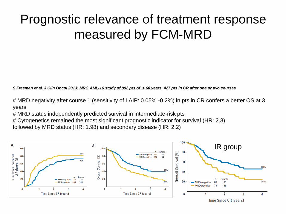
Prognostic relevance of treatment response
measured by FCM-MRD
S Freeman et al. J Clin Oncol 2013: MRC AML-16 study of 892 pts of > 60 years, 427 pts in CR after one or two courses
# MRD negativity after course 1 (sensitivity of LAIP: 0.05% -0.2%) in pts in CR confers a better OS at 3
years
# MRD status independently predicted survival in intermediate-risk pts
# Cytogenetics remained the most significant prognostic indicator for survival (HR: 2.3)
followed by MRD status (HR: 1.98) and secondary disease (HR: 2.2)
IR group

Prognostic relevance of treatment response
measured by FCM-MRD
IR group
M Terwijn et al. J Clin Oncol 2013: HOVON/SAKK study of 517 pts < 60 years; 389 pts available for analysis
# MRD > 0.1% after course 2 (23% of pts) was associated with Relapse of 72% at 4 years (HR: 2.97
(univariate analysis) and HR:2.6 (multivariate analysis)
Conclusions
• MRD is of prognostic significance and can be used for
risk stratification
• MRD (persistent high levels- rising levels) after front line
therapy predicts relapse
• Many patients without MRD relapse
Perspectives
• Studies are ongoing to investigate whether MRD assessment may be useful
to help make decisions concerning
1. Pre-emptive therapy after completion of therapy/ post transplant
2. Bone marrow transplantation in 1st remission
3. Treatment de-intensification
• Better understanding of the clonal architecture of AML is needed by targeted
sequencing of the diagnostic sample
• Implementation of newer methodologies such as next generation
sequencing / digital PCR
• Standardization and Further development of flow cytometry assays: mass
cytometry and automated analysis algorithms