mini-symposium thesynapticvesiclecyclerevisited ...€¦ · mini-symposium...
TRANSCRIPT

Mini-Symposium
The Synaptic Vesicle Cycle Revisited: New Insights into theModes and Mechanisms
X Natali L. Chanaday,1 X Michael A. Cousin,2 X Ira Milosevic,3 X Shigeki Watanabe,4 and X Jennifer R. Morgan5
1Department of Pharmacology, Vanderbilt University School of Medicine, Nashville, Tennessee 37232, 2Centre for Discovery Brain Sciences, University ofEdinburgh, Edinburgh, Scotland EH8 9XD, 3European Neuroscience Institute Gottingen, a Joint Initiative of the University Medical Center Gottingen andthe Max Planck Society, Gottingen, Germany 37077, 4Department of Cell Biology and Solomon H. Snyder Department of Neuroscience, Johns HopkinsSchool of Medicine, Baltimore, Maryland 21205, and 5Eugene Bell Center for Regenerative Biology and Tissue Engineering, Marine Biological Laboratory,Woods Hole, Massachusetts 02543
Neurotransmission is sustained by endocytosis and refilling of synaptic vesicles (SVs) locally within the presynapse. Until recently, aconsensus formed that after exocytosis, SVs are recovered by either fusion pore closure (kiss-and-run) or clathrin-mediated endocytosisdirectly from the plasma membrane. However, recent data have revealed that SV formation is more complex than previously envisaged.For example, two additional recycling pathways have been discovered, ultrafast endocytosis and activity-dependent bulk endocytosis, inwhich SVs are regenerated from the internalized membrane and synaptic endosomes. Furthermore, these diverse modes of endocytosisappear to influence both the molecular composition and subsequent physiological role of individual SVs. In addition, previously un-known complexity in SV refilling and reclustering has been revealed. This review presents a modern view of the SV life cycle and discusseshow neuronal subtype, physiological temperature, and individual activity patterns can recruit different endocytic modes to generate newSVs and sculpt subsequent presynaptic performance.
IntroductionDuring chemical neurotransmission, synaptic vesicles (SVs) fusewith the plasma membrane to release neurotransmitter. To sup-port high rates of release, synapses require a constant supply ofneurotransmitter-filled SVs. This supply is maintained primarilythrough membrane endocytosis, cargo sorting, and rapid refill-ing of newly formed SVs with neurotransmitter, which occurlocally within nerve terminals. This entire process of reconstitut-ing SVs after fusion is referred to as SV “recycling.” In contrast, denovo synthesis of SVs in the cell body and axonal transport tosynapses is usually too slow to support the high demand for neu-rotransmitter release. This review will focus mainly on the recy-cling aspect of the SV cycle. For mechanisms of exocytosis, pleaserefer to recent excellent reviews (Rizo and Xu, 2015; Chanadayand Kavalali, 2018a; Neher and Brose, 2018; Brunger et al., 2019;Dittman and Ryan, 2019).
Since the 1970s, the mechanisms by which SVs are recycledand trafficked at synapses have been intensely contested. On oneside, using the frog neuromuscular junction, Heuser and Reese(1973) reported that SVs are regenerated locally by the formation
of clathrin-coated vesicles at the periphery of active zones. Thus,they proposed that SV recycling occurs via clathrin-mediatedendocytosis (CME) from the plasma membrane (Fig. 1). Aroundthe same time, using similar experimental conditions, Ceccarelliet al. (1973) reported scant evidence of clathrin-coated vesicles atsynapses, but instead reported clear, uncoated vesicles potentiallybeing internalized at the active zone. They proposed that SVs canbe recycled by the reversal of an exocytic fusion pore, a model thatwas later termed “kiss-and-run” (Fig. 1) (Fesce et al., 1994). Thusensued a 40-year debate about how SVs are recycled and theunderlying mechanisms.
In the intervening decades, these models were further tested asnew molecular and imaging tools became available. However,instead of resolving the issue, two other models for SV recyclinghave emerged: activity-dependent bulk endocytosis (ADBE) andultrafast endocytosis. It is possible that all four mechanisms co-exist in nerve terminals, and are used differently depending onactivity levels or synapse type (Gan and Watanabe, 2018). Addingto the complexity is that SVs are functionally heterogeneous, de-fined by distinct molecular compositions (Crawford and Kava-lali, 2015). Thus, different recycling mechanisms may help to sortcargoes during SV reformation, ensuring that the proper molec-ular identity is maintained (Morgan et al., 2013). Additionally,recent data have revealed mechanistic insights into how newlyendocytosed SVs are refilled with neurotransmitter and subse-quently reclustered (Farsi et al., 2018; Milovanovic et al., 2018).In this review, we discuss these recent developments and, in do-ing so, present a modern view of the SV cycle with a specificemphasis on SV recycling mechanisms.
Received June 21, 2019; revised July 31, 2019; accepted Aug. 3, 2019.This work was supported by: Schram-Stiftung T287/25457 and Deutsche Forschungsgemeinschaft (Emmy
Noether Young Investigator Award MI-1702/1 to I.M.); the Wellcome Trust (204954/Z/16/Z to M.A.C.); the NationalScience Foundation (1727260 to S.W.), the National Institutes of Health (NINDS DP2 NS111133 and R01 NS105810 toS.W.); the McKnight Foundation (S.W.); the Sloan Foundation (S.W.); and the National Institutes of Health (NINDS/NIA R01 NS078165 to J.R.M. and NIMH R01 MH066198 to Dr. Ege Kavalali, which supports N.L.C.). We thank Drago-mir Milovanovic for helpful comments on this manuscript.
The authors declare no competing financial interests.Correspondence should be addressed to Jennifer R. Morgan at [email protected]://doi.org/10.1523/JNEUROSCI.1158-19.2019
Copyright © 2019 the authors
The Journal of Neuroscience, October 16, 2019 • 39(42):8209 – 8216 • 8209

SV heterogeneity and functional poolsAfter action potentials, SVs fuse andrelease neurotransmitter either in a time-locked or delayed manner. The time-locked, synchronous phase of releasetransmits fast and reliable signals, whiledelayed asynchronous release influencesnetwork parameters, including efficacy ofneurotransmission, synchronicity andplasticity (Otsu et al., 2004; Iremongerand Bains, 2016; Luo and Sudhof, 2017).In addition, SVs can fuse spontaneously inthe absence of action potentials, poten-tially affecting synapse formation and thestrength of the connection (Chanaday andKavalali, 2018a). Although not apparentfrom electron micrographs, increasing ev-idence suggests that visually identical SVs have different molecu-lar compositions, and this heterogeneity may underlie functionalorganization of vesicle pools that differentially participate in thethree phases of release (Chanaday and Kavalali, 2018a).
SVs are organized into four functional pools at presynapses:the readily releasable pool (RRP), recycling pool, reserve pool andresting pool. For synchronous neurotransmission, a subset of SVsare docked (physically in contact with the plasma membrane)and primed (ready-to-fuse upon calcium elevation) in a fusion-ready state at the active zone (Hammarlund et al., 2007; Sudhof,2013; Neher and Brose, 2018). Docked and primed SVs, locatednear the active zone, constitute the RRP that is immediately avail-able for fusion upon the arrival of action potentials (Holderith etal., 2012). To sustain neurotransmitter release, the RRP must beconstantly replenished with SVs (Guo et al., 2015) (Jonas, 2014).This replenishment can be accomplished by either rapid reuse offused vesicles or recruitment of new SVs from the “reserve pool”during prolonged periods of activity. Collectively, all SVs thatparticipate in activity-induced synaptic transmission comprisethe recycling pool (i.e., RRP and a fraction of the reserve pool)(Denker and Rizzoli, 2010), which is estimated to be �50% oftotal SVs (Kim and Ryan, 2010), but may be as few as 1–5%(Denker et al., 2011). The remaining SVs may be referred to as theresting pool because they are reluctant to be mobilized during/after stimulation (Chanaday and Kavalali, 2018a).
Within the recycling pool, distinct molecular machineries de-termine whether vesicles fuse synchronously or asynchronously.Synchronous fusion is mediated by the canonical neuronal Solu-ble NSF Attachment Protein Receptor (SNARE) complex, whichincludes the SV protein synaptobrevin 2 (syb-2/VAMP2) (Jahnand Fasshauer, 2012; Rizo and Sudhof, 2012) and the neuronalcalcium-sensing proteins synaptotagmins 1, 2, and 9. Synap-totagmins clamp the SNARE complex in the absence of an actionpotential and trigger synchronous fusion in response to localcalcium entry through voltage-gated calcium channels (Sudhof,2013). In contrast, asynchronous release is conferred by synap-totagmin 7 (Bacaj et al., 2013) or Doc2 (Yao et al., 2011) and bythe noncanonical SNARE VAMP4 (Raingo et al., 2012). Thesemolecular differences are likely maintained throughout the SVcycle. A recent report suggests that synaptotagmin 1 and 7 couplesynchronous and asynchronous release to a fast (1–2 s) or slow(several seconds) mode of endocytosis, respectively (Li et al.,2017). Moreover, the asynchronous SNARE VAMP4 is requiredfor ADBE after intense activity (Nicholson-Fish et al., 2015), dur-ing which asynchronous release becomes more prominent. Thus,
current evidence suggests that synchronous and asynchronousrelease is maintained through different SV recycling pathways.
SVs can also spontaneously fuse and recycle in the absence ofaction potentials (Kavalali, 2015). Although it is debated whetherspontaneous release draws from the recycling pool, resting pool,or its own pool (Sara et al., 2005; Fredj and Burrone, 2009), thepresence of noncanonical SNAREs VAMP4, VAMP7, and Vti1alikely defines whether particular vesicles fuse spontaneously(Ramirez and Kavalali, 2012; Bal et al., 2013; Chanaday and Kava-lali, 2018a). A number of calcium sensors have been proposed totrigger spontaneous neurotransmitter release, including theDoc2 family of proteins (Ramirez et al., 2017; Courtney et al.,2018). In addition to these molecular differences, endocytosis ofspontaneously fused SVs occurs at a faster timescale (�1 s) and ispartially calcium independent (Leitz and Kavalali, 2014), impli-cating a distinct mode of endocytosis. Thus, several modes ofendocytosis may maintain the SV supply at presynaptic terminalsto support distinct phases of neurotransmission.
Modes of SV recyclingAt present, at least four modes of SV recycling have been identi-fied, distinguished by their molecular mechanisms and speed:CME, kiss-and-run, ultrafast endocytosis, and ADBE (Fig. 2).
CME versus kiss-and-runOver the last two decades, many studies have focused on address-ing whether SVs are recycled via CME or kiss-and-run. The ki-netics and molecular requirements distinguish these two modesof endocytosis: CME is relatively slow (10 –30 s) and requires adistinct set of molecules (Fig. 2A) (Saheki and De Camilli, 2012;Milosevic, 2018). In contrast, kiss-and-run is fast (�1–2 s) anddoes not require clathrin-associated proteins. Thus, these fea-tures have been investigated extensively at model synapsesranging from those in invertebrates such as nematodes (Caeno-rhabditis elegans) (Nonet et al., 1999), fruit fly (Zhang et al., 1998;Heerssen et al., 2008), and squid (Morgan et al., 1999, 2000,2001), to vertebrates such as lampreys (Shupliakov et al., 1997;Walsh et al., 2018) and rodents (Granseth et al., 2006; Mani et al.,2007). At squid synapses, disrupting the functions of core clath-rin coat components, such as adaptor proteins (AP180, AP-2) orclathrin-uncoating proteins (Hsc70, auxilin) severely impairedneurotransmission, indicating an essential role for the clathrinpathway (Morgan et al., 1999, 2000, 2001). At mammalian nerveterminals, knock-down of clathrin-heavy chain suggested thatalmost all endocytosis is clathrin mediated (Granseth et al.,2006). A unified view from these studies is that SV recycling
kiss-and-run (K&R)?clathrin-mediated endocytosis (CME)
full-collapse fusion
active zonepostsynapse
Figure 1. Classical view of the SV cycle. Action potentials trigger fusion of SVs at the active zone. After formation of the fusionpore, resulting in neurotransmitter release, two options are possible: the pore can reclose via kiss-and-run (K&R) or it can expandirreversibly, leading to full collapse. Compensating for full-collapse fusion, specific SV proteins are recruited by adaptor proteins atthe periactive zone, triggering CME. Dynamin mediates vesicle scission, after which SVs are uncoated and refilled with neurotrans-mitters before being returned to the vesicle cluster.
8210 • J. Neurosci., October 16, 2019 • 39(42):8209 – 8216 Chanaday et al. • The Synaptic Vesicle Cycle Revisited
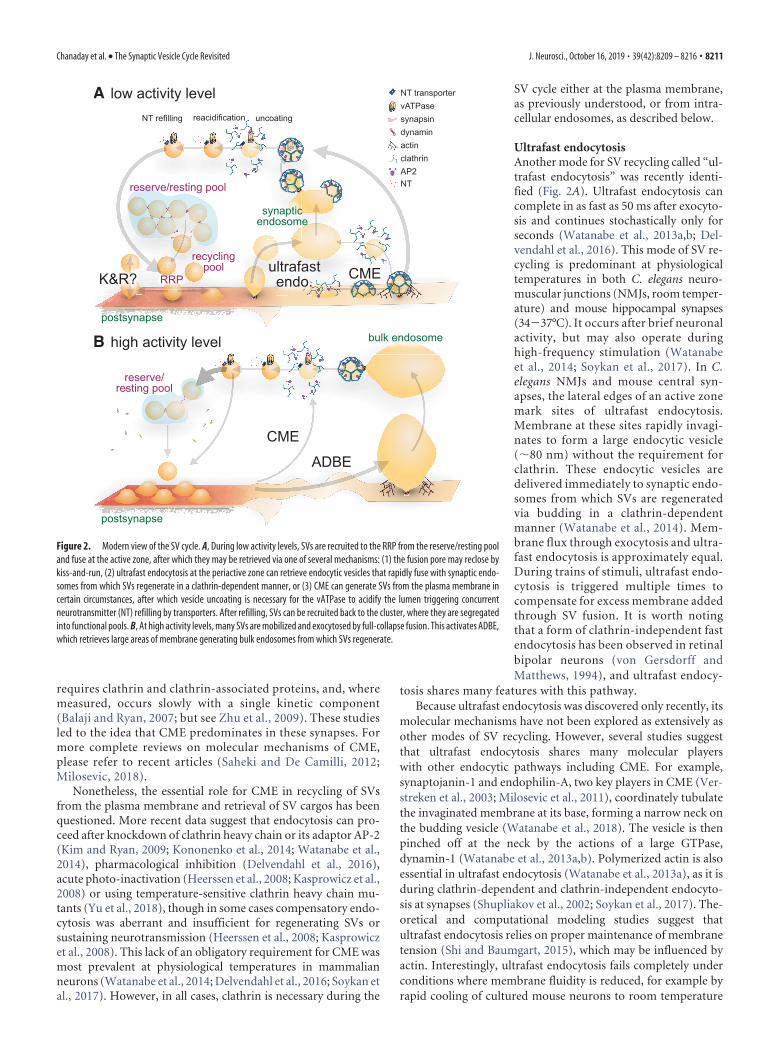
requires clathrin and clathrin-associated proteins, and, wheremeasured, occurs slowly with a single kinetic component(Balaji and Ryan, 2007; but see Zhu et al., 2009). These studiesled to the idea that CME predominates in these synapses. Formore complete reviews on molecular mechanisms of CME,please refer to recent articles (Saheki and De Camilli, 2012;Milosevic, 2018).
Nonetheless, the essential role for CME in recycling of SVsfrom the plasma membrane and retrieval of SV cargos has beenquestioned. More recent data suggest that endocytosis can pro-ceed after knockdown of clathrin heavy chain or its adaptor AP-2(Kim and Ryan, 2009; Kononenko et al., 2014; Watanabe et al.,2014), pharmacological inhibition (Delvendahl et al., 2016),acute photo-inactivation (Heerssen et al., 2008; Kasprowicz et al.,2008) or using temperature-sensitive clathrin heavy chain mu-tants (Yu et al., 2018), though in some cases compensatory endo-cytosis was aberrant and insufficient for regenerating SVs orsustaining neurotransmission (Heerssen et al., 2008; Kasprowiczet al., 2008). This lack of an obligatory requirement for CME wasmost prevalent at physiological temperatures in mammalianneurons (Watanabe et al., 2014; Delvendahl et al., 2016; Soykan etal., 2017). However, in all cases, clathrin is necessary during the
SV cycle either at the plasma membrane,as previously understood, or from intra-cellular endosomes, as described below.
Ultrafast endocytosisAnother mode for SV recycling called “ul-trafast endocytosis” was recently identi-fied (Fig. 2A). Ultrafast endocytosis cancomplete in as fast as 50 ms after exocyto-sis and continues stochastically only forseconds (Watanabe et al., 2013a,b; Del-vendahl et al., 2016). This mode of SV re-cycling is predominant at physiologicaltemperatures in both C. elegans neuro-muscular junctions (NMJs, room temper-ature) and mouse hippocampal synapses(34�37°C). It occurs after brief neuronalactivity, but may also operate duringhigh-frequency stimulation (Watanabeet al., 2014; Soykan et al., 2017). In C.elegans NMJs and mouse central syn-apses, the lateral edges of an active zonemark sites of ultrafast endocytosis.Membrane at these sites rapidly invagi-nates to form a large endocytic vesicle(�80 nm) without the requirement forclathrin. These endocytic vesicles aredelivered immediately to synaptic endo-somes from which SVs are regeneratedvia budding in a clathrin-dependentmanner (Watanabe et al., 2014). Mem-brane flux through exocytosis and ultra-fast endocytosis is approximately equal.During trains of stimuli, ultrafast endo-cytosis is triggered multiple times tocompensate for excess membrane addedthrough SV fusion. It is worth notingthat a form of clathrin-independent fastendocytosis has been observed in retinalbipolar neurons (von Gersdorff andMatthews, 1994), and ultrafast endocy-
tosis shares many features with this pathway.Because ultrafast endocytosis was discovered only recently, its
molecular mechanisms have not been explored as extensively asother modes of SV recycling. However, several studies suggestthat ultrafast endocytosis shares many molecular playerswith other endocytic pathways including CME. For example,synaptojanin-1 and endophilin-A, two key players in CME (Ver-streken et al., 2003; Milosevic et al., 2011), coordinately tubulatethe invaginated membrane at its base, forming a narrow neck onthe budding vesicle (Watanabe et al., 2018). The vesicle is thenpinched off at the neck by the actions of a large GTPase,dynamin-1 (Watanabe et al., 2013a,b). Polymerized actin is alsoessential in ultrafast endocytosis (Watanabe et al., 2013a), as it isduring clathrin-dependent and clathrin-independent endocyto-sis at synapses (Shupliakov et al., 2002; Soykan et al., 2017). The-oretical and computational modeling studies suggest thatultrafast endocytosis relies on proper maintenance of membranetension (Shi and Baumgart, 2015), which may be influenced byactin. Interestingly, ultrafast endocytosis fails completely underconditions where membrane fluidity is reduced, for example byrapid cooling of cultured mouse neurons to room temperature
reserve/resting pool
recyclingpool
reserve/resting pool
low activity level
high activity levelB
A
CME
dynamin
vATPaseNT transporter
clathrinAP2
synapsin
K&R?ultrafastendo. CME
synapticendosome
RRP
ADBE
bulk endosome
actin
postsynapse
postsynapse
uncoatingreacidificationNT refilling
NT
Figure 2. Modern view of the SV cycle. A, During low activity levels, SVs are recruited to the RRP from the reserve/resting pooland fuse at the active zone, after which they may be retrieved via one of several mechanisms: (1) the fusion pore may reclose bykiss-and-run, (2) ultrafast endocytosis at the periactive zone can retrieve endocytic vesicles that rapidly fuse with synaptic endo-somes from which SVs regenerate in a clathrin-dependent manner, or (3) CME can generate SVs from the plasma membrane incertain circumstances, after which vesicle uncoating is necessary for the vATPase to acidify the lumen triggering concurrentneurotransmitter (NT) refilling by transporters. After refilling, SVs can be recruited back to the cluster, where they are segregatedinto functional pools. B, At high activity levels, many SVs are mobilized and exocytosed by full-collapse fusion. This activates ADBE,which retrieves large areas of membrane generating bulk endosomes from which SVs regenerate.
Chanaday et al. • The Synaptic Vesicle Cycle Revisited J. Neurosci., October 16, 2019 • 39(42):8209 – 8216 • 8211

(Watanabe et al., 2014). Further studies are required to elucidatethe exact mechanism of ultrafast endocytosis.
Activity-dependent bulk endocytosisIn contrast to ultrafast endocytosis, longer bursts of intense ac-tivity trigger ADBE at invertebrate, amphibian, and mammaliansynapses (Clayton et al., 2008; Gan and Watanabe, 2018) and invivo (Korber et al., 2012). ADBE retrieves large areas of mem-brane within 1–2 s to form intracellular endosomes (average�150 nm) in a process that is clathrin-independent (Fig. 2B)(Clayton and Cousin, 2009; Kononenko and Haucke, 2015). Thisstrict coupling of ADBE to neuronal activity is due to the tran-sient activation of the calcium-dependent protein phosphatasecalcineurin (Kokotos and Cousin, 2015). Recent studies have alsohighlighted a key role for the actin cytoskeleton in ADBE (Wu etal., 2016; Soykan et al., 2017). This suggests that a rapid, actin-dependent invagination drives formation of the bulk endo-some, which may be coupled to neuronal activity by alteredmembrane tension during SV fusion events (Fig. 2B). Inhibi-tion of ADBE results in a modest relief of short-term depres-sion (Clayton et al., 2010; Smillie et al., 2013), potentially byincreasing the efficiency of SV cargo capture at the periactivezone. However, ADBE inhibition results in a reduced capacityto sustain neurotransmitter release in the longer term(Nicholson-Fish et al., 2015). When one considers the scopefor its bidirectional modulation (Smillie et al., 2013; Kokotoset al., 2018), this suggests that ADBE provides a plastic, scal-able mechanism to alter neuronal output.
Typical SV proteins (cargoes) such as VAMP2, synaptophy-sin, and vesicular glutamate transporter (v-Glut) are retrieved byADBE (Nicholson-Fish et al., 2015; Kokotos et al., 2018), thoughit is unclear whether this retrieval is direct or due to escape ofexcess cargo from saturated clustering mechanisms at the peri-active zone. Some cargoes, such as VAMP4, are preferentiallyaccumulated by ADBE, perhaps explaining why VAMP4 is alsoessential for this mode of endocytosis (Nicholson-Fish et al.,2015). Interestingly, the SV-associated calcium channel Flower,which is deposited into the plasma membrane during high activ-ity, may provide calcium influx to trigger ADBE and thus facili-tate the coupling of neuronal activity to ADBE (Yao et al., 2009,2017). Therefore, specific vesicle proteins may play direct roles inADBE rather than being passively retrieved.
After ADBE, subsequent SV budding from internalized mem-brane requires the efflux of previously accumulated extracellularcalcium, which is driven by endosomal acidification (Cheungand Cousin, 2013). Cargo selection most likely occurs at this step,since both the classical plasma membrane adaptor AP-2 and en-dosomal AP-1/AP-3 are required for SV generation from bulkendosomes (Kononenko et al., 2014; Kokotos and Cousin, 2015).Because endophilin-dependent recruitment of synaptojanin-1 isdetermined by membrane curvature (Chang-Ileto et al., 2011;Milosevic et al., 2011), this hybrid requirement for adaptors dur-ing ADBE may arise from heterogeneity in bulk endosome size(range: 100 –500 nm). With larger bulk endosomes, which haveshallower membrane curvature, endophilin and synaptojanin-1recruitment would be inefficient, resulting in stabilized PI(4,5)P2
and therefore enhanced AP-2-dependent cargo sorting, whereassmaller endosomes may use AP-1/AP-3. Consequently, the re-quirement of different adaptor proteins may result in SVs withvarying molecular compositions, resulting in the functional het-erogeneity discussed above (Silm et al., 2019).
Current view of SV recyclingAlthough decades of research implicate clathrin as an essentialplayer in the regeneration of SVs, the location of these events maybe dictated by stimulus intensity, temperature, and synapse type.In general, current data suggest that during lower activity levelsand at temperatures significantly lower than physiological tem-perature most endocytic events are clathrin mediated, sinceADBE is inactive and ultrafast endocytosis is highly temperature-sensitive (Fig. 2A). At near-physiological temperature, regardlessof stimulation, nascent plasma membrane sites of clathrin-mediated budding may be relocated to rapidly forming endo-somes, although exceptions do exist. For example, squid andlampreys, which live at cooler temperatures (4 –25°C), may useCME exclusively for recycling SVs (Gad et al., 1998; Morgan et al.,2000). Thus, SV recycling might have evolved to adapt to changesin activity and environmental conditions.
The essential requirement for clathrin during SV reformationmay underscore why mutations or alterations in the levels ofseveral well characterized clathrin-associated proteins are linkedto neurodegeneration. These include deficiency in membranecurvature sensing protein, endophilin-A, which is linked to age-dependent ataxia (Murdoch et al., 2016), as well as mutationsin phosphoinositide phosphatase synaptojanin-1 and putativetyrosine-protein phosphatase auxilin, which are linked to inher-ited forms of Parkinson’s disease or parkinsonism (Edvardson etal., 2012; Krebs et al., 2013). Similarly, a selective reduction ofthe clathrin adaptors AP180 and AP-2 has been reported inAlzheimer’s disease (Yao and Coleman, 1998). Thus, there arenumerous links between defects in the clathrin pathway andneurodegenerative diseases.
With several new endocytic models revealed, the debate on SVrecycling mechanisms is far from being resolved (Wu et al.,2014). Under all conditions discussed above, additional roles forkiss-and-run cannot be ruled out. The presence of kiss-and-run iswell established in non-neuronal secretory cells (Ales et al., 1999;Burgoyne et al., 2001). Although scarcer, several optical ap-proaches also indicate its existence at mammalian central syn-apses (Stevens and Williams, 2000; Zhang et al., 2009; Chanadayand Kavalali, 2018b). Given the modulatory nature of SV cycling,it would be important to understand at what stimulation fre-quency and temperature kiss-and-run is prevalent and whichmolecules stabilize the rapidly expanding fusion pore. With therefinement of tools and approaches, a better understanding ofthese processes will likely arise in coming years.
Mechanisms of SV (re)acidification and (re)fillingAfter endocytosis and vesicle reformation, newly formed SVsmust be refilled with neurotransmitter and made fusion-ready(Blakely and Edwards, 2012; Farsi et al., 2017). Regardless of themechanism of vesicle reformation, each SV must be rapidlyloaded with more than a thousand neurotransmitter molecules(Riveros et al., 1986; Burger et al., 1989). The key componentsthat execute neurotransmitter filling are the vacuolar H�-ATPase (vATPase) and the vesicular neurotransmitter transport-ers. The evolutionarily conserved vATPase is a large multiproteincomplex that consists of an integral V0 domain, which translo-cates protons across the membrane, and a peripheral V1 domainresponsible for ATP hydrolysis (Stevens and Forgac, 1997; Toei etal., 2010). The vesicular neurotransmitter transporters determineneurotransmitter content (Grønborg et al., 2010). These twogroups of proteins mediate distinct processes: the vATPase rap-idly forms an electrochemical gradient (��H�) across the mem-brane by pumping protons into the lumen of SVs with subsecond
8212 • J. Neurosci., October 16, 2019 • 39(42):8209 – 8216 Chanaday et al. • The Synaptic Vesicle Cycle Revisited

kinetics, whereas the transporters use this gradient to shuttle theneurotransmitter molecules into the SVs, although the exactloading mechanism differs depending on the charge of particularneurotransmitters (Blakely and Edwards, 2012). Nonetheless,under physiological conditions where ATP and neurotransmitterare abundant and readily available, these two processes likelyoccur in parallel.
Each SV isolated from mammalian brain contains manytens of copies of vesicular transporters, but only one or twocopies of the vATPase (Takamori et al., 2006). The recycling ofvATPases and neurotransmitter transporters must thereforebe tightly coupled with SV recycling, and at least one copy ofthe vATPase must be sorted into each SV to allow subsequentneurotransmitter loading in the vesicle. In addition, recycledSVs should contain a proper set of transporters, particularlywhen more than one type of neurotransmitter transporters areavailable in the same neurons (i.e., vesicular monoamine andglutamate transporters). Sorting of transporters requiresclathrin and multiple adaptor protein complexes (AP1, AP2,and AP3) (Onoa et al., 2010; Blakely and Edwards, 2012; Silmet al., 2019), again pointing to the essential roles of clathrin-mediated processes in SV recycling.
In addition to proper sorting of SV proteins, clathrin likelyplays an essential role in determining the timing of vesicle acidi-fication and thereby neurotransmitter loading. A recent studysuggests that reacidification of SVs relies on removal of clathrin-coats from vesicles, due to steric hindrance of the vATPase byclathrin cages (Farsi et al., 2018). Upon uncoating, vesicles rap-idly acidify, suggesting that the removal of clathrin-coats ensuresthat neurotransmitter is loaded as soon as SVs are reformed.Although partially filled SVs are fusion-competent, incompletelyfilled vesicles have a lower release probability (Rost et al., 2015).Thus, by ensuring proper loading of neurotransmitter into vesi-cles, fidelity of neurotransmission is maintained.
SV “maturation” and clusteringFinally, new SVs are captured into discrete SV clusters. Duringprolonged stimulation, vesicles are mobilized from these clustersto ensure continued neurotransmitter release. The primary com-ponents for vesicle clustering are the synapsins, which are highlyabundant phosphoproteins that reversibly associate with SVs (DeCamilli et al., 1983; Chi et al., 2001). Synapsins maintain thereserve pool via phosphorylation-dependent interactions withSVs and the actin cytoskeleton (Pieribone et al., 1995; Bloom etal., 2003; Gitler et al., 2008). Synapsins also functionally interactwith �-synuclein (Atias et al., 2019), peripheral Rab3 proteins(Giovedì et al., 2004), and other Rab GTPases and their interac-tors (Pavlos and Jahn, 2011), to regulate SV clustering. Impor-tantly, loss of function of synapsins is associated with a number ofneurological and neuropsychiatric disorders, including autism,schizophrenia, and epilepsy (Garcia et al., 2004; Porton et al.,2011; Greco et al., 2013).
One critical aspect of vesicle clustering that has remained un-clear is how all these proteins keep SVs clustered together whilestill allowing vesicle mobility. A recent study suggested that SVclusters represent an example of liquid condensates— distinctphase of liquid in aqueous environment, where lipid vesicles arecaptured by proteins of the interweaving matrix (Milovanovicand De Camilli, 2017). Indeed, synapsin was shown to organizevesicles in clusters in vitro by liquid–liquid phase separation,thereby suggesting that SV clustering at the presynaptic terminalcan be explained at least in part by the phase separation principle(Milovanovic et al., 2018). In addition, some endocytic proteins,
including amphiphysin, dynamin-1, and intersectin-1, have beenfound among the matrix components connecting SVs at restingstate (Shupliakov et al., 2011), raising the possibility that the SVcluster may additionally provide a source for proteins involved invesicle recycling. Upon stimulation, these endocytic proteinstranslocate to the periactive zone, thus coupling the processes ofexocytosis and endocytosis (Evergren et al., 2004).
ConclusionsIn summary, it is now recognized that the SV cycle is much morecomplex than previously thought. Given how important neu-rotransmission is to survival, in hindsight, it may not be so sur-prising that synapses harbor multiple modes of SV exocytosis andendocytosis to ensure their fidelity despite differences in activitylevels and physiological temperatures and to accommodate dif-ferent release modes or synapse types. In cold-blooded animals,for example, the modes of SV recycling may shift seasonally as theanimals adapt to environmental changes in temperature. Emerg-ing evidence also suggests that the different modes of vesicle re-cycling may supply SVs that are “tuned” (in molecular terms) tothe function of the neuron. This might be especially important atsynapses with phasic versus tonic activity or with different rates ofspontaneous release, or at sensory synapses that require particu-larly fast forms of neurotransmission.
Given this new knowledge, it will become increasingly impor-tant to measure SV recycling under experimental conditions thatbest mimic the synapses’ normal physiology or, in cases wherethis is not known, across different temperatures and stimulationintensities. Likewise, as we go forward in different model systems,it is essential to determine when and where the clathrin machin-ery acts during SV recycling. Such studies may reveal a molecularconvergence between the different vesicle retrieval modes, orconversely highlight specific presynaptic adaptations driven bythe variables listed above. Given the rapidly changing field,there are likely to be additional significant advances in thecoming years that further illuminate the regulatory mecha-nisms of SV cycling and how they play together to ensureongoing neurotransmission.
ReferencesAles E, Tabares L, Poyato JM, Valero V, Lindau M, Alvarez de Toledo G
(1999) High calcium concentrations shift the mode of exocytosis to thekiss-and-run mechanism. Nat Cell Biol 1:40 – 44.
Atias M, Tevet Y, Sun J, Stavsky A, Tal S, Kahn J, Roy S, Gitler D (2019)Synapsins regulate alpha-synuclein functions. Proc Natl Acad Sci U S A116:11116 –11118.
Bacaj T, Wu D, Yang X, Morishita W, Zhou P, Xu W, Malenka RC, Sudhof TC(2013) Synaptotagmin-1 and synaptotagmin-7 trigger synchronous andasynchronous phases of neurotransmitter release. Neuron 80:947–959.
Balaji J, Ryan TA (2007) Single-vesicle imaging reveals that synaptic vesicleexocytosis and endocytosis are coupled by a single stochastic mode. ProcNatl Acad Sci U S A 104:20576 –20581.
Bal M, Leitz J, Reese AL, Ramirez DM, Durakoglugil M, Herz J, MonteggiaLM, Kavalali ET (2013) Reelin mobilizes a VAMP7-dependent synapticvesicle pool and selectively augments spontaneous neurotransmission.Neuron 80:934 –946.
Blakely RD, Edwards RH (2012) Vesicular and plasma membrane trans-porters for neurotransmitters. Cold Spring Harb Perspect Biol 4:a005595.
Bloom O, Evergren E, Tomilin N, Kjaerulff O, Low P, Brodin L, Pieribone VA,Greengard P, Shupliakov O (2003) Colocalization of synapsin and actinduring synaptic vesicle recycling. J Cell Biol 161:737–747.
Brunger AT, Choi UB, Lai Y, Leitz J, White KI, Zhou Q (2019) The pre-synaptic fusion machinery. Curr Opin Struct Biol 54:179 –188.
Burger PM, Mehl E, Cameron PL, Maycox PR, Baumert M, Lottspeich F, DeCamilli P, Jahn R (1989) Synaptic vesicles immunoisolated from rat ce-rebral cortex contain high levels of glutamate. Neuron 3:715–720.
Chanaday et al. • The Synaptic Vesicle Cycle Revisited J. Neurosci., October 16, 2019 • 39(42):8209 – 8216 • 8213

Burgoyne RD, Fisher RJ, Graham ME (2001) Regulation of kiss-and-runexocytosis. Trends Cell Biol 11:404 – 405.
Ceccarelli B, Hurlbut WP, Mauro A (1973) Turnover of transmitter andsynaptic vesicles at the frog neuromuscular junction. J Cell Biol 57:499 –524.
Chanaday NL, Kavalali ET (2018a) Optical detection of three modes of en-docytosis at hippocampal synapses. Elife 7:e36097.
Chanaday NL, Kavalali ET (2018b) Presynaptic origins of distinct modes ofneurotransmitter release. Curr Opin Neurobiol 51:119 –126.
Chang-Ileto B, Frere SG, Chan RB, Voronov SV, Roux A, Di Paolo G (2011)Synaptojanin 1-mediated PI(4,5)P2 hydrolysis is modulated by mem-brane curvature and facilitates membrane fission. Dev Cell 20:206 –218.
Cheung G, Cousin MA (2013) Synaptic vesicle generation from activity-dependent bulk endosomes requires calcium and calcineurin. J Neurosci33:3370 –3379.
Chi P, Greengard P, Ryan TA (2001) Synapsin dispersion and reclusteringduring synaptic activity. Nat Neurosci 4:1187–1193.
Clayton EL, Cousin MA (2009) The molecular physiology of activity-dependent bulk endocytosis of synaptic vesicles. J Neurochem 111:901–914.
Clayton EL, Evans GJ, Cousin MA (2008) Bulk synaptic vesicle endocytosisis rapidly triggered during strong stimulation. J Neurosci 28:6627– 6632.
Clayton EL, Sue N, Smillie KJ, O’Leary T, Bache N, Cheung G, Cole AR,Wyllie DJ, Sutherland C, Robinson PJ, Cousin MA (2010) Dynamin Iphosphorylation by GSK3 controls activity-dependent bulk endocytosisof synaptic vesicles. Nat Neurosci 13:845– 851.
Courtney NA, Briguglio JS, Bradberry MM, Greer C, Chapman ER (2018)Excitatory and inhibitory neurons utilize different Ca(2�) sensors andsources to regulate spontaneous release. Neuron 98:977–991.e5.
Crawford DC, Kavalali ET (2015) Molecular underpinnings of synaptic ves-icle pool heterogeneity. Traffic 16:338 –364.
De Camilli P, Harris SM Jr, Huttner WB, Greengard P (1983) Synapsin I(Protein I), a nerve terminal-specific phosphoprotein. II. its specific asso-ciation with synaptic vesicles demonstrated by immunocytochemistry inagarose-embedded synaptosomes. J Cell Biol 96:1355–1373.
Delvendahl I, Vyleta NP, von Gersdorff H, Hallermann S (2016) Fast,temperature-sensitive and clathrin-independent endocytosis at centralsynapses. Neuron 90:492– 498.
Denker A, Rizzoli SO (2010) Synaptic vesicle pools: an update. Front Syn-aptic Neurosci 2:135.
Denker A, Bethani I, Krohnert K, Korber C, Horstmann H, Wilhelm BG,Barysch SV, Kuner T, Neher E, Rizzoli SO (2011) A small pool of vesiclesmaintains synaptic activity in vivo. Proc Natl Acad Sci U S A 108:17177–17182.
Dittman JS, Ryan TA (2019) The control of release probability at nerve ter-minals. Nat Rev Neurosci 20:177–186.
Edvardson S, Cinnamon Y, Ta-Shma A, Shaag A, Yim YI, Zenvirt S, Jalas C,Lesage S, Brice A, Taraboulos A, Kaestner KH, Greene LE, Elpeleg O(2012) A deleterious mutation in DNAJC6 encoding the neuronal-specific clathrin-uncoating co-chaperone auxilin, is associated with juve-nile parkinsonism. PLoS One 7:e36458.
Evergren E, Marcucci M, Tomilin N, Low P, Slepnev V, Andersson F, Gad H,Brodin L, De Camilli P, Shupliakov O (2004) Amphiphysin is a compo-nent of clathrin coats formed during synaptic vesicle recycling at thelamprey giant synapse. Traffic 5:514 –528.
Farsi Z, Jahn R, Woehler A (2017) Proton electrochemical gradient: drivingand regulating neurotransmitter uptake. Bioessays. Advance online pub-lication. Retrieved April 6, 2017. doi: 10.1002/bies.201600240.
Farsi Z, Gowrisankaran S, Krunic M, Rammner B, Woehler A, Lafer EM, MimC, Jahn R, Milosevic I (2018) Clathrin coat controls synaptic vesicleacidification by blocking vacuolar ATPase activity. Elife 7:e32569.
Fesce R, Grohovaz F, Valtorta F, Meldolesi J (1994) Neurotransmitter re-lease: fusion or ‘kiss-and-run’? Trends Cell Biol 4:1– 4.
Fredj NB, Burrone J (2009) A resting pool of vesicles is responsible for spon-taneous vesicle fusion at the synapse. Nat Neurosci 12:751–758.
Gad H, Low P, Zotova E, Brodin L, Shupliakov O (1998) Dissociation be-tween Ca2�-triggered synaptic vesicle exocytosis and clathrin-mediatedendocytosis at a central synapse. Neuron 21:607– 616.
Gan Q, Watanabe S (2018) Synaptic vesicle endocytosis in different modelsystems. Front Cell Neurosci 12:171.
Garcia CC, Blair HJ, Seager M, Coulthard A, Tennant S, Buddles M, Curtis A,
Goodship JA (2004) Identification of a mutation in synapsin I, a synap-tic vesicle protein, in a family with epilepsy. J Med Genet 41:183–186.
Giovedì S, Vaccaro P, Valtorta F, Darchen F, Greengard P, Cesareni G, Ben-fenati F (2004) Synapsin is a novel Rab3 effector protein on small syn-aptic vesicles. I. Identification and characterization of the synapsin I-Rab3interactions in vitro and in intact nerve terminals. J Biol Chem 279:43760 – 43768.
Gitler D, Cheng Q, Greengard P, Augustine GJ (2008) Synapsin IIa controlsthe reserve pool of glutamatergic synaptic vesicles. J Neurosci 28:10835–10843.
Granseth B, Odermatt B, Royle SJ, Lagnado L (2006) Clathrin-mediatedendocytosis is the dominant mechanism of vesicle retrieval at hippocam-pal synapses. Neuron 51:773–786.
Greco B, Manago F, Tucci V, Kao HT, Valtorta F, Benfenati F (2013)Autism-related behavioral abnormalities in synapsin knockout mice. Be-hav Brain Res 251:65–74.
Grønborg M, Pavlos NJ, Brunk I, Chua JJ, Munster-Wandowski A, Riedel D,Ahnert-Hilger G, Urlaub H, Jahn R (2010) Quantitative comparison ofglutamatergic and GABAergic synaptic vesicles unveils selectivity for fewproteins including MAL2, a novel synaptic vesicle protein. J Neurosci30:2–12.
Guo J, Ge JL, Hao M, Sun ZC, Wu XS, Zhu JB, Wang W, Yao PT, Lin W, XueL (2015) A three-pool model dissecting readily releasable pool replen-ishment at the calyx of held. Sci Rep 5:9517.
Hammarlund M, Palfreyman MT, Watanabe S, Olsen S, Jorgensen EM(2007) Open syntaxin docks synaptic vesicles. PLoS Biol 5:e198.
Heerssen H, Fetter RD, Davis GW (2008) Clathrin dependence of synaptic-vesicle formation at the Drosophila neuromuscular junction. Curr Biol18:401– 409.
Heuser JE, Reese TS (1973) Evidence for recycling of synaptic vesicle mem-brane during transmitter release at the frog neuromuscular junction.J Cell Biol 57:315–344.
Holderith N, Lorincz A, Katona G, Rozsa B, Kulik A, Watanabe M, Nusser Z(2012) Release probability of hippocampal glutamatergic terminalsscales with the size of the active zone. Nat Neurosci 15:988 –997.
Iremonger KJ, Bains JS (2016) Asynchronous presynaptic glutamate releaseenhances neuronal excitability during the post-spike refractory period.J Physiol 594:1005–1015.
Jahn R, Fasshauer D (2012) Molecular machines governing exocytosis ofsynaptic vesicles. Nature 490:201–207.
Jonas EA (2014) Contributions of bcl-xL to acute and long term changes inbioenergetics during neuronal plasticity. Biochim Biophys Acta 1842:1168 –1178.
Kasprowicz J, Kuenen S, Miskiewicz K, Habets RL, Smitz L, Verstreken P(2008) Inactivation of clathrin heavy chain inhibits synaptic recyclingbut allows bulk membrane uptake. J Cell Biol 182:1007–1016.
Kavalali ET (2015) The mechanisms and functions of spontaneous neu-rotransmitter release. Nat Rev Neurosci 16:5–16.
Kim SH, Ryan TA (2009) Synaptic vesicle recycling at CNS snapses withoutAP-2. J Neurosci 29:3865–3874.
Kim SH, Ryan TA (2010) CDK5 serves as a major control point in neu-rotransmitter release. Neuron 67:797– 809.
Kokotos AC, Cousin MA (2015) Synaptic vesicle generation from centralnerve terminal endosomes. Traffic 16:229 –240.
Kokotos AC, Peltier J, Davenport EC, Trost M, Cousin MA (2018) Activity-dependent bulk endocytosis proteome reveals a key presynaptic role forthe monomeric GTPase Rab11. Proc Natl Acad Sci U S A 115:E10177–E10186.
Kononenko NL, Haucke V (2015) Molecular mechanisms of presynapticmembrane retrieval and synaptic vesicle reformation. Neuron 85:484 –496.
Kononenko NL, Puchkov D, Classen GA, Walter AM, Pechstein A, Sawade L,Kaempf N, Trimbuch T, Lorenz D, Rosenmund C, Maritzen T, Haucke V(2014) Clathrin/AP-2 mediate synaptic vesicle reformation fromendosome-like vacuoles but are not essential for membrane retrieval atcentral synapses. Neuron 82:981–988.
Korber C, Horstmann H, Satzler K, Kuner T (2012) Endocytic structuresand synaptic vesicle recycling at a central synapse in awake rats. Traffic13:1601–1611.
Krebs CE, Karkheiran S, Powell JC, Cao M, Makarov V, Darvish H, Di PaoloG, Walker RH, Shahidi GA, Buxbaum JD, De Camilli P, Yue Z, Paisan-Ruiz C (2013) The Sac1 domain of SYNJ1 identified mutated in a family
8214 • J. Neurosci., October 16, 2019 • 39(42):8209 – 8216 Chanaday et al. • The Synaptic Vesicle Cycle Revisited

with early-onset progressive parkinsonism with generalized seizures.Hum Mutat 34:1200 –1207.
Leitz J, Kavalali ET (2014) Fast retrieval and autonomous regulation of sin-gle spontaneously recycling synaptic vesicles. Elife 3:e03658.
Li YC, Chanaday NL, Xu W, Kavalali ET (2017) Synaptotagmin-1- andsynaptotagmin-7-dependent fusion mechanisms target synaptic vesiclesto kinetically distinct endocytic pathways. Neuron 93:616 – 631.e3.
Luo F, Sudhof TC (2017) Synaptotagmin-7-mediated asynchronous releaseboosts high-fidelity synchronous transmission at a central synapse. Neu-ron 94:826 – 839.e3.
Mani M, Lee SY, Lucast L, Cremona O, Di Paolo G, De Camilli P, Ryan TA(2007) The dual phosphatase activity of synaptojanin1 is required forboth efficient synaptic vesicle endocytosis and reavailability at nerve ter-minals. Neuron 56:1004 –1018.
Milosevic I (2018) Revisiting the role of clathrin-mediated endoytosis insynaptic vesicle recycling. Front Cell Neurosci 12:27.
Milosevic I, Giovedì S, Lou X, Raimondi A, Collesi C, Shen H, Paradise S,O’Toole E, Ferguson S, Cremona O, De Camilli P (2011) Recruitment ofendophilin to clathrin-coated pit necks is required for efficient vesicleuncoating after fission. Neuron 72:587– 601.
Milovanovic D, De Camilli P (2017) Synaptic vesicle clusters at synapses: adistinct liquid phase? Neuron 93:995–1002.
Milovanovic D, Wu Y, Bian X, De Camilli P (2018) A liquid phase of syn-apsin and lipid vesicles. Science 361:604 – 607.
Morgan JR, Zhao X, Womack M, Prasad K, Augustine GJ, Lafer EM (1999)A role for the clathrin assembly domain of AP180 in synaptic vesicleendocytosis. J Neurosci 19:10201–10212.
Morgan JR, Prasad K, Hao W, Augustine GJ, Lafer EM (2000) A conservedclathrin assembly motif essential for synaptic vesicle endocytosis. J Neu-rosci 20:8667– 8676.
Morgan JR, Prasad K, Jin S, Augustine GJ, Lafer EM (2001) Uncoating ofclathrin-coated vesicles in presynaptic terminals: roles for Hsc70 andauxilin. Neuron 32:289 –300.
Morgan JR, Comstra HS, Cohen M, Faundez V (2013) Presynaptic mem-brane retrieval and endosome biology: defining molecularly heteroge-neous synaptic vesicles. Cold Spring Harb Perspect Biol 5:a016915.
Murdoch JD, Rostosky CM, Gowrisankaran S, Arora AS, Soukup SF, Vidal R,Capece V, Freytag S, Fischer A, Verstreken P, Bonn S, Raimundo N,Milosevic I (2016) Endophilin-A deficiency induces the Foxo3a-Fbxo32network in the brain and causes dysregulation of autophagy and theubiquitin-proteasome system. Cell Rep 17:1071–1086.
Neher E, Brose N (2018) Dynamically primed synaptic vesicle states: key tounderstand synaptic short-term plasticity. Neuron 100:1283–1291.
Nicholson-Fish JC, Kokotos AC, Gillingwater TH, Smillie KJ, Cousin MA(2015) VAMP4 is an essential cargo molecule for activity-dependentbulk endocytosis. Neuron 88:973–984.
Nonet ML, Holgado AM, Brewer F, Serpe CJ, Norbeck BA, Holleran J, Wei L,Hartwieg E, Jorgensen EM, Alfonso A (1999) UNC-11, a Caenorhabditiselegans AP180 homologue, regulates the size and protein composition ofsynaptic vesicles. Mol Biol Cell 10:2343–2360.
Onoa B, Li H, Gagnon-Bartsch JA, Elias LA, Edwards RH (2010) Vesicularmonoamine and glutamate transporters select distinct synaptic vesiclerecycling pathways. J Neurosci 30:7917–7927.
Otsu Y, Shahrezaei V, Li B, Raymond LA, Delaney KR, Murphy TH (2004)Competition between phasic and asynchronous release for recovered syn-aptic vesicles at developing hippocampal autaptic synapses. J Neurosci24:420 – 433.
Pavlos NJ, Jahn R (2011) Distinct yet overlapping roles of rab GTPases onsynaptic vesicles. Small GTPases 2:77– 81.
Pieribone VA, Shupliakov O, Brodin L, Hilfiker-Rothenfluh S, Czernik AJ,Greengard P (1995) Distinct pools of synaptic vesicles in neurotrans-mitter release. Nature 375:493– 497.
Porton B, Wetsel WC, Kao HT (2011) Synapsin III: role in neuronal plas-ticity and disease. Semin Cell Dev Biol 22:416 – 424.
Raingo J, Khvotchev M, Liu P, Darios F, Li YC, Ramirez DM, Adachi M,Lemieux P, Toth K, Davletov B, Kavalali ET (2012) VAMP4 directs syn-aptic vesicles to a pool that selectively maintains asynchronous neu-rotransmission. Nat Neurosci 15:738 –745.
Ramirez DM, Kavalali ET (2012) The role of non-canonical SNAREs in syn-aptic vesicle recycling. Cell Logist 2:20 –27.
Ramirez DMO, Crawford DC, Chanaday NL, Trauterman B, Monteggia LM,Kavalali ET (2017) Loss of Doc2-dependent spontaneous neurotrans-
mission augments glutamatergic synaptic strength. J Neurosci 37:6224 –6230.
Riveros N, Fiedler J, Lagos N, Munoz C, Orrego F (1986) Glutamate in ratbrain cortex synaptic vesicles: influence of the vesicle isolation procedure.Brain Res 386:405– 408.
Rizo J, Sudhof TC (2012) The membrane fusion enigma: SNAREs, Sec1/Munc18 proteins, and their accomplices– guilty as charged? Annu RevCell Dev Biol 28:279 –308.
Rizo J, Xu J (2015) The synaptic vesicle release machinery. Annu Rev Bio-phys 44:339 –367.
Rost BR, Schneider F, Grauel MK, Wozny C, Bentz C, Blessing A, RosenmundT, Jentsch TJ, Schmitz D, Hegemann P, Rosenmund C (2015) Optoge-netic acidification of synaptic vesicles and lysosomes. Nat Neurosci18:1845–1852.
Saheki Y, De Camilli P (2012) Synaptic vesicle endocytosis. Cold SpringHarb Perspect Biol 4:a005645.
Sara Y, Virmani T, Deak F, Liu X, Kavalali ET (2005) An isolated pool ofvesicles recycles at rest and drives spontaneous neurotransmission. Neu-ron 45:563–573.
Shi Z, Baumgart T (2015) Membrane tension and peripheral protein densitymediate membrane shape transitions. Nat Commun 6:5974.
Shupliakov O, Low P, Grabs D, Gad H, Chen H, David C, Takei K, De CamilliP, Brodin L (1997) Synaptic vesicle endocytosis impaired by disruptionof dynamin-SH3 domain interactions. Science 276:259 –263.
Shupliakov O, Bloom O, Gustafsson JS, Kjaerulff O, Low P, Tomilin N, Pieri-bone VA, Greengard P, Brodin L (2002) Impaired recycling of synapticvesicles after acute perturbation of the presynaptic actin cytoskeleton.Proc Natl Acad Sci U S A 99:14476 –14481.
Shupliakov O, Haucke V, Pechstein A (2011) How synapsin I may clustersynaptic vesicles. Semin Cell Dev Biol 22:393–399.
Silm K, Yang J, Marcott PF, Asensio CS, Eriksen J, Guthrie DA, Newman AH,Ford CP, Edwards RH (2019) Synaptic vesicle recycling pathway deter-mines neurotransmitter content and release properties. Neuron 102:786 – 800.e5.
Smillie KJ, Pawson J, Perkins EM, Jackson M, Cousin MA (2013) Control ofsynaptic vesicle endocytosis by an extracellular signalling molecule. NatCommun 4:2394.
Soykan T, Kaempf N, Sakaba T, Vollweiter D, Goerdeler F, Puchkov D,Kononenko NL, Haucke V (2017) Synaptic vesicle endocytosis occurson multiple timescales and is mediated by formin-dependent actin assem-bly. Neuron 93:854 – 866.e4.
Stevens CF, Williams JH (2000) “Kiss and run” exocytosis at hippocampalsynapses. Proc Natl Acad Sci U S A 97:12828 –12833.
Stevens TH, Forgac M (1997) Structure, function and regulation of the vac-uolar (H�)-ATPase. Annu Rev Cell Dev Biol 13:779 – 808.
Sudhof TC (2013) Neurotransmitter release: the last millisecond in the lifeof a synaptic vesicle. Neuron 80:675– 690.
Takamori S, Holt M, Stenius K, Lemke EA, Grønborg M, Riedel D, Urlaub H,Schenck S, Brügger B, Ringler P, Müller SA, Rammner B, Gräter F, HubJS, De Groot BL, Mieskes G, Moriyama Y, Klingauf J, Grubmüller H,Heuser J, et al. (2006) Molecular anatomy of a trafficking organelle. Cell127:831– 846.
Toei M, Saum R, Forgac M (2010) Regulation and isoform function of theV-ATPases. Biochemistry 49:4715– 4723.
Verstreken P, Koh TW, Schulze KL, Zhai RG, Hiesinger PR, Zhou Y, MehtaSQ, Cao Y, Roos J, Bellen HJ (2003) Synaptojanin is recruited by endo-philin to promote synaptic vesicle uncoating. Neuron 40:733–748.
von Gersdorff H, Matthews G (1994) Dynamics of synaptic vesicle fusionand membrane retrieval in synaptic terminals. Nature 367:735–739.
Walsh RB, Bloom OE, Morgan JR (2018) Acute manipulations of clathrin-mediated endocytosis at presynaptic nerve terminals. Methods Mol Biol1847:65– 82.
Watanabe S, Rost BR, Camacho-Perez M, Davis MW, Sohl-Kielczynski B,Rosenmund C, Jorgensen EM (2013a) Ultrafast endocytosis at mousehippocampal synapses. Nature 504:242–247.
Watanabe S, Liu Q, Davis MW, Hollopeter G, Thomas N, Jorgensen NB,Jorgensen EM (2013b) Ultrafast endocytosis at Caenorhabditis elegansneuromuscular junctions. Elife 2:e00723.
Watanabe S, Trimbuch T, Camacho-Perez M, Rost BR, Brokowski B, Sohl-Kielczynski B, Felies A, Davis MW, Rosenmund C, Jorgensen EM (2014)Clathrin regenerates synaptic vesicles from endosomes. Nature 515:228 –233.
Chanaday et al. • The Synaptic Vesicle Cycle Revisited J. Neurosci., October 16, 2019 • 39(42):8209 – 8216 • 8215
Watanabe S, Mamer LE, Raychaudhuri S, Luvsanjav D, Eisen J, Trimbuch T,Sohl-Kielczynski B, Fenske P, Milosevic I, Rosenmund C, Jorgensen EM(2018) Synaptojanin and endophilin mediate neck formation during ul-trafast endocytosis. Neuron 98:1184 –1197.e6.
Wu LG, Hamid E, Shin W, Chiang HC (2014) Exocytosis and endocytosis:modes, functions, and coupling mechanisms. Annu Rev Physiol 76:301–331.
Wu XS, Lee SH, Sheng J, Zhang Z, Zhao WD, Wang D, Jin Y, Charnay P,Ervasti JM, Wu LG (2016) Actin is crucial for all kinetically distinguish-able forms of endocytosis at synapses. Neuron 92:1020 –1035.
Yao CK, Lin YQ, Ly CV, Ohyama T, Haueter CM, Moiseenkova-Bell VY,Wensel TG, Bellen HJ (2009) A synaptic vesicle-associated Ca2� chan-nel promotes endocytosis and couples exocytosis to endocytosis. Cell138:947–960.
Yao CK, Liu YT, Lee IC, Wang YT, Wu PY (2017) A Ca2� channel differ-entially regulates Clathrin-mediated and activity-dependent bulk endo-cytosis. PLoS Biol 15:e2000931.
Yao J, Gaffaney JD, Kwon SE, Chapman ER (2011) Doc2 is a Ca2� sensorrequired for asynchronous neurotransmitter release. Cell 147:666 – 677.
Yao PJ, Coleman PD (1998) Reduced O-glycosylated clathrin assembly pro-tein AP180: implication for synaptic vesicle recycling dysfunction in Alz-heimer’s disease. Neurosci Lett 252:33–36.
Yu SC, Janosi B, Liewald JF, Wabnig S, Gottschalk A (2018) Endophilin Aand B join forces with clathrin to mediate synaptic vesicle recycling inCaenorhabditis elegans. Front Mol Neurosci 11:196.
Zhang B, Koh YH, Beckstead RB, Budnik V, Ganetzky B, Bellen HJ (1998)Synaptic vesicle size and number are regulated by a clathrin adaptor pro-tein required for endocytosis. Neuron 21:1465–1475.
Zhang Q, Li Y, Tsien RW (2009) The dynamic control of kiss-and-runand vesicular reuse probed with single nanoparticles. Science 323:1448 –1453.
Zhu Y, Xu J, Heinemann SF (2009) Two pathways of synaptic vesicle re-trieval revealed by single-vesicle imaging. Neuron 61:397– 411.
8216 • J. Neurosci., October 16, 2019 • 39(42):8209 – 8216 Chanaday et al. • The Synaptic Vesicle Cycle Revisited