minerals to dairy cows with focus on calcium and magnesium ... · minerals to dairy cows with focus...
TRANSCRIPT

Minerals to Dairy Cows with Focus on Calcium and Magnesium Balance
Cecilia Kronqvist Faculty of Veterinary Medicine and Animal Science
Department of Animal Nutrition and Management
Uppsala
Doctoral Thesis
Swedish University of Agricultural Sciences
Uppsala 2011

Acta Universitatis agriculturae Sueciae
2011:78
ISSN 1652-6880
ISBN 978-91-576-7622-1
© 2011 Cecilia Kronqvist, Uppsala
Print: SLU Service/Repro, Uppsala 2011
Cover: Kor betande invid en fyr (Cows grazing by a lighthouse). By Maria
Nordqvist

Minerals to Dairy Cows with Focus on Calcium and Magnesium Balance
Abstract
Both clinical and subclinical deficiency of calcium and magnesium may cause
problems in dairy cows. Clinical hypocalcaemia most commonly occurs at calving and
onset of lactation and is associated with milk fever, while clinical hypomagnesaemia
occurs under certain dietary conditions. Factors affecting the calcium and magnesium
status in dairy cows were examined in this thesis.
The effect of dietary magnesium (0.19 and 0.43 % of dry matter) and potassium (1.9,
2.8, and 3.7 % of dry matter) on magnesium digestibility and magnesium balance was
assessed using a Latin square design with six lactating cows. The effect of supplying
dietary calcium to dry cows (0.49, 0.93, and 1.36 % of dry matter) on calcium and
magnesium homeostasis was investigated in 29 periparturient cows. A case-control
study was used to investigate whether mineral feeding during the last part of the dry
period differed between 30 herds with high incidence of milk fever, and 22 herds with
no milk fever. Finally, the effect of prepartum milking for 1 to 7 days on calcium
homeostasis was investigated in 15 cows around calving.
Magnesium uptake in lactating cows was found to depend on the level of dietary
magnesium, but not on the potassium concentration. Dietary calcium had no effect on
hypocalcaemia at calving, and was not different between herds with high milk fever
incidence and herds without milk fever. However, high levels of calcium in the diet
resulted in decreased magnesium absorption. High amounts of potassium in the diet
were associated with increased risk of high milk fever incidence, while high amounts of
dietary magnesium were associated with decreased risk of high milk fever incidence.
Prepartum milking decreased plasma calcium levels and activated the calcium
homeostatic mechanisms. However, there were no differences in the degree of hypo-
calcaemia at calving, and plasma concentration of calcium decreased within 1 h after
calving, indicating that factors other than milk removal alone were responsible for the
decrease in plasma calcium levels at calving. The conclusion was that cows should be
fed high amounts of magnesium and low amounts of potassium during the last part of
the dry period to avoid milk fever, while the potassium concentration in the diet of
lactating cows is of less importance regarding magnesium uptake. Milk removal affects
calcium homeostasis, but the effect on the risk of milk fever is unclear.
Keywords: Parturient paresis, CTx, parathyroid hormone, calcium, magnesium, cow
Author’s address: Cecilia Kronqvist, SLU, Department of Animal Nutrition and
Management, Kungsängen Research Centre, SE-753 23 Uppsala, Sweden
E-mail: [email protected]

Our knowledge is a little island in a great ocean of nonknowledge.
Isaac Bashevis Singer
Illustration: Maria Nordqvist

Contents List of Publications 7
Abbreviations 8
1 Background 9
2 Introduction 11 2.1 Factors determining mineral requirements of the dairy cow 11 2.2 Dietary minerals 12 2.3 Mineral uptake 12 2.4 Regulation of mineral homeostasis 14 2.5 Mineral outputs 15
2.5.1 Faeces 15 2.5.2 Urine 16 2.5.3 Milk 17
2.6 Mineral deficiencies 17 2.6.1 Subclinical deficiencies 17 2.6.2 Milk fever 18 2.6.3 Hypomagnesaemic tetany 20
3 Aims 22
4 Materials and methods 23 4.1 Studies conducted 23 4.2 Animals and management 24 4.3 Sampling and laboratory analyses 24 4.4 Estimations of daily mineral losses 26 4.5 Statistical analyses 27
5 Results 28 5.1 Uptake and excretion of minerals 28 5.2 Plasma minerals, parathyroid hormone and CTx 31 5.3 Milk fever 33
6 General discussion 34 6.1 Calcium 34

6.1.1 Calcium regulation when homeostasis is challenged 34 6.1.2 Milk fever and hypocalcaemia at calving 37
6.2 Magnesium 40 6.2.1 Improving uptake 41 6.2.2 Magnesium supply 42 6.2.3 Assessing magnesium supply 43 6.2.4 Magnesium at calving 45
6.3 Mineral losses in milk 45
7 Main conclusions 47
8 Implications 48
9 Future research 50
10 Svensk sammanfattning 51 10.1 Bakgrund 51 10.2 Sammanfattning av studierna 52 10.3 Resultat 52 10.4 Slutsatser 53
References 54
Acknowledgements 64

7
List of Publications
This thesis is based on the work contained in the following papers, referred to
by Roman numerals in the text:
I Holtenius, K., Kronqvist, C., Briland, E. and Spörndly, R. (2008).
Magnesium absorption by lactating dairy cows on a grass silage-based diet
supplied with different potassium and magnesium levels. Journal of Dairy
Science 91(2), 743-748.
II Kronqvist, C., Emanuelson, U., Spörndly, R. and Holtenius, K. (2011).
Effects of prepartum dietary calcium level on calcium and magnesium
metabolism in periparturient dairy cows. Journal of Dairy Science 94(3),
1365-1373.
III Kronqvist, C., Emanuelson, U., Tråvén, M., Spörndly, R. and Holtenius, K.
Relationship between incidence of milk fever and feeding of minerals
during the last three weeks of gestation (submitted).
IV Kronqvist, C., Ferneborg, S., Emanuelson, U. and Holtenius, K. Effects of
prepartum milking of dairy cows on calcium metabolism and colostrum
quality at start of milking and at calving (manuscript).
Papers I-II are reproduced with the permission of the publishers.

8
Abbreviations
CTx C-terminal crosslinked telopeptide of type 1 collagen
EDTA Ethylenediaminetetraacetic acid
ELISA Enzyme-linked immunosorbent assay
NRC National Research Council

9
1 Background
In animals, almost no chemical elements are present in pure form. Instead, they
are parts of chemical compounds or occur in ionised form. The term mineral as
used in nutrition refers to the inorganic chemical elements in the diet and
animal. These are the elements that, in theory, will remain after total com-
bustion of the material, i.e. all elements except for carbon, hydrogen, oxygen
and nitrogen. Based on the abundance in the body, the minerals are divided
into macro minerals and micro minerals, the latter sometimes referred to as
trace elements. The original basis for this categorisation was the analytical
methods used, providing possibilities to quantify the amount of macro
minerals, while the micro minerals could only be found, not quantified
(Godden, 1939). The terms macro and micro minerals have been defined
differently by different authors, but the term macro minerals always includes
calcium, phosphorus, magnesium, sodium, potassium, chloride and sulphur.
It has been known for several hundred years that the living body contains
mineral compounds, and that they are important for normal function. However,
it was not until the beginning of the 20th century that the mineral nutrition of
farm animals and the importance of the ash fraction of the diet were considered
(Langworthy, 1910). Earlier, the feeding of ruminants with normal diets
composed of grass and supplemented with grain was assumed to provide
sufficient mineral compounds, as long as the animal had access to sodium
chloride. However, it must be noted that the production rate of animals in those
days was much lower, especially that of animals in the Western world, where
production capacity has increased enormously.
Milk is the only product of animal origin that is naturally designed for
consumption, to provide the newborn offspring with the nutrients needed for
growth. The composition of milk from different species differs, and is adapted
to the specific requirements of the young. Minerals in the milk consumed by
the mammalian offspring are important for skeletal growth and development,
and milk production is associated with increased mineral requirements in the

10
mother. Changes in breeding, feeding and management of the dairy cow have
resulted in a large increase in the average milk yield. This increase in yield is
associated with increased demand for all nutrients, including minerals. This has
made the mineral composition of the dairy ration an important issue for the
farmer, regarding animal health as well as production and farm profit.
Milk from cows is not only a source of nutrients for the calf, but also a part
of the diet consumed by humans, and the minerals provided in milk constitutes
thus an important part of the daily intake of minerals. According to Food and
Agricultural Organization statistics from 2007, the average milk consumption
in the world is more than 0.2 litres per person and day, and the average
consumption in the Western world is at least twice that amount (FAO, 2011).
The minerals in milk, and thus the transfer of minerals from dairy cow feed, is
therefore important for human health and nutrition, and in some cultures, e.g.
in Europe, milk products may provide adults with the majority of the calcium
they require (Fardellone et al., 2010).
This thesis focuses on calcium and magnesium in dairy cows, as they are
two of the most abundant minerals in animals and need to be provided in
relatively high amounts in the diet of lactating animals. In addition, deficiency
of calcium or magnesium in dairy cows is associated with specific diseases,
and causes animal welfare problems and also financial losses for the farmer.

11
2 Introduction
Calcium is the most abundant mineral in the body, with 99 % of that present
being found in the bones. There, together with phosphorus, it forms
hydroxyapatite, which constitutes an important part of the skeletal structure of
vertebrates. The remaining calcium is mainly extracellular, where it is
important for normal muscle contractions and nerve function. The calcium in
the bones is not a static mass, but is in continuous exchange with the
extracellular fluids. The intracellular calcium fraction is involved in signalling
within the cell. In plasma, calcium is found as protein-bound calcium, calcium
bound in complexes with anions and ionised calcium. The fraction of ionised
calcium must be maintained at a steady level to ensure the correct function of
muscles and nerves.
Magnesium is the second most abundant intracellular cation, even though,
as with calcium, the majority of the magnesium in the body is bound in the
bones. Intracellular magnesium is important for several enzymes that regulate
the metabolism, and although the extracellular concentration is only 1 % of the
total magnesium in the body, magnesium plays an important role as an
extracellular ion for nerve transmission. Plasma magnesium, like calcium, is
also found in ionised form, protein-bound and in complexes, with the ionised
magnesium being the most active form.
2.1 Factors determining mineral requirements of the dairy cow
The mineral requirements of an animal are highly dependent on its
physiological state. For most dietary minerals, the current Nordic recommen-
dations for dairy cows are based on a factorial division of the requirements for
maintenance, growth, gestation and lactation (Nielsen and Volden, 2011). The
maintenance requirement is made up of the inevitable losses from the gastro-
intestinal tract and urine, and should also cover losses from e.g. sweat and hair.

12
The additional requirement for growth, gestation and lactation is the amount of
minerals incorporated in the body, placenta, foetus and foetal membranes, and
milk, and these can be quantified by determining the amount of minerals in the
tissues and milk. The exact requirements for maintenance are more difficult to
estimate than the requirements for production, and different techniques have
been used. These include extrapolation of regression lines obtained in experi-
ments with different mineral intakes to zero mineral intake (Field et al., 1958),
dilution of isotopes (House and Van Campen, 1971), and feeding diets with
artificially achieved low levels of minerals (Smith, 1959).
2.2 Dietary minerals
The level of minerals in forages varies according to properties of the soil, level
and type of fertiliser applied to the crop, botanical composition, and maturity
of the plant (Swift et al., 2007). Generally, forages contain high levels of potas-
sium, fairly high levels of calcium and lower levels of magnesium and phos-
phorus. In modern Swedish dairy production, a large part of the diet consists of
concentrate compounds such as cereals, oil seed residues or sugar beet pro-
ducts, at least for high producing cows in early lactation. The mineral compo-
sition of these varies; cereals are usually rich in phosphorus, oil seed residues
are rich in phosphorus and potassium, and sugar beet products are rich in cal-
cium and potassium. In several dairy cow diets, inorganic mineral supplements
are added to fulfil the dietary demands of the cow. Limestone, calcium or mag-
nesium phosphate, sodium chloride and magnesium oxide are common sources
of minerals included in concentrate mixtures and mineral supplements and, in
addition, sodium chloride is often fed at appetite.
2.3 Mineral uptake
The uptake of minerals occurs along the gastrointestinal tract and often
requires the mineral to be in dissolved and ionised form (Thilsing-Hansen et
al., 2002a; Xin et al., 1989). However, some minerals can be taken up in other
forms, e.g. sulphur is an important part of certain amino acids and may
therefore be absorbed with amino acids. The site of absorption depends on the
element, and several minerals are absorbed through the epithelium in different
segments of the gastrointestinal tract (Khorasani et al., 1997). The net
absorption is affected by the secretion of minerals into the gastro-intestinal
tract, which may be considerably higher than the uptake in some parts of the
intestine.

13
The main site for calcium absorption in ruminants is assumed to be the
small intestine, at least at moderate calcium intakes (Schröder and Breves,
2006). The digestibility of calcium is dependent on the diet, mainly the level of
dietary calcium, and the absorption is regulated by the hormone calcitriol
(Hove, 1984). Dietary phosphorus has been shown to negatively influence the
uptake of calcium, and this may result from altered calcitriol production
(Tanaka et al., 1973). There may also be a significant net absorption of calcium
prior to the abomasum (Schröder and Breves, 2006), and this occurs mainly
when the calcium concentration in the rumen is high (Khorasani et al., 1997).
In adult ruminants, the main site for magnesium absorption is the rumen.
Magnesium is assumed to be absorbed through two different mechanisms
(Martens and Schweigel, 2000). One of these is dependent on the potential
difference over the rumen epithelium, and is the main transport mechanism at
low magnesium concentrations. Magnesium can also be transported over the
epithelium independent of the potential difference. This transport has been
suggested to occur together with anions or in exchange of hydrogen ions
(Schweigel et al., 2000), and is mainly driven by the concentration of
magnesium in the rumen fluid (Martens and Schweigel, 2000).
There are several factors affecting the rate of magnesium transportation
over the epithelium. The main factor is the concentration of ionised magnesium
in the rumen fluid. This concentration is dependent on the level of magnesium
in the diet, but also on the solubility of magnesium in the rumen. The ruminal
pH also affects the solubility of magnesium, with better solubility at lower pH.
Dietary potassium has been shown to affect the uptake of magnesium in dry
cows (Jittakhot et al., 2004a) and lactating cows (Weiss, 2004). This has been
suggested to be due to the effect of potassium on the potential difference over
the membrane in the epithelial cells, and thereby increased resistance for
magnesium transport at high potassium concentrations in the rumen (Schweigel
et al., 1999). As magnesium is transported independent of rumen concentration
of potassium when rumen concentrations of magnesium are high, this
inhibition of the uptake can be fully compensated for by addition of
magnesium to the diet, and potassium concentration has its main importance
for the passage of magnesium over the epithelium at low magnesium intakes
(Care et al., 1984). The inhibition of magnesium uptake by addition of
potassium is also most pronounced at low initial potassium concentrations
(Jittakhot et al., 2004b; Leonhard-Marek and Martens, 1996). Other dietary
factors, such as the amount of ammonium in the rumen, dietary starch and
dietary calcium can also influence the uptake of magnesium (Schonewille et
al., 1997; Care et al., 1984). Magnesium can also be absorbed from the small
intestine to a varying extent, although there can often be net secretion of

14
magnesium in the small intestine, especially when the concentration of
magnesium in the digesta is low (Scott, 1965). In the large intestine there may
be significant absorption, occurring through passive transport of magnesium
over the epithelium (Ben-Ghedalia et al., 1975).
2.4 Regulation of mineral homeostasis
The levels of minerals in the body, especially in the extracellular and
intracellular fluids, are often of critical importance for the normal functions of
the body. Thus, it is necessary to maintain adequate levels. This can be done by
regulating the inflow of minerals, e.g. the uptake from the intestines to the
plasma, or by regulating the outflow, e.g. the renal filtration of minerals and
excretion of excess minerals with the urine.
The level of ionised calcium in plasma is carefully regulated by hormones.
The main hormones maintaining the calcium levels in plasma are parathyroid
hormone and calcitriol, which is the most active metabolite of vitamin D (Horst
et al., 1994). Depending on the level of ionised calcium in the extracellular
fluid, these hormones are produced to maintain the calcium at a balanced level.
The parathyroid gland, located on the surface of the thyroid gland, has as its
main purpose to maintain calcium homeostasis in the extracellular fluid by
releasing the protein parathyroid hormone (Fry et al., 1979). Calcium-sensing
receptors in the parathyroid gland register the level of ionised Ca in the blood,
and if the level decreases, parathyroid hormone is secreted within minutes. The
release of parathyroid hormone has been suggested to result in a shift in the
equilibrium in the bone surface, resulting in rapid release of calcium from
bones (Talmage and Mobley, 2008). In addition, parathyroid hormone
increases the renal threshold for calcium and stimulates the production of
calcitriol in the kidneys, which in turn increases the absorption of calcium in
the intestine (Yamagishi et al., 2006). Braithwaite and Riazuddin (1971)
demonstrated in sheep that the efficiency of calcium uptake from the diet was
higher when dietary calcium was low.
There are no hormonal systems that directly affect the absorption of
magnesium, although aldosterone, a hormone regulating sodium metabolism,
exerts an indirect influence by regulating the potassium concentration in saliva
and thus in the rumen (Charlton and Armstrong, 1989). Instead, magnesium is
absorbed irrespective of the requirements of the cow, and it is thus only the
excretion of magnesium from the body that can be regulated. The output of
magnesium through urine is thus used to maintain normomagnesaemia (Ram et
al., 1998), as long as the absorption is sufficient.

15
Some minerals are stored in body tissues, and can be released when there is
an increased need or decreased availability, e.g. from a dietary deficiency.
Bone tissues contain high amounts of minerals, and most of the magnesium,
calcium and phosphorus in the body forms part of the skeleton and helps
maintain the resistance to compression (Ravaglioli et al., 1996). The calcium in
skeletal structures is of major importance for the maintenance of a stable
concentration of ionised calcium in the extracellular fluids. The osteoclasts are
the cells that are mainly responsible for catabolism of collagen in bone, and
thus release of the minerals that are stored in bone tissue (Blair, 1998). This
catabolism is increased as a result of increased levels of parathyroid hormone,
but there is no known direct action of parathyroid hormone on the osteoclasts,
as they seem to lack receptors for this hormone (Fuller et al., 2007). Instead,
the action is suggested to be mediated by cytokines released from the
osteoblasts, the cells responsible for the formation of bone, as a response to
parathyroid hormone (Li et al., 2007a). The importance of the tissue stores of
minerals was shown by Ellenberger et al. (1932) in an experiment where cows
lost about 1 kg of calcium during the beginning of the lactation, but this
amount was fully regained during the last part of the lactation and dry period.
The magnesium in bones is released when the bone is resorbed and calcium is
released, but deficiency in magnesium intake does not induce bone resorption,
and there are studies showing that there are no possibilities to store magnesium
during periods of high supply and use it when the supply is insufficient (Ritter
et al., 1984). Martens and Schweigel (2000) have suggested that the absorption
of magnesium must therefore meet the outflow.
2.5 Mineral outputs
When the mineral metabolism of a dairy cow is in a steady state, the mineral
output corresponds to the inflow of minerals through the diet. If the mineral
balance is negative, the cow excretes more minerals than are contained in
intake. This is commonly the situation with calcium in the beginning of
lactation, and it is compensated for by a positive balance at the end of the
lactation, when some of the dietary calcium is retained in the body (Taylor et
al., 2009; Verdaris and Evans, 1976).
2.5.1 Faeces
A fraction of the minerals excreted via faeces comes from endogenous losses,
e.g. from sloughed intestinal cells and gastric juices. The extent of endogenous
faecal mineral excretion differs for different minerals and depends on the feed
intake of the cow (Martz et al., 1990). The faecal mineral output that is not of

16
endogenous origin comes from dietary minerals that are not absorbed in the
gastro-intestinal tract. For magnesium, and sometimes also for calcium, the
excretion of undigested minerals via faeces constitutes the main pathway of
mineral losses from the body. The extent of excretion of calcium depends on
the calcium content in the diet and the requirement of the animal. If dietary
calcium is limited, the production of calcitriol increases (Wilkens et al., 2010),
and this may result in more efficient uptake of calcium in the intestine, and
thus decreased faecal excretion of calcium (Hove, 1984). The digestibility of
the mineral, and thus faecal excretion, differs for different minerals (Khorasani
et al., 1997). The digestibility also differs among mineral sources, and e.g.
different magnesium sources used in mineral supplements have different
solubility. The solubility of magnesium oxide, which is the most used
inorganic source, is variable and thus the source of magnesium oxide has a
high influence on the absorption and faecal losses of magnesium (Xin et al.,
1989).
2.5.2 Urine
The excretion of minerals in the urine is dependent on the plasma levels of
minerals and on renal filtration and reabsorption. Some minerals are almost
entirely absorbed and then excreted through urine, while others are only found
in low concentrations in urine from healthy cows. Due to the homeostatic
control of calcium absorption, the excretion of calcium in urine is normally at
or below 1 g per day, so a large decrease in calcium output through urine does
not reduce the total calcium output to a large extent (Ramberg et al., 1976).
Nevertheless, under the influence of parathyroid hormone, this excretion is
decreased to almost zero. Urine output of calcium has generally been shown to
be independent of calcium in the diet (Taylor et al., 2009), although Ramberg
et al. (1976) observed a decreased calcium excretion in cows fed high levels of
calcium. Calcium excretion in urine can increase markedly when cows are fed
acidifying rations, due to an increased supply of calcium from the bone tissues
that is released when the cow tries to buffer the excess of hydrogen ions
(Bushinsky, 1989). The excretion of calcium in urine can then increase several-
fold and reach levels above 10 g per day (Charbonneau et al., 2008).
When magnesium is absorbed in excess, the surplus is efficiently excreted
in the urine, while when the diet is deficient in magnesium, the urinary
excretion of magnesium decreases. Mayland (1988) suggested that magnesium
excretion in urine of more than 0.1 g per litre, equivalent to about 2.5 g per day
for lactating cows indicates sufficient magnesium absorption. Low rates of
excretion of magnesium in urine can be associated with low levels of plasma

17
magnesium and, thus, increased susceptibility to clinical hypomagnesaemia
(O’Kelley and Fontenot, 1969).
2.5.3 Milk
The most abundant mineral in milk is potassium, with calcium in second place.
The calcium and magnesium concentration of milk varies with lactation stage
and is highest soon after parturition (Tsioulpas et al., 2007), but the
concentration is independent of mineral intake (Taylor et al., 2009). The
excretion of these minerals through milk is therefore highly dependent on the
milk yield. A large fraction of the calcium is secreted into milk through
exocytosis in association with the casein (Neville and Peaker, 1979), and the
calcium concentration shows a high correlation to the casein concentration
(Cerbulis and Farrell, 1976). Magnesium is to some extent also associated with
the casein micelles, and it is possible to alter the concentration of calcium and
magnesium in milk by genetic selection, perhaps due to the relatively high
heritability of casein concentration (van Hulzen et al., 2009). The calcium
concentration in milk differs among cattle breeds, with higher concentrations
for Jersey cows compared with e.g. Holsteins (Cerbulis and Farrell, 1976).
2.6 Mineral deficiencies
2.6.1 Subclinical deficiencies
Subclinical deficiencies of minerals are by definition hard to diagnose, and the
definition of a subclinical deficiency is often based on normal values of
minerals in e.g. plasma (Goff et al., 1996). The diagnosis of subclinical
hypocalcaemia is usually based on plasma or serum levels of calcium, and cut-
off points at total calcium levels of 2.0 mM (Reinhardt et al., 2011) and 1.88
mM (Goff et al., 1996) have been suggested. Such levels commonly occur in
dairy cows soon after calving (Reinhardt et al., 2011). It has been shown that
cows with subclinical hypocalcaemia have impaired rumen function and
depressed feed intake (Hansen et al., 2003), and it has been suggested that
subclinical hypocalcaemia at calving predisposes the cow to several other
diseases, due to effects of low calcium ion concentrations on the immune
functions and muscle contractions (Reinhardt et al., 2011; Kimura et al., 2006).
The diagnosis of subclinical hypomagnesaemia can be made by measuring
total concentration of plasma magnesium, where 0.74 mM has been suggested
to be a cut-off point (Mayland, 1988). However, some authors suggest that
total magnesium in plasma is not always decreased at magnesium deficiency,
and that the ionised magnesium level in plasma provides a more accurate
measure of the magnesium status (Arnaud, 2008; Speich et al., 1981). Urine

18
sampling and the determination of magnesium concentration has been
suggested as a simple way of determining magnesium status in cows (Mayland,
1988), and has been shown to be linearly related to magnesium uptake
(Martens and Schweigel, 2000). The cut-off point for urinary magnesium
excretion indicating sufficient magnesium supply has been set at 2.5 g per day
(Guéguen et al., 1989; Kemp and Geurink, 1978) or 0.1 g per litre (Mayland,
1988). In human medicine, subclinical hypomagnesaemia is associated with
diabetes, metabolic syndrome and cardiovascular disease (Musso, 2009;
Barbagallo and Dominguez, 2006), and may also have a negative impact on the
ability to cope with hypocalcaemia, since adequate magnesium level is
important for both the excretion of parathyroid hormone (Suh et al., 1973) and
the tissue responsiveness to parathyroid hormone (Reddy et al., 1973).
2.6.2 Milk fever
Milk fever, or parturient paresis, is second only to mastitis in terms of number
of veterinary treatments in Sweden (Swedish Dairy Association, 2010). The
disease, which has been recognised since the 18th century, is associated with
severe hypocalcaemia and occurs mainly in periparturient cows. Just prior to or
after calving, the cow shows symptoms including loss of appetite, reduced
body temperature and anxiety (Houe et al., 2001). As the disease progresses,
the cow becomes unable to rise and if no treatment is given, the majority of the
afflicted cows eventually die (Hibbs, 1950). Onset of lactation is related to the
development of milk fever, probably because calcium is one of the most
abundant minerals in cow’s milk and the concentration of calcium in colostrum
is almost double that in milk later in lactation (Tsioulpas et al., 2007). The
daily amount of calcium required for the foetus during the last days of
pregnancy corresponds to the amount of calcium required to produce less than
five litres of colostrum. Modern dairy cows, unlike their ancestors, often
produce much more colostrum than that, and the first milking is usually
associated with a much higher calcium loss than the calcium regulatory
mechanisms of the cow can replace immediately. Adaptation to the new
requirements starts immediately, with increases in parathyroid hormone and
calcitriol (Penner et al., 2008), but despite this, plasma concentrations of
ionised calcium may decrease to a level where milk fever develops. The
disease is a predisposing factor for several other periparturient diseases, such
as retained placenta, ketosis and mastitis (Correa et al., 1990), and may have a
negative influence on milk yield (Rajala-Schultz et al., 1999). Milk fever is
usually accompanied by increased concentrations of plasma magnesium
(Klimiene et al., 2005; Larsen et al., 2001). Cows suffering from milk fever
usually have equal or higher levels of parathyroid hormone than healthy cows

19
(Horst et al., 1978). The reason why some cows develop such severe
hypocalcaemia and are affected by milk fever is not entirely understood. Cows
of certain breeds, e.g. Jersey cows, and cows in high parities have an increased
risk of developing milk fever (Roche and Berry, 2006), while heifers rarely
contract milk fever.
The treatment for milk fever today usually consists of intravenous infusion
of calcium borogluconate, which provides calcium ions and keeps the plasma
calcium concentration at an adequate level until the metabolism of the cow has
adjusted to the new calcium requirements. Earlier, insufflation of air into the
udder was successfully used to treat milk fever, see Figure 1. Although the
cause of milk fever was unknown when this treatment was invented, the reason
for insufflation was to increase the pressure in the udder, thus decreasing milk
secretion, as explained by Mohler (1904). This was effective because the
decreased milk secretion resulted in a decrease in calcium removal from
plasma. Insufflation of the udder has also recently been shown to improve the
recovery from milk fever in combination with intravenous infusion of calcium
(Andersen, 2003).
Figure 1. Equipment for insufflation of atmospheric air into the udder as a treatment for cows
suffering from milk fever. From Mohler (1904).
There is high variability in milk fever incidence among herds (McLaren et al.,
2006), which implies that there are possibilities to affect the milk fever
incidence by differences in management. Preventive measures against milk
fever, such as restricted milking after calving and oral supplementation with
calcium around parturition, are often applied in practice (Hansen et al., 2007;
Sørensen et al., 2002). The levels of minerals in the diet fed to dry cows during
the last part of gestation have been shown to be important for the incidence of
milk fever at parturition (Lean et al., 2006; Goings et al., 1974). A low level of
calcium during the last part of the gestation puts the cow in a calcium-saving
state and facilitates the adaptation to the high calcium requirements post
partum, given that the supply of calcium in the lactation ration is adequate

20
(Green et al., 1981). The range where a decrease in calcium is efficient in
preventing milk fever is not consistent among studies (Lean et al., 2006; Goff
and Horst, 1997; Shappell et al., 1987), but Thilsing-Hansen et al. (2002b)
reviewed a number of studies and suggested that dietary calcium had to be kept
below 20 g per day to prevent milk fever. Suboptimal intake of magnesium
increases the incidence of milk fever (Lean et al., 2006), and this may be due
to the negative effect of hypomagnesaemia on production and response to
parathyroid hormone. High intakes of potassium are also associated with high
incidence of milk fever (Goff and Horst, 1997), and this is explained by the
impact of potassium on the acid-base balance of the cow according to the
strong ion theory (Stewart, 1983). This theory suggests that when the amount
of absorbed cations (e.g. potassium) exceeds the amount of absorbed anions
(e.g. chloride), the cow will be shifted towards a more alkalotic state. On the
other hand, when the cow absorbs more negative ions, she will be pushed
towards a mild metabolic acidosis. This might have a positive effect on the
tissue response to parathyroid hormone (Krapf et al., 1992), and also increases
the degree of calcium mobilisation from bone tissue (Bushinsky et al., 2003). It
has been shown that cows in a more acidotic state, which can be measured by
the pH in the urine, have a decreased risk of developing milk fever (Seifi et al.,
2004).
2.6.3 Hypomagnesaemic tetany
Clinical hypomagnesaemia may occur when cows are grazing pasture under
certain conditions (Robinson et al., 1989), but may also occur at other times.
The disease is known by several names, such as grass tetany, grass staggers
and winter tetany, depending on the circumstances in which it occurs (Fontenot
et al., 1989). The condition is associated with tetany and, if severe, eventually
death, caused by a decrease in the magnesium levels in the cerebrospinal fluid
(McCoy et al., 2001), which is the result of decreased levels of magnesium in
the plasma. Experiments by Meyer and Scholz (1972, cit. Martens and
Schweigel, 2000) showed that plasma magnesium levels had to decrease below
0.5 mM before the magnesium concentration in the cerebrospinal fluid was
decreased, while Reynolds et al. (1984) found that changes in plasma
magnesium above that level did not affect the concentration of magnesium in
the cerebrospinal fluid. Hidiroglou et al. (1981) found that plasma magnesium
levels could decrease far below 0.5 mM without the cows showing any clinical
symptoms of hypomagnesaemic tetany. Hypomagnesaemia commonly occurs
as a result of impaired magnesium absorption, and may be exacerbated if the
animal is stressed, e.g. due to weather conditions or transportation. Magnesium
transport between the extracellular and intracellular compartment is affected by

21
stress (Ali et al., 2006), and it has been shown that the hormones related to
stress, e.g. adrenaline, decrease plasma magnesium level. Increases in insulin
levels in plasma are also associated with decreases in the concentration of
plasma magnesium, and the rapid change in plasma magnesium level has been
suggested to be caused by movements of magnesium ions from the plasma to
the cells (Miller et al., 1980). Clinical hypomagnesaemia, in contrast to milk
fever, also occurs in beef cows and, as it is associated with dietary factors and
sometimes external stressors, it may affect several cows in a herd at the same
time (Odette, 2005).

22
3 Aims
The overall aim of this thesis work was to increase the knowledge of calcium
and magnesium in the dairy cow. Specific aims were to investigate whether:
Addition of potassium to grass silage-based diets already high in potassium
results in lower uptake of magnesium in lactating cows.
Decreasing the amount of calcium prior to calving can improve calcium
status at calving, and whether it is associated with reduced incidence of
milk fever.
Differences in mineral feeding during the last part of the gestation period
among herds in Sweden are related to differences in milk fever frequency.
Milk removal prior to calving has the same effect on calcium homeostasis
as the onset of lactation occurring at parturition, and whether prepartum
milking can improve calcium status at calving.

23
4 Materials and methods
For a more detailed description of the methods used in the different studies, see
Papers I-IV.
4.1 Studies conducted
The papers upon which this thesis is based describe three different experiments
conducted at Kungsängen Research Centre in Uppsala, Sweden (Papers I, II
and IV) and one study based on information from commercial herds in Sweden
(Paper III). For the studies described in Papers I, II and IV, the animal
experimental procedures were approved by the local ethics committee in
Uppsala. In the following sections, the results obtained during the studies
described in Papers I-IV are attributed to Studies I-IV, respectively.
Paper I describes an experiment to determine the effect of an increase in
potassium concentration on the absorption and excretion of magnesium in
lactating cows. The experiment was arranged as a Latin square with 6 cows
and 6 treatments in a 2 x 3 factorial design, with 2 levels of magnesium (1.9
and 4.3 g per kg of dry matter) added as magnesium oxide, and 3 levels of
potassium (19, 27, and 36 g per kg of dry matter) in the form of potassium
bicarbonate. The adaptation period was 9 days and the sampling period was 5
days.
Paper II evaluates the effects of different calcium intakes during the dry
period on calcium homeostasis at calving. The experiment contained 29 cows.
The treatments consisted of 3 different levels of calcium (4.9, 9.3, and 13.6 g
per kg of dry matter) obtained by addition of ground limestone manually mixed
into the concentrate, and were applied during the last three weeks of gestation.
The cows were followed until 7 days in milk.
Paper III sought to determine whether the feeding of dry cows, particularly
in terms of the amount of minerals, was different in herds with high incidence

24
of milk fever and herds with no milk fever. The incidence of milk fever was
estimated as the reported milk fever treatments per cow per year. Paper III
comprises a case-control study based on information from the Swedish official
milk recording scheme and on dry cow feeding obtained through a
questionnaire. All herds had more than 44 cows enrolled in the official milk
recording scheme. Cases were herds that had a registered milk fever treatment
incidence of more than 8.8 % (n=30), and controls were herds with no
registered milk fever treatments (n=22).
Paper IV describes an experiment studying the effect of prepartal milk
removal on calcium homeostasis at start of prepartum milking and at calving.
The experiment contained 15 cows. Nine of the cows were subjected to twice
daily prepartum milking for 1 to 7 days prior to calving, and the remaining 6
cows served as controls. The cows were followed until 7 days after calving.
4.2 Animals and management
In Papers I, II and IV, cows of the Swedish Red breed belonging to the
Kungsängen research herd were used. In Paper I, the cows were in the 6th to
10th month of lactation and not pregnant. In Papers II and IV, the cows were
selected based on expected calving date, and none of them had previously been
treated for milk fever. They were all dry and pregnant in the 8th to 9th month
on entering the experiment. All cows were kept in individual tie stalls and were
fed in troughs during the experiments. The cows in Papers II and IV all calved
during the experiments, and were moved to a calving pen before calving. All
lactating cows were milked twice daily, and the cows were weighed once
during each experiment.
4.3 Sampling and laboratory analyses
In Papers I, II and IV, plasma was sampled during the experiment.
Approximately 10 ml of blood were drawn from a coccygeal vessel into a
vacuum tube with heparin as anticoagulant. The blood was centrifuged within
1 h at 1800 x g, and the plasma was harvested and stored in triplicate portions
at -20°C until analysis.
In Papers I, II and IV, urine samples were taken. The urine was obtained by
manual stimulation or at voluntary urination. The pH was measured in the
urine immediately after sampling using a pH meter that was calibrated daily
using buffers with pH 7 and 4. In Paper I, daily urinary samples were pooled
into one sample per experimental period. The samples were stored at -20°C
until analysis. In Paper IV, the urine samples were centrifuged at 200 x g to

25
make them cleaner by removing larger particles and transferred to new plastic
tubes before freezing.
In Papers I and II, faecal samples were taken to determine the concentration
and uptake of the minerals. Equally sized grab samples of faeces were taken at
defecation or by rectal stimulation twice daily during five subsequent days and
stored in plastic bags at -20°C. Samples from each cow and experimental
period were thawed and mixed, and a pooled sample was freeze-dried and
ground using a 1 mm sieve. In Paper I, faecal texture was determined using a
10-point scale on the faecal samples obtained in the morning of the last day of
each experimental period.
In Papers I and IV, milk yield was recorded and samples were taken. Milk
yield was measured for each milking during the last five days of the
experimental period (Paper I) or during the entire experiment (Paper IV). After
calving, when the cow had left the calving pen and later in lactation, yield
measurements and milk sampling were carried out using equipment from de
Laval (Tumba, Sweden), while prior to calving until the first milking after
calving, yield measurements and sampling were performed manually. A well
mixed sample of the milk was taken, preserved with bronopol (2-bromo-2-
nitro-1,3-propanediol) and stored at 4°C until analysis of fat, protein, lactose,
and density with mid-infra-red spectroscopy. In Paper I, equal amounts of the
milk samples preserved with bronopol were pooled into one sample per cow
and period, and stored at -20°C until analysis of magnesium. In Paper IV, an
additional sample from each milking was stored without additives at -20°C
until analysis of calcium and magnesium.
Samples of rumen fluid were collected once per cow and period in Paper I
using an oesophageal tube connected to a water jet pump, as described by
Odensten (2006). The rumen fluid was collected in a thermos and pH was
measured to ensure that there was no extensive contamination of saliva before
the sample was transferred to a plastic container and stored at -20°C until
analysis.
Analysis of calcium and magnesium in plasma, urine and rumen fluid was
performed using colorimetric methods with O-cresophthalein and Xylidyle
Blue (Randox, Antrim, UK; Stanbio, Boerne, USA). The analysis of
magnesium in urine was performed on acidified urine in Paper II, to dissolve
possible insoluble compounds binding to magnesium, according to the
instructions from the kit manufacturer. However, a comparison between results
obtained in untreated urine and acidified urine showed a high correlation and
equally high magnesium concentration in untreated urine and in acidified, and
thus analysis of untreated urine was considered to be equally accurate. Rumen

26
fluid was centrifuged at 13200 x g after thawing and the supernatant was
analysed.
Parathyroid hormone and C-terminal crosslinked telopeptide of type 1
collagen (CTx, a marker of collagen catabolism) in plasma were analysed
using commercial enzyme-linked immunosorbent assay (ELISA) kits
(Immutopics, San Clemente, USA; Nordic Biosciences, Herlev, Denmark,
respectively). For analysis of CTx, samples were diluted in the buffer provided
in the kit, according to the concentration of the samples and the analytical
range of the kit.
In Paper III, the experimental units were commercial herds. Data on herd
size, average parity, reported incidence of total treatments and milk fever
treatments and breed composition of the herds were obtained from the Swedish
Dairy Association, and data on feeding and management of the dry cows were
obtained from the farmer using a written questionnaire. Mineral analysis of the
silage fed to the dry cows during the last 3 weeks of gestation was required,
and data on mineral values of the other components of the diet were obtained
from the feed manufacturers or from Swedish feed tables (Spörndly, 2003).
Herds where the amount of forage fed was unknown were assigned the average
value of the other herds. Herds where the amount of mineral supplements fed
was unknown were not included. Maximum dry matter intake was assumed to
be 15 kg per cow and day, and herds where a higher intake was stated were not
included unless they fed a total mixed ration or high amounts of silage. The
intake was then recalculated to 15 kg. The farmers were asked about the use of
prophylactic treatment with oral calcium-containing preparations to prevent
milk fever and the categories of cows to which such treatment was applied.
4.4 Estimations of daily mineral losses
To estimate the daily excretion of minerals through faeces and urine in Papers
I, II and IV, intrinsic markers were used. For estimation of the digestibility of
minerals in Papers I and II, acid-insoluble ash was used as an indigestible
marker in the feed. The analysis was performed on dried feed components and
freeze-dried faeces according to Van Keulen and Young (1977). Estimations of
the daily urinary excretion to make it possible to calculate the loss of minerals
in the urine for Papers I, II and IV were made using the creatinine
concentration of the urine, analysed using a colorimetric method with picric
acid in an auto-analyser.The output of creatinine was assumed to be constant
and proportional to the body weight, with 29 mg creatinine per kg excreted
daily (Valadares et al., 1999). For the analysis of minerals in milk, the same
kits as for plasma, urine and rumen fluid were used. In the analysis of

27
magnesium in milk in Papers I and IV, the protein was precipitated with
trichloroacetic acid and the supernatant was analysed. In the analysis of
calcium in milk in Paper IV, the calcium in the sample was bound to
ethylenediaminetetraacetic acid (EDTA) to create a sample blank.
4.5 Statistical analyses
In all papers, the statistical software SAS (Version 9.1, SAS Institute, Cary,
USA) was used. In Paper I, the effects of the levels of magnesium and
potassium on the different variables regarding magnesium metabolism were
analysed using the MIXED procedure. The model contained treatment and
period as fixed factors and cow as a random effect. For Papers II and IV, the
MIXED procedure was used to model the effect of calcium level or prepartum
milking over time, with cow as a random effect and the covariation among
samples within cow over time modelled so that samples taken closer in time
were assumed to be more correlated. For Paper III, the risk of being a herd with
high milk fever incidence was modelled using a logistic regression in the
GENMOD procedure. More detailed descriptions of the statistical analyses can
be found in Papers I-IV.

28
5 Results
Detailed descriptions of the main results from the different studies can be
found in Papers I – IV.
5.1 Uptake and excretion of minerals
The apparent absorption of calcium was not significantly affected by different
calcium levels when fed to dry cows (Paper II), but cows fed the lowest level
of calcium excreted more calcium in the urine in the dry period. The potassium
intake did not affect the apparent absorption of magnesium in lactating cows
(Paper I). High magnesium intakes decreased the apparent absorption of
magnesium, independent of the level of potassium. Daily magnesium output in
urine was higher when the cows were fed high amounts of magnesium. High
intakes of calcium during the dry period decreased the apparent absorption of
magnesium (Paper II), and a low daily urinary output of magnesium further
indicated decreased absorption. After calving, urinary excretion of calcium
decreased rapidly, but returned to prepartum levels by one week postpartum
(unpublished data from Study II; Paper IV; Figure 2). Urine magnesium
excretion was increased compared with prepartum levels at the sampling 2
days after calving, and then decreased to levels below prepartum levels.
Prepartum milking did not affect daily urinary excretion of calcium or
magnesium (Paper IV).

29
Figure 2. Urine excretion around calving of calcium (squares) and magnesium (circles) in g per
day in 14 cows from Study IV. Data are shown as least squares means with standard errors of the
means.
There was a clear relationship between the magnesium excretion in urine (g per
day), estimated by the creatinine concentration and the body weight of the cow,
and the concentration of magnesium (g per litre) in urine samples from
lactating cows in Study I and dry cows in Studies II and IV (unpublished
results). These results are shown in Figure 3.
Figure 3. Relationship between total daily excretion of magnesium estimated by creatinine
excretion and concentration of magnesium in the urine of lactating cows in Study I (black) and
dry cows in Studies II and IV (red).
0
1
2
3
4
5
6
0.0
0.5
1.0
1.5
2.0
2.5
-2 0 2 4 6
Magnesiu
m, g p
er
day
Calc
ium
, g p
er
day
Day relative to calving
0
2
4
6
8
10
12
0 0.1 0.2 0.3 0.4 0.5
Magnesiu
m, g p
er
day
Magnesium, g per litre

30
The concentration of magnesium in milk from cows in mid to late lactation was
affected by dietary magnesium intake and the effect depended on the potassium
level. The concentration was higher at a low potassium level when the
magnesium intake was high, but higher at a high potassium level when the
magnesium intake was low. However, these differences were small and the
concentration of magnesium in milk was on a level of 0.09 g per litre (Paper I).
In periparturient cows, the initial magnesium concentration in milk was high
both when the cow was milked prepartum and postpartum for the first time,
0.34 and 0.24 g per litre in the first two milkings for prepartum and postpartum
milked cows, respectively. However, 1 week after calving the concentration
had decreased to 0.14 g per litre (Paper IV). The calcium concentration in
colostrum and milk was higher prepartum compared with postpartum, and was
higher on the day after calving compared with the rest of the first week
postpartum. Cows milked only after calving excreted between 8 and 35 g
calcium in the first milking which occurred 1 to 13 h after calving whereas
cows milked prepartum excreted between 0.3 and 18 g calcium in their first
milking, which occurred 1 to 7 days prior to calving (Figure 4, unpublished
data from Study IV). The corresponding amounts of magnesium were 1 - 7 g
for control cows, and 0.2 - 4 g for prepartum milked cows. The difference was
due to a much lower yield prepartum compared with the postpartum milked
cows. Milk yield was not affected by differences in magnesium or potassium
intake in mid to late lactation (Paper I), and prepartum milking did not affect
the milk yield during the first week postpartum (Paper IV).
Figure 4. Excretion of calcium and magnesium in the first milking from cows milked prepartum
and control cows milked after calving. Data are shown as mean values.
0
5
10
15
20
Control cows Prepartum milked cows
Milk
exc
reti
on
, g
Calcium
Magnesium

31
5.2 Plasma minerals, parathyroid hormone and CTx
Magnesium intake had an effect on the concentration of magnesium in plasma
in lactating cows, with a higher intake resulting in higher magnesium levels
(Paper I). The different calcium intakes prior to calving had no effect on the
concentration of plasma calcium prior to, at, or after calving; the calcium
homeostatic mechanisms measured by plasma parathyroid hormone level; or
bone mobilisation measured by the level of the collagen residual plasma CTx,
assumed to be a marker of calcium release from bones (Paper II). Cows fed
high concentrations of calcium prior to calving experienced hypomagnesaemia
after calving, as indicated by a plasma magnesium level below 0.74 mM, to a
greater extent than cows fed lower concentrations of calcium. Milk removal
prior to calving resulted in decreased plasma calcium concentration and
increased level of plasma parathyroid hormone at the sampling two days after
the start of prepartum milking and until calving (Paper IV). Plasma CTx level
increased from the sampling 24 h after milking until calving. After calving,
there was a further decrease in the level of plasma calcium and an increase in
plasma CTx concentration, while plasma parathyroid hormone level did not
increase further. However, there were no differences in the levels of calcium,
parathyroid hormone or CTx between prepartum milked cows and control
cows after calving. In Paper IV, there was no effect of prepartum milking on
the concentration of plasma magnesium, but in both Papers II and IV plasma
magnesium levels increased at calving, followed by a decrease to a minimum
level at the sampling 4 days after calving (results from analysis of data from
Studies II and IV, shown in Figure 5). At 7 days after calving, plasma
magnesium had returned to prepartum levels in Study II, while the level in
Study IV was still depressed compared with prepartum levels.

32
Figure 5. Plasma magnesium (mM) around calving in cows from Study II (circles) and Study IV
(squares). The treatments in each study are combined. Results are presented as least squares
means and standard error of the means.
Plasma calcium levels decreased within 1 h after calving compared with
prepartum levels (unpublished results from 11 cows from Studies II and IV that
were not milked prior to sampling), and the level of CTx increased compared
with prepartum levels at that time (Figure 6). There were no tendencies for a
decrease in plasma calcium concentration or an increase in the concentration of
CTx in the cows that were sampled just before calving (data not shown).
Figure 6. Plasma calcium (circles) and C-terminal telopeptide of type 1 collagen (CTx, squares)
around calving in 6 cows from Study II and 5 control cows from Study IV. The sample taken at
day 0 was taken within 1 h of calving, and none of the cows were milked or suckled at that time.
Data are shown as least squares means and standard error of the means.
0.7
0.75
0.8
0.85
0.9
0.95
1
1.05
1.1
1.15
-2 0 2 4 6
Pla
sma
Mg,
mM
Day relative to calving
1.8
1.9
2
2.1
2.2
2.3
2.4
2.5
0.3
0.5
0.7
0.9
1.1
1.3
1.5
1.7
1.9
-14 -7 0
Ca, m
M
CT
x, ng p
er
ml
Day relative to calving

33
5.3 Milk fever
Paper III indicated that dietary calcium was not of major importance for the
incidence of milk fever when feeding diets composed of commonly used
feedstuffs. However, an increase in potassium feeding during the last three
weeks of gestation was associated with a linear increase in the risk of being a
case herd, with potassium in the range 118 - 304 g potassium per day. Herds
feeding more than 26 g magnesium per day, corresponding to almost twice the
recommended amount (Nielsen and Volden, 2011), during the last three weeks
prior to calving had a decreased risk. Age, milk yield and breed composition
did not differ significantly between herds with high incidence of milk fever and
herds with no milk fever. There were no differences between case herds and
control herds in Paper III regarding the prophylactic use of oral calcium to
prevent milk fever. In most herds (87 %), the farmer stated that oral calcium
was used. In a few herds it was used to all cows but most often to a subset of
the cows, where the selection of cows given prophylaxis was based on e.g. age,
body condition and previous milk fever history. Only one cow in the animal
experiments in Papers II and IV was treated for milk fever with calcium
gluconate and magnesium chloride, and this was one of the control cows in
Paper IV, that had plasma Ca and Mg levels at calving of 1.5 and 1.2 mM,
respectively. Samples from that cow that were taken after treatment were not
included in the analysis of the results.

34
6 General discussion
6.1 Calcium
The high degree of regulation of calcium homeostasis in the cow maintains
plasma calcium at an adequate level in most situations, and as long as the
regulatory systems are functioning, plasma calcium level is maintained
independent of dietary calcium level. This was shown in Papers II and IV,
where plasma calcium was kept at a stable level until parturition or onset of
lactation, even when the dietary concentration of calcium was more than
doubled. This is further supported by results from studies in lactating cows
reported by Belyea et al. (1976) and Taylor et al. (2009). However, in Paper II,
increased intake of calcium resulted in decreased urine output of calcium. This
is in agreement with results for cows reported by Ramberg et al. (1976), but
the reason for the change in calcium excretion is not known. Theoretically,
increased intake of calcium ions would shift the cow towards a more alkalotic
state and thus decrease the calcium excretion through urine, but there were no
differences in urine pH among treatments in Paper II, indicating that the acid-
base status was similar in all treatments. The differences in calcium excretion
were small and did not have any important effect on total calcium balance. The
main period of interest regarding calcium supply in dairy cows is the
periparturient period, when a large proportion of cows develop transient
hypocalcaemia, as found in Papers II and IV and also by Reinhardt et al.
(2011). More than 3 % of dairy cows contract milk fever each year in Sweden
(Swedish Dairy Association, 2010).
6.1.1 Calcium regulation when homeostasis is challenged
In Papers II and IV, the level of CTx was increased already at the sampling 6 h
after calving, and remained on the same level for the rest of the first week of
lactation, indicating activated mobilisation of calcium from bones. The results

35
from cows that were sampled within 1 h of calving also showed increased
levels of CTx. Resorption of bone at calving has been shown to be of great
importance for maintaining plasma calcium above the level where clinical
symptoms of milk fever appear (Yarrington et al., 1976). However, the rapid
increase in CTx in the first 6 h after calving up to a level that seemed to be
stable during the rest of the first week after calving (Papers II and IV) is to
some extent different from measurements of the pattern of other bone
resorption markers in dairy cows (Liesegang et al., 1998), where the markers
for bone resorption showed a more gradual increase in concentration after
calving. Goff et al. (1986) suggested that 48 h of parathyroid stimuli were
needed to increase bone mobilisation, although they measured a urinary marker
of bone resorption. However, results from studies in humans reported by
Guillemant et al. (2003) and Prestwood et al. (2000) indicate that CTx is a
marker that is affected by both calcium status and alterations in e.g. steroid
hormones, and that the response is more evident and rapid compared with other
markers of bone catabolism. There are different mechanisms underlying the
metabolism of collagen (Everts et al., 1992), and the catabolism can be
mediated by matrix metalloproteinases or by cystein proteinases, one of these
being cathepsin K. In cell cultures, when cathepsin K is active, there is a
release of CTx from the collagen (Garnero et al., 2003), while the matrix
metalloproteinase activity releases other markers. The choice of marker can
thus be used to evaluate which pathway of collagen catabolism is active. The
rapid increase in the concentration of CTx that was found at calving in Papers
II and IV could be due to the decrease in plasma calcium level and/or
differences in the hormones associated with pregnancy. However, in Paper II,
no significant increase in parathyroid hormone level could be seen, while when
prepartum milking was started in Paper IV, there was a response in CTx
already at 24 h after the first milking, but the responses in plasma calcium and
parathyroid hormone level were only significant after two days of milking. The
lack of effect on parathyroid hormone at parturition in Paper II and the delay in
parathyroid hormone level increase at prepartum milking in Paper IV (although
the concentration of CTx increased rapidly at both occasions) indicates that
factors other than increased parathyroid hormone level affect calcium
mobilisation from bones as monitored by CTx level. Administration of
parathyroid hormone increases the release of CTx in humans (Schafer et al.,
2011). However, as osteoclasts and the bone resorption they mediate are also
affected by oestrogens, at least in birds (Oursler et al., 1991), they may be the
reason for an increase in CTx level at calving, when the level of oestrogen
drops markedly (Kurosaki et al., 2007). Results from Hollis et al. (1981)
indicate that oestrogen may have an impact on the development of milk fever,

36
with high levels of oestrogen increasing the risk of milk fever. van de Braak et
al. (1984) found that cows in late gestation responded less to an artificially
induced decrease in plasma calcium level than lactating cows. This may have
been the reason why plasma calcium level decreased and remained low when
cows were milked prepartum in Paper IV, compared with the transient decrease
occurring at parturition. It has also been shown that the effect of increased
parathyroid hormone level is less pronounced in cows just before calving
compared with after calving (Martig and Mayer, 1973). However, the increase
in the concentration of CTx at prepartum milking in Paper IV indicates that
there was an increased release of calcium from bones, starting even before the
decrease in plasma calcium level was significant.
Parathyroid hormone is the main hormone regulating the homeostasis of
calcium and has been shown to respond rapidly to changes in plasma calcium
level. In spite of this, no effect on the concentration of parathyroid hormone
could be seen at parturition in Paper II, even though plasma calcium level did
decrease rapidly. In Paper IV, parathyroid hormone level increased at the same
time as the concentration of plasma calcium decreased when prepartum
milking was started, and for control cows that were not milked until after
calving, parathyroid hormone level increased after calving. The level in
prepartum milked cows remained the same as on the day prior to calving, but
was elevated compared with pre-milking levels. A transient increase in
parathyroid hormone level occurring at calving is often reported (Thilsing et
al., 2007; Shappell et al., 1987). The reason for the absence of a significant
increase in Paper II is unclear, but the levels of parathyroid hormone found in
the analyses were low compared with the standards used for calibrating the
standard curve and this, together with a high variation between cows, might
have resulted in difficulties in detecting a significant increase. However, the
samples from Paper IV were analysed in the same way and there it was
possible to find a significant increase, despite the smaller number of animals.
The measured levels of parathyroid hormone were similar in the two
experiments. The lack of significant effect on the level of parathyroid hormone
at parturition in Paper II and the delay in parathyroid hormone level increase at
prepartum milking in Paper IV may indicate that there are factors other than
increased level of parathyroid hormone affecting calcium mobilisation from
bone as monitored by the levels of CTx. The osteoclasts express the same kind
of calcium-sensing receptors as the parathyroid gland, and there are indications
from studies with rabbit osteoclasts that the resorption of bone can be regulated
directly by the calcium concentration, without the influence of parathyroid
hormone (Kameda et al., 1998). However, the importance of this direct

37
regulation in maintaining calcium homeostasis under a hypocalcaemic
challenge like parturition in dairy cows remains to be determined.
6.1.2 Milk fever and hypocalcaemia at calving
Decreasing the calcium intake during the dry period is a method that is used in
practice to improve calcium status and decrease the risk of milk fever at
calving (Hansen et al., 2007). However, the results from Paper II did not show
any effects on the degree of hypocalcaemia when dietary calcium was varied in
a range that is achievable with commonly used Swedish feedstuffs, and
calcium intake was not different between herds with high or low incidences of
milk fever in Paper III. Some previous studies have demonstrated that a
decrease in calcium intake reduces the incidence of milk fever and
hypocalcaemia (Lean et al., 2006; Shappell et al., 1987; Green et al., 1981). It
has been shown in humans that calcium supplementation during the last half of
pregnancy results in higher losses of bone minerals during the first year after
childbirth, indicating that the higher calcium intake impairs the uptake of
calcium (Jarjou et al., 2010). Others have not been able to show any effect of
altered calcium intake on hypocalcaemia and milk fever (Kamiya et al., 2005;
Goff and Horst, 1997). It has been suggested that the calcium intake must be
below 20 g per day to affect the periparturient calcium metabolism in a positive
way and decrease the incidence of milk fever (Thilsing-Hansen et al., 2002b),
and with the feed stuff used in Sweden, this low level is difficult to achieve.
Although increases in the potassium concentration did not seem to have any
negative effect on magnesium uptake at high potassium levels in Paper I, there
was an association between high potassium intakes and the incidence of milk
fever in Paper III. This supports the conclusion of Lean et al. (2006) and Goff
and Horst (1997) that the increase in milk fever incidence found at high dietary
potassium concentrations is not due to a negative effect on the magnesium
status of the animal. In Paper III, the potassium concentrations were in the
range tested by Goff and Horst (1997), where differences in potassium intake
did not result in any differences in plasma magnesium around calving, so the
effect on milk fever incidence can be assumed to originate from the effect on
the acid-base status of the cow. However, results from Schonewille et al.
(1999) and Jittakhot et al. (2004a) indicate that dietary potassium at high levels
has a negative influence on magnesium absorption in dry cows. The effect of
potassium intake on the risk of high milk fever incidence in Paper III was
linear, at least over the range 118 to 304 g per day, corresponding to
concentrations of 11 to 30 g per kg of dry matter. Potassium is one of the
minerals that affects the acid-base status of the cow to a large extent (Rérat et
al., 2009), and it is thus possible that the difference in potassium concentration

38
corresponds to similar differences in the acid-base status of the cows. This is
supported by the results from lactating cows, where an increase in potassium
concentration in the diet increased the urine pH, indicating a more alkalotic
state of the cow (unpublished data from Study I). However, there were no
differences in urine pH between the medium and the high potassium
concentration, indicating that the effect on the acid-base status of the cow was
maximal already at a potassium concentration of 28 g per kg of dry matter.
Assuming that the strong ion theory is correct, this is in accordance with
studies on dry cows, lambs and steers, where increased potassium
concentrations or increases in dietary cation- anion difference only affected the
acid-base balance up to a certain point (Luebbe et al., 2011; Goff and Horst,
1997). Based on the diets in the case-control study (Paper III) and with normal
sodium, sulphur and cloride composition of Swedish feed stuffs (Spörndly,
2003; Pehrson et al., 1999), the strong cation-anion difference is usually above
the level suggested in the diet to transition cows to prevent them contracting
milk fever (McNeill et al., 2002). However, others have found a linear effect of
increasing the strong cation-anion difference on the incidence of milk fever
(Lean et al., 2006), which supports the findings in Paper III that potassium
intake was linearly related to milk fever incidence, even at high levels of
potassium.
Prepartum milking did not affect postparturient levels of plasma calcium,
parathyroid hormone or bone mobilisation measured as the concentration of
CTx in Paper IV. However, the cows that were prepartum milked had lower
concentration of plasma calcium prior to calving, so the decrease in plasma
calcium level was less pronounced, even if it decreased to the same level as in
control cows after calving. It can be speculated that the extent of the drop in
plasma calcium level is more important than the actual level. The drop was
relatively smaller in prepartum milked cows compared with control cows,
which may reduce the risk of developing milk fever. However, Larsen et al.
(2001) found that the degree of paresis was more severe in cows with low
plasma calcium level compared with cows with higher levels of plasma
calcium. Furthermore, Jørgensen et al. (1999) reviewed different studies and
concluded that the level of ionised calcium was similar when cows developed
signs of hypocalcaemia after EDTA infusion. The decrease in the level of
plasma calcium at calving was rapid and was evident shortly after parturition.
In contrast, when prepartum milking was started, no significant decrease in
plasma calcium level was seen until 2 days after milking began. This indicates
that the removal of milk is not the only determinant of plasma calcium level at
calving and thus is not of exclusive importance in the development of
hypocalcaemia and milk fever. Nevertheless, removal of milk prepartum

39
caused a decrease in the level of plasma calcium, which in a milk fever-prone
cow may aggravate the hypocalcaemia occurring at parturition, and perhaps
increase the risk of development of clinical hypocalcaemia. However, Smith et
al. (1948) investigated the incidence of milk fever and the degree of
hypocalcaemia in cows that were milked restrictedly after calving and cows
that were completely milked and found no positive effects of restricted
milking. To my knowledge, there are no more recent studies that have
evaluated the effects of restricted milking after calving on milk fever or
hypocalcaemia, although it is a measure that practitioners believe can decrease
the risk of milk fever (Sørensen et al., 2002).
In Paper II, high intakes of calcium decreased the uptake of magnesium
during the dry period, and this was associated with decreased excretion of
magnesium in the urine, indicating impaired magnesium status in the cows fed
high levels of calcium. Magnesium status of the cow may be important for the
capacity to cope with a challenge to calcium homeostasis (van de Braak et al.,
1986). Furthermore, Lean et al. (2006) found that dietary magnesium
concentration was linearly and negatively related to the incidence of milk
fever, i.e. with increasing predicted milk fever incidence when magnesium
concentration decreased. In Paper III, herds feeding low amounts of
magnesium during the dry period also had an increased risk of having a high
milk fever incidence. Thus, there may be a more indirect effect of calcium
intake, mediated by changes in magnesium uptake, on the risk of milk fever.
Most herds in Paper III used oral calcium supplements at calving to prevent
hypocalcaemia. This is to provide the cow with a high amount of easily
absorbable calcium ions to compensate for the drop in plasma calcium
concentration at calving, and has been shown to be efficient (Goff et al., 1996).
There was no difference in the use of prophylactic treatment between case and
control herds, and prophylaxis was not used more restrictedly in case herds,
according to statements given by the farmers. Hansen et al. (2007) found that
farmers applied preventive measures against milk fever irrespective of whether
they perceived it as a problem in the herd or not. Due to the beneficial effect of
oral calcium supplements around calving, the use of prophylactic treatments in
Paper III may have prevented milk fever, but as there was a large difference in
milk fever incidence between case and control herds, either the routines for the
application differed, or the use of prophylaxis cannot overcome suboptimal
feeding or management. Interestingly, there were no differences in the
proportion of cows in the third lactation or older between case and control
herds. Old cows have been shown to be more susceptible to parturient
hypocalcaemia (Reinhardt et al., 2011) and milk fever (Roche and Berry,
2006), but the results from Paper III indicate that the difference in milk fever

40
incidence in this study was not related to differences in age structure of the
herds. Breed composition of the herds in Paper III was also not found to differ
between case and control herds, although it is known that some breeds are
more susceptible to milk fever (Roche and Berry, 2006). The main breeds in
the 52 herds studied in Paper III were Swedish Holsteins and Swedish Reds,
and the difference in milk fever incidence between those breeds is not as
pronounced as the difference between e.g. Holsteins and Jerseys (Swedish
Dairy Association, 2010).
The extent of subclinical hypocalcaemia postpartum among the
experimental cows in Papers II and IV was high. Assessing hypocalcaemia
after calving in all cows from Papers II and IV according to the cut-off point
used by Reinhardt et al. (2011), 35 of 44 cows (80 %) had concentrations of
plasma calcium less than 2.0 mM in at least one sample during the week after
calving, and 21 cows (48 %) had plasma calcium levels below 1.88 mM, which
is the limit suggested by Goff et al. (1996). Although six cows showed plasma
calcium levels below 1.5 mM, which is the level at which Yarrington et al.
(1976) found that cows started to show signs of milk fever, only one cow
showed clinical signs of hypocalcaemia. These levels are associated with a
decrease in rumen motility (Daniel, 1983), and Hansen et al. (2003) found that
subclinical hypocalcaemia resulted in a decrease in feed intake. During the first
week after calving, cows are commonly in a negative energy balance
(Tamminga et al., 1997), which increases the risk of metabolic diseases
(Ospina et al., 2010). Milk fever has been associated with other periparturient
diseases such as ketosis (Correa et al., 1990), and subclinical hypocalcaemia
may thus also be a risk factor. The incidence of subclinical hypocalcaemia
among the cows at calving in Papers II and IV was higher than the level found
by Bigras-Poulin and Tremblay (1998) and Reinhardt et al. (2011). A reason
for this, apart from differences in the populations studied, may be differences
in sampling intensity between those studies, when the cows were sampled once
after calving, and Papers II and IV, where there were several samplings after
calving. The lowest plasma calcium level in cows from both Papers II and IV
was found 24 h after calving, and therefore, the extent of subclinical
hypocalcaemia could be quantified by plasma sampling 24 h after calving.
6.2 Magnesium
Magnesium absorption from the rumen to the blood is regulated by more
mechanistic means and is not as well regulated as e.g. hormonal control of
calcium absorption. This can be seen in the plasma magnesium levels in Papers
II and IV, where there was a larger variation in plasma magnesium levels in

41
dry cows compared with plasma calcium levels, with different levels in the two
experiments. If magnesium absorption from the diet is insufficient, the cow has
no possibilities to mobilise the magnesium stored in the bones and adequate
absorption is thus necessary to maintain the magnesium levels in the cells and
in the extracellular fluid.
6.2.1 Improving uptake
Because the magnesium supply is solely dependent on the uptake of
magnesium from the diet, and because the factors regulating magnesium
absorption are most often diet related, many studies have been made of dietary
factors affecting magnesium absorption in ruminants (Jittakhot et al., 2004a;
Jittakhot et al., 2004b; Care et al., 1984). In Paper I, with lactating cows,
increasing the intake of magnesium decreased the digestibility, although in
absolute figures the cows fed higher amounts of magnesium still absorbed
more magnesium, resulting in increased outputs of magnesium in the urine but
also a more positive magnesium balance. This is in agreement with results
from other studies in sheep (Ram et al., 1998). However, in dry cows,
increased magnesium intake has been reported to result in increased uptake of
magnesium (Jittakhot et al., 2004a). This is explained by the different
mechanisms of magnesium uptake from the rumen, with one transport
mechanism that is effective at low concentrations of magnesium, and another
occurring when the magnesium concentration gradient is large enough. The
magnesium concentrations in the diets fed in Paper I were much lower than the
concentrations fed by Jittakhot et al. (2004a), and this may be the reason for
the differences in results, as the rumen concentration of magnesium was
probably different. High intakes of magnesium may result in diarrhoea in cattle
(Chester-Jones et al., 1990). This was supported by the finding that the faeces
of the lactating cows in Paper I were judged to be wetter when the cows were
fed a higher level of magnesium. This can be one of the factors limiting the
level of dietary magnesium, especially in lactating cows.
In Paper II, the cows fed high amounts of calcium had a decreased uptake
of magnesium during the dry period, while the decreased urinary magnesium
excretion indicates that the absorption of magnesium was also impaired. In
normal feeding, the majority of the magnesium absorption is from the rumen,
while calcium is absorbed mainly from the intestines. However, when high
levels of calcium are fed, a significant net absorption of calcium occurs in the
rumen (Khorasani et al., 1997). In rats, it has been shown that high intake of
calcium decreases the absorption of magnesium (Behar, 1975), and this has
been suggested to result from competition for the transport mechanism that can
be utilised for both calcium and magnesium (Li et al., 2007b; Shachter and
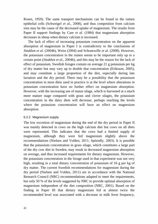
42
Rosen, 1959). The same transport mechanisms can be found in the rumen
epithelial cells (Schweigel et al., 2008), and thus competition from calcium
ions may be the cause of the decreased uptake of magnesium. The results from
Paper II support findings by Care et al. (1984) that magnesium absorption
decreases in sheep when dietary calcium is increased.
The lack of effect of increasing potassium concentration on the apparent
absorption of magnesium in Paper I is contradictory to the conclusions of
Jittakhot et al. (2004b), Weiss (2004) and Schonewille et al. (2008). However,
the potassium concentration in the rumen seems to be important only up to a
certain point (Jittakhot et al., 2004b), and this may be the reason for the lack of
effect of potassium. Swedish forages contain on average 21 g potassium per kg
of dry matter but may vary up to double that concentration (Eriksson, 2005),
and may constitute a large proportion of the diet, especially during late
lactation and the dry period. There may be a possibility that the potassium
concentration in most diets used in practice is at the level where alterations in
potassium concentration have no further effect on magnesium absorption.
However, with the increasing use of maize silage, which is harvested at a much
more mature stage compared with grass and clover silages, the potassium
concentration in the dairy diets will decrease, perhaps reaching the levels
where the potassium concentration will have an effect on magnesium
absorption.
6.2.2 Magnesium supply
The low excretion of magnesium during the end of the dry period in Paper II
was mainly detected in cows on the high calcium diet but cows on all diets
were represented. This indicates that the cows had a limited supply of
magnesium, although they were fed magnesium slightly above the
recommendations (Nielsen and Volden, 2011; Spörndly, 2003). It is possible
that the potassium concentration in grass silage, which constitutes a large part
of the dry cow diet in Sweden, may result in decreased magnesium absorption
on average, and thus increased requirements for dietary magnesium. However,
the potassium concentration in the forage used in that experiment was not very
high, resulting in a total dietary concentration of potassium of 16 g per kg of
dry matter. The current Swedish recommendations for magnesium during the
dry period (Nielsen and Volden, 2011) are in accordance with the National
Research Council (NRC) recommendations adapted to meet the requirements,
but only 50 % of the levels suggested by NRC to provide optimal absorption of
magnesium independent of the diet composition (NRC, 2001). Based on the
finding in Paper III that dietary magnesium fed at almost twice the
recommended level was associated with a decrease in milk fever frequency,

43
there may be reasons to exceed the minimum recommendations of magnesium
to dry cows, at least during the last weeks of gestation. It remains to be
determined whether this beneficial effect of extra magnesium is due to
compensation for decreased absorption of magnesium when the forage
proportion in the diet is high, underestimation of the magnesium requirement
during the last part of the pregnancy, or some other reason.
6.2.3 Assessing magnesium supply
In order to prevent production losses due to subclinical magnesium deficiency
and to detect cows and herds at risk of developing hypomagnesaemic tetany,
there is a need to evaluate whether the dietary magnesium provision is
adequate. This is best done by assessing the magnesium status at herd level.
The level of magnesium in plasma is considered to be a specific but not very
sensitive tool to detect subclinical hypomagnesaemia in humans (Malon et al.,
2004), and Schweigel et al. (2009) found that cows with plasma magnesium
levels considered to be normal still showed indications of depleted intracellular
magnesium pools. In Paper II, there were no differences in plasma magnesium
concentration in the dry period among cows with different calcium intakes,
although the cows fed high calcium had a decreased uptake of magnesium and
a lower excretion of magnesium in the urine. However, after calving, cows that
had been fed the high calcium diet showed lower plasma magnesium levels
compared with cows on the other treatments, although the feeding at that time
was the same. It is possible that this was the result of a gradual depletion of the
intracellular magnesium stores, which was reflected in plasma only after
calving when the output of magnesium increased. The urinary excretion of
magnesium was then a more sensitive indicator of inadequate magnesium
supply through the diet in the dry period. The possibility of using the
intracellular compartment as a magnesium store would perhaps explain parts of
the highly positive magnesium balances found in Paper I, and positive
magnesium balances have also been found in studies with dry cows (Jittakhot
et al., 2004a) and sheep (Ram et al., 1998), although their estimated
magnesium retention was numerically much smaller. The different magnesium
intakes in Paper I resulted in small but significant differences in plasma
magnesium levels. This is in agreement with results from studies with
periparturient cows (Shappell et al., 1987), but Jittakhot et al. (2004a) did not
see any significant effect of magnesium in the diet of dry cows on plasma
magnesium levels. Urine sampling seems to be a more reliable way to assess
magnesium status than plasma sampling, but the assessment has to be done
using an adequate cut-off point. Guéguen et al. (1989) suggested a minimum
loss of 1.8-2.4 g magnesium per day, while Mayland (1988) suggested

44
excretion of less than 0.1 g magnesium per litre of urine as indicating an
insufficient magnesium supply. However, the calculated daily excretion of
urine in dry cows from Papers II and IV varied widely, probably due to
differences in water excretion, which shows the difficulties in assessing total
magnesium output through urine and thus magnesium status only based on the
concentration of magnesium in urine. The variability in urine excretion, as
measured by variability in creatinine excretion, indicates that several cows
should be sampled to give an adequate assessment of the magnesium status in
dry cows in a herd, as long as only the magnesium concentration in the urine is
measured. However, there was a clear relationship between the total excretion
of magnesium and the concentration, especially at low concentrations. Cows
excreting less than 0.1 g magnesium per litre, the level suggested by Mayland
(1988), were likely to have a total excretion of less than 2.5 g magnesium per
day, which was the amount suggested by Kemp and Geurink (1978) and
Guéguen et al. (1989). As magnesium concentration in a spot sample of urine
is easier to determine than the total daily excretion of magnesium, this may be
the preferred method, at least to assess magnesium status at herd level. The
lactating cows in Paper I had a higher daily excretion of magnesium compared
with dry cows at the same concentration of magnesium in urine, which was
most evident at high total magnesium excretions. This was caused by a higher
amount of urine, and it can be speculated that the efficiency of kidney
reabsorption of magnesium may depend more on the concentration of
magnesium in the renal filtrate than on the total excretion of magnesium. If so,
the concentration of magnesium in the urine is a better marker of the
magnesium status than the daily total excretion, as a high volume of urine with
low concentration of magnesium can result in high total excretion.
Furthermore, samples from the lactating cows with concentrations below 0.1 g
per litre corresponded to total amounts below 2.5 g per day, so this
concentration could be used to test the magnesium status in both lactating and
dry cows.
Due to the impact of magnesium status on the adaptations to hypocalcaemia
in dairy cows (van de Braak et al., 1986), and also the high outflow of
magnesium after calving caused by the high concentration of magnesium in
colostrum, the magnesium status in periparturient cows is of major importance.
Goff (2008) suggests that several cows should be sampled within 12 h after
parturition to determine the herd magnesium status, and that plasma
magnesium levels less than 0.8 mM on that occasion suggest an inadequate
magnesium supply.

45
6.2.4 Magnesium at calving
In both Papers II and IV, a transient increase in plasma magnesium level was
found shortly after calving. This has also been reported in other studies (Goff
and Horst, 1997; Green et al., 1981), and results from Thilsing-Hansen et al.
(2002a) and Goff et al. (2002) indicate that the increase in magnesium is
related to the decrease in plasma calcium level occurring at calving. The reason
for this has been suggested to be the elevated renal threshold for calcium when
parathyroid hormone is excreted as a response to the decrease in plasma
calcium level, resulting in a similar increase in the renal threshold for
magnesium (Goff, 2008). Results from Taylor et al. (2008) suggest that there
may be an effect of parathyroid hormone levels at calving on the concentration
of plasma magnesium. However, in Paper IV, urinary excretion of magnesium
was not decreased, even though urinary calcium excretion decreased
immediately after calving. As the concentration of magnesium in plasma is
very low compared with the concentration of intracellular magnesium, the
results from Paper IV may suggest that the peak in magnesium occurring at
calving may depend on movements of magnesium between the intracellular
and extracellular compartment, rather than on decreased excretion. At 2 to 4
days after calving, the concentration of plasma magnesium was depressed
compared with prepartum levels in both Papers II and IV. However, the levels
were slightly higher both pre- and postpartum in Paper IV compared to Paper
II, probably reflecting a higher magnesium supply through the prepartum diet.
A decrease in plasma magnesium concentration after calving has also been
seen in other studies (Rérat et al., 2009; Verdaris and Evans, 1976). Urine
magnesium excretion was lowest 7 days after parturition in Paper IV, probably
aa a result of the decreased plasma magnesium concentration found after
calving.
6.3 Mineral losses in milk
The daily calcium and magnesium losses in milk immediately after calving in
Paper IV constituted more than half the calcium and magnesium requirement
of the cows, based on the maintenance requirements estimated in the Nordic
feed recommendations (Nielsen and Volden, 2011). The concentration of
calcium and magnesium in milk is much higher than the concentration in
plasma, which makes active transport of minerals into milk necessary. A large
part of the calcium is associated with the casein micelles and is needed for
coagulation (Horne, 1998), and this is also the case for some of the
magnesium. There is also a high concentration of ionised minerals in milk.
Mineral feeding has been shown not to alter the macro mineral concentration in

46
milk (Salih et al., 1987), and the amount of magnesium had no large impact on
the magnesium concentration in milk in Paper I. However, dietary
supplementation of minerals could result in increased milk production, thus
resulting in an increase in total mineral output in milk, as shown with micro
minerals by Rabiee et al. (2010). In Paper IV, the calcium and magnesium
concentration in milk was higher when the cows were milked prepartum
compared with after calving. After calving, the concentration was highest on
the first day of milking, and then decreased. This decrease in mineral
concentration after calving has also been found in earlier studies (Tsioulpas et
al., 2007). However, the main determinant of calcium excretion in milk after
calving in Paper IV was the milk yield obtained, and as the milk yield
increased, this resulted in an increase in the total excretion of calcium during
the first week of lactation.
The daily milk output of magnesium during the first week after calving,
both from cows milked prepartum and from control cows in Paper IV, was
almost twice the daily output of magnesium in milk from the cows in mid to
late lactation in Paper I. This was due to both higher milk yield and much
higher magnesium concentration in milk in the first week of lactation. This
emphasises the importance of a sufficient magnesium supply during early
lactation, and may even explain the decrease in plasma magnesium level found
during the first week of lactation in both Papers II and IV. In Paper I, magne-
sium excreted in milk constituted only up to 50 % of the magnesium apparently
absorbed by the cows with low magnesium intake, and a much smaller propor-
tion on high magnesium intake, while the urinary excretion of magnesium in-
creased. This is in agreement with results from Kamiya et al. (2010). Calcium
in milk often constitutes the major part of the absorbed calcium (Kamiya et al.,
2010; Taylor et al., 2009), and this difference between the minerals is probably
an effect of the different control of homeostasis, where calcium absorption is
under hormonal control while magnesium homeostasis is regulated only by
excretion, and may thus be absorbed more in excess. The concentration of
calcium in colostrum from cows in Paper IV was lower than the levels found
by Tsioulpas et al. (2007). However, the excretion of calcium in the first
milking after calving in the control cows was almost twice the daily amount of
calcium retained in conceptus at the end of gestation (House and Bell, 1993).
The excretion of magnesium in the first milking was more than 10-fold the
amount required for pregnancy.

47
7 Main conclusions
Addition of potassium to diets already high in potassium had no negative
effect on magnesium uptake in lactating cows.
High calcium intake during the last weeks of gestation resulted in decreased
magnesium absorption.
Calcium content in the dry cow diet did not affect calcium homeostasis at
parturition. It was also not an explanation for the difference in milk fever
incidence among Swedish herds.
High milk fever incidence in Swedish herds may partly be explained by low
levels of dietary magnesium and high amounts of potassium in the forage
fed to dry cows.
Milk removal prior to calving decreased the level of plasma calcium and
activated calcium mobilisation, but did not prevent the decrease in plasma
calcium level at calving.

48
8 Implications
To maintain good animal health and production when managing high-yielding
dairy cows in intense production systems, mineral supplementation of the cows
should be considered. The results presented in this thesis show that there are
possibilities to improve both the calcium and magnesium status in the dairy
cow, but also demonstrate the importance of diet composition to maintain
adequate levels of minerals, especially at a challenge such as parturition and
onset of lactation.
To improve the calcium availability in periparturient cows, the silage used
for dry cows during the last part of gestation should preferably have a low con-
centration of potassium, which to some extent can be achieved by harvesting
the plants at a more mature stage. The decrease in nutritional value that results
from later harvesting is often not a problem for dry cows, because their feed
consumption capacity is usually high. Furthermore, to prevent milk fever,
magnesium supply should be higher than the current recommendations during
the last part of gestation. This is more important than trying to reduce the
calcium concentration of the diet, at least as long as the calcium level still re-
mains above about 45 g per day. The removal of milk negatively affects
plasma calcium levels, but it is unclear whether restricted milking after calving
will prevent milk fever, as most of the decrease in plasma calcium levels at
calving seems to be unrelated to milk removal, and a decrease in plasma
calcium concentrations can be seen within 1 h after calving.
To ensure an adequate magnesium supply during lactation and the peri-
parturient period, magnesium intake should be sufficient. Silages that are early
cut and thus have high concentrations of potassium can be fed to lactating cows
without adverse effects on magnesium absorption. However, high calcium
intakes, at least during the last part of gestation, may lead to insufficient
absorption of magnesium and thus result in hypomagnesaemia after calving. To
monitor the magnesium status of a herd, urine samples should be preferred

49
over plasma samples due to the observable response of urinary magnesium to
increases in magnesium intake and the relative insensitivity of plasma
magnesium levels to decreases in absorption.

50
9 Future research
The studies included in this thesis identified several areas for future research:
As dietary magnesium at almost twice the recommended level appeared to
be preventive in the development of milk fever, the level of magnesium that
should be fed to dry cows to prevent milk fever and hypocalcaemia should
be investigated.
Bone mobilisation, as measured by the release of CTx into plasma,
increased rapidly at calving as well as at start of milk removal prepartum. It
should therefore be investigated whether calcium release from bones
increases immediately at calving, and also the factors on which onset of
calcium mobilisation depends.
It should be examined whether the amount of milk that is removed after
calving influences the extent of hypocalcaemia and the risk of milk fever.
Dietary calcium seemed to negatively influence magnesium absorption in
dry cows. It is thus of interest to determine the extent to which calcium
affects magnesium absorption, and whether this interaction is of importance
under practical conditions.
Further studies on the relationship between intracellular and extracellular
magnesium in the periparturient period would be of interest.

51
10 Svensk sammanfattning
10.1 Bakgrund
Dagens mjölkkor har en hög omsättning av mineraler eftersom de producerar
så mycket mjölk. Både kalcium och magnesium är viktiga för många av krop-
pens funktioner, till exempel muskel- och nervfunktioner, och eftersom de
förekommer i hög koncentration i mjölken måste de tillföras via fodret i stor
mängd för att täcka kons behov. Kalciumbalansen regleras med hjälp av hor-
moner, framför allt parathormon, som när det utsöndras leder till ökat kalcium-
upptag i tarmen, minskad utsöndring av kalcium i urinen och ökad frisättning
av kalcium från skelettet. Magnesiumbalansen regleras framför allt via utsönd-
ringen i urinen, medan upptaget i förmagarna bland annat beror på koncentra-
tionen av magnesium och andra joner, till exempel kalium, i våmmen. Akut
brist på kalcium är oftast kopplat till kalvningsförlamning, och drabbar varje år
över 10 000 svenska kor. Även om en hel del kalcium går åt till fostertillväxten
är behovet mycket större hos en lakterande mjölkko än hos en dräktig. Då
mjölkproduktionen startar tas kalcium från blodet, och plasmanivån sjunker
(hypokalcemi). De kalciummobiliserande mekanismerna aktiveras, men hos
vissa kor sjunker kalciumnivån trots det så lågt att muskelfunktionen blir så
försämrad att de inte kan resa sig. Akut brist på magnesium beror oftast på ett
för dåligt upptag i våmmen, ibland i kombination med stress som leder till att
magnesiumhalten i plasma sjunker ytterligare beroende på att magnesium
transporteras in i cellerna. En låg magnesiumhalt i plasman kan leda till en
sänkt magnesiumhalt i cerebrospinalvätskan, som i sin tur är kopplat till kram-
per. Även subkliniska brister av både kalcium och magnesium kan ge problem,
som dock är svårare att upptäcka. Fodrets innehåll av bl.a. kalcium, magnesium
och kalium påverkar risken för att kor ska drabbas av både akut och subklinisk
brist av kalcium och magnesium, och syftet med avhandlingen var att studera

52
hur kalcium- och magnesiumomsättningen hos mjölkkor kan påverkas och
underlättas.
10.2 Sammanfattning av studierna
Avhandlingen är uppbyggd av fyra olika studier. Tre av dem gjordes i försöks-
besättningen på Kungsängens forskningscentrum i Uppsala, och en gjordes
med hjälp av data från Svensk Mjölk och en enkät utskickad till utvalda
svenska besättningar med mer än 45 kor. I den första studien användes 6 kor i
laktationsmånad 6 eller senare för att undersöka effekterna av ett högt kalium-
intag på magnesiumupptaget hos lakterande kor. Försöket var upplagt med en
changeover design, med tre nivåer av kalium (19, 28 och 37 g per kg torrsub-
stans) och två nivåer av magnesium (1.9 och 4.3 g per kg torrsubstans), och
alla kor fick varje behandlingskombination under två veckor. I det andra försö-
ket ingick 29 dräktiga kor under sista delen av sinperioden och fram till en
vecka efter kalvning. Effekten av kalciumintag (4.9, 9.3 och 13.6 g kalcium per
kg torrsubstans) under sista delen av sinperioden på kalcium- och magnesium-
balansen runt kalvning undersöktes. Den tredje studien gjordes för att hitta
samband mellan utfodringen under sinperioden och risken för kalvningsför-
lamning. Genom en enkät erhölls information om utfodringen under sista delen
av sintiden i 30 besättningar med hög andel veterinärbehandlade kalvnings-
förlamningar och 22 besättningar med låg andel veterinärbehandlade
kalvningsförlamningar. I det fjärde försöket mjölkades 9 kor under 1 till 7
dagar före kalvning, och kalciumbalansen jämfördes med den hos 6 kor som
bara mjölkades efter kalvning.
10.3 Resultat
Resultaten från studierna tydde på att höga kaliumhalter i foderstaten under
laktationen inte medförde sämre upptag av magnesium vid de kaliumhalter
som användes. Högre intag av magnesium ledde till ett ökat upptag av magne-
sium, något som resulterade i en ökad magnesiumutsöndring i urinen. Beräknat
som en andel av magnesiumintaget var upptaget dock sämre vid det högre
intaget. En sänkt kalciumhalt i foderstaten innan kalvningen minskade inte
risken för hypokalcemi och kalvningsförlamning, åtminstone inte när kalcium-
halten låg mellan 4 och 13 g per kg torrsubstans. Däremot påverkade höga
kalciumhalter magnesiumupptaget negativt, och eftersom magnesium är viktigt
för att parathormon ska utsöndras och fungera effektivt kan höga kalciumhalter
väntas ha en indirekt påverkan på risken för kalvningsförlamning. Ett högt
magnesiumintag, omkring dubbla mängden mot NorFors rekommendation

53
under de sista tre veckorna av sinperioden minskade risken för kalvnings-
förlamning i besättningen, medan ökande intag av kalium ledde till ökad risk
för kalvningsförlamning i besättningen. Kalcium- och magnesiumutsöndringen
via mjölken påverkades framför allt av mängden mjölk kon producerade, och
den första råmjölken innehöll högre halter av både kalcium och magnesium än
mjölk senare i laktationen. De kalciumförluster som uppstod via mjölken när
laktationen startade ledde till en påfrestning på kalciumomsättningen, men
mjölkning före kalvning förberedde inte kon på kalciumförlusterna vid kalv-
ningen, eftersom kalciumhalten i blodet sjönk till samma nivå som hos kor som
inte var mjölkade före kalvningen. Det verkade finnas ytterligare faktorer mer
än enbart förlusten av mjölk som påverkade kalciumbalansen runt kalvning
eftersom kalciumhalten hade sjunkit betydligt redan en timme efter kalvningen,
samtidigt som nedbrytningen av benvävnad, och därigenom förmodligen fri-
sättandet av kalcium, hade ökat markant.
10.4 Slutsatser
De slutsatser som kunde dras var att högt kaliumintag, i intervallet 19 – 37 g
per kg torrsubstans, inte spelar någon roll för magnesiumupptaget hos lakte-
rande kor. Däremot bör sinkornas kaliumintag begränsas, i alla fall under den
sista delen av sinperioden, för att förebygga problem med kalvningsförlam-
ning. Magnesium bör utfodras på en högre nivå än de gällande rekommenda-
tionerna för att förebygga problem med kalvningsförlamning, medan varia-
tioner i kalciumnivån i sintidsfoderstaten inte spelar så stor roll vare sig för
graden av hypokalcemi vid kalvning eller för risken för kalvningsförlamning.
För att underlätta sinkornas upptag av magnesium bör kalciumintaget dock inte
vara alltför högt. Att mjölka korna ledde till en påverkan på kalciumbalansen,
men eftersom det verkade finnas andra faktorer än urmjölkning som också
påverkade plasmahalten av kalcium vid kalvning är det osäkert hur stor påver-
kan urmjölkningen har på risken för kalvningsförlamning.

54
References
Ali, B. H., Al-Quarawi, A. A. and Mousa, H. M. (2006) Stress associated with road transportation
in desert sheep and goats, and the effect of pretreatment with xylazine or sodium betaine.
Research in Veterinary Science 80, 343-348.
Andersen, P. (2003) Udder insufflations of cows with parturient paresis – A forgotten treatment
with a bright future? Acta Veterinaria Scandinavica. Supplementum 97, 75.
Arnaud, M. J. (2008) Update on the assessment of magnesium status. British Journal of Nutrition
99, supplement 3, S24-S36.
Barbagallo, M. and Dominguez, L. J. (2006) Magnesium metabolism in type 2 diabetes mellitus,
metabolic syndrome and insulin resistance. Archives of Biochemistry and Biophysics 458, 40-
47.
Behar, J. (1975) Effect of calcium on magnesium absorption. American Journal of Physiology
229, 1590-1595.
Belyea, R. L., Coppock, C. E. and Lake, G. B. (1976) Effects of a low calcium diet on feed
intake, milk production, and response to blood calcium challenge in lactating Holstein cows.
Journal of Dairy Science 59, 1068-1077.
Ben-Ghedalia, D., Tagari, H., Zamwel, S. and Bondi, A. (1975) Solubility and net exchange of
calcium, magnesium and phosphorus in digesta flowing along the gut of the sheep. British
Journal of Nutrition 33, 87-94.
Bigras-Poulin, M. and Tremblay, A. (1998) An epidemiological study of calcium metabolism in
non-paretic postparturient Holstein cows. Preventive Veterinary Medicine 35, 195-207.
Blair, H. C. (1998) How the osteoclast degrades bone. BioEssays 20, 837-846.
Braithwaite, G. D. and Riazuddin, S. (1971) The effect of age and level of dietary calcium intake
on calcium metabolism in sheep. The British Journal of Nutrition 26, 215-225.
Bushinsky, D. A. (1989) Net calcium efflux from live bone during chronic metabolic, but not
respiratory, acidosis. The American Journal of Physiology 256, F836-F842.
Bushinsky, D. A., Smith, S. B., Gavrilov, K. L., Gavrilov, L. F., Li, J. and Levi-Setti, R. (2003)
Chronic acidosis-induced alteration in bone bicarbonate and phosphate. American Journal of
Physiology. Renal Physiology 285, F532-F539.
Care, A. D., Brown, R. C., Farrar, A. R. and Pickard, D. W. (1984) Magnesium absorption from
the digestive tract of sheep. Quarterly Journal of Experimental Physiology 69, 577-587.

55
Cerbulis, J. and Farrell jr, H. M. (1976) Composition of the milks of dairy cattle. II. Ash, calcium,
magnesium, and phosphorus. Journal of Dairy Science 59, 589-593.
Charbonneau, E., Chouinard, P. Y., Tremblay, G. F., Allard, G. and Pellerin, D. (2008) Hay to
reduce dietary cation-anion difference for dry dairy cows. Journal of Dairy Science 91, 1585-
1596.
Charlton, J. A. and Armstrong, D. G. (1989) The effect of an intravenous infusion of aldosterone
upon magnesium metabolism in the sheep. Quarterly Journal of Experimental Physiology 74,
329-337.
Chester-Jones, H., Fontenot, J. P. and Veit, H. P. (1990) Physiological and pathological effects of
feeding high levels of magnesium to steers. Journal of Animal Science 68, 4400-4413.
Correa, M. T., Curtis, C. R., Erb, H. N., Scarlett, J. M. and Smith, R. D. (1990) An ecological
analysis of risk factors for postpartum disorders of Holstein-Friesian cows from thirty-two
New York farms. Journal of Dairy Science 73, 1515-1524.
Daniel, R. C. W. (1983) Motility of the rumen and abomasums during hypocalcemia. Canadian
Journal of Comparative Medicine 47, 276-280.
Ellenberger, H. B., Newlander, J. A. and Jones, C. H. (1932) Calcium and phosphorus
requirements to dairy cows. II. Weekly balances through lactation and gestation periods.
Bullentin - Vermont Agricultural Experiment Station 342, June 1932.
Eriksson, H. (2005) Mineraler – vallfodrets innehåll och mjölkkornas behov. Nytt från
Institutionen för Norrländsk Jordbruksvetenskap, Husdjur 3, 2005.
Everts, V., Delaissé, J. M., Korper, W., Niehof, A., Vaes, G. and Beertsen, W. (1992)
Degradation of collagen in the bone-resorbing compartment underlying the osteoclast involves
both cysteine-proteinases and matrix metalloproteinases. Journal of Cellular Physiology 150,
221-231.
FAO (2011) FAOSTAT, Food and Agriculture Organization of the United Nations. [online].
Available from: faostat.fao.org [Accessed 2011-08-06]
Fardellone, P., Cotté, F. E., Roux, C., Lespessailles, E., Mercier, F. and Gaudin, A. F. (2010)
Calcium intake and the risk of osteoporosis and fractures in French women. Joint, Bone, Spine
77, 154-158.
Field, A. C., McCallum, J. W. and Butler, E. J. (1958) Studies on magnesium in ruminant
nutrition. Balance experiments on sheep with herbage from fields associated with lactation
tetany and from control pastures. British Journal of Nutrition 12, 433-446.
Fontenot, J. P., Allen, V. G., Bunce, G. E. and Goff, J. P. (1989) Factors influencing magnesium
absorption and metabolism in ruminants. Journal of Animal Science 67, 3445-3455.
Fry, J. M., Curnow, D. H., Gutteridge, D. H. and Retallack, R. W. (1979) Effect of chronic
parathyroidectomy on calcium metabolism in the lactating rat. Journal of Endocrinology 82,
323-330.
Fuller, K., Kirstein, B. and Chambers, T. J. (2007) Regulation and enzymatic basis of bone
resorption by human osteoclasts. Clinical Science 112, 567-575.
Garnero, P., Ferreras, M., Karsdal, M. A., Nicamhlaoibh, R., Risteli, J., Borel, O., Qvist, P.,
Delmas, P. D., Foged, N. G. and Delaissé, J. M. (2003) The Type I collagen fragments ICTP
and CTX reveal distinct enzymatic pathways of bone collagen degradation. Journal of bone
and mineral research 18, 859-867.

56
Godden, W. (1939) “Trace” elements in human and animal nutrition. Journal of the Society of
Chemical Industry 58, 791-796.
Goff, J. P. (2008) The monitoring, prevention, and treatment of milk fever and subclinical
hypocalcemia in dairy cows. Veterinary Journal 176, 50-57.
Goff, J. P., Littledike, E. T. and Horst, R. L. (1986) Effect of synthetic bovine parathyroid
hormone in dairy cows: prevention of hypocalcemic parturient paresis. Journal of Dairy
Science 69, 2278-2289.
Goff, J.P., Horst, R. L., Jardon, P. W., Boreli, C. and Wedam, J. (1996) Field trials of an oral
calcium propionate paste as an aid to prevent milk fever in periparturient dairy cows. Journal
of Dairy Science 79, 378-383.
Goff, J. P. and Horst, R. L. (1997) Effects of the addition of potassium or sodium, but not
calcium, to prepartum rations on milk fever in dairy cows. Journal of Dairy Science 80, 176-
186.
Goff, J.P., Kimura, K. and Horst, R. L. (2002) Effect of mastectomy on milk fever, energy, and
vitamins A, E, and beta-carotene status at parturition. Journal of Dairy Science 85, 1427-
1436.
Goings, R. L., Jacobson, N. L., Beitz, D. C., Littledike, E. T. and Wiggers, K. D. (1974)
Prevention of parturient paresis by a prepartum, calcium-deficient diet. Journal of Dairy
Science 57, 1184-1188.
Green, H. B., Horst, R. L., Beitz, D. C. and Littledike, E. T. (1981) Vitamin D metabolites in
plasma of cows fed a prepartum low-calcium diet for prevention of parturient hypocalcemia.
Journal of Dairy Science 64, 217-226.
Guéguen, L., Lamand, M. and Meschy, F. (1989) Mineral requirements. In Jarridge, R. (ed):
Ruminant nutrition. Recommendation, allowances & feed tables. INRA, John Libbey
Eurotext, London-Paris, p. 52.
Guillemant, J. A., Accarie, C. M., de la Gueronniere, V. and Guillemant, S. E. (2003) Different
acute response of serum type I collagen telopeptides, CTX, NTX and ICTP, after repeated
ingestion of calcium. Clinica Chimica Acta 337, 35-41.
Hansen, S. S., Nørgaard, P., Pedersen, C., Jørgensen, R. J., Mellau, L. S. and Enemark, J D.
(2003) The effect of subclinical hypocalcaemia induced by Na2EDTA on the feed intake and
chewing activity of dairy cows. Veterinary Research Communications 27, 193-205.
Hansen, S. S., Ersbøll, A. K., Blom, J. Y. and Jørgensen, R. J. (2007) Preventive strategies and
risk factors for milk fever in Danish dairy herds: a questionnaire survey. Preventive
Veterinary Medicine 80, 271-286.
Hibbs, J. W. (1950) Milk fever (parturient paresis) in dairy cows – A review. Journal of Dairy
Science 33, 758-789.
Hidiroglou, M., Thompson, B. K., Ho, S. K. and Proulx, J. G. (1981) Hypomagnesemia in beef
cows wintered in Ohio. Canadian Journal of Comparative Medicine 45, 124-129.
Hollis, B. W., Draper, H. H., Burton, J. H. and Etches, R. J. (1981) A hormonal assessment of
bovine parturient paresis: evidence for a role of oestrogen. The Journal of Endocrinology 88,
161-171.
Horne, D. S. (1998) Casein Interactions: Casting light on the black boxes, the structure in dairy
products. International Dairy Journal 8, 171-177.

57
Horst, R. L., Jorgensen, D. A. and DeLuca, H. F. (1978) Plasma 1,25-dihydroxyvitamin D and
parathyroid hormone levels in paretic dairy cows. The American Journal of Physiology 235,
E634-E637.
Horst, R. L., Goff, J. P. and Reinhardt, T. A. (1994) Calcium and vitamin D metabolism in the
dairy cow. Journal of Dairy Science 77, 1936-1951.
Houe, H., Østergaard, S., Thilsing-Hansen, T., Jørgensen, R. J., Larsen, T., Sørensen, J. T.,
Agger, J. F. and Blom, J. Y. (2001) Milk fever and subclinical hypocalcemia – an evaluation
of parameters on incidence risk, diagnosis, risk factors and biological effects as input for a
decision support system for disease control. Acta Veterinaria Scandinavica 42, 1-29.
House, W. A. and Bell, A. W. (1993) Mineral accretion in the fetus and adnexa during late
gestation in Holstein cows. Journal of Dairy Science 76, 2999-3010.
House, W. A. and Van Campen, D. (1971) Magnesium metabolism of sheep fed different levels
of potassium and citric acid. The Journal of Nutrition 101, 1483-1492.
Hove, K. (1984) Effects of 1 alpha-hydroxylated metabolites of cholecalciferol on intestinal
radiocalcium absorption in goats. The British Journal of Nutrition 51, 157-164.
Jarjou, L. M., Laskey, M. A., Sawo, Y., Goldberg, G. R., Cole, T. J. and Prentice, A. (2010)
Effect of calcium supplementation in pregnancy on maternal bone outcomes in women with a
low calcium intake. The American Journal of Clinical Nutrition 92, 450-457.
Jittakhot, S., Schonewille, J. T., Wouterse, H., Yuangklang, C. and Beynen, A. C. (2004a)
Apparent magnesium absorption in dry cows fed at 3 levels of potassium and 2 levels of
magnesium intake. Journal of Dairy Science 87, 379-385.
Jittakhot , S., Schonewille, J. T., Wouterse, H. S., Yuanklang, C. and Beynen, A. C. (2004b) The
relationships between potassium intakes, transmural potential difference of the rumen
epithelium and magnesium absorption in wethers. British Journal of Nutrition 91, 183-189.
Jørgensen, R. J., Nyengaard, N. R., Daniel, R. C. W., Mellau, L. S. B. and Enemark, J. M. D.
(1999) Induced hypocalcemia by Na2EDTA infusion: A review. Journal of Veterinary
Medicine Series A 46, 389-407.
Kameda, T., Mano, H., Yamada, Y., Takai, H., Amizuka, N., Kobori, M., Izumi, N., Kawashima,
H., Ozawa, H., Ikeda, K., Kameda, A., Hakeda, Y. and Kumegawa, M. (1998) Calcium-
sensing receptor in mature osteoclasts, which are bone resorbing cells. Biochemical and
Biophysical Research Communications 245, 419-422.
Kamiya, Y., Kamiya, M., Tanaka, M. and Shioya, S. (2005) Effects of calcium intake and parity
on plasma minerals and bone turnover around parturition. Animal Science Journal 76, 325-
330.
Kamiya, Y., Kamiya, M. and Tanaka, M. (2010) The effect of high ambient temperature on Ca, P
and Mg balance and bone turnover in high-yielding dairy cows. Animal Science Journal 81,
482-486.
Kemp, A. and Geurink, J. H. (1978) Grassland farming and minerals in cattle. Netherlands
Journal of Agricultural Science 26, 161-169.
Khorasani, G. R., Janzen, R. A., McGill, W. B. and Kennelly, J. J. (1997) Site and extent of
mineral absorption in lactating cows fed whole-crop cereal grain silage or alfalfa silage.
Journal of Animal Science 75, 239-248.

58
Kimura, K., Reinhardt, T. A. and Goff, J. P. (2006) Parturition and hypocalcemia blunts calcium
signals in immune cells of dairy cattle. Journal of Dairy Science 89, 2588-2595.
Klimiene, I., Spakauskas, V. and Matusevicius, A. (2005) Correlation of different biochemical
parameters in blood sera of healthy and sick cows. Veterinary Research Communications 29,
95-102.
Krapf, R., Jaeger, P. and Hulter, H. N. (1992) Chronic respiratory alkalosis induces renal PTH-
resistance, hyperphosphatemia and hypocalcemia in humans. Kidney International 42, 727-
734.
Kurosaki, N., Yamato, O., Sasamoto, Y., Mori, F., Imoto, S., Kojima, T., Yamasaki, M. and
Maede, Y. (2007) Clinico-pathological findings in peripartum dairy cows fed anion salts
lowering the dietary cation-anion difference: Involvement of serum inorganic phosphorus,
chloride and plasma estrogen concentrations in milk fever. Japanese Journal of Veterinary
Research 55, 3-12.
Langworthy, C. F. (1910) Suggestions regarding research work in animal nutrition. Journal of
Animal Science 1, 25-27.
Larsen, T., Møller, G. and Bellio, R. (2001) Evaluation of clinical and clinical chemical
parameters in periparturient cows. Journal of Dairy Science 84, 1749-1758.
Lean, I. J., DeGaris, P. J., McNeill, D. M. and Block, E. (2006) Hypocalcemia in dairy cows:
Meta-analysis and dietary cation anion difference theory revisited. Journal of Dairy Science
89, 669-684.
Leonhard-Marek, S. and Martens, H. (1996) Effects of potassium on magnesium transport across
rumen epithelium. American Journal of Physiology 271, G1034-G1038.
Li, X, Qin, L., Bergenstock, M., Bevelock, L. M., Novack, D. V. and Partridge, N. C. (2007a)
Parathyroid hormone stimulates osteoblastic expression of MCP-1 to recruit and increase the
fusion of pre/osteoclasts. The Journal of Biological Chemistry 282, 33098-33106.
Li, M., Du, J., Jiang, J., Ratzan, W., Su, L.-T., Runnels, L. R. and Yue, L. (2007b) Molecular
determinants of Mg2+
and Ca2+
permeability and pH sensitivity in TRPM6 and TRPM7. The
Journal of Biological Chemistry 282, 25817-25830.
Liesegang, A., Sassi, M.-L., Risteli, J., Eicher, R., Wanner, M. and Riond, J.-L. (1998)
Comparison of bone resorption markers during hypocalcemia in dairy cows. Journal of Dairy
Science 81, 2614-2622.
Luebbe, M. K., Erickson, G. E., Klopfenstein, T. J., Greenquist, M. A. and Benton, J. R. (2011)
Effect of dietary cation-anion difference on urinary pH, feedlot performance, nitrogen mass
balance, and manure pH in open feedlot pens. Journal of Animal Science 89, 489-500.
Malon, A., Brockmann, C., Fijalkowska-Morawska, J., Rob, P. and Maj-Zurawska, M. (2004)
Ionized magnesium in erythrocytes – the best magnesium parameter to observe hypo- or
hypermagnesemia. Clinica Chimica Acta 349, 67-73.
Martens, H. and Schweigel, M. (2000) Pathophysiology of grass tetany and other
hypomagnesemias. Implications for clinical management. The Veterinary Clinics of North
America. Food Animal Practice 16, 339-368.
Martig, J. and Mayer, G. P. (1973) Diminished hypercalcemic response to parathyroid extract in
prepartum cows. Journal of Dairy Science 56, 1042-1046.

59
Martz, F. A., Belo, A. T., Weiss, M. F., Belyea, R. L. and Goff, J. P. (1990) True absorption of
calcium and phosphorus from alfalfa and corn silage when fed to lactating cows. Journal of
Dairy Science 73, 1288-1295.
Mayland, H. F. (1988) Grass tetany. In Church, D. C. (ed): The ruminant animal. Digestive
physiology and nutrition, Prentice Hall, Englewood Cliffs, p 511-531.
McCoy, M. A., Hutchingson, T., Davison, G., Fitzpatrick, D. A., Rice, D. A., Kennedy, D. G.
(2001) Postmortem biochemical markers of experimentally induced hypomagnesaemic tetany
in cattle. The Veterinary Record 148, 268-273.
McLaren, C. J., Lissemore, K. D., Diffield, T. F., Leslie, K. E., Kelton, D. F. and Grexton, B.
(2006) The relationship between herd level disease incidence and return over feed index in
Ontario dairy herds. The Canadian Veterinary Journal 47, 767-773.
McNeill, D. M., Roche, J. R., McLachlan, B. P. and Stockdale, C. R. (2002) Nutritional strategies
for the prevention of hypocalcaemia at calving for dairy cows in pasture-based systems.
Australian Journal of Agricultural Research 53, 755-770.
Meyer, H. and Scholz, H. (1972) Studies on the pathogenesis of hypomagnesemic tetany. I.
Relationships between the magnesium contents in blood and cerebrospinal fluid. Deutsche
Tierartzliche Wochenschrift 79, 55-61.
Miller, J. K., Madsen, F. C., Lentz, D. E., Wong, W.-O., Ramsey, N., Tysinger, C. E. and
Hansard, S. L. (1980) Blood plasma magnesium, potassium, glucose, and immunoreactive
insulin changes in cows moved abruptly from barn feeding to early spring pasture. Journal of
Dairy Science 63, 1073-1079.
Mohler, J. R. (1904) Milk fever: Its simple and successful treatment. Farmers Bullentin No. 206.
U.S. Department of Agriculture, Washington Government Printing Office.
Musso, C. G. (2009) Magnesium metabolism in health and disease. International Urology and
Nephrology 41, 357-362.
Neville, M. C. and Peaker, M. (1979) The secretion of calcium and phosphorus into milk. The
Journal of Physiology 290, 59-67.
Nielsen, N. I. and Volden, H. (2011) Animal requirements and recommendations. In Volden, H.
(ed): NorFor – The Nordic feed evaluating system EAAP publication No. 130, Wageningen
Academic Publishers, p 105-111.
NRC (2001) Nutrient Requirements of Dairy Cattle. Seventh revised edition, National Academy
Press, Whashington.
O’Kelley, R. J. and Fontenot, J. P. (1969) Effects of feeding different magnesium levels to drylot-
fed lactating beef cows. Journal of Animal Science 29, 959-966.
Odensten, M. (2006) Drying off the dairy cow. PhD dissertation 2006:18, Swedish University of
Agricultural Sciences.
Odette, O. (2005) Grass tetany in a herd of beef cows. The Canadian Veterinary Journal 46, 732-
734.
Ospina, P. A., Nydam, D. V., Stokol, T. and Overton, T. R. (2010) Association between the
proportion of sampled transition cows with increased nonesterified fatty acids and β-
hydroxybutyrate and disease incidence, pregnancy rate, and milk production at the herd level.
Journal of Dairy Science 93, 3593-3601.

60
Oursler, M. J., Osdoby, P., Pyfferoen, J., Riggs, B. L. and Spelsberg, T. C. (1991) Avian
osteoclasts as estrogen target cells. Proceedings of the National Academy of Science of the
United States of America 88, 6613-6617.
Pehrson, B., Svensson, C., Gruvaeus, I. and Virkki, M. (1999) The influence of acidic diets on the
acid-base balance of dry cows and the effect of fertilization on the mineral content of grass.
Journal of Dairy Science 82, 1310-1316.
Penner, G. B., Tremblay, G. F., Dow, T. and Oba, M. (2008) Timothy hay with a low dietary
cation-anion difference improves calcium homeostasis in periparturient Holstein cows.
Journal of Dairy Science 91, 1959-1968.
Prestwood, K. M., Kenny, A. M., Unson, C. and Kulldorff, M. (2000) The effect of low dose
micronized 17ss-estradiol on bone turnover, sex hormone levels, and side effects in older
women: a randomized, double blind, placebo-controlled study. The Journal of Clinical
Endocrinology and Metabolism 85, 4462-4469.
Rabiee, A. R., Lean, I. J., Stevenson, M. A. and Socha, M. T. (2010) Effects of feeding organic
trace minerals on milk production and reproductive performance in lactating dairy cows: a
meta-analysis. Journal of Dairy Science 93, 4239-4251.
Rajala-Schultz, P. J., Gröhn, Y. T. and McCulloch, C. E. (1999) Effects of milk fever, ketosis,
and lameness on milk yield in dairy cows. Journal of Dairy Science 82, 288-294.
Ram, L., Schonewille, J. T., Martens, H., Van’t Klooster, A. T. and Beynen, A. C. (1998)
Magnesium absorption by wethers fed potassium bicarbonate in combination with different
dietary magnesium concentrations. Journal of Dairy Science 81, 2485-2492.
Ramberg jr, C. F., Mayer, G. P., Kronfeld, D. S. and Potts jr, J. T. (1976) Dietary calcium,
calcium kinetics and plasma parathyroid hormone concentration in cows. The Journal of
Nutrition 106, 671-679.
Ravaglioli, A., Krajewski, A., Celotti, G. C., Piancastelli, A., Baccini, B., Montanari, L., Zama,
G. and Piombi, L. (1996) Mineral evolution of bone. Biomaterials 17, 617-622.
Reddy, C. R., Coburn, J. W., Hartenbower, D. L., Friedler, R. M., Brickman, A. S., Massry, S. G.
and Jowsey, J. (1973) Studies on mechanisms of hypocalcemia of magnesium depletion. The
Journal of Clinical Investigation 52, 3000-3010.
Reinhardt, T. A., Lippolis, J. D., McCluskey, B. J., Goff, J. P. and Horst, R. L. (2011) Prevalence
of subclinical hypocalcemia in dairy herds. Veterinary Journal 188, 122-124.
Rérat, M., Philipp, A., Hess, H. D. and Liesegang, A. (2009) Effect of different potassium levels
in hay on acid-base status and mineral balance in periparturient dairy cows. Journal of Dairy
Science 92, 6123-6133.
Reynolds, C. K., Bell, M. C. and Sims, M. H. (1984) Changes in plasma, red blood cell and
cerebrospinal fluid mineral concentrations in calved during magnesium depletion followed by
repletion with rectally infused magnesium chloride. The Journal of Nutrition 114, 1334-1341.
Ritter, R. J., Boling, J. A. and Gay, N. (1984) Labile magnesium reserves in beef cows subjected
to different prepasture supplementation regimes. Journal of Animal Science 59, 197-203.
Robinson, D. L., Kappel, L. C. and Boling, J. A. (1989) Management practices to overcome the
incidence of grass tetany. Journal of Animal Science 67, 3470-3484.

61
Roche, J. R. and Berry, D. P. (2006) Periparturient climatic, animal, and management factors
influencing the incidence of milk fever in grazing systems. Journal of Dairy Science 89, 2775-
2783.
Salih, Y., McDowell, L. R., Hentges, J. F., Mason jr, R. M. and Wilcox, C. J. (1987) Mineral
content of milk, colostrum, and serum as affected by physiological state and mineral
supplementation. Journal of Dairy Science 70, 608-612.
Schafer, A. L., Palermo, L., Bauer, D. C., Bilezikian, J. P., Sellmeyer, D. E. and Black, D. M.
(2011) Consistency of bone turnover marker and calcium responses of parathyroid hormone
(1-84) therapy in postmenopausal osteoporosis. Journal of Clinical Densitometry 14, 68-73.
Schonewille, J. T., Ram, L., Van’t Klooster, A. T., Wouterse, H. and Beynen, A. C. (1997) Native
corn starch versus either cellulose or glucose in the diet and the effects on apparent
magnesium absorption in goats. Journal of Dairy Science 80, 1738-1743.
Schonewille, J. T., Van’t Klooster, A. T., Wouterse, H. and Beynen, A. C. (1999) Effects of
intrinsic potassium in artificially dried grass and supplemental potassium bicarbonate on
apparent magnesium absorption in dry cows. Journal of Dairy Science 82, 1824-1830.
Schonewille, J. T., Everts, H., Jittakhot, S. and Beynen, A. C. (2008) Quantitative prediction of
magnesium absorption in dairy cows. Journal of Dairy Science 91, 271-278.
Schröder, B. and Breves, G. (2006) Mechanisms and regulation of calcium absorption from the
gastrointestinal tract in pigs and ruminants: comparative aspects with special emphasis on
hypocalcemia in dairy cows. Animal Health Research Reviews 7, 31-41.
Schweigel, M, Lang, I and Martens, H. (1999) Mg2+
transport in sheep rumen epithelium:
evidence for an electrodiffusive uptake mechanism. The American Journal of Physiology 277,
G976-G982.
Schweigel, M., Vormann, J. and Martens, H. (2000) Mechanisms of Mg2+
transport in cultured
ruminal epithelial cells. American Journal of Physiology. Gastrointestinal and Liver
Physiology 278, G400-G408.
Schweigel, M., Kolisek, M., Nikolic, Z. and Kuzinski, J. (2008) Expression and functional
activity of the Na/Mg exchanger, TRPM7 and MagT1 are changed to regulate Mg
homeostasis and transport in rumen epithelial cells. Magnesium Research 21, 118-123.
Schweigel, M., Voigt, J. and Mohr, E. (2009) Indication of intracellular magnesium deficiency in
lactating dairy cows revealed by magnesium loading and renal fractional excretion. Journal of
Animal Physiology and Animal Nutrition 93, 105-112.
Scott, D. (1965) Factors influencing the secretion and absorption of calcium and magnesium in
the small intestine of the sheep. Quarterly Journal of Experimental Physiology and Cognate
Medical Sciences 50, 312-329.
Seifi, H. A., Mohri, M. and Kalamati Zadeh, J. (2004) Use of pre-partum urine pH to predict the
risk of milk fever in dairy cows. The Veterinary Journal 167, 281-285.
Shachter, D. and Rosen, S. M. (1959) Active transport of Ca45 by the small intestine and its
dependence on vitamin D. The American Journal of Physiology 196, 357-362.
Shappell, N. W., Herbein, J. H., Deftos, L. J. and Aiello, R. J. (1987) Effects of dietary calcium
and age on parathyroid hormone, calcitonin and serum and milk minerals in the periparturient
dairy cow. The Journal of Nutrition 117, 201-207.

62
Smith, R. H. (1959) Calcium and magnesium metabolism in calves. 3. Endogenous faecal
excretion and absorption of magnesium. The Biochemical Journal 71, 306-311.
Smith, V. R., Niedermeier, R. P. and Hansen, R. G. (1948) Parturient paresis. II. The effect of
partial versus complete milking upon the total blood serum calcium of dairy cows at
parturition. Journal of Dairy Science 31, 173-177.
Speich, M., Bousquet, B. and Nicolas, G. (1981) Reference values for ionized, complexed, and
protein-bound plasma magnesium in men and women. Clinical Chemistry 27, 246-248.
Spörndly, R. (2003) Fodertabeller för idisslare (Feed tables for ruminants). Report 257.
Department of Animal Nutrition and Management, Swedish University of Agricultural
Sciences, Uppsala, Sweden.
Stewart, P. A. (1983) Modern quantitative acid-base chemistry. Canadian Journal of Physiology
and Pharmacology 61, 1444-1461.
Suh, S. M., Tashijan jr, A. H., Matuso, N., Parkinson, D. K. and Fraser, D. (1973) Pathogenesis of
hypocalcemia in primary hypomagnesemia: normal end-organ responsiveness to parathyroid
hormone, impaired parathyroid gland function. The Journal of Clinical Investigations 52, 153-
160.
Swedish Dairy Association (2010) Husdjursstatistik 2010, Cattle statistics 2010. Swedish Dairy
Association, Stockholm.
Swift, M. L., Bittman, S., Hunt, D. E. and Kowalenko, C. G. (2007) The effect of formulation and
amount of potassium fertilizer on macromineral concentration and cation-anion difference in
tall fescue. Journal of Dairy Science 90, 1063-1072.
Sørensen, J. T., Østergard, S., Houe, H. and Hindhede, J. (2002) Expert opinions of strategies for
milk fever control. Preventive Veterinary Medicine 55, 69-78.
Talmage, R. V. and Mobley, H. T. (2008) Calcium homeostasis: reassessment of the actions of
parathyroid hormone. General and Comparative Endocrinology 156, 1-8.
Tamminga, S., Luteijn, P. A. and Meijer, R. G. M. (1997) Changes in composition and energy
content of liveweight loss in dairy cows with time after parturition. Livestock Production
Science 52, 31-38.
Tanaka, R., Frank, H. and DeLuca, H. F. (1973) Intestinal calcium transport: stimulation by low
phosphorus diets. Science 181, 564-566.
Taylor, M. S., Knowlton, K. F., McGilliard, M. L., Seymour, W. M. and Herbein, J. H. (2008)
Blood mineral, hormone, and osteocalcin responses of multiparous Jersey cows to an oral
dose of 25-hydroxyvitamin D3 or vitamin D3 before parturition. Journal of Dairy Science 91,
2408-2416.
Taylor, M. S., Knowlton, K. F., McGilliard, M. L., Swecker, W. S., Ferguson, J. D., Wu, Z. and
Hanigan, M. D. (2009) Dietary calcium has little effect on mineral balance and bone mineral
metabolism through twenty weeks of lactation in Holstein cows. Journal of Dairy Science 92,
223-237.
Thilsing, T., Larsen, T., Jørgensen, R. J. and Houe, H. (2007) The effect of dietary calcium and
phosphorus supplementation in zeolite A treated dry cows on periparturient calcium and
phosphorus homeostasis. Journal of Veterinary Medicine. A, Physiology, Pathology, Clinical
Medicine 54, 82-91.

63
Thilsing-Hansen, T., Jørgensen, R. J., Enemark, J. M. and Larsen, T. (2002a) The effect of zeolite
A supplementation in the dry period on periparturient calcium, phosphorus, and magnesium
homeostasis. Journal of Dairy Science 85, 1855-1862.
Thilsing-Hansen, T., Jørgensen, R. J. and Østergaard, S. (2002b) Milk fever control principles: a
review. Acta Veterinaria Scandinavica 43, 1-19.
Tsioulpas, A., Grandinson, A. S. and Lewis, M. J. (2007) Changes in physical properties of
bovine milk from the colostrum period to early lactation. Journal of Dairy Science 90, 5012-
5017.
Valadares, R. F. D., Broderick, G. A., Valadares Filho, S. C. and Clayton, M. K. (1999) Effect of
replacing alfalfa silage with high moisture corn on ruminal protein synthesis estimated from
excretion of total purine derivatives. Journal of Dairy Science 82, 2686-2696.
van de Braak, A. E., van’t Klooster, A. T., van Hal-van Gestel, J. C. and Malestein, A. (1984)
Influence of stage of lactation and calcium level of the ration on mobilisation rate of calcium
and excretion of hydroxyproline in urine in dairy cows. Studies with Na2EDTA infusions in
monozygotic twins. Zentralblatt für Veterinärmedizin. Reihe A 31, 725-739
van de Braak, A. E., van’t Klooster, E. T. and Malestein, A. (1986) Influence of a deficient supply
of magnesium during the dry period on the rate of calcium mobilization by dairy cows at
parturition. Research in Veterinary Science 42, 101-108.
van Hulzen, K. J., Sprong, R. C., van der Meen, R. and van Arendonk, J. A. (2009) Genetic and
nongenetic variation in concentration of selenium, calcium, potassium, zink, magnesium, and
phosphorus in milk of Dutch Holstein-Friesian cows. Journal of Dairy Science 92, 5754-5759.
Van Keulen, J. and Young, B. A. (1977) Evaluation of acid-insoluble ash as a natural marker in
ruminant digestibility studies. Journal of Animal Science 44, 282-287.
Verdaris, J. M. and Evans, J. L. (1976) Diet calcium and pH versus mineral balance in Holstein
cows 84 days pre- to 2 days postpartum. Journal of Dairy Science 59, 1271-1277.
Weiss, W.P. (2004) Macromineral digestion by lactating dairy cows: factors affecting digestibility
of magnesium. Journal of Dairy Science 87, 2167-2171.
Wilkens, M. R., Mrochen, N., Breves, G. and Schröder, B. (2010) Effects of 1,25-
dihydroxyvitamin D3 on calcium and phosphorus homeostasis in sheep fed diets either
adequate or restricted in calcium content. Domestic Animal Endocrinology 38, 190-199.
Xin, Z., Tucker, W. B. and Henken, R. W. (1989) Effect of reactivity rate and particle size of
magnesium oxide on magnesium availability, acid-base balance, mineral metabolism, and
milking performance of dairy cows. Journal of Dairy Science 72, 462-470.
Yamagishi, N., Miyazaki, M. and Naito, Y. (2006) The expression of genes for transepithelial
calcium-transporting proteins in the bovine duodenum. The Veterinary Journal 171, 363-366.
Yarrington, J. T., Capen, C. C., Black, H. E., Re, R., Potts, J. T. jr and Geho, W. B. (1976)
Experimental parturient hypocalcemia in cows following prepartal chemical inhibition of bone
resorption. The American Journal of Pathology 83, 569-588.

64
Acknowledgements
The Swedish Farmers Foundation for Agricultural Research, the Council for
PhD education (FUR) of the Swedish University of Agricultural Sciences and
the Department of Animal Nutrition and Management are gratefully
acknowledged for providing funding for the studies, travel grants and research
facilities.
There are several people that have made this thesis work possible. I will not try
to mention all of them, but I would especially like to thank:
My main supervisor, Kjell Holtenius, for letting me take part in the project and
for always having time for helping me and answering my questions. Without
you I would never have started the PhD-studies. Ulf Emanuelson, my co-
supervisor, for patiently guiding me through the jungle of statistics over and
over again, and also for your never ending support in the writing process. Rolf
Spörndly, my co-supervisor, for always finding the weak points in the work
and helping me improve them. Madeleine Tråvén, my co-supervisor, for taking
the time to answer all my veterinary questions.
Börje Ericson, for always helping me and being available for my questions
(and there have been quite a few). Håkan Wallin, for help with the analyses
and for being available for discussions. The people working in the lab for help
with sampling, preparation, analyses and for guiding me in the lab facilities.
Anne-Marie, Anita and Kerstin, for helping me with all my practical problems.
All people working in the barn with the cows, for helping me in the
experiments. Special thanks to Susanne, Ann-Sofie, Hillevi, Peter, Robert,
Bertil, Sela, and last, but not least, Gunilla.
All the staff at Kungsängen for nice discussions at the coffee-table, food
making evenings and movie nights. Maggan, head of the department, for caring
for the staff and contributing to the good working environment.

65
All former and present and future PhD-students, both at HUV and other
departments that I have gotten to know, for your company and for letting me be
a part of the group.
Brian O, Steve R and Mary M for helping me improve the language in the
papers and the thesis.
Harry and Anders for taking me outside sometimes during the writing process,
and Camilla and Mia for your attempts to improve my nutrition at lunch.
Mia also for your support in all kinds of things, including listening to all my
complaints, nightly assistance with the sampling of the cows, and for reading
and improving the manuscript in several ways, and giving this thesis the
beautiful cover picture.
Anna, for your help with the writing and for your support and understanding
throughout the process.
Sabine, for your help in the prepartum milking experiment and all things
related to it.
Elin, Anna and Malla for helping me through the undergraduate studies, and
for believing in this thesis long before I even started as a PhD-student. Elin
also for the nice cooperation during the magnesium experiment.
Jonathan for helping me improve the thesis, both the text and the layout.
Emily, for showing interest in my work, and for helping me with the sampling.
Mats and the other people at Stefans Lantbruk in Ramsjö for increasing my
interest in dairy cows. Mats also for helping me improve the questionnaire in
the case-control study.
The participants in the studies: The farmers in the case-control study for
participating and putting a lot of effort on giving me the information I asked
for. The cows at Kungsängen, for patiently providing all the sample material I
needed.
Mamma, pappa, Fredrik, Gustav and Jonas for helping me with various things,
like driving me, feeding me and keeping me company in the barn at night when
needed.

66
I would also like to thank my relatives and friends for keeping my mind away
from work, at least partially. It might not have improved the thesis as much as
it has improved my life.