mineralogy and microbial diversity of the microbialites in
TRANSCRIPT

Mineralogy and Microbial Diversity of the Microbialitesin the Hypersaline Storr’s Lake, the Bahamas
Varun G. Paul,1 David J. Wronkiewicz,1 Melanie R. Mormile,2 and Jamie S. Foster3
Abstract
Microbialites found in the low-light-intensity, hypersaline waters of Storr’s Lake (SL), San Salvador Island, theBahamas, were investigated with respect to their morphology, mineralogy, and microbial diversity. Previouslydescribed microbialite morphologies, as well as a newly identified ‘‘multi-cuspate’’ morphology, were observed atvarious depths. Electron microscopy analysis revealed the presence of angular, blocky, and needle-shaped crystalswith mineralized cyanobacterial filaments and remains of exopolymeric substances. X-ray diffraction studiesconfirmed the presence of both Mg-calcite and aragonite in the plateau-mushroom and pinnacle mound micro-bialites, whereas only Mg-calcite was identified in the other microbialite morphotypes. A comprehensive molecularanalysis using barcoded pyrosequencing of five different microbial mat communities identified at least 12 dominantbacterial phyla. Cyanobacteria were generally low in abundance and ranged from *0.01% in the deeper pinnaclemounds to *3.2% in the shallow calcareous knobs. Other photosynthetic members included green nonsulfurbacteria of the phylum Chloroflexi and purple sulfur bacteria of the class Gammaproteobacteria. All mat typescontained significant amounts of sulfate-reducing and dehalogenating bacteria. The low light intensity reaching thedeeper microbialites, the lack of dominant cyanobacteria, and the abundance of sulfate reducers and Chloroflexicollectively suggest that sulfate reduction and anoxygenic photosynthetic processes influence the carbonate bio-mineralization process in these systems. Key Words: Microbial mats—Microbial diversity—Biomineralization.Astrobiology 16, 282–300.
1. Introduction
Modern microbialites, such as stromatolites andthrombolites, are organo-sedimentary structures com-
posed of lithified carbonate or siliceous material (Burne andMoore, 1987; Riding, 1991; Dupraz et al., 2009). Microbialitesrepresent one of the oldest ecosystems on Earth (*3.5 Ga;Hofmann et al., 1999) and presently form in a wide range ofhabitats from coastal marine (e.g., Reid et al., 2000), hyper-saline (e.g., Skyring and Bauld, 1990; Glunk et al., 2011),freshwater lakes (e.g., Ferris et al., 1997; Freytet and Verrec-chia, 1998; Laval et al., 2000), to deep-sea methane seeps (e.g.,Bailey et al., 2009). Microbial mats, usually present on thesurface of the microbialites, are considered to be the initiatorsand propagators of microbialite mineral structures and includea diverse microbial community involved in the trapping,binding, and precipitation of carbonate minerals. The micro-bial population, availability of sunlight, microbe-producedexopolymeric substances (EPS), wave/tidal energy in the en-vironment, water depth, and the chemistry of the surrounding
aqueous environment are some of the key factors that deter-mine the growth and lithification potential of microbialites(Reid et al., 2000; Riding, 2000; Dupraz et al., 2004, 2006,2013; Visscher and Stolz, 2005; Braissant et al., 2007).
Microbial mats that have the propensity to form micro-bialite structures typically consist of six major functionalguilds of microorganisms (Van Gemerden, 1993; Duprazand Visscher, 2005) and include (i) oxygenic photoauto-trophs, such as cyanobacteria that dominate the upper layersof the microbial mats; (ii) oxygenic heterotrophs that gainenergy from the breakdown of organics; (iii) anoxygenicphotoautotrophs that comprise purple and green bacteria;(iv) anaerobic heterotrophs, such as sulfate-reducing bacte-ria (SRB) capable of oxidizing organic material by couplingan electron transfer process with compounds like SO4
2-; (v)sulfide-oxidizing bacteria that oxidize reduced sulfur com-pounds with O2 or NO3
- while fixing CO2 into organiccompounds; and (vi) fermenters, degrading organic carbonto generate energy along with SRB. Cycling of various or-ganic and inorganic energy sources is important in the mat
1Department of Geological Sciences, Missouri University of Science and Technology, Rolla, Missouri.2Department of Biological Sciences, Missouri University of Science and Technology, Rolla, Missouri.3Department of Microbiology and Cell Science, University of Florida, Space Life Science Lab, Merritt Island, Florida.
ASTROBIOLOGYVolume 16, Number 4, 2016ª Mary Ann Liebert, Inc.DOI: 10.1089/ast.2015.1326
282

ecosystem. For example, hydrogen production by cyano-bacteria and consumption by members of Chloroflexi andSRB have been shown to be important metabolic processesin the functioning of a hypersaline mat community fromGuerrero Negro, Mexico (Lee et al., 2014).
Among the above-listed populations, the activity of oxy-genic and anoxygenic phototrophs and SRB can result inthe net precipitation of carbonate minerals (Visscher et al.,2000; Dupraz and Visscher, 2005; Dupraz et al., 2009). Inmodern stromatolites, oxygenic photosynthesis by cyano-bacteria has been identified as the primary metabolism ofthe microbial mat community that leads to carbonate pre-cipitation. In contrast, sulfide oxidizers and fermentative andaerobic heterotrophs will promote the dissolution of car-bonate minerals (Dupraz and Visscher, 2005). The com-bined metabolic activities of the microbial mat communitiescoupled with the environmental conditions thus determinethe net precipitation potential of the microbialites. Sulfurcycling between sulfate reducers and sulfide oxidizers hasbeen evidenced to play a key role in the precipitation ofcarbonate minerals (Visscher et al., 1998; Dupraz and Vis-scher, 2005; Breitbart et al., 2009). Sulfate-reducing bac-terial activity increases the alkalinity (HCO3-) and therebypropagates carbonate precipitation, while the reactions in-volving sulfide oxidizers result in net dissolution (Visscheret al., 1998). However, factors such as day-night cycle,presence/absence of oxygen, and the resulting complete orpartial oxidation of HS- further control the carbonate min-eral precipitation potential due to the activity of these twogroups of microorganisms (Dupraz and Visscher, 2005).
Phototrophic communities synthesize EPS, the productionof which is directly influenced by light availability (Dechoet al., 2005). Precipitation and lithification are subsequentlyinfluenced through the decomposition of the EPS, which re-leases bound bicarbonate and calcium ions. In addition, fa-vorable sites within the EPS matrix can serve as nucleationsites and templates for CaCO3 precipitation (Dupraz andVisscher, 2005). Not all microbial mats, however, undergomineral precipitation and lithification. For example, the hy-persaline mat communities of Guerrero Negro, Mexico, harbor
many of the same functional guilds of microbes found in mi-crobialites yet do not undergo lithification (Ley et al., 2006).
The mineralogy constituting modern microbialites can varyfrom carbonate minerals, such as Mg-calcite, and aragonite(Neumann et al., 1989; Reid et al., 2000), to siliceous deposits( Jones et al., 1998), while fossil microbialites also containchert and dolomite due to diagenetic changes (Sommers et al.,2000). Dolomite has been shown to form diagenetically inmicrobial mats, especially along anoxic sediment layers(Furman et al., 1993; Vasconcelos and McKenzie, 1997).
Storr’s Lake (SL), located on the eastern portion of SanSalvador Island, the Bahamas (Fig. 1), formed during thelate Holocene sea level rise, when a depression on the eastside of the island was flooded and subsequent evaporiticconditions caused the lake to become seasonally hypersaline(Neumann et al., 1989; Zabielski, 1991). The lake is shal-low, with a maximum depth of <2 m (Mann and Nelson,1989), and is slightly alkaline and extremely turbid. Thesalinity of the lake water generally fluctuates depending onevaporation rates and the frequency of rain. The turbidity inthe lake is caused by floating planktonic material and ben-thic components that become suspended after they trap gasbubbles released from microbial metabolism (Mann andNelson, 1989; Neumann et al., 1989). Storr’s Lake is anunusual system due to the growth of microbialites in shallowwater, in spite of the limited light conditions caused by highturbidity.
Four distinct microbialite morphologies in SL have beenpreviously identified in discreet zones (Mann and Nelson,1989; Neumann et al., 1989). Moving along a transect fromwestern shore to the lake center, the first morphotype isclotted thrombolitic pie mounds. These are located in shal-low water and periodically undergo desiccation during dryperiods with low lake levels when they are subaerially ex-posed. As the lake continues to deepen, there is a pointwhere the bottom exhibits a slight elevation and calcare-ous knobs begin to appear (the ‘‘bulbous crust’’ of Neumannet al., 1989). The upper parts of the *15 cm calcareousknobs may also be subaerially exposed during periods oflow water level. Continuing farther into the lake, the water
FIG. 1. Overview of the collection sites at SL. (A) Google map of San Salvador Island with SL located along the easternmargin of the island (white box). Bar = 2 km. (B) Magnified image of white box region depicting the northern section of SL.The line indicated within the black box represents the approximate transect along which the water and microbialite sampleswere collected. Bar = 500 m. (C) Magnified Google map image of the black box in (B) showing the locations where thedifferent microbialites and microbial mats were sampled in 2011 and 2012.
MINERALOGY AND DIVERSITY OF STORR’S LAKE MICROBIALITES 283

depth increases, and flat-topped plateau stromatolites withhard calcified structures firmly attached to the carbonatesubstrate of the Pleistocene Cockburn sequence are observedat 10–15 cm below the water surface. In the deepest locationsof the lake, the club-shaped, columnar, pinnacle mounds,which extend up to 70 cm in length, are found. Note that thewater level varied between the different studies. In addition tothe hard-calcareous microbialites, there are three nonlithifiedto weakly lithified microbial mat types that have been profiledin the lake: (i) near-shore ectoplasmic pie mounds, (ii) anoffshore mat that covered the calcareous knobs, and (iii) athick, leathery, ‘‘cheesecake’’ microbial mat present in be-tween, and on top of, the deeper microbialites (Neumannet al., 1989). The deeper, larger microbialites have beensuggested to be covered all year long under the bottom ge-latinous sediments and may have become, or are becoming,inactive (termed a ‘‘sub-fossil’’), and some of them exhibitfeatures of diagenetic alteration (Dupraz et al., 2006, 2013;Fowler, 2011). Submerged mats in SL exhibit a low rate ofphotosynthesis and nitrogen fixation relative to open marinemicrobialites (Paerl et al., 2003; Dupraz et al., 2013).
Previous morphological studies of the microbial popula-tion in SL have identified several taxa within the differentmats found in the lake bottom and the shallow calcareousknob microbialites, including cyanobacteria, such as Scyto-nema, Gloeocapsa, Schizothrix, Phormidium, Microcoleus,Calothrix, Spirulina, colorless sulfur bacteria Beggiatoa,purple sulfur bacteria Chromatium, and so on, and diatomslike Navicula and Nitzschia (Mann and Nelson, 1989; Neu-mann et al., 1989; Pentecost, 1989). A 16S rRNA geneanalysis of the microbial population of the near-shore cal-careous knob also identified at least five genera of SRBincluding Desulfovibrio, Desulfobulbus, Desulfococcus, De-sulfobacter, and Desulfobacterium (Brigmon et al., 2006).The Brigmon study also indicated that the cyanobacteria werereported to be dead in the evaporitic mats present in thenonphotic zone of SL, whereas nonphotosynthetic bacteriawere dominant in these regions.
Most modern microbialites studied to date (Burne andMoore, 1987; Reid et al., 2000; Visscher et al., 2000), withthe notable exception of deep-sea occurrences (Bailey et al.,2009) and those at Pavilion Lake (Laval et al., 2000; Limet al., 2009), are located in shallow, clear-water environ-ments. Identification of microbialites in locations such as SLhas broadened our understanding of the range of aquatichabitats, where these organo-sedimentary structures havedeveloped throughout recent and ancient Earth history. Thehigh-turbidity waters of SL limit sunlight availability, espe-cially to the deeper-water microbialites, and thus likely im-pede the development of their phototrophic population.The lake water with a low visibility of approximately 10 cmfurther hinders any assessment of the spatial extent of thevarious microbial morphotypes. Determining the light avail-ability at different depths, the water chemistry, and the mi-crobial mat composition is critical to understanding thecomplex role of the various biogeochemical components onthe development of the microbialites. Although several pre-vious studies have examined the microbialites in this lakeenvironment, there has been no comprehensive analysis of thediversity in the different types of microbial mats associatedwith the microbialites. In this study, we applied 16S rRNAgene sequencing to assess the microbial diversity of the
surface mats associated with both the shallow and deepermicrobialites. Additionally, we describe a new morphotype ofmicrobialite, the ‘‘multi-cuspate’’ type, which was discoveredin the lake; and we present new results of our investigationson several of the lake properties including the water chem-istry, the intensity of sunlight penetrating into the water, andthe mineralogy of the microbialites. The overall goal of thisstudy was to build upon previous studies on SL to enhanceour understanding of the biogeochemical processes that in-fluence the characteristics of the water, and the mineralogyand microbial mats associated with hypersaline microbialites.
2. Materials and Methodology
2.1. Site description and sample collection
Samples were collected from SL, San Salvador Island ofthe Bahamas, along a roughly east-west transect along thewestern shore during three visits in April 2011, June 2012,and June 2013 (Fig. 1). Year 2011 was relatively dry for SanSalvador Island, with the total rainfall for the months ofFebruary and March recorded at only *1.0 cm. In 2012 and2013, the lake received a substantial increase in the amountof freshwater inflow due to heavy seasonal rains. The totalrainfall recorded for the combined months of May and June2012 and 2013 were *34.0 and *22.2 cm, respectively(source: http://www.accuweather.com/en/bs/the-bahamas-weather). The inflow of brackish water near our samplinglocation is funneled through a conduit running beneath theroadway (Fig. 2A) that connects SL to the small ephemeralponds lying to the west of the road. The inflow of brackishwater from the west causes the lake to exhibit two visuallydifferent zones: a relatively clear, brown tannin-colored andlower-salinity zone closer to the western shore, and a turbid,high-salinity zone farther into the lake (Fig. 2B). The mixingzone between the two water types moves farther from shorefollowing periods of high rainfall and runoff. The high-turbidity and high-salinity water exhibits a characteristicreddish-brown color that is commonly observed in manyother lakes in the Bahamian Islands and other marginalmarine systems (Figs. 1A and 2C).
The water depth relative to the microbialites was mea-sured with a centimeter-marked PVC pipe at random loca-tions along a transect. The length between the top surface ofthe lake sediments and the water surface was noted as thewater depth. The transect was started near a human-madeconduit inlet to the lake on the western shore to CactusIsland in the northern portion of the lake (Fig. 1B). Oursampling transect was similar to that of Mann and Nelson(1989, section B-B’) and Neumann et al. (1989). Watersamples (*20–50 mL) were collected at different locationsalong the transect, and several water depth sample profileswere also collected. For comparison purposes, ocean waterwas also collected in 2012, directly south of SL, from DimBay. Microbialite and water samples were collected in 2011and 2012, and only water samples were collected in 2013.Five different microbialites were identified and sampled:calcareous knobs, plateau-mushroom, pinnacle mound,cauliflower-top mushroom, and a newly identified type,‘‘multi-cuspate.’’ The microbial mats found on the surfaceof the microbialites ranged in thickness from a few milli-meters to *1 cm and were uniform to sporadic in theirdistribution on the surface. The offshore ‘‘cheesecake’’
284 PAUL ET AL.

leathery mats, first described by Neumann et al. (1989),were ubiquitous across deeper portions of the lake bottomand were previously suggested to envelop the surface ofmicrobialite heads at the deeper locations. Though this wastrue in some cases, we observed that the heads generallyrose above the sediment surface and were covered by amicrobial mat that was thinner than the leathery mats. Mi-crobial mat samples were collected from the top surfaces ofthe four microbialite morphotypes, as well as the offshore,leathery ‘‘cheesecake’’ mat in April 2011 and June 2012. Thesurface of the shallow-water calcareous knob microbialiteshad occasional eukaryotic algal material growing over them,but the algae were avoided during sampling as much aspossible. Replicate mat samples were aseptically collected(5–10 g each) and immediately stored in a nucleic acidpreservation solution (LifeGuard Soil Preservation solution,MoBio Laboratories, Carlsbad, CA, USA). The mat sampleswere transported back to the Gerace Research Centre (GRC)lab on San Salvador Island and placed in a freezer within 8 hof sampling. They were then transported to the MissouriUniversity of Science and Technology (Missouri S&T),where the mats were stored at -20�C until processed.
2.2. Characterization of Storr’s Lake water
The water samples were analyzed on site and at the lab-oratory in GRC, or they were transported to Missouri S&Tfor further laboratory analysis. In situ water measurementsincluded pH and redox potential (Accumet AP115 portablepH meter, Fisher, Pittsburgh, PA, USA), light intensity(described below), conductivity, temperature (WTW Cond333i, Weilheim, Germany), and dissolved oxygen (AccumetAP64, Fisher Scientific, Houston, TX, USA). Alkalinity, Ca,and total hardness measurements were performed by col-orimetric titration (HACH, Loveland, CO, USA). Watersamples for alkalinity measurements were filled up com-pletely in a 1 L Nalgene bottle, capped tightly, and shieldedfrom sunlight to prevent heating. Measurements of alkalinitywere performed anywhere within 1 (on shore) to 6 h (atGRC) after sample collection. Samples were diluted 1:100with deionized water before testing for their Ca-Mg hard-ness values. The water samples for cation and anion analysiswere filtered on site with disposable 0.45 lm cellulose ac-etate syringe filters and later returned to the Missouri S&T
laboratory. Major element cations (Ca, Mg, Si, K, Na, Fe,Mn, and Al) were analyzed with a Perkin-Elmer Optima2000 DV inductively coupled plasma–optical emissionspectrometer at Missouri S&T, and anions were analyzedwith a Dionex DX-120 ion chromatography unit at VHGLabs (Manchester, NH). Analysis of standards before, during,and after measurements indicated an accuracy of better than–0.05 pH units for the 7.0 pH standard (i.e., –0.7% error ona log pH scale) and a precision better than 99.8%. An av-erage error percentage of –8.2% and –3.2% was calculatedfor the conductivity probe when checked with 1008 and100.8 mS/cm standards, with precisions better than 95.5%and 92.1%, respectively. For the inductively coupled plasma–optical emission spectrometry (ICP-OES) analysis, commer-cially traceable standard solutions were used for calibrationand for quality control checks during each run. Using thestandard checks, we observed the overall average accuracywith the ICP-OES analysis to be within –5.0% error range for1.0 ppm and –6.0% for 10 ppm standards. Precision values,reported as relative standard deviation obtained between du-plicate measurements of randomly chosen samples, rangedfrom –97.0% for most elements, while Na, Al, Si in certainsamples had a precision better than –90.0%. The PHREEQsoftware program was used to calculate saturation index (SI)values in SL water (Saini-Eidukat and Yahin, 1999).
Turbidity and light penetration depths were qualitativelyevaluated during the 2012 and 2013 sampling trips with anunderwater video camera and quantitatively ex situ with aHACH nephelometric turbidity units (NTU) light-scatteringdevice, or in situ with a Secchi disk, and light intensityloggers from HOBO (Cape Cod, MA, USA). The waterproofHOBO light meters were placed at several locations at thebottom of the lake, and the data were recorded with theHOBOware program. The HOBO probes were attached tometal spikes by using monofilament thread and anchoredinto the bottom sediment of SL adjacent to the microbialitemounds. The probes were also attached to floating fishingbobbers to allow the units to be relocated and recovered. Todetermine the long-term settling rate of the particles in thelake water, the turbidity of water was measured at regularintervals under controlled, enclosed conditions. The watersample collected in 2012 was taken in a clean glass vial andallowed to remain in the HACH NTU light-scattering devicefor a 6-month period at room temperature, as turbidity values
FIG. 2. (A) The conduit located on the western shore of SL through which the brackish meteoric runoff water entered thelake. (B) Image of the lake from the western shore in 2013 showing the mixing zone between the two different waterturbidity zones with the transition shown by arrows. Distance from shore to the transition zone is *60 m. Cactus Islandforms the shoreline in the background in right half of photograph. (C) Image taken in 2011 of a calcareous knob microbialitewith its head sticking out of the reddish-brown lake water.
MINERALOGY AND DIVERSITY OF STORR’S LAKE MICROBIALITES 285
were measured. A comparison was also made to illite (illite-bearing shale, Fithian, Illinois) and smectite clay (Wards Sci-entific and Clay Mineral Society Standard, STX-1) samplessuspended separately into a 5% synthetic seawater solution.
2.3. Mineralogy and microscopy of the microbialites
The X-ray diffraction (XRD) technique was employed todetermine the mineralogy of the microbialites. The first set ofanalyses included homogenized microbialite samples collectedfrom *1 cm3 partitioned replicate sections from random po-sitions within each of the five microbialite heads, including thewalls, nodes, and highly compacted bottom layer. These sec-tions were used to provide an overall average compositesample for each microbialite. The microbialites that displayedpeaks for both Mg-calcite and aragonite were characterized inmore detail both vertically and laterally by collecting indi-vidual samples from carefully partitioned *2 cm3 regions atdifferent positions throughout half-sectioned microbialitemounds. All samples were hand crushed to a powdery con-sistency with an agate mortar and pestle, rinsed three times withdeionized water to remove soluble salts and low-density or-ganic particles, and then dried in the oven at 55�C overnight.Samples were scanned between 6� and 90� 2-theta at a scan rateof 2.8� (2-theta) per minute using a PANalytical X’Pert Promultipurpose X-ray diffractometer (PANalytical, Netherlands)with a CuKa radiation source.
High-precision elemental analysis was performed onacid-digested samples of the pinnacle mound and plateau-mushroom microbialites by ICP-OES. The digestions wereperformed on identical sample splits from the second set ofindividual XRD analysis samples. Approximately 0.25 g ofeach sample was rinsed with deionized water, dried over-night in an oven at 55�C, weighed to 0.01 g accuracy, andthen digested in a 10 mL volume solution of 5% high-purityUltrex HNO3. The digested solution was filtered through a0.45 lm cellulose acetate syringe filter; then major andminor element cations (Ca, Mg, and Sr) were determined bythe Perkin Elmer Optima 2000 DV ICP-OES system. Theaccuracy of the analysis with respect to solutions of therespective standards is discussed earlier for all elements.
Scanning electron microscopy–energy dispersive spectros-copy (SEM-EDS) analytical techniques were used to providea semiquantitative elemental composition and spatial-morphological analysis of the crystals present in the solidmicrobialites. Samples were sputter coated with Au/Pd for2 min to prevent charging and then were analyzed by a HitachiS4700 SEM-EDS system. A rhombohedral grain of hydro-thermal vein calcite was used as a positive control standard forSEM-EDS analysis. The plateau-mushroom microbialite wassubjected to two additional analyses: (i) a reflected light mi-croscopic analysis conducted on thick, epoxy-coated cuttingsof the top 2 cm calcified portion of the microbialite to identifythe association and characteristics of the grains and (ii) anoptical microscopic analysis of the microbial mat present onthe surface of the microbialite to study the relationship be-tween the microorganisms and minerals.
2.4. DNA extraction and 16S rRNA gene analysis
To determine the composition of the microbial matcommunity in SL, a 16S rRNA–based gene analysis byusing multiplex barcoded pyrosequencing method was per-
formed. Genomic DNA (UltraClean Soil DNA Isolation Kit,MoBio Laboratories, Carlsbad, CA, USA) was extracted intriplicate from microbial mats associated with the calcareousknobs, plateau-mushroom, cauliflower-top mushroom, andpinnacle mound microbialites, as well as a nonlithifyingcheesecake microbial mat. The extracted DNA was sent toMoGene, LC (St. Louis, MO, USA) for 16S rRNA gene am-plification and sequencing. Amplicon libraries were generatedby using barcoded primers that target the V1–V3 region of the16S rRNA gene. The bacterial primers used were 27-ForwardAGRGTTTGATCMTGGCTCAG (Weisburg et al., 1991)and 518-Reverse CGTATTACCGCGGCTGCTGG (Muyzeret al., 1993). The primers incorporated the Titanium Lib-Aadaptor and barcode sequences specific for the individualsamples (Supplementary Table S1; Supplementary Informa-tion is available online at www.liebertonline.com/ast).
The polymerase chain reaction (PCR) mixture included1X Master mix (MOLZYM Mastermix 16S Basic, Molzym,Bremen, Germany), 0.2 lM each of the barcoded forwardand reverse primers, 0.51 U Taq DNA polymerase (MolTaq,Molzym), 30 ng of the template DNA, and DNA-free waterfor a final volume of 25 lL. Conditions for PCR were 96�Cfor 5 min, 25 cycles of 95�C for 10 s, 56�C for 30 s, and72�C for 30 s, followed by a single cycle of 72�C for 5 min.The amplicon libraries were normalized, pooled, and thenpyrosequenced with Titanium chemistry (454 GS-FLX,Roche, Branford, CT, USA).
The bioinformatics software program QIIME was used forprocessing, cluster analysis, and classification of the raw se-quences (Caporaso et al., 2010). High-quality reads wereseparated based on the barcode sequences and analyzed forphylogenetic origin by comparison to reference databaseGreengenes (DeSantis et al., 2006). The ChimerSlayer pro-gram available in QIIME was used to perform a Chimeracheck (Haas et al., 2011). Community analysis and compar-ison of the mat types were performed phylogenetically byusing Unifrac jackknifed environmental clustering thatcompares distances between communities (Lozupone andKnight, 2005). QIIME was also used to calculate the alphadiversity index parameters, such as Chao1, Shannon, andobserved species. The program randomly generated a subsetof sequences (termed ‘‘equalized’’), and 10 iterations wererun for each diversity indices. The sequencing reads obtainedfrom SL were submitted to NCBI Sequence Read Archive(SRA) with accession number SRP031628. Note that the se-quences obtained from plateau-mushroom mats were sub-mitted as ‘‘plateau,’’ and the cauliflower-top mushroom matsequences were submitted as ‘‘mushroom.’’
3. Results
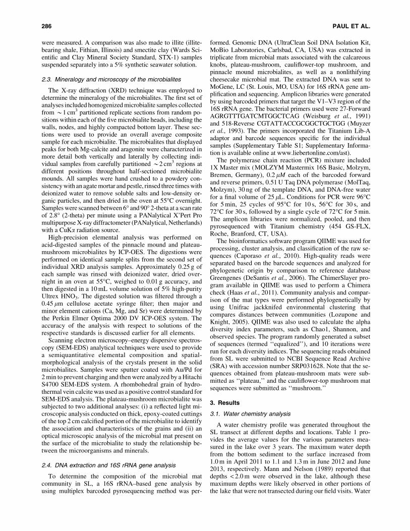
3.1. Water chemistry analysis
A water chemistry profile was generated throughout theSL transect at different depths and locations. Table 1 pro-vides the average values for the various parameters mea-sured in the lake over 3 years. The maximum water depthfrom the bottom sediment to the surface increased from1.0 m in April 2011 to 1.1 and 1.3 m in June 2012 and June2013, respectively. Mann and Nelson (1989) reported thatdepths < 2.0 m were observed in the lake, although thesemaximum depths were likely observed in other portions ofthe lake that were not transected during our field visits. Water
286 PAUL ET AL.
parameters fluctuated between each sampling visit. Forexample, the average pH changed from 8.22 – 0.05 to8.58 – 0.08 and back to 8.22 – 0.2 in the 3 years of sampling.The corresponding conductivity readings in April 2011, June2012, and June 2013 were 138 mS/cm (*92 ppt), 64 mS/cm(*43 ppt), and 59 mS/cm (*39 ppt), respectively, whileocean water collected from nearby Dim Bay in 2012 had aconductivity value of 53.9 mS/cm (*36 ppt). Water con-ductivity was measured during cursory visits to the island inMay 2004, 2005, and 2009. The conductivity values duringthese 3 years were 98.3 mS/cm (*66 ppt), 108 mS/cm(*72 ppt), and 40.6 mS/cm (*26 ppt), respectively.
Calcium and magnesium hardness was measured in 2012and 2013. The average calcium hardness in 2012 was1328 – 40 mg/L as CaCO3 (532 – 16 mg/L Ca), and that ofmagnesium was 6953 – 296 mg/L as MgCO3 (2005 – 85 mg/L Mg). The respective Ca and Mg concentrations, as de-tected by ICP-OES, were 507 – 16 and 1681 – 49 ppm (mg/L). Alkalinity did not change much in the 3 years, with thehighest value in 2011 of 178 – 8 mg/L and lowest in 2013 of140 – 27 mg/L. Dissolved oxygen consistently showed adecreasing trend from the shallow to deeper water levels.For example, in 2011, the average dissolved oxygen de-creased from 5.0 (– 0.6) mg/L in shallow waters to 4.1(– 0.7) mg/L at *25 cm, 3.3 (– 0.8) mg/L at *50 cm, 3.4(– 0.4) mg/L at *75 cm, and 2.2 mg/L (– 0.1) at *100 cm.Quantitative readings for dissolved oxygen could not bemade in 2012 and 2013 due to error in instrument calibra-tion. A vertical and lateral profile of the water characteristicsis provided in the Supplementary Information along withcomparison with previous investigations in the lake.
Saturation index values were calculated for carbonateminerals with the equilibrium-based PHREEQ softwareprogram and the measured chemical parameters of SL
water. The calculated SI values indicate that the waterconditions always remained supersaturated with respect toboth aragonite and calcite (SI > 0.0). For aragonite, the SIvalues in 2011, 2012, and 2013 averaged 1.13, 0.94, and0.95, respectively. The corresponding calcite SI values were1.27, 1.08, and 1.09. The lower 2012 and 2013 SI values forboth carbonate phases reflect the influence of a heavyrainfall event that occurred before and during water sam-pling visits to the lake.
Particulate material in SL water was examined by opti-cal microscopy and found to be dominated by a variety ofalgae, bacteria, diatoms, and dinoflagellates, all of whichcontributed to the high turbidity (Supplementary Fig. S1).Comparisons made between settling rates of particulatematerial from SL with illite and smectite clay samplessuspended in a 5% synthetic seawater solution are shownin Fig. 3. The turbidity values indicated that the lake watersample took *180 days to reduce from a turbidity value of121 to 8 NTU. In contrast, illite and smectite clays in the5% seawater solution took only 11 and 6 h, respectively, togo from a starting turbidity of approximately 450 to 8NTU.
3.2. Microbialite morphology and distribution
A representative transect of the distribution of micro-bialites identified in the northern section of SL is shown inFig. 4. Several of the microbialite types have been previ-ously described (Mann and Nelson, 1989; Neumann et al.,1989) and include calcareous knobs (Fig. 5A), plateau-mushroom shaped (Fig. 5B), pinnacle mound (Fig. 5C), andcauliflower-top mushroom shaped (Fig. 5D) stromatolites.Although previously described, we observed that the near-shore calcareous knobs or bulbous crust (Neumann et al.,
Table 1. Water Parameters Measured during the Three Sampling Years
Parameters
March31–April 01, 2011
June 16–20,2012
May 30–June 3,2013
SeawaterJune 16, 2012
15:00 (EST) 15:00 (EST) 16:00 (EST) 14:00 (EST)
Max. water depth *1 m *1.1 m *1.3 m –pH 8.22 (– 0.05) 8.58 (– 0.08) 8.22 (– 0.20) 8.26Eh (mV) -60 (– 2) -80 (– 12) -73 (– 14) -55Alkalinity (mg/L as CaCO3) 178 (– 8) 151 (– 1) 140 (– 27) 113Ca hardness (mg/L as CaCO3) – 1,328 (– 40) 1,150 (– 233) 1,060Mg hardness (mg/L as CaCO3) – 6,953 (– 296) 6,340 (– 466) 5,840Temperature (�C) 27.5 (– 1.0) 28.7 (– 1.2) 24.6 (– 0.2) 28Conductivity (mS/cm) 138 (– 4.5) 64 (– 1.3) 59 (– 2.3) 53.9Salinity (ppt) 92.1 (– 3.0) 42.7 (– 0.9) 39.4 (– 1.5) 36.0Dissolved oxygen (mg/L) 3.86 (– 1.07) 5.72 (– 1.80) 3.11 4.70Turbidity (NTU) 169 (– 15) 96 (– 12) 135 (– 12) 5Calcium (ppm)* 990 (– 32) 503 (– 15) 689 (– 52) 386Magnesium (ppm)* 2,873 (– 84) 1,676 (– 43) 2,033 (– 108) 1,361Sodium (ppm)* 25,133 (– 275) 14,422 (– 280) 16,692 (– 807) 11,811Potassium (ppm)* 926 (– 27) 520 (– 15) 629 (– 28) 417Strontium (ppm)* 22 (– 1.5) – – –Chloride (ppm){ – 24,075 (– 624) – 19,000Sulfate (ppm){ – 3,910 (– 94) – 2,700
Note: The average values of 6–20 measurements from different depths and locations each year are shown with their standard deviation.Concentration of calcium, magnesium, sodium, and potassium was analyzed using ICP-OES (*); the anions chloride and sulfate were analyzedusing ion chromatography ({) only for the 2012 samples. Near-shore water measurements in brackish water zone are not included in the average2013 values. Salinity was calculated by multiplying the conductivity value with 0.667 to obtain parts per thousand (ppt) value (Boyd, 2002).
MINERALOGY AND DIVERSITY OF STORR’S LAKE MICROBIALITES 287

1989) exhibited two distinct features: (i) a lower, darker-colored, fused-granular, carbonate structure (*6 to 7 cm inthickness) by which the microbialite was attached to thelake bottom and (ii) an upper, lighter-colored carbonatestructure (*5 cm thick) that had a smoother, continuouscarbonate buildup compared to the granular bottom portion.A microbial mat with sporadic eukaryotic algal material wasfound between the grooves and on the uppermost surface.
It is important to note that both the cauliflower-topmushroom and the plateau-mushroom types described herepossess the same overall external morphological appearancebut varied in certain surface characteristics as describedbelow (and also Supplementary Fig. S3). Additionally, thecauliflower-top mushroom reported here is the same asthe ‘‘mushroom type’’ described by Neumann et al. (1989).The new terminologies, that is, plateau-mushroom andcauliflower-top mushroom types, were required to differ-entiate between the two morphologically different types ofmushroom microbialites identified in our study. Bothplateau-mushroom and cauliflower-top mushroom typeswere found at similar depths of 70–100 cm. The cauliflower-
top mushroom microbialite had several bulging calciumcarbonate knobs on the top surface, each resembling indi-vidual, smaller mounds, similar to the mushroom knobsreported by Neumann et al. (1989). These mounds began todisappear (*3 to 4 cm from the top) and were followed bycontinuous horizontal calcium carbonate laminations withintermittent branching or thrombolitic nodes. The lamina-tions ended in the bottom stalk, which was very fragile andcrumbled easily.
The uppermost surficial crust of the pinnacle moundmicrobialite was a continuous layer that was 0.5–1 cm thickon which the microbial mats were found in a scatteredmanner (Fig. 5C). A thrombolitic morphology involving acombination of branched and massive features that were *7to 8 cm thick was observed directly below the upper crust.These thrombolitic features were replaced below by a highlycompacted region that extended between 5 and 6 cm. In itsbottom-most region, the pinnacle mound exhibited stro-matolitic layering for 1–2 cm. The single pinnacle moundthat we collected was broken off as it was pulled from thesoft muddy sediment at the base of the lake; thus the exact
FIG. 4. Overview of the distribution of the microbialites in SL showing the approximate locations where the microbialitesand water samples were collected in previous studies (Mann and Nelson, 1989; Neumann et al., 1989) and currentinvestigation. Horizontal distance from the western shore to Cactus Island is *450 m.
FIG. 3. Turbidity measurements in controlled environment, comparing SL particle settling rates with smectite and illiteclays suspended into a 5% synthetic seawater solution. The relatively flat horizontal settling trends at the beginning of eachtest reflect the time taken for the suspended particles to move from the top of the water column in the vials to below the levelof the light-scattering detector.
288 PAUL ET AL.

length of this type of microbialite could have been longerthan that described here (i.e., *17 cm). The frequency oflithified microbialite structures decreased in the deepestportions of the lake, except for the occasional, broken cal-cified knobs that were submerged in *30 cm thick gelati-nous bottom material (termed as ooze).
Moving toward Cactus Island, another microbialite mor-photype was identified at a depth of about 46–50 cm, herereferred to as the ‘‘multi-cuspate.’’ The distribution of themulti-cuspate microbialites was fairly continuous from thepoint it began to appear but was less frequent in the shallowwater toward the shore of Cactus Island. The multi-cuspatemicrobialites were morphologically different from the othermorphotypes owing to their more distinguished protrusionsand sharper edges (Fig. 5E). The closest resemblance was tothe calcareous knobs that were located at similar depths buton the other end of the transect along the western shore ofSL. The multi-cuspate structures were well lithified and didnot crumble easily (Supplementary Fig. S4). The bottom 1or 2 cm of the multi-cuspate microbialite was layered andseemed to represent the locations by which it was anchoredto the lake bottom sediment. Similar bottom layering wasalso found in the pinnacle mound microbialite. On top ofthis layered profile, the microbialite began to form a massivecarbonate structure, which eventually proliferated to resultin sharper edges of varying lengths, resembling severalfused cusplike formations. These large (*3 to 5 cm) pro-trusions distinguished these microbialites from the calcare-ous knobs. The multi-cuspate microbialites possessedlimited microbial mat material on the top surface unlike theother microbialite types found in SL, with occasional ge-latinous green and pink material found scattered on thegrooves and flatter surfaces.
A well-laminated, cohesive, but weakly lithified and flat-lying ‘‘cheesecake’’ microbial mat was found to be presenton top of the gelatinous material (ooze) in most lake-bottom
locations (Fig. 5F). This mat type was particularly abundantin the region between the plateau-mushroom and pinnaclemounds, and the multi-cuspate microbialite and Cactus Is-land shore. The laminations in the cheesecake mat extend toabout 7–8 cm depth from the top of the sediment layer. Aloose and friable, calcified layer existed below the lamina-tions. The laminations disappeared below this calcified layerand were replaced by the soft yet cohesive gelatinous ma-terial that extends below for approximately 30 cm, till itreaches the hard, bottom layer of the lake. The cheesecakemat was the only microbial mat collected that was not as-sociated with the lithified microbialite heads.
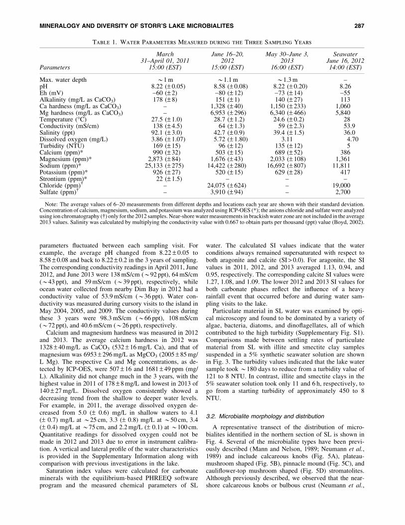
3.3. Light penetration in Storr’s Lake water
Due to the high turbidity of the SL water, it was essentialto determine the amount of light available for the photo-trophs in the microbial mats located at different depths inthe water. The Secchi disk method was used to determinelight penetration at two similar locations, where the cal-careous knob microbialites were identified ( June 2012). Thewhite portion of disks was not visible below depths of 17–26 cm, and the black portion disappeared below depths of15–20 cm. Neumann et al. (1989) reported depths of 46 cmbelow which the Secchi disk disappeared during their De-cember 1987 study.
Light intensity was also measured by HOBO pendantloggers with results shown in Fig. 6. In 2012, the probe wasinitially set into the location adjacent to the top of calcare-ous knob microbialites at a depth of 40 cm. The Day 1readings were collected on an overcast and mostly cloudyday and recorded a maximum signal of *1150 to 1200lumens/ft2 (107–112 lumens/m2). The probe was removedfrom the water and exposed to direct sunlight above thewater surface during Day 2, where a maximum reading of*20,000 lumens/ft2 (1,860 lumens/m2) was obtained. The
FIG. 5. Microbialites and microbial mats collected from SL. (A) Calcareous knobs; bar = 12 cm. (B) Plateau-mushroom;bar = 10 cm. (C) Half-sectioned pinnacle mound; bar = 7.5 cm. (D) Cauliflower-top mushroom; bar = 10 cm. (E) Multi-cuspate type; bar = 5 cm. (F) Cohesive but nonlithified cheesecake mat shown in cross-section profile; bar = 7.5 cm.
MINERALOGY AND DIVERSITY OF STORR’S LAKE MICROBIALITES 289
same day, the probe was then moved and inserted at thesame height as the top of a plateau-mushroom microbialiteat a depth of about 100 cm and kept in this position for2 days. The weather patterns changed from partly cloudy onDays 2 and 3 to scattered rain on Day 4. The light intensityduring Days 3 and 4 at the plateau position varied from 200to 600 lumens/ft2, or between 1% and 3% of the surfacelight intensity.
In 2013, the probe was placed at *95 cm next to the top ofthe plateau-mushroom shaped microbialite and left in placefor 6 days. The highest intensity of light at this depth wasrecorded at 12 lumens/ft2 (1.12 lumens/m2) relative to thesurface, where a maximum intensity of *5100 lumens/ft2
was obtained. This represents a decrease in luminosity with96 cm of water depth of approximately 99.8%. The lowerlight intensity values in 2013 as opposed to 2012 were due tothe heavy overcast conditions. Unfortunately, the overcastand rainy conditions present during both our 2012 and 2013sampling intervals prevented us from making any recordingsunder full sunlight conditions with the HOBO pendants.
3.4. Mineralogy and microscopic characterizationof the microbialites
Magnesium calcite was the dominant mineral found inhomogenized composite samples of all five microbialitetypes prepared and analyzed separately by XRD. Aragonitewas detected only in the pinnacle mound, plateau-mushroom, and in smaller quantities (*3%) in the multi-cuspate type.
A second set of XRD analyses was conducted, with in-dividual samples being collected from multiple positionswithin the plateau-mushroom and pinnacle mound micro-bialites to discern whether they displayed any spatial vari-ability with respect to both aragonite and Mg-calcitecontents (Fig. 7). The pinnacle mound (PM) microbialite
contained aragonite in the top 1 cm surface section (PM-1b-C); however, no aragonite was detected in the sample col-lected at a 2 cm depth below the top surface (PM-2b-C) andin the central core of the sample located 9 cm below thesurface. Aragonite percentages were highest at the base ofthis microbialite (PM-3b-S to PM-4b-C) and at the lateralsurfaces (PM-6-S). In the plateau-type microbialites, bothlateral surficial (Plateau-S) and core (Plateau-C) sampleshad some of the highest proportions of aragonite detected inSL microbialites, though the aragonite/calcite ratios de-creased outward; 76/24 for core versus 48/52 for surficialsample, respectively. Although the aragonite/calcite ratiosvaried from site to site within the microbialites, no particulartrend was observed with regard to water depth or distancefrom the shore in the microbialites that were evaluated.
Insoluble components were not detected as residues byvisual inspection following deionized water rinse (to removesalts) and a 5% nitric acid digestion step performed on 11sections of the microbialite heads. Calcite with variable mol% Mg (7.5–15.6%) was identified as the dominant mineraltype in all samples analyzed by combined ICP-OES(Table 2) and XRD analyses (Fig. 7). Concentrations of Siand Na were at, or just above, detection limit (i.e., 10 ppm),while Fe, K, Mn, and Al concentrations were all belowdetection. An explanation for the correlation between theelemental concentration and XRD mineralogy is provided inSupplementary Fig. S5.
Preliminary investigation with reflected light microscopyof thick, epoxy-coated sections of the pinnacle mound mi-crobialites revealed grains that were generally subrounded,sand-sized, and present in a layered format (SupplementaryFig. S6). Many of the rounded grains appeared to be zoned,often with one overgrowth layer, rarely two, around a cen-tral core grain. Most grains were in contact with each otherin the 2-D X-sections. However, in a couple of locations,these grains were isolated and not in proximity with the
FIG. 6. Light-intensity data collected using HOBO logger anchored into the bottom sediment and set at a depth equivalentto that of the microbialite heads. The graph shows the data from day and night cycles recorded in (A) 2012: Day 1, probeplaced next to calcareous knobs (at 40 cm depth); Day 2, probe exposed to direct sunlight above the water (the y axis in theplot is cut off at 5000 lumens/ft2, but the actual value when exposed to sunlight reached *20,000 lumens/ft2); Day 3, probeleft at 100 cm depth; Day 4, probe removed from lake water—the small spike at *1800 lumens/ft2 was due to exposure tosurface sunlight under overcast conditions. (B) 2013: Probe emplacement at a depth of *95 cm for the entire 6 days. Thespiked value at the end of the run at Day 6 was produced when the probe was exposed to sunlight under overcast conditions.The y axis in the plot is cut off at 20 lumens/ft2, but the total subaerial light intensity on Day 6 was *5100 lumens/ft2.
290 PAUL ET AL.

neighboring grains. A couple of grains of Homotrema ru-brum could be seen. Porosity, as seen by epoxy-filled cav-ities, was also observed.
Microscopic investigation of the microbial mat in theplateau-mushroom microbialite indicated that, in addition to
the microorganisms, there were abundant gelatinous features(EPS) with various dark, amorphous grains (data not shown).These irregular grains reacted with 1 N HCl and producedCO2 bubbles, indicating that those were carbonate minerals.Some of the EPS material also exhibited effervescence with
FIG. 7. Microbialite samples depicting the various positions where samples were collected for XRD analysis. (A) Half-sectioned pinnacle mound (PM) and (B) half-sectioned plateau-mushroom shaped microbialites. Samples were collectedeither in the center, C, or at surface, S. Numbers to the right of the sample locations in (A) and (B) correspond to thearagonite:calcite percent ratio as determined by XRD analysis. Examples of XRD pattern for two of the samples, PM-1b-C(C) and Plateau-C (D), show peaks for aragonite and calcite, shown by the letters A and C, respectively.
Table 2. ICP-OES Results for the Acid-Digested Microbialite Solid Samples Showing
the Profile Positions as Noted in Figure 7, the S mM Values of Ca, Mg, and Sr Recovered
from the Digestion of 0.25 g of Solid in 10 mL of Solvent
Sample
mMMolar
Ca:Mg ratioMolar
Mg:Ca ratio Mol % Mg Total Sr (mM)Ca Mg
Pinnacle moundPM-1b-C 201 35.2 5.7 0.18 14.9 0.06PM-2b-C 212 33.0 6.4 0.16 13.6 0.11PM-3b-S 207 16.7 12.4 0.08 7.5 0.26PM-4b-C 195 34.7 5.6 0.18 15.6 0.13PM-5-C 211 36.2 5.8 0.17 14.6 0.07PM-6-S 216 34.6 6.3 0.16 13.7 0.07
PlateauPlateau-top-C 203 32.9 6.2 0.16 14.2 0.09Plateau-middle-C 219 34.5 6.3 0.16 13.7 0.10Plateau-bottom-S 217 32.4 6.7 0.15 12.7 0.18Plateau-C 215 18.3 11.7 0.09 7.9 0.29Plateau-S 227 21.0 10.8 0.09 8.4 0.22
Vein calcite 259 0.09 2742 Insignificant Insignificant Not analyzed
The molar ratio of Ca:Mg and the molar Mg percentage in the samples is also shown.
MINERALOGY AND DIVERSITY OF STORR’S LAKE MICROBIALITES 291

HCl, revealing that these polymeric substances might alsohave carbonate material present in them. Similarly, examinationof the water column showed rare, dark, carbonate particlesin the midst of dispersed microbial and EPS assemblage.
Scanning electron microscopy–energy dispersive spec-troscopy analysis of the microbialites showed a variety ofinternal and external morphological features (Fig. 8). Themicrobialites were dominated by calcium carbonate crystals,with 0.5–3.9 atom % Mg. The morphology of the crystalsranged from irregular to angular, blocky, needle- andtriangular-shaped, with calcium carbonate–encrusted cya-nobacterial filaments and remains of EPS. The plateau-mushroom and pinnacle mound microbialite exhibitedoccasional needle-shaped structures, suggesting an arago-nitic mineralogy (Fig. 8A). Remains of diatoms, such asNavicula, were also observed to be present (Fig. 8B).
3.5. Microbial mat diversity
Cultivation-independent microbial diversity analyseswere performed on the five different types of microbial mats(i.e., calcareous knob, plateau-mushroom, pinnacle mound,cauliflower-top mushroom shaped, and cheesecake) isolatedfrom SL. Amplicon libraries of the partial 16S rRNA genes(V1–V3 region) were sequenced and clustered into opera-tional taxonomic units (OTUs) at 97% similarity or greater.The summary of the data obtained from the pyrosequencingmethod is shown in Table 3. A total of 126,576 barcodedsequences was recovered after sequence trimming and fil-tering by using mixed and dots filters. The mixed filter re-moved reads that occurred as a result of simultaneouslysequencing a mixture of different DNA molecules, whereasthe dots filter removed sequences with three successivenucleotide flows that recorded no incorporation. The aver-age read length of the sequences was 471 bp. The five dif-ferent mat types had between 15,782 and 31,862 sequencereads and were clustered into OTUs with QIIME software(Caporaso et al., 2010).
Alpha diversity index values (Table 3) were calculatedbased on equalized sequences and indicate that all five mi-crobial mat types exhibited high diversity. Chao 1 values,which were used to estimate species richness, indicate that
the deeper-water plateau-mushroom microbial mat had thelargest estimated diversity (20,915), whereas the lowest wasfor the shallow-water calcareous knobs (7,743). The mat onthe plateau-mushroom also had the highest Shannon indexvalues (12.03). The Chao1 values correspond well with theShannon index and observed species values. The distribu-tion of species was most even in the plateau type and closelyfollowed by the other microbial mats as shown by the eq-uitability values.
Among the OTUs identified, only 24% of the recoveredsequences were shared between all five microbial mats,suggesting that each mat type had a distinctive community.The relatedness between the five microbial mat communitieswas also calculated with beta diversity analysis. A distancetree was constructed by using the unweighted pair groupmethod with arithmetic mean (UPGMA). Jackknife analysisallowed repeated subsampling of a subset of data from eachsample, providing a final robust distance tree (Fig. 9A). Themat on the shallow-water calcareous knob was most similarto the plateau-mushroom microbial mat in terms of micro-bial population. These two mats clustered closest with thecheesecake microbial mat that was found on top of the ge-latinous material covering the lake bottom at deeper-waterlocations. The deep-water pinnacle mound microbialitegrouped closely with the cauliflower-top mushroom micro-bialite. Both of these latter two types of microbialites arelocated farther away from the western shore of the lake andare submerged to greater water depths.
The bacterial composition of the five mats fell into 12major bacterial phyla, each distributed in various relativepercentages among the mats (Fig. 9B). Though a largeamount of sequences was recovered, the analysis of only apartial length (V1–V3) of the entire 16S rRNA gene allowedidentification only up to the family level of taxonomy, withfew sequences reaching only to level of class or order. Thecheesecake microbial mat had approximately one-third(36%) of its OTUs as unclassified, which was the highestamong the five mat types. The shallow-water calcareousknob had the lowest percentage (12%) of unclassified bac-teria present. Chloroflexi constituted approximately half(*46%) of the entire bacterial population identified in boththe deeper-water cauliflower-top mushroom and pinnacle
FIG. 8. Scanning electron microscope images of the microbialite structures. (A) Acicular crystals, characteristic ofaragonite, found in the lower section of plateau-mushroom microbialite. (B) Diatoms in the microbialites of SL. Remains ofEPS material are indicated by black arrows.
292 PAUL ET AL.

mound mats, while calcareous knob, plateau-mushroom, andcheesecake types had only 16–20% Chloroflexi. Within thephylum Chloroflexi, anaerobic halorespirers, Dehalo-coccoidaceae made up to 24% in the cauliflower-topmushroom and 21% in the pinnacle mound. Other members
included several bacteria under the class Anaerolineae andorder Dehalococcoidetes (family Dehalococcoidaceae fallsunder this order). Members of the yet-uncultured OPB11order, which is thought to include several green nonsulfurbacteria (Hugenholtz et al., 1998), were present in notable
Table 3. Summary of the Bacterial 16S rRNA Gene Diversity Analysis by the Pyrosequencing Method
Calcareous knobs Plateau Cheesecake Mushroom Pinnacle mound
Sequences 31,862 30,608 28,082 20,242 15,782OTUsa 5,636 12,261 9,096 6,859 4,897Singletonsa 3,083 8,118 5,872 4,493 3,075Doubletonsa 852 1,774 1,355 1,020 795Equalized sequencesb 14,500 14,500 14,500 14,500 14,500Chao1c 7,743 (– 236) 20,915 (– 725) 15,697 (– 347) 14,218 (– 262) 10,507 (– 183)Chao1 confidenced 7,343/8,054 20,217/22,302 15,209/16,213 13,873/14,547 10,260/10,800Shannonc 10.07 (– 0.02) 12.03 (– 0.02) 11.25 (– 0.02) 10.90 (– 0.01) 10.49 (– 0.01)Observed speciesc 3,595 (– 37) 7,340 (– 41) 5,840 (– 42) 5,501 (– 23) 4,646 (– 14)Equitability 0.82 0.92 0.88 0.87 0.86% Coveragee 90.3 73.4 79.1 77.8 80.5
aValues calculated based on 97% similarity threshold.bNumber of equalized sequences used to generate the diversity indices, such as Chao1, Shannon, observed species.cAverage values – standard deviation of diversity using 10 iterations of randomized sequences.dChao1 lower and upper confidence limits at 95%.eBased on Good’s coverage estimator.
FIG. 9. (A) Dendrogram showing the relationship between the different microbial mats in SL acquired by jackknifedUPGMA clustering method. The scale bar indicates the evolutionary distance between the communities and has a value of0.05, meaning 0.05 nucleotide substitutions per sequence position. (B) Relative percentage abundance of major bacterialtaxa detected in five SL microbial mats by analysis of the 16S rRNA gene. Twelve different major taxa are reported alongwith significant percentages of candidate phyla and unclassified bacteria. Note: Plateau indicates plateau-mushroom mi-crobialite, and mushroom indicates cauliflower-top mushroom microbialite.
MINERALOGY AND DIVERSITY OF STORR’S LAKE MICROBIALITES 293

quantity, especially in the pinnacle mound and cheesecakemat (*9% in each). The class Deltaproteobacteria includesseveral SRB that were enriched in the shallow-water cal-careous knob (21%), followed by the cheesecake mat (16%).The lowest percentage of Deltaproteobacteria occurred inthe deeper-water pinnacle mound (5%) and cauliflower-topmushroom (6%) mat types. The SRB family, Desulfo-bacteraceae, dominated the recovered Deltaproteobacteriasequences. For example, 16% of the 21% of Deltaproteo-bacteria were represented by Desulfobacteraceae. Othersignificant SRB members in this class included family De-sulfohalobiaceae and Desulfovibrionaceae. The other taxarepresented in all mat types included the Spirochaetes,Planctomycetes, Alphaproteobacteria, Gammaproteobacte-ria, Bacteroidetes, and Caldithrix. Purple nonsulfur bacteriaof families Rhodospirillaceae and Rhodobacteraceae be-longing to the class Alphaproteobacteria were detected onlyin the plateau-mushroom and calcareous knob microbialites.
Several members of the phyla discussed above includehalophilic and brackish-living bacteria, with many of themalso being photosynthetic (e.g., order Chromatiales andfamily Chromatiaceae in the Gammaproteobacteria) andable to survive in anoxygenic environments (Imhoff, 2006).Vibrionaceae is yet another anaerobic, phototrophic Gam-maproteobacteria that represented *4% of the total cal-careous knob population. Cyanobacteria were detected atlow levels in all the mat types ranging between <1.0% and3.0%. The highest levels of cyanobacteria were observed inthe shallow-water calcareous knobs (*3.0%) and includedfamilies Cyanobacteriaceae, Pseudanabaenaceae, and Phor-midiaceae, and order Chroococcales. The other mat typeshad less than 1.0% relative abundance of cyanobacteria.Other taxa, such as Acidobacteria, Verrucomicrobia, andLentisphareae were below 1.0%, while Firmicutes, Actino-bacteria, Chlorobi, Elusimicrobia, Betaproteobacteria, andEpsilonproteobacteria were present at less than 0.1% (Sup-plementary Table S2). A relatively high proportion of themat population was classified as candidate phyla, whichinclude bacteria that have not yet been cultured in the lab-oratory but whose existence has been observed through 16SrRNA gene analysis. For example, 11% and 8.0% of thecauliflower-top mushroom and pinnacle mound population,respectively, were represented by the order HMMVPog-54.These have previously been identified in methane-rich bio-film communities with a high diversity of Deltaproteo-bacteria (Losekann et al., 2007). Additionally, bacteria fromthe phyla WS3 (Dojka et al., 1998) occupied anywhere from*0.6% in cauliflower-top mushroom shaped to a maximumof *3.4% in calcareous knobs.
4. Discussion
4.1. Physical, chemical, and optical characterizationof Storr’s Lake water
Water parameters, such as depth, salinity, local turbidity,and so on, in SL exhibited high fluctuations that are pri-marily influenced by rainfall events (Table 1). The mainbody of water was always turbid during field analysis, but arelatively clear-water brackish lens zone developed adjacentto the shoreline following periods of rainfall. The associatedbrackish-saline mixing zone migrated outward into thedeeper portions of the lake during periods of high meteoric
water runoff (Fig. 2B). Previous studies have reported thatrainfall events can cause considerable dilution of the lakewater and result in lowered salinity and an increase in themetabolism of the phototrophic community in the lake(Pinckney et al., 1995; Paerl et al., 2003). The lowering ofsalinity following rain events was confirmed in the presentstudy, and we believe that our May 2009 recording of*26 ppt is the lowest salinity reading ever recorded for SL.This lower-salinity period can enable opportunistic gastro-pods and fish species to occasionally colonize the lake(Garrett, 1970; Supplementary Fig. S2).
The reddish-brown color in the lake is imparted by twosources: (i) the organic tannins brought in by the brackishwater draining into the lake that were derived from decayingplant material and (ii) the presence of planktonic commu-nities of halophilic algae and bacteria, which are usuallyenriched in carotenoid pigments that contribute to the red-dish coloration (Mann and Nelson, 1989; Oren and Rodrıguez-Valera, 2001). Bacteria, algae, and most organic materialhave a net negative surface charge, causing these particles torepel from each other, enabling the material to becomecolloidal and remain suspended rather than settling to thelake bottom. The long-term suspension of particles in thewater (Fig. 3) supports the colloidal nature of the particles.Motile planktonic microorganisms seeking sunlight for theirmetabolism would also move toward the surface of thewater during day. Neumann et al. (1989) suggested thatoxygen gas released from the gelatinous thromboliticmounds also carried some of the mat material to the surface.A similar process, possibly involving other gas componentsas well, could be presumed to occur with the microbial matsat deeper depths. Additionally, high wind activity seen in thelake could possibly agitate the water and thus keep theparticles suspended, although the stratification observed indissolved oxygen profiles suggests that complete and rapidvertical mixing is hindered. It should be noted that tidal flowhas only a minimal effect on the lake due to limited con-nectivity to the ocean, though abnormal high or low tidescould influence the lake parameters (Mann and Nelson,1989). Density changes due to seasonal or diurnal warming-cooling and evaporative concentration of dissolved com-ponents also could conceivably play a role in water mixing.In addition to the above factors, freshwater inflow duringrain and storm events would also affect the changes seen inturbidity, pH, salinity, and other water parameters.
The average turbidity was 96 (– 12) and 135 (– 12) NTU,during 2012 and 2013, respectively. Likewise, the light in-tensity reaching depths of *100 cm in both these years was200–600 and 12 lumens/ft2, respectively, ranging from 0.2%to 3.0% of the intensity of sunlight reaching the surface ofthe lake. The limited sunlight reaching the bottom wasscattered between particles as it penetrated through thewater column (Figs. 3 and 6). It is thus interesting that somephotosynthetic communities survive even in the deepermicrobialites that are located in the dim-light conditions(Fig. 9B). In one recent study, cyanobacterial mats showedmaximum photosynthetic efficiency under limited lightconditions (Al-Najjar et al., 2012). The wavelengths of thepenetrating light are also important in governing the pho-totrophic system. For a cyanobacterial photosynthetic com-munity to function, it requires light of wavelengths between400 and 700 nm (Jørgensen et al., 1987). However, some
294 PAUL ET AL.

photosynthetic communities are capable of surviving inwavelengths of the extended spectrum (UV and IR), havingdeveloped specialized pigments and enzymes capable ofusing wavelength fractions (Castenholz and Garcıa-Pichel,2000; Kuhl et al., 2005). Phototrophic green sulfur bacteria,for example, contain light-harvesting chlorosomes that makethem well-adapted to survive in low-light conditions (Fri-gaard and Bryant, 2004). The light probe used for SL wateranalysis measured a full array of wavelengths from 200 to1100 nm but unfortunately does not differentiate betweenthe different wavelengths. However, in the case of SL, theshorter-wavelength UV spectrum would be expected to bemore penetrating and, thus, extend to deeper sections of thelake. Also, the small amount of light reaching to depths of*1.0 m appears to be able to support a photosyntheticcommunity on the microbialites based upon our microbialdiversity results (Fig. 9B). Future investigations into thespectrum of light used by the microbial communities in SLwould be of great interest.
4.2. Distribution and mineralogy of microbialites
The external morphology of the cauliflower-top mushroommicrobialite identified in our study was similar to the previ-ously reported plateau-mushroom type (Mann and Nelson,1989; Neumann et al., 1989), but closer observation revealeddifferences in laminations and appearances (SupplementaryFig. S3). Hence, the cauliflower-top mushroom and plateau-mushroom were separately categorized in this study. In ad-dition to the previously identified microbialites, we describeda new morphotype of the microbialite termed ‘‘multi-cuspate’’ that had limited microbial mat material. The lackof prominent microbial mat development on the surface ofthe multi-cuspate microbialites was not a reflection of waterdepth, since these were located at almost the same waterlevel as the calcareous knobs. Since microbial mat coverageand thereby the activity of mat organisms are inferred to beinadequate, the multi-cuspate type could be categorized as a‘‘sub-fossil.’’ The exact reason for the limited microbial matcover on the multi-cuspate microbialite is still unknown andis worthy of further investigation.
Similar to other environments that harbor microbialites,the overall microbialite morphology is generally accepted tobe primarily controlled by environmental factors, though themicroorganisms play the role of actual accretion and fabricdevelopment (Dupraz et al., 2006). One of the major con-trols is the water depth of SL, and in turn the availability oflight for the microorganisms, especially phototrophs. Theflat top of the slightly deeper plateau microbialite has alsobeen interpreted as the lowest water level in the lake (Mannand Nelson, 1989). The bulging nature toward the top of themicrobialites indicates that the area closest to the surface isthe most productive region, where maximum light is ac-cessible and water depth is conducive.
A considerable amount of aragonite was identified inthe pinnacle mound and plateau-mushroom microbialites(Fig. 7). All previous studies of SL microbialites have re-ported only the presence of Mg-calcite and no aragonite(Hattin, 1983; Pentecost, 1989; Neumann et al., 1989;Fowler, 2011; Dupraz et al., 2013). We observed that themol % Mg in the carbonate minerals of the pinnacle mound–type microbialites initially decreased from the top to middle
positions (PM-1b-C to PM-3b-S) but then increased, movingfrom the middle toward the bottom of the microbialite (PM-4b-C). The aragonite percentage decreased from PM-1b-Cto PM-2b-C and PM-5-C and then increased again, reachingthe highest values with samples PM-4b-C and PM-6-S (22%and 24% aragonite, respectively; Fig. 7A and 7B; Table 2).It should be noted that the bottom, calcified, ground sub-strate of the lake, on which the microbialites develop, isaragonitic in composition. However, the microbialite sam-ples chosen for XRD analysis were carefully sectioned onlyfrom the microbialite structures themselves (exact locationsas shown in Fig. 7), thereby rendering any influence of thehard lake substrate negligible.
The presence of aragonite was further confirmed by thecorrelation seen between Sr and Mg concentration (Table 2,and Supplementary Fig. S5). The large radius of Sr2+ (1.18 A)is more readily accepted into the aragonite structure, whilethe smaller Mg2+ ion (0.72 A) gets preferentially incorporatedas a replacement for Ca2+ (1.00 A) in calcite (Krauskopf andBird, 1994). Calcite can accept a higher proportion of Mg inits crystal structure relative to aragonite, especially when thecalcite is precipitated in biologically mediated reactions(Weiner and Dove, 2003). Aragonite does not readily acceptMg in its crystal structure; thus the aragonite content shouldbe inversely correlated with Mg concentration. The resultsobtained generally support this expectation, but there wereclear anomalies. For example, both the aragonite percentageand Mg content reach their maximum values with samplePM-4b-C (22% aragonite and 15.1 mol % Mg; Table 2).Therefore, the Mg/Ca ratios may be controlled by processesassociated with calcite formation rather than exclusionaryprocesses arising from the formation of aragonite.
Two sources are likely attributed to the origin of arago-nite in the microbialites: an in situ biogeochemical precip-itation and/or detrital grains that are blown over from thenearby East Beach and incorporated into the structure. Thesaturation state of the carbonate minerals in water has beenshown to be an essential component in determining micro-bialite development (Dupraz and Visscher, 2005). The SIvalues for current SL water samples as determined with thePHREEQ program indicate that calcite precipitation is fa-vored over aragonite, based on equilibrium modeling.Though the microbial mats can drastically alter the avail-ability of ions, such as calcium and bicarbonate in the mi-croenvironment that they create (Dupraz et al., 2009), thecharacteristics of the water above them are also important incontrolling the mineralogy of the microbialites. The calciteand aragonitic composition in SL microbialites could thusbe partially or completely dependent on the past composi-tion of the lake water. Storr’s Lake began to form around3100 years ago due to shifting tidal sands that closed inlets(Zabielski, 1991). Carbon dating revealed that the micro-bialites began to form at least 2310 years ago (Paull et al.,1992). Terrestrial and evaporative processes likely wouldhave increasingly influenced the lake water compositionafter the basin became isolated from direct tidal input. Giventhe fluctuations observed in the lake water parameters, in-cluding ‘‘freshening events’’ and periods of hypersalinitynoted in previous studies (Pinckney et al., 1995; Paerl et al.,2003; Park, 2012) and this investigation, it is likely that thelake has experienced similar and possibly even more drasticchanges in its composition during its 3100-year history.
MINERALOGY AND DIVERSITY OF STORR’S LAKE MICROBIALITES 295
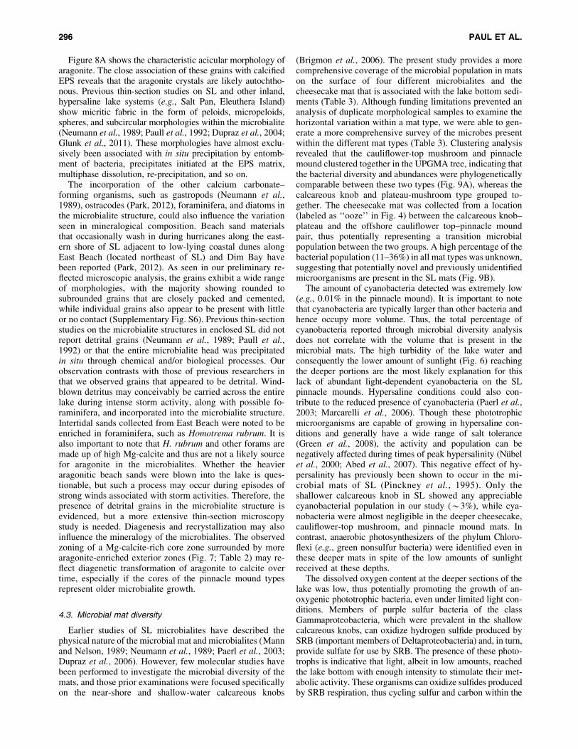
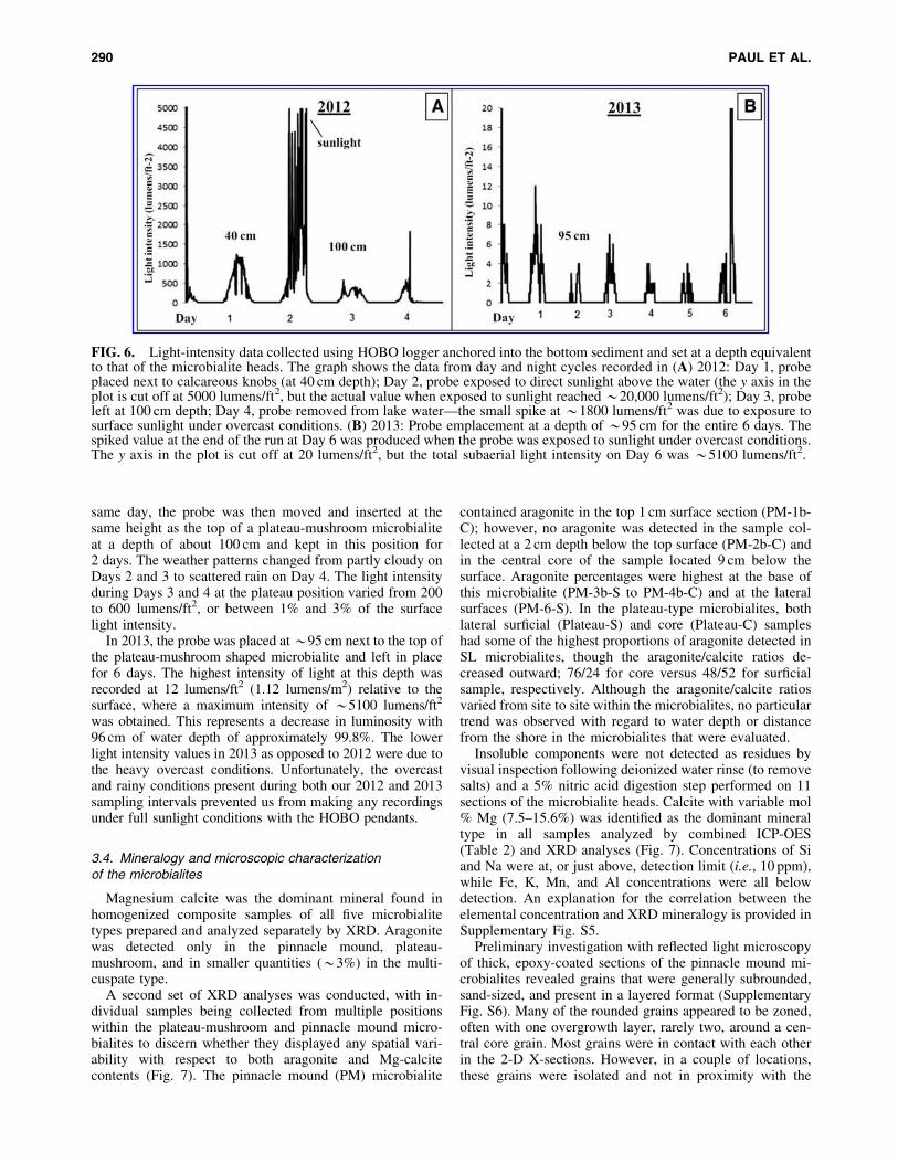
Figure 8A shows the characteristic acicular morphology ofaragonite. The close association of these grains with calcifiedEPS reveals that the aragonite crystals are likely autochtho-nous. Previous thin-section studies on SL and other inland,hypersaline lake systems (e.g., Salt Pan, Eleuthera Island)show micritic fabric in the form of peloids, micropeloids,spheres, and subcircular morphologies within the microbialite(Neumann et al., 1989; Paull et al., 1992; Dupraz et al., 2004;Glunk et al., 2011). These morphologies have almost exclu-sively been associated with in situ precipitation by entomb-ment of bacteria, precipitates initiated at the EPS matrix,multiphase dissolution, re-precipitation, and so on.
The incorporation of the other calcium carbonate–forming organisms, such as gastropods (Neumann et al.,1989), ostracodes (Park, 2012), foraminifera, and diatoms inthe microbialite structure, could also influence the variationseen in mineralogical composition. Beach sand materialsthat occasionally wash in during hurricanes along the east-ern shore of SL adjacent to low-lying coastal dunes alongEast Beach (located northeast of SL) and Dim Bay havebeen reported (Park, 2012). As seen in our preliminary re-flected microscopic analysis, the grains exhibit a wide rangeof morphologies, with the majority showing rounded tosubrounded grains that are closely packed and cemented,while individual grains also appear to be present with littleor no contact (Supplementary Fig. S6). Previous thin-sectionstudies on the microbialite structures in enclosed SL did notreport detrital grains (Neumann et al., 1989; Paull et al.,1992) or that the entire microbialite head was precipitatedin situ through chemical and/or biological processes. Ourobservation contrasts with those of previous researchers inthat we observed grains that appeared to be detrital. Wind-blown detritus may conceivably be carried across the entirelake during intense storm activity, along with possible fo-raminifera, and incorporated into the microbialite structure.Intertidal sands collected from East Beach were noted to beenriched in foraminifera, such as Homotrema rubrum. It isalso important to note that H. rubrum and other forams aremade up of high Mg-calcite and thus are not a likely sourcefor aragonite in the microbialites. Whether the heavieraragonitic beach sands were blown into the lake is ques-tionable, but such a process may occur during episodes ofstrong winds associated with storm activities. Therefore, thepresence of detrital grains in the microbialite structure isevidenced, but a more extensive thin-section microscopystudy is needed. Diagenesis and recrystallization may alsoinfluence the mineralogy of the microbialites. The observedzoning of a Mg-calcite-rich core zone surrounded by morearagonite-enriched exterior zones (Fig. 7; Table 2) may re-flect diagenetic transformation of aragonite to calcite overtime, especially if the cores of the pinnacle mound typesrepresent older microbialite growth.
4.3. Microbial mat diversity
Earlier studies of SL microbialites have described thephysical nature of the microbial mat and microbialites (Mannand Nelson, 1989; Neumann et al., 1989; Paerl et al., 2003;Dupraz et al., 2006). However, few molecular studies havebeen performed to investigate the microbial diversity of themats, and those prior examinations were focused specificallyon the near-shore and shallow-water calcareous knobs
(Brigmon et al., 2006). The present study provides a morecomprehensive coverage of the microbial population in matson the surface of four different microbialites and thecheesecake mat that is associated with the lake bottom sedi-ments (Table 3). Although funding limitations prevented ananalysis of duplicate morphological samples to examine thehorizontal variation within a mat type, we were able to gen-erate a more comprehensive survey of the microbes presentwithin the different mat types (Table 3). Clustering analysisrevealed that the cauliflower-top mushroom and pinnaclemound clustered together in the UPGMA tree, indicating thatthe bacterial diversity and abundances were phylogeneticallycomparable between these two types (Fig. 9A), whereas thecalcareous knob and plateau-mushroom type grouped to-gether. The cheesecake mat was collected from a location(labeled as ‘‘ooze’’ in Fig. 4) between the calcareous knob–plateau and the offshore cauliflower top–pinnacle moundpair, thus potentially representing a transition microbialpopulation between the two groups. A high percentage of thebacterial population (11–36%) in all mat types was unknown,suggesting that potentially novel and previously unidentifiedmicroorganisms are present in the SL mats (Fig. 9B).
The amount of cyanobacteria detected was extremely low(e.g., 0.01% in the pinnacle mound). It is important to notethat cyanobacteria are typically larger than other bacteria andhence occupy more volume. Thus, the total percentage ofcyanobacteria reported through microbial diversity analysisdoes not correlate with the volume that is present in themicrobial mats. The high turbidity of the lake water andconsequently the lower amount of sunlight (Fig. 6) reachingthe deeper portions are the most likely explanation for thislack of abundant light-dependent cyanobacteria on the SLpinnacle mounds. Hypersaline conditions could also con-tribute to the reduced presence of cyanobacteria (Paerl et al.,2003; Marcarelli et al., 2006). Though these phototrophicmicroorganisms are capable of growing in hypersaline con-ditions and generally have a wide range of salt tolerance(Green et al., 2008), the activity and population can benegatively affected during times of peak hypersalinity (Nubelet al., 2000; Abed et al., 2007). This negative effect of hy-persalinity has previously been shown to occur in the mi-crobial mats of SL (Pinckney et al., 1995). Only theshallower calcareous knob in SL showed any appreciablecyanobacterial population in our study (*3%), while cya-nobacteria were almost negligible in the deeper cheesecake,cauliflower-top mushroom, and pinnacle mound mats. Incontrast, anaerobic photosynthesizers of the phylum Chloro-flexi (e.g., green nonsulfur bacteria) were identified even inthese deeper mats in spite of the low amounts of sunlightreceived at these depths.
The dissolved oxygen content at the deeper sections of thelake was low, thus potentially promoting the growth of an-oxygenic phototrophic bacteria, even under limited light con-ditions. Members of purple sulfur bacteria of the classGammaproteobacteria, which were prevalent in the shallowcalcareous knobs, can oxidize hydrogen sulfide produced bySRB (important members of Deltaproteobacteria) and, in turn,provide sulfate for use by SRB. The presence of these photo-trophs is indicative that light, albeit in low amounts, reachedthe lake bottom with enough intensity to stimulate their met-abolic activity. These organisms can oxidize sulfides producedby SRB respiration, thus cycling sulfur and carbon within the
296 PAUL ET AL.
microbial mat system. Fermenters, such as members of theclass Phycisphaerae (maximum of 7% in the cheesecake mi-crobial mat) and phyla Spirochaetes were also present in the SLmicrobial mats, indicating the metabolically diverse popula-tion present (Magot et al., 1997; Fukunaga et al., 2009; Du-binina et al., 2011; Lee et al., 2014).
4.4. Microbial influence on carbonate mineralprecipitation and lithification
The results from the microscopic investigations of themicrobial mat in the plateau-mushroom microbialite and thewater column reveal that at least some carbonate particles inthe suspended water column could settle on the microbialitesurface. However, this finding does not rule out the in situcarbonate biomineralization (by the microorganisms) and/ororgano-mineralization (the influence of organic matrix, EPS,etc.) in the microbial mats. Another line of evidence for theoccurrence of biomineralization can be gathered from thepreferred orientation of the crystals of acicular aragonite(Fig. 8A), indicating that the minerals were precipitated bymicrobial activity (Weiner and Dove, 2003).
The microbialites at the deeper locations in SL have beensuggested to be inactive or ‘‘sub-fossilized’’ (Dupraz et al.,2006, 2013; Fowler, 2011). However, carbonate mineral–precipitating microbial communities, including anaerobicphototrophs and sulfate reducers (except for cyanobacteria),were detected in all mats sampled (Fig. 9B), although it wasnot clear whether these microorganisms are actively con-tributing to the precipitation on the microbialite head. In theabsence of cyanobacteria, carbonate mineralization can stillpotentially occur with the activity of SRB and anoxygenicphototrophs (Visscher et al., 2000; Dupraz and Visscher,2005). Considering the possibility that only shallow-watercalcareous knob microbialites are the ‘‘actively’’ formingmicrobialites, the SRB families in class Deltaproteobacteria(*18%) were present in higher number than the phylumCyanobacteria (*3.2%) in these mats. This large repre-sentation of Deltaproteobacteria indicates that the SRB arelikely playing an active role in carbonate mineral precipi-tation (Nitti et al., 2012). As the SRB and anoxygenicphototrophs were present in significant percentages in allmat types, it is possible that these two groups influencecarbonate mineralization in SL. The role of SRB in theprecipitation of carbonate minerals in microbialites has beenverified by using the 35SO4
2- coated Ag-foil technique,where maximum SRB activity has been shown to precipitatea calcified laminae (Visscher et al., 2000). Studies on therole of anoxygenic phototrophs on carbonate precipitationhave been gaining more attention (Bundeleva et al., 2012;Nitti et al., 2012), and this investigation further helpsstrengthen the importance of this microbial group as a keymember in a microbialite mat community. It is important tonote that, in addition to the abundance or even presence ofmicroorganisms capable of carbonate mineral precipitation,knowledge of their metabolic rates and the gene expressionpattern over spatial and temporal extent in the microbial matis essential to better understand the actual mechanisms in-volved (Dupraz and Visscher, 2005).
Storr’s Lake microbialites reveal that a microbial com-munity with a significant lack of cyanobacteria can still beinvolved in the precipitation of carbonate minerals. We
show here that sulfate reducers and anoxygenic phototrophs,as part of the microbial mat community, could be capable ofsustaining carbonate mineral precipitation and the relatedgrowth that occurs in a microbialite. This understandingwidens the ecology and associated metabolic functioning ofvarious microbial communities within the mat that con-tributed to the formation of ancient microbialites. For ex-ample, in an ancient environment with limited oxygen in theocean and atmosphere, anoxygenic phototrophs and sulfatereducers could have been key members in a microbialitecommunity. Our study further highlights the complexity ofthe microbial mat system in the SL environment, whereseveral biogeochemical components are suspected to play aninteractive role in the precipitation and lithification of themicrobialites.
5. Conclusions
Our research builds upon previous analysis and interpre-tation of (i) the water parameters, (ii) microbialite distri-bution and mineralogy, and (iii) the microbial matcomposition through the application of molecular tools.Light-penetration studies revealed that the turbid water re-stricted >97% of the luminosity to the deeper sections of thelake. One of our main findings was the identification ofaragonite as a significant part of the microbialite mineral-ogical composition in the plateau-mushroom and pinnaclemound microbialites. Secondly, a new microbialite mor-phology, ‘‘multi-cuspate,’’ was discovered near Cactus Is-land and categorized as a subfossil because of the lack ofprominent microbial mat on its surface. Thirdly, our mo-lecular diversity analysis of the microbial mats, the majorfocus of this investigation, revealed that the cyanobacterialpopulation was generally low and almost negligible in cer-tain deeper-water microbial mats. The lack of prominentcyanobacterial populations can be explained by the lessintense sunlight reaching those depths. On the other hand, aplethora of anaerobic phototrophs exist in these deeperwater mats, indicating that factors such as oxygen avail-ability or the lack of it is controlling the distribution of thesephototrophs in the dim-light conditions under which theyexist. Though most reported microbialite communities aretypically found in clear waters, the SL microbialites presentan anomaly to this view as they exist under highly turbidand light-limiting conditions. Hence, these and other un-common examples further necessitate the exploration ofpotential unique and diverse environments in which mi-crobialites could be found. Investigations on SL micro-bialites have potential implications for understandingpossible life on other planets and in applied areas, such ascarbon sequestration, where microbial activity could en-hance the conversion of CO2 gas into carbonate minerals inthe absence of light. A better knowledge of such micro-bialite systems could also provide useful clues with regardto past climatic and environmental conditions and act as apossible indicator of local or global climate in the future.
Acknowledgments
Funding for this study was provided by the Department ofEnergy–National Energy Technology Lab under agreementDE-FE0002416. Travel expenses were partially covered byDonald Radcliffe Trust–Geology and Geophysics Department,
MINERALOGY AND DIVERSITY OF STORR’S LAKE MICROBIALITES 297

Missouri S&T. We thank the staff of the Gerace ResearchCentre, San Salvador Island, for providing logistics, transpor-tation and laboratory access while we were stationed on theisland, and the Bahamas Environment Science & TechnologyCommission (BEST) for permission to sample and exportmicrobialite samples. We also thank the Advanced MaterialCharacterization Laboratory (AMCL), Materials ResearchCenter (MRC), and Environmental Research Center (ERC) atMissouri S&T for access to SEM, XRD, and ICP-OES in-strumentation, respectively. We thank Joshua Silverstein andCarlos Sanchez (Missouri S&T) for help with XRD samplepreparation and optical microscopy, respectively. We alsothank Shreya Ghosh (Missouri S&T) for her insightful dis-cussions on the use of QIIME for our bioinformatics analyses.
Author Disclosure Statement
No competing financial interests exist.
References
Abed, R.M., Kohls, K., and de Beer, D. (2007) Effect of salinitychanges on the bacterial diversity, photosynthesis and oxygenconsumption of cyanobacterial mats from an intertidal flat ofthe Arabian Gulf. Environ Microbiol 9:1384–1392.
Al-Najjar, M.A., de Beer, D., Kuhl, M., and Polerecky, L.(2012) Light utilization efficiency in photosynthetic micro-bial mats. Environ Microbiol 14:982–992.
Bailey, J.V., Orphan, V.J., Joye, S.B., and Corsetti, F.A. (2009)Chemotrophic microbial mats and their potential for preser-vation in the rock record. Astrobiology 9:843–859.
Boyd, C.E. (2002, February) Specific conductance, alternativesalinity measurement. The Advocate 70–71.
Braissant, O., Decho, A.W., Dupraz, C., Glunk, C., Przekop,K.M., and Visscher, P.T. (2007) Exopolymeric substances ofsulfate-reducing bacteria: interactions with calcium at alka-line pH and implication for formation of carbonate minerals.Geobiology 5:401–411.
Breitbart, M., Hoare, A., Nitti, A., Siefert, J., Haynes, M.,Dinsdale, E., Edwards, R., Souza, V., Rohwer, F., and Hol-lander, D. (2009) Metagenomic and stable isotopic analysesof modern freshwater microbialites in Cuatro Cienegas,Mexico. Environ Microbiol 11:16–34.
Brigmon, R.L., Smith, G., Morris, P., Byrne, M., and McKay,D. (2006) Microbial ecology in modern stromatolites fromSan Salvador, Bahamas. In Proceedings of the Twelfth Sym-posium on the Geology of the Bahamas and Other CarbonateRegions, edited by R.L. Davis and D.W. Gamble, GeraceResearch Centre, San Salvador, Bahamas, pp 1–12.
Bundeleva, I.A., Shirokova, L.S., Benezeth, P., Pokrovsky,O.S., Kompantseva, E.I., and Balor, S. (2012) Calcium car-bonate precipitation by anoxygenic phototrophic bacteria.Chem Geol 291:116–131.
Burne, R.V. and Moore, L.S. (1987) Microbialites: organose-dimentary deposits of benthic microbial communities. Pa-laios 2:241–254.
Caporaso, J.G., Kuczynski, J., Stombaugh, J., Bittinger, K., Bush-man, F.D., Costello, E.K., Fierer, N., Pena, A.G., Goodrich, J.K.,and Gordon, J.I. (2010) QIIME allows analysis of high-throughput community sequencing data. Nat Methods 7:335–336.
Castenholz, R.W. and Garcıa-Pichel, F. (2000) Cyanobacterialresponses to UV-radiation. In The Ecology of Cyanobacteria:Their Diversity in Time and Space, edited by B.A. Whittonand M. Potts, Kluwer, Dordrecht, pp 591–611.
Decho, A.W., Visscher, P.T., and Reid, R.P. (2005) Productionand cycling of natural microbial exopolymers (EPS) within amarine stromatolite. Palaeogeogr Palaeoclimatol Palaeoecol219:71–86.
DeSantis, T.Z., Hugenholtz, P., Larsen, N., Rojas, M., Brodie,E.L., Keller, K., Huber, T., Dalevi, D., Hu, P., and Andersen,G.L. (2006) Greengenes, a chimera-checked 16S rRNA genedatabase and workbench compatible with ARB. Appl EnvironMicrobiol 72:5069–5072.
Dojka, M.A., Hugenholtz, P., Haack, S.K., and Pace, N.R.(1998) Microbial diversity in a hydrocarbon-and chlorinated-solvent-contaminated aquifer undergoing intrinsic bioreme-diation. Appl Environ Microbiol 64:3869–3877.
Dubinina, G., Grabovich, M., Leshcheva, N., Rainey, F.A., andGavrish, E. (2011) Spirochaeta perfilievii sp. nov., anoxygen-tolerant, sulfide-oxidizing, sulfur-and thiosulfate-reducing spirochaete isolated from a saline spring. Int J SystEvol Microbiol 61:110–117.
Dupraz, C. and Visscher, P.T. (2005) Microbial lithification inmarine stromatolites and hypersaline mats. Trends Microbiol13:429–438.
Dupraz, C., Visscher, P.T., Baumgartner, L., and Reid, R.(2004) Microbe-mineral interactions: early carbonate pre-cipitation in a hypersaline lake (Eleuthera Island, Bahamas).Sedimentology 51:745–765.
Dupraz, C., Pattisina, R., and Verrecchia, E.P. (2006) Translation ofenergy into morphology: simulation of stromatolite morpho-space using a stochastic model. Sediment Geol 185:185–203.
Dupraz, C., Reid, R.P., Braissant, O., Decho, A.W., Norman, R.S.,and Visscher, P.T. (2009) Processes of carbonate precipitation inmodern microbial mats. Earth-Science Reviews 96:141–162.
Dupraz, C., Fowler, A., Tobias, C., and Visscher, P.T. (2013)Stromatolitic knobs in Storr’s Lake (San Salvador, Bahamas):a model system for formation and alteration of laminae.Geobiology 11:527–548.
Ferris, F.G., Thompson, J.B., and Beveridge, T.J. (1997)Modern freshwater microbialites from Kelly Lake, BritishColumbia, Canada. Palaios 12:213–219.
Fowler, A.J. (2011) Stromatolitic knobs in Storr’s Lake, SanSalvador, Bahamas: insights into organomineralization.Master’s thesis, University of Connecticut, Storrs, CT.
Freytet, P. and Verrecchia, E.P. (1998) Freshwater organismsthat build stromatolites: a synopsis of biocrystallization byprokaryotic and eukaryotic algae. Sedimentology 45:535–563.
Frigaard, N.U. and Bryant, D.A. (2004) Seeing green bacteria ina new light: genomics-enabled studies of the photosyntheticapparatus in green sulfur bacteria and filamentous anoxygenicphototrophic bacteria. Arch Microbiol 182:265–276.
Fukunaga, Y., Kurahashi, M., Sakiyama, Y., Ohuchi, M., Yo-kota, A., and Harayama, S. (2009) Phycisphaera mikurensisgen. nov., sp. nov., isolated from a marine alga, and proposalof Phycisphaeraceae fam. nov., Phycisphaerales ord. nov. andPhycisphaerae classis nov. in the phylum Planctomycetes.J Gen Appl Microbiol 55:267–275.
Furman, F.C., Woody, R.E., Rasberry, M.A., Keller, D.J., andGregg, J.M. (1993) Carbonate and evaporite mineralogy ofHolocene (<1900 RCYBP) sediments at Salt Pond, San Sal-vador Island, Bahamas: preliminary study. In Proceedings ofthe Sixth Symposium on the Geology of the Bahamas, editedby B.N. White, Bahamian Field Station, San Salvador, Ba-hamas, pp 47–54.
Garrett, P. (1970) Phanerozoic stromatolites: noncompetitiveecologic restriction by grazing and burrowing animals. Sci-ence 169:171–173.
298 PAUL ET AL.

Glunk, C., Dupraz, C., Braissant, O., Gallagher, K.L., Verrec-chia, E.P., and Visscher, P.T. (2011) Microbially mediatedcarbonate precipitation in a hypersaline lake, Big Pond(Eleuthera, Bahamas). Sedimentology 58:720–736.
Green, S.J., Blackford, C., Bucki, P., Jahnke, L.L., and Prufert-Bebout, L. (2008) A salinity and sulfate manipulation ofhypersaline microbial mats reveals stasis in the cyano-bacterial community structure. ISME J 2:457–470.
Haas, B.J., Gevers, D., Earl, A.M., Feldgarden, M., Ward, D.V.,Giannoukos, G., Ciulla, D., Tabbaa, D., Highlander, S.K., So-dergren, E., Methe, B., DeSantis, T.Z., Human MicrobiomeConsortium, Petrosino, J.F., Knight, R., and Birren, B.W. (2011)Chimeric 16S rRNA sequence formation and detection inSanger and 454-pyrosequenced PCR amplicons. Genome Res21:494–504.
Hattin, D.E. (1983) Holocene lithification of carbonate sedi-ments San Salvador Island, Bahamas. In Proceedings of theFirst Symposium on the Geology of the Bahamas, CollegeCenter of the Finger Lakes, Bahamian Field Station, SanSalvador, Bahamas, pp 41–51.
Hofmann, H., Grey, K., Hickman, A., and Thorpe, R. (1999)Origin of 3.45 Ga coniform stromatolites in WarrawoonaGroup, Western Australia. Geol Soc Am Bull 111:1256–1262.
Hugenholtz, P., Pitulle, C., Hershberger, K.L., and Pace, N.R.(1998) Novel division level bacterial diversity in a Yellow-stone hot spring. J Bacteriol 180:366–376.
Imhoff, J. (2006) The Chromatiaceae. In The Prokaryotes, Vol. 6,3rd ed., edited by M. Dworkin, S. Falkow, E. Rosenberg, K.-H.Schleifer, and E. Stackebrandt, Springer-Verlag, New York, pp846–873.
Jones, B., Renaut, R.W., and Rosen, M.R. (1998) Microbialbiofacies in hot-spring sinters; a model based on Ohaaki Pool,North Island, New Zealand. Journal of Sedimentary Research68:413–434.
Jørgensen, B., Cohen, Y., and Des Marais, D. (1987) Photo-synthetic action spectra and adaptation to spectral light dis-tribution in a benthic cyanobacterial mat. Appl EnvironMicrobiol 53:879–886.
Krauskopf, K.B. and Bird, D.K. (1994) Introduction to Geo-chemistry, 3rd ed., McGraw-Hill, New York.
Kuhl, M., Chen, M., Ralph, P.J., Schreiber, U., and Larkum,A.W. (2005) Ecology: a niche for cyanobacteria containingchlorophyll d. Nature 433:820.
Laval, B., Cady, S.L., Pollack, J.C., McKay, C.P., Bird, J.S.,Grotzinger, J.P., Ford, D.C., and Bohm, H.R. (2000) Modernfreshwater microbialite analogues for ancient dendritic reefstructures. Nature 407:626–629.
Lee, J.Z., Burow, L.C., Woebken, D., Everroad, R.C., Kubo,M.D., Spormann, A.M., Weber, P.K., Pett-Ridge, J., Bebout,B.M., and Hoehler, T.M. (2014) Fermentation couplesChloroflexi and sulfate-reducing bacteria to Cyanobacteria inhypersaline microbial mats. Front Microbiol 5:1–17.
Ley, R.E., Harris, J.K., Wilcox, J., Spear, J.R., Miller, S.R.,Bebout, B.M., Maresca, J.A., Bryant, D.A., Sogin, M.L., andPace, N.R. (2006) Unexpected diversity and complexity ofthe Guerrero Negro hypersaline microbial mat. Appl EnvironMicrobiol 72:3685–3695.
Lim, D.S., Laval, B.E., Slater, G., Antoniades, D., Forrest, A.L.,Pike, W., Pieters, R., Saffari, M., Reid, D., Schulze-Makuch, D.,Andersen, D., and McKay, C.P. (2009) Limnology of PavilionLake, BC, Canada—characterization of a microbialite formingenvironment. Fundamental and Applied Limnology/Archiv furHydrobiologie 173:329–351.
Losekann, T., Knittel, K., Nadalig, T., Fuchs, B., Niemann, H.,Boetius, A., and Amann, R. (2007) Diversity and abundance ofaerobic and anaerobic methane oxidizers at the Haakon MosbyMud Volcano, Barents Sea. Appl Environ Microbiol 73:3348–3362.
Lozupone, C. and Knight, R. (2005) UniFrac: a new phyloge-netic method for comparing microbial communities. ApplEnviron Microbiol 71:8228–8235.
Magot, M., Fardeau, M.L., Arnauld, O., Lanau, C., Ollivier, B.,Thomas, P., and Patel, B. (1997) Spirochaeta smaragdinaesp. nov., a new mesophilic strictly anaerobic spirochete froman oil field. FEMS Microbiol Lett 155:185–191.
Mann, C.J. and Nelson, W.M. (1989) Microbialitic structures inStorr’s lake, San Salvador Island, Bahama Islands. Palaios4:287–293.
Marcarelli, A.M., Wurtsbaugh, W.A., and Griset, O. (2006)Salinity controls phytoplankton response to nutrient enrich-ment in the Great Salt Lake, Utah, USA. Can J Fish Aquat Sci63:2236–2248.
Muyzer, G., de Waal, E.C., and Uitterlinden, A.G. (1993)Profiling of complex microbial populations by denaturinggradient gel electrophoresis analysis of polymerase chainreaction-amplified genes coding for 16S rRNA. Appl EnvironMicrobiol 59:695–700.
Neumann, C., Bebout, B., McNeese, L., Paull, C., and Paerl, H.(1989) Modern stromatolites and associated mats: San Sal-vador, Bahamas. In Proceedings of the Fourth Symposium onthe Geology of the Bahamas, edited by J.E. Mylroie, Baha-mian Field Station, San Salvador, Bahamas, pp 243–254.
Nitti, A., Daniels, C.A., Siefert, J., Souza, V., Hollander, D., andBreitbart, M. (2012) Spatially resolved genomic, stable isoto-pic, and lipid analyses of a modern freshwater microbialitefrom Cuatro Cienegas, Mexico. Astrobiology 12:685–698.
Nubel, U., Garcıa-Pichel, F., Clavero, E., and Muyzer, G.(2000) Matching molecular diversity and ecophysiology ofbenthic cyanobacteria and diatoms in communities along asalinity gradient. Environ Microbiol 2:217–226.
Oren, A. and Rodrıguez-Valera, F. (2001) The contribution ofhalophilic bacteria to the red coloration of saltern crystallizerponds (1). FEMS Microbiol Ecol 36:123–130.
Paerl, H.W., Steppe, T.F., Buchan, K.C., and Potts, M. (2003)Hypersaline cyanobacterial mats as indicators of elevatedtropical hurricane activity and associated climate change.AMBIO: A Journal of the Human Environment 32:87–90.
Park, L.E. (2012) Comparing two long-term hurricane fre-quency and intensity records from San Salvador Island, Ba-hamas. Journal of Coastal Research 28:891–902.
Paull, C., Neumann, A., Bebout, B., Zabielski, V., and Showers,W. (1992) Growth rate and stable isotopic character ofmodern stromatolites from San Salvador, Bahamas. Palaeo-geogr Palaeoclimatol Palaeoecol 95:335–344.
Pentecost, A. (1989) Observations on the Scytonema mats of SanSalvador, Bahamas. In Proceedings of the Fourth Symposiumon the Geology of the Bahamas, edited by J.E. Mylroie, Ba-hamian Field Station, San Salvador, Bahamas, pp 295–302.
Pinckney, J., Paerl, H., and Bebout, B. (1995) Salinity control ofbenthic microbial mat community production in a Bahamianhypersaline lagoon. J Exp Mar Bio Ecol 187:223–237.
Reid, R.P., Visscher, P.T., Decho, A.W., Stolz, J.F., Bebout, B.,Dupraz, C., Macintyre, I., Paerl, H., Pinckney, J., and Prufert-Bebout, L. (2000) The role of microbes in accretion, lami-nation and early lithification of modern marine stromatolites.Nature 406:989–992.
MINERALOGY AND DIVERSITY OF STORR’S LAKE MICROBIALITES 299
Riding, R. (1991) Calcareous Algae and Stromatolites,Springer-Verlag, Berlin.
Riding, R. (2000) Microbial carbonates: the geological recordof calcified bacterial-algal mats and biofilms. Sedimentology47:179–214.
Saini-Eidukat, B. and Yahin, A. (1999) Web-PHREEQ: a WWWinstructional tool for modeling the distribution of chemicalspecies in water. Comput Geosci 25:347–353.
Skyring, G. and Bauld, J. (1990) Microbial mats in Australiancoastal environments. Adv Microb Ecol 11:461–498.
Sommers, M., Awramik, S., and Woo, K. (2000) Evidence forinitial calcite-aragonite composition of Lower Algal ChertMember ooids and stromatolites, Paleoproterozoic GunflintFormation, Ontario, Canada. Can J Earth Sci 37:1229–1243.
Van Gemerden, H. (1993) Microbial mats: a joint venture. MarGeol 113:3–25.
Vasconcelos, C. and McKenzie, J.A. (1997) Microbial media-tion of modern dolomite precipitation and diagenesis underanoxic conditions (Lagoa Vermelha, Rio de Janeiro, Brazil).Journal of Sedimentary Research 67:378–390.
Visscher, P.T. and Stolz, J.F. (2005) Microbial mats as biore-actors: populations, processes, and products. PalaeogeogrPalaeoclimatol Palaeoecol 219:87–100.
Visscher, P.T., Reid, R.P., Bebout, B.M., Hoeft, S.E., Macintyre,I.G., and Thompson, J.A. (1998) Formation of lithified micriticlaminae in modern marine stromatolites (Bahamas): the role ofsulfur cycling. Am Mineral 83:1482–1493.
Visscher, P.T., Reid, R.P., and Bebout, B.M. (2000) Microscaleobservations of sulfate reduction: correlation of microbialactivity with lithified micritic laminae in modern marinestromatolites. Geology 28:919–922.
Weiner, S. and Dove, P.M. (2003) An overview of biominer-alization processes and the problem of the vital effect. Re-views in Mineralogy and Geochemistry 54:1–29.
Weisburg, W.G., Barns, S.M., Pelletier, D.A., and Lane, D.J.(1991) 16S ribosomal DNA amplification for phylogeneticstudy. J Bacteriol 173:697–703.
Zabielski, V.P. (1991) The depositional history of Storr’s LakeSan Salvador Island, Bahamas. PhD dissertation, Universityof North Carolina at Chapel Hill, Chapel Hill, NC.
Address correspondence to:Varun G. Paul
Department of Geological SciencesMissouri University of Science and Technology
146 McNutt HallRolla, MO 65409
E-mail: [email protected]
Submitted 26 March 2015Accepted 26 November 2015
Abbreviations Used
EPS ¼ exopolymeric substancesGRC ¼ Gerace Research Centre
ICP-OES ¼ inductively coupled plasma–opticalemission spectrometry
Missouri S&T ¼ Missouri University of Scienceand Technology
NTU ¼ nephelometric turbidity unitsOTUs ¼ operational taxonomic units
PCR ¼ polymerase chain reactionPM ¼ pinnacle mound
SEM-EDS ¼ scanning electron microscopy–energydispersive spectroscopy
SI ¼ saturation indexSL ¼ Storr’s Lake
SRB ¼ sulfate-reducing bacteriaUPGMA ¼ unweighted pair group method
with arithmetic meanXRD ¼ X-ray diffraction
300 PAUL ET AL.