might lead one to believe. furthermore, its importance lies in the and
TRANSCRIPT
STREPTOCOCCUS HEPATITIS *
H. E. MACMAMON, M.D., AN F. B. MAI.wRY, M.D.
(From th PaAogical Laboratory of th Boston City Hospil, Bostox, Ms.)
Infectious lesions of the liver do not play as important a r6le asthose of a toxic nature, but they occur in considerable variety, andsome of them, syphilis and tuberculosis for instance, are of consider-able importance in the pathology of this organ. It is the object ofthis paper to present several examples of a type of infection of theliver which may not be so infrequent as examination of the literaturemight lead one to believe. Furthermore, its importance lies in thefact that it may throw light on certain instances of acute yellowatrophy, and on the type of cirrhosis which may follow that lesion ifrecovery takes place.The degenerative and inflammatory changes of the liver that may
accompany streptococcus infection with and without a septicemiavary considerably and show no one characteristic histological lesionthat may be considered as specific for this organism.
AcuTE Toxic PATTTIS
The more common changes which are found in the liver in cases ofstreptococcus infection are of a degenerative and inflammatorynature and are usually ascribed to the effect of toxins circulatingwithin the blood stream. These lesions may be distributed diffuselyand uniformly throughout the liver, or they may appear quiteirregularly. Such lesions as the latter have been described by Hellyunder the name "septische Leberfileckung." This is characterizedgrossly by the presence of anemic-like zones throughout the liver.Histologically one sees changes in both the liver cells and endothelialcells. The former are swollen, granular, intensely stained, and fre-quently distended with fat. The endothelial cells are larger than nor-mal, and show both proliferation and desquamation. The sinusoidscontain less blood than the surrounding areas, and the perisinusoidal
* Presented at the meeting of the American Association of Pathologists and Bac-teriologists, Cleveland, Ohio, April 3, 1931.
Received for publication April 21, 1931.
299

3ACMAHON AND XALLORY
spaces are moderately edematous. A similar histological change maybe found throughout the entire liver, and such a picture has beendescribed by Rissle2 as a "diffuse serous hepatitis."
I instances where the liver has been more severely injured, theliver cells in the central zones of the lobules may show dissociationnecrobiosis and necrosis. The last cange is not infrequently asso-ciated with an extravasation of red blood cells and an infiltration ofpolymorphonuclear and endothelial leucocytes.
In another group of cases, the changes within the lobule may benegligible, the outstanding lesion being almost entirely confined tothe portal areas. Here one finds an acute exudative inflammatoryreaction in the portal connective tissue characterized by an infiltra-tion of polymorphonudear leucocytes, strands of fibrin, edema andswelling of the collagen fibrils. The liver cells bordering this areamay show early degenerative changes.
Occasionally one finds an almost pure lymphocytic infiltration ofthe portal area, a picture first described by Friedreich and vonGaffky,3 and later spoken of by Virchow4 as "Lymphome." Rissleregards this lesion as an inflammatory hyperplasia, in the sense of anincreased resorptive activity against toxins passing from the dam-aged lobule into the portal areas.Whereas we have depicted two rather distinct groups of lesions,
one occurring within the lobule and the other in the portal area, wedo not imply that both may not be found together- on the con-trary this is a fairly common finding. However, to repeat what wehave already mentioned, none of these lesions is the result of theactual presence of the streptococcus within the liver, but is due tothe presence, either directly or indirectly, of a circulating toxinwithin the blood stream.
AcTE INFECTIOUS HEPATTS
Gastou 5 in I893, in his description of "foie infecte," was the firstto point out that a diffuse, acutp inflammation of the portal areaswas not uncommonly a ed with focal intralobular hepatitis,and in a child diagnosed dinically as having diphtheria, which termi-nated fatally, he demonstrated in the liver both an infiltration of theportal oDnnective tissue and inflammatory foci (containing cocc inchains) within the lobules.
300
STRtEPTOCOCCUS EEPATITIS
It is with this latter lesion-a lesion described recently byLandO as an "acute focal necrotizing hepatitis" -that we areprincipally interested in this report, because we believe that in someinstances, but not in all, it is the result of the actual presence of thebacteria within the liver.In the year following Gastou's report, Babes 7 reported four cases
of fulminting streptococcus septicemia showing widespread grossand microscopic degenerative and necrotic changes in the liver. Ineach case streptococci were obtained from the blood, and in all casesbut one these organisms were demonstrable in the liver. The his-tological lesion which he described in three of the cases resembledacute yellow atrophy in the very early stages, showing in additionthe sinusoids distended with mononudear cells containing strepto-cocci. The lobules were made up of trabeculae of large, swollen,granular, eosin-staining necrotic liver cells. In the fourth case, apatient who had been jaundiced for some time, the liver grosslyresembled a later stage of acute yellow atrophy. It was s ,shrivelled, soft and red. Histologically much of the parenchyma haddisappeared and bile ducts had begun to proliferate and extend intothe lobules. Streptococci were neither demonstrable culturally norin the fixed preparation, although they were recovered from theheart's blood and from several other organs. Babes made no attemptto explain acute yellow atrophy on an infectious basis, but thoughtthat perhaps occasional cases might be of infectious origin. Further-more, in explaining the absence of streptococci in the liver in thelast case he believed that the organism had produced its destructivelesion, and subsequently disappeared.
Bingel,8 in reporting the pathological findings within the liver ineight fatal cases of scarlet fever, found in two of them irregularlyscattered foci of necrotic liver cells infiltrated with an inflammatoryexudate. Bacteria were neither isolated nor were they seen withinthe lesions.
Baginsky9 reported an interesting case of interstitial hepatitis withwidespread but isolated necrosis and inflammation of the liverparenchyma occurring in a child io years of age. Clinically it was apicture of a generalized septicemia of ten days' duration, andstreptoccci were obtained from both blood culture and liver punc-ture. Grossly the liver was soft, grayish yellow, doudy and swollen.Syphilis and tuberculosis, as well as such chemical poisons as alcohol
301
3XMLAHON AND UALLORY
and phosphorus, were definitely ruled out. Smears made directlyfrom the lesions within the liver at the time of the autopsy revealedstreptoc . In view of this finding, even though organisms werenot demonstrable within the fixed tissue preparations, the authorfelt that the lesions were probably infectious in origin.Land6 reported two cases of acute focal necrotizing hepatitis, and
in both, the lesions varied in size and appeared as grayish yellowares against a darker background. Microscopically the lesionsvaried in size from sa foci scattered within lobules to muchlarger areas involving several lobules. These showed dissociationof both reticulum and liver cells, degeneration and necrosis ofthe liver cells, together with an infiltration of mononudear leuco-cytes. Even in the most extensive areas, the vessels and bile ductstogether with the interstitial tissue comprising the portal areas weremoderately well preserved. Both cases were considered as infec-tious; streptococcus hemolyticus was recovered from the blood of oneof these, but organisms were not demonstrated within the lesions ofeither.
Aschoff10 refers to the lesions already described by Lande, andadds that similar lesions may be found in other infectious diseases aswell as in streptococcus infection. Furthermore, he points out thatprobably many foci, often considered as pure necrosis, may be con-sidered in this class.
Rissle, in his description of focal and acute inflammatory lesionsof the liver, states that this group of lesions constitutes a relativelyuncommon finding, and in autopsies in which one might anticpatethese lesions, such as in cases of septicopyemias, they are usuallylacking.Thomson U speaks of a type of infectious jaundice in newborn
children caused by the streptocccus being aurried to the liver fromthe umbilicus and giving rise to an acute hepatitis. An interestingpoint which he brings out, and one which has a bearing on one ofour cases, is that the umbilicus, although the primary point of in-fection, may show no external sign of inflammation.
Rolleston,l2 in discussing the etiology of icterus gravis, indudesstreptococci among the etiological agents in producing the chngeswthin the liver, but feels that the lesion is the result of a generalizedtoxemia striking a liver that is already lacking in vitality, ratherthan the direct result of bacteria being present within the organ. He
302

STREPTOCOCCUS HEPATIIS
states that vanous organisms have been found within the liver butnone so constantly as to justify definite condusions.One obtains little aid in the study of these acute, non-suppurative,
diffuse and focal forms of hepatitis involving the parenchyma of t-heliver from textbooks of pathology and medicne. Certainly one gainsthe impression that if streptococci reach the liver they either pro-duce no demonstrable lesion, or an abscess results. Karsner,u how-ever, does devote a paragraph to acute non-suppurative inflamma-tion of the liver, in which he descibes two types. In one thelivershows doudly swelling, and histologically one finds an acute poly-morphonuclear leucocytic exudate in the portal areas: in the secondtype, descrbed as acute interstitial hepatitis in contrast to the firstwhich he cals acute parenchymatous, one finds mononudear leu-cocytes and lymphocytes instead of polymorphonuclear leucocytes.The production of abscesses within the liver by the streptococcus
is mentioned in almost all textbooks of pathology, and although nota common finding at autopsy it is one that is generally accepted asbeing beyond dispute. One finds in the literature isolated reports ofabscess formation following streptococcus infection; perhaps amongthe earliest recorded cases is one by Roger 14 in I896. The patient, ayoung woman 30 years of age, complained of severe abdominal paindemanding surgical interference. A large abscess was revealed in thetubo-ovarian region from which streptoccci were grown in pureculture. The convalescence was poor and the patient died wthin afew days. An autopsy revealed abscesses in the liver in addition tothe pelvic condition. Cultures taken from both sources demon-strated an organism in pure culture similar to that which was ob-tained a few days earlier at the operating table.In the more recent literature mention is made by Thomson u
of the same conditions occurring in cildren following infection ofthe umbilical vein.
Before descrbing the cases which we have found of this acute focaland diffuse non-suppurative type of hepatitis, we wish to consider anentirely different form of hepatitis that is essentially chronic andprogressive.
CQRONIC INFECTIOUS HIEPATITISSuggestions of a chronic progressive inflammatory process in the
liver going on to form a true cirrhosis and ascribed to streptococcicolon bacilli and other bacteria are not infequently encountered in
303
30 ACAON AND MALLORY
the literature.1'-% 17,I& 1,, 2,22' In fact attempts have been madeto show that any and all types of cirrhosis could be explained on aninfectious basis. Today, however, such an idea seems absurd. Theetiological and morphological classifications of cirrhosis have beenmore accurately determined, and specific lesions of the liver of achronic progressive inflammatory nature can usually be groupedamong the more common and well recognized types of cirrhos.
Siredey " in I886 described lesions in the liver both degenerativeand inflammatory in nature in cases of diphtheria and scarlet fever,and believed that the inflammatory reaction may become chronicand acoount for crtain instances of sclerosis found in later years.He also remarked that patients with an alcoholic history are predis-posed to inflammatory lesions within the liver, stating that the alco-hol may lower the vitality of the cells, making them more susceptibleto infection.A year later Mogk u reported a case of cirrhosis occur in a
young cild who, eight weeks before death, suffered a very severeattac of scarlet fever. The liver at autopsy showed irregular areasof necrosis, an inflammatory exudate and a proliferation of connec-tive tissue. The most interesting finding in this case, however, wasthat chains of streptoocci were demonstrated within the morerecent lesions.At a pathological meeting several years later this case of Mogkl's
was thoroughly discussed by SChlichthorst 25 who, admitting theprobability that the changes in the liver were part of the infectiousdisease, found it difficult to believe that streptococci could persist inthe liver for so many weeks.Henoch 26 was convinced of the important r6le infectious diseases
play in the etiology of cirrhosis of the liver. He observed in the verysevere cases of measles and scarlet fever signs and symptoms indica-tive of pathology within the liver. These lesions, he pointed out,could either completely disappear with full restoration of the liver,or could persist long after the acute infection, as an interstitialhepatitis. Histologically he found in such cases a moderate hepa-tosis, proliferation of the portal connective tissue and dilatation ofthe small ducts, and looked upon this form of hepatitis as beingquite capable of developing into a true cirrhosis.
Folger n reported an unusual case of hypertrophic cirrhosis of theliver in a child who had been jaundiced for weeks. The liver was
304

STREPTOCOCCUS HTS
definitely drrhotic, and showed advanced sclerosis and a massiveproduction of small bile ducts. Streptococi were demonstrable inthe liver and other organs, but were accepted by Folger with con-siderable doubt as having any direct bearing on the lesion within theliver.
Bingel 8 firmly believed that certain cases of cirrhosis in children,alcohol and syphilis being excluded, could be directly linked up withchanges in the liver which may occur in severe epidemics of scarletfever. He reported a case in a young child, 9 years of age, whohadrecently suffered a very severe angina, probably of scrlet fever ori-gin, accompanied by pain in the right upper quadrant. The conva-lescence was poor and seven months later jaundice appeared, to-gether with gastric distress and fever. A few days later the childdied. The liver was unifornly firm, sclerotic and irregularly lobu-lated. Microscopically it resembled somewhat the alcoholic type ofcirrhosis, but in addition the fibrosis extended quite often betweengroups of liver cells. There was a reconstruction of the liver pa-renchyma, marked increase m connective tissue and a somewhatirregularly distnrbuted, , round cell infiltration.Very recently Moon 28 reported two cases showing a progressive
type of crrhosis which he considered as being infectious in origin.One of these occurred in a patient, aged 12 years, who had beendiagnosed clinically as having "Banti's disease." The spleen andliver were of about equal size, each weighing just under iooO gm.The liver, which was firm and nodular, was diagnosed as atrophiccirrhosis. Histologially a section stained for bacteria showed cocciin pairs throughout this organ. The autopsy unfortunately was donea considerable number of hours postmortem; thereby lessening tosome extent the importance attnbuted to these organisms within theliver. The second case was in a boy I4years of age. The family his-tory is worthy of note in that several of the children had alreadydied from cirrhosis of the liver. This child's spleen and liver werelarge, and in addition he showed marked anemia, moderate leuco-penia, increasing ascites and shortly before death a slight degree ofjaundice. No clinical diagnosis was made. At autopsy the liver waslarge, firm, and showed a hobnail granular surface. Sections of livertissue showed cocci in areas of more recent degeneration and necro-sis, and in addition a pure culture of streptococcus hemolyticus wasobtained from the liver at the time of the autopsy.
305
36XXAHON AND MALLORY
An attempt to ascribe certain dcrnic inflammatory lesions of theliver to streptocci, acting locally, is of course open to crticm evenwhere the organism may be demonstrable within the lesions of theliver, since such a bacteriological finding could occur in a terminalbacteremia Cases assumed to be the result of streptococcus infec-tion, purely on the basis of a clinical history- such cases in whichthe o are not demonstrable, the supposition being that theyhave died and disappeared- are subject to even greater criticismand can be accepted only with reserve.
HEALED INFECTIOUS HEPATITIS
The last point which we must now consider is the gross and histo-logical canges which one may find in the healed stages of theseacute and chronic inflammatory processes within the liver. Wherethe acute lesions are small there is evidence to show that there isprobably complete structural and functional restoration; yet asRossle says, "we know too little about the fate of these small areas ofnecrosis and the associated liver changes that may appear with vari-ous infectious diseases." The large areas in which all of the liver cellsin one or more lobules have been destroyed probably show incom-plete regeneration, and heal by sclerosis. Such healed lesions arecharacterized by the presence of one or more rather sharply circum-scribed and irregularly distributed areas composed of connectivetissue, bile ducts and regenerated liver tissue; the latter in some casesis entirely absent. This picture, though usually focal in distribution,resembles very closely the healed stage of true acute yellow atrophyof non-infectious onrgin.Apropos of this, is an unusual case of cirrhosis, reported several
years ago by R6ssIle,29 occurring in an elderly male. The liver wasquite normal, except for a single isolated area of sclerosis about thesize of the palm of the hand lying on the anterior surface of the rightlobe. This was irregular, yellowish brown and extended into theliver several finger breadths. Sections from different parts of theliver were carefully eained histologically and except in the areasof sclerosis showed no noteworthy change. The lesion itself showeddefinite cirrhosis, with reconstruction of the remainng liver paren-chyma, increase in connective tissue and bile ducts and a small roundcell infiltration. No visible explanation was found for this rather
306
STREPTOCOCUS HEPATITS
rare occurrence and the author suggested that it was probably a typeof cirrhosis that could be exlained on the basis of an embolic toxic-infectious process.
MATERIAL FOR STUDY
The material which forms the basis for this work was obtainedfrom several of the larger hospitals of Boston, and the cases whichare reported below have been selected from several thousand au-topsies. We have induded only those cases of streptococcus hepa-titis which can best be explained as the result of the actual presenceof the orgaism within the liver, and the five cases selected are fairlyrepresentative of the types of pathology one may encounter. Wehave purposely omitted those showing the more common and wellrecognized degenerative and inflammatory changes so often seen ininstances of generalized toxemia of streptococcus origin.We have considered the lesions in the liver as occurring in three
rather characteristic forms: the acute stage with necrosis of livercells accompanied by a cellular exudate; a chronic lesion showingdegeneration and necrosis on the one hand and active proliferationof liver cells, bile ducts and connective tissue on the other; and lastlythe healed stage, from which 11 signs of an active inflammatory reac-tion have disappeared. As examples of the acute lesion three casesare fully reported. The remaining two cases represent the chronicand healed lesions. We are quite aware of the criticim that may bedirected against these latter cases, and for this reason they are pre-sented not as definitely proved examples of what the streptococcuscan do, but only as possibilities that may result when the processbecomes chronic, and lastly when it has entirely healed.
CASE REPORTSCAS i. (C. H A. 2-30), an apparently healthy, white male infant, aged
8 days, developed an abscess near the right wrist Two days later this was in-cised and ded. On the third day after the operation the child suddenly de-velope difficulty in breathing, cied almost oDntinually, and passed five loosegreen stools during that night The folowing morning he was admitted to thehospital dangerously ll In addition to the lesion on the wrist, an minationrevealed sgns of bronchopneumonia, a distended abdomen and a protrudingumbilicus covered with a pigmented crust Death ocurred a few hours afteradmission, and an autopsy was performed one and a half hours postmortem.The peritoneal cavity contained an excess of amber-colored fluid in which
were dumps of thick, white, purulnt materiaL Smears of this revealed chains of
307
0cN AND MALLORY
streptooCCi lNke wall of the umbiical vein was thickened and the lumen con-tained pus
The liver, weight I79 gm., was enlarged and smooth. The sinus venosus waspatent, though the wall was thickened and surrounded by a wide area of nec-rotic tissue extending from the wall into the liver substance. Smaler areas, yel-lowish red and varying in size from a pinhead to in. in dieter, were scat-tered thou t the liver.The spleen, weight 38 gm., was lare, soft and of the septic type.No noteworthy lesions w found in the heart, lungs and other viscer Te
bacteriologl exam tion m both peritoneal cavity and liver a positiveforst s hemolyticus
Anatomical Diagnoses: Acute infectious hepatitis; acute peritonitis; pyo-phlebitis of the umbiical vein; culitis of the right wrist and omphalits
HISTOLOGICAL EXAMINATION
The umbilical vein shows an inflammatory reaction with a fibrin-ous thrombus attached to its inner surface, and an infiltration ofendothelial leucocytes, lymphocytes and plasma cells in its wall.Within the liver some of the branches of the portal vein are dis-
tended with endothelial leucocytes containing numerous strepto-cocci, together with fibrin, polymorphonudear leucoytes and freestreptococci. Many of the smniallest branches of the portal vein con-tain endothelial leucocytes with many streptococci within them.The liver lobules show scattered foci of hematopoiesis. The out-
standing lesion present consists of necrotic liver cells occurng singlyand in small groups. They extend to the portal vessels and to thecentral veins but are most numerous in the intermediate zones.Some lobules show many more of these lesions than others. Thenecrotic cells tend to stain deeply with eosin and the nuclei are moreor less pyknotic. Others have lost their nuclei and are being sur-rounded or invaded by endothelial leucocytes. In certain areaswhich may involve one or more complete lobules, all of the liver cellshave disappeared, leaving only the stroma infiltrated with numerousendothelial leucocytes. There is no evidence in any of the lobules ofa toxic central necrosis, and no abscesses are present.Ihe most noticeable feature in the sections stained by the Gram-
Weigert method is the presence of large numbers of streptococci,chiefly in the endothelial cells lining the sinusDids, but also to someextent within the vessels. In the areas where all the liver cells havebeen killed off and have disappeared, the stroma is infiltrated withendothelial leucocytes containing fairly numerous streptococci.
308
STEPTOCOCCUS HPATIS
In this case the necrosis of the liver cells and the inflammatoryreaction are evidently due to the direct action of the tox liberatedby the organisms present in the lesion. This toxin has destroyed themore highly specalized liver cells, leaving the endothelial cells andfibroblasts relatively uninjured.
If the child had overcome this infection and lived, the streptococciand necrotic liver cells would have been removed and the histologicalpicture would then suggest a rather late stage of acute yellowatrophy. In many places only the stroma and portal vessels wouldhave remained, whereas in areas where liver cells had escaped necro-sis, regeneration would have occurred. The end result would havebeen a cirrhosis corresponding to that so often seen following acutetoxic hepatitis.
CAM 2. (U 25-23), a female infant who had suffered a prolonged and difficultdelivery and died on the fifth day after birth A postmortem e tion dis-closed a hemorha into the cerebeUar fossa, with extension down the spinalcanal and out into the loose tssue of the neck.The liver, weight rSo gmi, was normal in size, shape and conistence and dark
reddish brown. On the upper surface of the right lobe were two round areas,one 3 an. in diameter and the other 2 aC. Both were slightly depressed beeaththe normal surfac, and yellowislh brown.On section these depressed areas were see to extend about I.5 cm. into th
liver parenchyma. Thir yellowish brown clut surfae we striated with darkred lines ting distended capilla In istence, these ar were dis-tinctly softer than the surrmding tissue.A culture from the heart's blood yielded streptoaocus hemolyticus in pure
cultureAnaomical Diagnoss: nfratentorial hemorrhage into cerebellarfossa. Focal
necrosis of liver.
ISToLoGICAL ExAMATiON
Sections from the greater portion of the liver show a fewsa focof hematopoiesis but nothing abnormal beyond the presence of smalland medium sized fat droplets in some of the liver cells. There is notoxic central necrosis. The two lesions descnrbed in the gross ex-amination show a very different condition. Necrosis of liver cells isextensive and in many of the lobules all of them have been killed.In other lobules they remain around central or portal vessels, or arescattered in small groups within the lobule. Masses of necrotic cellsare still present and are slowly being invaded and surrounded byendothelial leucocytes and gradually dissolved. The terminal bile
309
AHON AND IALLORY
ducts in the portal systems are prominent, but they have not yetbegun to grow toward the centers of the lobules. Around them are afew polymorphonudear leucocytes, eosinophiles and lymphocytes.Examination for organisms in the fixed preparations, especially for
streptocci, was entirely negative.The decidedly focal character of the two lesions present in the
liver strongly suggests that they are of infectious rather than toxicorigin. The lesion is in the reparative stage and the causal agent hasbeen destroyed and removed. There remain two foci showing theearly healing stages of acute yellow atrophy. In time these wouldhave ferminated in areas of sclerosis.
CASE 3. (B. C. H. OI-46), a white female, aged 20 years, was operated onFeb. 2, I9OI and the left ovary removed. On March i6, about six weeks later,at a second operation the right ovary and tube were excised and a diagnosis ofacute purulent salpingitis made. Death occurred a week after this second opera-tion and at the postmortem examination made twelve hours later, acute sal-pingitis of the left tube, a pelvic abscess, and a localized peritonitis of the pelviswere found.
The liver, weight 2320gm., wasmuch enlarged, smooth and mottled yellowishbrown. On section the middle portions of the lobules were yellow and sur-rounded by narrow red zones.
Cultures from the heart's blood, spleen, kidneys and liver showed a strepto-coccus.
Anatomical Diagnoses: Pelvic peritonitis, septicemia.
HISTOLOGICAL EXA1MIATION
Many of the liver cells contain s:mall to medium sized fat vacuoles;occasionally single large vacuoles are present. Necrosis of liver cellsis extensive and diffuse and involves an irregular zone about eachportal area from two to ten cells in width, but as a rule leaves one ortwo rows of cells adjacent to the portal area comparatively unin-jured. Vlewed in relation to the central vein of the lobule it could becalled a zonal necrosis involving principally the periphery of thelobule. Occasionally the necrosis reaches the portal connective tis-sue, less often it extends here and there to the central vein. There isno evidence anywhere of a toxic central necrosis, even in its earlieststages. The cytoplasm of the necrotic cells is finely granular andstrongly eosinophilic. In an occasional normal liver cell adjoiningthe necrotic zone a mitotic figure can be found. In one section threewere grouped dosely together.
310
STREPTOCOCCUS PATITIS
The necrotic cells are surrounded and to some extent invaded bypolymorphonudear and endothelial leucocytes. The former oftencollect in considerable numbers but no abscesses are found. Theyare present also in the portal connective tissue together with en-dothelial cells and lymphocytes. Here and there branches of theportal vein are distended with clots, evidently of postmortem onrgin,consisting of fibrin, polymorphonuclear and endothelial leucocytes.Inspissated bile is present in some of the bile capillaries near thecentral vein.A striking feature of this lesion is the presence of masses of strepto-
cocci most often within and adjoining the zones of necrotic livercells. They are found in endothelial cells lining the sinusoids andalso in the vessels, extending along them and often filling them.
In this liver we have a lesion uniformly distributed in every lobule,in close relation to the portal areas but occasionally reaching thecentral vein. This uniformity of distribution would suggest a toxicorigin. On the other hand, the numerous dumps of cocci situatedin the endothelial cells and in the sinusoids adjoining the affectedliver cells strongly favor the view that, in part at least, they bear acausal relationship to the necrotic cells.The dinical history of this case resembles very dosely the case
reported by Roger 14 in I896. In his case, however, the pathologicalanatomy differed in that the liver was riddled with abscesses.Ihere is another explanation for this zonal necrosis based on ex-
perimental work done by Opie 30 which will be discussed more fullybeow. He found that by producing a bacteremia in an animalwhose liver he had previously injured, he invariably produced arather characteristic midzonal form of necrosis. Certainly the morecommon severe lesion caused by the streptococcus toxin is a centralnecrosis, and of that there is not the slightest evidence in this liver.
CAsE 4. (P. M. H. 970, M 1153), a white ma, 56 years of age, was op-erated upon for apendicitis and made an uneventful recovery. Ten monthslater the gall-bladder contg two concretions was removed. The liver wasreported to be small and a piece was excised which histologically showed nothingabnormaL Seven weeks later the patient developed chills and fever which per-sisted for a month. Jaundice later appeared; and finally ascites developed whichrequired tapping on two oc . At this time the liver was observed to bedefinitely enlarged. The patient's conditionprl gradualy and eight anda half months after the operation on the gall-ladder he died with an extensivec osis of the liver.
311
3A2 LAHON AND IALLORY
At autopsy the tsues were all deeply jaundiced, the abdomen was distendedand the peritoneal cavity cotained 6ooo cc. of slightly doudy yellowish fluidcontaining flakes Of fibrinS
Thne liver, weight I92o gm., extended I cn. below the costal margin (Odadhesions joined the anteior surfac of the liver to the under rf of thediaphragm. The left lobe was reduced to a small scanred puckered mass of con-nective tissue lying to the lit of the cownary li t, which, when sectioned,revealed sr tissue, blood vessels and large bile ducts.The right lobe was yellowish in color with a fairly nodular surface. It cat
with in sed reanc, exposng the fresh surfices noduls of deep goldenyellow which change to gen on exposure to air.
The ema g vcra, with the etion of the spleen which was enlargeand rather lax and weighed 4oo gn., showed no noteworthy cangesAnamica Diagnoses: Cirhosis of the liver, peritonitis, asdtes and aundice.
HISTOLOGICAL EXAxmNATION
The liver presents a most unusual appearance. The orginallobular architecture is almost entirely replaced by wide interlacingtracts of proliferatin bile ducts and connective tissue which isolatesmall nodules of liver cells composed partly of remnants of formerlobules together with regenerated trbeculae showing no orderlystructure. These young bile ducts form a most intricate meshwork ofchannels among themselves, often encircling small groups of livercells. The bile ducts are definitely abnormal, resembling somewhatthe structures seen in primary bile duct tumors. The cells andnudei are parallel with the luinina, both are distinctly elongated, in-stead of being rather cuboidal with the nudei at right angles to thelunina. Occasional dusters of streptococci can be found among theliver cells, some are extracellular, others intracellular, apparentlywithin the cytoplasm of endothelial cells lining the sinusoids. Inother fields are sa groups of degenerating and necrotic liver cellswhich are infiltrated with polymorphonudear and endothelial leu-coctes. The interstitial tissue is increased, especially about the newformed bile ducts and also, although to a lesser degree, among theregenerated liver cells. The stroma everywhere is infiltrated withneutrophilic leuccytes, endothelial leucocytes, lymphocytes, oc-casional eosinophiles and nests of plama cells.The striking feature of this case is the extraordinary number of
bile ducts present. They are so prominent that they suggest thepossibility of a tumor but evidently are not. Small patches of asomewhat imilar bile duct formation occur in other forms of cirr-
312
STREPTOCOCCUS HEPATITIS
hosis such as the pigment and syphilitic types, but to nothing likethe extent and amount present in this case.In the relatively common type of toxic cirrhosis following acute
yellow atrophy, the bile ducts at the periphery of each lobule growfor a certain distance toward the center and then stop. In the adultthey do not produce liver cells and they do not extend indefinitely.The history of this case strongly suggests infection of the liver.
"Seven weeks after cholecystectomy the patient had chills and feverwhich persisted for a month. After two months he became jaun-diced, and one month later developed ascites. He died from cirrhosisof the liver eight and a hal months after the operation of chole-cystectomy."Unfortunately the liver was not cultured at the time of the au-
topsy. The ins of cocci seen in the stained sections may repre-sent simply a terminal bacteremia, or, and what seems to us not im-probable, they may have been present in the liver for weeks, possiblyhaving gained entrance to the organ at the time of the chole-cystectomy. There seems to be no other way to explain it. It wouldmean a chronic lesion due to a streptococcus of moderate virulencecausing widespread, but not extensive and rapid necrosis.
CAS 5. (B. C. H. 98-2 II), a young woman who died of periousanThe immediate dinical history is irrelevant insofar as it has no bearing on an oldhealed inflammatory process which was found within the liver.The liver was of normal size and color, but revealed both beneath the capsule
and on the cut surface minute grayish areas suggesting small scars.
HISTOLOGICAL EXAMINATION
These small scar-like areas consisted of contracted lobules con-taining numerous bile ducts but no liver cells. They strongly sug-gest healed patches of acute yellow atrophy and so far as can bedetermined from their size, shape, isolation and distribution, aremuch more likely to have been of infectious than of toxic origin.The lesions at first suggested multiple adenomas of bile duct deriva-tion, but careful study of them later disclosed the contracted lobulararrangement with the portal vessels still evident.
3I13
31 MHON AND IALLORY
REPORT OF EXPIKETAL WORK
If we are willing to postulate that streptococc acting locally canproduce a definite inflammatory reaction in the liver with degenera-tion and necrosis of the parenchyma, then one might ask what ex-perimental evidence we have to substantiate such a daim. Further-more, can we assume in certain cases of streptococcus infectionin which one finds inflammatory foci within the liver, but no organ-isms, that such lesions are actually infectious in origin, only theorganisms have been rapidly killed and removed?We have employed rabbits in our experimental work, but there
are certain dangers in attempting to correlate pathological lesionsproduced in lower animals with those seen in man, in that we havehere two biological systems of quite different constitution. Theproblem becomes still more complicated when we add to these athird living organism- namely an orgamism so complex and van-able as the streptococcus. In attempting, therefore, to answer thesetwo questions, namely, can streptococci produce imilar changes inanimals, and how long may the organisms survive within these le-sions, we report the results of our work with considerable reserve.Among the earliest investigators in this field of research was
Wyssokowitsch n who as early as i886 showed that the endothelialcells of the liver were capable of phagocytosis and would take upbacteria that were injected into the crculating blood.Many years later this work of Wyssokowitsch's was repeated by
Nathan,3 using not alone bacteria, but collargol and other sub-stances as well. He verified the earlier work on phagocytosis by theKupffer cells, and demonstrated an active proliferation of thesecells followed by a desquamation into the circulating blood.Roger 14 in I896, after isolating a streptococcus from abscesses
within the liver, made an emulsion with saline and injected thissubcutaneously and intravenously into rabbits, but produced nomarked demonstrable reaction.Weaver 3 in I9oo produced a type of cirrhosis in guinea pigs by
inoculating a strain of B. coli into the portal vein. This type ofcirr-hosis was characterized by an increase in bile ducts and perilobularconnective tissue. His results indicate how critical one must be ininterpreting the results obtained in lower animals, because the sameorganism produced absolutely no lesions in rabbits.
3I14
STREPTOCOCCUS HEPATITIS
A year later Hektoen " reported certain results, confirming thework of Weaver. He produced a similar lesion, using a second or-ganism belonging to the B. d-iphtheria group, and was able to demon-strate the organisms within the early lesions.As far as we can determine from a review of the literature, Opie30
was the first to attempt to produce lesions experimentally within theliver by injecting streptococci. When he injected a suspension ofstreptococci intravenously into dogs, the only appreciable change hefound in the liver was a slight deposition of fat within the liver cells.However, if he first inoculated dogs with a small amount of chloro-form or phosphorus- an amount which he previously determinedto be incapable of producing destructive lesions wthin the liver-and then followed this a few days later with an intravenous injectionof streptococci, he invariably produced extensive midzonal and cen-tral necrosis- a type of lesion resembling that of acute yellowatrophy. He explained these lesions on the basis of a combined in-toxication, making no attempt to link up the changes with the pres-ence of organisms within the liver.Recently Moon28 in an attempt to substantiate his claim that a
strain of streptococcus hemolyticus, which he had isolated from theliver of a young child with cirrhosis was the causative agent in thisdisease, injected a suspension subcutaneously and intraperitoneallyinto rabbits and produced degenerative lesions in the liver in whichhe demonstrated the organisms.Regarding the second question relative to the period of survival
of organisms within inflammatory lesions in the liver, some informa-tion is to be found in a report by Schwarz.3 This worker injectedinto mice an organism of the diphtheroid group which he had iso-lated from a child's liver. He killed these mice at intervals of one toseven days following the injection. After twenty-four hours the liverwas riddled with minute inflammatory foci teeming with organisms;as the interval of time increased, the number of organisms dimin-ished, and after seven days he was unable to demonstrate organ-isms within the lesion.Kyes N studied the fate of pneumococci after intravenous inocu-
lation into the pigeon, which is naturally resistant to this organism.He made careful studies of various organs and found that the organ-isms were quickly removed from the circulating blood and localizedwithin the endothelial cells of both spleen and liver. In addition he
3I15
M&CMAHON AND YALLORY
showed that at the end of one hundred and twelve hours the cocciwere completely destroyed.We began our experimental work using a group of guinea pigs, but
because the livers of these animals were practically refractory to in-fection, it was necesy to select rabbits, which proved to be moresatisfactory.A pure culture of streptococcus hemolyticus which had been iso-
lated from the throat of a patient with scarlet fever was usedthroughout the experiments. On blood agar the colonies were quitetypical, being small, gray and opaque and surrounded by a widedear ring of hemolysis.A saline suspension was made from a growth on blood agar slants
as we wished to inject the organism as free from toxin as possible.The quantity injected varied from 3 to 4 cc. of a moderately heavyuniform suspension.As a control, an equal quantity of a similar suspension that had
been heated to 6o0 C for one hour, and proved sterile, was introducedinto the rabbit's liver under precisely the same conditions. Theseanimals lived and showed neither gross nor histological lesions withinthe liver.The operating technique was simplified as much as possible. The
abdomen was shaved and using asceptic precautions a small openingwas made into the peritoneal cavity. A loop of the small intestinewas drawn through the opening, thereby exposing branches of themesenteric artery and portal vein. The vein was freed for aboutI cm. from the surrounding fat and connective tissue and ligated atthe distal end of this exposed section. A second ligature was looselytied about the vein proximal to the first. The next step was to injectthe organisms into the vein, and for this a small Luer syringe with aNo. 24 gauge needle proved most suitable. Just before withdrawingthe needle the proxmal ligature was tightly tied. The intestineswere replaced and the abdominal wall closed with a double row ofcontinuous sutures.The first rabbit died after eighteen hours, the second after twenty.
The third, which made a good recovery and appeared quite healthy,was killed at the end of forty-eight hours. The fourth rabbit alsomade a good recovery but was killed after five days.Blood cultures taken from the ear veins of these animals at the end
of twelve hours showed streptococc in pure culture. Further cul-tures taken after forty-eight and seventy-two hours were negative.
3I6
STREPTOCOCCUS HEPATITIS
The autopsy blood cultures from the first and second rabbits,which had died at the end of eighteen and twenty hours respectively,were sterile. Cultures taken directly from the liver in both casesyielded a few typical colonies on a blood agar plate. Smears madefrom scrapings of the freshly cut surfaces of both livers, showed inaddition to liver cells, chains of coca and many polymorphonudearleucocytes. A blood culture taken from the third rabbit at the timeof the autopsy forty-eght hours after the injection was sterile, whileonly three colonies were grown on a blood agar plate after beingheavily streaked with a swab that had been inserted deeply into theliver parenchyma. A smear made directly from the liver at thistime showed liver cells, cellular d6bris, polymorphonudear andendothelial leucocytes, and a few poorly stained and questionableclusters of organisms suggesting streptococci.The fourth rabbit, from which sterile blood cultures had been
obtained at forty-eight and seventy-two hours after the injection,was autopsied at the end of five days. Cultures taken from the heart,and liver, as well as smears from the latter organ revealed no strepto-coca.In sumng up our bacteriological results we find that at twenty-
four hours living organisms had to a great extent disappeared fromthe circulating blood and the liver; at forty-eight hours the numberof viable streptococci in the liver was practically negligible, andlastly at the end of five days all cultures from different sources, aswell as snears taken directly from the liver showed no trace ofstreptococci.
IThe rapidity with which streptococci have been destroyed in theliver may largely explain some of the histological characteristics ofthe lesions, and particularly the fact which has been previouslystressed in ths paper that it has often been quite impossible todemonstrate organisms histologically in the lesions.Gross Examination: Within the first twenty-four hours there was
little to suggest any severe injury to the liver. The organ was normalin size, the capsule smooth and the cut surface uniformly congestedand rather soft. In the forty-eight hour animal, small areas beneaththe capsule showed up as a yellowish stippling or a very fine networkof delicate lines that were slightly raised above the surrounding livertissue. The cut surface varied considerably in different lobes; someareas showed simply congestion, edema, and a loss of the finer mark-
3I17
318M1AHON AND MALLORY
ings, other areas were traversed by thin yellow lines, and still otherswere mottled with small yellowish foc which varied in size andcontour. After five days, the liver was normal in size, moderatelyfirm and dark reddish brown. The surface was smooth, except forthree yellowish depressed areas, each about 2 to 3 mm. in diameter.
Microscopic Examiniton: In Rabbit I, dead at the end of eight-een hours, one finds chains of cocci up to a dozen or more in manyof the endothelial cells lining the sinusoids. The reaction to theseconsists in an accumulation of polymorphonudear and endothelialleucocytes, together with small clumps of fibrin within the vessels.Where leucocytes have clustered in adjoining sinusoids, the isolatedliver cells often show necrobiotic changes and necrosis. Such lesionsare found in any part of the lobule, even adjoining the hepatic vein,but they occur most abundantly at the periphery of the lobulesdose to the portal vessels. These peripheral lesions may suggestin their extent and distribution, snall zones of infarction, but theinflammatory reaction which is uniformly distributed throughoutthis damaged area differentiates them dearly from bland areas ofinfarction.
In Rabbit 2, dead at the end of twenty hours, streptococci aremore difficult to find. The lesions are more numerous and a littlelarger. Many of the necrotic liver cells have already disappeared,and their places are occupied by minute islet-like collections ofendothelial leucocytes.In Rabbit 3, killed forty-eight hours after the injection, numerous
large and small lesions are present. Some nearly equal the size of alobule. These large lesions are composed of necrotic liver cellsamong which polymorphonudear and endothelial leucocytes are in-vading and digesting the celluar debris. At the periphery of theselesions the necrotic liver cells have largely disappeared, and hereaccumulations of endothelial leucocytes are more prominent thancentrally where the polymorphonuclear leucocytes are in greaterevidence. The maller lesions represent a later stage in this repara-tive process, and are merely nests of endothelial leucocytes whichhave removed the dead liver cells. A very few streptococci can stillbe found in a few of the lesions.In Rabbit 4, killed after five days, only small lesions are present.
They consist of accumulations of endothelial leucocytes and signifya late stage of repair. Either the lesions were originally very small,
3I8
STREPTOCOCCUS HEPATrTIS
or neighboring liver cells have regenerated and replaced those thatwere destroyed.In summarizing these histological lesions we find that they are
essentally destructive and focal in character, and in none of the liverswas there a suggestion of a lesion uniformly limited to the centralzones of all lobules such as is seen in toxic hepatitis resulting fromchemical intoxication or severe bacterial toxemias. In other wordswe are dealing with a pathological condition of the liver in which theinjury is the result of the immediate presence of the organisms withinthe lesions themselves.
PAMOLOGICAL PHYSIOLOGY
Before entering into a general discussion of inflammatory lesionswithin the liver, it seems not at all irrelevant at this point to saya word about the pathological physiology of the liver in cases ofstreptococcus infection. Since this organ constitutes the largestgland of internal metabolism within the body and is directly or in-directly involved in the metabolism of proteins, fats and carbohy-drates, any alteration in function of the liver cell, with or withouthistological signs, might be considered as sufficient to interfere withthe metabolism of any one or all of these substances. We shall notgo into this phase of the problem fully, but merely mention a few ofthe important functional changes which at times are maifested asdinical signs and symptoms.
HIildebrandt 37 in I9IO, made the observation that many cases ofscarlet fever showed an appreciable increase in the urobilin contentof the urine. Such patients may show no trace of jaundice and nodemonstrable bilirubinuria. He considered two possibilities in ex-plaining this urobilinuria: first and more important as the result of afunctional insufficiency on the part of the liver cell, and secondly,though probably merely as a contributory factor, by the increaseddestruction of red blood cells. He reported the findings in one liverfrom one case which terminated fatally. This organ was large,swollen and edematous, and microscopically revealed scatteredpatches of necrosis. He described these changes as a form of "pa-renchymatous hepatitis," and quoted Litter who believed that ininstances where destruction was more widespread the liver wouldsimulate the picture of acute yellow atrophy and the patient would
3I19

MACXAHON AND MALLORY
show corresponding clinical signs and symptoms, with jaundice andincreased bilirubin in the circulating blood.ShelenZ 38 several years later followed up this work of Hilde-
brandt, and reported that the liver would appear to be more severelyaffected in some epidemics of scarlet fever than in others. Thissimulates an observation which has been made repeatedly in regardto inflammatory lesions of the kidney complicating scarlet fever.This investigator reported a fatal case of scarlet fever in a youhgchild who showed a very high urobilin excretion. The liver paren-chyma revealed a slight degenerative change, and a diffuse inter-stitial hepatitis.In cases of streptococcus infection there are usually no constant
clinical signs and symptoms referable to functional or morphologicalchanges within the liver. That is, the liver is involved to such aslight degree as to be clinically unrecognizable.Where the liver is damaged it is probable that the secretion of
urobilin is not the only functional alteration of secretion that occursin the more severe infections. Smyth and Whipple " have demon-strated the marked influence that mild chloroform poisoning has onbile salt secretion. Dogs, which had received small doses of chloro-form, did not show the slightest clinical indisposition, and yet thebile salt secretion was greatly reduced. Microscopic amination ofthe livers at this time showed only very slight degenerative changeswithin the liver cells in the central zones of the lobules.Whipple and Smith 4 have indicated that an important function
of the liver cell is to group together the animo acids to join the pre-cursors of hemoglobin which in turn are utilized by the marrow cellsin turning out the finished red blood cells into the circulation. Thisfunction of the liver cell, like that of bile salt seacetion and the secre-tion of urobilin and bilirubin, is diminished in liver cells showingslight degenerative changes and would therefore probably be alteredin streptococcus hepatitis.
DISCuSSION
One of the most interesting characteristics of the liver is thereistance it shows to infection. Indeed how often at autopsy onesees acute suppurative inflammatory lesions in different parts of thebody, severe septicemias and septicopyemias, diffse inflammatory
320
STREPTOCOCCUS HEPATITIS
reactions within the gastro-intestinal tract, sloughing tumors andragged ulcers- and yet the liver apparently unharmed. Certainlywe may say that the manner in which the liver handles massive in-fection is scarcely to be seen in any other organ in the body. Thisvery fact, namely the rarity of inflammatory lesions within the liver,coupled with the rather unusual character of these lesions when theydo occur, makes the study of this organ one of the most interestingproblems in general pathology.What structures are to be found in the liver which are not com-
mon to most organs which may play an important r6le in preventinginflammations from gaining a foothold? Certainly the most impor-tant factor is the presence of a very highly developed and healthysystem of endothelial cells. But it is not alone the mesenchymalportion of the liver that is bound up with this protective mechanism,it is deinitely supported by the integrity of the liver cells and a veryrich blood supply. But after all, this protection has its limits: we seethis in the occasional colon infection extending out from the terminalbile ducts, in the metastatic abscesses following suppurative phle-bitis of the portal vein, in the amebic and actinomyces infections,and also in tuberculosis, syphilis, typhoid and others, and lastly, aswe have attempted to point out in this paper, we see this occasion-ally in streptococcus infection.We have presented the lesions found in five different livers. Two
of the three acute cases showed a rather acute inflammatory reactionwith degeneration, necrosis of liver cells and an active cellular infil-tration in which streptococi were demonstrable within the lesions.The remaining two livers contain very unusual lesions which may
be considered distinctly puzzling. One of these is probably a varietyof a chronic lesion due to the immediate presence of streptococciwithin the liver, whereas the other may perhaps be considered ahealed type of lesion due to a imilar organism.These five cases, two of them certainly and perhaps all, due to the
imediate preence and action of streptococci, are presented for thepurpose of calling attention to these pathological processes whichmight be overlooked or misinterpreted.In our short series of experiments we have attempted- with cer-
tain reservations- to correlate lesions which may be produced inanimals with lesions found in man. Iastly, in regard to the sur-vival period of streptococc within the animal liver, considerable
321

3 AHON AND MALLORY
light is thrown on the interpretation of the rarity with which one candemonstrate organisms within the lesions in man.
It should be pointed out dearly that absolutely no attempt hasbeen made to explain cirrhosis of the liver in its broad sense, as achronic or healed inflammatory lesion of infectious origin. On thecontrary, the points that are particularly emphsized are first thatan acute inflammation can occur in the liver as a result of the actualpresence of streptococci within this organ; and second, that with ex-tensive destruction of liver cells, followed by a reparative prolifera-tion of connective tissue and bile ducts, wide tracts of sclerosis canbe produced, giving a picture which resembles in many respects thehealed stages of acute yellow atrophy.
SUMMARY
i. The more common inflammatory changes in the liver in casesof streptococcus infection with and without a septicemia are de-scribed.
2. Emphasis is laid on a less common lesion of which three casesare given in detail. This is characterized by focal or diffuse areas ofliver tissue showing necrobiotic changes and necrosis, infiltratedwith an inflammatory exudate. A Gram-Weigert stain shows strep-tococci in large numbers in the lesions of two of these livers.
3. The similarity of this lesion to the histological picture at timesencountered in acute yellow atrophy is discussed, and the suggestionis made that a careful bacteriological search of the liver in the fixedpreparation together with a culture of the liver at the time of the au-topsy might reveal bacteria within the lesions more commonly thanis suspected- particularly in those cases of so-called acute yellowatrophy showing a vety irregular distribution of the lesion- a con-dition that is extremely difficult to explain purely on the basis ofa circulating toxin in the blood.
4. Another case is described with a chronic inflammatory reactionwithin the liver, showing on the one hand degeneration, necrosis,exudation and bacteria, and on the other a very active proliferationof bile ducts and connective tissue. This case is presented more fordiscussion than as a proved case of chronic progressive cirrhosis ofinfectious origin.
322
STREPTOCOCCUS HEPATITIS 323
5. The last point that is considered is the histological and grosschanges which one may find in the healed stage of these acute andchronic inflammatory lesions.
6. The second part of the paper is devoted to the results of expen-mental work. A streptococcus obtained from an early case of scarletfever, was injected free of toxin into one of the radides of the portalveins of both guinea pigs and rabbits. The animals were killed atvarying intervals, and the lesions produced, together with the resultsof bacteriological studies, are fully described and compared with thelesions seen in human cases.
REFERENCES
i. Helly, K. Uber die septische Leberfleckung. Verhand. d. deutsch. path.Gcsedsch., I909, 13, 3I2.
2. R6ssle, R. Entziindungen der Leber. Handbuch der iellen patho-logischen Anatomie und Histologie, Henke, F., and Lubarsch, 0. Berlin,1930, 5, No. I, 243.
3. Friedreich and von Gaffky. Lymphomat6se Infiltration des interlobulirenBindegewebes. Handbuch von Ziemssen, 8, 2, 259.
4. Virchow, R. Uber Hepatitis haenorrhagica. Charid-Ann., I879, 6, 665,I88o, 7, 8oo.
5. Gastou, P. L. Du foie infecte. These de Paris, I893.6. Land6, K. Uber akute herdf6rmige nekrotiserende Hepatitis. Frankfut.
Ztsckr. f. Path., I926,34, 221.
7. Babes, V. Ueber die durch streptokoiken bedingte acute Leberentartung.Vird,ows Arch. f. path. Anat., i894, 36, I.
8. Bingel, A. Uber Lebercirrhose im Kindesalter nach Scariatina. Jahrb. f.Kinderk., I907, 65, 393.
9. Baginsky, A. Fall von interstitielaer Hepatitis mit Finscbhmeg desLeberparenchyms. Arch. f. Kinderk., I9I6, 65, 324.
io. Aschoff, L. Herdf6rmige nekrotizierende Hepatitis. Pathologische Ana-tomie, 1927, 2, 876.
xi. Thomson, John. Infectious jaundice in newly-born children. System ofMedicine, Albutt and Rolleston, 1908,4, pt. I, I01.
12. Rofleston, H. D. Icterus gravis. Oxford System of Medicine, 1927,3,315.13. Karsner, H. T. Acute non-suppurative inflammations. Human Pathology,
1929, Ed. 2,676.14. Roger, H. Abcis streptococciques du foie. Presse Md., I896, 4, No. 107,
37-
324 MAcHAHON AND MALLORY
is. Heineke, H. Zur Kenntnis der primirn bfliixen Lebercirrhose (hy-pertropische Leberirrhxse). Beitr. s. path. Ana. u. s. aUg. PaLk., 1897,22, 259.
i6. Adami, J. G. On the bactericidal functions of the lver and the etiology ofprogressive hepatic cirrhosis. Brit. M. J., I898, 2, 1215.
17. Adami, J. G. The bacteriology of prgressive cirrhosis of the liver. Lancd,I898,2,396.
I8. Boinet Sur l'origine infectieuse de la cirrhose hypertrophique biliare(type Hanot). Arch. gdn. de ui., i898, 9, 385.
I9. Abbott, M. E. On the bacteriology of a case ofp sive portal cirrhosis.J. Path. & Bad., I900, 6, 315.
2o. Kirikow, N. N. Zur Parasitologie der Hanot'schen Krankheit (hyper-trophischen iischen Lebercirrhose). Pdersburg med. Wchnschr.,I9oo, I7, No. 37, 353, 36I-
21. Mallory, F. B. Cirrhosis of the liver. Five different types of lesions fromwhich it may arise. BuU. Jokns Hopkins Hosp., I9I1, 22, 69.
22. MacMahon, H. E. Infectious drrhosis. Am. J. Path., I93I, 7, 77.23. Siredey, A. Contnbution a l'etude des alt&rations du foie dan les maladies
infectieuses. Rev. de m*l., i886, 6, 465.24. Mogk. Uber Lebercirrhose im Kindesalter. Diss. Giessen, I887.25. SchlichthorSt. Uber die Lebeirrhose iindlichen und jugendlichen
Alter. Inaug. Diss., Marburg, I897.26. Henoch. Ueber Lebercirrhose bei Kindern. ChariM-Ann., i886, 13, 636.27. Folger, C. Hypertrophische Lebercrrhose im Kindesalter. Jahrb. f.
Kinderh., I900, 52, 673.28. Moon, V. H. Infection as a cause of juvenile crrhosis. Am. J. Med. Sc.,
1929, 177, 68i.
29. R6ssle, R Ueber einen isolierten irrhotischen Herd der Leber. Verhandi.d. desch. path. Geselsch., 1907, II, 332.
30. Opie, E. L. On the relation of combined intotion and bacterial infectionto necrosis of the liver, acute yellow atrophy and cirrhosis. J. Exper.Med., 1910, 12,367.
31. Wyssokowitsch, W. Ueber die Schicksale der in's Blut injicirten Mlhro-organismen im K&rper der Warmbhiter. Ztskr. f. Hyg., I886, 1, 3.
32. Nathan, M. La cellule de Kupffer ses r6actions expeimentales et patho-]ogiques. J. de Panal. t de la physiol., I9o8, 44, 208.
33. Weaver, G. H. Cirrhosis by injecting portal vein with B. coli, Johns Hop-kins Hosp. Rep., 1900, 9, 297.
34. Hekctoen, L. Exprimental ballary cirrhosis of the liver. J. Path. & Bad.,1901, 7, 214.
35. Schwarz, L. Anatomisches und Exprimentelles fiber mnliare Nekrosen derLeber von Siuglinge. Virchows Arck. f. path. Anal., 1925, 254, 203.
STREPTOCOCCUS HEPATIS 325
36. Kyes, P. The naturl resistance of the pigeon to the pnewmococcus J.Infect. Dis., 19I6, I8, 277.
37. Hildebrandt, W. Das klinishe Verhalten der Leber bei Scharlach Mun-cken md. Wcknsckr., I9IO, 57, No. 48, 2512.
38. Schel, C. Weitere Beobactungen iiber die Uroblinogenreaktion imHame ha kran}ker. Med. Klin., 1913, 9, No. i6, 622.
39. Smyth, F. S., and Whipple, G. H. Bile salt metabolism. Influence ofchlorofoim and phosphorus on bile fistula dogs. J. Biol. Ckcm., 1924, 59,623.
40. Whipple, G. H., and Smith, H. P. Bile salt metabolism. Liver injury andliver stimulation. J. Bid. Chcm., 1930, 89, 727.

DESCRIPTON OF PLATES
PLATE 55
FIG. I. Case I. Masses of streptococi, largely in endothelial leucocytes,within a portal vem. x iooo.
FIG. 2. Case i. The sinusoids of the liver contain numerous streptococci ofwhich many aremclded in the lining edothelial cells and in endotheliallucyts fiing the vessels x IOoo.
AxCA-N JOURNAL OF PATHOLOGY-. VOL. VII
_ _|_|| 4> -:iA. )6 ,_L4L:
...s ...
"Ii-i"-
kJ~
MacMahon and Mallory Streptococcus Hepatitis
PLATE 56
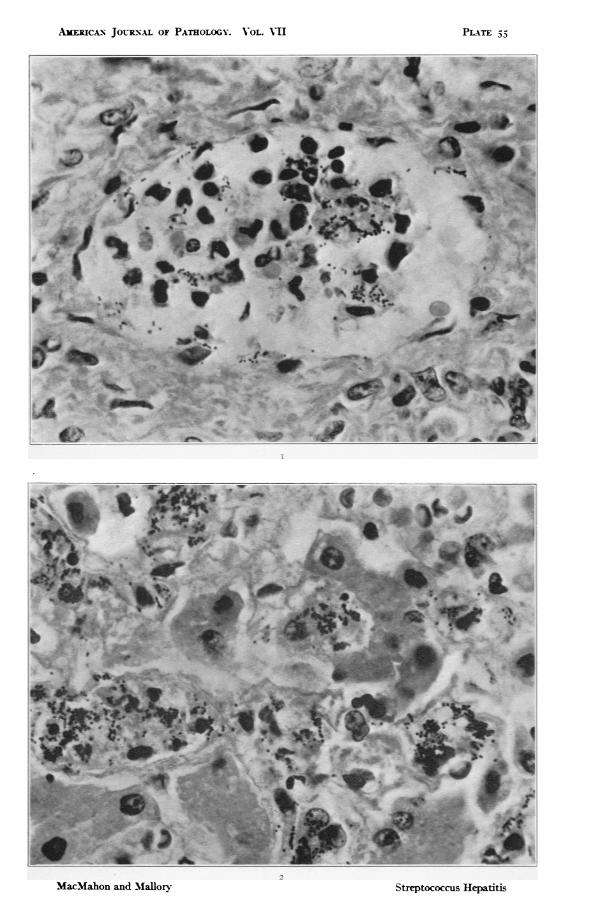
FIG. 3. Case i. An area in a lobule where the necrotic liver cells have to a largeextent disappeared. Streptococci are still persistent in moderate numbers,largely in endothelial leucocvtes. x IOoo.
FIG. 4. Case 2. The edge of one of the two areas of necrosis involving manylobules. -Al of the liver cells have been killed and are being removed bvthe action of leucocytes. Onlv the bile ducts and stroma persist. Theadjoining liver tissue is uninjured. x40.

AMERICA-N JOURN-L OF PATHOLOGY. VOL. VIIP
--v,a,@s .,, >A;> o. _ _. = ., . . ., , .W ; ^ s _F-F_-r SF n ^ _Kv _ .* .zES | _ > . + _ . +* _;SK L. K 31 _h:;ifi::. _B. . ei w i _E|;* w * 4 .e 4.wSt.tt;w oS |;_ ' # v- <. .-bow}iMe _ b
MacMahon and 'Mallory
PLAX 5,6
Streptococcus Hepatitis
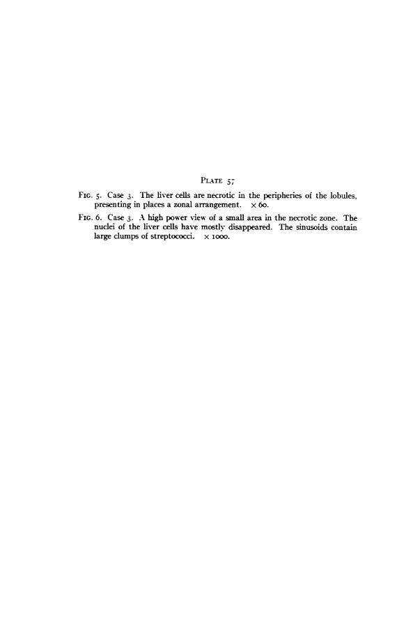
PLATE Z--
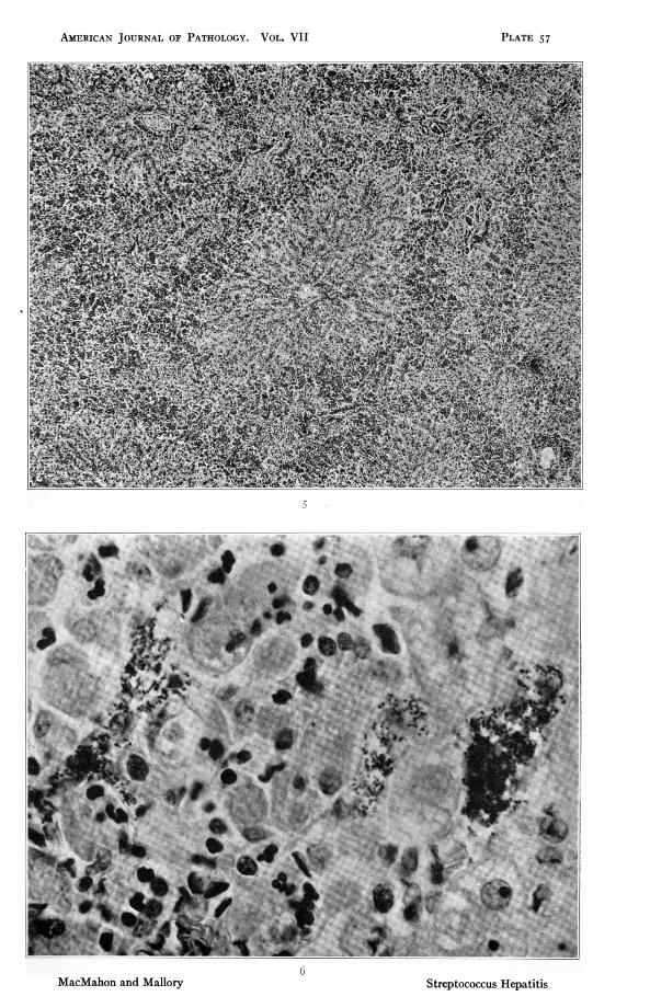
FIG. 5. Case 3. The liver cells are necrotic in the peripheries of the lobules,presenting in places a zonal arrangement. x 6o.
FIG. 6. Case 3. A high power view of a small area in the necrotic zone. Thenudei of the liver cells have mostly disappeared. The sinusoids containlarge dumps of streptococci. x iOOO.
AMERICAN JOURNAL OF PATHOLOGY. VOL. VII
6MacMahon and Mallory
PLATE 5 7
Streptococcus Hepatitis
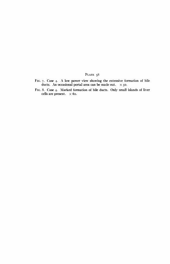
PLATE 58
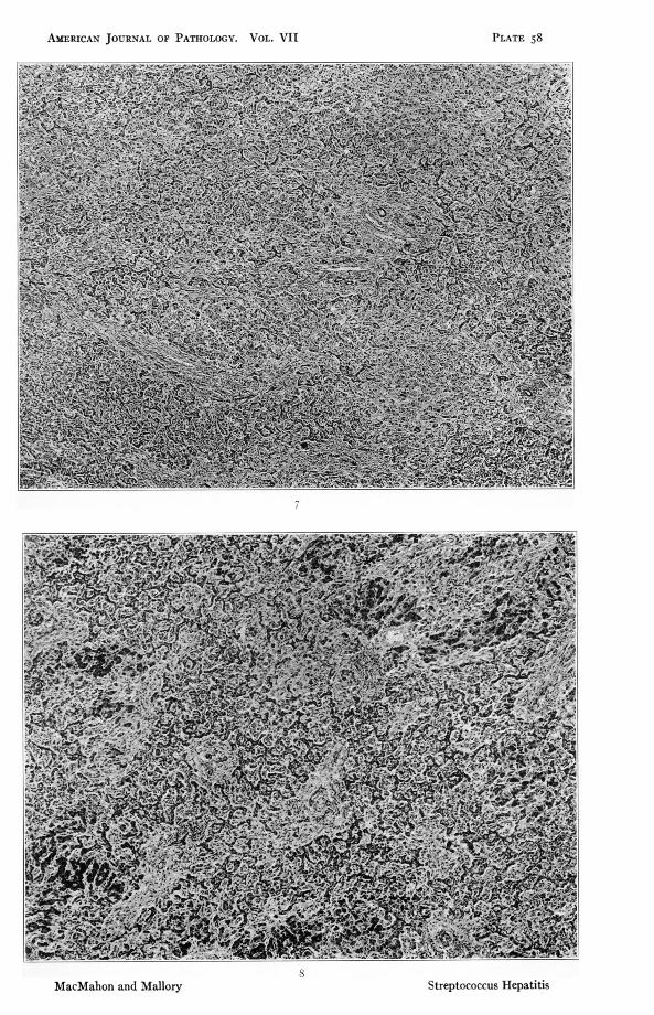
FIG. 7. Case 4. A low power view showing the extensive formation of bileducts. An occasional portal area can be made out. x 50.
FIG. 8. Case 4. AMarked formation of bile ducts. Onlv small islands of livercells are present. x 6o.
AMERICAN JOURNAL OF PATHOLOGY. VOL. VII
8MacMahon and Mallory
PLATE 58
i
Streptococcus Hepatitis

PLATE 59
FIG. 9. Case 4. High power view of network of bile ducts. The stroma is fairlvabundant. x 500.
FIG. IO. Case 4. Mlasses of streptococci mostlv contained within endothelialcells lining the sinusoids. x IOoo.

AMERICAN JOURNAL OF PATHOLOGY. VOL. VI1
'l;,;;:s'-q.. . I° .iJ _v
-.-,m
Mfac-%lahon and Mlallorv
PLATE 59
t 444L
Streptococcus Hepatitis