microtubules and microtubule-associated proteins
TRANSCRIPT
Microtubules and Microtubule-AssociatedProteins
Holly V. Goodson and Erin M. Jonasson
Department of Chemistry and Biochemistry, University of Notre Dame, Notre Dame, Indiana 46556
Correspondence: [email protected]
SUMMARY
Microtubules act as “railways” for motor-driven intracellular transport, interact with accessoryproteins to assemble into larger structures such as the mitotic spindle, and provide an organi-zational framework to the rest of the cell. Key to these functions is the fact that microtubules are“dynamic.” As with actin, the polymer dynamics are driven by nucleotide hydrolysis andinfluenced by a host of specialized regulatory proteins, including microtubule-associatedproteins. However, microtubule turnover involves a surprising behavior—termed dynamicinstability—in which individual polymers switch stochastically between growth and depoly-merization. Dynamic instability allows microtubules to explore intracellular space and re-model in response to intracellular and extracellular cues. Here, we review how such instabilityis central to the assembly of many microtubule-based structures and to the robust functioningof the microtubule cytoskeleton.
Outline
1 Introduction
2 Physical attributes of microtublesand tubulin
3 Structures formed from microtubules
4 Microtubule assembly and dynamics
5 Microtubule-binding proteins
6 Conclusion
References
Editors: Thomas D. Pollard and Robert D. Goldman
Additional Perspectives on The Cytoskeleton available at www.cshperspectives.org
Copyright # 2018 Cold Spring Harbor Laboratory Press; all rights reserved; doi: 10.1101/cshperspect.a022608
Cite this article as Cold Spring Harb Perspect Biol 2018;10:a022608 1
on January 28, 2022 - Published by Cold Spring Harbor Laboratory Press http://cshperspectives.cshlp.org/Downloaded from
1 INTRODUCTION
Along with actin (Pollard 2016) and intermediate filaments(Herrmann and Aebi 2016; Hol and Capetanaki 2016;Jacob et al. 2016; Jones et al. 2016; Yuan et al. 2016), mi-crotubules (Figs. 1 and 2) constitute one of the three mainclasses of cytoskeletal filaments in eukaryotic cells. Micro-tubules are found in all characterized eukaryotic organ-isms. Thus, the last common ancestor of eukaryotes hadmicrotubules; this ancestor also had the dynein and kinesinmotors that operate on the microtubule cytoskeleton(Sweeney and Holzbaur 2016). Many prokaryotes have atleast one gene homologous to that encoding tubulin, themost common of which encodes FtsZ, a protein that formspolymers involved in cytokinesis. These observations sug-gest that the tubulin gene family appeared very early, per-haps in the last common ancestor of all forms of life onEarth (Pollard and Goldman 2016). Even without knowinganything else about microtubules, the maintenance ofthese structures and their constituent proteins across sucha large span of time and in highly divergent organismsindicates that they have a fundamental role in eukaryoticcell biology.
Indeed, microtubules and their accessory proteins formthe mitotic spindle—the dynamic self-organized machinethat separates the chromosomes during mitosis, arguablythe most important of all eukaryotic cell processes (McIn-tosh 2016). In addition, complexes of microtubules andmotors form the core of cilia and flagella (Viswanadhaet al. 2016), making microtubules essential for the motilityof many organisms, including numerous protists and mostmetazoan sperm. Microtubules also provide tracks for mo-tors that catalyze the movement of organelles, transportvesicles, and other structures. This microtubule-based in-tracellular transport contributes to the efficient function ofmany organisms and cell types, but it is crucial for thedramatically elongated neurons of animals (reviewed byBarlan and Gelfand 2016). Microtubules also play funda-mental roles in cell organization by localizing organellesand establishing the polarity of a wide variety of cells inboth animals and plants.
How do microtubules contribute to these diverse cel-lular activities? As reviewed here and elsewhere, there aremany answers to this question, only some of which are wellunderstood. One central theme is that the dynamic behav-ior of microtubule polymers is essential to many micro-tubule-based processes. Briefly, structures assembled frommicrotubules and actin filaments usually have longer life-times than the individual polymers from which they areassembled. In both cases, hydrolysis of nucleotides boundto the polymer subunits drives their turnover. However,the patterns of turnover differ. Actin filaments in vivo
typically grow at their barbed end and disassemble attheir pointed end as a result of multiple reactions, includ-ing severing by accessory proteins.1 In contrast, micro-tubule polymers usually (but not exclusively) display asurprising behavior termed “dynamic instability,” in whichindividual polymers switch stochastically (i.e., randomly)between growth and shortening. As explained below,this dynamic instability behavior is fundamental tomany of the functions and properties of the microtubulecytoskeleton.
Because microtubule function and behavior derive ul-timately from the structure and biochemistry of the micro-tubule filaments, this review starts with a description oftheir structure and biochemistry. It then discusses the sub-cellular structures that form from microtubules and re-turns to examine in more depth dynamic instability andits mechanism. Finally, we shall discuss the proteins thatregulate microtubule dynamics and interact with microtu-bules to interface with the rest of the cell.
2 PHYSICAL ATTRIBUTES OF MICROTUBLESAND TUBULIN
2.1 Microtubule Structure
Microtubules are assembled from heterodimers of a- andb-tubulin into long hollow polymers that are �25 nm wideand range in length from ,1 mm to .100 mm (Fig. 2).These heterodimeric tubulin subunits are referred to asab-tubulin, tubulin dimers, or simply “tubulin.”
Microtubule structure is most straightforwardly de-scribed as comprising approximately 13 linear protofila-ments (PFs) that are associated laterally and closed into ahollow tube. The resulting polymer is polar, with a fast-growing plus end that has exposed b-tubulin and a slow-growing minus end with exposed a-tubulin (Fig. 2A).
In typical 13-PF microtubules, the boundary where thetube closes, unlike the other interfaces between PFs, createsa “seam” where the lateral interactions among PFs differfrom those elsewhere in the microtubule (Fig. 2A). Thisseam is generally believed to be a weak point of the micro-tubule structure, although there is some evidence to thecontrary (Sui and Downing 2010). Although most micro-tubules in vivo have 13 PFs, there are some exceptions, andmicrotubules assembled in vitro can have a wide range ofPF numbers. One striking difference between the typical13-PF microtubules and those with 15 or 16 PFs is that the
1In vitro, purified actin can by itself slowly treadmill (undergo assembly at oneend and disassembly at the other). In vivo treadmilling of the actin network isdriven by proteins that regulate assembly, capping, severing, and depoly-merization (reviewed by Pollard 2016).
H.V. Goodson and E.M. Jonasson
2 Cite this article as Cold Spring Harb Perspect Biol 2018;10:a022608
on January 28, 2022 - Published by Cold Spring Harbor Laboratory Press http://cshperspectives.cshlp.org/Downloaded from
vd
afl
mb
fnvfl
pfl
cfl5 μm
N
A
C
E
G H
F
D
B
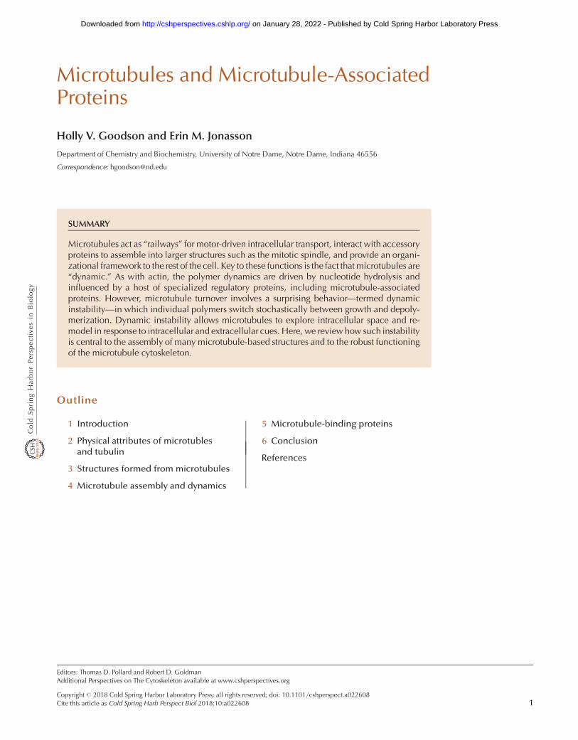
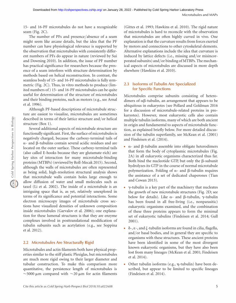
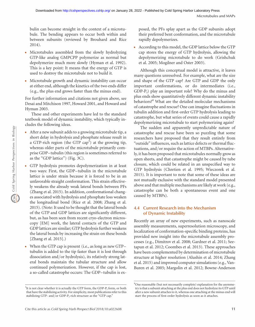
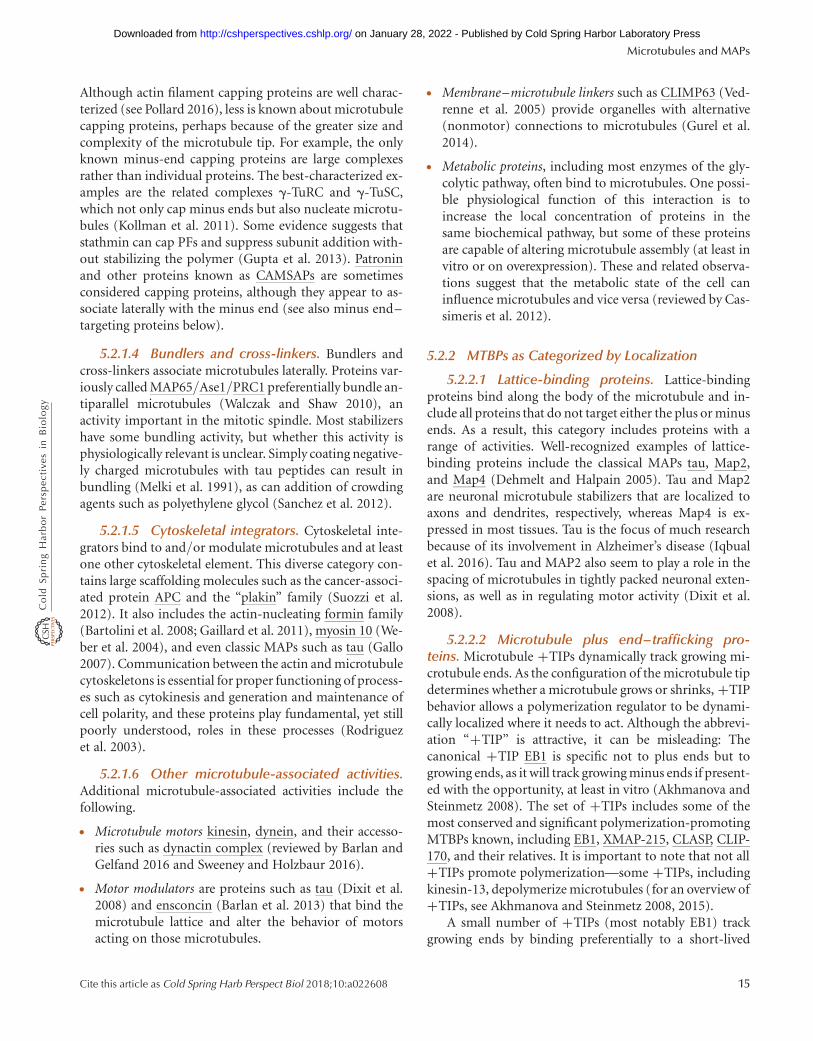
Figure 1. The microtubule cytoskeleton in various cell types. Each pair of panels contains a fluorescence microscopyimage of a specific cell/group of cells (left) with a cartoon depicting the generalized microtubule organization in thatcell type (right). The color schemes for the microscope images are described below. In the cartoons, microtubules areshown in green, the DNA in blue, and centrosomes in red. Noncentrosomal microtubule nucleation machineryexists in many cell types (see text) but is not depicted. (A) Radial microtubule array in interphase cells. Microtubules(green), DNA (blue), microtubule-organizing center (MTOC; red). (B) Columnar microtubule array in polarizedepithelial cells (green-fluorescent protein [GFP]–tubulin expressed in Madin–Darby canine kidney [MDCK] cells).(C) Microtubules in a neuronal growth cone. Microtubules (green) and actin filaments (red). (D) Cortical micro-tubule array in plant cells (GFP–tubulin expressed in Arabidopsis cells). (E) Fission yeast interphase microtubules(GFP–tubulin). (F) Microtubule cytoskeleton in Giardia. Microtubules (red), DNA (blue). (G) Animal cell mitoticspindle. Microtubules (green) and DNA (blue). (H ) Metaphase plant mitotic spindle. Microtubules (green) andDNA (blue). (A, Reproduced from Gundersen laboratory website [http://www.columbia.edu/~wc2383/pictures.html]; B, reprinted from Reilein et al. 2005; C, reprinted from Kalil et al. 2011; D, reprinted from Ehrhardt and Shaw2006, with permission of Annual Review of Plant Biology; E, reprinted, with permission, from Chang and Martin2009, # Cold Spring Harbor Laboratory Press; F, reprinted, with permission, from Dawson 2010, # John Wiley &Sons Inc.; G, left image, reprinted from O’Connell and Khodjakov 2007, with permission from Elsevier, originallyfrom Cell Motility and Cytoskeleton [1999] V.43[3] [cover], with permission from Wiley; H, reproduced from Yu et al.1999.)
Microtubules and MAPs
Cite this article as Cold Spring Harb Perspect Biol 2018;10:a022608 3
on January 28, 2022 - Published by Cold Spring Harbor Laboratory Press http://cshperspectives.cshlp.org/Downloaded from
Seam
12 PFs 13 PFs 15 PFs orgreater
2-start 3-start 4-start
γ-TuRC
Lateral γ-αcontact
Attachmentfactors
MTOC
Seam
Seam Seam
A lattice B lattice
Seam
Plus endA B
C D
Lateral
Longitudinal
MicrotubuleProtofilamentαβ heterodimer
α
β
Minus end
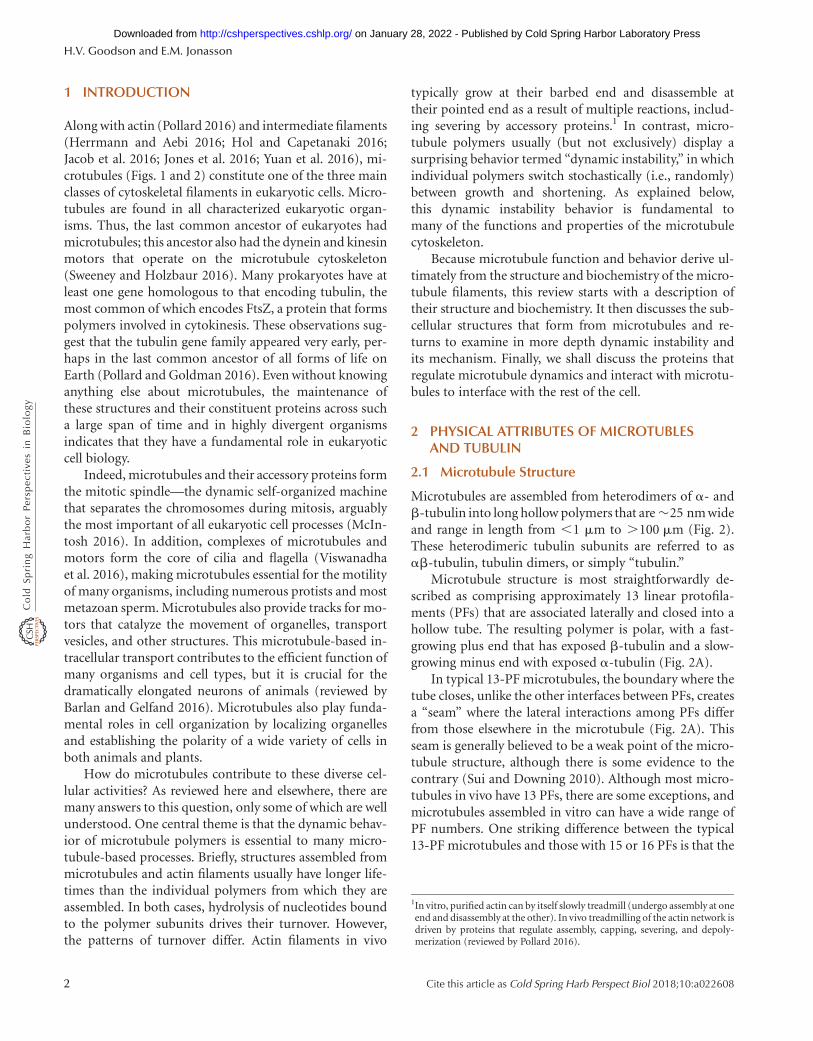
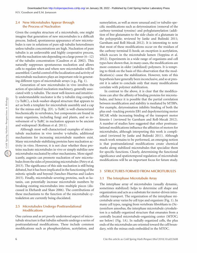
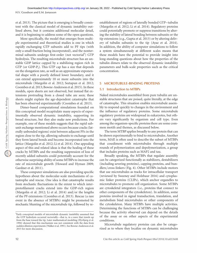
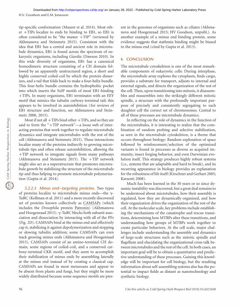
Figure 2. Microtubule structure. (A,B) Key aspects of microtubule structure, as indicated. (C) Diagram of therelationship between protofilament number and microtubule structure. (D) Model of the g-tubulin ring complex(g-TuRC) associated with the minus end of a microtubule (gray). MTOC, microtubule-organizing center. (A,B,Modified from Kollman et al. 2011, with permission from Macmillan Publishers; C, modified, with permission,from Amos 2004, with permission of The Royal Society of Chemistry, http://dx.doi.org/10.1039/B403634D.)
BOX 1. DESCRIPTION OF MICROTUBULE STRUCTURE IN TERMS OF LATTICES AND HELICES
The main body of a typical 13-PF microtubule can be de-scribed as being composed of a so-called B lattice, in whicha-subunits are next to a-subunits (a–a) and b next to b (b–b).However, at the seam, the a-subunits associate laterally withb-subunits in the adjacent PFs (a–b) in an A lattice (Fig. 2A,B).The lattice structure changes at the seam because each PF inthe main body of a microtubule is shifted slightly relative to itsneighbor, resulting in an offset at the seam of 1.5 dimers for a13-PF microtubule (Fig. 2C). Changing the number of PFschanges the offset, so that the offset for microtubules with 15or 16 PFs is two full subunits, resulting in a situation in whichthere is no discernable seam (these microtubules compriseentirely B lattice: a–a and b–b).
In addition, microtubules are sometimes described as he-lices, although the presence of a seam means that a 13-PFmicrotubule is not a true helix; it just appears to be one inlow-resolution electron microcopy images in which a- andb-tubulin are indistinguishable. Given this ambiguity, a 13-PF microtubule can be described as a left-handed three-start
helix because each of the three monomers in the 1.5 tubulindimer offset at the seam can be viewed as starting a newhelix (Fig. 2C). As noted above, changing the number of PFschanges the offset, so that the offset for microtubules with15 or 16 PFs is two full subunits. This situation results in afour-start helix, in which, as noted above, B lattice is founduniformly through the microtubule (Fig. 2C; see also Amos2004).
Although considering microtubules as (pseudo)helices canbe useful in structural studies, it is usually more informative toview microtubules as PF-based structures because a consensusis building that microtubules grow by adding subunits to theselinear PFs, not by extending the helices. This idea is based, inpart, on evidence that the longitudinal interactions betweensubunits within a PF are stronger and more extensive than thelateral bonds between subunits in different PFs (Sept et al.2003; Zhang et al. 2015). The predominance of these longitu-dinal bonds means that interactions within a given PFare moresignificant than those within a given helix.
H.V. Goodson and E.M. Jonasson
4 Cite this article as Cold Spring Harb Perspect Biol 2018;10:a022608
on January 28, 2022 - Published by Cold Spring Harbor Laboratory Press http://cshperspectives.cshlp.org/Downloaded from
15- and 16-PF microtubules do not have a recognizableseam (Fig. 2C).
The number of PFs and presence/absence of a seammight seem like arcane details, but the idea that the PFnumber can have physiological relevance is supported bythe observation that microtubules with consistently differ-ent numbers of PFs do appear in nature (reviewed by Suiand Downing 2010). In addition, the issue of PF numberhas practical significance for researchers because the pres-ence of a seam interferes with structure determination bymethods based on helical reconstruction. In contrast, theseamless body of 15- and 16-PF microtubules is fully sym-metric (Fig. 2C). Thus, in vitro methods to produce (lim-ited numbers of ) 15- and 16-PF microtubules can be quiteuseful for determination of the structure of microtubulesand their binding proteins, such as motors (e.g., see Arnalet al. 1996).
Although PF-based descriptions of microtubule struc-ture are easiest to visualize, microtubules are sometimesdescribed in terms of their lattice structure and/or helicalstructure (Box 1).
Several additional aspects of microtubule structure arefunctionally significant. First, the surface of microtubules isnegatively charged, because the carboxy-terminal tails ofa- and b-tubulins contain several acidic residues and arelocated on the outer surface. These carboxy-terminal tails(also called E-hooks because they are glutamate-rich) arekey sites of interaction for many microtubule-bindingproteins (MTBPs) (reviewed by Roll-Mecak 2015). Second,although the walls of microtubules are often representedas being solid, high-resolution structural analysis showsthat microtubule walls contain holes large enough toallow diffusion of water and small molecules such astaxol (Li et al. 2002). The inside of a microtubule is anintriguing space that is, as yet, relatively unexplored interms of its significance and potential interactions. Someelectron microscopy images of microtubule cross sec-tions have visualized densities of unknown compositioninside microtubules (Garvalov et al. 2006); one explana-tion for these lumenal structures is that they are enzymecomplexes involved in posttranslational modification oftubulin subunits such as acetylation (e.g., see Soppinaet al. 2012).
2.2 Microtubules Are Structurally Rigid
Microtubules and actin filaments both have physical prop-erties similar to the stiff plastic Plexiglas, but microtubulesare much more rigid owing to their larger diameter andtubular construction. To make this comparison morequantitative, the persistence length of microtubules is�5000 mm compared with �20 mm for actin filaments
(Gittes et al. 1993; Hawkins et al. 2010). The rigid natureof microtubules is hard to reconcile with the observationthat microtubules are often highly curved in vivo. Oneexplanation is that the curvature results from forces exertedby motors and connections to other cytoskeletal elements.Alternative explanations include the idea that curvature isinduced by lattice defects (i.e., missing and/or misincor-porated subunits) and/or binding of MTBPs. The mechan-ical aspects of microtubules are discussed in more depthelsewhere (Hawkins et al. 2010).
2.3 Isoforms of Tubulin Are Specializedfor Specific Functions
Microtubules comprise subunits consisting of hetero-dimers of ab-tubulin, an arrangement that appears to beubiquitous in eukaryotes (see Pollard and Goldman 2016for a discussion of microtubule-related proteins in pro-karyotes). However, most eukaryotic cells also containmultiple tubulin isoforms, many of which are both ancientin origin and fundamental to aspects of microtubule func-tion, as explained briefly below. For more detailed discus-sion of the tubulin superfamily, see McKean et al. (2001)and Findeisen et al. (2014).
† a- and b-tubulin assemble into obligate heterodimersthat form the body of cytoplasmic microtubules (Fig.2A) in all eukaryotic organisms characterized thus far.Both bind the nucleotide GTP, but only the b-subunithydrolyzes its GTP in the course of normal microtubulepolymerization. Folding of a- and b-tubulin requiresthe assistance of a set of dedicated chaperones (Tianand Cowan 2013).
† g-tubulin is a key part of the machinery that nucleatesthe growth of new microtubule structures (Fig. 2D; seebelow for details). Like a- and b-tubulin, g-tubulinhas been found in all free-living (i.e., nonparasitic)eukaryotic organisms examined, and the combinationof these three proteins appears to form the minimalset of eukaryotic tubulins (Findeisen et al. 2014; Gull2001).
† d-, 1-, and z-tubulin isoforms are found in cilia, flagella,and/or basal bodies, and in general they are specific toorganisms with these structures. These ancient proteinshave been identified in some of the most divergentknown eukaryotic organisms, but they have also beenlost from many lineages (McKean et al. 2001; Findeisenet al. 2014).
† Other tubulin isoforms (e.g., h-tubulin) have been de-scribed, but appear to be limited to specific lineages(Findeisen et al. 2014).
Microtubules and MAPs
Cite this article as Cold Spring Harb Perspect Biol 2018;10:a022608 5
on January 28, 2022 - Published by Cold Spring Harbor Laboratory Press http://cshperspectives.cshlp.org/Downloaded from
2.4 New Microtubules Appear throughthe Process of Nucleation
Given the complex structure of a microtubule, one mightimagine that generation of new microtubules is a difficultprocess. Indeed, spontaneous appearance of new microtu-bules is rare in solutions of pure ab-tubulin heterodimersunless tubulin concentrations are high. Nucleation of puretubulin is an unfavorable and highly cooperative process,with the nucleation rate depending on a large power (6–12)of the tubulin concentration (Caudron et al. 2002). Thisnaturally suppresses spontaneous nucleation and allowscells to regulate when and where new microtubules will beassembled. Careful control of the localization and activity ofmicrotubule nucleators plays an important role in generat-ing different types of microtubule arrays (e.g., Fig. 1).
Generation of new microtubules in vivo involves theaction of specialized nucleation machinery, generally asso-ciated with g-tubulin. The most well-known and intuitive-ly understandable nucleator is the g-tubulin ring complex(g-TuRC), a lock-washer-shaped structure that appears toact as both a template for microtubule assembly and a capfor the minus end (Fig. 2D). g-TuRC is well-characterizedbiochemically in vertebrates, but components are found inmany organisms, including fungi and plants, and so in-volvement of g-TuRC in nucleation appears to be ancientand widespread (Kollman et al. 2011).
Although most well-characterized examples of micro-tubule nucleation in vivo involve g-tubulin, additionalmechanisms for increasing microtubule numbers exist.Many microtubule-stabilizing proteins have nucleation ac-tivity in vitro. However, it is not clear whether these pro-teins nucleate microtubules in vivo or simply stabilize newmicrotubules nucleated by other mechanisms. More signif-icantly, augmin can promote nucleation of new microtu-bules from the sides of preexisting microtubules (Petry et al.2013). The significance of this side nucleation is still beingdebated, but it has been implicated in the functioning of themitotic spindle and beyond (Sanchez-Huertas and Luders2015). Finally, microtubule-severing proteins, such as ka-tanin, can potentially increase microtubule numbers bybreaking existing microtubules into multiple pieces (dis-cussed in Ehrhardt and Shaw 2006). The contributions ofthese mechanisms to the function of the microtubule cy-toskeleton are currently being elucidated.
2.5 Microtubules Undergo PosttranslationalModifications
One curious and as yet poorly understood aspect of micro-tubule structure is that tubulin subunits undergo a series ofposttranslational modifications. These include commonmodifications such as phosphorylation, acetylation, and
sumoylation, as well as more unusual and/or tubulin-spe-cific modifications such as detyrosination (removal of thecarboxy-terminal tyrosine) and polyglutamylation (addi-tion of free glutamates to the side-chain of a glutamate inthe polypeptide; reviewed by Janke and Bulinski 2011;Garnham and Roll-Mecak 2012). It is interesting to notethat most of these modifications occur on the residues ofthe carboxy-terminal E-hook; an exception is acetylation,which occurs in the microtubule lumen (Soppina et al.2012). Experiments in a wide range of organisms and celltypes have shown that, in many cases, the modifications aremore common in older (stabilized) polymers. It is tempt-ing to think on the basis of this observation that the mod-ification(s) cause the stabilization. However, tests of thishypothesis have generally been inconclusive, and so at pres-ent it is safest to conclude only that many modificationscorrelate with polymer stabilization.
In contrast to the above, it is clear that the modifica-tions can alter the affinity of binding proteins for microtu-bules, and hence it is possible that the partial correlationbetween modification and stability is mediated by MTBPs.For example, detyrosination inhibits binding of both theplus end–tracking protein EB1 and depolymerizing motorMCAK while increasing binding of the transport motorkinesin-1 (reviewed by Garnham and Roll-Mecak 2012).A number of studies have suggested that other posttrans-lational modifications influence the affinities of motors formicrotubules, although interpreting this work is compli-cated (reviewed by Janke and Bulinski 2011). Althoughmuch work remains to be performed, an emerging notionis that posttranslational modifications create chemicalmarks along stabilized microtubules that specialize themfor specific functions. Clearly, determining the functionalsignificance and spatiotemporal regulation of microtubulemodifications will be an important focus for future study.
3 STRUCTURES FORMED FROM MICROTUBULES
3.1 The Interphase Microtubule Array
The interphase array of microtubules (usually dynamic,sometimes stabilized) helps to determine cell shape andorganization and acts as a substrate for motor-driven intra-cellular transport. The organization of the interphase mi-crotubule array varies by cell type and organism (Fig. 1). Inmany cell types, ranging from vertebrate fibroblasts to Dic-tyostelium amoebas, the interphase microtubule cytoskele-ton is a radially organized structure that emanates from acentrally located microtubule-organizing center (MTOC;see below) (Fig. 1A). In radially organized cells, the plusends of the microtubules are oriented toward the cell boun-dary, with the minus ends embedded in the MTOC.
H.V. Goodson and E.M. Jonasson
6 Cite this article as Cold Spring Harb Perspect Biol 2018;10:a022608
on January 28, 2022 - Published by Cold Spring Harbor Laboratory Press http://cshperspectives.cshlp.org/Downloaded from
Although radial organization of the microtubule cyto-skeleton is common and is sometimes presented as canon-ical, it is by no means universal. As one example of adifferent arrangement, the microtubule array in vertebratepolarized epithelial cells has a more parallel organization,with the microtubule minus ends located toward the apicalmembrane and the plus ends located toward the base of thecell (Fig. 1B) (Bartolini and Gundersen 2006). In higher-plant cells (e.g., Arabidopsis), microtubules are found incortically associated parallel arrays oriented transverse tothe axis of cell elongation (Fig. 1D) (Ehrhardt and Shaw2006).
The organization of the microtubule array is importantin most cells because it plays a central role in determiningthe organization of the rest of the cell. For example, whenmicrotubules are oriented radially, as in fibroblasts, themembranes of the Golgi apparatus are generally locatednear the MTOC, with most other membranes distributedmore peripherally. In contrast, the parallel microtubulearrays endow polarized epithelial cells with a more linearinternal organization, with the Golgi membranes at theapical face, and other membranes located more basolater-ally (Bartolini and Gundersen 2006). In these cells andothers, loss of microtubules leads to loss of normal internalorganization, as well as other aspects of cell polarity (de
Forges et al. 2012). For example, many animal cells canmove without microtubules, but lose directionality (Gan-guly et al. 2012), and plant cells with depolymerized mi-crotubules grow aberrantly (Ehrhardt and Shaw 2006).
How is microtubule organization established andmaintained? These processes are not yet well understood,but the microtubule array, as observed in a particular cell,emerges from interactions between dynamic microtubules,their nucleators, their regulators, microtubule motors, andthe cell boundary (Box 2).
3.2 Other Subcellular Microtubule-Based Structures
3.2.1 Mitotic Spindle
The mitotic spindle (Fig. 1G,H) is the complex andbeautiful self-assembled machine that separates the chro-mosomes in all eukaryotic cells. The mitotic spindle com-prises dynamic microtubules, a wide array of motors, and aseries of other microtubule-associated proteins (MAPs).The mitotic spindle forms when the interphase microtu-bule array undergoes a dramatic reorganization upon entryinto mitosis. The assembly, activities, and properties of themitotic spindle have been, and continue to be, the subjectsof intense study, and they are discussed in more detailelsewhere (McIntosh 2016).
BOX 2. HOW IS THE ORGANIZATION OF THE MICROTUBULE NETWORK ESTABLISHED AND MAINTAINED?
The obvious answer is that microtubule organization dependson localization of the microtubule organizing center, butfunctional bipolar spindles can form in cells even after thecentrosomes have been removed by microsurgery (Khodjakovand Rieder 2001), and a number of cell types (most obviouslyplant cells) lack centrosomes (Ehrhardt and Shaw 2006).Moreover, spindle-like bipolar structures can form around ar-tificial chromosomes in vitro in the absence of centrosomes(Heald et al. 1996). Examination of the localization of g-tu-bulin in these and other noncentrosomal systems leads to arelated proposal: Perhaps microtubule organization dependson the localization of the microtubule nucleators. Centralizednucleation machinery correlates with radial organization, anddistributed nucleation machinery correlates with distributednetworks (Fig. 1). Although compelling, this explanation rais-es the next question: What determines the localization of thenucleators?
A hint to resolving this conundrum is provided by theobservation that mixtures of stabilized microtubules and pu-rified active motors can spontaneously self-organize into arange of different structures; the details of these structuresdepend on the specific activities and ratios of the proteinsinvolved (Surrey et al. 2001). Motor-driven microtubule orga-nization is also involved in the formation of dynamic radial
microtubule arrays in melanophore cell fragments (Vorobjevet al. 2001).
However, it is important to remember that other aspects ofthe cellular environment can also influence the microtubulecytoskeleton. For instance, the pigment granules in melano-phores appear to participate in microtubule organization (Vor-objev et al. 2001), and the Golgi apparatus can nucleatemicrotubules and/or act as a MTOC (de Forges et al. 2012).Moreover, the signal transduction pathways that were original-ly hypothesized by Mitchison and Kirschner to allow selectivestabilization of microtubules near particular regions of the cellboundary (Kirschner and Mitchison 1986) are becoming elu-cidated (Akhmanova et al. 2009). Finally, it is important tounderstand that the physical barrier presented by the cellboundary can itself influence microtubule organization anddynamics (Maly and Borisy 2002; Gregoretti et al. 2006; Dog-terom and Surrey 2013).
In summary, it is becoming apparent that the interphasemicrotubule array emerges from dynamic interactions be-tween microtubules, motors, their regulators, and their physi-cal environment. Gaining a deeper understanding of howparticular large-scale structures emerge from local interactionswill likely require computational modeling and other ap-proaches used to study complex systems.
Microtubules and MAPs
Cite this article as Cold Spring Harb Perspect Biol 2018;10:a022608 7
on January 28, 2022 - Published by Cold Spring Harbor Laboratory Press http://cshperspectives.cshlp.org/Downloaded from
3.2.2 Microtubule-Organizing Centers,Centrosomes, and Spindle Pole Bodies
MTOCs, centrosomes, and spindle pole bodies (SPBs) arevarious names given to localized foci of microtubule-nu-cleating machinery (see Vertii et al. 2016). “Microtubule-organizing center” is a term that applies to all of thesestructures, whereas “centrosome” usually applies more spe-cifically to the perinuclear MTOC of radially organizedcells, and “spindle pole body” applies to the nuclear-mem-brane-embedded MTOC of fungi such as the buddingyeast Saccharomyces cerevisiae and the fission yeast Schizo-saccharomyces pombe. Although centrosomes and SPBshave similar functions in terms of microtubule nucleationand contain many similar proteins, their ultrastructure isquite different (Ada and Kilmartin 2000). MTOCs containg-tubulin and the g-TURC (Fig. 2D), but can contain acomplex array of other proteins, such as motors and +TIPs(microtubule plus end–tracking proteins; see Sec. 5 be-low), and can include centrioles (see below). In the past,centrioles were thought to be fundamental to the functionof MTOCs, but many organisms (e.g., most higher plants)lack centrioles, and fly mutants lacking centrioles developin a largely normal way (Basto et al. 2006). One explanationfor the frequent association between centrioles and centro-somes is that the colocalization of these structures keepsthe microtubule nucleating activities organized in asingle focus.
3.2.3 Flagella and Cilia
Flagella and cilia (reviewed by Viswanadha et al. 2016) arecomplex organelles that comprise highly organized ar-rangements of microtubules, motors, and other proteins.Structurally they are similar, but they can differ in termsof attributes such as their function, motion, length, anddetails of their protein composition. Flagella and ciliaare highly conserved and ancient organelles, existing insimilar form in organisms ranging from human to someof the most divergent protists (Carvalho-Santos et al.2010).
3.2.4 Centrioles and Basal Bodies
Centrioles and basal bodies are complex structures thattypically comprise nine sets of triplet microtubules and aset of well-conserved associated proteins (Vertii et al. 2016),although with some variation (Carvalho-Santos et al.2011). They are found at the base of flagella and cilia (wherethey are called basal bodies) and in centrosomes (wherethey are called centrioles). Centrioles and basal bodiescan interconvert as cells pass through the cell cycle. Centri-oles undergo a mysterious form of replication in which new
centrioles appear at 90˚ angles from the parent centrioles,and this replication is typically tightly coordinated with cellreplication and assembly of the mitotic spindle (Nigg andStearns 2011).
3.2.5 The Midbody
The midbody is an enigmatic structure comprising bun-dled microtubules and associated proteins derived fromthe mitotic spindle. The midbody forms during cytokine-sis at the point of abscission (separation) of the twodaughter cells. Midbodies have often been viewed as wastedepots for the cell-division process, but increasing evi-dence suggests that these structures are transient organelleswith still-mysterious functions of their own (Chen et al.2013).
3.2.6 Organism-Specific Structures
Protists contain a wide variety of complex microtubule-based structures that are important for their viabilityand/or pathogenicity. Striking examples include the Toxo-plasma conoid (Morrissette 2015), the Giardia ventral disk(i.e., the suction plate) (Fig. 1F) (Schwartz et al. 2012), andthe cilia array of ciliates (Winey et al. 2012). The diversity ofcellular architecture in protists is remarkable, and themechanisms leading to generation of these structures areonly beginning to be defined (Slabodnick and Marshall2014).
4 MICROTUBULE ASSEMBLY AND DYNAMICS
4.1 Introduction to Microtubule Dynamics
The term “cytoskeleton” brings to mind a static structure,an idea that is reinforced by immunofluorescence imagessuch as those in Figure 1. Nothing could be farther from thetruth: As with actin (reviewed by Pollard 2016), the micro-tubule cytoskeleton in eukaryotic cells is constantly turningover in a process that is driven by nucleotide hydrolysis(GTP hydrolysis in the case of microtubules). However,the energy of the nucleotides is not used to build actinfilaments and microtubules but rather to destroy them.This concept can be shown by replacing the GTP inthe tubulin subunits with the slowly hydrolyzable GTPanalog GMPCPP (guanylyl 5′-a,b-methylenediphospho-nate): The microtubules grow, but fail to disassemble nor-mally (Hyman et al. 1992). Where does the energy forbuilding the filament come from? The answer is that as-sembly of GTP–tubulin into microtubules is a spontane-ous process that is driven primarily by the hydrophobiceffect (Vulevic and Correia 1997), like many other formsof macromolecular assembly.
H.V. Goodson and E.M. Jonasson
8 Cite this article as Cold Spring Harb Perspect Biol 2018;10:a022608
on January 28, 2022 - Published by Cold Spring Harbor Laboratory Press http://cshperspectives.cshlp.org/Downloaded from

Microtubules in most cell types display a behaviorknown as dynamic instability, in which the ends of indi-vidual polymers transition randomly between periods ofgrowth and shortening (Fig. 3). In many animal cells, theminus ends of most microtubules are embedded in theMTOC (see discussion above), and so dynamic instabilityoccurs primarily at the plus ends, but uncapped minusends can also show dynamic instability, at least in vitro(Fig. 3A). Some microtubules can also treadmill, a behaviorthat is particularly important in the cortical microtubulearray of plants (Ehrhardt and Shaw 2006) but is also seen inmore-limited cases in animal cells (e.g., Rodionov andBorisy 1997; Vorobjev et al. 2001).
The constant turnover of cytoskeletal structures mightseem wasteful—just consider the amount of GTP that is“burned” by a single microtubule undergoing dynamicinstability. Why would cells expend so much energyconstantly destroying structures that they have just built?It turns out that microtubule turnover is necessary formany aspects of cell physiology and is an essential aspect
of the microtubule cytoskeleton. Some examples includethe following.
† The dynamic nature of the microtubule cytoskeletonallows cells to adapt to changes in cell shape and envi-ronment.
† The random probing generated by dynamic instabilityallows individual microtubules to explore cellular spaceand bring the microtubule “train tracks” into contactwith cargo such as vesicles, organelles, and chromo-somes, which are too large to diffuse effectively in thehighly crowded environment of the cytoplasm.
† Spatially localized (“selective”) stabilization of dynamicmicrotubules provides a mechanism for generatingmorphological change in response to internal or exter-nal signals (Kirschner and Mitchison 1986).
† The combination of spatial exploration and selectivestabilization (sometimes summarized by the phrase
Time
2 μm
1 m
in
Minusend
GTPcap
GDPlattice
Rescue
Minus end
Growing Shortening
Catastrophe
Plusend
Plus end
GMPCPPseed
Tim
e
RescueMT
leng
th
Gro
wth
Shortening
v
vv
Catastrophe
v
MT lengthA
C
B
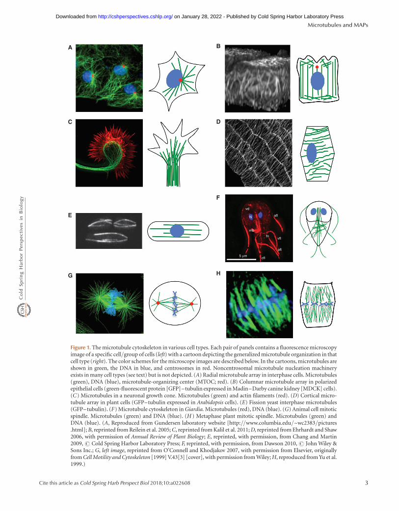
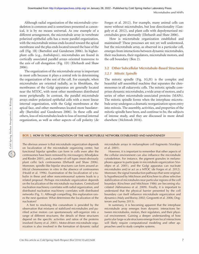
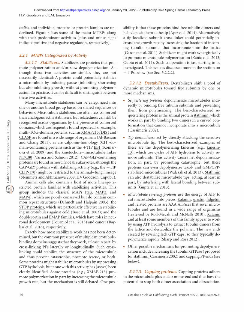
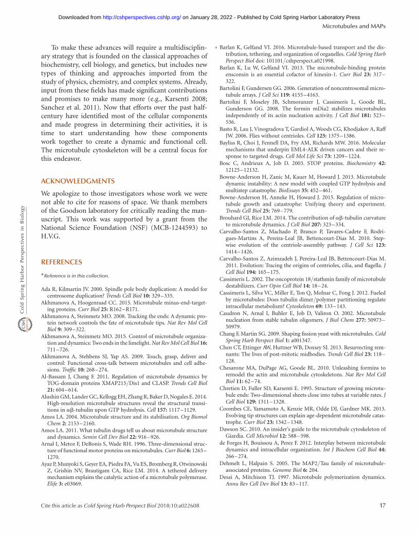
Figure 3. Microtubule (MT) dynamics and assembly. (A) Kymograph (length/time plot derived from a movie) of amicrotubule undergoing dynamic instability in vitro, with dynamics at both the minus (left) and plus (right) ends.Green represents Alexa488-labeled tubulin, and red represents tetra-rhodamine-labeled tubulin GMPCPP (guanylyl5′-a,b-methylenediphosphonate)-stabilized microtubule seeds. (B) Cartoon of a length-history plot (also called alife-history plot) of a microtubule undergoing dynamic instability. The key processes of microtubule dynamics areindicated. (C) Standard model of dynamic instability. As long as the microtubule has a GTP cap, it can grow, but ittransitions to rapid depolymerization (catastrophe) on loss of the GTP cap. (A, Adapted, with permission, fromZanic et al. 2013.)
Microtubules and MAPs
Cite this article as Cold Spring Harb Perspect Biol 2018;10:a022608 9
on January 28, 2022 - Published by Cold Spring Harbor Laboratory Press http://cshperspectives.cshlp.org/Downloaded from

“search–capture”) plays a fundamental role in the self-assembly of structures such as the mitotic spindle (re-viewed by Mogilner et al. 2006).
The process of building and maintaining such a dynamicmicrotubule cytoskeleton might be energy-intensive, but itis profoundly robust—in other words, it is unlikely to faileven if significant perturbations (e.g., changes in cell shape,number of microtubules) occur to the system. This reali-zation is important for understanding how the cytoskele-ton works, but it also has implications for understandingthe process by which the cytoskeleton evolved (Kirschnerand Gerhart 2005).
4.2 Microtubule Assembly Can Be Alteredby Changes to the Environmentand by Drugs
Microtubules assembled from pure GTP–tubulin are re-markably unstable polymers—reduce the concentration ofGTP–tubulin subunits for a few seconds and they disap-pear. Rapid depolymerization can also be induced by re-ducing the temperature or shifting other aspects of theenvironment (e.g., an increase in Ca2+). The transientand unstable nature of microtubules is all the more strikingwhen considered alongside the ability of these structures towithstand physical perturbations (bending, tension), asoutlined above.
Researchers can manipulate the assembly state of mi-crotubules through the use of small molecules. The nat-ural product taxol and its relatives induce microtubuleassembly and can stabilize microtubules against dilu-tion-induced depolymerization and (to a lesser degree)cold temperatures, whereas molecules such as nocoda-zole, colchicine, vinblastine, and vincristine destabilizemicrotubules. The mechanism of taxol-mediated stabili-zation has been unclear (Amos 2011), but recent high-resolution structural data might be resolving this issue(Alushin et al. 2014).
Microtubule-altering drugs are very important inagriculture and medicine, especially cancer chemother-apy, at least partly because of the role microtubules playin spindle assembly. Microtubule-directed drugs can besomewhat organism specific, and some commerciallysignificant compounds target microtubules of fungi orplants (e.g., Benomyl). Hence, work continues on de-veloping new compounds that target microtubules(Amos 2011). Researchers are also developing drugsthat target MTBPs—for example, motors (as possiblecancer-fighting agents) and tau (as possible treatmentsfor neurodegenerative disease) (Rath and Kozielski2012).
4.3 Mechanism of Microtubule Dynamic Instability
As discussed above, the apparently random2 transitionsbetween growth and depolymerization that characterizemicrotubule dynamic instability are functionally signifi-cant, but they are also intriguing. Transitions from growthto depolymerization are termed “catastrophes,” whereasthose from depolymerization to growth are called “rescues”(Fig. 3B). What could cause such abrupt switching? Inother words, what is the “mechanism” of dynamic insta-bility? The answers to these questions have become clearerin the 30 years that have passed since dynamic instabilitywas first recognized (Mitchison and Kirschner 1984), butsome important questions still remain.
Some generally accepted experimental observations rel-evant to the mechanism of dynamic instability include thefollowing.
† As discussed above, soluble tubulin binds to GTP. Thea-subunit binds GTP without hydrolyzing or exchanging.The exchangeable GTP on the b-subunit hydrolyzes andreleases its phosphate quickly after polymerization (withrates on the order of 0.5/sec) but slowly in the absence ofpolymer (Melki et al. 1998; see also Margolin et al. 2012and Seetapun et al. 2012).
† Assembly promotes hydrolysis of the GTP bound to thesubunits. This occurs because the incoming subunitfunctions as a GAP (GTPase-activating protein) forthe subunit by completing its nucleotide-active site (No-gales et al. 1998).
† GTP–tubulin will assemble into microtubules if thefree-tubulin concentration is sufficiently high (i.e.,above the critical concentration). GDP–tubulin willnot assemble into more than oligomers (its critical con-centration is impractically high; Howard 2001).
† Growing microtubules have slightly curved PFs and/orsheet-like extensions at their tips (Chretien et al. 1995),but depolymerizing microtubules have at their tipstightly curled “ram’s horns” consisting of curved PFsthat appear to be peeling off the microtubules. IsolatedGDP–tubulin can form rings similar to these rams’horns.
† These observations led early on to the idea that GTP–tubulin and GDP–tubulin have different preferred con-formations. Initially, it was proposed that GTP–tubulinis straight and GDP–tubulin is curved. Later structuraldata suggested that both are curved, but that GTP–tu-
2The transitions are often described as random, but this is not completelyaccurate: Microtubules that have been growing longer are more likely toundergo catastrophe (Odde et al. 1995; Coombes et al. 2013).
H.V. Goodson and E.M. Jonasson
10 Cite this article as Cold Spring Harb Perspect Biol 2018;10:a022608
on January 28, 2022 - Published by Cold Spring Harbor Laboratory Press http://cshperspectives.cshlp.org/Downloaded from
bulin can become straight in the context of a microtu-bule. The bending appears to occur both within andbetween subunits (reviewed by Brouhard and Rice2014).
† Microtubules assembled from the slowly hydrolyzingGTP-like analog GMPCPP polymerize as normal butdepolymerize much more slowly (Hyman et al. 1992).This is a key point: It means that the energy of GTP isused to destroy the microtubule not to build it.
† Microtubule growth and dynamic instability can occurat either end, although the kinetics of the two ends differ(e.g., the plus end grows faster than the minus end).
For further information and citations not given above, seeDesai and Mitchison 1997, Howard 2001, and Howard andHyman 2003.
These and other experiments have led to the standardtextbook model of dynamic instability, which typically in-cludes the following ideas.
† After a new subunit adds to a growing microtubule tip, ashort delay in hydrolysis and phosphate release result ina GTP-rich region (the GTP cap3) at the growing tip,whereas older parts of the microtubule primarily com-prise GDP–tubulin (this region is sometimes referred toas the “GDP lattice”) (Fig. 3C).
† GTP hydrolysis promotes depolymerization in at leasttwo ways: First, the GDP–tubulin in the microtubulelattice is under strain because it is forced to be in anunfavorable straight conformation. This strain effective-ly weakens the already weak lateral bonds between PFs(Zhang et al. 2015). In addition, conformational chang-es associated with hydrolysis and phosphate loss weakenthe longitudinal bond (Rice et al. 2008; Zhang et al.2015). (Note: It used to be thought that the lateral bondsof the GTP and GDP lattices are significantly different,but, as has been seen from recent cryo-electron micros-copy [EM] work, the lateral contacts of the GTP andGDP lattices are similar; GTP hydrolysis further weakensthe lateral bonds by increasing the strain on these bonds[Zhang et al. 2015].)
† When the GTP cap is present (i.e., as long as new GTP–tubulin is added to the tip faster than it is lost throughdissociation and/or hydrolysis), its relatively strong lat-eral bonds maintain the tubular structure and allowcontinued polymerization. However, if the cap is lost,a so-called catastrophe occurs: The GDP–tubulin is ex-
posed, the PFs splay apart as the GDP subunits adopttheir preferred bent conformation, and the microtubulerapidly depolymerizes.
† According to this model, the GDP lattice below the GTPcap stores the energy of GTP hydrolysis, allowing thedepolymerizing microtubule to do work (Grishchuket al. 2005; Mogilner and Oster 2003).
Although this conceptual model is attractive, it leavesmany questions unresolved. For example, what are the sizeand shape of the GTP cap? Are GTP and GDP the onlyimportant conformations, or do intermediates (i.e.,GDP-Pi) play an important role? Why do the minus andplus ends show quantitatively different dynamic instabilitybehaviors?4 What are the detailed molecular mechanismsof catastrophe and rescue? One can imagine fluctuations intubulin addition and first-order GTP hydrolysis leading tocatastrophe, but what series of events could cause a rapidlydepolymerizing microtubule to start polymerizing again?
The sudden and apparently unpredictable nature ofcatastrophe and rescue have been so puzzling that someresearchers have proposed that they result entirely from“outside” influences, such as lattice defects or thermal fluc-tuations, and/or require the action of MTBPs. Alternative-ly, it has been proposed that microtubules normally grow asopen sheets, and that catastrophe might be caused by tubeclosure, which could be related in an unspecified way toGTP hydrolysis (Chretien et al. 1995; Wieczorek et al.2015). It is important to note that some of these ideas arenot mutually exclusive with the standard model presentedabove and that multiple mechanisms are likely at work (e.g.,catastrophe can be both a spontaneous event and onecaused by MTBPs).
4.4 Current Research into the Mechanismof Dynamic Instability
Recently an array of new experiments, such as nanoscaleassembly measurements, superresolution microscopy, andlocalization of conformation-specific binding proteins, hasprovided new insight into the microtubule assembly pro-cesses (e.g., Dimitrov et al. 2008; Gardner et al. 2011; See-tapun et al. 2012; Coombes et al. 2013). These approacheshave been complemented by determination of microtubulestructure at higher resolution (Alushin et al. 2014; Zhanget al. 2015) and improved computer simulations (e.g., Van-Buren et al. 2005; Margolin et al. 2012; Bowne-Anderson
3It is not clear whether it is actually the GTP form, the GDP-Pi form, or boththat have the stabilizing activity. For simplicity, most publications refer to thisstabilizing GTP- and/or GDP-Pi-rich structure as the “GTP cap.”
4One reasonable (but not necessarily complete) explanation for the asymme-try is that a subunit attaching at the plus end does not hydrolyze its GTP untilafter a new subunit attaches to it, whereas one attaching at the minus end willstart the process of first-order hydrolysis as soon as it attaches.
Microtubules and MAPs
Cite this article as Cold Spring Harb Perspect Biol 2018;10:a022608 11
on January 28, 2022 - Published by Cold Spring Harbor Laboratory Press http://cshperspectives.cshlp.org/Downloaded from
et al. 2013). The picture that is emerging is broadly consis-tent with the classical model of dynamic instability out-lined above, but it contains additional molecular detail,and it is beginning to address some of the open questions.
More specifically, the model that emerges from multi-ple experimental and theoretical studies is one in whichrapidly exchanging GTP subunits add to PF tips (withonly a small fraction being incorporated), and the nonter-minal subunits undergo first-order (not vectorial5) GTPhydrolysis. The resulting microtubule structure has an un-stable GDP lattice capped by a stabilizing region rich inGTP (or GDP-Pi). This GTP cap has a size that dependson the elongation rate, as well as an approximate exponen-tial shape with a poorly defined lower boundary, and itcan extend approximately 10 or more subunits into themicrotubule (Margolin et al. 2012; Seetapun et al. 2012;Coombes et al. 2013; Bowne-Anderson et al. 2015). In thesemodels, open sheets are not observed, but instead flat ex-tensions protruding from a closed tube are seen; thesesheets might explain the age-dependent catastrophe thathas been observed experimentally (Coombes et al. 2013).
Dimer-based computational simulations founded onthis conceptual model recapitulate many aspects of exper-imentally observed dynamic instability, supporting itsbroad structure, but they also make new predictions. Forexample, one of these models suggests that the rapid sub-unit exchange mentioned above occurs because cracks (lat-erally unbonded regions) exist between adjacent PFs in theregion close to the tip, allowing subunits to exchange untilthey form lateral bonds and become incorporated into thelattice (Margolin et al. 2012; Li et al. 2014). One appealingaspect of this and related ideas is that the healing of thesecracks by MTBPs and the resulting suppression of loss ofrecently added subunits could potentially account for theotherwise surprising ability of some MTBPs to increase therate of microtubule growth (Howard and Hyman 2009;Gardner et al. 2011).
These computer simulations are also providing specifichypotheses about the molecular-scale mechanisms of ca-tastrophe and rescue. One idea is that catastrophe resultsfrom stochastic fluctuations in the extent to which inter-protofilament cracks extend into the GDP-rich region(Margolin et al. 2012; Li et al. 2014) and/or the lengthsof the PF extensions (Coombes et al. 2013). Rescue (a rareevent in the absence of MTBPs) might be promoted bystochastic blunting of the microtubule tip, followed by re-
establishment of regions of laterally bonded GTP–tubulin(Margolin et al. 2012; Li et al. 2014). Regulatory proteinscould potentially promote or suppress transitions by alter-ing the stability of lateral bonding between subunits or thetip extensions (e.g., Gupta et al. 2013) or by altering deliv-ery of tubulin subunits to the tip (Ayaz et al. 2014).In addition, the ability of computer simulations to followa system simultaneously at different scales means thatthese models have the potential to provide insight intolong-standing questions about how the properties of thetubulin dimers relate to the observed dynamic instabilityparameters and bulk-scale properties such as the criticalconcentration.
5 MICROTUBULE-BINDING PROTEINS
5.1 Introduction to MTBPs
Naked microtubules assembled from pure tubulin are un-stable structures that are poised, quite literally, at the edgeof catastrophe. This situation enables microtubule assem-bly to respond quickly to changes in the environment andthe influence of regulatory proteins. Some microtubuleregulatory proteins are widespread in eukaryotes, but oth-ers vary significantly by organism and cell type. Evenamong the organism-specific proteins there are some com-mon motifs and themes, as discussed below.
The term MTBP applies broadly to any protein that canbe shown experimentally to bind to microtubules. Anotherterm, MAP, is often used to describe the subset of MTBPsthat cosediment with microtubules through multiplerounds of polymerization and depolymerization, a groupthat includes proteins such as MAP2 and tau.
Broadly speaking, the MTBPs that regulate assemblycan be categorized functionally as stabilizers, destabilizers(including severing proteins), capping proteins, and bun-dlers/cross-linkers (Fig. 4). Other MTBPs include motorsthat use microtubules as tracks for intracellular transport(reviewed by Sweeney and Holzbaur 2016) and cytoplas-mic linker proteins (CLIPs), which anchor organelles tomicrotubules to promote cell organization. Some MTBPsare cytoskeletal integrators (i.e., proteins that connect toother components of the cytoskeleton). In addition, someproteins involved in signal transduction, translation, andmetabolism bind microtubules or other components ofthe cytoskeleton. Many MTBPs have multiple activities.Determining the functions of MTBPs can be challengingbecause the activity observed can depend on the detailsof the assay or on other aspects of the experimentalconditions.
Microtubule regulatory proteins can also be catego-rized as to where they localize on dynamic microtubules
5Early conceptual models of microtubule dynamic instability assumed thatthe GTP hydrolysis occurred vectorially—that is, in a wave that travels upfrom the base toward the tip. Later mathematical modeling (Flyvbjerg et al.1994) showed that this mechanism was not consistent with the data fromsudden dilution experiments (Walker et al. 1991). See Bowne-Anderson et al.2015 for more discussion.
H.V. Goodson and E.M. Jonasson
12 Cite this article as Cold Spring Harb Perspect Biol 2018;10:a022608
on January 28, 2022 - Published by Cold Spring Harbor Laboratory Press http://cshperspectives.cshlp.org/Downloaded from
(Fig. 4). Lattice-binding proteins associate with microtu-bules along their length, whereas end-binding proteins lo-calize more specifically to one or both of the microtubuleextremities. Microtubule +TIPs are a subset of end-bind-ing proteins that dynamically track growing microtubuleends, which in vivo are typically the plus ends (Akhmanovaand Steinmetz 2015). The recently recognized microtubule
minus end–targeting proteins are specifically recruited tominus ends (Akhmanova and Hoogenraad 2015).
5.2 Specific Classes of MTBPs
For the purposes of the discussion that follows, major clas-ses of MTBPs are listed in the headings, subclasses are in
Stathmin
Minusend Seam
GDP lattice
Lattice binding
Tubulin dimerbinding proteins
CLIP170, XMAP215,tau
Stathmin
GTP GDP
Plusend
GTP cap
Stathmin, kinesin 8and 13 (depolymerizingmotor proteins)
EB1, XMAP215,CLASP, CLIP170,APC, doublecortin
+ TIP network
Stabilizers: tau, Map2,Map4, Map1b
Destabilizers: katanin,severing proteins
Bundlers and cross-linkers:MAP65/ASE1/PRC1
Motor regulators: tau, ensconsin
γ-TuRC, patronin/CAMSAPs, +TIPnetwork (in vitro)
Minus end bindingproteins
–
–
–
–
+
+
+
+
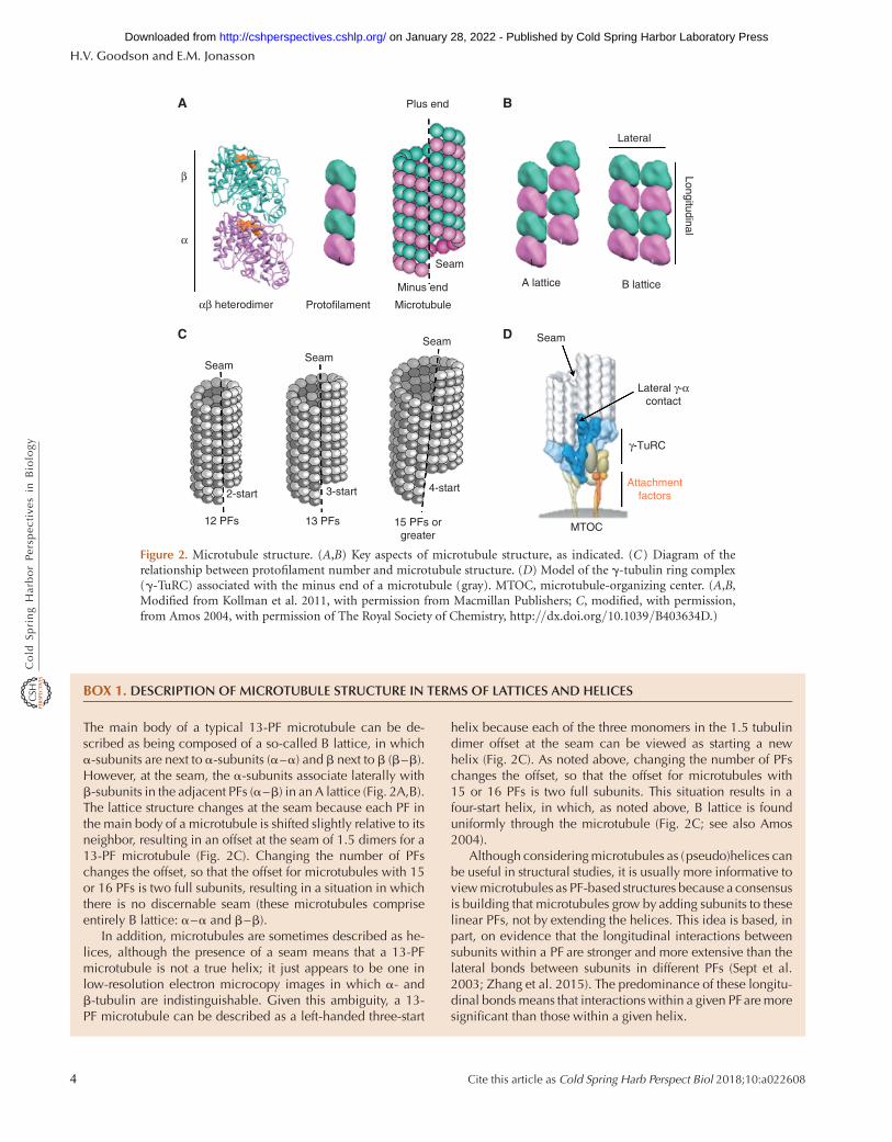
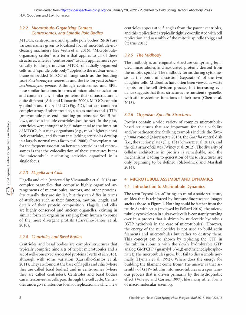
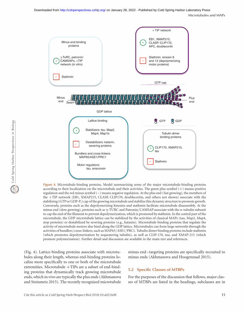
Figure 4. Microtubule-binding proteins. Model summarizing some of the major microtubule-binding proteinsaccording to their localization on the microtubule and their activities. The green plus symbol (+) means positiveregulation and the red minus symbol (–) means negative regulation. At the plus end (fast growing), the members ofthe +TIP network (EB1, XMAP215, CLASP, CLIP170, doublecortin, and others not shown) associate with thestabilizing (GTPor GDP-Pi) cap of the growing microtubule and stabilize this dynamic structure to promote growth.Conversely, proteins such as the depolymerizing kinesins and stathmin facilitate microtubule disassembly. At theminus end (slow growing), proteins such as g-TURC and Patronin/CAMSAP associate with the a-tubulin subunitto cap the end of the filament to prevent depolymerization, which is promoted by stathmin. In the central part of themicrotubule, the GDP microtubule lattice can be stabilized by the activities of classical MAPs (tau, Map2, Map4,stop proteins) or destabilized by severing proteins (e.g., katanin). Microtubule-binding proteins that regulate theactivity of microtubule motors also bind along the GDP lattice. Microtubules can form large networks through theactivities of bundlers/cross-linkers, such as MAP65/ASE1/PRC1. Tubulin dimer binding proteins include stathmin(which promotes depolymerization by sequestering tubulin), as well as CLIP-170, tau, and XMAP-215 (whichpromote polymerization). Further detail and discussion are available in the main text and references.
Microtubules and MAPs
Cite this article as Cold Spring Harb Perspect Biol 2018;10:a022608 13
on January 28, 2022 - Published by Cold Spring Harbor Laboratory Press http://cshperspectives.cshlp.org/Downloaded from
italics, and individual proteins or protein families are un-derlined. Figure 4 lists some of the major MTBPs alongwith their predominant activities (plus and minus signsindicate positive and negative regulation, respectively).
5.2.1 MTBPs Categorized by Activity
5.2.1.1 Stabilizers. Stabilizers are proteins that pro-mote polymerization and/or slow depolymerization. Al-though these two activities are similar, they are notnecessarily identical: A protein could potentially stabilizea microtubule by inducing pause (inhibiting shorteningbut also inhibiting growth) without promoting polymeri-zation. In practice, it can be difficult to distinguish betweenthese two activities.
Many microtubule stabilizers can be categorized intoone or another broad group based on shared sequences orbehaviors. Microtubule stabilizers are often less conservedthan analogous actin stabilizers, but relatedness can still berecognized across organisms by the presence of conserveddomains,which are frequently found repeated. Forexample,multi-TOG-domain proteins, such as XMAP215/DIS1 andCLASP, are found in a wide range of organisms (Al-Bassamand Chang 2011), as are calponin-homology (CH)-do-main-containing proteins such as the +TIP EB1 (Komar-ova et al. 2009) and the kinetochore–microtubule linkerNDC80 (Varma and Salmon 2012). CAP-GLY-containingproteins are found in most if not all eukaryotes, although theCAP-GLY proteins with stabilizing activity (e.g., the +TIPCLIP-170) might be restricted to the animal–fungi lineage(Steinmetz and Akhmanova 2008; HV Goodson, unpubl.).
Animal cells also contain a host of more lineage-re-stricted protein families with stabilizing activities. Thisgroup includes the classical MAPs (tau, MAP2, andMAP4), which are poorly conserved but do contain com-mon repeat structures (Dehmelt and Halpain 2005); theSTOP proteins, which are particularly effective in stabiliz-ing microtubules against cold (Bosc et al. 2003); and thedoublecortin and EMAP families, which have roles in neu-ronal development (Fourniol et al. 2013) and cancer (Bay-liss et al. 2016), respectively.
Exactly how most stabilizers work has not been deter-mined, but the common presence of multiple microtubule-binding domains suggests that they work, at least in part, bycross-linking PFs laterally or longitudinally. Such cross-linking could stabilize the structure of the microtubuleand thus prevent catastrophe, promote rescue, or both.Some proteins might stabilize microtubules by suppressingGTP hydrolysis, but none with this activity has (as yet) beenclearly identified. Some proteins (e.g., XMAP-215) pro-mote polymerization in part by increasing the microtubulegrowth rate, but the mechanism is still debated. One pos-
sibility is that these proteins bind free tubulin dimers andhelp deposit them at the tip (Ayaz et al. 2014). Alternatively,a tip-localized subunit cross-linker could potentially in-crease the growth rate by increasing the fraction of incom-ing tubulin subunits that incorporate into the lattice(Gardner et al. 2011). Stabilizers might work synergisticallyto promote microtubule polymerization (Zanic et al. 2013;Gupta et al. 2014). Such cooperation is just starting to beinvestigated. This issue is discussed more in the section on+TIPs below (see Sec. 5.2.2.2).
5.2.1.2 Destabilizers. Destabilizers shift a pool ofdynamic microtubules toward free subunits by one ormore mechanisms.
† Sequestering proteins depolymerize microtubules indi-rectly by binding free tubulin subunits and preventingthem from polymerizing. The best-characterized se-questering protein is the animal protein stathmin, whichworks in part by binding two dimers in a curved con-formation that cannot incorporate into a microtubule(Cassimeris 2002).
† Tip destabilizers act by directly attacking the sensitivemicrotubule tip. The best-characterized examples ofthese are the depolymerizing kinesins (e.g., kinesin-13), which use cycles of ATP hydrolysis to actively re-move subunits. This activity causes net depolymeriza-tion, in part, by promoting catastrophe, but theseproteins can even depolymerize taxol- and GMPCPP-stabilized microtubules (Walczak et al. 2013). Stathmincan also destabilize microtubule tips, acting, at least inpart, by interfering with lateral bonding between sub-units (Gupta et al. 2013).
† Microtubule severing proteins use the energy of ATP tocut microtubules into pieces. Katanin, spastin, fidgetin,and related proteins are AAA ATPases that sever micro-tubules and are found in a wide range of organisms(reviewed by Roll-Mecak and McNally 2010). Kataninand at least some members of this family appear to workby using ATP hydrolysis to extract tubulin dimers fromthe lattice and destabilize the polymer. The new endscreated by severing lack GTP caps, so they typically de-polymerize rapidly (Sharp and Ross 2012).
† Other possible mechanisms for promoting depolymeri-zation include increasing the tubulin GTPase (proposedfor stathmin; Cassimeris 2002) and capping PF ends (seebelow).
5.2.1.3 Capping proteins. Capping proteins adhereto the microtubule plus end or minus end and thus have thepotential to stop both dimer association and dissociation.
H.V. Goodson and E.M. Jonasson
14 Cite this article as Cold Spring Harb Perspect Biol 2018;10:a022608
on January 28, 2022 - Published by Cold Spring Harbor Laboratory Press http://cshperspectives.cshlp.org/Downloaded from
Although actin filament capping proteins are well charac-terized (see Pollard 2016), less is known about microtubulecapping proteins, perhaps because of the greater size andcomplexity of the microtubule tip. For example, the onlyknown minus-end capping proteins are large complexesrather than individual proteins. The best-characterized ex-amples are the related complexes g-TuRC and g-TuSC,which not only cap minus ends but also nucleate microtu-bules (Kollman et al. 2011). Some evidence suggests thatstathmin can cap PFs and suppress subunit addition with-out stabilizing the polymer (Gupta et al. 2013). Patroninand other proteins known as CAMSAPs are sometimesconsidered capping proteins, although they appear to as-sociate laterally with the minus end (see also minus end–targeting proteins below).
5.2.1.4 Bundlers and cross-linkers. Bundlers andcross-linkers associate microtubules laterally. Proteins var-iously called MAP65/Ase1/PRC1 preferentially bundle an-tiparallel microtubules (Walczak and Shaw 2010), anactivity important in the mitotic spindle. Most stabilizershave some bundling activity, but whether this activity isphysiologically relevant is unclear. Simply coating negative-ly charged microtubules with tau peptides can result inbundling (Melki et al. 1991), as can addition of crowdingagents such as polyethylene glycol (Sanchez et al. 2012).
5.2.1.5 Cytoskeletal integrators. Cytoskeletal inte-grators bind to and/or modulate microtubules and at leastone other cytoskeletal element. This diverse category con-tains large scaffolding molecules such as the cancer-associ-ated protein APC and the “plakin” family (Suozzi et al.2012). It also includes the actin-nucleating formin family(Bartolini et al. 2008; Gaillard et al. 2011), myosin 10 (We-ber et al. 2004), and even classic MAPs such as tau (Gallo2007). Communication between the actin and microtubulecytoskeletons is essential for proper functioning of process-es such as cytokinesis and generation and maintenance ofcell polarity, and these proteins play fundamental, yet stillpoorly understood, roles in these processes (Rodriguezet al. 2003).
5.2.1.6 Other microtubule-associated activities.Additional microtubule-associated activities include thefollowing.
† Microtubule motors kinesin, dynein, and their accesso-ries such as dynactin complex (reviewed by Barlan andGelfand 2016 and Sweeney and Holzbaur 2016).
† Motor modulators are proteins such as tau (Dixit et al.2008) and ensconcin (Barlan et al. 2013) that bind themicrotubule lattice and alter the behavior of motorsacting on those microtubules.
† Membrane–microtubule linkers such as CLIMP63 (Ved-renne et al. 2005) provide organelles with alternative(nonmotor) connections to microtubules (Gurel et al.2014).
† Metabolic proteins, including most enzymes of the gly-colytic pathway, often bind to microtubules. One possi-ble physiological function of this interaction is toincrease the local concentration of proteins in thesame biochemical pathway, but some of these proteinsare capable of altering microtubule assembly (at least invitro or on overexpression). These and related observa-tions suggest that the metabolic state of the cell caninfluence microtubules and vice versa (reviewed by Cas-simeris et al. 2012).
5.2.2 MTBPs as Categorized by Localization
5.2.2.1 Lattice-binding proteins. Lattice-bindingproteins bind along the body of the microtubule and in-clude all proteins that do not target either the plus or minusends. As a result, this category includes proteins with arange of activities. Well-recognized examples of lattice-binding proteins include the classical MAPs tau, Map2,and Map4 (Dehmelt and Halpain 2005). Tau and Map2are neuronal microtubule stabilizers that are localized toaxons and dendrites, respectively, whereas Map4 is ex-pressed in most tissues. Tau is the focus of much researchbecause of its involvement in Alzheimer’s disease (Iqbualet al. 2016). Tau and MAP2 also seem to play a role in thespacing of microtubules in tightly packed neuronal exten-sions, as well as in regulating motor activity (Dixit et al.2008).
5.2.2.2 Microtubule plus end–trafficking pro-teins. Microtubule +TIPs dynamically track growing mi-crotubule ends. As the configuration of the microtubule tipdetermines whether a microtubule grows or shrinks, +TIPbehavior allows a polymerization regulator to be dynami-cally localized where it needs to act. Although the abbrevi-ation “+TIP” is attractive, it can be misleading: Thecanonical +TIP EB1 is specific not to plus ends but togrowing ends, as it will track growing minus ends if present-ed with the opportunity, at least in vitro (Akhmanova andSteinmetz 2008). The set of +TIPs includes some of themost conserved and significant polymerization-promotingMTBPs known, including EB1, XMAP-215, CLASP, CLIP-170, and their relatives. It is important to note that not all+TIPs promote polymerization—some +TIPs, includingkinesin-13, depolymerize microtubules (for an overview of+TIPs, see Akhmanova and Steinmetz 2008, 2015).
A small number of +TIPs (most notably EB1) trackgrowing ends by binding preferentially to a short-lived
Microtubules and MAPs
Cite this article as Cold Spring Harb Perspect Biol 2018;10:a022608 15
on January 28, 2022 - Published by Cold Spring Harbor Laboratory Press http://cshperspectives.cshlp.org/Downloaded from
tip-specific conformation (Maurer et al. 2014). Most oth-er +TIPs localize to ends by binding to EB1, so EB1 isoften considered to be “the master +TIP” (reviewed byAkhmanova and Steinmetz 2015). Consistent with theidea that EB1 has a central and ancient role in microtu-bule dynamics, EB1 is found across the spectrum of eu-karyotic organisms, including Giardia (Dawson 2010). Inthis wide diversity of organisms, EB1 has a canonicalhomodimeric structure consisting of a CH domain fol-lowed by an apparently unstructured region, a short andhighly conserved coiled-coil by which the protein dimer-izes, and a tail that folds back to make a four-helix bundle.This four-helix bundle contains the hydrophobic pocketinto which inserts the SxIP motifs of most EB1-binding+TIPs. In many organisms, EB1 terminates with an EEYmotif that mimics the tubulin carboxy-terminal tail; thisappears to be involved in autoinhibition (for reviews ofEB1 structure and function, see Akhmanova and Stein-metz 2008, 2015).
Most if not all +TIPs bind other +TIPs, and so they aresaid to form the “+TIP network”—a loose web of inter-acting proteins that work together to regulate microtubuledynamics and integrate microtubules with the rest of thecell (Akhmanova and Steinmetz 2015). These interactionslocalize many of the proteins indirectly to growing micro-tubule tips and often release autoinhibition, allowing the+TIP network to integrate signals from many pathways(Akhmanova and Steinmetz 2015). The +TIP networkmight also act as a superstructure that promotes microtu-bule growth by stabilizing the structure of the microtubuletip and thus helping to promote microtubule polymeriza-tion (Gupta et al. 2014).
5.2.2.3 Minus end–targeting proteins. Two typesof proteins localize to microtubule minus ends—the g-TuRC (Kollman et al. 2011) and a more recently discoveredset of proteins known collectively as CAMSAPs (whichincludes the Drosophila protein Patronin) (Akhmanovaand Hoogenraad 2015). g-TuRC blocks both subunit asso-ciation and dissociation by interacting with all of the PFs(Fig. 2D). CAMSAPs bind at the minus end and effectivelycap it, stabilizing it against depolymerization and stoppingor slowing tubulin addition; some CAMSAPs can eventrack growing minus ends (Akhmanova and Hoogenraad2015). CAMSAPs consist of an amino-terminal CH do-main, some regions of coiled-coil, and a conserved car-boxy-terminal CKK motif, and they seem to accomplishtheir stabilization of minus ends by assembling laterallyat the minus end instead of by creating a classical cap.CAMSAPs are found in most metazoans and appear tobe absent from plants and fungi, but they might be morewidely distributed because some sequence motifs are pres-
ent in the genomes of organisms such as ciliates (Akhma-nova and Hoogenraad 2015; HV Goodson, unpubl.). Asanother example of a minus end binding protein, someevidence suggests that stathmin binding might be biasedto the minus end (cited by Gupta et al. 2013).
6 CONCLUSION
The microtubule cytoskeleton is one of the most remark-able components of eukaryotic cells: During interphase,the microtubule array explores the cytoplasm, finds cargo,provides a substrate for transport, adjusts to internal andexternal signals, and directs the organization of the rest ofthe cell. Then, upon transitioning into mitosis, it disassem-bles and reassembles into the strikingly different mitoticspindle, a structure with the profoundly important pur-pose of precisely and consistently segregating to eachdaughter cell the correct set of chromosomes. Central toall of these processes are microtubule dynamics.
In reflecting on the role of dynamics in the function ofthe microtubules, it is interesting to realize that the com-bination of random probing and selective stabilization,as seen in the microtubule cytoskeleton, is a theme thatoccurs throughout biology: Random exploration of spacefollowed by reinforcement/selection of the optimizedvariants is found in processes as diverse as acquired im-munity, insect forging behavior, and even Darwinian evo-lution itself. This strategy produces highly robust systems(i.e., systems that are adaptable and hard to break), and itsrecurring appearance in biology provides an explanationfor the robustness of life itself (Kirschner and Gerhart 2005;Karsenti 2008).
Much has been learned in the 30 years or so since dy-namic instability was discovered, but a great deal remains tobe understood about microtubules, how their assembly isregulated, how they are dynamically organized, and howtheir organization drives the organization of the rest of thecell. At the molecular scale, key problems include establish-ing the mechanisms of the catastrophe and rescue transi-tions, determining how MTBPs alter these transitions, andunderstanding how groups of MTBPs work together tocreate particular behaviors. At the cell scale, major chal-lenges include understanding the assembly and dynamicsof large-scale structures such as the mitotic spindle andflagellum and elucidating the organizational cross talk be-tween microtubules and the rest of the cell. In both cases, animportant goal will be to obtain a quantitative and predic-tive understanding of these processes. Gaining this knowl-edge will be important for cell biology, but the resultinginformation about self-assembling systems also has the po-tential to impact fields as distant as nanotechnology andsynthetic biology.
H.V. Goodson and E.M. Jonasson
16 Cite this article as Cold Spring Harb Perspect Biol 2018;10:a022608
on January 28, 2022 - Published by Cold Spring Harbor Laboratory Press http://cshperspectives.cshlp.org/Downloaded from
To make these advances will require a multidisciplin-ary strategy that is founded on the classical approaches ofbiochemistry, cell biology, and genetics, but includes newtypes of thinking and approaches imported from thestudy of physics, chemistry, and complex systems. Already,input from these fields has made significant contributionsand promises to make many more (e.g., Karsenti 2008;Sanchez et al. 2011). Now that efforts over the past half-century have identified most of the cellular componentsand made progress in determining their activities, it istime to start understanding how these componentswork together to create a dynamic and functional cell.The microtubule cytoskeleton will be a central focus forthis endeavor.
ACKNOWLEDGMENTS
We apologize to those investigators whose work we werenot able to cite for reasons of space. We thank membersof the Goodson laboratory for critically reading the man-uscript. This work was supported by a grant from theNational Science Foundation (NSF) (MCB-1244593) toH.V.G.
REFERENCES
∗Reference is in this collection.
Ada R, Kilmartin JV. 2000. Spindle pole body duplication: A model forcentrosome duplication? Trends Cell Biol 10: 329–335.
Akhmanova A, Hoogenraad CC. 2015. Microtubule minus-end-target-ing proteins. Curr Biol 25: R162–R171.
Akhmanova A, Steinmetz MO. 2008. Tracking the ends: A dynamic pro-tein network controls the fate of microtubule tips. Nat Rev Mol CellBiol 9: 309–322.
Akhmanova A, Steinmetz MO. 2015. Control of microtubule organiza-tion and dynamics: Two ends in the limelight. Nat Rev Mol Cell Biol 16:711–726.
Akhmanova A, Stehbens SJ, Yap AS. 2009. Touch, grasp, deliver andcontrol: Functional cross-talk between microtubules and cell adhe-sions. Traffic 10: 268–274.
Al-Bassam J, Chang F. 2011. Regulation of microtubule dynamics byTOG-domain proteins XMAP215/Dis1 and CLASP. Trends Cell Biol21: 604–614.
Alushin GM, Lander GC, Kellogg EH, Zhang R, Baker D, Nogales E. 2014.High-resolution microtubule structures reveal the structural transi-tions in ab-tubulin upon GTP hydrolysis. Cell 157: 1117–1129.
Amos LA. 2004. Microtubule structure and its stabilisation. Org BiomolChem 2: 2153–2160.
Amos LA. 2011. What tubulin drugs tell us about microtubule structureand dynamics. Semin Cell Dev Biol 22: 916–926.
Arnal I, Metoz F, DeBonis S, Wade RH. 1996. Three-dimensional struc-ture of functional motor proteins on microtubules. Curr Biol 6: 1265–1270.
Ayaz P, Munyoki S, Geyer EA, Piedra FA, Vu ES, Bromberg R, OtwinowskiZ, Grishin NV, Brautigam CA, Rice LM. 2014. A tethered deliverymechanism explains the catalytic action of a microtubule polymerase.Elife 3: e03069.
∗ Barlan K, Gelfand VI. 2016. Microtubule-based transport and the dis-tribution, tethering, and organization of organelles. Cold Spring HarbPerspect Biol doi: 101101/cshperspect.a021998.
Barlan K, Lu W, Gelfand VI. 2013. The microtubule-binding proteinensconsin is an essential cofactor of kinesin-1. Curr Biol 23: 317–322.
Bartolini F, Gundersen GG. 2006. Generation of noncentrosomal micro-tubule arrays. J Cell Sci 119: 4155–4163.
Bartolini F, Moseley JB, Schmoranzer J, Cassimeris L, Goode BL,Gunderson GG. 2008. The formin mDia2 stabilizes microtubulesindependently of its actin nucleation activity. J Cell Biol 181: 523–536.
Basto R, Lau J, Vinogradova T, Gardiol A, Woods CG, Khodjakov A, RaffJW. 2006. Flies without centrioles. Cell 125: 1375–1386.
Bayliss R, Choi J, Fennell DA, Fry AM, Richards MW. 2016. Molecularmechanisms that underpin EML4-ALK driven cancers and their re-sponse to targeted drugs. Cell Mol Life Sci 73: 1209–1224.
Bosc C, Andrieux A, Job D. 2003. STOP proteins. Biochemistry 42:12125–12132.
Bowne-Anderson H, Zanic M, Kauer M, Howard J. 2013. Microtubuledynamic instability: A new model with coupled GTP hydrolysis andmultistep catastrophe. BioEssays 35: 452–461.
Bowne-Anderson H, Anneke H, Howard J. 2015. Regulation of micro-tubule growth and catastrophe: Unifying theory and experiment.Trends Cell Biol 25: 769–779.
Brouhard GJ, Rice LM. 2014. The contribution of ab-tubulin curvatureto microtubule dynamics. J Cell Biol 207: 323–334.
Carvalho-Santos Z, Machado P, Branco P, Tavares-Cadete F, Rodri-gues-Martins A, Pereira-Leal JB, Bettencourt-Dias M. 2010. Step-wise evolution of the centriole-assembly pathway. J Cell Sci 123:1414–1426.
Carvalho-Santos Z, Azimzadeh J, Pereira-Leal JB, Bettencourt-Dias M.2011. Evolution: Tracing the origins of centrioles, cilia, and flagella. JCell Biol 194: 165–175.
Cassimeris L. 2002. The oncoprotein 18/stathmin family of microtubuledestabilizers. Curr Opin Cell Biol 14: 18–24.
Cassimeris L, Silva VC, Miller E, Ton Q, Molnar C, Fong J. 2012. Fueledby microtubules: Does tubulin dimer/polymer partitioning regulateintracellular metabolism? Cytoskeleton 69: 133–143.
Caudron N, Arnal I, Buhler E, Job D, Valiron O. 2002. Microtubulenucleation from stable tubulin oligomers. J Biol Chem 277: 50973–50979.
Chang F, Martin SG. 2009. Shaping fission yeast with microtubules. ColdSpring Harb Perspect Biol 1: a001347.
Chen CT, Ettinger AW, Huttner WB, Doxsey SJ. 2013. Resurrecting rem-nants: The lives of post-mitotic midbodies. Trends Cell Biol 23: 118–128.
Chesarone MA, DuPage AG, Goode BL. 2010. Unleashing formins toremodel the actin and microtubule cytoskeletons. Nat Rev Mol CellBiol 11: 62–74.
Chretien D, Fuller SD, Karsenti E. 1995. Structure of growing microtu-bule ends: Two-dimensional sheets close into tubes at variable rates. JCell Biol 129: 1311–1328.
Coombes CE, Yamamoto A, Kenzie MR, Odde DJ, Gardner MK. 2013.Evolving tip structures can explain age-dependent microtubule catas-trophe. Curr Biol 23: 1342–1348.
Dawson SC. 2010. An insider’s guide to the microtubule cytoskeleton ofGiardia. Cell Microbiol 12: 588–598.
de Forges H, Bouissou A, Perez F. 2012. Interplay between microtubuledynamics and intracellular organization. Int J Biochem Cell Biol 44:266–274.
Dehmelt L, Halpain S. 2005. The MAP2/Tau family of microtubule-associated proteins. Genome Biol 6: 204.
Desai A, Mitchison TJ. 1997. Microtubule polymerization dynamics.Annu Rev Cell Dev Biol 13: 83–117.
Microtubules and MAPs
Cite this article as Cold Spring Harb Perspect Biol 2018;10:a022608 17
on January 28, 2022 - Published by Cold Spring Harbor Laboratory Press http://cshperspectives.cshlp.org/Downloaded from
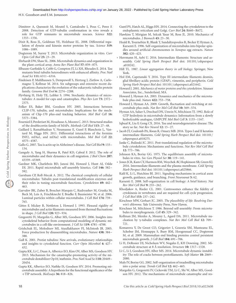
Dimitrov A, Quesnoit M, Moutel S, Cantaloube I, Pous C, Perez F.2008. Detection of GTP-tubulin conformation in vivo reveals arole for GTP remnants in microtubule rescues. Science 322:1353–1356.
Dixit R, Ross JL, Goldman YE, Holzbaur EL. 2008. Differential regu-lation of dynein and kinesin motor proteins by tau. Science 319:1086–1089.
Dogterom M, Surrey T. 2013. Microtubule organization in vitro. CurrOpin Cell Biol 25: 23–29.
Ehrhardt DW, Shaw SL. 2006. Microtubule dynamics and organization inthe plant cortical array. Annu Rev Plant Biol 57: 859–875.
Elbaum-Garfinkle S, Cobb G, Compton JT, Li XH, Rhoades E. 2014. Taumutants bind tubulin heterodimers with enhanced affinity. Proc NatlAcad Sci 111: 6311–6316.
Findeisen P, Muhlhausen S, Dempewolf S, Hertzog J, Zietlow A, Carlo-magno T, Kollmar M. 2014. Six subgroups and extensive recent du-plications characterize the evolution of the eukaryotic tubulin proteinfamily. Genome Biol Evol 6: 2274–2288.
Flyvbjerg H, Holy TE, Leibler S. 1994. Stochastic dynamics of micro-tubules: A model for caps and catastrophes. Phys Rev Lett 73: 2372–2375.
Folker ES, Baker BM, Goodson HV. 2005. Interactions betweenCLIP-170, tubulin, and microtubules: Implications for the mech-anism of Clip-170 plus-end tracking behavior. Mol Biol Cell 16:5373–5384.
Fourniol F, Perderiset M, Houdusse A, Moores C. 2013. Structural studiesof the doublecortin family of MAPs. Methods Cell Biol 115: 27–48.
Gaillard J, Ramabhadran V, Neumanne E, Gurel P, Blanchoin L, Van-tard M, Higgs HN. 2011. Differential interactions of the forminsINF2, mDia1, and mDia2 with microtubules. Mol Biol Cell 22:4575–4587.
Gallo G. 2007. Tau is actin up in Alzheimer’s disease. Nat Cell Biol 9: 133–134.
Ganguly A, Yang H, Sharma R, Patel KD, Cabral F. 2012. The role ofmicrotubules and their dynamics in cell migration. J Biol Chem 287:43359–43369.
Gardner MK, Charlebois BD, Janosi IM, Howard J, Hunt AJ, OddeDJ. 2011. Rapid microtubule self-assembly kinetics. Cell 146: 582–592.
Garnham CP, Roll-Mecak A. 2012. The chemical complexity of cellularmicrotubules: Tubulin post-translational modification enzymes andtheir roles in tuning microtubule functions. Cytoskeleton 69: 442–463.
Garvalov BK, Zuber B, Bouchet-Marquis C, Kudryashev M, Gruska M,Beck M, Leis A, Frischknecht F, Bradke F, Baumeister W, et al. 2006.Luminal particles within cellular microtubules. J Cell Biol 174: 759–765.
Gittes F, Mickey B, Nettleton J, Howard J. 1993. Flexural rigidity ofmicrotubules and actin filaments measured from thermal fluctuationsin shape. J Cell Biol 120: 923–934.
Gregoretti IV, Margolin G, Alber MS, Goodson HV. 2006. Insights intocytoskeletal behavior from computational modeling of dynamic mi-crotubules in a cell-like environment. J Cell Sci 119: 4781–4788.
Grishchuk EL, Molodtsov MI, Ataullakhanov FI, McIntosh JR. 2005.Force production by disassembling microtubules. Nature 438: 384–388.
Gull K. 2001. Protist tubulins: New arrivals, evolutionary relationshipsand insights to cytoskeletal function. Curr Opin Microbiol 4: 427–432.
Gupta KK, Li C, Duan A, Alberico EO, Kim OV, Alber MS, Goodson HV.2013. Mechanism for the catastrophe-promoting activity of the mi-crotubule destabilizer Op18/stathmin. Proc Natl Acad Sci 110: 20449–20454.
Gupta KK, Alberico EO, Nathke IS, Goodson HV. 2014. Promoting mi-crotubule assembly: A hypothesis for the functional significance of the+TIP network. BioEssays 36: 818–826.
Gurel PS, Hatch AL, Higgs HN. 2014. Connecting the cytoskeleton to theendoplasmic reticulum and Golgi. Curr Biol 24: R660–R672.
Hawkins T, Mirigian M, Selcuk Yasar M, Ross JL. 2010. Mechanics ofmicrotubules. J Biomech 43: 23–30.
Heald R, Tournebize R, Blank T, Sandaltzopoulos R, Becker P, Hyman A,Karsenti E. 1996. Self-organization of microtubules into bipolar spin-dles around artificial chromosomes in Xenopus egg extracts. Nature382: 420–425.
∗ Herrmann H, Aebi U. 2016. Intermediate filaments: Structure and as-sembly. Cold Spring Harb Perspect Biol doi: 101101/cshperspect.a018242.
Hill TL. 1987. Linear aggregation theory in cell biology. Springer, NewYork.
∗ Hol EM, Capetanaki Y. 2016. Type III intermediate filaments desmin,glial fibrillary acidic protein (GFAP), vimentin, and peripherin. ColdSpring Harb Perspect Biol doi: 101101/cshperspect.a021642.
Howard J. 2001. Mechanics of motor proteins and the cytoskeleton. SinauerAssociates, Inc., Sunderland, MA.
Howard J, Hyman AA. 2003. Dynamics and mechanics of the microtu-bule plus end. Nature 422: 753–758.
Howard J, Hyman AA. 2009. Growth, fluctuation and switching at mi-crotubule plus ends. Nat Rev Mol Cell Biol 10: 569–574.
Hyman AA, Salser S, Drechsel DN, Unwin N, Mitchison TJ. 1992. Role ofGTP hydrolysis in microtubule dynamics: Information from a slowlyhydrolyzable analogue, GMPCPP. Mol Biol Cell 3: 1155–1167.
Iqbual K, Liu F, Gong CX. 2016. Tau and neurodegenerative disease: Thestory so far. Nat Rev Neurol 12: 15–27.
∗ Jacob JT, Coulomb PA, Kwan R, Omary MB. 2016. Types I and II keratinsintermediate filaments. Cold Spring Harb Perspect Biol doi: 101101/cshperspect.a018275.
Janke C, Bulinski JC. 2011. Post-translational regulation of the microtu-bule cytoskeleton: Mechanisms and functions. Nat Rev Mol Cell Biol12: 773–786.
Johnson KA, Borisy GG. 1975. The equilibrium assembly of microtu-bules in vitro. Soc Gen Physiol Ser 30: 119–141.
∗ Jones JCR, Kam CY, Harmon RM, Woychek AV, Hopkinson SB, Green KJ.2016. Intermediate filaments and the plasma membrane. Cold SpringHarb Perspect Biol doi: 101101/cshperspect.a025866.
Kalil K, Li L, Hutchins BI. 2011. Signaling mechanisms in cortical axongrowth, guidance, and branching. Front Neuroanat 5: 62.
Karsenti E. 2008. Self-organization in cell biology: A brief history. NatRev Mol Cell Biol 9: 255–262.
Khodjakov A, Rieder CL. 2001. Centrosomes enhance the fidelity ofcytokinesis in vertebrates and are required for cell cycle progression.J Cell Biol 153: 237–242.
Kirschner MW, Gerhart JC. 2005. The plausibility of life: Resolving Dar-win’s dilemma. Yale University Press, New Haven.
Kirschner M, Mitchison T. 1986. Beyond self-assembly: From microtu-bules to morphogenesis. Cell 45: 329–342.
Kollman JM, Merdes A, Mourey L, Agard DA. 2011. Microtubule nu-cleation by g-tubulin complexes. Nat Rev Mol Cell Biol 12: 709–721.
Komarova Y, De Groot CO, Grigoriev I, Gouveia SM, Munteanu EL,Schober JM, Honnappa S, Buey RM, Hoogenraad CC, DogteromM, et al. 2009. Mammalian end binding proteins control persistentmicrotubule growth. J Cell Biol 184: 691–706.
Li H, DeRosier DJ, Nicholson WV, Nogales E, KH Downing. 2002. Mi-crotubule structure at 8 A resolution. Structure 10: 1317–1328.
Li C, Li J, Goodson HV, Alber MS. 2014. Microtubule dynamic instabil-ity: The role of cracks between protofilaments. Soft Matter 10: 2069–2080.
Maly IV, Borisy GG. 2002. Self-organization of treadmilling microtubulesinto a polar array. Trends Cell Biol 12: 462–465.
Margolin G, Gregoretti IV, Cickovski TM, Li C, Shi W, Alber MS, Good-son HV. 2012. The mechanisms of microtubule catastrophe and res-
H.V. Goodson and E.M. Jonasson
18 Cite this article as Cold Spring Harb Perspect Biol 2018;10:a022608
on January 28, 2022 - Published by Cold Spring Harbor Laboratory Press http://cshperspectives.cshlp.org/Downloaded from
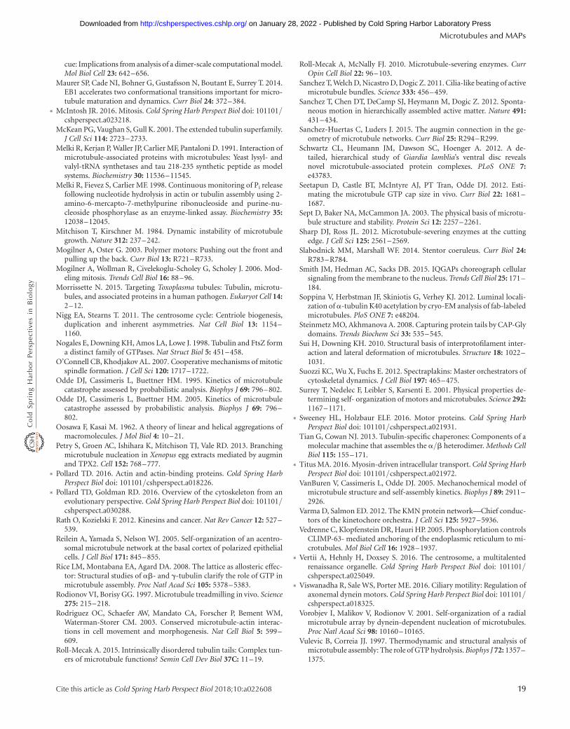
cue: Implications from analysis of a dimer-scale computational model.Mol Biol Cell 23: 642–656.
Maurer SP, Cade NI, Bohner G, Gustafsson N, Boutant E, Surrey T. 2014.EB1 accelerates two conformational transitions important for micro-tubule maturation and dynamics. Curr Biol 24: 372–384.
∗ McIntosh JR. 2016. Mitosis. Cold Spring Harb Perspect Biol doi: 101101/cshperspect.a023218.
McKean PG, Vaughan S, Gull K. 2001. The extended tubulin superfamily.J Cell Sci 114: 2723–2733.
Melki R, Kerjan P, Waller JP, Carlier MF, Pantaloni D. 1991. Interaction ofmicrotubule-associated proteins with microtubules: Yeast lysyl- andvalyl-tRNA synthetases and tau 218-235 synthetic peptide as modelsystems. Biochemistry 30: 11536–11545.
Melki R, Fievez S, Carlier MF. 1998. Continuous monitoring of Pi releasefollowing nucleotide hydrolysis in actin or tubulin assembly using 2-amino-6-mercapto-7-methylpurine ribonucleoside and purine-nu-cleoside phosphorylase as an enzyme-linked assay. Biochemistry 35:12038–12045.
Mitchison T, Kirschner M. 1984. Dynamic instability of microtubulegrowth. Nature 312: 237–242.
Mogilner A, Oster G. 2003. Polymer motors: Pushing out the front andpulling up the back. Curr Biol 13: R721–R733.
Mogilner A, Wollman R, Civelekoglu-Scholey G, Scholey J. 2006. Mod-eling mitosis. Trends Cell Biol 16: 88–96.
Morrissette N. 2015. Targeting Toxoplasma tubules: Tubulin, microtu-bules, and associated proteins in a human pathogen. Eukaryot Cell 14:2–12.
Nigg EA, Stearns T. 2011. The centrosome cycle: Centriole biogenesis,duplication and inherent asymmetries. Nat Cell Biol 13: 1154–1160.
Nogales E, Downing KH, Amos LA, Lowe J. 1998. Tubulin and FtsZ forma distinct family of GTPases. Nat Struct Biol 5: 451–458.
O’Connell CB, Khodjakov AL. 2007. Cooperative mechanisms of mitoticspindle formation. J Cell Sci 120: 1717–1722.
Odde DJ, Cassimeris L, Buettner HM. 1995. Kinetics of microtubulecatastrophe assessed by probabilistic analysis. Biophys J 69: 796–802.
Odde DJ, Cassimeris L, Buettner HM. 2005. Kinetics of microtubulecatastrophe assessed by probabilistic analysis. Biophys J 69: 796–802.
Oosawa F, Kasai M. 1962. A theory of linear and helical aggregations ofmacromolecules. J Mol Biol 4: 10–21.
Petry S, Groen AC, Ishihara K, Mitchison TJ, Vale RD. 2013. Branchingmicrotubule nucleation in Xenopus egg extracts mediated by augminand TPX2. Cell 152: 768–777.
∗ Pollard TD. 2016. Actin and actin-binding proteins. Cold Spring HarbPerspect Biol doi: 101101/cshperspect.a018226.
∗ Pollard TD, Goldman RD. 2016. Overview of the cytoskeleton from anevolutionary perspective. Cold Spring Harb Perspect Biol doi: 101101/cshperspect.a030288.
Rath O, Kozielski F. 2012. Kinesins and cancer. Nat Rev Cancer 12: 527–539.
Reilein A, Yamada S, Nelson WJ. 2005. Self-organization of an acentro-somal microtubule network at the basal cortex of polarized epithelialcells. J Cell Biol 171: 845–855.
Rice LM, Montabana EA, Agard DA. 2008. The lattice as allosteric effec-tor: Structural studies of ab- and g-tubulin clarify the role of GTP inmicrotubule assembly. Proc Natl Acad Sci 105: 5378–5383.
Rodionov VI, Borisy GG. 1997. Microtubule treadmilling in vivo. Science275: 215–218.
Rodriguez OC, Schaefer AW, Mandato CA, Forscher P, Bement WM,Waterman-Storer CM. 2003. Conserved microtubule-actin interac-tions in cell movement and morphogenesis. Nat Cell Biol 5: 599–609.
Roll-Mecak A. 2015. Intrinsically disordered tubulin tails: Complex tun-ers of microtubule functions? Semin Cell Dev Biol 37C: 11–19.
Roll-Mecak A, McNally FJ. 2010. Microtubule-severing enzymes. CurrOpin Cell Biol 22: 96–103.
Sanchez T, Welch D, Nicastro D, Dogic Z. 2011. Cilia-like beating of activemicrotubule bundles. Science 333: 456–459.
Sanchez T, Chen DT, DeCamp SJ, Heymann M, Dogic Z. 2012. Sponta-neous motion in hierarchically assembled active matter. Nature 491:431–434.
Sanchez-Huertas C, Luders J. 2015. The augmin connection in the ge-ometry of microtubule networks. Curr Biol 25: R294–R299.
Schwartz CL, Heumann JM, Dawson SC, Hoenger A. 2012. A de-tailed, hierarchical study of Giardia lamblia’s ventral disc revealsnovel microtubule-associated protein complexes. PLoS ONE 7:e43783.
Seetapun D, Castle BT, McIntyre AJ, PT Tran, Odde DJ. 2012. Esti-mating the microtubule GTP cap size in vivo. Curr Biol 22: 1681–1687.
Sept D, Baker NA, McCammon JA. 2003. The physical basis of microtu-bule structure and stability. Protein Sci 12: 2257–2261.
Sharp DJ, Ross JL. 2012. Microtubule-severing enzymes at the cuttingedge. J Cell Sci 125: 2561–2569.
Slabodnick MM, Marshall WF. 2014. Stentor coeruleus. Curr Biol 24:R783–R784.
Smith JM, Hedman AC, Sacks DB. 2015. IQGAPs choreograph cellularsignaling from the membrane to the nucleus. Trends Cell Biol 25: 171–184.
Soppina V, Herbstman JF, Skiniotis G, Verhey KJ. 2012. Luminal locali-zation of a-tubulin K40 acetylation by cryo-EM analysis of fab-labeledmicrotubules. PloS ONE 7: e48204.
Steinmetz MO, Akhmanova A. 2008. Capturing protein tails by CAP-Glydomains. Trends Biochem Sci 33: 535–545.
Sui H, Downing KH. 2010. Structural basis of interprotofilament inter-action and lateral deformation of microtubules. Structure 18: 1022–1031.
Suozzi KC, Wu X, Fuchs E. 2012. Spectraplakins: Master orchestrators ofcytoskeletal dynamics. J Cell Biol 197: 465–475.
Surrey T, Nedelec F, Leibler S, Karsenti E. 2001. Physical properties de-termining self- organization of motors and microtubules. Science 292:1167–1171.
∗ Sweeney HL, Holzbaur ELF. 2016. Motor proteins. Cold Spring HarbPerspect Biol doi: 101101/cshperspect.a021931.
Tian G, Cowan NJ. 2013. Tubulin-specific chaperones: Components of amolecular machine that assembles the a/b heterodimer. Methods CellBiol 115: 155–171.
∗ Titus MA. 2016. Myosin-driven intracellular transport. Cold Spring HarbPerspect Biol doi: 101101/cshperspect.a021972.
VanBuren V, Cassimeris L, Odde DJ. 2005. Mechanochemical model ofmicrotubule structure and self-assembly kinetics. Biophys J 89: 2911–2926.
Varma D, Salmon ED. 2012. The KMN protein network—Chief conduc-tors of the kinetochore orchestra. J Cell Sci 125: 5927–5936.
Vedrenne C, Klopfenstein DR, Hauri HP. 2005. Phosphorylation controlsCLIMP-63- mediated anchoring of the endoplasmic reticulum to mi-crotubules. Mol Biol Cell 16: 1928–1937.
∗ Vertii A, Hehnly H, Doxsey S. 2016. The centrosome, a multitalentedrenaissance organelle. Cold Spring Harb Perspect Biol doi: 101101/cshperspect.a025049.
∗ Viswanadha R, Sale WS, Porter ME. 2016. Ciliary motility: Regulation ofaxonemal dynein motors. Cold Spring Harb Perspect Biol doi: 101101/cshperspect.a018325.
Vorobjev I, Malikov V, Rodionov V. 2001. Self-organization of a radialmicrotubule array by dynein-dependent nucleation of microtubules.Proc Natl Acad Sci 98: 10160–10165.
Vulevic B, Correia JJ. 1997. Thermodynamic and structural analysis ofmicrotubule assembly: The role of GTP hydrolysis. Biophys J 72: 1357–1375.
Microtubules and MAPs
Cite this article as Cold Spring Harb Perspect Biol 2018;10:a022608 19
on January 28, 2022 - Published by Cold Spring Harbor Laboratory Press http://cshperspectives.cshlp.org/Downloaded from
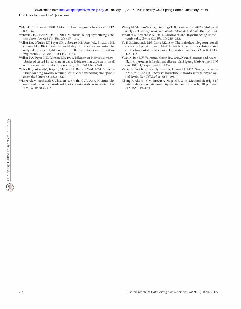
Walczak CE, Shaw SL. 2010. A MAP for bundling microtubules. Cell 142:364–367.
Walczak CE, Gayek S, Ohi R. 2013. Microtubule-depolymerizing kine-sins. Annu Rev Cell Dev Biol 29: 417–441.
Walker RA, O’Brien ET, Pryer NK, Soboeiro MF, Voter WA, Erickson HP,Salmon ED. 1988. Dynamic instability of individual microtubulesanalyzed by video light microscopy: Rate constants and transitionfrequencies. J Cell Biol 107: 1437–1448.
Walker RA, Pryer NK, Salmon ED. 1991. Dilution of individual micro-tubules observed in real time in vitro: Evidence that cap size is smalland independent of elongation rate. J Cell Biol 114: 73–81.
Weber KL, Sokac AM, Berg JS, Cheney RE, Bement WM. 2004. A micro-tubule-binding myosin required for nuclear anchoring and spindleassembly. Nature 431: 325–329.
Wieczorek M, Bechstedt S, Chaaban S, Brouhard GJ. 2015. Microtubule-associated proteins control the kinetics of microtubule nucleation. NatCell Biol 17: 907–916.
Winey M, Stemm-Wolf AJ, Giddings THJ, Pearson CG. 2012. Cytologicalanalysis of Tetrahymena thermophila. Methods Cell Biol 109: 357–378.
Woolner S, Bement WM. 2009. Unconventional myosins acting uncon-ventionally. Trends Cell Biol 19: 245–252.
Yu HG, Muszynski MG, Dawe RK. 1999. The maize homologue of the cellcycle checkpoint protein MAD2 reveals kinetochore substrate andcontrasting mitotic and meiotic localization patterns. J Cell Biol 145:425–435.
∗ Yuan A, Rao MV, Veeranna, Nixon RA. 2016. Neurofilaments and neuro-filament proteins in health and disease. Cold Spring Harb Perspect Bioldoi: 101101/cshperspect.a018309.
Zanic M, Widlund PO, Hyman AA, Howard J. 2013. Synergy betweenXMAP215 and EB1 increases microtubule growth rates to physiolog-ical levels. Nat Cell Biol 15: 688–693.
Zhang R, Alushin GM, Brown A, Nogales E. 2015. Mechanistic origin ofmicrotubule dynamic instability and its modulations by EB proteins.Cell 162: 849–859.
H.V. Goodson and E.M. Jonasson
20 Cite this article as Cold Spring Harb Perspect Biol 2018;10:a022608
on January 28, 2022 - Published by Cold Spring Harbor Laboratory Press http://cshperspectives.cshlp.org/Downloaded from
2018; doi: 10.1101/cshperspect.a022608Cold Spring Harb Perspect Biol Holly V. Goodson and Erin M. Jonasson Microtubules and Microtubule-Associated Proteins
Subject Collection The Cytoskeleton
ProteinsMicrotubules and Microtubule-Associated
Holly V. Goodson and Erin M. JonassonEvolutionary PerspectiveOverview of the Cytoskeleton from an
Thomas D. Pollard and Robert D. GoldmanMotor Proteins
H. Lee Sweeney and Erika L.F. HolzbaurTypes I and II Keratin Intermediate Filaments
Kwan, et al.Justin T. Jacob, Pierre A. Coulombe, Raymond
Myosin-Driven Intracellular TransportMargaret A. Titus
Muscle ContractionH. Lee Sweeney and David W. Hammers
The Actin Cytoskeleton and Actin-Based MotilityTatyana Svitkina
PeripherinFibrillary Acidic Protein (GFAP), Vimentin, and Type III Intermediate Filaments Desmin, Glial
Elly M. Hol and Yassemi Capetanaki
CellsMechanical Properties of the Cytoskeleton and
WeitzAdrian F. Pegoraro, Paul Janmey and David A.
Cytokinesis in Metazoa and FungiMichael Glotzer
Motility during Regeneration and Wound HealingIntermediate Filaments and the Regulation of Cell
Fang Cheng and John E. ErikssonMotorsCiliary Motility: Regulation of Axonemal Dynein
E. PorterRasagnya Viswanadha, Winfield S. Sale and Mary
Intermediate Filaments and the Plasma Membrane
Harmon, et al.Jonathan C.R. Jones, Chen Yuan Kam, Robert M.
Neighboring CellsInteractions with the Extracellular Matrix and Actin-Based Adhesion Modules Mediate Cell
Nelson, et al.Alexia I. Bachir, Alan Rick Horwitz, W. James
Intracellular Motility of Intermediate Filaments
WindofferRudolf E. Leube, Marcin Moch and Reinhard Tethering, and Organization of Organelles
Microtubule-Based Transport and the Distribution,
Kari Barlan and Vladimir I. Gelfand
http://cshperspectives.cshlp.org/cgi/collection/ For additional articles in this collection, see
Copyright © 2018 Cold Spring Harbor Laboratory Press; all rights reserved
on January 28, 2022 - Published by Cold Spring Harbor Laboratory Press http://cshperspectives.cshlp.org/Downloaded from
http://cshperspectives.cshlp.org/cgi/collection/ For additional articles in this collection, see
Copyright © 2018 Cold Spring Harbor Laboratory Press; all rights reserved
on January 28, 2022 - Published by Cold Spring Harbor Laboratory Press http://cshperspectives.cshlp.org/Downloaded from