micropropagation and acclimatization of aloepolyphylla and
TRANSCRIPT

Micropropagation and acclimatization of
Aloe polyphylla and Platycerium bifurcatum
BY
JUDE CHINEDU CHUKWUJEKWU
submitted in fulfilment of the requirements for the degree of
(Master of science)
in the Research Centre for Plant Growth and Development, School of Botany and
Zoology, Faculty of Science and Agriculture.
University of Natal, Pietermaritzburg
June, 2001
PREFACE
With great pleasure, I wish to declare that this thesis, submitted for the degree of Master
of Science in the Research Centre for Plant Growth and Development, School of Botany
and Zoology, University of Natal, Pietermaritzburg, except where the work of others is
acknowledged, is the result of my own investigation.
JUDE CHINEDU CHUKWUJEKWUJUNE 2001
We certify that the above statement is correct.
............. ~ ..~ .PROFES R J VAN STADEN
(SUPERVISOR)
.................A·~ .MISS C W FENNELL(CO-SUPERVISOR)
PUBLICATIONS FROM THIS THESIS
CHUKWUJEKWU JC, FENNELL CW and VAN STADEN J (2001)
Micropropagation and acclimatization of Aloe polyphyJla (In preparation).
CONFERENCE CONTRIBUTIONS FROM THISTHESIS
CHUKWUJEKWU JC, FENNELL CW and VAN STADEN J (2000)
Micropropagation and acclimatization of Aloe polyphyJla (paper). Second Annual
Research Centre Meeting, University of Natal, Pietermaritzburg.
11

ACKNOWLEDGEMENTS
Iwish to express my sincere appreciation to Professor J. Van Staden (supervisor)
and Miss C W Fennell (co-supervisor) fortheir guidance during this investigation
and in the preparation of this manuscript.
To Or. Mia Abrie, thank you for taking me through tissue culture for the first time.
It was really great.
To my parents and Dr. C I Chukwujekwu, thank you for your encouragement and
financial support throughout my years of study.
To every member of the Research Centre for Plant Growth and Development,
especially Cathy Ford, Georgina Arthur and Noxwenda Makunga, for your ever
available assistance. You were all a source of inspiration to me.
Lastly, but not the least, to Almighty God, whom by his grace made it possible for
me to successfully complete this work. I give you all the glory and honour.
111

ABSTRACT
Shoot cultures of Aloe polyphylla were initiated from young shoot explants of in
vitro grown plants. The basal medium was MS medium (MURASHIGE and
SKOOG, 1962), supplemented with 100 mgl-1myo-inositol, and 30 gl-1 sucrose.
Agar (0.8 %) was used as the gelling agent. Different cytokinins, singly or in
combination with auxins (IBA and NAA), were tested for shoot proliferation
activity. All the cytokinins tested (kinetin, zeatin, iP, and BA) gave a good shoot
proliferation response. The optimal concentrations for shoot proliferation of each
of the cytokinins tested were: zeatin (0.5 mgl-1), kinetin (1.5 mgl-1), iP (1.0 mgl-1)
and BA (1.5 mgl-1). In combination with auxins, the optimal combinations were
kinetin/NAA (2.0/0.1 mgl-1), kinetinllBA (1.5/1.0 mgl-1), zeatinllBA (1.0/0.5 mgl-1),
zeatin/NAA (1.0/1.0 mgl-1), BA/IBA (1.0/1.0 mgl-1), BA/NAA (1.5/0.1 mgl-1).
Although it gave the highest number of shoots per explant, BA was responsible
for hyperhydricity.
Temperature and sucrose also influenced shoot proliferation. The optimal
temperature was 25°C, while 30 gl-1 was the optimal concentration of sucrose for
shoot proliferation. Plants rooted well in plant growth regulator-free MS medium.
Amongst the potting mixtures tested, soil: sand: vermiculite (1:1:1 v/v) was the
best with 98 % plantlet survival.
In the second part of this project, Platycerium bifurcatum cultures were
established using leaf explants. The basal medium was MS medium
(MURASHIGE and SKOOG, 1962), supplemented with 100 mgl-1 myo-inositol
and 30 gl-1 sucrose. For bud initiation, 1.0 mgl-1BA was used, while 0.8 % agar
was used as the gelling agent. Three different strengths of MS medium (full, half,
and one-quarter strength) without plant growth regulators were tested for further
bud growth and development. Half-strength MS proved to be the best for further
IV

bud growth and development. Rooting was best achieved in one-quarter strength
MS medium without plant growth regulators. In vitro grown plantlets were
successfully acclimatized using peat as the potting medium.
v

CONTENTS
PREFACE i
PUBLICATIONS FROM THIS THESiS ii
CONFERENCE CONTRIBUTIONS FROM THIS THESIS ii
ACKNOWLEDGEMENTS iii
ABSTRACT ivI •
CONTENTS Vl
LIST OF ABBREVIATIONS USED ix
LIST OF TABLES x
LIST OF FIGURES xi
LISTOF PLATES xv
1.Generalliterature review (Introduction)
1.1 Literature review of the genus Aloe 1,- -------
1.2 Horticultural and other uses of Aloe. 4
1.3 Conservation 5
1.4 Conventional propagation . . . . . . . . . . . . . . . . . . . . . . . . . . . . . . .. 5
1.5 Review of tissue culture of the genus Aloe. . . . . . . . . . . . . . . . . .. 6
1.5.1 Shoot -tip and meristem culture. . . . . . . . . . . . . . . . . . . .. 7
1.5.2 Callus culture . . . . . . . . . . . . . . . . . . . . . . . . . . . . . . . . .. 11
1.5.3 Other in vitro work . . . . . . . . . . . . . . . . . . . . . . . . . . . . .. 13
1.6 Literature review of the genus Platycerium . . . . . . . . . . . . . . . .. 13
1.7 Horticultural and other uses of Platycerium . . . . . . . . . . . . . . . .. 14
1.8 Conventional propagation 14
1.9 Review of tissue culture of the genus Platycerium 16
1.9.1 Shoot - tip and meristem culture. . . . . . . . . . . . . . . . . .. 16
1.9.2 Callus culture 16
VI
2. Shoot Culture of Aloe polyphylla
2.1 Introduction. . . . . . . . . . . . . . . . . . . . . . . . . . . . . . . . . . . . . . . . .. 22
2.1.1 Objectives 22
2.2 Materials and methods . . . . . . . . . . . . . . . . . . . . . . . . . . . . . . . .. 23
2.2.1 Decontamination procedures and aseptic techniques. .. 23
2.2.2 Explant source. . . . . . . . . . . . . . . . . . . . . . . . . . . . . . . .. 24
2.2.3 Media and supplements 24
2.2.4 Environmental conditions 27
2.2.5 Acclimatization . . . . . . . . . . . . . . . . . . . . . . . . . . . . . . .. 28
2.3 Results and Discussion 29
2.3.1 Decontamination procedures. . . . . . . . . . . . . . . . . . . . .. 29
2.3.2 Media and supplements 29
2.3.2.1 Effects of plant growth regulators on regeneration 29
2.3.2.2 Effects of sucrose on regeneration 42
2.3.3 Temperature 45
2.3.3.1 Effects of temperature on shoot regeneration 45
2.3.4. Acclimatization 46
2.3.5 Conclusions. . . . . . . . . . . . . . . . . . . . . . . . . . . . . . . . . .. 48
3. Tissue culture of Platycerium bifurcatum
3.1 Introduction 50
3.1.1 Objectives 50
3.2 Material and methods. . . . . . . . . . . . . . . . . . . . . . . . . . . . . . . . .. 51
3.2.1 Decontamination procedures and aseptic techniques .. 51
3.2.2 Explant source 51
3.2.3 Media and supplements . . . . . . . . . . . . . . . . . . . . . . . .. 52
3.2.4 Environmental conditions 52
3.2.5 Acclimatization . . . . . . . . . . . . . . . . . . . . . . . . . . . . . . .. 52
3.3 Results and Discussion 52
vu

3.3.1 Decontamination procedures. . . . . . . . . . . . . . . . . . . . .. 52
3.3.2 Media and supplements . . . . . . . . . . . . . . . . . . . . . . . .. 54
3.3.3 Acclimatization 55
3.3.4 Conclusions 56
4. General Conclusions 57
References 59
V111

LIST OF ABBREVIATIONS USED
BA Benzyladenine
iP isopentenyladenine
K Kinetin
Z Zeatin
IBA indole-3-butyric acid
NAA ex: - naphthaleneacetic acid
IX

LIST OF TABLES
Page
Table 1 8
Summary of in vitro work on Aloe species.
Table 2 17
Summary of in vitro work on Platycerium species.
Table 3 26
Different constituents of the basal medium used throughout this study as
described by Murashige and Skoog (1962) (MS).
Table 4 49
Mean number of roots produced by Aloe polyphylla at different strengths of MS
medium.
Table 5 56
Survival percentage of Platycerium bifurcatum plantlets obtained with different
potting mixtures.
x

LIST OF FIGURES
Page
Figure 1 3
The morphology of Aloe polyphylla (A) showing the clockwise arrangement ofthe
leaves, (B) the counterclockwise arrangement of leaves and a flowering shoot
with the arrangement of flowers at the tip of the inflorescence.
Figure2 15
The morphology of Platycerium bifurcatum showing its typical forked fertile leaves
that provide its excellent ornamental qualities.
Figure3 31
Effect of various concentrations of kinetin on shoot and root proliferation of Aloe
polyphylla in vitro.
Figure4 31
Effect of various concentrations of BA on shoot and root proliferation of Aloe
polyphylla in vitro.
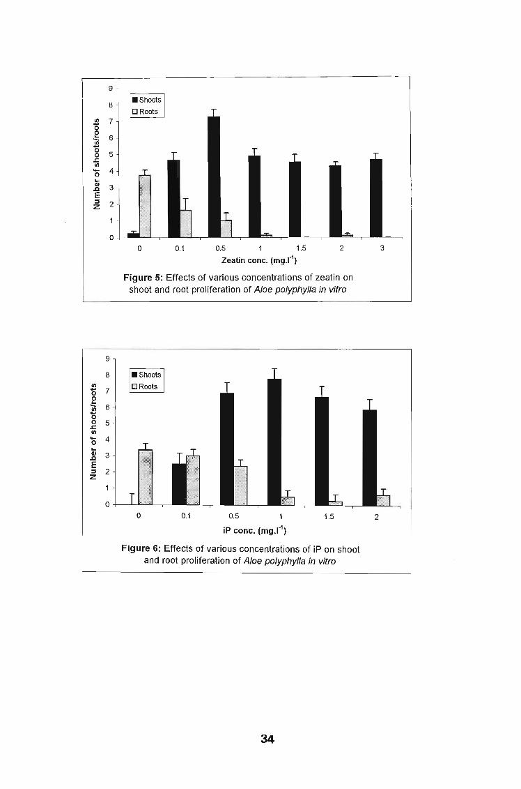
Figure5 34
Effect of various concentrations of zeatin on shoot and root proliferation of Aloe
polyphylla in vitro.
Figure6 34
Effect of various concentrations of iP on shoot and root proliferation of Aloe
polyphylla in vitro.
Xl
Figure7 36
Effect of various combinations of kinetin/IBA on shoot proliferation of Aloe
polyphylla in vitro.
FigureS 36
Effect of various combinations of kinetin/NAA on shoot proliferation of Aloe
polyphylla in vitro.
Figure9 37
Effect of various combinations of zeatin/IBA on shoot proliferation of Aloe
polyphylla in vitro.
Figure 10 37
Effect of various combinations of zeatin/NAA on shoot proliferation of Aloe
polyphylla in vitro.
Figure 11 39
Effect of various combinations of BA/IBA on shoot proliferation of Aloe polyphylla
in vitro.
Figure 12 39
Effect of various combinations of BAlNAA on shoot proliferation ofAloe polyphylla
in vitro.
Figure 13 44
Effect of various concentrations of sucrose on shoot proliferation of Aloe
polyphylla in vitro.
Xli

Figure 14 44
Effect of various temperatures on shoot proliferation of Aloe polyphylla in vitro.
Xlll

LIST OF PLATES
Page
Plate 1 , 32
Multiple, stunted and light green shoots obtained after 5 weeks of treating cultures
with 3.0 mgl-1 of BA.
Plate 2 32
Multiple shoot production with 1.5 mgl-1 kinetin in the culture medium.
Plate3 32
Callus production at the base of an explant with 3.0 mgl-1 kinetin in the basal
medium.
Plate4 35
Multiple shoot production with cytokinins tested. (A) 0.5 mgl-1 zeatin, (B) 1.0 mgl-1
iP.
PlateS 35
Multiple shoot production obtained with different combinations of kinetin and auxin
in the basal medium. (A) kinetin/NAA (2.0/0.1 mgl-1), (B) kinetin/lBA (1.5/1.0 mgl
1).
Plate6 38
Multiple shoot production obtained with different combinations of zeatin and auxin
in the basal medium. (A) zeatin/NAA (1.0/1.0 mgl-1), (B) zeatin/IBA (1.0/0.5 mgl-1).
Plate7 38
Multiple shoot production obtained with different combinations of BA and auxin
XlV

in the basal medium. (A) BA/NAA (1.5/0.1 mgl-1), (B) BA/IBA (1.0/1.0 mgl-1
).
PlateS 43
Effect of sucrose on shoot proliferation of Aloe polyphyJla in vitro. (A) at 0 % level,
no shoot proliferation, (B) at 3 % level, multiple shoot proliferation.
Plate9 43
Effect of temperature on shoot proliferation of Aloe polyphyJla in vitro. (A) at1 0 °c
shoot proliferation was near zero, (B) at 20°C a sharp increase in shoot
proliferation, (C) at 30°C, shoot proliferation was inhibited.
Plate 10 47
(A) Aloe polyphyJla rooting in plant growth regulator- free MS medium, (B) fully
acclimatized Aloe polyphyJla plants.
Plate 11 55
(A) Platycerium bifurcatum cultured in half-strength MS medium, (B) in full
strength MS medium, (C) one-quarter strength MS medium, (0) fully acclimatized
plants.
xv

Chapter One
General literature review
1.1 Literature review of the genus Aloe
Introduction ~_-- - ...-.
The importancaQtpJ.anlsJQJ:n,an~jngg§DJlOtJ~5LQV~Lempha§ts~~. We aIL<!~~nd----------- ""'
upon.plants forJll:l~~~~d~.:-::U1<e fQgd,__medicine_,.cJQthing, fuel and furniture.
Ifshould not be surprising therefore that much human endeavour has been aimed
at producing and improving useful plants. Important in this endeavour has been
the development of techniques for cultivating plant cells and tissues in vitro
plant tissue culture.
Plant tissue culture is a technique through which any plant part is cultured on a
sterile nutrient medium in controlled light and temperature with the purpose of
obtaining growth. The idea of plant tissue culture originated from the cell theory
that was formulated by Schwann in 1839. Tissue culture techniques have for
decades played a great role in the micropropagation of horticultural and
ornamental plants.. In fact, the first ever successful plant tissue culture was
achieved in horticultural plants (ALTMAN and ZIV, 1997). These techniques have
been widely used in disease elimination and vegetative propagation ( HUSSEY,
1979).
Tissue culture propagation presents one solution to the problems encountered
with conventional propagation. It usually ensures that the desired characteristics
of a selected plant or plants are retained through clonal propagation. The rate of
multiplication of plants has also been shown to be enhanced considerably using
this method. It also allows year round production of plants. In commercial
nurseries, it can also be used to rationalize the growing space usually constrained
for the preservation of stock plants. In addition, when properly accomplished,
1

tissue culture may be employed forthe reproduction and maintenance of disease
free plants (MURASHIGE, 1974). In view of the above benefits of tissue culture,
experimentation on the in vitro propagation of Aloe polyphylla was embarked on.
It was expected that in this way the devastation of native populations could be
averted.
Plants belonging to the family Liliaceae are cultured for their high medicinal and
ornamental value ( VIJ et al., 1980). Aloe, a member of the Asphodelaceae of the
Liliaceae is cultivated in gardens for its unassuming succulent foliage as well as
for aloin - an important drug (ROY and SARKAR, 1991). Although the genus Aloe
has for many decades been recognised for its medicinal and ornamental values,
it is only since research activities have disclosed that some members of this
genus possess some medicinal compounds, that an interest in the
pharmacological potential of these plants has developed. This has resulted in an
increasing demand for aloes. In the case of Aloe polyphylla, and in fact for many
threatened species, n.9~od has been devised to ensure a continuou_~__~ypply. . -~-----
of these plants for commercial and research activities. Wild resources of many_.'__~.W_ .._.__·_--
species are rapidly declining.
Aloe polyphylla (spiral aloe), also known as the Kharetsa, belongs to the family
Asphodelaceae. This species of Aloe is a succulent perennial with a rounded
rosette of 75-150 mostly erect leaves measuring up to 80 cm across; arranged
in five spiral rows, clockwise or counterclockwise ( Fig. 1A). The grey-green
coloured leaves are egg-shaped and very fleshy, 20-30 cm long and 6-10 cm
. broad when mature, nearly flat above and unevenly ridged below, and with rather
soft white teeth on the margin. A flowering shoot extends 50-60 cm high,
branching from near the base, with flowers crowded on the branch tips (Fig. 1B).
Each flower has a narrow, triangular bract 2-3 cm long, and a cylindrical corolla
45-55 mm long. The flowers can be pale red to salmon pink or, very rarely,
2
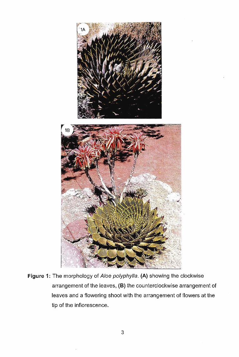
Figure 1: The morphology of Aloe polyphylla. (A) showing the clockwise
arrangement of the leaves, (B) the counterclockwise arrangement of
leaves and a flowering shoot with the arrangement of flowers at the
tip of the inflorescence.
3

yellow. Flowering occurs from August through December, peaking in September
and October (EMANOIL, 1994).
Aloe polyphylla, one of the most spectacular plants of southern Africa, is found
in Lesotho, with a major concentration in the Thaba Putsoa Range and Maseru
area of the Drakensberg mountains. It grows at elevations of 2230-2720 m on
steep basalt slopes with loose rock. I~ thrive~Jt]~~e9.§--,#h~r~lts (q01§. C!re._k~p-t
moist in summer by a continual flow of water, where there are mostly low shrubs,
and where rainfall is around 1100 mm ann~a\ II~. c (,"'Ot '\ _~, ( \ Q, Ij~' 11"\ "" a.,. \"" e ,,", n_ (\ <:lE: ,.~)\A. \, I:~... lA 1.... ~;.Ir, J 'J~, •
..... ~' \ . /_'\~_i~)'\I'~,(~ t.,\J n ~\( ,~..~\ El ,I rn ·eo! ! (, Q I r::J t\..'!f~ifc/'· II-S
1.2 Horticultural and other uses of Aloe- \ e. '~U"1--U\~ e o~ 0, tQe he ift L~ <' I 1 I '::,
f'nEe\,.; Cc:, \ \ 'j -\ 0{ (c n.J.·I.;{ l' J 4' .\
Apart from their ornamental value, Aloe species have long been known for their
medicinal properties. These plan!~_have been cultured recently for their high
medicinal and ornamental value.beaf juice-.eJ_Aloe have been used medicinally
~n~s (VAN WYK and SMITH, 1996). Amongst the species known to
possess some medicinal properties are Aloe barbadensis, Aloe ferox, Aloe
aristata, Aloe candelabrum, Aloe coopperi, Aloe maculata, Aloe sessiliflora, Aloe
marlothii, and Aloe Iinearifolia (VAN WYK et al., 1997; HUTCHINGS et al., 1996).
Tb.e mediCi~J&+AIDe+-mJMfafJ!dlC& of A,oeLra is widely used in
tb.e c_os.metic a d__har a.ceuti.cai,g9Jtstrj.e.s (VIJ et al., 1980). In traditional
medicine, the le e~ ru!Joots of most'~p-ecies~,en 9QH~EUn wa~~r-~~e !aken.- .: b~em(,~ ------ - ,as a laxatIVe. They are also_ talsen ._fOL..Clrthritis, eczema, conjunctivitis,------------------------- ,------ .. "-.- -~-~---- - .~ .,......." ------ _. -~.- - -
h ertension an~ stress. Leaf sap of several species is applied externally to treat
skin irritations, bruises and burns. The leaves of Aloe marlothii are popular in
snuff mixtures. The main purgative element is the anthrone C-glycoside aloin,
while the wound-healing properties are attributed to glycoprotein and to hydrating,
insulating and protective effects (HUTCHINGS et al., 1996; VAN WYK et al.,~. 16
1997). Due to lhe belie.l.byJ~!sfrom Lesotho that Aloe (Polyphylla )has some~. -----._,~---..••.•--'..._. --"'-'>-"-~'-~. -
m~r:op-el=t-ie.Sj-tAeY'·GhQp.Q.fUb.e.Je.a:ves-a.r}d .P.J.Rce them in water for poultry- 1oN""-{,-", r .., 'r-e lJ e '-~'='='~'-'--'-"~
4
consumption (KOOPOWITZ and KAYE, 1990). They also make a kind of
concoction with the leaves together with other herbs which they also believe. .-(.-improves o~':~iflJ.rIl.Y~¥~!~m-'lvhenconsurned. However, these claims are yet" -- --_.-
to be proved scientifically since no study has been done to confir~ t~es~ beliefs.t'XQ1 b .. , \ .,.:2"IV"" __/
1.3 CgoseJX?J!Q!}.......~~- ,- " ......
With its large rosette oftriangular grey-green leaves arranged in five spiral series
and coral-coloured flowers, Aloe polyphylla is very desirable. There has been
over-exploitation of Aloe polyphylla in its natural habitat. The trade in wild
collected plants has been a contentious issue for many decades. Aloe polyphylla,-
which is the national flower of Lesotho is endangered according to World
Conservation Monitoring Units' Re,d List of Threatened Plants. This is due to the
indiscriminate collection of the wild population. Apart from the collection of plants
for the ornamental trade, locals, who believe that the leaves contain medicinal
properties have been chopping them up. Other factors that could have contributed
to the decline in numbers of Aloe polyphylla include urban and industrial
expansion, agricultural development, afforestation and mining activities. To
protect these plants, the Lesotho government prohibits export of both seeds and
plants. It is illegal to remove plants from their natural habitats. This legislation
w~s put in place in 1938 to reduce the likelihood of extinction (EMANOIL, 1994).
Conservation of these ornamentals depends on the availability of propagation
material to the horticulturist. This can be realised by successful rapid cultivation
of these plants to meet the ever growing demand for the horticultural trade, and
also guaranteeing the availability of plant material for propagation.
1.4 Conventional propagation
Conventionally, Aloe polyphylla is propagated both sexually and vegetatively.
Sexually, it is propagated through seeds. Though it produces a large number of
5

seeds, only about half are viable owing to the fact that its pollinator, the Malachite
sunbird, is endangered. Although sexual propagation is regarded as a most
efficient and economical method of propagating plants, once seed dormancy
problems have been overcome, the major disadvantage of this method is that it
results in genetic variability. Besides, it is normally time and space consuming,
and seasonally controlled (HARTMANN and KESTER, 1983). Vegetative
propagation is through offshooting of pups which is very rare in Aloe polyphylla.
Occasionally, a plant may split in two, and it requires two years to grow apart.
Furthermore, owing to the fact that Aloe polyphylla apparently requires special
soil and moisture for growth, conventional propagation may be difficult.
1.5 Review of tissue culture of the genus Aloe
Plant tissue culture is a generic description which embraces plant protoplast,
plant cell, plant tissue and, plant organ culture. These various types of culture
involve, as a common factor, the growth of decontaminated plant material in an
aseptic environment, such as sterilized nutrient medium in a test tube or other
culture vessel (DE FOSSARD, 1981). In vitro techniques are generally used in
plant improvement which includes recovery of virus free clones, haploid cultures,
embryo culture, production of chimeras, preservation of valuable germplasm,
mutation initiation and selection, screening for disease, toxin and stress
resistance, in vitro pollination and fertilization, and protoplast culture. Other uses
are production of pharmaceuticals and other natural compounds; movement of
plant material from one country to another, all year round production of clones,
and demonstration of natural interactions between bacteria and plants such as in
bacterially induced tumours. and symbiotic nitrogen fixation in legumes
(MURASHIGE, 1974; SPIEGEL-ROYand KOCHBA, 1977; GRESSHOFF, 1978;
DE FOSSARD, 1981).
6
Tissue culture has been employed extensively for the rapid clonal propagation of
plants (MURASHIGE, 1974; VASIL and VASIL, 1980). The technique is
particularly useful for plants where the rate of multiplication is slow.
Micropropagation of ornamentals is the most widely used tissue culture
technique. In vitro cultures have been used as a tool in the acquisition of
knowledge of some physiological characteristics of many plants, such as
nutritional requirements, endogenous hormone production, and other
developmental aspects of these plants. Not much work has been done on the in
vitro culture of Aloe species owing to the fact that primary culture establishment
is difficult because phenolic substances are secreted by the explants (ROY and
SARKAR, 1991). The tissue culture protocols available for Aloe are shown in
Table 1.
1.5.1 Shoot-tip and meristem culture
Meristem culture is a technique in which a small piece of the stem tip is cultured
in appropriate nutrient medium. Only the apical dome and one or two leaf
primodia are used. In shoot tip culture, a portion of the sub-apical region is
involved ( HUSSEY, 1979).
This technique was first developed by Morel and Martin (1952) for Dahlia
(DEBERGH, 1994). In ornamentals, meristem culture was one of the first, and is
still the most widely used procedure in commercial applications of tissue culture.
The purpose of meristem culture is to free plants from virus infection. It is
believed that a virus does not easily invade or reproduce in the very young cells
of the shoot meristem (HUSSEY, 1979; DE FOSSARD, 1981). Besides
eradicating viruses, Pelargonium meristem culture, combined with appropriate
testing by immunofluorescence, has been effective in the production of mother
plants free of Xanthomonas pelargonii ( REUTHER, 1983). However, this
technique does have some limitations. According to Debergh, et al. (1990) a good
7

Table 1: Summary of in vitro work on Aloe species
Species Explant Medium & Supplements Growth Response Reference
MS + 3% sucroseAloe arborescens + 0.02- 0.1 mgl-1kinetin + 1.8-9.2 Callus formation Kawai et al. (1993)Mill. Axillary mgl-1NAA
shootsCorneau et al.MS medium Micropropagation(1994)
MS + magnetic fluid 2° shoot productionRhizogenesis
Leaf tissue, Not given Micropropagation Groenewald et al.Aloe pretoriensis stem (1979)Pole Evans segments
LS + 0.2 mgl-12,4-0 +1 mgl-1 Callus formation that Groenewald et al.Seeds kinetin later developed into (1976)
+ 0.1 mgl-1p-aminobenzoic acid shoots and roots+ 100 mgl-1tyrosine + 3 gl-1 casein
hydrolysate
Aloe species leaves, MS + 1.0 mgl-1NAA + 1.0 mgl-1 BA Callus induction and Hyashi (1987)(23 species) perianth, growth
floral stalks
Aloe variegata L. Stem apex MS + supplements Lateral bud formation Zhao (1990)
8

Table 1 contd.
Species Explant Medium & Supplements Growth Response Reference
MS+ 1 mgl-1 2,4-0 + 0.2 mgl-1 kinetin Callus induction Roy & Sarkar
~ (1991 )+ 0.1 mgl-1 PABA Best for callus growth
+ 0.02 mgl-1 2,4-0 + 1 mgl-1 kinetin Shoot formation
MS + 3 mgl-1 BA Bud inductionZhou et al. (1999)
Aloe vera L. Apical and MS + 0.3 mgl-1 NAA Rooting
(Aloe axillary budsbarbadensis Mill.) MS + 3 mgl-1 BA + 0.2 mgl-1 NAA Adventitious shoot
inductionMS + 5% sucrose + 1 mgl-1 BA + 5 Rapid proliferation of Feng et al. (2000)mgl-1 adenine + 0.2 mgl-1 NAA shoots
MS (half-strength) + 0.1 mgl-1 BA + Rooting2-3 mgl-1 IBA
Meristem MS + 30% sucrose + 0.2 mgl-1 2,4-tips o + 0.5 mgl-1 kinetin Multiple shoot Natali et al. (1990)
production
9

Table 1 contd.
Species Explants Medium & Supplements Growth Response Reference
Modified MS + 30% sucrose
Multiple + 1.0 mgl-1 IBA Adventitious andnodes of axillary bud growth Meyer and Van
Aloe vera L. plants (8 - 10 Maximum bud growth & Staden (1991)(Aloe cm) rootingbarbadensisMill.) + 1.0 mgl-1 IAA Axillary bud formation
Stem MS + 2 mgl-1 Zeatin + 0.5 mgl-1 Plantlets formed on thesegments NAA cell masses on callus Gui et al. (1990)
surface
Modified MS+ NN vitamins + 30 gl-1Aloe vera L. & Immature sucroseAloe harlona inflorescence +1.2 mgl-1zeatin Multiple shootDanguy production Richwine et al.
(1995)+ 0.52 mgl-1BA Multiple shoot
production
10

correlation exists between the size ofthe explant and the degree of contamination
for a given virus. He also pointed out that the results also depend to a large extent
on the physiological status of the stock plant and, thus, vary with the season.
Furthermore, meristem culture has no value when it is not combined with
thorough testing of the plant material afterwards (DEBERGH, 1994). Key
protocols for meristem culture must be combined with some experimental
treatment like chemotherapy, thermotherapy, or cryotherapy (SHARP and
LARSEN, 1979; DEBERGH, 1994).
Available protocols for meristem culture of Aloe include the works of Natali et al.
(1990), and Hirimburegam and Gamage (1995) with Aloe vera . Plantlet
regeneration was achieved by both researchers on Murashige and Skoog medium
(MS) (MURASHIGE and SKOOG, 1962), but with different growth regulators.
Natali et al. (1990), used 0.25 mg 1-1 2,4-D and 0.5 mg 1-1 kinetin while
Hirimburegama and Gamage (1995), used 0.18 mg 1-1 IAA and 2.25 mg 1-1 BA.
1.5.2 Callus culture
Callus - proliferation of cells to form an unorganized mass of tissue - is produced
when the nutrient medium contains fairly high concentrations of hormones,
particularly auxins. The development of a callus from a fragment oftissue may be
divided into three stages; induction of cellular division, continued proliferation and
dedifferentiation, and structural and physiological re-differentiation (YEOMAN and
AITCHISON, 1973). Nearly all callus cUlture_~__are derived from two major types
of cells in an explant; those of vascular cambium wh+ch may already be in a state
of division, and a variety of parenchyma cells which are inactive and have to be
induced to divide ( YEOMAN, 1973).This type of culture however, is usually
avoided because of its association with genetic instability (HUSSEY, 1979; DE
FOSSARD, 1981).
Polyploid cells with various types of chromosomal variations from the normal for
11

the species have often been reported for callus cultures, and thus plants which
are induced to form from such cultures, either by induction of adventitious buds
or by embryogenesis, may arise from cells of abnormal type. These abnormalities
may involve flower colour, leaf shape, growth rate or other characters which
would mean that the plants would be off-type. These 'disadvantages' of callus
culture not withstanding, are particularly useful for analysing the influence of
various factors on plant organogenesis. However, the presence of cells without
the normal genome does not necessarily prevent the formation of adventitious
buds and embryos with the normal chromosomal type of the species. According
to Hussey (1979), changes in a callus are unpredictable and often the capacity
to regenerate shoots is lost. However, the tendency for callus to develop
abnormal cells varies according to the species and media on which it is grown.
The fast-growing calli are generally the most likely to produce abnormal plants.
On the other hand, the variation in genome could lead to the formation of
interesting and variable new strains ( DE FOSSARD, 1981).
According to the literature, the first organogenetically active Aloe callus was
reported by Groenewald et al. (1975). Seed segments were used as explants. LS
medium (L1NSMAIER and SKOOG, 1965) was used with 2,4
dichlorophenoxyacetic acid (0.2 mgl-1) and kinetin (1 mgl-1) incorporated into the
medium. After three to four weeks calli were observed at the cut edges of some
of the seed fragments. After eight to ten weeks regeneration of shoots and roots
were observed as well. In 1988, Castorena-Sanchez et al., reported
micropropagation of Aloe vera using callus cultures. Using DNA
microdensitometry, they found that the morphogenetic ability of callus is
correlated with nuclear DNA content in callus cultured in vitro. Micropropagation
was obtained only from calli containing close to the normal 2C or 4C amount of
DNA per nucleus. Cavallini et al. (1993) later gave credence to this work by
reporting the chromosome number of six plants regenerated from Aloe vera callus
12

culture. Five of the six plants were diploid and one was tetraploid. Racchi (1988)
while studying the biosynthesis of secondary products of Aloe ferox in vitro,
achieved indirect organogenesis in callus obtained from root and embryo explants
of this species. Cultures were established on NN (NITSCH and NITSCH, 1967)
and MS (MURASHIGE and SKOOG, 1962) media. However, from the reported
findings on callus cultures of Aloe species, it is evident that the plant growth
regulators 2,4-dichlorophenoxyacetic acid and kinetin play major roles in callus
induction and plant regeneration respectively.
1.5.3 Other in vitro work
Besides shoot tip, meristem and stem segments, other plant parts that have been
used as explants in in vitro work of Aloe include leaf tissue, immature
inflorescences, and floral stalks. Groenewald et al. (1979) achieved
micropropagation of Aloe pretoriensis using leaf tissue as an explant source.
Richwine et al. (1995) working with Aloe vera and Aloe harJona achieved multiple
shoot production using immature inflorescences as explants. To achieve multiple
shoot production, they used modified MS (MURASHIGE and SKOOG, 1962)
medium with NN (NITSCH and NITSCH, 1967) vitamins supplemented with 1.2
mg 1-1 zeatin or 0.52 mg 1-1 BA.
1.6 Literature review on the genus Platycerium
Introduction
Platycerium bifurcatum (Cav) C. Chr. is one of fifteen species of the genus
Platycerium belonging to the plant Polypodiaceae. It is a widespread and much
diversified species occurring in southern Australia, extending beyond the tropics
into the warm-temperate sub-tropical areas of eastern Australia. Generally it is
found growing in areas with a pronounced dry season at an altitude of up to 2000
meters or more (HENNIPMAN and ROOS, 1982).
Platycerium bifurcatum (Cav) C. Chr. commonly known as elkhorn is an epiphytic
13

fern. As in other ferns, this species consists of two independent generations: the
small, simple haploid gametophyte that is limited to moist environments and the
large, morphological complex, diploid sporophyte. The gametophyte bears the
sexual organs that carry out sexual reproduction, while the large and complex
sporophyte on the other hand bears elaborate fertile leaves that facilitate spore
dispersion (FERNANDEZ et al., 1999). Platycerium bifurcatum is characterized
by typical forked fertile leaves that provides excellent ornamental qualities (Fig.
2). The frond is dimorphic in nature, comprises the foliage fronds which become
detached when they are old, and the nest fronds that remain on the plant even
after they turn brown. The nest fronds help trap leaf litter from the surrounding
trees (HENNIPMAN and RODS, 1982; CHIN, 1997). According to Hoshizaki
(1972) the dimorphism exhibited by the fronds is a response to the epiphytic
habitat, ensuring better absorption of water and nutrients by humus collection as
well as a better protection of the roots.
1.7 Horticultural and other uses of Platycerium
The excellent ornamental qualities provided by the leaves of this plant makes it
a desirable plant. The fronds are widely used in flower bouquets and flower
arrangements because of their lacy appearance. They are attached to wayside
trees or used as indoor plants (CHIN, 1997). Due to their attractive appearance,
they have a popular following worldwide and are eagerly sought after by
gardeners.
1.8 Conventional propagation
Platycerium bifurcatumis propagated conventionally both asexually (by offshoots)
and sexually (by spores). Asexually, the offshoots arise as a result of branching
of the short creeping rhizomes which are normally not seen as they are hidden
beneath layers of nest fronds. Offshoots also arise from roots quite readily. Plants
are easily increased by removal of these offshoots (HENNEN and SHEEHAN,
14
Figure 2: The morphology of a Platycerium bifurcatum plant showing its typical
forked fertile leaves that provide excellent ornamental qualities.
15
1978; CHIN, 1997). Sexually, Platycerium bifurcatum is propagated through
spores. This technique is most often used by horticulturists but it is generally slow
(THENTZ and MONCOUSIN, 1984) and phytopathological problems are common
(LANE, 1981).
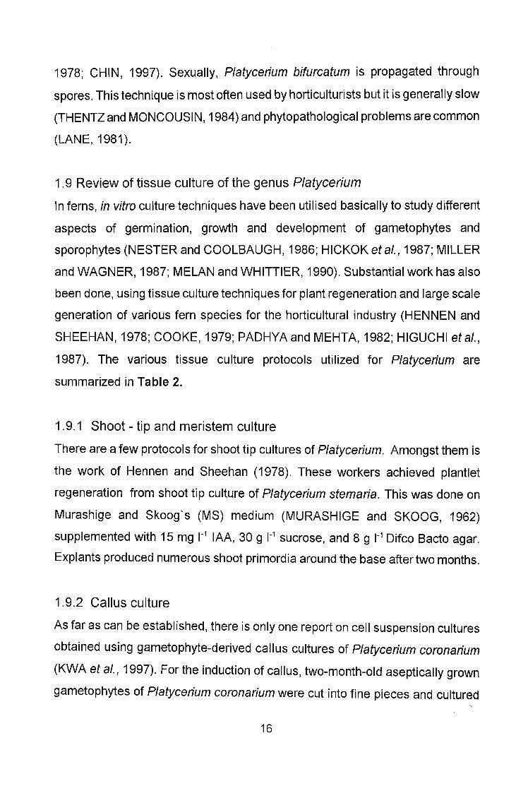
1.9 Review of tissue culture of the genus Platycerium
In ferns, in vitro culture techniques have been utilised basically to study different
aspects of germination, growth and development of gametophytes and
sporophytes (NESTER and COOLBAUGH, 1986; HICKOK et al., 1987; MILLER
and WAGNER, 1987; MELAN and WHITTlER, 1990). Substantial work has also
been done, using tissue culture techniques for plant regeneration and large scale
generation of various fern species for the horticultural industry (HENNEN and
SHEEHAN, 1978; COOKE, 1979; PADHYA and MEHTA, 1982; HIGUCHI et al.,
1987). The various tissue culture protocols utilized for Platycerium are
summarized in Table 2.
1.9.1 Shoot - tip and meristem culture
There are a few protocols for shoot tip cultures of Platycerium. Amongst them is
the work of Hennen and Sheehan (1978). These workers achieved plantlet
regeneration from shoot tip culture of Platycerium stemaria. This was done on
Murashige and Skoog's (MS) medium (MURASHIGE and SKOOG, 1962)
supplemented with 15 mg 1-1 IAA, 30 g 1-1 sucrose, and 8 g 1-1 Difco Bacto agar.
Explants produced numerous shoot primordia around the base after two months.
1.9.2 Callus culture
As far as can be established, there is only one report on cell suspension cultures
obtained using gametophyte-derived callus cultures of Platycerium coronarium
(KWA et al., 1997). For the induction of callus, two-month-old aseptically grown
gametophytes of Platycerium coronarium were cut into fine pieces and cultured
16

Table 2 : Summary of in vitro work on Platycerium species
Species Explant Media & Supplements Growth Response Reference
Modified Miller medium (Miller and Increase in dry weight of Camloh andMiller, (1961) explant (gametophyte) Gogala (1992)
In vitro grown +4% sucrosegametophytes
MS medium (half strength) Subsequent development Knauss (1976)+3% sucrose + 0.8% agar. of sporophytes on soil
surface
MS + 3% sucrose + 0 or 1.1 mgl-1 BA Adventitious bud Camloha et al.
Platyceriumdevelopment (1994)
bifurcatum (Cav) Modified MS + 0.02, 0.2, 2.0, 20 mgl- Rhizoid and shoot Camloh et al.C. Chr. 1Jasmonic acid (JA) development (1999)
Leaves Culture medium + 0.1 mgl-1 Kinetin + Bud induction Jambor et al.0.1 mgl-1 NAA (1995)
Culture medium + active charcoal + Rooting2 mgl-1 NAA
Modified MS medium + 3% sucrose Adventitious bud formation Camloh et al.+ 8 gl-1 agar without growth hormone (1991)
Not given MS + 3% sucrose + 0.9% agar + 0.1 Optimal shoot Pevalek (1996)mgl-1 NAA + 2.0 mgl-1 kinetin multiplication
17

Table 2 contd.
Species Explant Media & Supplements Growth Response Reference
Scales Modified MS by Hennen and Outgrowth development Ambrozic andSheehan, (1978) + 0.01 - 3 % that later developed into Camloh (1997)sucrose + 0.8% agar buds
IAA or NAA in culture medium Multiple shoot formation Kim et al. (1996)Sporelings
NAA in the culture medium Rooting
Platycerium Knop's medium (Miller and Greany) Spore germination Camloh et al.bifurcatum (Cav.) (1996)C. Chr. Spores Knop's medium + Jasmonic acid
(JA) Increase in length andnumber of rhizoids
Spores and Modified MS medium Micropropagation Gleba andaxillary shoots Gordzievskaya
MS medium without growth Rooting (1987)regulators or MS medium (1/10strength)
18

Table 2 contd.
Species Explant Media & Supplements Growth Response Reference
MS + 2% sucrose + 2 gl-1 gelrite + Callus formation4.4 mgl-12,4-0
MS medium + 1.1 mgl-12,4-0 + 2% Establishment of cellsucrose suspension with increase Kwa et al. (1997)
in fresh weightPieces ofgametophytes MS + 2.2 mgl-1kinetin Development and
proliferation of callus
Platycerium MS medium without growth hormone Morphogenesis intocoronarium gametophytes and(Koenig) Desv. sporophytes
MS medium + 2% sucrose 0.2% Formation of bud-likeGelrite structures that later
Rhizomes and developed into Kwa et al. (1995)fronds sporophytes
MS medium + 1.1 mgl-1 NAA Formation of sporophytes
19
Table 2 contd.
Species Explant Media & Supplements Growth Response Reference
Platycerium In vitro grown MS medium + 3% sucrose + 0.8% Multiplication of plantlets Cooke (1979)species sporophyte agar
plants
Adventitious bud formationat the base of the explant; Hennen and Sheehan
Platycerium MS medium + 30% sucrose + 0.8% on roots formed in culture; (1978)stemaria Shoot tip agar and on the fronds(Beauvois) produced on the explant inDesu. contact with the medium
Platycerium Modified de Fossards (1976) Development of prothallus, Bourne (1994)superbum Spores medium sporophytic plants and(Beauvois) adventitious budsDesu.
20

on Murashige and Skoog (MS) medium (MURASHIGE and SKOOG, 1962)
supplemented with 2 % sucrose and 4.4 mg 1-1 2,4-dichlorophenoxyacetic acid.
On plantlet differentiation, cells subcultured on MS medium supplemented with
2.2 mg 1-1 kinetin gave rise to two types of callus- dark green and pale green. The
pale green callus developed into sporophytes when subcultured onto basal MS
medium.
21
Chapter Two
Shoot Culture of Aloe polyphylla
2.1 Introduction
Shoot culture uses apices of lateral or main shoots, from actively growing shoots
or dormant buds, as explants for shoot multiplication. This is the most important
method of micropropagation. It is widely used commercially, mainly because of
its independence from climate, weather and seasons ( GEORGE, 1993). Shoot
cultures have also been widely used for clonal propagation of ornamental
flowering plants, for conservation of genetically defined stocks, and as
experimental material in biochemical, physiological, and genetic investigations
(BINDING and KRUMBIEGE-SCHROEREN, 1984). They are desirable due to
their high multiplication rates and sufficient genetic stability in contrast to callus
cultures.
2.1.1 Objectives
Aloe polyphylla, which is considered to be highly endangered (EMANOIL,1994)
has once been propagated in vitro (ABRIE and VAN STADEN, 2001). The
following study was therefore initiated in an attempt to optimize the tissue culture
protocol for differentiation, regeneration, and multiplication of Aloe polyphylla
plants. The aim of this research work however, was to optimise the different
stages of a tissue culture protocol of Aloe polyphylla laying emphasis on
multiplication and acclimatization. Considering the endangered status of this
plant, a very successful optimised protocol will definitely play a major role in
saving this important plant for mankind while at the same time optimizing
production for horticultural exploitation.
22

2.2 Materials and methods
2.2.1 Decontamination procedures and aseptic techniques.
Contaminants can cause large losses during micropropagation and their control
is usually the most frequent and a difficult problem encountered in
micropropagation. It is very important to detect and eliminate contaminating
organisms before they are transferred to many culture vessels during routine
subcultures. This is of utmost importance if contamination is to be avoided.
Plants are invariably infested externally with fungi, bacteria, yeasts and animal
pests (GEORGE, 1993). Other sources of contamination include the working area,
culture media, and working instruments. Generally, the plant material is
decontaminated with different concentrations of sodium hypochloride (NaOCI)
depending on the plant material.
Prior to use, the surface of the laminar flow bench was swabbed down with 95%
ethyl alcohol and the interior sprayed with the same alcohol. All glassware,
instruments and media were steam-sterilized in an autoclave at a pressure of one
bar and temperature of 121 QC for 20 minutes. Instruments in use on the bench
were placed in a beaker containing 95% ethanol and were flamed repeatedly
using a spirit burner during the course ofthe work. A bead sterilizer was also used
for the instruments. The worker's hands and forearms were washed thoroughly
with soap and water and repeatedly sprayed with alcohol during the period of
work. The mouth of all culture vessels was flamed before and after positioning of
the explant on the medium.
The explants used for this study were Aloe polyphylla shoots (2-3 cm long) taken
from in vitro grown plantlets. Due to large contamination rates experienced in the
preliminary experiments, even when other aseptic procedures were followed,
explants were later re-decontaminated with 1% NaOCI (Jik at 3.5%). This was
23

done by dipping the explants in 1% NaOCI for three minutes with continuous
agitation of the solution to ensure efficient distribution of the sterilant over the
plant material. This was followed by three rinses in autoclaved distilled water. On
completion of surface decontamination, the plant material, was placed on a sterile
petri dish for the removal of the outer damaged material. All further dissection
took place on sterile petri dishes and the explants, thus prepared, were
transferred to the culture vessels containing the nutrient medium.
2.2.2 Explant source
Totipotentiality is probably characteristic of all plant cells, but its expression may
be greater for particular cells. Familiarity with a cultivar's peculiarities are
frequently helpful in seeking explant sources (MURASHIGE, 1976). The choice
of a suitable explant is essential for successful tissue culture. The size, origin, and
physiological status of an explant can also affect its response in culture. For this
research project, shoots, 2-3 cm long with a maximum of five leaves taken from
in vitro grown plantlets were used.
2.2.3 Media and supplements
For plant tissue culture, many different media have been developed. The origin
of these media was mostly determined by the different nutrient requirements of
different plants. The choice of a medium for a plant normally depends on media
used for closely related species. Generally, these media are composed of a
mixture of inorganic nutrients and organic components which include sucrose and
sometimes plant growth regulators, depending on what kind of response the
researcher wants to achieve. Apart from the common constituents of a medium,
some unidentified supplements such as yeast extract, juices, pulps and extracts
from various fruits have been used to improve growth and development. The pH
of the medium is usually adjusted by the addition of dilute hydrochloric acid (HCI)
or sodium hydroxide (NaOH) to a pH range of between 5 and 6 prior to
24
autoclaving.
The standard culture medium used throughout this study contained full strength
of the macro-nutrients, micro-nutrients and vitamins as described by Murashige
and Skoog (1962). Details of these constituents are presented in Table 3. Forthe
present research work, all constituents of this medium besides sucrose were
made up as stock solutions. These solutions were obtained by dissolving the
required amounts of analytical grade macro-nutrients, micro-nutrients and
vitamins in distilled water, and making the final volume up to 1000 ml (1 litre). All
stocks were stored in glass containers at 5°C. Those stocks that contained light
sensitive constituents such as vitamin complexes, were stored in containers
wrapped in aluminium foil to exclude light.
To obtain the complete culture medium, stock solutions were combined in
volumes as shown in the last column of Table 3. This was supplemented with 30
gl-1 sucrose, 0.1 gl-1 myo-inositol, and made to volume with distilled water. The pH
of the medium was adjusted to 5.8 using sodium hydroxide (NaOH). To each litre
of medium, 0.8 g of agar was added to solidify it. This was dissolved in the
medium by steaming in a microwave for about ten minutes priorto dispensing into
the culture vessels. All cultures were initiated in 25 mm by 80 mm glass tubes,
each glass tube containing 12 ml of medium. Re-decontaminated shoots were
inoculated onto the basal medium and tubes sealed with Cap-O-Test tops.
Cultures were incubated at 25 ± 2°C with continuous flourescent light at a photon
flux density (400-700 nM) of 30 - 50 IJmol m-2 S-1
Shoot explants were placed on the basal medium, each supplemented with
various concentrations of plant growth regulators - benzyladenine (BA), kinetin,
zeatin or isopentenyladenine (iP). In another set of experiments explants were
again placed on the basal medium supplemented with various concentrations of
plant growth regulators in the following combinations: kinetin and 0<_
25

Table 3: Revised MURASHIGE and SKOOG (1962) nutrient medium
STOCK SOLUTION CHEMICAL MASS 9 500 ml-1 mlSTOCKSTOCK SOLUTION SOLUTION USED 1-1
MEDIUM
1 NH4N03 82.5 10
2 KN03 47.5 20
3 CaCI2 17.2 10
4 MgS04·7H2O 18.5 10
5 NaFeEDTA 2.0 10
6 KH2P04 8.5 10
7 H3B03 0.310 10MnS04.4Hp 1.115ZnS04·7Hp 0.430KI 0.04
8 NaMo04·2H2O 0.0125 10CUS04·5H2O 0.0013CoCI2·6H2O 0.0013
9 Thiamine. HCI 0.005 10Nicotinic acid 0.025Pyridoxin. HCI 0.025Glycine 0.100
Additional Sucrose 30 gl·l medium
Agar 8 gl-1 medium
Myo - inositol 0.1 gl-1 medium
pH adjusted to 5.8 with NaOH
26
naphthaleneacetic acid (NAA) or indole-3-butyric acid (IBA), zeatin and NAA or
IBA, BA and NAA or IBA. Cultures were incubated at 25 ± 2°C with continuous
flourescent light at a photon flux density (400 - 700 nM) of 30 - 50 IJmol m-2S-1.
Eight replicates were used per experiment.
In yet another set of experiments, to determine the effect of sucrose concentration
on shoot proliferation, explants were placed on the basal medium (full strength
MS medium) supplemented with 0.5 mgl-1 of zeatin and different concentrations
of sucrose. Cultures were incubated at 25±2°C under continuous flourescent light
at a photon flux density (400 - 700 nM) of 30 - 50 IJmol m-2S-1. Eight replicates
were used per treatment.
2.2.4 Environmental conditions
The major factors that are always considered in the culture environment are
temperature, light, and relative humidity. The average constant growth room
temperature is normally 25°C, (GEORGE, 1993). However, explant
establishment, culture growth, plantlet development and morphogenesis have all
been found to be temperature-dependent. In the past, relative humidity was never
thought to be an important environmental factor, until it was discovered to have
an effect on the hyperhydricity of cultured shoots and plantlets. Thus relative
humidity within a culture vessel depends on its temperature and that of the
medium (GEORGE, 1993). Light is considered as a complicated factor which can
be divided into light intensity, photoperiod, and spectral composition. Normally,
light is needed for photosynthesis, photomorphogenesis, and photoperiod. Since
cultures are supplied with enough carbohydrate, and phototropism is not of great
importance, light is critically important in plant tissue culture for
photomorphogenesis.
In this part of the research work an effort was made to study the effect of
27

temperature on shoot proliferation in order to determine the optimal temperature
for this activity. Explants were cultured on the basal medium supplemented with
0.5 mg 1-1 zeatin. Cultures were incubated under standard light conditions at
different temperatures. Eight replicates were used per treatment.
2.2.5 Acclimatization
Acclimatization or "hardening-off' is a process by which in vitro propagated plants
are made to adapt to an in vivo environment. It is basically concerned with
rooting, either in vitro or in vivo, and transfer to non-sterile conditions with
humidity control and temperature (DUNSTAN and TURNER, 1984). This is very
important considering that the waxy cuticle and stomata of in vitro grown plants
are inadequate or non-functional (SUTTER and LANGHANS, 1979; BRAINERD
and FUCHIGAMI, 1981; WETZSTEIN and SOMMER, 1982). However, the rate
of transpiration is always high in such leaves when subjected to the variable
humidity of the in vivo environment. There is also reason to believe that the high
sucrose and salt medium that is often used with in vitro grown cultures limits the
photoautotrophic capacity of leafy shoots (WETZSTEIN and SOMMER, 1982).
In vitro rooted plantlets of Aloe polyphylla were first washed in water to remove
excess agar since it has been found that sucrose and other organic compounds
trapped by agar in the proximity of roots cause plantlets to be infected by disease
causing organisms or be damaged by toxic microbial metabolites (GEORGE,
1993). Plantlets were then potted in different planting mixtures - full potting soil;
1 potting soil: 1 sand: 1 vermiculite (v: v: v); 1 sand: 1 peat: ) perlite (v: v: v).
Plants were kept in the mist house (with over-head sprinklers) and at a
temperature of 24°C ± 2°C for five weeks after which they were transferred to the
green house.
28
2.3 Results and Discussion
2.3.1 Decontamination procedures
As the explants were taken from in vitro grown plantlets, it was deemed
unnecessary to decontaminate them. However, when the first set of shoot cultures
was established, many shoots were lost due to fungal contamination. An attempt
was made to save the contaminated cultures by re-decontaminating them in 1%
and 2%, NaOCI and spore-kill respectively. This led to the death of most of the
explants while some of the surviving ones were still contaminated.
At this stage, it became inevitable to discard this set of explants and a new set of
explants was employed. The new set of explants from the same source was now
successfully decontaminated by dipping them in 1% NaOCI for five minutes
followed by three rinses in autoclaved distilled water. They were successfully
established without observing any contamination. In subsequent experiments, this
procedure was adopted and it provided good results.
2.3.2 Media and supplements
Since this study was concerned with optimizing the tissue culture protocol for Aloe
polyphylla, the number of healthy shoots produced by individual plant growth
regulators or in combination in the basal medium was recorded.
2.3.2.1 Effects of plant growth regulators on regeneration
The response of explants to different plant growth regulators singly or in
combination varied. The response with respect to shoot proliferation (both aXillary
and adventitious shoots) is shown in Figures 3 to 14 and Plates 1 to 10. Shoots
were produced on explants with the inclusion of the cytokinins BA, kinetin, iP, or
zeatin singly or in combination with the auxins IBA or NAA in the basal medium.
Both axillary and adventitious shoots were formed.
29
However, of all the cytokinins tested, BA gave the highest shoot proliferation
response. After about 15 days the base of the explants became swollen forming
adventitious buds which later developed into shoots. A yellowish callus was
observed at the base of the explants from where most of the adventitious shoots
seemed to originate. Callus formation increased with an increase in BA
concentration. BA (1.5 mg 1-1) in the basal medium gave the highest average
number of shoots (12 shoots per explant) (Fig.3). This was not statistically
different from the 10 and 9 shoots per explant produced by 0.5 mg 1-1 and 1.0 mg
1-1 BA respectively in the basal medium. Hyperhydricity was very pronounced
especially in cultures on media with 1.0, 1.5, 2.0, or 3.0 mg 1-1 BA. Most of the
shoots were stunted and light green, quite unlike cultures with 0.5 or 0.1 mg 1-1 BA
(Plate 1). Generally, cultures on media with 0.5, or 0.1 mg 1-1 looked healthier than
cultures with higher concentrations of BA. Browning occurred in about 7% of
cultures.
With kinetin alone in the culture, multiple shoot production was observed (Plate
2). A yellowish callus was also noticed at the base of the explant which tended to
increase in mass with an increase in kinetin concentration. This was first observed
in cultures with 1.5 mg 1-1 kinetin which happens to be the optimal concentration
for shoot proliferation (8 shoots per explant) (Fig.4 ). However, callus
development was much more prominent in the culture with higher concentrations
of kinetin (2 and 3 mg 1-1) (Plate 3. ). Both axillary and adventitious shoots were
formed, the adventitious shoots originated from the base of the explants. Of all the
shoots produced in cultures with 1.5, 2.0 and 3.0 mg 1-1 of kinetin, 6, 12, 25 %
were adventitious in origin respectively. Browning was witnessed in very few
cultures contrary to its severity in Aloe vera cultures reported by Natali et al.
(1990), while hyperhydricity developed sporadically in cultures with 2 and 3 mgl-1
kinetin.
30

14
12
Ul
<5 10~-Ul....g 8~
Ul...~ 6<ll.0E~ 4
2
o320.1 0.5 1 1.5
BA conc. (mg.r1)
Figure 3 : Effects of various concentrations of BA onshoot and root proliferation of Aloe polyphylla in vitro
o
10
9
Ul 8<5~ 7-Ul
<5 6o-;; 5...~ 4<ll.0E 3::J
Z 2
oo 0.1 0.5 1.5 2 3
Kinetin conc. (mg.r1)
Figure 4: Effects of various concentrations of kinetin onshoot and root proliferation of Aloe polyphylla in vitro
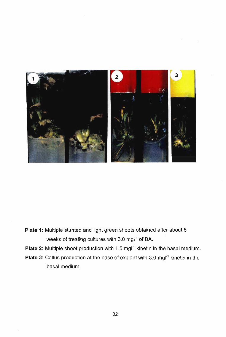
Plate 1: Multiple stunted and light green shoots obtained after about 5
weeks of treating cultures with 3.0 mgl-1 of BA.
Plate 2: Multiple shoot production with 1.5 mgl-1 kinetin in the basal medium.
Plate 3: Callus production at the base of explant with 3.0 mgl-1 kinetin in the
basal medium.
32

Unlike other cytokinins the optimal concentration for shoot proliferation with zeatin
alone in the basal medium was 0.5 mg 1-1 producing an average of 7 shoots per
explant (Fig. 5); statistically this was different from other concentrations. Shoots
were healthy and dark green (Plate 4A). Again formation of a yellowish callus was
observed with 0.5 mg 1-1 zeatin. Callus formation increased with an increase in
zeatin concentration. The number of adventitious shoots observed also increased
with an increase in cytokinin concentration. Cultures containing 0.5 mg 1-1 zeatin
had the least percentage of adventitious shoots (5 %) of the total number of
shoots formed, compared with the 88 % occurring in cultures with 3.0 mg 1-1 of
zeatin. Hyperhydricity was observed in about 2 % of cultures, occurring in cultures
containing higher concentrations of 3 mg 1-1 of zeatin.
With the cytokinin iP alone in the basal medium, similar results to BA were
obtained except that fewer shoots were formed and hyperhydricity was not as
pronounced as in cultures with BA. The optimal iP concentration for shoot
proliferation was 1.0 mg 1-1 producing an average of 8 shoots per explant (Fig. 6),
(Plate 48).
Multiple shoot formation was observed in cultures with the cytokinins - BA, zeatin,
or kinetin in combination with the auxins - NAA or IBA. Shoot proliferation activity
of kinetin alone in the basal medium was better than when it was used in
combination with either IBA or NAA. The optimal combination of both (ie
kinetin/NAA and kinetin/lBA) for shoot proliferation was 2.0/0.1 and 1.5/1.0 mg 1-1
respectively, producing an average number of 5 and 7 shoots per explant
respectively (Figs. 7 & a), (Plate SA & 58). There was a sporadic occurrence of
hyperhydricity and browning. No development of the yellowish callus was noticed.
Zeatin/lBA and zeatin/NAA combinations, like other cytokinin/auxin combinations
tested, also produced a good number of healthy shoots. The optimal zeatin/IBA
and zeatin/NAA combinations were 1.0/0.5 mg 1-1 (7 shoots per explant) and
1.0/1.0 mg 1-1 (7 shoots per explant) (Figs. 9 & 10). Browning and hyperhydricity
33
9
8
.l!! 7o.g 6.l!!g 5
.s::.l/I
'0 4
.8 3E~ 2
o320.1o 0.5 1 1.5
Zeatin conc. (mg.r1)
Figure 5: Effects of various concentrations of zeatin onshoot and root proliferation of Aloe polyphylla in vitro
9
8I/)
'0 7EUi 6'0.,g 5I/)
'0 4...1l 3E::J 2z
o21.50.1o 0.5 1
iP conc. (mg.r1)
Figure 6: Effects of various concentrations of iP on shootand root proliferation of Aloe polyphylla in vitro
34

Plate 4: Multiple shoot production with cytokinins tested. (A) 0.5 mgl-1 of
zeatin, (B) 1.0 mgl-1 of iP.
Plate 5: Multiple shoot production obtained with different combinations of
kinetin and auxin in the basal medium. (A) kinetin/NAA (2.0/0.1 mgl-1),
(B) kinetin/lBA (1.5/1.0 mgl-1).
35

8
7
6
1/1+'g 5J::1/1
'0 4...C1l.c 3E:::JZ
2
1
o
6
5
.l!! 4ooJ::1/1
'0 3...C1l.c~ 2Z
o
I_Shoots I
A BeD E F G HKinetin/IBA cone. (mg.r1
)
Figure 7: Effect of various combinations of kinetin/lBA onshoot proliferation of A/oe po/yphylla in vitro
I_Shoots I
A BeD E F G H
Kinetin/NAA cone. (mg.r1)
Figure 8: Effect of various combinations of kinetin/NAA onshoot proliferation of A/oe po/yphylla in vitro
Kinetin/NAA & KinetinllBA (mg/I)A -1.5/0.1 C -1.5/1.0B - 1.5/0.5 D - 1.5/2.0
36
E - 2.0/0.1F - 2.0/0.5
G - 2.0/1.0H - 2.0/2.0

9
8
7Ul
"0 6o
..s::Ul 5....o~ 4
.Q
E 3:rZ
2
o
I_Shoots I
HGBA C D E F
Zeatin/IBA cone. (mg.r1)
Figure 9: Effect of various combinations of zeatin/IBA onshoot proliferation of Aloe polyphylla in vitro
9
8 I_Shoots I
7Ul+'g 6..s::Ul 5....0..Ql 4
.Q
E~ 3
2
1
0
A B C D E F
Zeatin/NAA cone. (mg.r1)
G H
Figure 10: Effect of various combinations of zeatin/NAA onshoot proliferation of Aloe polyphylla in vitro
Zeatin/NAA & Zeatin/ BA (mgll)A - 0.5/0.1 C - 0.5/1.0B - 0.5/0.5 D - 0.5/2.0
E -1.0/0.1F -1.0/0.5
G -1.0/1.0H -1.0/2.0
37
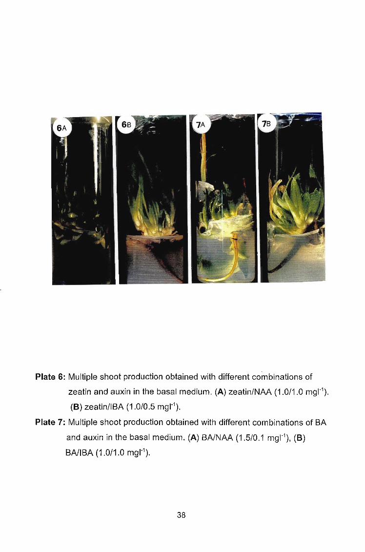
Plate 6: Multiple shoot production obtained with different combinations of
zeatin and auxin in the basal medium. (A) zeatin/NAA (1.0/1.0 mgl-1).
(8) zeatin/IBA (1.0/0.5 mgl-1).
Plate 7: Multiple shoot production obtained with different combinations of BA
and auxin in the basal medium. (A) BA/NAA (1.5/0.1 mgl-1), (8)
BA/IBA (1.0/1.0 mgl-1).
38

14
I_Shoots I12
Cfl10...
00
..c: 8Cfl-0...Ql 6..cE::::lZ 4
2
0A BeD E F G H
BA/IBA conc. (mg.r1)
Figure 11: Effect of various combinations of BA/IBA onshoot proliferation of Aloe polyphylla in vitro
12
I_Shoots I10
~ 800
..c:Cfl
'0 6...Ql
..c
~ 4z
2
0A B C 0 E F G H
BA/NAA conc. (mg.r1)
Figure 12: Effect of various combinations of BA/NAA onshoot proliferation of Aloe polyphylla in vitro
BA/NAA & BA/IBA (mgll)
A -1.0/0.1 C -1.0/1.0B - 1.010.5 D - 1.0/2.0
39
E -1.5/0.1F -1.5/0.5
G -1.5/1.0H -1.5/2.0

were not observed. Shoots produced were healthy, having a dark green
appearance (Plate 6A & 68). Multiple shoot formation was also observed with
BA/lBA and BA/NAA combinations. The optimal combinations were 1.0/1.0 mg
1-1 (11 shoots per explant) and 1.5/0.1 mg 1-1 (9 shoots per explant) (Figs. 11 &
12), (Plate 7A & 78).
Few culture media have been used in tissue culture of species of Aloe, with
different explants employed. Shoot and meristem tip explants of Aloe have been
cultured on MS (MURASHIGE and SKOOG, 1962) medium (NATAL! etal., 1990;
ROY and SARKAR,1991; GUI et al., 1990; ZHAO, 1990; KAWAI et al., 1993;
CORNEANU etal., 1994; HIRIMBUREGAMAand GAMAGE, 1995) and modified
MS medium (MEYER and VAN STADEN, 1991; RICHWINE et al., 1995). The
number of proliferating shoots is an important factor to consider when developing
an optimal tissue culture protocol. In this research, the presence or absence of a
certain plant growth regulator influenced the number of proliferating shoots. The
results of this study showed that shoot proliferation was improved by the addition
of plant growth regulators, although some explants formed shoots on hormone
free medium. The potential of these explants to produce shoots in hormone- free
medium could probably be attributed to the presence of endogenous hormones.
Results showed that the presence of cytokinins - BA, kinetin, zeatin or iP alone
or in combination with auxins - NAA or IBA in the basal medium increased shoot
proliferation activity at different levels. Previous workers reported that cytokinins
alone or in combination with auxins positively influenced shoot proliferation in
Aloe species (GUI et al., 1990; NATAL! et al., 1990; ROY and SARKAR, 1991;
HIRIMBUREGAM and GAMAGE, 1995; RICHWINE et al., 1995~ FENG et al.,
2000). Auxins (NAA and IBA) alone in the basal medium did not promote shoot
proliferation unlike the report of MEYER and VAN STADEN (1991).
40

Results also showed that a high level of cytokinin (2 and 3 mg 1-1) adversely
affected shoot proliferation, usually promoting the formation of a yellowish callus
at the base of explants. BA which showed the best shoot proliferation potential
was associated with hyperhydricity, and most of the shoots so formed were
stunted. Hyperhydricity was observed in cultures with 1.0, 1.5, 2.0, or 3.0 mg 1-1
of BA. BA is known for its hyperhydricity-inducing tendencies in shoot cultures
(LESHEM et al., 1988; LI et al., 1997; TSAY and DREW, 1998; THOMAS et al.,
2000). Zeatin was another cytokinin that showed high shoot proliferation activity.
Unlike BA, the shoots were elongated and hyperhydricity was very low, occurring
only in cultures with 2 and 3 mg 1-1 zeatin. The optimal concentration (0.5 mg 1-1)
for shoot proliferation was much lower than other cytokinins. Kinetin and iP also
produced good results except that iP was also associated with hyperhydricity.
However, the effect was not as pronounced as for BA. The shoots were not
stunted as with BA. The results also showed BA to be slightly more active with
respect to number of proliferating shoots than other cytokinins tested
(MURASHIGE, 1974).
Combinations of BA and NAA or BA and IBA proved to be better than BA alone
in the basal medium in terms of reducing the incidence of hyperhydricity and
callus formation. This could be attributed to the balancing of the cytokinin/auxin
ratio. Zeatin in combination with IBA or NAA was slightly better than zeatin alone
with respect to shoot formation. In contrast, kinetin alone in the basal medium was
better than when in combination with either NAA or IBA with regards to shoot
proliferation. Generally, there was not much difference in shoot proliferation
activities between the tested cytokinins alone or when used in combination with
auxins. Most previous workers reported that combinations of cytokinins and
auxins were better than cytokinins alone in the basal medium with respect to
shoot proliferation (NATALI et al., 1990; ROY and SARKAR, 1991;
HIRIMBUREGAM and GAMAGE, 1995; FENG, 2000). This contrasts with the
41

results of this study. It was only Richwine et al. (1995) who reported multiple
shoot production for Aloe vera and Aloe harlona with kinetin or BA alone in the
basal medium. In this study all the cytokinins singly or in combination with IBA or
NAA showed multiple shoot production.
Browning was not a severe problem since the explants used were young shoots.
This might be as a result of the fact that juvenile tissues tend to have a greater
capacity for restoration (MURASHIGE and SKOOG, 1962) or produce less
phenols (ROY and SARKAR, 1991).
2.3.2.2 Effects of sucrose on regeneration
The presence of sucrose in the basal medium had a tremendous effect on shoot
proliferation. At 0% level, there was hardly any shoot proliferation (Fig.13), (Plate
8A). After about 30 days of incubation, explants showed no sign of shoot
proliferation nor rooting despite the presence of 0.5 mg 1-1 zeatin in the basal
medium. With 1% of sucrose in the basal medium, after about 30 days of
incubation, shoot proliferation was observed. An average of 5 shoots per explant
was obtained (Fig.13). When the sucrose concentration was increased to 3 %,
there was a significant increase in the number of shoots produced (8 shoots per
explant), (Fig.13), (Plate 8B), but there was no significant increase in the number
of shoots obtained when the sucrose concentration was increased further to 4 %
(Fig.13). However, when the concentration was increased to 5 % and finally 6 %,
shoot proliferation declined significantly (Fig.13).
Carbohydrates are generally added to culture media to serve as a carbon and
energy source. Besides, they also play an important role in the regulation of the
external osmotic potential. In most cultured plant tissue, sucrose is the primary
substrate for respiration that produces carbon dioxide on which metabolism and
growth of the tissue depends (THORPE and MEIER,1972; DODOS and
42

Plate 8: Effect of sucrose on shoot proliferation of Aloe polyphylla in vitro. (A)
at 0 % level, no shoot proliferation, (8) at 3 % level, multiple shoot
proliferation.
Plate 9: Effect of temperature on shoot proliferation of Aloe polyphylla in vitro.
(A) at 10°C, shoot proliferation was near zero, (8) at 20°C, a sharp
increase in shoot proliferation, (C) at 30°C, shoot proliferation was
inhibited.
43

9
8 I_Shoots IIII
7-0 60.&:.III 5-0.. 4Ql.cE 3::::JZ
2
1
00 10 20 30 40 50 60
Sucrose concentration (g.r1)
Figure 13: Effect of various levels of sucrose on shootproliferation of Aloe polyphylla in vitro
14
12 _BA Shoots
I!Z Shoots
10 EJK ShootsIII
'00 8.&:.III-0.. 6Ql.cE::::JZ 4
2
010 20 25
Temperature (oC)30
Figure 14: Effect of various temperatures on shootproliferation of Aloe polyphylla in vitro

ROBERTS, 1985; LANGFORD and WAINWRIGHT, 1988; GEORGE, 1993). The
results of this study showed no proliferation of shoots by the explants at 0%
sucrose after about five weeks. Explants did however, not die. These results were
obtained irrespective of the presence or absence of 0.5 mg 1-1 of zeatin in the
medium. According to Thorpe and Murashige (1968 and 1970) the main function
of carbohydrate (sucrose) in the medium is to serve as a readily available source
of energy for the initiation of shoot primodia and their subsequent development.
Results also showed a marked increase in shoot proliferation by explants at
sucrose concentration of 3 and 4 %. The number of shoots formed at these
sucrose levels did not statistically differ. Regeneration of shoots at these levels
of sucrose could be credited to the fact that metabolism and growth of the tissues
were feasible due to the respiratory activities of the tissues made possible by the
presence of a suitable substrate (sucrose) (THORPE and MURASHIGE, 1968,
1970). There was a significant decrease in shoot proliferation by explants at
higher sucrose concentrations. Langford and Wainwright (1988) reported in their
study that the greater the sucrose concentration in the medium, the less carbon
dioxide was taken up per unit chlorophyll, perhaps indicating a greater chlorophyll
efficiency at the lower sucrose concentrations.
2.3.3 Temperature
2.3.3.1 Effects of temperature on shoot regeneration
Temperature was observed to exert some effect on shoot proliferation. At 1Doe,
with zeatin (0.5 mg 1-1) in the medium, shoot proliferation was near zero (Fig. 14),
(Plate 9A). This same result was also obtained when zeatin was substituted with
either BA (1.5 mgl-1) or kinetin (1.5 mgl-1
). But when cultures were incubated at
20°C, shoot proliferation activity increased sharply with either zeatin, BA, or
kinetin in the medium (Fig.14), (Plate 98). The optimal temperature for shoot
proliferation activity with BA in the medium was 25°C. However, with zeatin or
',,- 45

kinetin in the medium, the number of shoots produced at 20°C and 25°C
respectively were not statistically different (Fig.14). At 30°C, shoot proliferation
activity was generally inhibited irrespective of the cytokinin in the basal medium.
The few shoots produced at this temperature (30°C), were light green and shorter
in length when compared to shoots obtained in cultures incubated at 20°C or
25°C (Plate 9C).
Maintaining in vitro cultures at a relatively high temperature reduced the efficacy
of cytokinin which is basically responsible for shoot regeneration
(GEORGE, 1993). The few regenerated shoots at 30°C were shorter in length than
shoots obtained in cultures incubated at 20°C and 25°C, and the leaves were light
green. These results also showed that the temperature (30°C) which is above the
determined optimal temperature for shoot regeneration was detrimental to shoot
regeneration and growth. Previous workers have shown that temperature is one
of the deciding factors for shoot multiplication and growth (FONNESBECH et al.,
1979; PIERIK et al., 1988; HORN etal., 1988; MEYER and VAN STADEN, 1991;
PUDDEPHAT et al., 1997).
2.3.4. Acclimatization
The results of various experiments carried out on rooting showed that rooting can
best be achieved on MS (MURASHIGE and SKOOG, 1962) medium free of any
plant growth regulator (Plate 10A). Plantlets commenced rooting within the first
two weeks of incubation. Subsequently, rooted plantlets were planted into three
different potting mixtures and were successfully acclimatized in the mist house for
about four weeks before being transferred to the greenhouse (Plate 108). Of all
the potting mixtures employed, the highest survival, (98%) of plantlets, was
obtained with a soil:sand:vermiculite mixture (1:1:1 v/v). It was also observed that
mist house acclimatized Aloe polyphylla plants needed to be kept in at least 70
% shaded greenhouse for about six weeks to avoid direct sunlight at this early
46

Plate 10: (A) Aloe polyphylla rooting in plant growth regulator- free MS medium,
(B) fully acclimatized Aloe polyphylla plants.
47

stage of development.
Results of this study showed that plantlets rooted well in vitro in a hormone-free
medium. Generally, auxins are included in the medium at this stage for rooting,
however there was no exogenous auxin in the medium and the plantlets rooted
well. Previous studies on various species belonging to the Liliaceae showed that
they can form roots in a hormone-free medium (NATAL! et al., 1990; ROY and
SARKAR, 1991; RICHWINE et al., 1995). Since rooting is generally believed to
be enhanced by auxins, it appears that the level of endogenous auxins in these
species is high enough to promote rooting.
The higher survival percentage observed with the potting soil: sand: vermiculite
(1: 1: 1 v/v) mixture when compared to peat, and potting soil respectively could
mean that Aloe polyphylla survive better in a well aerated and drained soil
mixture. Acclimatized plants (about eight months old) so far do not show any
physical aberration.
2.3.5 Conclusion
At the end of this study, one could say that the cytokinins - kinetin, zeatin or BA
alone enhanced shoot proliferation better than cytokinins and auxins in
combination. In all optimal concentrations (cytokinin singly or in combination with
auxin), both axillary and adventitious shoots were formed. Callus formation and
hyperhydricity were very pronounced with BA, unlike the other cytokinins, and
most shoots so formed with BA in the basal medium were stunted. Plantlets
rooted better in a plant growth regulator- free MS medium than in MS with an
auxin or in different strengths of MS medium (Table 4). The optimal temperature
for shoot proliferation was observed at 25°C, though there was no significant
difference with the number of shoots produced in cultures incubated at 20°C.
Again, the optimal sucrose concentration for shoot proliferation was 3%. As for
48

potting mixture, the best for in vitro grown Aloe polyphylla was sand: soil:
vermiculite (1:1:1 v/v). Mist house acclimatized plants need to be kept in at least
a 70 % shaded greenhouse for about six weeks to avoid direct sunlight which
tends to be harmful to the plants at this early stage of development.
Table 4: Mean number of roots produced by Aloe polyphylla at different
strengths of MS medium.
MS Mean no of No of Std. Std. Error of
medium Roots replicates Deviation Means
Full S 3.375 8 0.9161 0.3239
Half S 3.125 8 0.991 0.3504
One- 2.125 8 0.8345 0.295
quarter S
Total 2.875 24 1.0347 0.2112
(S - Strength).
The clonal micropropagation method described here is very efficient in terms of
both the rate and number of shoots produced and the rapidity at which new rooted
shoots are obtained when compared with normal vegetative propagation. This
protocol may prove to be a solution to the problem of declining numbers of Aloe
polyphylla in nature.
49

Chapter Three
Tissue culture of Platycerium bifurcatum
3.1 Introduction
Tissue culture techniques have been employed not only in the propagation of
Platycerium bifurcatum, but also in understanding its nutritional, physical and
chemical requirements for growth and development. Previous tissue culture
studies have focussed on the in vitro germination of spores (KNAUSS, 1976;
BOURNE, 1994); the effects of nutrients and physical and chemical factors on
growth and development of the prothallus, and on sporophyte formation
(FERNANDEZ et al., 1996; 1997) and the homogenization of gametophytes or
sporophytes as an aid in sporophyte production (KNAUSS, 1976; COOKE, 1979;
FINNIE and VAN STADEN, 1987; FERNANDEZ et al., 1999).
3.1.1 Objectives
Platycerium bifurcatum is conventionally propagated sexually by spores and
asexually through root bud development (RICHARDS et al., 1983). According to
Lane (1981), phytopathological problems are very common with extra vitro fern
propagation from spores. Various workers have studied the in vitro propagation
of Platycerium bifurcatum, but it seems that growth of the sporophyte in vitro is
slow. It takes an average of six months to get to rooting size (JAMBOR, 1995).
The strength of the MS medium has been shown to affect organogenesis and
subsequent growth of plantlets in vitro (GEORGE, 1993). Therefore the objective
of this part of the project was to investigate the effect of different strengths of the
MS medium on organogenesis and subsequent growth of Platycerium bifurcatum,
and different potting media on acclimatization.
50

3.2 Materials and methods
3.2.1 Decontamination procedures and aseptic techniques
Disinfection of plant materials is necessary in order to eradicate surface
microorganisms. The presence of any contaminant will interfere with the growth
of explants or cultures. Normally, prior washing of the explants with soap and
water or dipping in ethanol is recommended to induce adequate wetting and
initial cleaning. Most commonly a dilute solution of sodium hypochloride (0.25
2.63%) is used as a disinfectant, and an emulsifier such as Tween 20 is added
at the rate of one drop per 100 ml solution. Vegetative explants are usually
decontaminated for 10- 20 minutes, followed by a rinse in autoclaved distilled
water to remove residual disinfectant. Mechanical agitation using a stirrer is
sometimes helpful to dislodge air bubbles and facilitate an even distribution of
~the disinfectant over the explant.
3.2\2 Explant source
Th~ choice of a suitable explant is essential for successful tissue culture. Thej
size, origin, and physiological status of an explant can also affect its response in
culture. The explants used for this study were leaves of Platycerium bifurcatum
collected from the Botanical Garden of the University of Natal (Pietermaritzburg).
Leaves were rinsed under running water, soaked in 70% ethanol for a minute,
washed in 1% NaOCI solution with a few drops of Tween 20 for ten minutes.
Explants were afterwards rinsed three times in autoclaved distilled water. fan
completion of surface re-decontamination of the plant material, the explant was
placed on a sterile petri dish for the removal of all the brown and discoloured
areas. Leaves were cut into 1 cm squares. All further dissection took place on
sterile petri dishes and the explants thus prepared were transferred to the culture
vessels containing the nutrient medium.
51

3.2.3 Media and supplements
Leaf explants were placed on the basal medium supplemented with 1.0 mgl-1 of
BA for adventitious bud proliferation.
In the subsequent experiment, after the initiation of adventitious buds, the effect
of different strengths (full, half, and one-quarter strength) of the mineral salt
constituents of the basal medium on the growth of plantletS"Vv'as investigated.
3.2.4 Environmental conditions
Cultures were incubated at 25 ± 2°C with a 16 h light, 8 h dark photoperiod. Light
was provided by cool white fluorescent tubes at flux density (400-700 nM) of 30
50 ~mol m-2 S-1.
3.2.5. Acclimatization
In vitro rooted plantlets of Platycerium bifurcatum were first washed in water to
remove excess agar since sucrose and other organic compounds trapped by
agar in the proximity of roots cause plantlets to be infected by disease, causing
organisms to be damaged by toxic microbial metabolites (GEORGE, 1993).
Plantlets were then potted in different planting mixtures - peat; 1 peat: 1
vermiculite: 1 sand. Plants were kept in the mist house (at a temperature of 24°C
± 2°C) for six weeks, after which they were transferred to the green house.
3.3 Results and Discussion
3.3.1 Decontamination procedures
The decontamination procedure -soaking explants in 70 % ethanol for a minute,
and afterwards washing explants in 1 % NaOCI solution with a few drops of
Tween 20 described in 3.2.1 was very successful. Almost 80 % of the explants
were decontaminated. Due to the success achieved with this decontamination
procedure no further effort was made to try other decontamination procedures.
52

Plate 11: (A) Platycerium bifurcatum cultured in half-strength MS medium,
(B) in full strength MS medium, (C) one-quarter strength MS medium,
(0) fully acclimatized plants.
53
3.3.2 Media and supplements
For this part of the project, the number of proliferating buds was not recorded
since it was not the focus of the experiment. The focus of the experiment was the
growth and subsequent development of the plantlets. The results are therefore
presented in visual form only.
Adventitious buds appeared on the leaf surface without callus formation in the
basal medium supplemented with 1.0 mg 1-1 BA. After adventitious bud initiation,
further bud proliferation was observed on full strength and half-strength MS
media. Adventitious buds were isolated and subcultured monthly on different
strengths of MS media (full strength, half-strength, and one-quarter strength MS
media). On half-strength MS medium, bud growth was very vigorous (Plate 11A),
and plantlets rooted well in this medium. With full strength MS medium, bud
growth was observed (Plate 11 B) although it was not as pronounced as bud
growth observed in half-strength MS medium. There was root initiation in this
medium, but fewer roots were present compared to the half-strength MS medium
treatment. When buds were subcultured in one-quarter strength MS medium, bud
growth was the slowest. The leaves were yellow (Plate 11 Cl. Root development
was the best in this medium.
Results showed that after bud initiation on a growth regulator containing MS
medium, further growth and development of buds can best be achieved in half
strength MS medium without growth regulators. Further proliferation of buds
observed after subculturing on a plant growth regulator - free MS medium could
be attributed to either a carry-over effect of BA, or the ability of explants to
proliferate buds in a plant growth regulator - free medium, as reported by
previous workers (CAMLOH and GOGALA, 1991; CAMLOHA et al., 1993;
BOURNE, 1994). Generally, research has shown that organogenesis is plant
growth regulator-controlled. However, the subsequent organogenesis of buds
54

without exogenous growth regulators might reflect high endogenous growth
regulator levels.
3.3.3 Acclimatization
The strength of the MS medium played a major role in the rooting of Platycerium
bifurcatum in vitro. Plantlets subcultured on one-quarter strength of MS medium
rooted better than plantlets subcultured on the other two media (full strength and
half-strength MS respectively). This result was obtained without any plant growth
regulator in the medium. This suggests that the plantlets might have had
sufficient endogenous auxins to initiate rooting. Results obtained are similar to
that of Gleba and Gordzievskaya (1987), while working on in vitro propagation of
Platycerium bifurcatum. They also successfully achieved rooting in MS medium
without plant growth regulators and in MS medium diluted ten-fold.
Rooted plantlets of about 3-4 cm in height were isolated and planted into two
different potting mixtures (peat; peat: vermiculite: sand (1: 1: 1) v/v). They were
acclimatized in the mist house for about six weeks before being transferred to the
greenhouse (Plate 11 D). Highest survival (90 %) of plantlets was obtained with
peat alone (Table 5). By comparing the water retaining ability ofthe two different
potting mixtures, it is clear that Platycerium bifurcatum thrived better on a high
water retaining potting mixture. Successfully acclimatized plants are doing well
in the greenhouse.
55

Table 5: Percentage survival of in vitro grown Platycerium bifurcatum planted out
on two different potting mixtures
Potting mixture No. of No. of survived Survival (%)
transplanted plants
plants
Peat 40 36 90
PeatVermiculite: 40 22 56
sand(1:1:1 v/v)
3.3.4 Conclusion
Platycerium bifurcatum showed a high mUltiplication rate using leaf explants. The
subsequent isolation and subculturing of plantlets were, however, labour
intensive. Although growth of plantlets in half-strength MS medium was
satisfactory, rooting was better in one,.quarter strength MS than the half-strength
MS medium. To ensure better rooting of plantlets, it is best to monthly isolate
plantlets from half-strength MS medium and then subculture them on one-quarter
strength medium for about five weeks.
Peat proved a better potting mixture in terms of plantlet survival.
56

Chapter Four
General Conclusion
Having presented the results of this study in Chapters two and three, it is
necessary to briefly conclude the salient points of this investigation. Many rare
and endangered plant species are propagated in vitro because they do not
respond well to conventional methods of propagation. As far as I know there is
only one report on tissue culture of Aloe polyphyffa (ABRIE and VAN STADEN,
2001) despite its spectacular ornamental and 'medicinal' values. ArLQp-tim~~_~t
tissue culture protocol was developed to produce a large number of plants of this- ----------- ------------rare sp~cie.~_ of~!C?e.:.Shoot explantscfedvearrom-in vlfro grownplcintlets were--".~-_.".--
inoculated onto MURASHIGE SKOOG (MS) (1962) medium supplemented with
100 mg 1-1 myo-inositol, 3 % sucrose, 0.8 % agar, and different concentrations of
various cytokinins singly or in combination with the auxins naphthaleactic acid
(NAA) or indole-butryic acid (IBA). These explants produced multiple shoots. The
average number of plantlets produced per explant was high, ranging from six to
ten. There was a yellowish callus formed at the base of the explants especially
when 3.0 mg 1-1 of the cytokinins (kinetin, iP and zeatin) were used.
Hyperhydricity was encountered with BA in the mUltiplication medium.
Hyperhydricity with BA needs further research because of the fact that BA
induced the highest average number of plantlets.
Sucrose and temperature proved to be amongst the most important determining
factors in shoot proliferation. Sucrose at the 3 % level and temperature at 2SoC
were ideal for shoot proliferation. Plantlets were successfully rooted in a
hormone-free MS medium and subsequently acclimatized. This protocol is
assumed to be effective considering the high regeneration percentage of explants
and also the high number of plantlets produced. The ease with which rooting was
achieved without any auxin in the medium makes the protocol desirable.
57
Plantlets cultured in vitro are highly susceptible to dessication once transferred
to soil. Survival of plants depends on their ability to carry out photosynthesis and
withstand water loss. Aloe polyphylla showed a good ability to carry out
photosynthesis and withstand water loss. This is reflected in the high survival
rates (almost 100 %). Aloe polyphylla remains an endangered species. However,
the future of this species is now less precarious due to the success of the
multiplication protocol now available.
In the second investigation, using leaf explants, Platycerium bifurcatum showed
a high multiplication rate. Growth and rooting of plantlets were best achieved in
half- and one-quarter strength MURASHIGE and SKOOG (MS) (1962) media,
each supplemented with 100 mg 1-1 myo-inositol, 3 % sucrose, and 0.8 % agar.
The growth of plantlets was very satisfactory. Plantlets were successfully
acclimatized. The major problem to be solved is the labour intense monthly
subculturing of the plantlets. This problem must be addressed in the future.
58

References
ABRIE AL and VAN STADEN J (2001) Micropropagation of the endangered Aloepolyphylla. Plant Growth Regulation 33: 19-23.
ALTMAN A and ZIV M (1997) Horticultural biotechnology: A historical perspectiveand future prospects. Acta Horticulturae 447: 31-35.
AMBROZIC D and CAMLOH M (1997) Gametophytic and sporophytic regenerationof Platycerium. Annals of Botany 80: 23-28.
BINDING Hand KRUMBIEGEL-SCHWEREN G (1984) Clonal propagation: shootcultures. Cell Culture and Somatic Cell Genetic of Plants, Vo!. 1, 43-48.
BOURNE RA (1994) The development of a program of commercial productionof staghorns from plant tissue culture. Combined Proceedings InternationalPlant propagators' Society 44: 90-93.
BRAINERD KE and FUCHIGAMI LH (1981) Acclimatization of aseptically culturedApple plants to low relative humidity. Journal of American Society ofHorticultural Science 106: 515-518
CAMLOH M, RAVNIKAR M and ZEL J (1996) Jasmonic acid promotes division offern protoplasts, elongation rhizoids and early development ofgametophytes. Physiologia Plantarum 97: 659-664.
CAMLOH M, VILHAR B, ZELJ and RAVNIKAR M (1999) Jasmonicacid stimulatesdevelopment of rhizoids and shoots in fern leaf culture. Journal ofPlantPhysiology 155: 798-801.
CAMLOHA M and GOGALA N (1991) Platycerium bifurcatum - adventitious budand root formation without growth regulators in vitro. Acta Horticulturae 289:89-90.
CAMLOHA M and GOGALA N (1992) In vitro culture of Platycerium bifurcatumgametophytes. Scientia Horticulturae 51: 343-346.
59

CAMLOHA M, GOGALA N and RODE J (1994) Plant regeneration from leafexplants of the fern Platycerium bifurcatum in vitro. Scientia Horticulturae 56:257-266.
CASTORENA SANCHEZ I, NATALI Land CAVALLlNI A (1988) In vitro culture ofAloe barbadensis: morphogenetic ability and nuclear DNA content. PlantScience. 55:53-59.
CAVALLlNI A, NATALI L, CIONINI G, SASSOLl 0 and CASTORENA-SANCHEZI (1993) In vitro culture of Aloe barbadensis: quantitative DNAvariation in regenerated plants. Plant Science Limerick 91: 223-229.
CHIN WY (1997) Ferns of the tropics. Times editions, Singapore.
COOKE RC (1979) Homogenization as an aid in tissue culture propagation ofPlatycerium and Davallia. HortScience 14: 21-22.
CORNEANU M, CORNEANU G, BADICA C, MINEA R, BICA D and VEKAS L(1994) In vitro organogenesis of Aloe aborescens. Revue Roumainede Biologie Vegetale 39: 45-52.
DE FOSSARD RA (1981) Plant Tissue Culture propagation. Filmfiche Corporation,Sydney.
DEBERGH P, ROGGEMANS J and STANDAERT-DEMETSENAERE R (1990)Tissue culture in relation to ornamental plants. In: Plant Tissue Culture:Applications and Limitations. (Ed) Bhojwani SS, Elsevier, Amsterdam. Pp161-189.
DEBERGH P (1994) In vitro culture of ornamentals: In: Plant Cell and TissueCulture. (Eds) Vasil KI and Thorpe TA, Kluwer Academic Publishers,Dordrecht, Netherlands. Pp 561-572.
DODDS JH and ROBERTS LW (1985). Experiments in Plant Tissue Culture, 2nd
edition, Cambridge University Press, Cambridge.
DUNSTAN DI and TURNER KE (1984) The acclimatization of micropropagatedplants: In: Cell Culture and Somatic Genetics of Plants. (Ed) Vasil IK, vol.1Laboratory Procedures and their Applications, Academic Press, Inc.,Orlando, Florida.
60

EMANOIL M (1994) Encyclopedia of Endangered Species. Gale Research inc.,Michigan. Pp 1091-1092.
FENG F, LI HB, LU QF and XIE JY (2000) Tissue Culture of Aloe spp. Journal ofSouthwest Agricultural University 22: 157-159.
FERNANDEZ H, BERTRAND AM and SANCHEZ-TAMES R (1999) Biological andnutritional aspects involved in fern multiplication. Plant Cell, Tissue andOrgan Culture 56:211-214.
FERNANDEZ H, BERTRAND AM and SANCHEZ-TAMES R (1996) Influence oftissue culture conditions on apogamy in Dryopteris affinis. Plant Cell, Tissueand Organ Culture 45: 93-97.
FERNANDEZ H, BERTRAND AM and SANCHEZ-TAMES R (1997) Germinationin cultured gametophytes of Osmunda regalis. Plant Cell Report16: 358-362.
FINNIE JF and VAN STADEN J (1987) Multiplication of the tree fern Cyatheadregei. Horticultural science 22: 665.
FONNESBECH A, FONNESBECH M and BREDMOSE N (1979) Influence ofcytokinins and temperature on development of Asporagus plumosis shoottip in vitro. Physiology Plant 45: 73- 76.
GEORGE EF (1993) Plant Propagation by Tissue Culture: Part 1: The Technology,2nd edition, Exegetics Ltd., Everseley.
GLEBA DM and GORDZIEVSKAYA LP (1987) Propagation of Platyceriumbifurcatum in in vitro culture. Introducktsiya i Rastenii 7: 59-61.
GRESSHOF PM (1978) Phytohormones and Growth and Differentiation of Cells andTissues Cultured in vitro. In: Phytohormones and Related Compounds - AComprehensive Treatise. Voll!. Phytohormones and the Development ofHigher Plants. (Eds) Letham OS, Godwin PB and Higgins JJV. Elsevier/NorthHolland Biomedical Press, Amsterdam, Oxford, New York. Pp 1-22.
GROENEWALD EG, KOELEMAN A and WESSELS DCJ (1979) The use of tissuecultures in the propagation and possible hybridization of Aloes and relatedplants. Aloe 17: 37-40
61

GROENEWALO EG, KOELEMAN A and WESSELS OCJ (1975) Callus formationand plant regeneration from seed tissue of Aloe pretoriensis. Zeitschift JurPflangenphysiology 75: 270-272.
GROENEWALO EG, WESSELS OCJ and KOELEMAN A (1976) Embryoidformation in callus culture of Aloe pretoriensis. South African Journal ofScience 72: 89-90.
GUI YL, XU TV, GU SR, L1U SQ, ZHANG Z, SUN GO and ZHANG Q (1990)Studies on stem tissue culture and organogenesis of Aloe vera.Acta Botanica Sinica 32: 606- 610.
HARTMANN HT and KESTER DE (1983) Plant Propagation: Principles andPractices. Prentice - Hall, Englewood Cliffs, New Jersey. Pp 53-149.
HAYASHI M (1987) Callus characteristics and classification of Haworthia and alliedgenera. South African Journal of Botany 53: 411-423.
HENNEN GR and SHEEHAN TJ (1978) In vitro propagation of Platyceriumstemaria. HortScience 13: 245.
HENNIPMAN E and ROOS MC (1982) A Monograph ofthe Fern Genus Platycerium(Polypodiaceae). North Holland PUblishing Company, Amsterdam.
HICKOK LG, WARNE TR and SLOCUM MK (1987) Ceratopteris richardii:Applications for experimental plant biology. American Journal of Botany74: 1303-1316.
HIGUCHI H, AMAKI Wand SUZUKI S (1987) In vitro propagation of Nephrolepiscordifolia. Presl. Scientia Horticulturae 32: 105-113.
HIRIMBUREGAMA K and GAMAGE N (1995) In vitro mUltiplication of Aloe verameristem tips for mass propagation. Horticultural Science 27: 15-18.
HORN W, SCHLEGEL G and KARIN J (1988) Micropropagation of roses(Rosa hybr.). Acta Horticulturae 226: 623-625.
HOSHIZAKI JH (1972) Morphology and phylogeny of Platycerium species.Biotropica 4: 93-117.
62

HUSSEY G (1979) Tissue culture and its application to plant propagation.Plantsman 1: 133-145.
HUTCHINGS A, SCOTT AH, LEWIS G and CUNNINGHAM A (1996) Zulumedicinal plants: an inventory, University of Natal Press, Pietermaritzburg.
JAMBOR BE, CSILLAG A, MARTA RA and CSIZMADIA G (1995) In vitroregeneration and propagation of Platycerium bifurcatum. Acta AgronomicaHungarica 43: 1-2.
KAWAI K, BEPPU H, KOIKE T, FUJITA K and MARUNOUCHI T (1993) Tissueculture of Aloe arborescens. International Congress of Phytotherapy.Phytotherapy Research 7: 5-10.
KIM JK, HWANG IT and KIM HE (1996) Micropropagation by in vitro spore cultureand potting medium in Platycerium bifurcatum. RDA Journal ofAgriculturalScience - Biotechnology 38: 267-272.
KNAUSS JF (1976) A partial tissue culture method for a pathogen- freepropagation of selected ferns from spores. Proceedings of Florida StateHorticultural Society 89: 363-365.
KOOPOWITZ Hand KAYE H (1990) Plant Extinction: A Global Crisis. (Ed.) KneesS, Christopher Helm, London. Pp 190-191.
KWA SH, WEE YC, L1M TM and KUMAR pp (1997) Morphogenetic plasticity ofcallus reinitiated from cell suspension cultures of the fern Platyceriumcoronarium. Plant Cell, Tissue and Organ Culture 48: 37-44.
KWA SH, WEE YC and KUMAR pp (1995) Role of ethylene in the production ofsporophytes from Platycerium coronarium (Koenig) Desv. Frond andrhizome pieces cultured in vitro. Journal of Plant Growth Regulation14:183-189.
LANE BC (1981) A procedure for propagating ferns from spores using a nutrientagar solution. Combined Proceedings of International Plant PropagatorsSociety 30: 94-97.
63

LANGFORD PJ and WAINWRIGHT H (1988) Influence of sucrose concentrationon the photosynthetic ability of in vitro grown rose shoots. Acta Horticulturae227: 305-307.
LESHAM S, WERKER E and SHALEV DP (1988) The effect of cytokinins onvitrification in melon and carnation. Annals of Botany 62: 271-276.
LI Y, WANG LH, YE MM and SHEN DL (1997) The factors influencing vitrificationof tissue-cultured carnation plantlets. Plant Physiology Communications33: 256-258.
L1NSMAIER EM and SKOOG F (1965) Organic growth factor requirements oftobacco tissue cultures. Physiologia Plantarum 18: 100-125.
MELAN MA and WHITTlER DP (1990) Effects of inorganic nitrogen sources onspore germination and gametophyte growth in Botrychium dissectum. PlantCell Envir. 13: 477-482.
MEYER HJ and VAN STADEN J (1991) Rapid in vitro propagation of Aloebarbadensis. Plant Cell, Tissue and Organ Culture 26: 167-171.
MILLER JH and WAGNER PM (1987) Co-requirement of calcium and potassiumin the germination of spores of the fern Onoclea sensibilis. American Journalof Botany 74: 1585-1589.
MOREL GM and MARTIN G (1952) Guerison de Dahlia ~tteints d' une maladie avirus. C. R. lebd. Seance. Acad. Sci. Paris 235: 1324-1325.
MURASHIGE T (1974) Plant propagation through tissue culture. Annual Review ofPlant Physiology 25:135-165.
MURASHIGE T (1976) Clonal Crops through Tissue Culture. In: Proceedings inlife sciences: Plant tissue culture and its biotechnological applications. (Eds)Barz W, Reinhard E and Zenk MH, Springer-Verlag, Berlin. Pp 393-403.
MURASHIGE T and SKOOG F (1962) A revised medium for rapid growth andbioassay with tobacco tissue cultures. Physiologia Plantarum 15: 473-497.
64

NATALI L, SANCHEZ IC and CAVALLlNI A (1990) In vitro culture of Aloebarbadensis: micropropagation from vegetative meristems. Plant Cell,Tissue and Organ Culture 20:71-74.
NESTER JE and COOLBAUGH RC (1986) Factors influencing spore germinationand early gametophyte development in Anemia mexicana and AnemiaphyJlitidis. Plant Physiology 82: 230-235.
NITSCH C and NITSCH JP (1967) The induction of flowering in vitro in stemsegments of Plumbago indica L. Planta 72: 355-370.
PADHYA MA and MEHTA AR (1982) Propagation of fern (Nephrolepis) throughtissue culture. Plant Cell Reports 1: 261-263.
PEVALEK KB (1996) Effect of sucrose and agar concentration, and medium pHon staghorn fern (Platycerium bifurcatum (Chr.) C. Cav.) Shootmultiplication. Horticultural Science 28: 3-4, 18-20.
PIERIK RLM, STEEGMANS HHM, ELlAS AA, STIEKEMA OTJ and VAN DERVELDE AJ (1988) Vegetative propagation of Syringa vulgaris L. in vitro.
Acta Horticulturae 226: 195-101.
PUDDEPHAT J, ALDERSON PG and WRIGHT NA (1997) Influence of explantsource, plant growth regulators and culture environment on cultureinitiation and establishment of Quercus robur L. in vitro. Journal ofExperimental Botany 48 (309): 951-962.
RACCHI ML (1988) Using in vitro culture to study the biosynthesis of secondaryproducts in Aloe ferox. Rivista di Agriculture Subtropicale e Tropicale 82(4):707-714.
REUTHER G (1983) Propagation of disease-free Pelargonium cultivars by tissueculture. Acta Horticulturae 131: 311 - 319.
RICHARDS JH, BECK Z and HIRSCH AM (1983) Structural investigations ofasexual reproduction in Nephrolepsis exaltata and Platycerium bifurcatum.American Journal of Botany 70: 993-1001.
65

RICHWINE AM, TIPTON JL and THOMPSON GA (1995) Establishment of Aloe,Gasteria, and Haworthia shoot cultures from inflorescence explants.HortScience 30: 1443-1444.
ROY SC and SARKAR A (1991) In vitro regeneration and micropropagation of Aloevera. Scientia Horticulturae 47:107-113.
SCHWANN T (1839) Mikrocopische Untersuchungen uber die U bereinstimmung inder Thiere und Pflanzen, Oswalds, Berlin.
SHARP WR and LARSEN PO (1979) Plant Cell and Tissue Culture: Currentapplications and potentials. In: Plant Cell and Tissue Culture. Principles andApplications, ed. Sharp, W. R., Larsen, P. 0., Paddock, E. F. andRaghanvan, V., Ohio state university, Columbus. Pp 115-120.
SPIEGEL-ROY P and KOCHBA J (1977) Application of Tissue Culture for PlantImprovement: In: Proceedings in Life Sciences, Plant Tissue Culture and itsBio-technological Applications. (Eds) Barz W, Reinhard E and Zenk MH,Springer-Verlag, New York. Pp 404-411.
SUTTER E and LANGHANS RW (1979) Epicuticular wax formation on carnationplantlets regenerated from shoot tip culture. Journal ofAmerican Society ofHorticultural Sciences 104: 493-496
THENTZ M and MONCOUSIN C (1984) In vitro micropropagation of Platyceriumbifurcatum. Revue Horticole Suisse 57: 293-297.
THOMAS P, MYTHILI JB and SHIVASHANKARA KS (2000) Explant, medium andvessel aeration affect the incidence of hyperhydricity and recovery of normalplantlets in triploid watermelon. Journal of Horticultural Science andBiotechnology 75:19-25.
THORPE TA and MEIER DD (1972) Starch Metabolism, Respiration, and ShootFormation in Tobacco callus Culture. Physiologia Plantarum 27: 365-369.
THORPE TA and MURASHIGE T (1968) Starch accumulation in shoot formingtobacco callus cultures. Science 160: 421-422.
66

THORPE TA and MURASHIGE T (1970) Some histochemical changes underlyingshoot initiation in tobacco callus cultures. Canadian Journal of Botany 48:277-285.
TSAY HS and DREW RA (1998) Effect of medium compositions at differentrecultures on vitrification of carnation (Dianthus caryophyllus) in vitro shootproliferation. Acta Horticulturae 461: 243-249.
VAN WYK BE, VAN OUDTSHOORN Band GERICKE N (1997) Medicinal plantsof South Africa. Briza pUblications, Pretoria.
VAN WYK B and SMITH G (1996) Guide to the Aloes of South Africa. Brizapublications, Pretoria. Pp 154-155.
VASIL IK and VASIL V (1980) Clonal propagation. International Review ofCytology11A: 145-173.
VIJ SP, SHARMA M and TOOR IS (1980) Cytogenetical investigation into somegarden ornamentals 11. The genus Aloe. Cytologia 45:515-532.
WETZSTEIN HY and SOMMER HE (1982) Leaf anatomy of tissue-culturedLiquidambar stryaciflua (Hammelidaceae) during acclimatization. AmericanJournal of Botany 69: 1579-1586
YEOMAN MM (1973) Tissue (callus) cultures - techniques. In: Plant tissue and cellculture. Botanical monographs. Vol. 11. (Ed) Street HE, Blackwell scientificpublications, Oxford. Pp 31-58.
YEOMAN MM and AITCHISON PA (1973) Growth patterns in tissue (callus)cultures. In: Plant tissue and cell culture. Botanical monographs. Vol. 11. (Ed)Street HE, Blackwell scientific publications, Oxford. Pp 240-267.
ZHAO M (1990) Rapid propagation of Aloe variegata by tissue culture. Journal ofShenyang Agricultural University 21: 339-340.
ZHOU GY, DING HF, SHI WM and CHENG L (1999) Fast asexual propagation ofAloe vera. Acta Horticulturae Sinica 26: 410-411.
67