microparticles released by senescent endothelial...
TRANSCRIPT

Microparticles released by senescent endothelial cells promote premature
endothelial senescence associated with an impaired NO formation and
oxidative stress
Malak Abbas a,b PhD, Laurence Jesel a,c MD, Cyri l Auger a PhD , Grazielle C Silva a PhD, Valérie Schini-Kerth a PhD, Olivier Morel a,c PhD, MD , Florence Toti a,b PhD.
aUMR CNRS 7213 Laboratoire de Biophotonique et Pharmacologie, Faculté de Pharmacie, Université de Strasbourg, Illkirch, France. bEA 7293 Stress Vasculaire et Tissulaire en Transplantation, Faculté de Pharmacie, Université de Strasbourg. Illkirch, France. cPôle d’Activité Médico-Chirurgicale Cardio-Vasculaire, Nouvel Hôpital Civil, Centre Hospitalier Universitaire, Strasbourg, France. Circulating levels of endothelial microparticles (EMPs) are elevated in vascular diseases such as coronary artery disease, peripheral vascular disease or rejection after heart transplantation. EMPs, besides being relevant biomarkers, might also contribute to the development of endothelial dysfunction and vascular damage. This study examined whether the induction of endothelial senescence is associated with EMPs shedding, and if senescence-related EMPs promote endothelial senescence and prothrombotic changes of the endothelial cell. Replicative senescence was induced by serial passaging of primary cultures of porcine coronary artery endothelial cells (ECs) up to the third passage (P3). Cells retained normal phenotype of ECs at P1 and P2 whereas at P3 an increased proportion exhibited features of senescence with cell cycle arrest in the G0/G1 phase and senescence-associated β-galactosidase (SA-β-gal) activity. In addition, senescence was associated with increased EMP shedding and no detectable apoptosis. Exposure of ECs at P1 to EMPs collected from the conditioned medium of ECs at P3 induced cell cycle arrest and increased SA-β-Gal activity in target cells. Furthermore, EMPs isolated from ECs at P3 increased cellular oxidative stress and the expression of the senescence markers p53 and its downstream targets p21 and p16. Moreover, EMPs reduced the ability of ECs to inhibit platelet aggregation and up-regulated TF expression and activity at EC cell membrane. The present findings indicate that endothelial senescence favors the increased shedding of EMPs, which, in turn, promote premature senescence and an endothelial prothrombotic phenotype. The response to senescence-related EMPs involves oxidative stress, the up-regulation of p53 and p21, and a reduced formation of nitric oxide and up-regulation of TF. Our data further suggest that EMPs released by senescent endothelial cells may contribute to the development of an endothelial dysfunction and thrombogenicity.
JCI 2015 Talks 1
New contrast agents for targeted Biomedical Imaging
Mohamed. F. Attia, Nicolas. Anton, and Thierry. F. Vandamme
University of Strasbourg, CNRS 7199, Conception et Applications de Molécules Bioactives, Faculté de Pharmacie, 74 route du Rhin, 67401 Illkirch Cedex
In spite of the progresses of the imagers’ efficiency (which used for biomedical diagnosis in order
to detect pathogens and specific cellular, tissular and molecular targets), notably X-ray and optical
modality, their use and potentials are still dramatically limited by the low efficiency and toxicity of
contrast agents.1 This study presents the development of new contrast agents overcoming these
limitations, based on non-toxic nano-emulsions highly loaded in contrasting materials, intended to
fluorescence tomography and/or computed tomography (CT) preclinical imaging. The success of the
formulation of such contrast agents relies on several interdependent challenges: (i) Designing efficient
and cost-effective contrast that are easy to synthesize and that can be loaded at high concentrations
in nanoparticles. (ii) Developing formulations of the contrast agents without organic solvents and
specific mechanical device. (iii) Adjusting the nanoparticle surface to allow high stability of the
nanoparticles (at least several months), good bioavailability and efficient targeting. (iv) Minimal toxicity
of the contrast agent.2
This study deals with the development of new
generations of contrast agent for biomedical imaging based
on nano-emulsions templates. Compared to common and
clinical contrast agents, nano-emulsions brings new real
advantages like a long circulation in blood, the control of the
biodistribution and pharmacokinetics, and the absence of
toxicity. Contrast agents were formulated as lipid nano-
emulsions that consisted in a lipid core, surrounded by a
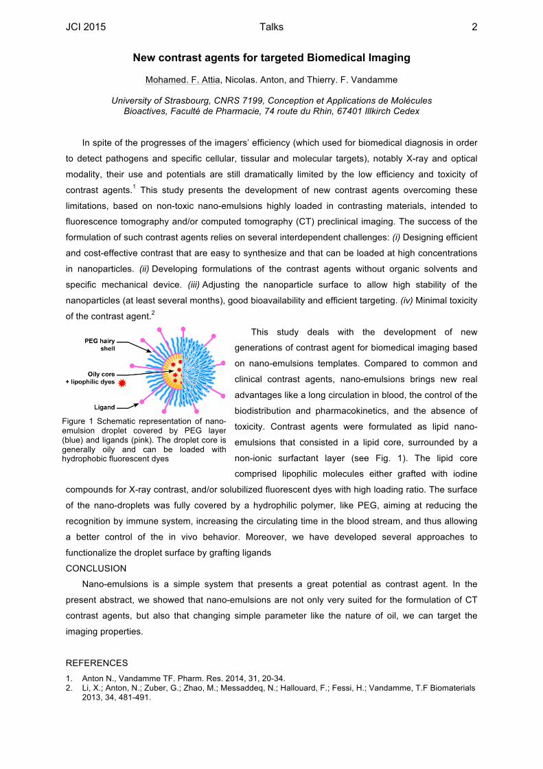
non-ionic surfactant layer (see Fig. 1). The lipid core
comprised lipophilic molecules either grafted with iodine
compounds for X-ray contrast, and/or solubilized fluorescent dyes with high loading ratio. The surface
of the nano-droplets was fully covered by a hydrophilic polymer, like PEG, aiming at reducing the
recognition by immune system, increasing the circulating time in the blood stream, and thus allowing
a better control of the in vivo behavior. Moreover, we have developed several approaches to
functionalize the droplet surface by grafting ligands
CONCLUSION
Nano-emulsions is a simple system that presents a great potential as contrast agent. In the
present abstract, we showed that nano-emulsions are not only very suited for the formulation of CT
contrast agents, but also that changing simple parameter like the nature of oil, we can target the
imaging properties.
REFERENCES
1. Anton N., Vandamme TF. Pharm. Res. 2014, 31, 20-34. 2. Li, X.; Anton, N.; Zuber, G.; Zhao, M.; Messaddeq, N.; Hallouard, F.; Fessi, H.; Vandamme, T.F Biomaterials
2013, 34, 481-491.
Figure 1 Schematic representation of nano-emulsion droplet covered by PEG layer (blue) and ligands (pink). The droplet core is generally oily and can be loaded with hydrophobic fluorescent dyes
JCI 2015 Talks 2
Implication of alpha5 beta1 integrin in resistance to anti-EGFR therapies in glioblastoma
A.F Blandin, G. Renner, F. Noulet, S. Martin, L. Choulier, N. Etienne-Seloum, I. Lelong Rebel, M. Dontenwill, M. Lehmann
Equipe Signalisation Cellulaire, UMR7213 LBP, Faculté de Pharmacie, Illkirch
Introduction:
Glioblastoma multiforme (GBM) is the most common primary brain tumor. Amplification and mutation
of the epidermal growth factor receptor (EGFR) is detected in about 50% of patients with GBM.
Clinical trials using anti-EGFR therapies for the treatment of GBM reveal limited efficacy.
Overexpression of the fibronectin receptor, α5β1 integrin, is associated with a poor prognosis for
patients and the integrin triggers resistance to chemotherapy. Integrins can cross-talk with tyrosine
kinase growth factor receptors to promote cell growth survival and migration. The aim of my thesis is
to characterize the functional interaction between EGFR and α5β1 integrin in GBM cells and
determine its potential involvement in resistance to anti-EGFR targeted therapy.
Results:
We genetically modified a glioblastoma cell line, to overexpress (U87-a5+) or repress (U87-a5-) a5
integrin expression. We first examined the impact of EGFR/a5 crosstalk on cell migration with anti-
EGFR drugs (Cetuximab® and Gefitinib®) used in clinical trials. Using Boyden chamber assay with a
fibronectin coating, we showed that U87-a5+ cells are resistants to Cetuximab® and Gefitinib®
activity. By contrast, the loss of α5 integrin sensitizes U87 MG cells towards anti-EGFR drugs.
Inhibition of a5 integrin with specifics inhibitors (RGD mimetic antagonist and antibody) restores the
sensitivity of U87-a5+ cells toward Cetuximab® demonstrating the importance of a5 integrin in
resistance to anti-EGFR drugs in glioblastoma. Next, we examined the impact of a5 integrin
expression on collective cell migration of spheroids. We examined the propensity of cells to migrate
out of spheroids onto fibronectin coating. The addition of Gefitinib® during cell migration reduces by
half the migration of a5 deficient cells compared to control cells. By contrast, U87-a5+ cells are totally
resistant to anti-migratory activity of Gefitinib®. We then tested the implication of a5 integrin in EGFR
trafficking in U87-MG cells resistant to Gefitinib® action during cell migration. Gefitinib® treatment
induces a translocation of a5b1 integrin from focal adhesions to intracellular vesicles which are not
identified for the moment. Moreover, the addition of Gefitinib® modifies the EGFR localization in U87-
a5+ spheroids but not in U87-a5- spheroids.
Conclusion:
Using two models of migration, we showed that α5 integrin drives U87-MG cells resistance to anti-
EGFR drugs. A drug association targeted EGFR and α5β1 might be a new therapeutic option to
overcome resistance in brain tumors which overexpress a5b1 integrin. Moreover, Gefitinib® treatment
increases the internalization of a5b1 integrin and the EGFR trafficking seems to be implicated in
resistance to anti-EGFR drugs in glioblastoma cells.
JCI 2015 Talks 3
Optical dissection of gating in P2X receptors
Chloé Habermacher1, Adeline Martz1, Nicolas Calimet2, Laurie Peverini1, Damien Lemoine1, Alexandre Specht1, Marco Cecchini2 and Thomas Grutter1
1 CNRS, UMR7199, Laboratoire de Conception et Application de Molécules Bioactives, Equipe de
chimie et neurobiologie moléculaire, Université de Strasbourg, F-67400 Illkirch, France 2 CNRS, UMR7006, Laboratoire d’Ingénierie des fonctions moléculaires, Institut de Science et
d’Ingénierie Supramoléculaires, F-67083 Strasbourg, France
P2X receptors are ligand-gated ion channels activated by extracellular ATP. They form trimeric pores
selective to cations and are involved in different physiological and pathological functions, including
neuromodulation, neuropathic pain and vascular remodeling (1). Crystal structures were solved
recently (2) but they give little insight into the dynamic motions involved in channel gating.
Complementary functional approaches have thus to be developed to address this issue. Here we
present a new method, which uses photoisomerizable azobenzene-containing crosslinkers as
“molecular rulers” to investigate channel gating. We have engineered P2X receptors to obtain optical
control of the channel: they can be opened and closed by irradiation in the absence of their native
ligand. The cis-trans isomerization of the covalently tethered bis-maleimide azobenzene-containing
derivatives between substituted cysteine residues induces movements of the transmembrane helices
that are similar to those induced by ATP. With these valuable experimentally derived interatomic
distances, we confirm the X-ray predicted expansion of the extracellular part of the channel during
opening and further identify a new mechanism of channel gating. Our novel approach can thus be
extended to any membrane-embedded proteins for investigating physiological dynamic motions.
1. Lemoine, D., et al., Ligand-gated ion channels: new insights into neurological disorders and ligand recognition. Chem Rev., 2012. 112 (12) : p. 6285-318. 2. Hattori, M., and Gouaux, E., Molecular mechanism of ATP binding and ion channel activation in P2X receptors. Nature, 2012. 485 : p. 207-12.
JCI 2015 Talks 4

Palladium-catalyzed synthesis of bridged Phe-Gly dipeptide to access novel ligands of Translocator Protein 18kDa (TSPO).
François Hallé,1 Imane lejri,2 Irene Marginedas,3 Christelle Doebelin,1 Séverine Schneider,1 Christian Klein,4 Michel Maitre,4 Martine Schmitt,1 Guy Mensah,4 Mariano Ostuni,3 Anne Eckert,2 Frédéric Bihel1*
1 CNRS, University of Strasbourg, UMR7200, Faculté de pharmacie, 74, route du Rhin, 67401 Illkirch 2 University of Basel, Dept of Biomedicine, Psychiatric University Clinics, Wilhem, CH-4012 Basel 3 UMRS665 - Institut National de la transfusion sanguine, 6, rue Alexandre Cabanel, 75015 Paris
4 INSERM, University of Strasbourg, U1119, Faculté de medicine, 11 rue Humann, 67000 Strasbourg.
We identified compound NCS1008 (1) as a good ligand of the Translocator Protein 18kDa (TSPO). As
many ligands of this protein, the druggability of NCS1008 is pretty poor as this compound is planar and
contains a high number of aromatic rings leading to low water solubility.
With the goal to improve druggability, we designed a bridged Phe-Gly dipeptide (2) which presents
the same pharmacophoric pattern as NCS 1008. This non-natural rigidified dipeptide constitutes a new
unplanar scaffold for TSPO. The bridged dipeptide is obtained through an intramolecular Buchwald-
Hartwig cross-coupling reaction starting from a 2-bromophenylalanine-glycine derivative (3), and we
developed conditions that induce chemoselectivity leading to either indoline (4) or 3,4-dihydroquinolinone
(5) derivatives. Performed under mild conditions, no racemization was observed during cyclization. A
preliminary mechanistic study was performed to identify the parameters leading to the cyclization
chemoselectivity.
Following an unconventional strategy, we started from a planar heterocyclic compound to design a
novel unplanar bridged dipeptide, affording a new class of TSPO ligands.
JCI 2015 Talks 5

Interaction of Gag (NCp7) in human immunodeficiency virus HIV-1
with ribosomic protein RpL7: complex characterization and role(s) in
replicative cycle Karnib Hassan1, Boutant Emmanuel1, Hala El Mekdad1, Biedma Marina2, Réal
Eléonore1, Sharma Kamal1, Mély Yves1, Moog Christiane2 and de Rocquigny
Hugues1
1Laboratoire de Biophotonique et Pharmacologie – UMR7213, Faculté de Pharmacie,
Illkirch.
2 Institut de Virologie, Inserm U 1109, Strasbourg
Gag is a polyprotein composed by various domains such as matrix (MA), capsid (CA),
nucleocapsid (NCp7) and p6 as well as peptides SP1 and SP2 surrounding NC. Gag
interact with gRNA (1) to initiate it dimerization, a mandatory step for the production of
infectious particles and also interacts with large scale of cellular proteins (ALIX,
Staufen…). Among them, our team was interested by the human ribosomal protein called
RpL7, a multifunctional protein playing important roles in ribosome biogenesis and other
extra-ribosomal activities like regulation of translation(2). We demonstrated that Gag
interacts with RpL7 by coIP and yeast 2-Hybrid throught the NCp7 domain of Gag with
the N- and C- terminal domains of RpL7 in an RNA independent manner. We also
confirmed the presence pf RpL7 in viral particles. To characterize the role of this
complex an in vitro study was initiated, using a model measuring nucleic acid chaperone
activity (3); we demonstrate that RpL7 actively chaperones cTAR-dTAR duplex while
Gag was less efficient in similar conditions. More importantly, we show that Gag-RpL7
accelerate this hybridization. In conclusion, we discovered and characterized a new
cellular partner of Gag, the RpL7. We propose a model whereby Gag could recruit a
cellular protein to offset the low level of Gag chaperone activity and help in gRNA
dimerization, key step of encapsidation of viral genome.
Bibliographie
(1)de Rocquigny. (2015).. J mol Bio, 1481-83. (2) Robledo, S. (2008). RNA, 1919-21. (3)Godet J.
(2010). RNA biol, 687-96
JCI 2015 Talks 6

Identification and characterization of a novel GPR103 antagonist
Glenn Marie Le Coz1, V Utard1, I Bertin1, M Schmitt2, JJ Bourguignon2, F Bihel2, F Simonin1
1UMR7242, Biotechnologie et Signalisation Cellulaire, Illkirch. 2UMR7200, Laboratoire d’Innovation Thérapeutique, Illkirch
Chronic pain is a common health issue that remains difficult to treat. Opiates represent the
standard treatment used in clinic but their chronic use leads to the development of several adverse
side effects including hyperalgesia (enhancement of the pain perception) and tolerance (decrease of
the analgesic effects over time). It has been proposed that stimulation of opioid receptors triggers
activation of anti-opioid systems that in turns produce hyperalgesia thus diminishing the net analgesic
effect of the opioid agonist (tolerance). This process has been evidenced in vivo both in rats and in
man where acute and prolonged opioid treatments induce a long lasting hyperalgesia. Anti-opioid
receptor antagonists could therefore represent a promising strategy for limiting the development of
pain hypersensitivity and analgesic tolerance associated with chronic opiates treatments. Several
systems have been shown to display anti-opioid properties including RF-amide neuropeptides family
and their receptors. These peptides have an Arg-Phe-NH2 motif at their C-terminal and act through 5
specific GPCRs (G protein coupled receptors). However, pharmacological tools, particularly
antagonists, are missing for several of these receptors including the receptor of 26RFa GPR103.
In collaboration with a team of chemists, we therefore decided to identify a GPR103 small ligand
capable of antagonizing the effect of 26 RFa both in vitro and in vivo. In a first step, we screened a
chemical library of RF-amide derivatives (± 2000 molecules) on recombinant human GPR103 receptor
stably expressed in CHO cells. From this screening procedure we found several molecules that
display significant affinity for GPR103. We further characterized the in vitro pharmacological profile of
these hits and identify RF10 that display a good affinity (< 100 nM) for GPR103 as well as a high
selectivity and antagonist activity at this receptor. In a second part, we tested the effect of RF10 in vivo
in a mouse model. We measured the pain sensitivity to heat after the injection of either 26RFa (2.5
nmol, i.c.v.) alone or coadministered with RF10 (1 nmol, i.c.v.). As expected, we observed that 26RFa
induced significant hyperalgesia while RF10 completely prevented hyperalgesia induced by this
peptide. Altogether, our results indicate that RF10 represents a good pharmacological tool to study the
involvement of GPR103 in the modulation of nociception and opiates analgesia.
JCI 2015 Talks 7

New antimalarial flavones: from screening to in vivo proof of concept
Flore Nardella1,2, Valérie Collot3, Silvia Stiebing3, Marcel Kaiser4, Martine Schmitt1, Ermanno Candolfi2, Catherine Vonthron-Sénécheau1
1- Equipe Chimie Biologie Intégrative - Laboratoire d’Innovation Thérapeutique, UMR CNRS-Unistra 7200, Faculté de Pharmacie, 67401 Illkirch Cedex, France
2- Institut de Parasitologie et de Pathologie Tropicale de Strasbourg, Faculté de Médecine, 67000 Strasbourg, France
3- Centre d’Etudes et de Recherches sur le Médicament en Normandie, Université de Caen Basse-Normandie, 14032 Caen Cedex, France
4- Swiss Tropical and Public Health Institute, 4051 Basel, Switzerland
Malaria is the deadliest parasitic disease with almost 600.000 deaths every year1. The parasite
(Plasmodium sp.) is transmitted by a female Anopheles mosquito during a blood meal. It undertakes
then a complex life cycle in humans: first in the liver, then in the red blood cells. This erythrocytic cycle
is responsible for the symptoms like fever, sweat and shivering. Death occurs in complicated cases
due to severe anemia, kidney failure, or coma.
Only a few drugs cure malaria, and on top of that the parasite is resistant to most of them. The first
line treatment—artemisinin—is not an exception: in 2008, Noedl and colleagues reported the
emergence of partial resistance to artemisinin in South-East Asia2. By the past, progress in decreasing
the mortality has already been reversed because of resistance spreading from Asia to Africa.
Artemisinin resistance seems to follow the same march: resistance has already spread to Myanmar
near the Indian frontier3. To conduct its malaria eradication plan by 2050, WHO needs new fast acting
drugs with original mechanisms of action.
After the isolation of an active biflavonoid from Campnosperma panamense (Anacardiaceae, IC50 =
480 nM in vitro on P. falciparum K1 multi-resistant strain)4, we developed novel simplified synthetic
analogs (MR series) with improved pharmacological and pharmacokinetic profiles. Two of them (MR70
and MR87) exhibit a partial in vivo antimalarial activity. They reduce parasitaemia by 35% to 70%
respectively on day 4 on a murine model (P. berghei ANKA, dosing regimen of 100 mg/kg for 4 days).
But these compounds showed no significant improvement in terms of survival.
MR70 is parasiticidal on early blood stages of P. falciparum in less than 30 minutes. Interestingly,
these stages are specifically the ones that are resistant to artemisinin5. Further investigation is needed
to optimize in vivo activity and to understand the underlying mechanism(s) of action of these
compounds.
1 World malaria report, 2013, WHO 2 Noedl H, Se Y, Schaecher K, Smith BL, Socheat D, Fukuda MM: Evidence of artemisinin-resistant malaria in western Cambodia. N Engl J Med (2008). 3 Tun KM, et al.: Spread of artemisinin-resistant Plasmodium falciparum in Myanmar: a cross-sectional survey of the K13 molecular marker. Lancet Infect Dis (2015). 4 Weniger et al.: A bioactive biflavonoid from Campnosperma panamense. Fitoterapia (2004). 5 Witkowski B, et al.: Novel phenotypic assays for the detection of artemisinin-resistant Plasmodium falciparum malaria in Cambodia: in-vitro and ex-vivo drug-response studies. Lancet Infect Dis (2013).
JCI 2015 Talks 8
Role of Toll like receptor 7 and plasmacytoid dendritic cells in mouse models of experimental arthritis
Nehmar Ramzi1, Alsaleh Ghada1, Bahram Siamak1, Georgel Philippe1
1 INSERM UMR_S 1109, Fédération de Médecine Translationnelle (FMTS), Université de Strasbourg, Centre de Recherche en Immunologie et Hématologie, 1, Place de l’Hôpital 67085 Strasbourg Cedex
France
Introduction: Among immune cells, plasmacytoid dendritic cells (pDCs) are the main type I IFN producers following activation (i.e. upon viral challenge). These cells, which are also characterized by their expression of the endosomal TLRs (TLR3, 7/8 and 9) are central in the initiation of the inflammatory response and as such, are involved in the etiology of several chronic inflammatory diseases. Surprisingly however, anti-inflammatory effects of type I IFN were also considered [1], especially IFN β, which is known for its beneficial effect in murine models of rheumatoid arthritis (RA) [2]. In addition, circulating pDCs depletion in RA patients was observed [3], suggesting a protective role for these cells. In this work, we aimed at a better characterization of the role of these cells in murine models of RA to clarify these contradictory observations.
Methods: Induction of arthritis was made by arthritogenic (K/BxN ) serum transfer [4] and upon repeated injection of heterologous collagen (CIA) [5]. Symptoms were evaluated by visual scoring and measurement of the joints with a caliper. IKAROS L/L mice are hypomorphic mutants in a C57Bl/6 background which exhibit reduced numbers in peripheral pDCs [6]. DBA/1 mice were used in the CIA model. Cellular infiltrates were analyzed by FACS and cytokines expression with ELISA and RTqPCR. Bone histological evaluation used TRAP staining. Results: IKAROS L/L (pDC-deficient) mice showed exacerbation of inflammatory and arthritic symptoms after arthritogenic serum transfer (as seen upon measurements and scoring, quantification of pro-inflammatory cytokines, histological analysis of the erosion). In wild-type animals, topical application of a TLR7 agonist which induces Type I IFN production reduces articular inflammation in K/BxN and CIA arthritis models and concomitant pDCs recruitment at inflammatory sites. Our results suggest a beneficial role of pDCs in arthritis
Conclusion: Alternative pDC depletion models (genetic, Ab-mediated) as well as Tlr7-/- mice will be investigated to confirm these results. Our data, which suggest possible interactions between pDCs and other cells present in the inflamed, arthritic joint, could lead to new therapeutics options.
1. Benveniste EN, Qin H: Type I Interferons as Anti-Inflammatory Mediators. Sci Signal 2007, 2007:pe70. 2. Van Holten J, Reedquist K, Sattonet-Roche P, Smeets TJM, Plater-Zyberk C, Vervoordeldonk MJ, Tak PP: Treatment with recombinant interferon-beta reduces inflammation and slows cartilage destruction in the collagen-induced arthritis model of rheumatoid arthritis. Arthritis Res Ther 2004, 6:R239–249. 3. Jongbloed SL, Lebre MC, Fraser AR, Gracie JA, Sturrock RD, Tak PP, McInnes IB: Enumeration and phenotypical analysis of distinct dendritic cell subsets in psoriatic arthritis and rheumatoid arthritis. Arthritis Res Ther 2005, 8:R15. 4. Monach PA, Mathis D, Benoist C: The K/BxN arthritis model. Curr Protoc Immunol Ed John E Coligan Al 2008, Chapter 15:Unit 15.22. 5. Brand DD, Latham KA, Rosloniec EF: Collagen-induced arthritis. Nat Protoc 2007, 2:1269–1275. 6. Allman D, Dalod M, Asselin-Paturel C, Delale T, Robbins SH, Trinchieri G, Biron CA, Kastner P, Chan S: Ikaros is required for plasmacytoid dendritic cell differentiation. Blood 2006, 108:4025–4034.
JCI 2015 Talks 9
Genetic analysis of epidermal cell mechanical properties during C. elegans embryonic elongation
Gabriella Pásti, Christelle Gally, Julien Pontabry, Michel Labouesse
Equippe Labouesse, IGBMC - UMR7104 - INSERM U964
The contribution of mechanical forces to development is gaining wider acceptance. But the detailed molecular
mechanism by which cells sense and respond to forces remains elusive. To investigate those issues, we focus on C.
elegans embryonic elongation, a process controlled by cell shape changes transforming a ball of cells into a tube-
shaped animal. C. elegans embryonic elongation consists of two phases. First, it is driven by the epidermal
actomyosin. Second, muscles promote elongation through a recently reported mechanotransduction pathway
involving CeHDs, which mechanically link muscles to the epidermis.
PAK-1 (p21-activated kinase) is a key regulator of both phases: it is a good starting point to further dissect the
molecular landscape of elongation. To reach this aim, we have two approaches. First, we carried out a systematic
search for genetic interactions by an RNAi screen, targeting 356 genes in a pak-1(Ø) mutant. Second, we looked for
potential PAK-1 interactors by a yeast two-hybrid screen. We tested the in vivo relevance of the most interesting
candidate, the α-spectrin SPC-1. We established a genetic interaction between the two genes, showing that double
mutants display a novel elongation defect, whereby they retract to a lima bean-like shape after reaching the 1.5-fold
stage. Retraction is not observed if muscles are defective, suggesting it is induced by muscle twitching. Spinning disk
time-lapse analysis showed that the elastic properties normally displayed by epidermal cells in response to muscle
twitching input are affected in double mutants. To further confirm these results, we are using laser ablations in the
dorsoventral epidermis to measure epidermal viscoelastic properties.
Consistent with the genetic interaction, in vivo expression studies revealed a co-localization between the two proteins
at the apical level of the epidermal cells. Furthermore, we found that the SH3 domain of SPC-1 interacting with PAK-1
in vitro is essential for its function in vivo, and that loss of SPC-1 disturbed PAK-1::GFP localization.
To define why embryos retract, we examined important elongation players and found that in the double deficient
embryos the actin cytoskeleton and the CeHDs are getting gradually affected as the retraction appears and evolves.
Finally, we performed a supplementary genetic screen that allowed us to reproduce the retraction in different genetic
contexts. Thereby we have identified novel molecular players acting together with spc-1 and pak-1, triggering a
retraction.
Altogether, we found that the SPC-1—PAK-1 interaction is important for C. elegans embryonic elongation. Moreover,
we suggest it modulates the elastic properties of dorso-ventral epidermal cells submitted to external mechanical
stress, helping to stabilize them between consecutive inputs.
JCI 2015 Talks 10

Physiopathology of Tubular aggregate myopathy
Georges Arielle PECHE, Johann BÖHM, Catherine KOCH, Jocelyn LAPORTE
Pathophysiology of neuromuscular diseases, IGBMC- UMR7104 - INSERM U964
Tubular aggregate myopathies (TAM) are progressive muscle disorders with an autosomal
dominant inheritance and characterized by abnormal accumulations of membrane tubules in muscle
fibers. Our team identified STIM1 as the first gene implicated in TAM. STIM1 regulates calcium
homeostasis through a mechanism known as store-operated calcium entry (SOCE). Upon stimulation,
calcium is released from the sarcoplasmic reticulum to the cytoplasm, where it triggers muscle
contraction and acts as a second messenger controlling growth and differentiation. In case of calcium
store depletion, STIM1 unfolds, oligomerizes and thereby activates the calcium entry channel ORAI1
to trigger extracellular calcium entry. We demonstrated that the identified STIM1 mutations strongly
impact on the calcium level in TAM myoblasts. However, the nature, formation and pathogenicity of
the tubular aggregates are not yet defined and the link between STIM1 mutations and muscle
dysfunction is not understood.
My PhD project aims to investigate the physiopathological mechanisms underlying TAM in both
cellular and animal models. In order to analyze the formation and physiological impact of the tubular
aggregates, I used correlative light-electron microscopy (CLEM) to monitor the aggregates in muscle
cells overexpressing wild type or mutant STIM1 constructs. I observed that the STIM1 clusters
correspond to stacks of membrane layers, and I will next compare and quantify the clusters size and
membrane composition at different time points post transfection in order to assess the timeframe and
phases of tubular aggregate formation.
There is currently no mammalian model for tubular aggregate myopathy. I therefore plan to use
the AAV system to analyze the impact of different STIM1 mutations on muscle structure and function
in STIM1+/skm- (= heterozygous knockout in skeletal muscle) mice. I generated STIM1 AAV constructs
harboring different mutations that will be injected in the tibialis anterior and I will perform histological,
ultrastructural and immunofluorescence analyses on the muscle at different time points post-
transduction to decipher the sequence of events leading to TAM. In order to correlate molecular and
cellular alterations with disease development and muscle function, the transduced mice will
furthermore undergo general and specific force measurements in response to nerve and muscle
stimulation at different time points post-transduction.
In conclusion, my project is expected to provide new insights in the physiological mechanisms leading
to calcium-related muscle dysfunction and TAM, and might suggest targets for therapeutic
approaches.
JCI 2015 Talks 11

Cyclohydrocarbonylation-Based Strategy towards Novel N-Heterocyclic Scaffolds Derived from Aza-diketopiperazines
Pierre-Michel Regenass1, Jean-François Margathe1, André Mann1, Jean Suffert1, Marcel Hibert1,
Nicolas Girard1 and Dominique Bonnet1
1Laboratoire d’Innovation Thérapeutique, UMR7200 CNRS/Université de Strasbourg, Faculté de Pharmacie, 74 route du Rhin, 67412 Illkirch, France
Privileged structures represent an ideal source of new biologically active
molecules. Among these compounds, 2,5-diketopiperazines (DKP) are represented
in a large array of structurally diverse natural products and display interesting
therapeutic properties1,2. As reported for aza-peptides3, the replacement of one Cα-
stereogenic center by a planar nitrogen could have a profound impact in both the
chemical and biological properties of DKP and could offer new potential opportunities
for drug discovery and chemical biology.
DKP Aza-DKP
Herein, we will present an efficient diversity-oriented and stereoselective synthesis
of novel heterobicyclic and tricyclic compounds derived from this scaffold. To access
such structures, we have explored a strategy based on Rh(I)-catalyzed
hydroformylative cyclohydrocarbonylation (CHC)4 of allyl-substituted aza-DKP. To
facilitate the diastereoselective access to novel aza-DKP platforms, a one pot
reaction combining 5-step process was also envisaged starting from allyl carbazate.
[1] Martins, M. B.; Carvalho, I. Tetrahedron 2007, 63 (40), 9923−9932. [2] McCleland, K.; Milne, P. J.; Lucieto, F. R.; Frost, C.; Brauns, S. C.; Van De Venter, M.; Du Plessis, J.; Dyason, K. J. Pharm. Pharmacol. 2004, 56 (9), 1143−1153. [3] Zega, A. Curr. Med. Chem. 2005, 12: 589-597. [4] Varchi, G.; Ojima, I. Curr. Org. Chem. 2006, 10, 1341–1362.
NNH
R1
R3
O
O
R2 NNH
R1
R3
O
O
R2 N
NN
O
O
R2 N
n
nR1 NR2
NONHBoc
COOMe
R1OMe
R = OMeR = COOH, NHBoc
one pot5-step process
2 steps
JCI 2015 Talks 12
Development of design tools for biosystem engineering
Elise Rosati1, Morgan Madec1, François Pêcheux2,3, Yves Gendrault1,4, Christophe Lallement1, Jacques Haiech5
1 Laboratoire des Sciences de l’Ingénieur, de l’Informatique et de l’Imagerie (ICube), UMR 7357,
Equipe SMH, 300 Boulevard Sébastien Brandt, F-67412 Illkirch, 2 UPMC Univ Paris 06, UMR 7606, LIP6, F-75005, Paris, 3 CNRS, UMR 7606, LIP6, F-75005 Paris, 4 ECAM Strasbourg-Europe - 2 rue de
Madrid – F-67300 Schiltigheim, 5 Laboratoire d’Innovation Thérapeutique (LIT), UMR 7200, 74 route du Rhin - F-67400 Illkirch.
Over the past fifteen years, synthetic biology, a new scientific field at the interface between
biotechnologies and engineering sciences has developed rapidly. The goal of synthetic biology
is to create new biological functions by a rational assembly of artificial or natural biological
parts [1].
Along with the improvement of technological material and processes, focus should be put
on the improvement of design tools (in silico simulations) and methodologies. As this rational
approach used to design genetic networks is very similar to the one used in microelectronics,
our team chose to take advantage of the valuable know-how of microelectronics scientists
aquired with 40 years of experience in creating adapted design tools [2]. By using electronic
formalism, we work on modeling biological systems and adapting the generic electronic
workflow to synthetic biology by enriching existing tools to support multi-domain design with
biological models or by developing new tools for synthetic biology.
[1] D. Endy, “Foundations for engineering biology.,” Nature, vol. 438, no. 7067, pp. 449–53, Nov. 2005.
[2] Y. Gendrault, M. Madec, C. Lallement, F. Pecheux, and J. Haiech, “Synthetic biology methodology and model
refinement based on microelectronic modeling tools and languages.,” Biotechnol. J., vol. 6, no. 7, pp. 796–806, Jul. 2011.
JCI 2015 Talks 13

Isomorphic fluorescent nucleoside analogs: applications in protein/nucleic acid interactions
Marianna Sholokh,1 Rajhans Sharma,1 Dongwon Shin,2 Ranjan Das,3 Yitzhak Tor,2 and Yves Mély1 1Laboratoire de Biophotonique et Pharmacologie, UMR 7213 CNRS, Université de Strasbourg, Illkirch,
2Department of Chemistry and Biochemistry, University of California, San Diego,
3Department of
Chemistry, West Bengal State University, India
Monitoring site-selective conformational changes in nucleic acids by fluorescence techniques is
highly challenging due to the lack of appropriate fluorescent nucleic acid analogs. The most explored
nucleoside analog, 2-aminopurine (2Ap),1 has been used in countless assays though it suffers from
very low quantum yield, especially when included in double strands. Moreover, its residual emission
frequently does not represent biologically relevant information.
To circumvent these limitations, a nearly perfect fluorescent substituent of the guanine base
thienodeoxyguanosine (dthG) was designed.2,3 Using steady-state and time-resolved fluorescence
spectroscopy, we compared the ability of 2Ap and dthG to substitute and provide faithful structural and
dynamical information on a key G residue in the () DNA copy of the HIV-1 primer binding site
sequence. dthG fluorescent nucleoside, in contrast to 2Ap, was found to fully preserve the stability of
the labeled hairpin and the duplex, and also showed advantageous photophysical properties. In further
contrast to 2Ap, the fluorescently detected dthG species represent the predominantly populated G
conformers that allow studying their relevant dynamics. Being able to perfectly substitute G residues,
dthG opens a large area of possible applications and will be particularly used in studying the annealing
mechanism of ()/(+) PBS DNA, an HIV-1 reverse transcription key step.
1. Ward, D.C.; Reich, E.; Stryer, L. J. Biol. Chem. 1969, 244, 1228. 2. Park, S.; Otomo, H.; Zheng, L.; Sugiyama, H. Chem. Commun. 2014, 50, 1573. 3. Sholokh, M.; Sharma, R.; Shin, D.; Das, R.; Zaporozhets, O. A.; Tor, Y.; Mély, Y. J. Am. Chem. Soc.
2015, 137, 3185.
JCI 2015 Talks 14
IChem-PIC – 3D mapping and classification of protein-protein interfaces.
Franck Da Silva and Didier Rognan
Laboratoire d'innovation thérapeutique UMR 7200 faculté de pharmacie, Illkirch Graffenstaden
Modulating protein-protein interactions by low molecular-weight ligands is a novel and promising
approach in drug discovery, opening novel therapeutic avenues and extending the scope of applicability
of currently known macromolecular targets. Detection and characterization of protein-protein interfaces
(PPi) is a key but at the moment there are few tools to study protein-protein interfaces. Nowadays we
can only detect a PPi but not characterize their properties for drug design.
Thus our work is to develop a software which can analyze and characterize PPis. The development
of this software begins by the detection of biological interfaces. In protein structures we can observe two
types of interface, biological and crystallographic. It is essential to separate them to retain the
biologically relevant ones. This separation is determined by a machine-learning model generated from
known x-ray structures.
We characterize interactions between proteins with an in-house tool [1]. It describes all non-covalent
interactions between two chains of a protein by type (hydrophobic, hydrogen bond, ionic bond and
aromatic interaction). We used a Random Forest algorithm to separate 300 known protein-protein
Interfaces in two classes. We optimized the model parameters by cross-validation and test it on 4
external data sets. The first one is composed by 100 PPis manually curated in the lab. The 3 others are
data sets from the literature currently used for the study of PPi. We remove all redundancy on the 4
sets.
Our Model shows that some descriptors (number of protein-protein interactions, proportion of fully
buried hydrophobic contacts) are key to distinguish biological from crystallographic interfaces. The
model is robust and presents an accuracy between 75 and 90% depending on the external data set
used.
We herewith present a novel computational approach (available online) to distinguish between
biologically-relevant and crystallographic protein-protein interfaces. IChem-PIC is the only approach
able to predict with the same accuracy the two categories of PPi.
Our project aims at charting, for the first time, the ensemble of all druggable protein-protein interfaces
of known 3D structures as well as their allosteric binding sites. With all these data we wish to screen
commercial compound libraries to find novel compounds that can interact, stabilize or inhibit protein-
protein interactions.
1. DESAPHY J., RAIMBAUD E., DUCROT T., ROGNAN D. J.Chem. Inf. Model. 2013, 53, 623-637.
JCI 2015 Talks 15

The effect of urapidil, an alpha-1 adrenoceptor antagonist and a 5-HT1A agonist, on vascular tone of isolated porcine coronary and pulmonary arteries, rat
aorta, and human pulmonary artery
C. Bopp1, 2, C. Auger1, P. Diemunsch2, V.B. Schini-Kerth1
1 Laboratoire de Biophotonique et Pharmacologie, UMR CNRS 7213, Faculté de Pharmacie,
Université de Strasbourg, Strasbourg, France,2 CHU de Hautepierre, Strasbourg
While being effective and well tolerated, urapidil (Eupressyl®, Takeda Pharmaceutical Company
Limited, Osaka, Japan) an antihypertensive drug has restrained indications due to its incompletely
known pharmacodynamics. Recent evidence suggests that urapidil may also be of interest in the
treatment of hypertension associated with preeclamptic toxaemia and in hypertensive patients with
respiratory disease. Urapidil acts as a selective α1-adrenoreceptor antagonist and as an agonist of 5-
HT1A receptors. On the other hand, serotonin has been shown to induce endothelium-dependent
relaxations of several types of blood vessels involving 5-hydroxytryptamine (5-HT) receptors.
Therefore, the aim of the present study was to determine the ability of urapidil to cause relaxations of
different types of blood vessels including the pig coronary and pulmonary artery, the rat aorta and the
human pulmonary artery, using vascular reactivity studies.
36 pulmonary and coronary arteries from pigs, 22 aortae from rats and 9 human pulmonary arteries
were cut into rings, which were suspended in organ chambers containing oxygenated Krebs
bicarbonate solution. The endothelium was removed mechanically in some rings. The pig rings were
contracted with U46619, a thromboxane mimetic, the aortic rings with endothelin-1, and the human
pulmonary artery rings with U46619 before construction of a concentration-relaxation curve either to
urapidil or serotonin. Serotonin but not urapidil and the 5-HT1A receptor agonist 8-OH-DPAT, induced a concentration-
dependent relaxation in the porcine coronary and pulmonary artery rings with an intact endothelium (P
<0.05). Urapidil (10-5 M) markedly inhibited phenylephrine-induced contraction in rat aortic rings with
and without endothelium with a more pronounced effect in rings without endothelium. Both serotonin
and 8-OH-DPAT did not induce relaxation of rat aortic rings with endothelium. Serotonin and
phenylephrine but not urapidil caused concentration-dependent contractions in human pulmonary
artery rings.
In conclusion, urapidil in contrast to serotonin failed to cause relaxation of porcine coronary and
pulmonary artery rings. The present findings, while confirming that urapidil is a potent inhibitor of α1-
adrenoceptor-induced contraction, did not support a role of 5-HT1A receptor activation in the control of
the vascular tone in the three types of blood vessels studied, in response to urapidil. Moreover, the
inhibitory effect of urapidil is more pronounced in rings without endothelium suggesting that urapidil
may target preferentially arteries with an endothelial dysfunction. Such an effect might be of particular
interest in managing hypertension associated with preeclamptic toxaemia and pulmonary disease.
[1] J. Gross G, Shuttler K, Xin X, hanft G. J cardiovascular Pharmacol 1990: 15 suppl 7
[2] Cohen R.A., Vanhoutte P.M. Circulation. 1995; 92:3337–3349
JCI 2015 Talks 16