micromechanical studies of mitotic chromosomes · micromechanical studies of mitotic chromosomes...
TRANSCRIPT

Micromechanical studies of mitotic chromosomes
John F. Marko*
Department of Physics and Astronomy, and Department of Biochemistry, Molecular Biology and Cell Biology,Northwestern University, Evanston, IL, 60208, USA; E-mail: [email protected]* Correspondence
Key words: chromatin, chromosome, chromosome mechanics, mitosis, single-molecule experiments
Abstract
Mitotic chromosomes respond elastically to forces in the nanonewton range, a property important to transduction
of stresses used as mechanical regulatory signals during cell division. In addition to being important biologically,
chromosome elasticity can be used as a tool for investigating the folding of chromatin. This paper reviews
experiments studying stretching and bending stiffness of mitotic chromosomes, plus experiments where changes
in chromosome elasticity resulting from chemical and enzyme treatments were used to analyse connectivity of
chromatin inside chromosomes. Experiments with nucleases indicate that non-DNA elements constraining
mitotic chromatin must be isolated from one another, leading to the conclusion that mitotic chromosomes have a
chromatin Fnetwork_ or Fgel_ organization, with stretches of chromatin strung between Fcrosslinking_ points. The
as-yet unresolved questions of the identities of the putative chromatin crosslinkers and their organization inside
mitotic chromosomes are discussed.
Abbreviations
BAF barrier to autointegration factor
CAP chromosome-associated protein
Muk Mukaku, Japanese for anucleate
MukBEF E. coli condensin complex
NEB nuclear envelope breakdown
SMC structural maintenance of chromosomes complex
topo II topoisomerase II
Introduction
How DNA in eukaryote cells is physically organized
into chromosomes is largely a mystery. During
interphase, different chromosomes occupy different
nuclear regions or Fterritories_ (Cremer et al. 1993,
1996, 2000, Cremer & Cremer 2001, Bolzer et al.2005). Interphase chromosomes are highly dynamic,
with active chromatin in an open conformation and
undergoing continual exchange of protein compo-
nents (Misteli et al. 2000, Phair & Misteli 2000).
Chromosomal loci are known to be mobile (Marshall
et al. 1997, Bystricky et al. 2004, Levi et al. 2005),
and recently it has been shown that gene positioning
affects gene expression (Brickner & Walter 2004,
Ahmed & Brickner 2007, Akhtar & Gasser 2007,
Lanctot et al. 2007).
During mitosis, gene expression stops, and chromo-
somes are folded into their segregated mitotic forms
(Figure 1). Although condensin structural mainte-
nance of chromosome (SMC) protein complexes are
essential to mitotic condensation (Strunnikov et al.1993, Hirano & Mitchison 1994, Strunnikov et al.1995, Hirano 2006), exactly how these proteins act
on chromatin is unknown. Given the many different
models for mitotic chromosome structure (Saitoh
& Laemmli 1994, Houchmandzadeh & Dimitrov
1999, Kimura et al. 1999, Machado & Andrew
Chromosome Research (2008) 16:469–497 # Springer 2008DOI: 10.1007/s10577-008-1233-7

2000, Maeshima & Laemmli 2003, Dietzel et al.2004, Kireeva et al. 2004, Hirano 2006, Sheval and
Polyakov 2006, Fukui & Uchiyama 2007), it is clear
that we do not yet understand large-scale chromo-
some folding (Belmont 2006).
Our lack of understanding of chromosome organi-
zation is rooted in physical properties of chromatin.
The dynamic nature of chromosomes resulting from
gene positioning, cell-cycle reorganization, and con-
tinual thermal (Brownian) motions indicates that
chromatin structure must be described statistically,
rather than in terms of precise folds. Furthermore,
chromatin and chromosomes are soft materials, with
rigidities far less than those of the molecules from
which they are composed, leading to the pitfall that
large-scale structure of chromosomes can be altered
by preparations which leave protein and DNA
molecular structures intact.
This paper reviews studies of structural and
mechanical properties of mitotic chromosomes,
focusing on micropipette-based micromanipulation
experiments which permit measurement of changes
in mechanical properties to assay structural changes
driven biochemically. Most of these types of experi-
ments have studied prometaphase or metaphase
chromosomes of animals; many experiments that will
be discussed below involve amphibian chromosomes
from the newt Notophthalmus viridescens (Hutchison
& Pardue 1975) and the frog Xenopus laevis. These
are model organisms for study of mitosis simply
because their chromosomes are large. In the case of
Xenopus, there is the additional feature that egg
extracts may be used to assemble chromosomes,
mitotic apparatus, and nuclei in vitro (Almouzni &
Wolffe 1993, Maresca et al. 2005).
The next section of this paper (Mitotic Chromosome
Architecture) summarizes molecular-biological under-
standing of mitotic chromosome organization. The
third section (Micromanipulation and Mechanics of
Mitotic Chromosomes) discusses experimental meth-
ods for measuring chromosome elasticity, and reviews
experimental results for stretching and bending elastic-
ity, emphasizing the surprisingly low bending stiffness
of chromatids reconstituted using Xenopus egg extracts
relative to the bending stiffness of chromosomes
extracted from cells. That section also discusses the
ability of mitotic chromosomes to be rapidly unfolded
and reversibly refolded by shifts in ionic concentration.
The fourth section of the paper (Molecular
Connectivity of Mitotic Chromosomes) focuses on
the question of how mitotic chromosomes are held
together, and on experiments with enzymes that
illuminate this question. A Fchromatin network_ or
Fgel_ model is suggested by these results, where
Figure 1. Cell division in newt epithelial cell, phase-contrast imaging. (A) Early prophase; (B) late prophase showing long and thin prophase
chromosomes; (C) spindle-aligned metaphase chromosomes; (D) separation of chromatids at anaphase; (E) telophase chromosomes
beginning to decondense; (F) interphase nuclei in daughter cells. Bar=20 mm. (Images courtesy of M.G. Poirier).
470 J. F. Marko

chromatin is intermittently crosslinked to itself. A
short review of the fascinating phenomenon of
interchromosome linkers is included. The final
section (Implications for Chromosome Structure) dis-
cusses the consequences of biochemical and biophys-
ical data for models of mitotic chromosome folding.
Mitotic chromosome architecture
Proteomic experiments are moving towards provid-
ing a comprehensive catalogue of mitotic chromo-
some proteins (Uchiyama et al. 2004, 2005, Fukui &
Uchiyama 2007). Here only a subset of the proteins in
the mitotic chromosome will be discussed (Figure 2),
from a coarse-grained structural and biophysical point
of view.
Chromatin fibre
Chromosomes are composed of chromatin fibre,
which consists of DNA complexed with histones
into repeated nucleosome units. The structure of the
entire nucleosome (147 bp of DNA plus core
histones) has been determined using X-ray crystal-
lography (Luger et al. 1997). Each õ10 nm-diameter
nucleosome contains 147 bp of DNA wrapped
around eight core histone proteins (two each of
histones H2A, H2B, H3 and H4, for a total octamer
mass of roughly 95 kDa). Given one nucleosome
every õ180 bp of DNA (of mass 110 kDa), in a
chromosome the total mass of core histones is about
the same as that of DNA.
Formation of a nucleosome reduces the total 60 nm
contour length of 180 bp of DNA to roughly 10 nm.
Figure 2. Cartoons of elements of eukaryote mitotic chromosomes, sketched roughly to the same scale. Upper left: DNA is complexed with
histones to form nucleosomes, which then fold into chromatin fibre containing roughly 100 bp/nm; linker histones are not shown. Upper
right: small HMGB1 and BAF-1 proteins bend and crosslink DNA; a larger (10 nm) topo II is shown bound to one DNA, while passing a
second DNA through it. Lower left: much larger (50 nm) condensin I complex composed of two long SMCs plus a bridging kleisin unit and
two additional accessory proteins. Condensin II is not shown; its structure is similar to that of I (see text). Lower right: a cohesin complex
composed of long SMCs plus kleisin and accessory units has a large open structure with a hole large enough to pass 30 nm chromatin fibre.
Micromechanical studies of mitotic chromosomes 471

Thus naked DNA, with 3 bp/nm, can be compacted
into a string of nucleosomes with roughly 20 bp/nm,
a linear compaction of about 6-fold.
Nucleosomes have associated with them linker
histones (H1 or H5, õ20 kDa). Linker histones have
long been thought to be to be involved in compac-
tion of chromatin fibre to a folded 30 nm-thick form
(Thoma et al. 1979), but the details of how this occurs
remain poorly understood. The questions of linker
histone to nucleosome stoichiometry (Woodcock et al.2006), and exactly how linker histone binds to
chromatin are not settled (Brown et al. 2006).
An often-used estimate is that when compacted
into 30 nm form, there are about 6 nucleosomes per
10 nm of chromatin fiber length, or 100 bp/nm, about
30-fold shorter than the original DNA. Recent X-ray
studies of crystallized nucleosome arrays (Dorigo et al.2004) and tetranucleosomes (Schalch et al. 2005)
support this estimate, and suggest that the 30 nm
fiber has a two-start helix organization, with linker
DNA in the interior of the fiber. This is in accord
with neutron scattering experiments on chromatin
containing histone H1 which indicate that linker
histone is concentrated in the fiber center (Graziano
et al. 1994). Daban (2000) has emphasized that the
known very high DNA concentration in chromo-
somes may provide useful constraints on models of
chromatin fibre folding.
Chromatin fibre structure is sensitive to ionic
conditions. When chromatin fibres are extracted into
solution at sub-physiological 10 mM univalent ion
concentration, they are observed in the electron micro-
scope as 10 nm-thick Fbeads-on-a-string_. As one
approaches the more physiological level of 100Y150
mM univalent ions, nucleosomes stack into the 30 nm
fibre. At physiological ionic strength, lateral internu-
cleosomal attractions also tend to lead to aggregation of
isolated fibres (van Holde 1988). This sensitivity
indicates that nucleosomeYnucleosome interactions
have a strong electrostatic component, and also
indicates that chromatin is soft and easily deformed.
This softness and consequent variable structure of
chromatin has made it difficult to arrive at consensus
regarding the folded structure of the 30 nm fibre.
Many small DNA-binding proteins are found in
chromatin, e.g., transcription factors. Two small
proteins that are likely to be important to chromatin
folding are high-mobility-group (HMG) proteins, and
the barrier-to-integration-factor (BAF-1) protein
(Margalit et al. 2007), which are present in mitotic
chromosomes in numbers comparable to those of
histones (Uchiyama et al. 2005). HMG proteins are
divided into three families with different functions
(Hock et al. 2007): HMGA proteins bind AT-rich
DNA, HMGB proteins bend DNA (Thomas & Travers
2001), and HMGN proteins reorganize nucleosomes.
Intriguingly, BAF-1 molecules (10 kDa) organize
into dimers, with two DNA-binding domains capable
of interacting with two different DNA helices.
BAF-1 has been demonstrated to be able to link
DNA helices (Lee & Craigie 1998, Bradley et al.2005). BAF-1 is an abundant chromosomal protein
capable of noncovalently crosslinking chromatin.
Topoisomerase II
Topo II is a large dimeric protein (each polypeptide
chain is õ175 kDa) responsible for passing DNA
through DNA in an ATP-dependent manner, so as to
resolve DNA entanglements such as those between
sister chromatids resulting from DNA replication.
Remarkably, topo II has been demonstrated to be
able to drive entanglements below the number
expected for random strand passages, indicating that
it can use energy liberated during ATP hydrolysis to
selectively remove DNA entanglements (Rybenkov
et al. 1997).
Topo II_s activity is essential to condensation and
segregation of mitotic chromosomes (Hirano &
Mitchison 1993). Metazoan cells contain topo IIaand b isoforms; during mitosis topo IIa is mainly
resident on chromosomes, while topo IIb is mainly
cytoplasmic (Meyer et al. 1997). Observations of
GFP-topo IIa fusion proteins in vivo show it to rapidly
exchange on and off chromosomes (Christensen et al.2002, Tavormina et al. 2002). Estimates for amounts
on mitotic chromosomes vary over the range of one
topo IIa for every 20Y50 kb of DNA (Gasser et al.1986, Fukui & Uchiyama 2007).
Topo II has been suggested not only to efficiently
disentangle DNA but also to play a structural role in
mitotic chromosomes. Analysis of non-histone pro-
teins in mitotic chromosomes found Fscaffold protein
I_ (Lewis & Laemmli 1982), later identified as topo
II (Earnshaw et al. 1985, Gasser et al. 1986).
Electron-microscopic studies have indicated that topo
II can bind a crossover of two DNAs (Zechiedrich &
Osheroff 1990), and topo II has been observed to be
able to recondense protease-decondensed chromo-
somes (Bojanowski et al. 1998). Immunofluores-
472 J. F. Marko

cence experiments have observed topo II localized in
chromatid-axial patterns in mitotic chromosomes
(Boy de la Tour & Laemmli 1988, Saitoh & Laemmli
1994, Maeshima & Laemmli 2003, Kireeva et al.2004, Maeshima et al. 2005) (Figure 3).
However, although topo II is required for assembly
of mitotic chromatids using Xenopus egg extracts,
500 mM univalent salt treatment extracts topo II after
assembly, without causing noticeable changes in
chromatid structure (Hirano & Mitchison 1993).
Topo II does not appear to be essential for mainte-
nance of mitotic chromosome structure.
Condensin structural maintenance of chromosome(SMC) complexes
SMC proteins are large (õ150 kDa) ATPases folded
into long (50 nm) coiled-coils terminated by globular
domains (Figure 2). Together with additional
Fkleisin_ and accessory proteins, SMCs form large
complexes that play major roles in chromosome
condensation and segregation, as well as in other
aspects of chromosome dynamics. The SMC-kleisin
complexes of interest here are the condensin and
cohesin SMC complexes (Nasmyth & Haering 2005,
Hirano 2006).
Condensins consist of two SMCs (a heterodimer of
a 135 kDa SMC2 and a 150 kDa SMC4) complexed
with a lighter kleisin unit (70 kDa) and at least two
additional accessory units (Figure 2). Condensins
were first characterized in yeast (Strunnikov et al.1993, 1995) and Xenopus (Hirano & Mitchison
1994). It was soon realized that one of the SMCs
had been identified as non-histone Fscaffold protein
II_ from mitotic chromosomes (Lewis & Laemmli
1982). Electron microscopy (EM) indicates that the
two SMCs bind together to form a hinged structure
nearly 100 nm in length if extended, suggesting a
function as a chromatinYchromatin linker. Experi-
ments with Xenopus egg extracts established that the
Figure 3. Condensin and topo II distributions on HeLa metaphase chromosomes. Chromosomes were stained with DAPI (blue), anti-topo IIa(green), anti-condensin I (aBa, red in panels A, B), and anti-condensin II (Eg7, red in panel C). Antibody signals occur along the chromatid
axis, with condensin and topo II in alternating or coiled regions. (D) A higher magnification image of the box of panel A; (E) and (F) show
individual antibody signals of (D). (G) A higher magnification image of the boxed region of (B). (H) A side of the boxed region of (B)
obtained from a series of images taken along the focusing axis. Bars=1 mm. Reprinted from Developmental Cell 4, Maeshima K and Laemmli
UK, A two-step scaffolding model for mitotic chromosome assembly, Pages 467Y80, Copyright (2003), with permission from Elsevier and
Cell Press.
Micromechanical studies of mitotic chromosomes 473

SMC units were essential for establishment and main-
tenance of mitotic chromatid structure (Hirano &
Mitchison 1994).
Further experiments with Xenopus egg extracts
and human cells revealed that two distinct conden-
sins (I and II) are involved in mitotic chromosome
condensation (Ono et al. 2003). These two com-
plexes are built on the same SMC2-SMC4 hetero-
dimer, but have different kleisin and accessory units
(condensin II, not shown in Figure 2, contains
distinct CAP-H2, CAP-D3 and CAP-G2 units).
Condensin I and II appear to have distinct architec-
tural functions; in human cells, depletion of the
condensin-I-specific G subunit led to poorly con-
densed, fat and fuzzy metaphase chromosomes, while
depletion of the condensin-II-specific G2 subunit led
to Fcurly_ chromosomes (Ono et al. 2003).
The dynamics of the two condensin units are quite
different. Vertebrate condensin II loads onto chro-
mosomes in the nucleus, participating in prophase
chromosome condensation, while condensin I is
cytoplasmic, and loads onto chromosomes only after
nuclear envelope breakdown (NEB) (Maeshima &
Laemmli 2003, Hirota et al. 2004, Ono et al. 2004).
Photobleaching experiments find condensin II to be
immobile on human chromosomes even during pro-
phase, while condensin I is highly mobile, exchanging
on a roughly 4-minute timescale throughout mitosis
(Gerlich et al. 2006).
The roles and dynamics of different condensin
complexes may be different in different organisms.
Yeast contains only condensin II. In Drosophila,
condensin I has been reported to dominate mitotic
chromosome condensation and to be necessary for
stability of chromosomes during mitosis (Oliveira
et al. 2005). It is loaded onto chromosomes in early
prophase, and remains highly dynamic throughout
mitosis, undergoing bindingYunbinding turnover on a
timescale of a few minutes (Oliveira et al. 2007).
Estimates of the number of condensin complexes
on mitotic chromosomes are in the range of 1 per
10Y30 kb of DNA (Takemoto et al. 2004, Fukui &
Uchiyama 2007). In chromosomes assembled using
Xenopus extracts, it has been estimated that there is
one condensin per 5Y10 kb (Kimura & Hirano 1997);
10 kb contains about 60 nucleosomes, or about
100 nm of 30 nm chromatin fibre.
Condensin activity on individual tethered DNA
molecules has been observed. Strick et al. (2004)
carried out experiments with purified Xenopus con-
densin I, finding that the complex was able to
condense single DNAs by roughly 30 nm steps, in
an ATP-dependent fashion. This result establishes that
condensin has an ATP-dependent DNA-condensing
function in a biochemically defined system.
A second and important single-DNA experiment
of (Strick et al. 2004) started by introducing
condensin without ATP; no condensation occurred.
Then, all condensin in solution was washed away.
Finally, ATP alone was introduced, triggering step-
wise condensation of DNA. Thus, condensin is able
to associate with DNA in the absence of ATP, and
then after ATP becomes available, to reorganize
along DNA so as to condense it. A second in ciscapability of condensin is generation of chiral knots
and supercoiling along DNA (Kimura & Hirano
1997, Kimura et al. 1999, Petrushenko et al. 2006).
In vivo experiments suggest that condensin sub-
units may to some extent be dispensable. In a
conditional knockout system (Hudson et al. 2003)
observed that in the absence of one of the condensin
SMCs, mitotic chromosome condensation was
delayed but eventually proceeded. When isolated,
the mitotic chromosomes appeared more easily
damaged, and less mechanically robust. (Gerlich et al.2006) found that depletion of non-SMC condensin
I or II subunits led to problems with chromosome
segregation, and made it more likely that chromo-
somes would be damaged by spindle forces during
mitosis; however, chromosome condensation did occur.
Cohesin SMC complexes
Eukaryote cells also contain cohesin complexes,
which like condensins are based on a heterodimer
of ~50 nm-long coiled-coil SMC proteins and a
kleisin unit, plus additional subunits (Figure 2)
(Nasmyth & Haering 2005). Cohesins have a more
open, ring-like form, appearing in EM studies as
asymmetric polygons (Nasmyth & Haering 2005)
large enough to encircle chromatin fibres (Haering
et al. 2002, Gruber et al. 2003).
Cohesins associate with DNA before S-phase
(Ivanov & Nasmyth 2005, Lengronne et al. 2006).
After DNA replication, cohesins link the sister DNAs
together, holding them together until anaphase, when
a regulated protease cuts the cohesion, allowing
sister separation (Gruber et al. 2003, Wirth et al.
474 J. F. Marko

2006). Remarkably, in yeast it has been established
the spatial distribution of cohesion units changes
after their initial loading, eventually becoming
concentrated at regions of convergent transcription
spaced by roughly 10Y15 kb (Glynn et al. 2004,
Lengronne et al. 2004). Experiments of Haering
et al. (2002), Gruber et al. (2003) and Ivanov &
Nasmyth (2005) support a model whereby cohesins
topologically link sister chromatids together, and are
able to slide during their redistribution. Other authors
have presented evidence suggesting that cohesin
binds to individual chromatids (Milutinovich et al.2007), and that cohesins are reorganized by tran-
scription-driven dissociation (Bausch et al. 2007).
In metazoan cells, much of the cohesin initially
loaded is removed following S-phase, during pro-
phase and prometaphase. However, an appreciable
amount of cohesin remains near centromeres
(Waizenegger et al. 2000), and at least some cohesin
stays bound along arms of vertebrate mitotic chro-
matids up to the point when anaphase segregation
occurs (Gimenez-Abian et al. 2004).
Universality of SMC complexes
SMC-containing complexes are also found in prokar-
yotes, the prime example being the MukBEF com-
plex in E. coli, which is based on a homodimer of the
MukB SMC. MukB was identified genetically via a
chromosome segregation defect (Niki et al. 1991).
Overexpression of MukB has been observed to cause
chromosome overcondensation in vivo (Wang et al.2006). Estimates of 1000 bsSMC condensins in
B. subtilis (Lindow et al. 2002) suggest that there is
roughly one bacterial condensin per 10 kb of
(replicated) DNA, not terribly different from the
eukaryote ratio. SMCs are also found in archaeal
species (Hirano 2005), making them a chromosomal
protein that can be found in all three domains of life.
Large-scale mitotic chromosome organization
Mitotic animal chromosomes have a Fnoodle_ shape
at metaphase (Figure 1), with two parallel chromatids
(Figures 3 and 4) held together by cohesins. The
longest human metaphase chromosome is roughly
10 mm long, and slightly less than 2 mm in width,
with 250 Mb of DNA in each linear chromatid. The
longest metaphase newt (Notophthalmus viridescens)
chromosome is about 20 mm long (Hutchison &
Pardue 1975) and slightly more than 2 mm in width.
Here the focus is on chromatin packing along the arms
of the chromosome without discussion of the special-
ized chromatin folding at centromeres (Carroll &
Straight 2006).
Our understanding of chromatin folding in mitotic
chromosomes at sub-optical scales (G200 nm) is
largely based on EM studies. EM visualization of
DNA loops extending from a protein-rich chromo-
some body after histone depletion (Paulson &
Laemmli 1977), plus visualization of structures
consistent with a loop organization in serial-sectioned
fixed cells (Marsden & Laemmli 1979, Adolph 1980,
1981, Adolph & Phelps 1982, Adolph et al. 1986,
Maeshima et al. 2005), support a model for mitotic
chromosome structure based on chromatin loops
interconnected by a non-histone-protein-rich
Fscaffold_ (Marsden & Laemmli 1979). In human
cells, mitotic loops observed in EM experiments are
50Y100 kb in size.
Other EM studies suggest a hierarchical folding
formed from a succession of coils or folds at
progressively larger length scales (Belmont et al.1987, 1989, Strukov et al. 2003). Proposals have also
been made for mitotic chromosome structure which
combine loop and helix folding motifs (Rattner &
Lin 1985, Saitoh & Laemmli 1993, 1994), and which
include an axial Fglue_ acting on a hierarchically
folded chromosome (Kireeva et al. 2004).
The general idea that folded domains of chromatin
are attached to a chromatid-axial structure is sup-
ported by many studies which have observed axial
distribution of non-histone chromosome structural
proteins (Figures 3 and 4). As mentioned above, topo
II has been observed to be axially or helically
organized in mitotic chromosomes (Boy de la Tour
& Laemmli 1988, Sumner 1996, Tavormina et al.2002, Cuvier & Hirano 2003, Maeshima & Laemmli
2003, Kireeva et al. 2004, Maeshima et al. 2005),
although the degree to which an axial distribution is
observed appears sensitive to experimental details
(Hirano & Mitchison 1993).
Immunofluorescence studies of expanded chromo-
somes by (Earnshaw & Heck 1985, Maeshima &
Laemmli 2003, Gassmann et al. 2004) revealed a
punctuate, discontinous distribution of topo II.
Live-cell experiments with dyed topo II (Swedlow
et al. 1993) and GFP fusion proteins (Christensen
Micromechanical studies of mitotic chromosomes 475

et al. 2002, Tavormina et al. 2002) disagree as to the
degree of its axial localization. Swedlow et al.(1993), Sumner (1996) have suggested that topo II
will be found where DNA interlocks occur, which in
conjunction with topo II_s dynamic exchange on and
off chromosomes might be responsible for the
variability in axial localization observed experimen-
tally. The data might be unified if a portion of topo
IIa is rather stably bound along chromatid axes, with
less stably bound topo IIa populating the chromatid
exteriors.
Condensin units have been observed to be axially
organized in mitotic chromatids (Figures 3 and 4)
(Hirano & Mitchison 1994, Maeshima & Laemmli
2003, Ono et al. 2003, 2004, Kireeva et al. 2004,
Maeshima et al. 2005). Immunofluorescence studies
indicate that in animal cells, condensin II may be
localized nearer to the chromatid axis than condensin
I (Ono et al. 2003), reflecting the loading of con-
densin II before condensin I. The same study
suggests that condensin I and II may have alternating
or helically interwound axial distributions (Figure 4)
Figure 4. Condensin I and condensin II distribution on HeLa chromosomes. (A) Metaphase HeLa chromosome stained with DAPI (blue) and
biotinylated anti-hCAP-G (condensin I) and anti-hCAP-G2 (condensin II). Condensins occupy chromatid-axial distributions, with condensin I
and II in separate alternating or coiled regions. Right panels show merged images. Bar=2 mm. (B) Higher-magnification images of boxed
regions of (A) show alternating condensin I and II domains, with condensin I (green in merge) possibly exterior to condensin II (red). Bar=
2 mm. Reprinted from Cell 115, Ono T, Fang Y, Spector DL and Hirano T, Spatial and temporal regulation of Condensins I and II in mitotic
chromosome assembly in human cells, Pages 109Y21, Copyright (2003), with permission from Elsevier and Cell Press.
476 J. F. Marko

(Ono et al. 2003). A similar alternating distribution
along chromatid axes was observed for condensins
and axial topo II (Figure 3) (Maeshima & Laemmli
2003).
Experiments with Xenopus egg extracts have
shown that varying the amount of linker histone
dramatically affects large-scale mitotic chromatid
structure assembled in vitro. Comparison of experi-
ments with native extracts, linker-histone-depleted
extracts, and mock-depleted extracts revealed that
absence of linker histone resulted in an approxi-
mately 2-fold-longer chromatid (Maresca et al. 2005,
Maresca & Heald 2006). When linker histone was
added to the depleted extracts, a shorter chromatid was
recovered. This important result shows that local
changes to chromatin fibre folding strongly affect
global mitotic chromatid folding, inan anisotropic way.
Micromanipulation and mechanics of mitotic
chromosomes
Chromosome-stretching experiments
Micromanipulation of chromosomes can be useful in
studying chromosome structure, through observation
of how modification or removal of specific molecules
impacts chromosome mechanics. Several of the
experiments discussed in the previous section used
observations of qualitative changes in chromosome
mechanical stability following interference with or
depletion of condensin (Hirano & Mitchison 1994,
Hudson et al. 2003, Ono et al. 2003, Gerlich et al.2006) to infer their chromosome-folding functions.
Measurement of chromosome mechanical properties
is also important to understanding the mitotic
apparatus, which is in part regulated via roughly
nanonewton forces (1 nN=10j9 newtons; a newton
approximately is the gravitational force exerted by a
100 gram mass on the earth) generated in chromo-
somes during mitosis (Nicklas 1983, Skibbens et al.1995, Skibbens & Salmon 1997, Nicklas et al. 2001,
Gardner et al. 2005). Chromosome mechanics has
been suggested to play a variety of roles in mitotic
and meiotic chromosome dynamics (Kleckner 1995,
1996, Marko & Siggia 1997, Kleckner et al. 2004).
Methods have been developed to isolate and to
carry out stretching experiments on single prometa-
phase chromosomes, using glass micropipettes
(Houchmandzadeh et al. 1997, Poirier et al. 2000,
Poirier & Marko 2002b, 2003, Marko & Poirier
2003, Pope et al. 2006). The method is broadly
similar to microneedle-based manipulation of meiot-
ic metaphase chromosomes inside grasshopper sper-
matocytes (Nicklas 1963, 1983), as well as to classic
studies of lampbrush chromosome mechanics (Callan
1954). Manipulation experiments without force mea-
surement have been done by Maniotis et al. (1997,
2005) and Bojanowski et al. (1998) who have
developed methods for taking whole genomes out
of cells using microneedles, and exposing them to
changes in buffer conditions and enzymes.
For single-chromosome experiments we culture
cells in open dishes, prepared on thin, ethanol-
cleaned, but otherwise untreated, glass microscope
slides, onto which rubber O-rings (õ25 mm diame-
ter) are affixed by wax. These slides are kept in an
incubator until use; 80% confluent samples are
typically used for experiments. Experiments are done
using an inverted microscope (Olympus IX-70, 60�1.4 NA objective) with a motorized stage (Prior
Scientific, Rockland, MA, USA). The microscope
has a video camera connected to a PC computer with
a video frame grabber (National Scientific) which
allows real-time acquisition and analysis of images
(Figure 5).
Micropipettes used for manipulation and force
measurement are fabricated from borosilicate glass
capillaries (1 mm OD, 0.75 mm ID, World Precision
Instruments) using a puller (Sutter P-97) and home-
built forge. Micropipette taper can be adjusted over a
wide range to produce either very stiff or very easily
bent pipettes. We generally use stiff pipettes with
õ5 mm taper and a 2 mm end opening, and floppy
pipettes with õ12 mm taper. Floppy pipettes require
roughly 0.1 nN applied to their ends to be deflected
(bent) by about 1 mm, and are used for force
measurements. Stiff pipettes are not detectably bent
by the nanonewton forces used in our experiments.
The two pipettes for holding the ends of the
chromosome are attached to motorized three-axis
micromanipulators (Sutter Instruments MP-285).
These can be controlled manually or automatically
with a Labview (National Instruments) program. The
pipettes are mounted in holders connected to flexible
tubing which terminates in reservoirs of 10 ml
volume that have manually adjustable position;
manual adjustment of the height of the reservoirs
by a few centimetres allows either suction or flow to
be generated.
Micromechanical studies of mitotic chromosomes 477
A third pipette with a wider aperture (10 mm) for
spraying reagents onto chromosomes is attached to a
mechanical micromanipulator (Taurus, World Preci-
sion Instruments). This allows rapid introduction of
any reagent by flow, while observing changes in
mechanical properties of an individual chromosome.
When flow is stopped, the tiny volume of chemicals
diffuses rapidly away, rapidly turning off the reaction.
The procedure for isolating an individual chromo-
some is straightforward (Figure 6). A stiff micropi-
pette is filled with a 0.05% solution of Triton X-100
(Fisher) in 60% phosphate-buffered saline (PBS,
Cambrex; for mammalian cells undiluted, PBS is
used). This pipette is positioned close to the edge of a
cell identified to be in prometaphase. A few seconds
of spraying produces a hole in the cell membrane,
through which the chromosomes flow out into the
extracellular medium. At this point, a second, floppy
pipette filled with 60% PBS is used to catch one end
of one of the chromosomes: the chromosomes adhere
tightly to the untreated glass surface. The other end
of the chromosome is next detached from the other
chromosomes, and sucked into a stiff moveable
pipette.
The result is that a single isolated chromosome is
suspended between the two micropipettes in the
extracellular medium, ready for mechanical study.
Stretching elasticity measurements proceed by mov-
ing the stiff pipette, and by observation of deflection
of the stationary floppy pipette (Figure 7). Positions
of pipettes are computed in real time using Labview
(National Instruments) programs which determine
the amount the images of the pipettes shift with time.
Experiments begin with measurement of the force
(deflection) generated on the initial, Fnative_ chro-
mosome by its stretching, then spraying of one or
more reagents onto the chromosome, followed by
measurement of the possibly modified force-versus-
extension response of the chromosome. At the end of
the experiment, the force-measuring floppy pipette is
removed and cleaned, and has its force constant
calibrated. This calibration is done by pushing the
floppy pipette against a stiffer pipette with known
force constant; this calibration pipette has itself been
calibrated in the same way. Through a sequence of
three or four calibrations against progressively stiffer
pipettes, one obtains a pipette that can have its stiff-
ness measured by pushing it against an analytical
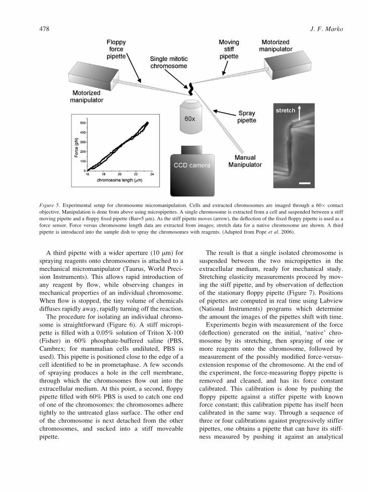
Figure 5. Experimental setup for chromosome micromanipulation. Cells and extracted chromosomes are imaged through a 60� contact
objective. Manipulation is done from above using micropipettes. A single chromosome is extracted from a cell and suspended between a stiff
moving pipette and a floppy fixed pipette (Bar=5 mm). As the stiff pipette moves (arrow), the deflection of the fixed floppy pipette is used as a
force sensor. Force versus chromosome length data are extracted from images; stretch data for a native chromosome are shown. A third
pipette is introduced into the sample dish to spray the chromosomes with reagents. (Adapted from Pope et al. 2006).
478 J. F. Marko

balance with a micrometer (Poirier et al. 2000).
Calibration of the force-measuring pipette stiffness
allows the deflectionYextension data to be converted
to forceYextension. Typical native newt chromosome
data are shown in Figure 5, lower left.
Our group has carried out experiments of this
type on chromosomes extracted from cells.
Houchmandzadeh & Dimitrov (1999) and Almagro
et al. (2004) have used similar techniques to study
unreplicated chromatids assembled using Xenopusegg extracts.
Mitotic chromosome stretching elasticity
Mitotic chromosomes have robust elasticity, return-
ing to native length even after 5-fold extensions
(Nicklas 1983, Houchmandzadeh et al. 1997, Poirier
et al. 2000). During mitosis, chromosomes are often
doubled in length by spindle-generated forces on the
order of 1 nN in large animal or insect cells (Nicklas
1983). The extensibility of mitotic chromosomes has
been used to increase the resolution of chromosome
banding (Claussen et al. 1994).
Nicklas made the first measurements of the
elasticity of mitotic chromosomes (actually meiotic
metaphase I and II chromosomes), using micro-
needles to push and hook chromosomes insidegrasshopper cells, by pushing on the cell membrane
(Nicklas 1963, 1983). Bending of the microneedle
provided a way to measure forces, and Nicklas found
that roughly nanonewton forces caused chromo-
somes to be stretched to double their native length
in vivo.
Our experiments on newt and Xenopus mitotic
chromosomes removed from cells and manipulated
with micropipettes showed that mitotic newt
Figure 6. Isolation of metaphase chromosome from dividing newt cell. Upper left: cell is sprayed with dilute solution of Triton surfactant
which destabilizes cell membrane. Upper centre: chromosomes flow out of cell. Upper right and lower left: one chromosome is captured in a
pipette using suction. Lower centre: chromosome is moved away from cell by right pipette, note stretching of isolated chromosome caused by
attachment to other chromosomes. Lower right: chromosome is attached to second pipette using suction. A video version of this procedure is
available at http://markolab.bmbcb.northwestern.edu/ Bar=10 mm. (Images courtesy of M.G. Poirier).
Micromechanical studies of mitotic chromosomes 479
chromosomes could be doubled in length by roughly
1 nN forces (Houchmandzadeh et al. 1997, Poirier
et al. 2000, Poirier & Marko 2003), in good accord
with Nicklas (1963). We also found stretching force
to vary nearly linearly with extension for elongations
up to four times the native length, allowing us to
summarize the elastic response with a number, the
Fforce constant_ or slope of the force-versus-elonga-
tion curve. We emphasize that for newt chromosomes,
if one makes sufficiently slow extensionYrelaxation
cycles, the same forces are measured during retraction
as during extension; i.e., no hysteresis or irreversibility
is observed. Similar results were obtained for chro-
matids reconstituted using Xenopus egg extracts
(Houchmandzadeh & Dimitrov 1999): forces of
about 1 nN were required to double chromatid
length, with well-defined linear reversible elasticity.
Interestingly, a broad distribution of chromosome
force constants is obtained from single-chromosome
stretching experiments (Nicklas 1983, Poirier &
Marko 2003); it is not clear whether this variation
is due to mitotic stage or is chromosome-specific.
This level of force (1 nN) on a whole newt
chromosome is insufficient to remove histones from
DNA. Stretching experiments on assembled chroma-
tin fibers in buffer (typically 10 to 100 mM NaCl,
pH 7.5) (Cui & Bustamante 2000, Bennink et al.2001, Brower-Toland et al. 2002) show that nucle-
osome removal occurs only when tension exceeds
roughly 10 piconewtons (pN) (note 1 pN=10j3 nN;
individual protein motors such as kinesin generate
pN forces). By contrast, in a whole newt chromo-
some of õ1 mm radius and therefore õ3 mm2 cross-
sectional area, several thousand 30 nm chromatin
fibres pass through each chromosome cross-section.
Therefore, forces of a few nanonewtons on a whole
chromosome reduce to roughly 1 pN forces per
chromatin fibre, insufficient force to dislodge histo-
nes. However, this level of force is sufficient to
stretch out chromatin fibre from a folded (30 nm) to
an extended (10 nm or Bbeads-on-a-string^) confor-
mation; in this force range chromatin fibres have been
observed to display a nearly linear force-versus-
extension response (Cui & Bustamante 2000,
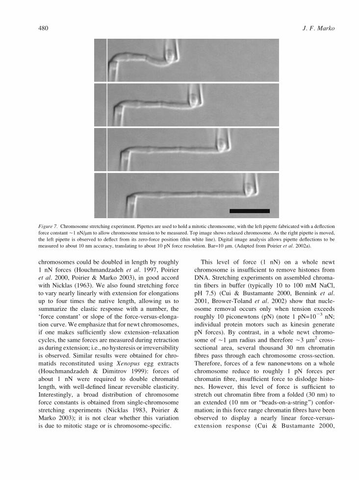
Figure 7. Chromosome stretching experiment. Pipettes are used to hold a mitotic chromosome, with the left pipette fabricated with a deflection
force constant õ1 nN/mm to allow chromosome tension to be measured. Top image shows relaxed chromosome. As the right pipette is moved,
the left pipette is observed to deflect from its zero-force position (thin white line). Digital image analysis allows pipette deflections to be
measured to about 10 nm accuracy, translating to about 10 pN force resolution. Bar=10 mm. (Adapted from Poirier et al. 2002a).
480 J. F. Marko

Bennink et al. 2001). More detailed estimates
indicate that the linear reversible elastic range of
stretching of whole chromosomes can be attributed to
this folding elasticity of the 30 nm chromatin fibre
(Cocco et al. 2003). However, one should keep in
mind that the possibility that at least part of whole-
chromosome elasticity may arise from the opening of
large-scale chromatin folding.
To describe the elastic properties of a material, one
often quotes its elastic modulus. This expresses what
stress (force per area) would be required to double an
object_s length, if the initial linear elasticity were
extrapolated. For a mitotic chromosome, this stress is
about 500 pascals (Nicklas & Staehly 1967, Poirier
et al. 2000, Poirier & Marko 2002b) (1 pascal (Pa)=1
newton/meter2 is the SI unit of pressure and stress).
A 500 Pa modulus is low, even for a very loose high-
polymer gel: 1% agarose gels have moduli of about
10 kPa (10 000 Pa), plexiglass and folded biomole-
cules (B-DNA and globular protein domains) have
moduli near 1 GPa (109 Pa), and covalently bonded
materials (metals, glasses) have moduli of about 10
GPa. The modulus is useful because it expresses the
strength of the interactions holding a material
together, in a way which is independent of size or
shape. Table 1 lists moduli of the mitotic chromo-
somes studied to date.
Mitotic chromosomes have a modulus roughly a
millionth of the modulus of the molecules from which
they are composed, indicating that they are relatively
loosely internally linked. The extreme extensibility of
up to five times without apparent damage indicates that
the internal structure must involve loosely compacted
domains of chromatin that can readily unfold under
force. Further evidence for unfolding of polymer-like
folded domains is given by dynamic experiments that
show a slow, viscous response to applied forces con-
sistent with the elastic response of a flexible polymer
network (Poirier et al. 2000, 2001, Poirier & Marko
2002a). All of our elastic experiments require very
slow (100 s) extensionYrelaxation cycles to stay in
mechanical equilibrium; sufficiently fast stretching
can cause irreversible changes to chromosomes
(Houchmandzadeh et al. 1997).
By contrast, following very slow extension to 5-fold
or greater extensions and forces in the 10Y20 nN
range, mitotic chromosomes are permanently length-
ened, suggesting that internal Flinks_ holding chro-
matin in its compacted form are being broken
(Poirier et al. 2000); similar irreversible elasticity is Tab
le1.
Ph
ysi
cal
pro
per
ties
of
mit
oti
cch
rom
oso
mes
.R
anges
for
val
ues
ind
icat
eth
ew
idth
of
dis
trib
uti
on
of
mea
sure
dv
alu
es,
and
no
tm
easu
rem
ent
erro
rs
Ch
rom
oso
me
type
Ex
per
imen
tco
nd
itio
ns
Str
etch
ing
(Yo
un
g)
mod
ulu
s(P
a)B
endin
gri
gid
ity
(Jm
)R
efer
ence
s
Dro
soph
ila
met
aph
ase
chro
mo
som
eIn
vivo
ND
õ6�
10j
24
Mar
shal
let
al.
(20
01
)
Gra
ssh
op
per
met
aph
ase
Ian
dan
aph
ase
Ich
rom
oso
me
Invi
vo2
00Y1
00
0(a
vg
=4
30
)N
DN
ick
las
(19
67,
19
83
)
New
t(N
.vi
ride
scen
s)p
rom
etap
has
ech
rom
oso
me
Cel
lcu
ltu
rem
ediu
m1
00Y1
00
01Y3�
10j
22
Ho
uch
man
dza
deh
eta
l.(1
99
7),
Po
irie
ret
al.
(20
00,
20
02a,
b),
Po
pe
etal
.(2
00
6)
New
tp
rom
etap
has
ech
rom
oso
me
Invi
voN
D2Y5�
10j
23
Po
irie
ret
al.
(20
02
a)
Xen
opus
pro
met
aphas
ech
rom
oso
me
Cel
lcu
ltu
rem
ediu
m2
00Y8
00
0.5Y2�
10j
23
Po
irie
ret
al.
(20
02
a)
Xen
opus
anap
has
ech
rom
atid
Cel
lcu
ltu
rem
ediu
mõ
30
0õ
5�
10j
24
Po
irie
ret
al.
(20
02
a)
Xen
opus
reco
nst
itu
ted
chro
mat
idB
uff
erE
B1
00
01
.2�
10j
26
Ho
uch
man
dza
deh
&D
imit
rov
(19
99),
Alm
agro
etal
.(2
00
4)
ND
ind
icat
esq
uan
tity
no
td
irec
tly
mea
sure
d.
Micromechanical studies of mitotic chromosomes 481

seen for unreplicated mitotic chromatids following
sufficient extension (Houchmandzadeh & Dimitrov
1999). After slow extensions beyond about 30 times
native length followed by relaxation, mitotic chro-
mosomes end up not only longer than native but also
wider (Figure 8) (Poirier et al. 2000). This suggests
that if sufficient numbers of chromatin interconnects
are broken up, the now less constrained chromatin
swells up. Using fluorescent antibodies, histone
content was observed not to change appreciably
during this experiment (Poirier et al. 2000).
Note that experiments in the irreversible stretching
regime involved forces of at most 20 nN, corre-
sponding to forces of several piconewtons per chro-
matin fibre by the cross-section argument mentioned
above. This is insufficient force to quickly break
chemical bonds (Grandbois et al. 1999), but it is
sufficient to break proteinYDNA and proteinYprotein
interactions. The irreversible stretching behaviour of
chromosomes is most likely due to disruption of
chromatin-crosslinking elements.
Mitotic chromosome bending stiffness
The utility of the bending stiffness of a chromosome
is that it can be measured without applying external
stresses. Any small flexible rod will undergo random
bending fluctuations at room temperature by thermal
forces. The approach of measuring thermal bending
fluctuations has been widely used to study mechan-
ical properties of biopolymers and biopolymer com-
plexes (e.g. Gittes et al. 1993). One usually measures
the length over which thermally excited bends occur,
or the Bpersistence length[ (Gittes et al. 1993,
Houchmandzadeh & Dimitrov 1999). The bending
stiffness is just the persistence length times a thermal
energy factor (kBT=4�10j21 joules; here T is
absolute temperature, essentially the same for all
laboratory temperatures). Thus, the bending constant
is measured in joule-metres (J m) (Table 1).
This approach has been applied to mitotic chro-
mosomes. When prometaphase chromosomes are
isolated from either newt or Xenopus cells, very
small bending fluctuations are observed: the
Fpersistence length_ obtained from quantitative anal-
ysis is found to be many times the length of the
chromosome (Poirier et al. 2002a). However, when
Xenopus chromatids assembled using egg extracts are
observed (after dilution into suitable buffer to avoid
non-thermal fluctuations generated by condensins
and other ATPases), one sees drastic thermal bending
fluctuations by large angles, and one measures a per-
sistence length of roughly 2 mm, much shorter than
the 20 mm-long chromatids (Houchmandzadeh &
Dimitrov 1999). Reconstituted Xenopus chromatids
have a bending stiffness about 500 times less than
Xenopus chromosomes (Poirier et al. 2002a), indi-
cating a profound difference in internal structure
between unreplicated egg-extract chromatids and pro-
metaphase chromosomes from differentiated cells.
A rod made of a material with a well-defined
elastic stretching modulus has a bending stiffness
which is proportional to that modulus times the
fourth power of the cross-section radius (times a
numerical factor close to 1 in value; see Poirier &
Marko (2003). Given the numbers for the stretching
modulus and the bending stiffness for chromosomes,
one can ask whether they are consistent with this
uniform-elastic-medium result. The result is that for
newt and Xenopus chromosomes from tissue culture
Figure 8. Extreme overextension of newt chromosome leads to swollen chromosome. Top: native chromosome before overextension.
Bottom: chromosome after a series of slow extensions to 40 times native length by a peak force of 16 nN; chromosome is permanently
lengthened by approximately 5-fold, and widened approximately 1.5-fold. Bar=10 mm. (Adapted from Poirier et al. 2000).
482 J. F. Marko
cells, the bending stiffnesses are consistent with their
being made of a uniform elastic medium with stretch-
ing modulus of 500 Pa (Poirier et al. 2002a).
In contrast, the Xenopus egg-extract chromatids
are thus about 500 times easier to bend than we
would expect for a uniform elastic medium, suggest-
ing that egg-extract chromatids have the organization
of a halo of chromatin attached to a very thin internal
elastic structure, i.e. with no crosslinking in the
exterior halo region (Houchmandzadeh & Dimitrov
1999). If two such chromatids were linked together
by cohesins as in the prometaphase chromosomes,
the resulting structure would be much more difficult
to bend, possibly explaning the large difference in
bending modulus between egg-extract chromatids
and somatic-cell chromosomes.
Observation of bending fluctuations has also been
used to estimate stretching modulus in systems where
stretching experiments would be very difficult or
impossible owing to the small size of the chromo-
somes involved. Marshall et al. (2001) used bending
fluctuations of chromosomes in colchicine-poisoned
cells to estimate the elastic modulus of mitotic
Drosophila embryo chromosomes: a value of roughly
10 Pa was obtained, significantly smaller than the
500 Pa measured for amphibian tissue culture cell
chromosomes. It would be quite interesting to know
the corresponding stretching modulus; recall that
Drosophila chromosomes are thought to be dominated
by highly dynamic condensin I (Oliveira et al. 2007).
A more detailed discussion of chromosome
mechanics can be found in Poirier & Marko (2003).
Reversible folding and unfolding of mitoticchromosomes by salt
As discussed above, chromatin can readily be made
to unfold from 30 nm to 10 nm fibre form by shifting
univalent salt concentration to low (10 mM) values at
which electrostatic repulsion overwhelms nucleo-
some stacking interactions (van Holde 1988). This
type of experiment can be carried out with whole
chromosomes, with dramatic results. Maniotis et al.(1997) reported that mitotic chromosomes could be
abruptly decondensed and recondensed merely by
shifting salt concentration. An older literature
concerning this general type of experiment also
indicates that mitotic chromosomes can be hyper-
condensed or greatly decondensed by shifts in salt
concentration (Cole 1967, Zelenin et al. 1979).
In a series of experiments in which univalent and
multivalent salts were sprayed onto newt mitotic
chromosomes, not only that mitotic chromosomes
could be hypercondensed or decondensed on a few-
second timescale, but furthermore chromosome
elastic response after such treatments matched the
pre-treatment response, suggesting refolding to a
near-native state with little or no loss of protein
(Poirier et al. 2002b). For univalent salt (NaCl), both
low-salt (G100 mM) and high-salt (9100 mM)
conditions led to chromosome unfolding. Thus,
maximum chromosome compaction as a function of
NaCl concentration occurred for essentially physio-
logical (100 mM) levels. At low salt, decondensation
can be understood in terms of electrostatic repulsion
driving adjacent nucleosomes apart, essentially
unfolding 30 nm chromatin to the 10 nm form. At
high salt, the simplest explanation is that attractive
electrostatic interactions favouring chromosome
compaction become screened by the high charge
density, leading to expansion of the chromosome.
This unfolding is dramatic; for 400 mM NaCl, a newt
chromosome reaches a volume roughly 5 times larger
than its native state.
For divalent salt (MgCl2), different results were
found: low divalent concentrations (10 mM) led to
compaction of the chromosome (the opposite effect
of the univalent salt), possibly due to Mg2+-mediated
attractions between single negative charges along chro-
matin fibres. At high divalent concentrations (100 mM),
chromosome expansion was observed, again likely due
to screening out of charge interactions. In all cases
where univalent and divalent salts were used, the
chromosomes rapidly recovered their native elasticity
when the flow of ions was stopped.
These experiments indicate that, far from being
tightly bound together, chromatin in mitotic chromo-
somes is greatly compacted by relatively weak
electrostatic interactions which can be easily dis-
rupted. The native state can easily be recovered
following its disruption. Interestingly, by use of
trivalent cations, the volume of a chromosome can
be reduced by about a third. Thus, the native state is
well below its maximum density; much of the visible
mitotic chromatid volume is mobile small molecule
species, presumably mostly water. Notably, both the
unfolding (expansion) and hypercondensation (con-
traction) driven by salt were always observed to be
isotropic, with length changed by the same factor as
width (Poirier et al. 2002b).
Micromechanical studies of mitotic chromosomes 483

Molecular connectivity of mitotic chromosomes
Nucleases disintegrate mitotic chromosomes
The elasticity experiments described in the previous
section show that mitotic chromosomes can be
reversibly extended up to five times their native
length, indicating that the molecules holding them
together are themselves highly extensible. A main
question one can ask is whether this extensibility and
elasticity is due to DNA (chromatin) extensibility, or
whether chromosome elasticity comes from extensi-
bility of protein structures, e.g. SMCs. A closely
related question is whether the chromatin in a mitotic
chromosome is folded by being looped or attached to
a protein scaffold which is stably connected by
proteinYprotein interactions, or alternately whether
non-histone proteins which stabilize mitotic chroma-
tin are essentially disconnected from one another so
as to act as chromatin Fcrosslinks_.One way to attack these questions is to use enzyme
digestion to determine how the mechanical properties
of chromosomes are modified by cleavage of differ-
ent molecular components. Classic experiments of
this type (Callan & Macgregor 1958, Macgregor &
Callan 1962) showed that DNAase fragmented
amphibian lampbrush chromosomes (meiotic pro-
phase), and that this was not done by RNAase and
Figure 9. MNase digestion severs mitotic chromosome. Native chromosome rapidly loses elastic stiffness (note bending of chromosome by
flow after 240 s of MNase exposure) and is then cleaved. Bar=10 mm. (Adapted from Poirier & Marko 2002c).
484 J. F. Marko

proteases. Gall (1963) used DNAase-cleavage
experiments to determine that lampbrush chromo-
somes contained four parallel DNA molecules (i.e.
the four chromatids present at meiotic pachytene).
Later experiments studied the access of restriction
enzymes to loop domains in lampbrush chromosomes
(Gould et al. 1976).
A few groups have followed this general approach
to examine the effect of cutting nucleic acid on
mechanical properties of individual mitotic chromo-
somes. Digestion of DNA has been shown to disrupt
mitotic chromatin (Cole 1967, Maniotis et al. 1997).
We wished to understand the process by which
mitotic chromosomes lost their mechanical conti-
nuity. Experiments with micrococcal nuclease on a
chromosome under low tension (roughly 0.1 nN)
revealed that cutting of DNA alone rapidly elimi-
nates chromosome elasticity. Chromosomes become
irreversibly extensible after even brief MNase expo-
sure. Sufficient digestion causes cleavage of the
whole chromosome (Figure 9), and finally collapse
of the remaining chromatin into a spherical droplet,
indicating all loss of elasticity and memory of its
original shape (Poirier & Marko 2002c).
If light MNase digestion is applied to a chromo-
some with no tension at all applied to it, one sees
little change in chromosome shape or global structure
(Figure 10, top panels). However, if the chromosome
is then extended, it has no elasticity at all, and
extends into a series of domains linked by very thin
threads (Figure 10, 0 s), possibly reflecting variations
in density of chromatin crosslinking. Restarting light
Figure 10. Chromosome unfolding after partial MNase digestion of newt chromosome. Top left: native chromosome; top right: chromosome
after MNase digestion. Second panel (0 s): chromosome is extended, and left end breaks up into a series of dense domains linked by thin
filaments. Third through sixth panels (50Y120 s) show result of MNase spray, which severs extended filament. Note that the remaining
segment of chromosome refolds. Bar=10 mm. (Adapted from Poirier & Marko 2002c).
Micromechanical studies of mitotic chromosomes 485

MNase digestion cleaves the chromosome in the
middle of the thin fibre (Figure 10, 67.5Y67.8 s). The
remaining chromatin string then folds back up into a
chromosome-like structure (120 s).
Cutting of DNA alone leads to complete dissolu-
tion of the mitotic chromosome, and therefore non-
histone proteins are not connected together (Poirier &
Marko 2002c). Instead, proteins such as topo II and
SMC complexes are disconnected from one another,
and must act as crosslinkers to form a Fgel_ or
Fnetwork_ of chromatin. Recent experiments on
reconstituted Xenopus chromatids obtained similar
results and conclusions (Almagro et al. 2004).
Blunt-cutting restriction enzymes allow a rough
estimate of the inter-crosslink distance (Poirier &
Marko 2002c). 4-Base-specificity cutters led to the
same result as for MNase (Figure 11). However,
6-base-specificity blunt cutters caused no change in
chromosome elasticity. Experiments with a restric-
tion enzyme with 5-base specificity led to a reduction
of chromosome elastic modulus but not to cleavage
of the chromosome. This series of experiments
indicates that 4-base specificity is sufficient to
entirely disconnect the chromatin inside a mitotic
newt chromosome. Given that restriction enzymes
are able to efficiently access only linker DNA
(Polach & Widom 1995), this corresponds to one
cut every õ3 kb. However, 6-base-specificity cutting
(one cut every õ40 kb in chromatin) is insufficient to
even reduce the elastic modulus. Based on these
results, the typical distance between crosslinking
elements in the newt chromosome was estimated to
be approximately 15 kb.
Proteases gradually expand but do not cleavechromosomes
Maniotis et al. (1997) showed that trypsin and
proteinase K treatment of whole genomes caused a
volume expansion of human mitotic chromosomes.
Figure 11. Digestion of newt mitotic chromosome by 4-base-specificity blunt-cutting restriction enzyme AluI. Initial (0 s) image shows
native chromosome under low tension (100 pN). As digestion proceeds, the force measuring pipette (right) relaxes, indicating that the
chromosome has lost elasticity (250 s). Additional digestion thins (275 s) and cleaves (300 s) the chromosome; additional digestion converts
the chromosome to a Fdroplet_ of chromatin fragments (390 s) and finally eliminates most of the chromosome outside the right pipette (1100 s).
Bar=10 mm. (Figures courtesy of M.G. Poirier).
486 J. F. Marko

Force-measurement experiments on Xenopus recon-
stituted chromatids (Almagro et al. 2004) showed
that the elastic stiffness was gradually reduced by
protein digestion.
Protease experiments on newt mitotic chromo-
somes obtained similar results: exposure to either
trypsin or proteinase K gradually decondensed and
softened chromosomes but without ever entirely
eliminating their elastic response or cleaving them
(Figure 12) (Pope et al. 2006). Protein digestion led
to a strongly anisotropic decondensation process,
with length increasing by a larger proportion than
width. It was also found that partial digestion of
mitotic chromosome protein induced sensitivity of
the elastic modulus to 6-base-specificity blunt-cutting
restriction enzymes. All of these effects are consis-
tent with a network organization of the mitotic
chromosome, with a strong degree of anisotropy of
folding to allow strong lengthening in response to
mild protein digestion (Kireeva et al. 2004, Pope
et al. 2006).
Interchromosome linkers
A feature of chromosome structure that is evident
whenever mitotic chromosomes are removed from
animal cells is that different chromosomes
(replicated chromatid pairs) are connected together
by thin, highly extensible filaments. These have
been observed in chromosome isolation experi-
ments for many years (Hoskins 1968, Korf &
Diacumakos 1978, Maniotis et al. 1997) but have
always been controversial since they contradict the
common wisdom that different chromosomes are
separate gene linkage units. Definitive observation
of such filaments inside a live cell has not been
reported, and observing these filaments outside the
cell always invites the criticism that they are
an artefact of chromosome isolation (Korf &
Diacumakos 1980).
A number of authors have reported that mitotic
interchromosome linkers are cut by nucleases
(Maniotis et al. 1997, Poirier & Marko 2003), and
Figure 12. Decondensation driven by digestion of protein in newt mitotic chromosome. (A) Progressive lengthening and widening of
chromosome resulting from increasing trypsin digestion; digestion time shown in seconds. Expansion is anisotropic, with length increasing
more than width. Chromosomes remain elastic during these digestion experiments. (B) Similar effects of proteinase K. Bars=5 mm. (Adapted
from Pope et al. 2006).
Micromechanical studies of mitotic chromosomes 487

therefore that they are based on DNA. In our
experiments, we almost always find these fibres
between mitotic newt chromosomes (note the chro-
mosome being stretched by an invisible fibre in the
lower middle panel of Figure 6); once in every few
dozen experiments we observe a loose chromosome
free of such linkers. We typically break linkers after
firmly attaching a chromosome to two pipettes.
Although their mechanical effects are obvious,
interchromosome linkers can barely be observed by
phase-contrast or DIC indicating that their thickness
is in the range of 100Y200 nm.
A study of chaffinch (bird) chromosomes revealed
filaments containing a centromeric satellite DNA
extending between nonhomologous metaphase chro-
mosomes (Saifitdinova et al. 2000, 2001). Interchro-
mosome filaments containing centromeric satellite
DNA and CENP protein have also been observed
in mouse tissue culture cells by Kuznetsova et al.(2007). The function of these interchromosome
filaments remains an enigma.
Implications for chromosome structure
Mitotic chromosomes are chromatin networks
Biochemical and biophysical results put constraints
onto models of how the mitotic chromosome is
folded. DNA digestion experiments indicate that the
basic organization of the mitotic chromosome is that
of a chromatin network or gel with non-DNA cross-
linking elements which are not bound to one another
(Poirier & Marko 2002c). Note that Fcrosslinking_does not necessarily imply covalent binding; the
chromatin crosslinkers of interest here may act via
non-covalent protein-DNA, protein-protein or even
topological interactions (Nasmyth & Haering 2005).
It must also be noted that digestion experiments do
not rule out an inhomogeneous spatial distribution of
crosslinks inside chromatids. However, recent EM
studies have observed a surprisingly regular network
of chromatin in the interior of egg-extract-assembled
chromosomes (Konig et al. 2007).
Chromosome elasticity experiments combined
with single-chromatin-fibre stretching experiments
are consistent with isolated scaffold elements. If the
crosslinks were bonded together into a contiguous
protein scaffold, one would not expect such a large
range of elastic force response, since folded proteins
are known to be relatively rigid; For example,
condensin-folded structures along single DNAs (Strick
et al. 2004) require 10 pN forces to be broken, and
coiled-coils require even higher 20 pN forces to be
uncoiled (Schwaiger et al. 2002). The known high
degree of extensibility of chromatin fibre (Cui &
Bustamante 2000, Bennink et al. 2001) can simply
explain the large extensibility of mitotic chromosomes
at relatively low forces, but only if chromatin cross-
linking elements are not bound to one another.
What are the crosslinking elements?
Current data suggest SMC complexes as prime
candidates for crosslinkers. Animal condensin units
can by themselves condense DNA (Strick et al.2004), and are essential to chromatid condensation in
the egg-extract system (Hirano & Mitchison 1994).
Depletion of condensins in cells impairs chromosome
condensation and causes chromosomes to be mechan-
ically weak (Hudson et al. 2003, Ono et al. 2003,
Hirota et al. 2004, Gerlich et al. 2006). Finally,
estimates for the numbers of condensins on animal
chromosomes are consistent with inter-crosslink
distances inferred from digestion experiments (Poirier
& Marko 2002c).
Cohesins have a chromatin-crosslinking function
in mitotic chromosomes, given that they hold sister
chromatids together, possibly by a topological mech-
anisms (Nasmyth & Haering 2005). They appear to
be mobile and affected by transcription in yeast
(Lengronne et al. 2004). Cohesins provide crosslinks
between sister chromatids which persist until anaphase.
It is possible that there are other as yet uncharac-
terized mitotic crosslinking elements, e.g. BAF-1,
given that condensin depletion experiments suggest
that the cell may have alternatives to condensins to
drive chromosome condensation (Hudson et al. 2003,
Gassmann et al. 2004, Hirota et al. 2004, Gerlich
et al. 2006).
SMC-crosslinked-chromatin-network modelof mitotic chromosome condensation
The results discussed above, combined with con-
clusions of Marsden & Laemmli (1979), Losada &
Hirano (2001), Lavoie et al. (2002), Maeshima &
Laemmli (2003), Strukov et al. (2003), Kireeva et al.(2004), Lavoie et al. (2004), Polyakov et al. (2006),
and Sheval and Polyakov (2006), suggest the follow-
488 J. F. Marko

ing scenario for vertebrate chromosome condensation
(Figure 13). Numbers are approximate, and apply to
the human case.
The first event is loading of cohesin onto unre-
plicated chromatin. Cohesins are then organized by a
process possibly coupled to transcription, into inter-
mittent clusters along replicated sister chromatids,
whose positions are programmed by DNA sequence
(Glynn et al. 2004, Lengronne et al. 2004, 2006).
This redistribution might also be coupled to DNA
replication, which has been suggested to drive
condensation and segregation of sister chromatids,
e.g. through extrusion of replicated DNA domains
(Pflumm 2002, Gotoh 2007). The key point is estab-
lishment of well-separated points of cohesion, pre-
ceding condensin activity.
Next, during prophase, condensin II is loaded, and
acts to condense the parallel sisters (Figure 13AYC).
If condensin II acts in cis along DNA as observed in
single molecule experiments (Strick et al. 2004), then
crosslinking and potential topological relinking of
sisters will not occur. Instead, remnant sister catena-
tion will be pushed out of the condensin-rich regions,
to form tight DNA crossings favoured by topo II
(Sumner 1996), and generating alternating condensin-
and topo II-rich regions (Maeshima & Laemmli 2003).
A plausible mechanism for condensin II to
accomplish chromatin condensation in cis is for it
to initially bind short, contiguous segments of
chromatin of length similar to its õ50 nm size (also
comparable to the õ30 nm persistence length of
chromatin fibre (Cui & Bustamante 2000, Dekker
et al. 2002)), and then to gradually translocate, or
alternately to stimulate binding of additional con-
densin units at neighbouring chromatin sites, so as to
progressively condense chromatin between cohesin
Fboundaries_ (Lavoie et al. 2002, 2004). The outcome
would be a series of segregated loop-like chromatin
domains, separated by cohesin clusters along the
chromatid axis, and a highly contracted chromosome.
These loop-like chromatin domains might be folded
or interwound by topological effects of condensin
(Kimura & Hirano 1997, Kimura et al. 1999) or by
binding of metal ions (Strick et al. 2001). Condensin
locations may be programmed: evidence exists
supporting defined yeast condensin binding sites
spaced by roughly 10 kb (Wang et al. 2005).
Local translocation over distances of tens of
kilobases would make the condensin II distribution
appear stationary at optical scales (Gerlich et al.
2006), while still generating a large amount of
compaction, and without adding links between sister
chromatids. Quite to the contrary, tension built up
between adjacent chromatids would drive topo II to
gradually segregate them (Marko & Siggia 1997,
Maeshima & Laemmli 2003).
This scheme organizes prophase chromatids into a
string of rosette-like Fchromomere_ structures
(Sumner 1991) of size similar to folding intermedi-
ates observed by Prusov et al. (1983), Zatsepina et al.(1983), Belmont et al. (1987, 1989), Belmont &
Bruce (1994), Kireeva et al. (2004), and Belmont
(2006). For human chromosomes, these proposed
structures contain about 1000 nm of 30 nm fibre
(100 kb of DNA), with a condensed volume of
roughly 106 nm3, and therefore with roughly a 100
nm diameter (Figure 13C).
These chromomeres can be folded or coiled (e.g.
like nucleosomes in 30 nm fibre) only if there is a
gradual loss of cohesin along chromatid arms: the
cohesins of Figure 13C will oppose longitudinal
condensation beyond roughly 1000 bp/nm, with
higher compaction factors requiring cohesin removal.
Removal of cohesin and further folding gives a fur-
ther 6-fold compaction, generating a 6000 bp/nm
mid-prophase chromatid (Figure 13D; note that the
gray balls represent chomomere units of roughly
100 nm diameter).
Volume conservation indicates that the chromo-
some will become thicker by an amount approxi-
mately equal to the square root of the length
compaction. For the 60-fold length compaction of
chromatin fibre into the human prophase chromatid
described above, this is a factor of 8 (times the 30 nm
fibre thickness), resulting in segregated prophase
chromatids that are 250 nm thick with a condensing
II-enriched core region (Kireeva et al. 2004).
Then, at NEB, condensin I binds, acting as a
highly mobile (Gerlich et al. 2006), reversible
chromatin crosslinker. Condensin I acting as a
reversible crosslinker in the chromosome interior
will drive chromatids to adopt a configuration with
lower surface area, driving longitudinal compaction
and transverse thickening after NEB (Figure 13E)
(Kireeva et al. 2004). This effect is in exact analogy
to surface tension driving the shape of a liquid
droplet to be spherical, but for a chromosome, the
underlying chromatin network will oppose formation
of a sphere, and will maintain an anisotropic shape.
The result will be a metaphase chromatid which is
Micromechanical studies of mitotic chromosomes 489

Figure 13. Model of human mitotic chromosome folding. (A) Part of the replicated sister chromatids (gray lines represent 30 nm fibre
containing 100 bp/nm). Roughly 1000 nm of 30 nm fibre (100 kb of DNA) is between successive cohesin domains (blue bars). (B) Binding of
condensin II (red bars) during early prophase begins to organize loops of chromatin, gradually shortening the chromosome; topo II (yellow
diamonds) binds to and resolves chromatid crossings trapped between condensin-rich regions. (C) At end of prophase, the chromatin length
between cohesion blocks has been absorbed into condensin-looped regions; the result is a chromatin Brosette^ of Bchromomere^ of roughly
100 nm diameter with a condensin II core. Topo II and adjacent looped regions of chromosome are not shown for clarity. (D) Parallel
chromatids corresponding to a series of rosette chromomeres organized as in C. Gray balls (100-nm-diameter) correspond to rosette/
chomomere structures of C (note change of scale relative to C.). Red balls indicate their condensin II-rich cores. Successive chromomeres are
stacked, folded or coiled to achieve a packing density of 6000 bp/nm, about 2000-fold higher than linear in DNA. (E) Binding of additional
bulk-chromatid-condensing factors including condensin I (green) compresses chromosome along length, increasing its width. Length
compaction at this stage is roughly 20 000 bp/nm, approaching 10 000-fold relative to DNA. (F) End-on view of prophase chromosome of D.;
chromatids contain chromomeres (gray circles) with condensin II-rich cores (red). (G) End-on view of metaphase chromosomes of E.;
addition of condensin I (green) compresses chromosome lengthwise, increasing chromatid diameters.
490 J. F. Marko

shortened and thickened relative to prometaphase,
with a condensin II-rich core, covered by a layer of
condensin I (Figure 13E).
The final metaphase length to width ratio will be
determined by mechanical balance of condensin I
condensation versus chromatin network elasticity.
Condensin-I-driven longitudinal compaction may
force buckling or folding of the central non-histone-
protein-rich chromatid axis (Saitoh & Laemmli 1993,
1994). Sufficient crosslinking by condensin I to drive
longitudinal compaction will also provide mechanical
stabilization: consistent with this, depletion of con-
densin I has been observed to significantly weaken
metaphase chromosomes (Gerlich et al. 2006).
In experiments where some condensin subunits
are depleted or mutated, condensation might still
be driven by partial condensin activity, or even by
other proteins (e.g. BAF-1, or direct nucleosomeYnucleosome attraction), leading to eventual segre-
gation of adjacent chromatids (Gerlich et al. 2006).
Note that trans-fibre crosslinking factors must bind
and unbind, either by being ATP-cycled or by simply
binding rather weakly, in order to ensure chromatid
segregation (Marko & Siggia 1997).
This model predicts trends across species. The
larger is the distance between cohesin domains, the
greater will be the length compaction. Metaphase
cohesin interdomain distances in vertebrates must be
much greater than the 15 kb distance observed in
yeast; for Xenopus the cohesin density has been
estimated to be one per 400 kb (Losada & Hirano
2001). Cohesin interdomain distances should corre-
late with mitotic loop size, and possibly with
convergent transcription domain size (Lengronne
et al. 2004) and replicon size (Buongiorno-Nardelli
et al. 1982). Notably, condensin binding sites in
yeast have been found to be correlated with DNA
replication landmarks (Wang et al. 2005).
Stretching and bending elasticity
In this model, cohesin and condensin I and II are not
bonded together but act as isolated chromatin cross-
linkers, so that a whole chromosome can be stretched
simply by affine stretching of chromatin fibres
between network nodes. As mentioned above, exten-
sion of individual chromatin fibres accounts for the
roughly nanonewton force needed to double the
length of a vertebrate chromosome. The 5-fold
reversible elastic response of whole chromosomes
correlates well with the 6-fold extension obtained
when chromatin is converted from 10 nm to 30 nm
form. Action of condensin I and possibly other
crosslinking mechanisms through the body of the
chromatid generates the mechanical coupling needed
to generate bending elasticity.
Beyond 5-fold extension, one can expect to start to
break crosslinking elements, at roughly 20 nN forces
(given 2000 fibres in parallel, this corresponds to
10 pN per condensin, the force required for con-
densin disruption (Strick et al. 2004)). The result will
be permanent chromosome lengthening and widen-
ing, with lengthening predominating owing to the
prometaphase condensin I-driven contraction (Poirier
et al. 2000).
The small bending stiffness of egg-extract-recon-
stituted chromatids (Houchmandzadeh et al. 1997)
may be a result of the unreplicated chromatids
having regions of individual chromatin fibres
between chromomeric domains which can act as
Fhinges_. Consistent with this, under large extension,
egg-extract chromatids extend by formation of thin,
extended fibres between thicker chromosome
domains (Houchmandzadeh & Dimitrov 1999), not
seen for comparable extensions of chromosomes
from animal cells (Poirier et al. 2000). Furthermore,
and also supporting this hypothesis, are observations
of Almagro et al. (2003) that the bending stiffness
varies along the egg-extract chromatids.
The difference between the egg-extract and
somatic-cell chromosomes may also be a conse-
quence of the embryonic developmental state of the
egg extracts. The condensin I to condensin II ratio in
egg extracts is about 5:1, while in somatic HeLa cells
it is closer to 1:1 (Ono et al. 2003). It has been
documented that metaphase chromosomes in Xen-opus embryos are twice as long as and substantially
narrower than those in swimming larvae (Micheli
et al. 1993), the difference in condensation perhaps
being due to different ratios of condensin I and II or
to cohesin domain or replicon size (Marilley &
Gassend-Bonnet 1989).
Micromechanical studies of mitotic chromosomes 491

Effects of cutting and removing molecules
In this model, cutting DNA sufficiently frequently
will result in loss of elasticity (due to disconnection
of chromatin), with cleavage of the chromosome
only if the crosslink elements are not bound together.
Insufficient cutting (less than one cut per crosslink)
will not change chromosome elasticity.
If protein is cut instead, histone tails and other
exposed protein structures along chromatin fibres
will be cleaved, causing chromatin fibre unfolding
and lengthening. This will drive gradual isotropic
expansion, similar to that observed for shifts in
univalent salt concentration (Poirier et al. 2002b).
However, crosslinkers will also be cleaved, possibly
causing a less symmetric effect: the hypothesis of a
condensin-I-driven longitudinal compaction dis-
cussed above can explain the anisotropic unfolding
of prometaphase newt chromosomes observed to
result from protease treatment (Pope et al. 2006).
Under the assumption that condensin I is the last
major crosslinker added to chromosomes, it will be
the crosslinker most exposed to general proteolysis.
Cutting condensin I will release constraints that
longitudinally compacted the chromosome, leading
to longitudinal expansion. Of course general prote-
olysis will also cut histone tails and other proteins
along chromatin which will contribute to the trans-
verse swelling observed during proteolysis of whole
chromosomes.
Specific cleavage of condensin (e.g. using engi-
neered kleisins with suitable specific protease sites)
on assembled metaphase chromosomes could be
instructive. There may be large differences between
cleavage of condensin I and condensin II units, given
their differing functions in chromosome compaction
(Ono et al. 2003). Based on the above model,
condensin I cleavage should produce lengthening
and narrowing, and reduction in elasticity of meta-
phase chromosomes, while condensin II cleavage
should produce little effect (since condensin I acts as
a more general stabilizer and crosslinker Fon top_ of
condensin II). However, simultaneous condensin I
plus condensin II cleavage should produce lengthen-
ing and widening similar to that seen by forced
chromosome unfolding (Poirier et al. 2000). Of
course, the possibility remains that additional cross-
linking elements added during prometaphase (e.g.
BAF-1 or other relatively small DNA-crosslinking
proteins) may be present in sufficient numbers to
maintain some chromosome integrity when conden-
sin is disrupted. An interesting question is how
having condensin I or II entirely absent, as in
C. elegans or yeast respectively, would affect
condensin-cleavage experiments.
Network, hierarchical folding and radial loop
models are unified by the scenario outlined above.
If histones are suddenly removed, there will be
release of DNA length and a large degree of swelling
of the chromosome, as a more severe and irreversible
version of the result of swelling of chromosomes by
high or low univalent salt concentrations (Poirier et al.2002b). In the case of histone removal, loop-like
domains of DNA will be observed (Paulson &
Laemmli 1977), a result of the domain structure of
early prophase condensation.
Acknowledgements
I am indebted to Professor Michael Poirier, Dr Lisa
Pope, and Mr Ryo Kawamura who carried out many of
the experiments discussed in this paper. I am also
grateful to Dr Tatsuya Hirano, Professor Rebecca
Heald, Professor Bahram Houchmandzadeh, Professor
Didier Chatenay and Professor Valentin Rybenkov for
their advice and assistance. This work was supported in
part by US NSF grants MCB-0240998, DMR-0715099
and PHY-0445565.
References
Adolph KW (1980) Isolation and structural organization of human
mitotic chromosomes. Chromosoma 76: 23Y33.
Adolph KW (1981) A serial sectioning study of the structure of
human mitotic chromosomes. Eur J Cell Biol 24: 146Y153.
Adolph KW, Kreisman LR, Kuehn RL (1986) Assembly of
chromatin fibers into metaphase chromosomes analyzed by
transmission electron microscopy and scanning electron micros-
copy. Biophys J 49: 221Y231.
Adolph KW, Phelps JP (1982) Role of non-histones in chromo-
some structure. Cell cycle variations in protein synthesis. J BiolChem 257: 9086Y9092.
Ahmed S, Brickner JH (2007) Regulation and epigenetic control of
transcription at the nuclear periphery. Trends Genet 23:
396Y402.
Akhtar A, Gasser SM (2007) The nuclear envelope and transcrip-
tional control. Nat Rev Genet 8: 507Y517.
Almagro S, Dimitrov S, Hirano T, Vallade M, Riveline D
(2003) Individual chromosomes as viscoelastic copolymers.
Europhysics Letters 63: 908Y914.
Almagro S, Riveline D, Hirano T, Houchmandzadeh B, Dimitrov S
(2004) The mitotic chromosome is an assembly of rigid elastic
492 J. F. Marko

axes organized by structural maintenance of chromosomes
(SMC) proteins and surrounded by a soft chromatin envelope.
J Biol Chem 279: 5118Y5126.
Almouzni G, Wolffe AP (1993) Nuclear assembly, structure, and
function: the use of Xenopus in vitro systems. Exp Cell Res 205:
1Y15.
Bausch C, Noone S, Henry JM, Goudenz K, Sanderson B, Seidel
C, Gerton JL (2007) Transcription alters chromosomal locations
of cohesin in S. cerevisiae. Mol Cell Biol 27: 8522Y8532.
Belmont AS (2006) Mitotic chromosome structure and condensa-
tion. Curr Opin Cell Biol 18: 632Y638.
Belmont AS, Bruce K (1994) Visualization of G1 chromosomes: a
folded, twisted, supercoiled chromonema model of interphase
chromatid structure. J Cell Biol 127: 287Y302.
Belmont AS, Sedat JW, Agard DA (1987) A three-dimensional
approach to mitotic chromosome structure: evidence for a
complex hierarchical organization. J Cell Biol 105: 77Y92.
Belmont AS, Braunfeld MB, Sedat JW, Agard DA (1989) Large-
scale chromatin structural domains within mitotic and inter-
phase chromosomes in vivo and in vitro. Chromosoma 98:
129Y143.
Bennink ML, Leuba SH, Leno GH, Zlatanova J, de Grooth BG,
Greve J (2001) Unfolding individual nucleosomes by stretching
single chromatin fibers with optical tweezers. Nat Struct Biol 8:
606Y610.
Bojanowski K, Maniotis AJ, Plisov S, Larsen AK, Ingber DE
(1998) DNA topoisomerase II can drive changes in higher order
chromosome architecture without enzymatically modifying
DNA. J Cell Biochem 69: 127Y142.
Bolzer A, Kreth G, Solovei I, Koehler D, Saracoglu K, Fauth C,
Muller S, Eils R, Cremer C, Speicher MR, Cremer T (2005)
Three-dimensional maps of all chromosomes in human male
fibroblast nuclei and prometaphase rosettes. PLoS Biol 3: e157.
Boy de la Tour E, Laemmli UK (1988) The metaphase scaffold is
helically folded: sister chromatids have predominantly opposite
helical handedness. Cell 55: 937Y944.
Bradley CM, Ronning DR, Ghirlando R, Craigie R, Dyda F (2005)
Structural basis for DNA bridging by barrier-to-autointegration
factor. Nat Struct Mol Biol 12: 935Y936.
Brickner JH, Walter P (2004) Gene recruitment of the activated
INO1 locus to the nuclear membrane. PLoS Biol 2: e342.
Brower-Toland BD, Smith CL, Yeh RC, Lis JT, Peterson CL,
Wang MD (2002) Mechanical disruption of individual nucleo-
somes reveals a reversible multistage release of DNA. Proc Natl
Acad Sci U S A 99: 1960Y1965.
Brown DT, Izard T, Misteli T (2006) Mapping the interaction
surface of linker histone H1(0) with the nucleosome of native
chromatin in vivo. Nat Struct Mol Biol 13: 250Y255.
Buongiorno-Nardelli M, Micheli G, Carri MT, Marilley M (1982)
A relationship between replicon size and supercoiled loop
domains in the eukaryotic genome. Nature 298: 100Y102.
Bystricky K, Heun P, Gehlen L, Langowski J, Gasser SM (2004)
Long-range compaction and flexibility of interphase chromatin
in budding yeast analyzed by high-resolution imaging techni-
ques. Proc Natl Acad Sci U S A 101: 16495Y16500.
Callan HG (1954). Recent work on the structure of cell nuclei. In:
Fine Structure of Cells. Symposium of the VIIIth Congress inCell Biology. Groningen: Noordhof, 89Y109.
Callan HG, Macgregor HC (1958) Action of deoxyribonuclease on
lampbrush chromosomes. Nature 181: 1479Y1480.
Carroll CW, Straight AF (2006) Centromere formation: from
epigenetics to self-assembly. Trends Cell Biol 16: 70Y78.
Christensen MO, Larsen MK, Barthelmes HU, Hock R, Andersen
CL, Kjeldsen E, Knudsen BR, Westergaard O, Boege F, Mielke
C (2002) Dynamics of human DNA topoisomerases IIalpha and
IIbeta in living cells. J Cell Biol 157: 31Y44.
Claussen U, Mazur A, Rubtsov N (1994) Chromosomes are
highly elastic and can be stretched. Cytogenet Cell Genet 66:
120Y125.
Cocco S, Marko JF, Monasson R, Sarkar A, Yan J (2003) Force-
extension behavior of folding polymers. Eur Phys J E Soft
Matter 10: 249Y263.
Cole A (1967) Chromosome Structure. New York: Dekker.
Cremer T, Cremer C (2001) Chromosome territories, nuclear
architecture and gene regulation in mammalian cells. Nat Rev
Genet 2: 292Y301.
Cremer T, Kurz A, Zirbel R et al. (1993) Role of chromosome
territories in the functional compartmentalization of the cell
nucleus. Cold Spring Harb Symp Quant Biol 58: 777Y792.
Cremer C, Munkel C, Granzow M, Jauch A, Dietzel S, Eils R,
Guan XY, Meltzer PS, Trent JM, Langowski J, Cremer T (1996)
Nuclear architecture and the induction of chromosomal aberra-
tions. Mutat Res 366: 97Y116.
Cremer T, Kreth G, Koester H, Fink RH, Heintzmenn R, Cremer
M, Solovei I, Zink D, Cremer C (2000) Chromosome territories,
interchromatin domain compartment, and nuclear matrix: an
integrated view of the functional nuclear architecture. Crit Rev
Eukaryot Gene Expr 10: 179Y212.
Cui Y, Bustamante C (2000) Pulling a single chromatin fiber
reveals the forces that maintain its higher-order structure. Proc
Natl Acad Sci U S A 97: 127Y132.
Cuvier O, Hirano T (2003) A role of topoisomerase II in linking
DNA replication to chromosome condensation. J Cell Biol 160:
645Y655.
Daban JR (2000) Physical constraints in the condensation of
eukaryotic chromosomes. Local concentration of DNA versus
linear packing ratio in higher order chromatin structures.
Biochemistry 39: 3861Y3866.
Dekker J, Rippe K, Dekker M, Kleckner N (2002) Capturing
chromosome conformation. Science 295: 1306Y1311.
Dietzel S, Zolghadr K, Hepperger C, Belmont AS (2004)
Differential large-scale chromatin compaction and intranuclear
positioning of transcribed versus non-transcribed transgene
arrays containing beta-globin regulatory sequences. J Cell Sci
117: 4603Y4614.
Dorigo B, Schalch T, Kulangara A, Duda S, Schroeder RR,
Richmond TJ (2004) Nucleosome arrays reveal the two-start
organization of the chromatin fiber. Science 306: 1571Y1573.
Earnshaw WC, Heck MM (1985) Localization of topoisomerase II
in mitotic chromosomes. J Cell Biol 100: 1716Y1725.
Earnshaw WC, Halligan B, Cooke CA, Heck MM, Liu LF (1985)
Topoisomerase II is a structural component of mitotic chromo-
some scaffolds. J Cell Biol 100: 1706Y1715.
Fukui K, Uchiyama S (2007) Chromosome protein framework
from proteome analysis of isolated human metaphase chromo-
somes. Chem Rec 7: 230Y237.
Gall JG (1963) Kinetics of deoxyribonuclease action on chromo-
somes. Nature 198: 36Y38.
Gardner MK, Pearson CG, Sprague BL, Zarzar TR, Bloom K,
Salmon ED, Odde DJ (2005) Tension-dependent regulation of
Micromechanical studies of mitotic chromosomes 493

microtubule dynamics at kinetochores can explain metaphase
congression in yeast. Mol Biol Cell 16: 3764Y3775.
Gasser SM, Laroche T, Falquet J, Boy de la Tour E, Laemmli UK
(1986) Metaphase chromosome structure. Involvement of top-
oisomerase II. J Mol Biol 188: 613Y629.
Gassmann R, Vagnarelli P, Hudson D, Earnshaw WC (2004)
Mitotic chromosome formation and the condensin paradox.
Exp Cell Res 296: 35Y42.
Gerlich D, Hirota T, Koch B, Peters JM, Ellenberg J (2006)
Condensin I stabilizes chromosomes mechanically through a
dynamic interaction in live cells. Curr Biol 16: 333Y344.
Gimenez-Abian JF, Sumara I, Hirota T, Hauf S, Gerlich D, dela
Torre C, Ellenberg J, Peters JM (2004) Regulation of sister
chromatid cohesion between chromosome arms. Curr Biol 14:
1187Y1193.
Gittes F, Mickey B, Nettleton J, Howard J (1993) Flexural rigidity
of microtubules and actin filaments measured from thermal
fluctuations in shape. J Cell Biol 120: 923Y934.
Glynn EF, Megee PC, Yu HG, Mistrot C, Unal E, Koshland DE,
DeRisi JL, Gerton JL (2004) Genome-wide mapping of the
cohesin complex in the yeast Saccharomyces cerevisiae. PLoS
Biol 2: e259.
Gotoh E (2007) Visualizing the dynamics of chromosome structure
formation coupled with DNA replication. Chromosoma 116:
453Y462.
Gould DC, Callan HG, Thomas CA Jr (1976) The actions of
restriction endonucleases on lampbrush chromosomes. J Cell Sci
21: 303Y313.
Grandbois M, Beyer M, Rief M, Clausen-Schaumann H, Gaub HE
(1999) How strong is a covalent bond? Science 283: 1727Y1730.
Graziano V, Gerchman SE, Schneider DK, Ramakrishnan V
(1994) Histone H1 is located in the interior of the chromatin
30-nm filament. Nature 368: 351Y354.
Gruber S, Haering CH, Nasmyth K (2003) Chromosomal cohesin
forms a ring. Cell 112: 765Y777.
Haering CH, Lowe J, Hochwagen A, Nasmyth K (2002) Molecular
architecture of SMC proteins and the yeast cohesin complex.
Mol Cell 9: 773Y788.
Hirano T (2005) SMC proteins and chromosome mechanics: from
bacteria to humans. Philos Trans R Soc Lond B Biol Sci 360:
507Y514.
Hirano T (2006) At the heart of the chromosome: SMC proteins in
action. Nat Rev Mol Cell Biol 7: 311Y322.
Hirano T, Mitchison TJ (1993) Topoisomerase II does not play a
scaffolding role in the organization of mitotic chromosomes
assembled in Xenopus egg extracts. J Cell Biol 120: 601Y612.
Hirano T, Mitchison TJ (1994) A heterodimeric coiled-coil protein
required for mitotic chromosome condensation in vitro. Cell 79:
449Y458.
Hirota T, Gerlich D, Koch B, Ellenberg J, Peters JM (2004)
Distinct functions of condensin I and II in mitotic chromosome
assembly. J Cell Sci 117: 6435Y6445.
Hock R, Furusawa T, Ueda T, Bustin M (2007) HMG chromo-
somal proteins in development and disease. Trends Cell Biol17: 72Y79.
Hoskins GC (1968) Sensitivity of micrurgically removed chro-
mosomal spindle fibres to enzyme disruption. Nature 217:
748Y750.
Houchmandzadeh B, Dimitrov S (1999) Elasticity measurements
show the existence of thin rigid cores inside mitotic chromo-
somes. J Cell Biol 145: 215Y223.
Houchmandzadeh B, Marko JF, Chatenay D, Libchaber A (1997)
Elasticity and structure of eukaryote chromosomes studied by
micromanipulation and micropipette aspiration. J Cell Biol 139:
1Y12.
Hudson DF, Vagnarelli P, Gassmann R, Earnshaw WC (2003)
Condensin is required for nonhistone protein assembly and
structural integrity of vertebrate mitotic chromosomes. Dev Cell
5: 323Y336.
Hutchison N, Pardue ML (1975) The mitotic chromosomes of
Notophthalmus (=Triturus) viridescens: localization of C band-
ing regions and DNA sequences complementary to 18S, 28S and
5S ribosomal RNA. Chromosoma 53: 51Y69.
Ivanov D, Nasmyth K (2005) A topological interaction between
cohesin rings and a circular minichromosome. Cell 122:
849Y860.
Kimura K, Hirano T (1997) ATP-dependent positive supercoiling
of DNA by 13S condensin: a biochemical implication for
chromosome condensation. Cell 90: 625Y634.
Kimura K, Rybenkov VV, Crisona NJ, Hirano T, Cozzarelli NR
(1999) 13S condensin actively reconfigures DNA by introducing
global positive writhe: implications for chromosome condensa-
tion. Cell 98: 239Y248.
Kireeva N, Lakonishok M, Kireev I, Hirano T, Belmont AS (2004)
Visualization of early chromosome condensation: a hierarchical
folding, axial glue model of chromosome structure. J Cell Biol166: 775Y785.
Kleckner N (1995) Interactions between and along chromosomes
during meiosis. Harvey Lect 91: 21Y45.
Kleckner N (1996) Meiosis: how could it work? Proc Natl AcadSci U S A 93: 8167Y8174.
Kleckner N, Zickler D, Jones GH, Dekker J, Padmore R, Henle J,
Hutchinson J (2004) A mechanical basis for chromosome
function. Proc Natl Acad Sci U S A 101: 12592Y12597.
Konig P, Braunfeld MB, Sedat JW, Agard DA (2007) The three-
dimensional structure of in vitro reconstituted Xenopus laevis
chromosomes by EM tomography. Chromosoma 116: 349Y372.
Korf BR, Diacumakos EG (1978) Microsurgically-extracted
metaphase chromosomes of the Indian muntjac examined with
phase contrast and scanning electron microscopy. Exp Cell Res
111: 83Y93.
Korf BR, Diacumakos EG (1980) Absence of true interchromo-
somal connectives in microsurgically isolated chromosomes.
Exp Cell Res 130: 377Y385.
Kuznetsova IS, Enukashvily NI, Noniashvili EM, Shatrova AN,
Aksenov ND, Zenin W, Dyban AP, Podgornaya DI (2007)
Evidence for the existence of satellite DNA-containing connec-
tion between metaphase chromosomes. J Cell Biochem 101:
1046Y1061.
Lanctot C, Cheutin T, Cremer M, Cavalli G, Cremer T (2007)
Dynamic genome architecture in the nuclear space: regulation of
gene expression in three dimensions. Nat Rev Genet 8: 104Y115.
Lavoie BD, Hogan E, Koshland D (2002) In vivo dissection of the
chromosome condensation machinery: reversibility of conden-
sation distinguishes contributions of condensin and cohesin. J
Cell Biol 156: 805Y815.
494 J. F. Marko

Lavoie BD, Hogan E, Koshland D (2004) In vivo requirements for
rDNA chromosome condensation reveal two cell-cycle-regulated
pathways for mitotic chromosome folding. Genes Dev 18: 76Y87.
Lee MS, Craigie R (1998) A previously unidentified host protein
protects retroviral DNA from autointegration. Proc Natl Acad
Sci U S A 95: 1528Y1533.
Lengronne A, Katou Y, Mori S, Yokobayashi S, Kelly GP, Itoh T,
Watanabe Y, Shirahige K, Uhlmann F (2004) Cohesin relocation
from sites of chromosomal loading to places of convergent
transcription. Nature 430: 573Y578.
Lengronne A, McIntyre J, Katou Y, Kanoh Y, Hopfner KP,
Shirahige K, Uhlmann F (2006) Establishment of sister
chromatid cohesion at the S. cerevisiae replication fork. MolCell 23: 787Y799.
Levi V, Ruan Q, Plutz M, Belmont AS, Gratton E (2005)
Chromatin dynamics in interphase cells revealed by tracking in
a two-photon excitation microscope. Biophys J 89: 4275Y4285.
Lewis CD, Laemmli UK (1982) Higher order metaphase chromo-
some structure: evidence for metalloprotein interactions. Cell
29: 171Y181.
Lindow JC, Kuwano M, Moriya S, Grossman AD (2002)
Subcellular localization of the Bacillus subtilis structural
maintenance of chromosomes (SMC) protein. Mol Microbiol
46: 997Y1009.
Losada A, Hirano T (2001) Shaping the metaphase chromosome:
coordination of cohesion and condensation. BioEssays 23:
924Y935.
Luger K, Mader AW, Richmond RK, Sargent DF, Richmond TJ
(1997) Crystal structure of the nucleosome core particle at 2.8 A
resolution. Nature 389: 251Y260.
Macgregor HC, Callan HG (1962) The actions of enzymes on
lampbrush chromosomes. Q J Microsc Sci 103: 173Y203.
Machado C, Andrew DJ (2000) Titin as a chromosomal protein.
Adv Exp Med Biol 481: 221Y232; discussion 32Y36.
Maeshima K, Laemmli UK (2003) A two-step scaffolding model
for mitotic chromosome assembly. Dev Cell 4: 467Y480.
Maeshima K, Eltsov M, Laemmli UK (2005) Chromosome
structure: improved immunolabeling for electron microscopy.
Chromosoma 114: 365Y375.
Maniotis AJ, Bojanowski K, Ingber DE (1997) Mechanical
continuity and reversible chromosome disassembly within intact
genomes removed from living cells. J Cell Biochem 65:
114Y130.
Maniotis AJ, Valyi-Nagy K, Karavitis J, Moses J, Boddipali V,
Wang Y, Nunez R, Setty S, Arbieva Z, Bissell MJ, Folberg R
(2005) Chromatin organization measured by AluI restriction
enzyme changes with malignancy and is regulated by the
extracellular matrix and the cytoskeleton. Am J Pathol 166:
1187Y1203.
Maresca TJ, Heald R (2006) The long and the short of it: linker
histone H1 is required for metaphase chromosome compaction.
Cell Cycle 5: 589Y591.
Maresca TJ, Freedman BS, Heald R (2005) Histone H1 is essential
for mitotic chromosome architecture and segregation in Xenopuslaevis egg extracts. J Cell Biol 169: 859Y869.
Margalit A, Brachner A, Gotzmann J, Foisner R, Gruenbaum Y
(2007) Barrier-to-autointegration factorVa BAFfling little pro-
tein. Trends Cell Biol 17: 202Y208.
Marilley M, Gassend-Bonnet G (1989) Supercoiled loop organi-
zation of genomic DNA: a close relationship between loop
domains, expression units, and replicon organization in rDNA
from Xenopus laevis. Exp Cell Res 180: 475Y489.
Marko JF, Poirier MG (2003) Micromechanics of chromatin and
chromosomes. Biochem Cell Biol 81: 209Y220.
Marko JF, Siggia ED (1997) Polymer models of meiotic and
mitotic chromosomes. Mol Biol Cell 8: 2217Y2231.
Marsden MP, Laemmli UK (1979) Metaphase chromosome
structure: evidence for a radial loop model. Cell 17: 849Y858.
Marshall WF, Straight A, Marko JF, Swedlow J, Dernburg A,
Belmont A, Murray AW, Agand DA, Sedat JW (1997)
Interphase chromosomes undergo constrained diffusional motion
in living cells. Curr Biol 7: 930Y939.
Marshall WF, Marko JF, Agard DA, Sedat JW (2001) Chromo-
some elasticity and mitotic polar ejection force measured in
living Drosophila embryos by four-dimensional microscopy-
based motion analysis. Curr Biol 11: 569Y578.
Meyer KN, Kjeldsen E, Straub T, Knudsen BR, Hickson ID,
Kikuchi A, Kreipe H, Boege F (1997) Cell cycle-coupled
relocation of types I and II topoisomerases and modulation of
catalytic enzyme activities. J Cell Biol 136: 775Y788.
Micheli G, Luzzatto ARC, Carri MT, Decapoa A, Pelliccia F
(1993) Chromosome length and DNA loop size during early
embryonic-development of Xenopus laevis. Chromosoma 102:
478Y483.
Milutinovich M, Unal E, Ward C, Skibbens RV, Koshland D
(2007) A multi-step pathway for the establishment of sister
chromatid cohesion. PLoS Genet 3: e12.
Misteli T, Gunjan A, Hock R, Bustin M, Brown DT (2000)
Dynamic binding of histone H1 to chromatin in living cells.
Nature 408: 877Y881.
Nasmyth K, Haering CH (2005) The structure and function of
SMC and kleisin complexes. Annu Rev Biochem 74: 595Y648.
Nicklas RB (1963) A quantitative study of chromosomal elasticity
and its influence on chromosome movement. Chromosoma 14:
276Y295.
Nicklas RB (1967) Chromosome micromanipulation. II. Induced
reorientation and the experimental control of segregation in
meiosis. Chromosoma 21: 17Y50.
Nicklas RB (1983) Measurements of the force produced by the
mitotic spindle in anaphase. J Cell Biol 97: 542Y548.
Nicklas RB, Staehly CA (1967) Chromosome micromanipulation.
I. The mechanics of chromosome attachment to the spindle.
Chromosoma 21: 1Y16.
Nicklas RB, Waters JC, Salmon ED, Ward SC (2001) Checkpoint
signals in grasshopper meiosis are sensitive to microtubule attach-
ment, but tension is still essential. J Cell Sci 114: 4173Y4183.
Niki H, Jaffe A, Imamura R, Ogura T, Hiraga S (1991) The new
gene mukB codes for a 177 kd protein with coiled-coil domains
involved in chromosome partitioning of E. coli. EMBO J 10:
183Y193.
Oliveira RA, Coelho PA, Sunkel CE (2005) The condensin I
subunit Barren/CAP-H is essential for the structural integrity of
centromeric heterochromatin during mitosis. Mol Cell Biol 25:
8971Y8984.
Oliveira RA, Heidmann S, Sunkel CE (2007) Condensin I binds
chromatin early in prophase and displays a highly dynamic
Micromechanical studies of mitotic chromosomes 495

association with Drosophila mitotic chromosomes. Chromosoma
116: 259Y274.
Ono T, Losada A, Hirano M, Myers MP, Neuwald AF, Hirano T
(2003) Differential contributions of condensin I and condensin II
to mitotic chromosome architecture in vertebrate cells. Cell 115:
109Y121.
Ono T, Fang Y, Spector DL, Hirano T (2004) Spatial and temporal
regulation of Condensins I and II in mitotic chromosome
assembly in human cells. Mol Biol Cell 15: 3296Y3308.
Paulson JR, Laemmli UK (1977) The structure of histone-depleted
metaphase chromosomes. Cell 12: 817Y828.
Petrushenko ZM, Lai CH, Rai R, Rybenkov VV (2006) DNA
reshaping by MukB. Right-handed knotting, left-handed super-
coiling. J Biol Chem 281: 4606Y4615.
Pflumm MF (2002) The role of DNA replication in chromosome
condensation. BioEssays 24: 411Y418.
Phair RD, Misteli T (2000) High mobility of proteins in the
mammalian cell nucleus. Nature 404: 604Y609.
Poirier MG, Marko JF (2002a) Effect of internal friction on
biofilament dynamics. Phys Rev Lett 88: 228103.
Poirier MG, Marko JF (2002b) Micromechanical studies of mitotic
chromosomes. J Muscle Res Cell Motil 23: 409Y431.
Poirier MG, Marko JF (2002c) Mitotic chromosomes are chroma-
tin networks without a mechanically contiguous protein scaffold.
Proc Natl Acad Sci U S A 99: 15393Y15397.
Poirier MG, Marko JF (2003) Micromechanical studies of mitotic
chromosomes. Curr Top Dev Biol 55: 75Y141.
Poirier M, Eroglu S, Chatenay D, Marko JF (2000) Reversible and
irreversible unfolding of mitotic newt chromosomes by applied
force. Mol Biol Cell 11: 269Y276.
Poirier MG, Nemani A, Gupta P, Eroglu S, Marko JF (2001)
Probing chromosome structure with dynamic force relaxation.
Phys Rev Lett 86: 360Y363.
Poirier MG, Eroglu S, Marko JF (2002a) The bending rigidity of
mitotic chromosomes. Mol Biol Cell 13: 2170Y2179.
Poirier MG, Monhait T, Marko JF (2002b) Reversible hyper-
condensation and decondensation of mitotic chromosomes
studied using combined chemical-micromechanical techniques.
J Cell Biochem 85: 422Y434.
Polach KJ, Widom J (1995) Mechanism of protein access to
specific DNA sequences in chromatin: a dynamic equilibrium
model for gene regulation. J Mol Biol 254: 130Y149.
Polyakov VY, Zatsepina OV, Kireev, II, Prusov AN, Fais DI,
Sheval EV, Koblyakova YV, Golyshev SA, Chentsov YS (2006)
Structural-functional model of the mitotic chromosome. Bio-
chemistry (Mosc) 71: 1Y9.
Pope LH, Xiong C, Marko JF (2006) Proteolysis of mitotic
chromosomes induces gradual and anisotropic decondensation
correlated with a reduction of elastic modulus and structural
sensitivity to rarely cutting restriction enzymes. Mol Biol Cell17: 104Y113.
Prusov AN, Polyakov V, Zatsepina OV, Chentsov Yu S, Fais D
(1983) Rosette-like structures from nuclei with condensed
(chromomeric) chromatin but not from nuclei with diffuse
(nucleomeric or nucleosomic) chromatin. Cell Biol Int Rep 7:
849Y858.
Rattner JB, Lin CC (1985) Radial loops and helical coils coexist in
metaphase chromosomes. Cell 42: 291Y296.
Rybenkov VV, Ullsperger C, Vologodskii AV, Cozzarelli NR
(1997) Simplification of DNA topology below equilibrium
values by type II topoisomerases. Science 277: 690Y693.
Saifitdinova AF, Timofejeva LP, Zhurov VG, Gaginskaya ER
(2000) A highly repeated FCP centromeric sequence from
chaffinch (Fringilla coelebs: Aves) genome is revealed within
interchromosomal connectives during mitosis. Tsitologiia 42:
581Y586.
Saifitdinova AF, Derjusheva SE, Malykh AG, Zhurov VG,
Andreeva TF, Gaginskaya ER (2001) Centromeric tandem
repeat from the chaffinch genome: isolation and molecular
characterization. Genome 44: 96Y103.
Saitoh Y, Laemmli UK (1993) From the chromosomal loops and
the scaffold to the classic bands of metaphase chromosomes.
Cold Spring Harb Symp Quant Biol 58: 755Y765.
Saitoh Y, Laemmli UK (1994) Metaphase chromosome structure:
bands arise from a differential folding path of the highly AT-rich
scaffold. Cell 76: 609Y622.
Schalch T, Duda S, Sargent DF, Richmond TJ (2005) X-ray
structure of a tetranucleosome and its implications for the
chromatin fibre. Nature 436: 138Y141.
Schwaiger I, Sattler C, Hostetter DR, Rief M (2002) The myosin
coiled-coil is a truly elastic protein structure. Nat Mater 1:
232Y235.
Sheval EV, Polyakov VY (2006) Visualization of the chromosome
scaffold and intermediates of loop domain compaction in
extracted mitotic cells. Cell Biol Int 30: 1028Y1040.
Skibbens RV, Salmon ED (1997) Micromanipulation of chromo-
somes in mitotic vertebrate tissue cells: tension controls the state
of kinetochore movement. Exp Cell Res 235: 314Y324.
Skibbens RV, Rieder CL, Salmon ED (1995) Kinetochore motility
after severing between sister centromeres using laser microsur-
gery: evidence that kinetochore directional instability and
position is regulated by tension. J Cell Sci 108(Pt 7): 2537Y2548.
Strick R, Strissel PL, Gavrilov K, Levi-Setti R (2001) Cation-
chromatin binding as shown by ion microscopy is essential for
the structural integrity of chromosomes. J Cell Biol 155:
899Y910.
Strick TR, Kawaguchi T, Hirano T (2004) Real-time detection of
single-molecule DNA compaction by condensin I. Curr Biol 14:
874Y880.
Strukov YG, Wang Y, Belmont AS (2003) Engineered chromo-
some regions with altered sequence composition demonstrate
hierarchical large-scale folding within metaphase chromosomes.
J Cell Biol 162: 23Y35.
Strunnikov AV, Larionov VL, Koshland D (1993) SMC1: an
essential yeast gene encoding a putative head-rod-tail protein is
required for nuclear division and defines a new ubiquitous
protein family. J Cell Biol 123: 1635Y1648.
Strunnikov AV, Hogan E, Koshland D (1995) SMC2, a Saccha-
romyces cerevisiae gene essential for chromosome segregation
and condensation, defines a subgroup within the SMC family.
Genes Dev 9: 587Y599.
Sumner AT (1991) Scanning electron microscopy of mammalian
chromosomes from prophase to telophase. Chromosoma 100:
410Y418.
Sumner AT (1996) The distribution of topoisomerase II on
mammalian chromosomes. Chromosome Res 4: 5Y14.
496 J. F. Marko
Swedlow JR, Sedat JW, Agard DA (1993) Multiple chromosomal
populations of topoisomerase II detected in vivo by time-lapse,
three-dimensional wide-field microscopy. Cell 73: 97Y108.
Takemoto A, Kimura K, Yokoyama S, Hanaoka F (2004) Cell
cycle-dependent phosphorylation, nuclear localization, and
activation of human condensin. J Biol Chem 279: 4551Y4559.
Tavormina PA, Come MG, Hudson JR, Mo YY, Beck WT,
Gorbsky GJ (2002) Rapid exchange of mammalian topoisomer-
ase II alpha at kinetochores and chromosome arms in mitosis.
J Cell Biol 158: 23Y29.
Thoma F, Koller T, Klug A (1979) Involvement of histone H1 in
the organization of the nucleosome and of the salt-dependent
superstructures of chromatin. J Cell Biol 83: 403Y427.
Thomas JO, Travers AA (2001) HMG1 and 2, and related Farchi-
tectural_ DNA-binding proteins. Trends Biochem Sci 26: 167Y174.
Uchiyama S, Kobayashi S, Takata H, Ishihara T, Hori N, Higashi
T, Hayashihara K, Sone T, Higo D, Nirasawa T, Takao T,
Matsunaga S, Fukui K (2004) Protein composition of human
metaphase chromosomes analyzed by two-dimensional electro-
phoreses. Cytogenet Genome Res 107: 49Y54.
Uchiyama S, Kobayashi S, Takata H, Ishihara T, Hori N, Higashi
T, Hayashihara K, Sone T, Higo D, Nirasawa T, Takao T,
Matsunaga S, Fukui K (2005) Proteome analysis of human
metaphase chromosomes. J Biol Chem 280: 16994Y17004.
van Holde KE (1988) Chromatin. New York: Springer.
Waizenegger IC, Hauf S, Meinke A, Peters JM (2000) Two distinct
pathways remove mammalian cohesin from chromosome arms
in prophase and from centromeres in anaphase. Cell 103:
399Y410.
Wang BD, Eyre D, Basrai M, Lichten M, Strunnikov A (2005)
Condensin binding at distinct and specific chromosomal sites in
the Saccharomyces cerevisiae genome. Mol Cell Biol 25:
7216Y7225.
Wang Q, Mordukhova EA, Edwards AL, Rybenkov VV (2006)
Chromosome condensation in the absence of the non-SMC
subunits of MukBEF. J Bacteriol 188: 4431Y4441.
Wirth KG, Wutz G, Kudo NR, Desdouets C, Zetterberg A,
Taghybeeglu S, Seznec J, Ducos GM, Ricci R, Firnberg N,
Peters JM, Nasmyth K (2006) Separase: a universal trigger for
sister chromatid disjunction but not chromosome cycle progres-
sion. J Cell Biol 172: 847Y860.
Woodcock CL, Skoultchi AI, Fan Y (2006) Role of linker histone
in chromatin structure and function: H1 stoichiometry and
nucleosome repeat length. Chromosome Res 14: 17Y25.
Zatsepina OV, Polyakov VY, Chentsov YS (1983) Chromonema
and chromomere Y structural units of mitotic and interphase
chromosomes. Chromosoma 88: 91Y97.
Zechiedrich EL, Osheroff N (1990) Eukaryotic topoisomerases
recognize nucleic acid topology by preferentially interacting
with DNA crossovers. EMBO J 9: 4555Y4562.
Zelenin MG, Poliakov V, Chentsov Iu S (1979) Induction of
reversible differential decondensation of mitotic chromosomes
using hypotonic solutions. Dokl Akad Nauk SSSR 247:
960Y962.
Micromechanical studies of mitotic chromosomes 497