microglia: actively surveying and shaping neuronal...
TRANSCRIPT
Microglia: actively surveying andshaping neuronal circuit structureand functionHiroaki Wake1*, Andrew J. Moorhouse2, Akiko Miyamoto3,4,and Junichi Nabekura3,4,5
1 Nervous System Development and Plasticity Section, The Eunice Kennedy Shriver National Institute of Child Health and Human
Development, Bethesda, MD, USA2 School of Medical Sciences, The University of New South Wales, Sydney, Australia3 Division of Homeostatic Development, National Institute for Physiological Sciences, Okazaki, Japan4 Department of Physiological Sciences, The Graduate School for Advanced Study, Hayama, Japan5 Core Research for Evolutional Science and Technology, Japan Science and Technology Agency, Saitama, Japan
Review
The traditional role of microglia has been in brain infec-tion and disease, phagocytosing debris and secretingfactors to modify disease progression. Recent evidenceextends their role to healthy brain homeostasis, includ-ing the regulation of cell death, synapse elimination,neurogenesis, and neuronal surveillance. These actionscontribute to the maturation and plasticity of neuralcircuits that ultimately shape behavior. Here we reviewmicroglial contributions to the development, plasticity,and maintenance of neural circuits with a focus oninteractions with synapses. We introduce this topic byreviewing recent studies on the migration and prolifera-tion of microglia within the brain, and conclude with theproposal that microglia dysfunction may adversely af-fect brain function, and thereby contribute to the devel-opment of psychiatric and neurological disorders.
IntroductionMicroglia were first characterized by Pio del Rio-Hortegain the 1920s and 1930s, who described their distinctmorphological phenotypes [1]. Many subsequent studieshave focused on microglia and their roles in different brainpathologies [2,3], where a marked transformation ofmicroglia takes place from a ramified to an amoeboidmorphology, associated with the secretion of neuroactivecompounds, the expression of various cell-surface recep-tors, proliferation, and phagocytosis. This has resulted inthe traditional view that microglial function is largelyassociated with disease, but with the implication thatmicroglial involvement in any pathology is secondary todisease formation and progression. Indeed, the observedphenotypic changes have led to a concept of dormantramified or ‘resting’ microglia in the healthy adult brain,and that only amoeboid or ‘activated’ microglia influencebrain function and pathology. However, this is clearly not
Corresponding author: Nabekura, J. ([email protected])* Current address: Division of Brain Circuits, National Institute for Basic Biology,
Okazaki, Japan.Keywords: microglia; synapse; psychiatric disease; glia; brain immune cells; synapticplasticity.
0166-2236/$ – see front matter � 2012 Elsevier Ltd. All rights reserved. http://dx.doi.org/10.101
entirely true, and recent studies indicate that both ‘rest-ing’ and ‘activated’ microglia (as defined by this morpho-logical phenotype) have physiological functions even in theabsence of pathologies. Consequently the concept of rest-ing and activated is misleading because multiple pheno-typic stages of microglia can influence neuronal structureand function. In this review we highlight recent findingsthat have advanced our understanding of microglia innormal central nervous system (CNS) homeostasis, andsuggest that microglia function to maintain neural cir-cuits. We further speculate that dysfunction of this normalhomeostatic role may contribute to disease.
Immigrants to the CNS take up long-term residenceThe embryonic origins of microglia have been hotly debat-ed over the past several decades [4]. The genetic andimmunohistochemical fingerprint of microglia stronglysupports the consensus that they are not derived fromthe same embryonic lineage (neuroectoderm) as neuronsand astrocytes, but instead share the same mesodermalorigin as macrophages and other hematopoietic cells [5–8].For example, mice deficient in PU.1, a transcription factorwhich controls the differentiation of myeloid cells intomacrophages, lack microglia [9]. Microglia migrate fromthis lineage into the CNS in early embryogenesis: macro-phage progenitors are found in the neuroepithelium ofmice at embryonic (E) days E8.5–10 [10,11], with substan-tial numbers being detected in the fourth ventricle at E10.5[11]. In rodents, peripheral macrophages and other mye-loid cells are derived from both hematopoietic stem cells(HSCs) and from a second population of yolk-sac progeni-tors which differentiate slightly earlier. Fate-mappingusing fluorescent traces expressed under the control ofan age-dependent promoter (i.e., Runx1) indicates thatthe adult microglia population are all derived from embry-onic progenitors present at E7.5 in the mouse [12], suggest-ing that this earlier differentiating yolk-sac population isthe major source of the resident adult microglia population.HSC-derived myeloid cells are dependent on a specifictranscription factor, MyB, for their maintenance and
6/j.tins.2012.11.007 Trends in Neurosciences, April 2013, Vol. 36, No. 4 209
Review Trends in Neurosciences April 2013, Vol. 36, No. 4
renewal in the adult, whereas yolk sac-derived monocytespersist in the absence of MyB [13,14]. Microglia in theadult brain parenchyma were unaffected by loss of MyB,confirming that they derive from this earlier yolk-sacpopulation [13,14]. Following migration, microglia popu-late the CNS with extensive proliferation [11,15]. Duringpostnatal (P) days P0–11 a 20-fold increase in the numberof microglia has been reported [16]. This migration andsubsequent mitotic phase is of prime importance for thesubsequent functions of microglia because the adult popu-lation in healthy brain is maintained by proliferationinstead of by infiltration of circulating myeloid progenitors[12,14,17]. Runx1 is an important transcription factor thatregulates the balance between amoeboid microglia, whichcharacterize the neonatal proliferative phase (and theinjured brain), and the ramified phenotype, which dom-inates the (healthy) adult brain [18]. Macrophage colony-stimulating factors (CSFs) have also been implicated in thecontrol of microglial proliferation [19–21]. Determining themechanisms that control this developmental proliferationand differentiation will be important for understandingmicroglial responses to brain disturbances.
Source of microglial expansion in diseaseAn increase in the number of microglia occurs in responseto brain injuries. Such reactive microgliosis is a feature ofboth acute injury and chronic or recurring neuronal dis-eases, including infections, facial nerve axotomy, Alzhei-mer’s disease (AD), Parkinson’s disease (PD), amyotrophiclateral sclerosis (ALS), and stroke [1,19,21]. The increasedmicroglial population stems from a variable mix of infil-tration of circulating monocytes, proliferation of the resi-dent population, and/or from migration from uninjuredregions. The extent to which these sources contributevaries with different injuries – facial nerve axotomy largelyinvolves proliferation of resident microglia, whereas theentorhinal cortex lesion model largely involves migration[22] and infiltration of circulating monocyte [23]. Infiltra-tion of circulating monocytes is particularly evident whenthe vasculature is disrupted, such as in trauma and stroke,although it is not exclusive to diseases that disrupt theblood–brain barrier (BBB) [4,19]. Circulating monocytescan enter the CNS in experimental autoimmune encepha-litis (EAE), although they did not persist and differentiateinto unchallenged, ramified microglia [24]. In diseasessuch as AD, PD, and ALS the BBB is intact, which presentsa very different scenario compared to EAE or trauma.Several studies addressing the source of microgliosis haveused bone-marrow irradiation and donor transplantation,a procedure which itself can disrupt the BBB to facilitateinfiltration of circulating monocytes (e.g., [12]). Indeed,circulating monocytes were found to make only a verysmall or negligible contribution to the local expansion ofmicroglia in experimental denervation or in neurodegen-erative disease [17]. Interestingly, the incidence of micro-gliomas is a very rare cause of CNS tumors, as compared toastrocytomas or oligodendrocytomas, suggesting a verytight regulation of microglial cell cycle [25]. Determiningthe extent of peripheral infiltration in specific conditions isan important consideration for the treatment of neurode-generative and autoimmune diseases, and may also be a
210
means to access the focus of CNS injury for gene therapy ordrug delivery.
Microglial regulation of neuronal numbers indevelopment and in the adult CNSDevelopmental programmed cell death
Following their establishment in the CNS, microglia regu-late neuronal circuit development. Programmed cell death(PCD) is an integral part of the refinements that take placeduring CNS development [26]. Amoeboid microglia havelong been known to phagocytose the apoptotic neuronsassociated with PCD [10,27,28], but the role of microgliaextends beyond simply cleaning up the debris. In develop-ing vertebrate spinal cord, cerebellum, and hippocampus,microglia play a more active role in that they promote PCD[29–31]. Several signaling interactions are involved, in-cluding microglia responding to neuronal ‘eat-me’ signals,microglia priming neurons for PCD, and microglia directlytriggering PCD by the release of neurotoxic substances[30]. For example, the majority of cerebellar Purkinje cellsundergoing developmental apoptosis are engulfed byamoeboid microglia that release superoxide ions to triggerthis PCD [31]. Abnormalities of cerebellar development,including alterations in neuronal numbers and neuralcircuit formation, result in deficiency of sensory motorcoordination and cerebellar ataxia [32]. Given their rolein cerebellar PCD, it would be interesting to investigate ifabnormalities in microglia function could contribute tothese (and other) developmental disorders. For example,a recent study reported elevated interleukin-6 (IL-6) levelsin cerebellar tissues from autistic children. When IL-6 wasoverexpressed in cultured cerebellar neurons and micro-glia, there was altered cell adhesion, migration, and synap-togenesis [33].
Adult neurogenesis
Only a small subset of neural progenitor cells arising fromthe subgranular zone of the hippocampal dentate gyrussurvive to become integrated into neuronal circuits [34].The majority die by apoptosis in the first few days, beingengulfed and phagocytosed by microglia [35]. Interesting-ly, this phagocytosis is by ramified or unchallenged ‘rest-ing’ microglia, as opposed to the amoeboid microglia thattypically mediate phagocytosis. Furthermore, phagocyto-sis by these ramified microglia involved the formation of aphagocytic pouch in distal extensions [35], as opposed toengulfment by the amoeboid cell soma. This again empha-sizes the traditional labeling of ramified microglia as ‘rest-ing’ is deceptive.
Microglia also release soluble substances, such as neu-rotrophic factors, to affect the extent of proliferation ofneural progenitor cells [36]. Given that the extent of neu-rogenesis and/or progenitor proliferation is increased inepilepsy [37], neurodegenerative disease [38], and stroke[39], and decreased in AD [40,41] and drug addiction [42],targeting microglia regulation of these processes may be-come a strategy to modulate the progression of thesediseases. Hippocampal neurogenesis is also part of normalbrain homeostasis, being affected by environmental en-richment, learning, and exercise [43]. The stimulation ofneurogenesis and microglia proliferation by environmental
Review Trends in Neurosciences April 2013, Vol. 36, No. 4
enrichment, and the subsequent increase in spatial learn-ing abilities, are impaired in immune-deficient mice [44].This study proposed that T cells infiltrated the neurogenicniches of the subventricular and subgranular zones toactivate microglia and enhance neurogenesis, suggestingmore broadly that immune status may affect learning andcognition. Genetic knockout of the microglial chemokinereceptor, Cx3CR1, resulted in decreased neurogenesis andimpairments in spatial learning and other behavioral andlearning tasks [45]. More directly, microglia derived fromexercised mice have been shown to enhance the number ofneural progenitor cells in cultured hippocampal neuronsderived from sedentary mice [46]. These results implicatemicroglia in the maintenance and plasticity of neuronalcircuits that are important for information processing andfor learning and memory. Interestingly, depletion of micro-glia from cultures derived from aged mice conversely re-duced the age-related decline in neurogenesis [46],indicating that microglia can differentially regulate neu-rogenesis.
In conclusion, ramified or unchallenged microglia areimplicated in adult neurogenesis and in the integration ofthese new neurons into neuronal circuits. This neuronalplasticity is important for learning, memory, and cognition.
Interactions between microglia and synapses in neuralcircuit formation and maintenanceThe discussion above has focused on the role of microglia inregulating neuronal numbers during development and inthe adult CNS. Neurons also need to be correctly wired upin neuronal circuits to mediate brain functions. This occursduring development in an activity-dependent fashion asexcessive synapses are eliminated or pruned, while keyfunctional synapses are strengthened [47,48]. Incorrectwiring of synapses and altered synapse morphology canbe associated with developmental disorders such as theautism spectrum disorders (ASDs) and with psychiatricdiseases such as schizophrenia and depression [49–52].
Microglia and synaptic stripping
The facial nerve axotomy model has become a useful modelin which to investigate the neuronal and synaptic plasticitythat occurs in response to injury [53]. Axotomy of the facialnerve results in a range of cellular changes that ultimatelyresult in apoptosis of a subset of the relevant motor neu-rons. Loss of afferent synaptic inputs is one of the earliestchanges, and this has been proposed to prevent hyperacti-vation of motor neurons and facilitate their survival[53,54]. Resident microglia markedly proliferate in thevicinity of the motor nuclei in response to the axotomy[54,55], and their processes become interspersed betweenthe pre- and postsynaptic elements, suggesting they areliterally ‘stripped’ off these synapses [56] (Figure 1a). Ac-tivation of microglia by cerebral inflammation also resultsin substantial loss of synapses on adjacent neuronal somas,suggesting a role of microglia in initiating synaptic strip-ping [57] (Figure 1b). Astrocytes, however, also show bio-chemical and morphological changes in response toaxotomy, and may be more important in the protectionof motor neurons from apoptosis [58]. The relative contri-butions of these glial cells, and any interactions between
them in injury-induced loss of synapses, needs furtherexploration, with species differences being an importantpoint to recognize [54,55]. Recently, the acute effects ofmicroglia activation by the proinflammatory molecule li-popolysaccharide (LPS) on synaptic transmission in brainslices was linked to astrocytes [59]. Bath application of LPSincreased spontaneous excitatory postsynaptic currentsand decreased the threshold for seizure-like bursting. Avariety of pharmacological and genetic manipulations havedemonstrated that microglial activation resulted in ATPrelease and the activation of purinergic receptors (P2Y1)on astrocytes, and this resulted in the subsequent releaseof glutamate and activation of presynaptic metabotropicglutamate receptors (mGluRs) [59]. There are a number ofother examples of important and close interplay betweendifferent glial cells in neuronal circuits [60,61].
Direct interactions between microglia and neurons
contribute to synaptic homeostasis and developmental
plasticity
Interactions between microglia and neurons at synapticsites have been directly visualized in vivo using two-photonmicroscopy [62–65]. The initial study was performed inyoung transgenic mice (aged 6–10 weeks) in which micro-glia were labeled by a fluorescent tag driven by the ionizedCa2+-binding adapter molecule 1 (Iba1) promoter [66],coupled with sparsely tagged layer 5 cortical neurons(M-line mice) [67]. Contacts between microglial processesand presynaptic and postsynaptic neuronal elements weredirectly imaged and semi-quantified. Microglia made fre-quent but brief contacts with synapses in control (i.e.,anaesthetized) conditions (Figure 1c). When brains weremade ischemic during the recording period by photothrom-bosis, the duration of these contacts increased markedly(from �5 to �60 min) in the penumbra of the ischemiccortex [65]. Ischemia results in a higher turnover rate ofpresynaptic boutons and postsynaptic spines, and somesynapses that had been contacted by microglia were sub-sequently lost [65]. It was suggested that microgliadetected the condition of synapses, and could identifysynapses to be eliminated during post-ischemia circuitremodeling.
Two subsequent studies confirmed these interactionsbetween microglia and synapses using high-resolutionmicroscopy. 3D reconstruction of serial-section electronmicroscopy revealed extensive contacts in the visual cortexof juvenile (4 weeks of age) mice between microglia andaxon terminals, spines, perisynaptic astrocytes, and withthe synaptic cleft. Almost every microglial cell made atleast one contact, and often multiple contacts, with theseelements [64] (Figure 1d). Time-lapse two-photon imagingrevealed that contacted spines were more likely to besubsequently eliminated, further supporting a role ofmicroglia in structural plasticity of synapses [64]. Stimu-lated-emission depletion (STED) microscopy and immuno-gold electron-microscopy data directly demonstrated thatmicroglia engulfed synapses in the postnatal (P15) mousehippocampus [68]. Both presynaptic and postsynapticimmunogold reactivity [measured by synaptosomal-asso-ciated protein 25 (SNAP25) and postsynaptic density pro-tein 95 (PSD95)-immunoreactivity, respectively] were
211
Motoneuron
AxotomyAc�vated microglia
Presynap�c terminals
Res�ng microglia
Dendrite
Axon
Dendrite
Microglialprocess
Bouton
Spine
Sensorydepriva�on
Bouton
SpineMicroglial process
Spine
LGN relay neuron dendrite
RGC axon from contralateral eye
C3CR3
RGC axon from ipsilateral eye
(a)
(c) (d)
(e) (f)
C1qKey:
Astrocyte
Pyramidal cell
Presynap�c bouton
Ac�vated microglia
BCG injec�on area
(b)
Outside of injec�on area
TRENDS in Neurosciences
Figure 1. Schematic summary of microglia and synapse interactions. (a) Axotomy of the facial nerve induces ‘synaptic stripping’ of presynaptic excitatory afferent terminals
projecting to the facial motor neurons, resulting in reduced neuronal hyperactivity. Microglia are strongly associated with this synaptic stripping, and this was the first
description of specific microglia–synapse interactions [56]. The schematic image illustrates microglial processes invading into the synaptic cleft and inserting between the
synapse and soma. (b) Microglia responding to cerebral inflammation [in response to heat-killed Bacillus Calmette–Guerin (BCG) treatment] reduced the number of axo-
somatic synapses on adjacent neurons, providing wider evidence that microglia can influence synaptic number [57]. The schematic illustrates a neuronal cell body
contacted by microglia with a much lower density of synapses as compared to neurons further away from the microglial contact region. This suggests that the responding
microglia have removed the synapses. (c) Direct contacts between resting microglia and presynaptic terminal boutons and postsynaptic spines observed in vivo by the use
of adapted two-photon microscopy and double-labeled transgenic mice. The schematic image shows microglia contacting both presynaptic terminals (purple arrow) and a
postsynaptic spine (blue arrow). Note bulb formation at the tip of the microglial process that forms before contact with synaptic structures [65]. (d) The contacts between
microglia and synapses are affected by physiological sensory inputs [64]. A schematic image demonstrating a microglia in direct contact with synapses, and also with an
astrocyte. Sensory deprivation results in increased microglia–synapse contacts. (e) Microglia have been directly demonstrated to phagocytose synapses in the developing
mouse hippocampus, engulfing both pre- and postsynaptic elements [68]. The schematic image shows part of the dendritic spine being engulfed by microglia. (f) The
pruning of retinal ganglion cell (RGC) synapses is activity-dependent, with RGC inputs from the eye made quiescent by tetrodotoxin injection (i.e., weak synapses)
undergoing an increased microglial engulfment, whereas inputs from afferents that were stimulated to increase their neuronal activity by forskolin injections (i.e., strong
synapses) were maintained and had a greater dominance within the target lateral geniculate nucleus (LGN). This pruning of synapses in the LGN was mediated by the
complement protein C3 and signaling via its receptor, CR3 [69].
Review Trends in Neurosciences April 2013, Vol. 36, No. 4
detected inside microglia [68] (Figure 1e). In the hippo-campus of a chemokine receptor (Cx3CR1) knockout mousethere was a transient decrease in microglia density and acorresponding transient increase in spine density, indicat-ing that engulfment of synapses by microglia was a criticalcomponent of the developmental pruning of synapses thatis required for circuit maturation [68]. Consistently, excit-atory synaptic transmission to CA1 neurons showed amore immature phenotype at this time-period in theknockout mice [68]. Microglia also engulf retinal ganglioncell synapses in the lateral geniculate nucleus duringdevelopmental pruning in the mouse visual system [69]
212
(Figure 1f) (discussed further below). Together, these find-ings suggest that microglia–synapse interactions signifi-cantly contribute to synaptic pruning during circuitmaturation, in synapse surveillance in the healthy brain,and in the rewiring of neuronal circuits following injury.
Activity-dependent interactions between microglia and
neural circuits
The close interactions between microglia and synapsesimply that signaling may take place between these ele-ments. Modulating electrical activity has given inconsis-tent results on microglial motility and synapse
Microglia
Acute intravitreal injec�on of TTX, reduces microglia-synapse
contact frequency.
Chronic dark adapta�on reducesmicroglial process mo�lity.
BCC, but not with TTX, increases microglial surveillance of neurons.
Outer
Inner
Neuronal excita�on increasesmicroglia process mo�lity.
(a)
(b)
(c)
(d)
17.5
Control
***
***
**
*
*
***
Mo�
lity
inde
x (%
)
Key:
Dark-adaptedDark-adapted + light
15.0
12.5
10.0
7.5
5.0
2.5
0.0
+BCC + TTX CTRL
100%
Total lengthOuter/inner ra�o
+BCC + TTX CTRL
/hr
1
****
0.8
0.6
0.4
0.2
032 ˚C 37 ˚C 37 ˚C
+ TTX
100%
Control +BCC
100
% C
hang
efr
om B
asel
ine
% C
hang
efr
om B
asel
ine
806040
20
0
5040302010
0-10-20
AMPA Kainate NMDA
BCC GABA
Δt(5,0) Δt(25,0)
Overall processvelocity
TRENDS in Neurosciences
Cont
act f
requ
ency
Figure 2. Microglia–synapse interactions and neuronal activity. A summary of the effects of acute and chronic pharmacological, physiological and pathological modulation of
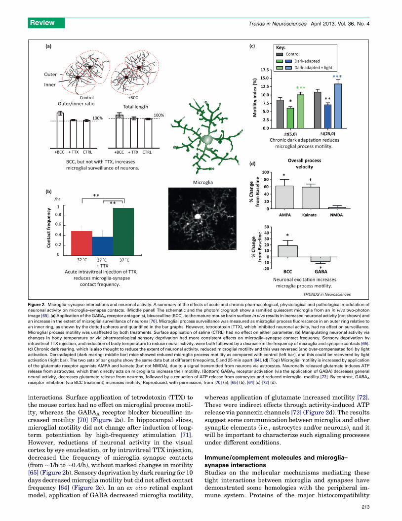
neuronal activity on microglia–synapse contacts. (Middle panel) The schematic and the photomicrograph show a ramified quiescent microglia from an in vivo two-photon
image [65]. (a) Application of the GABAA receptor antagonist, bicucculline (BCC), to the mature mouse brain surface in vivo results in increased neuronal activity (not shown) and
an increase in the extent of microglial surveillance of neurons [70]. Microglial process surveillance was measured as microglial process fluorescence in an outer ring relative to
an inner ring, as shown by the dotted spheres and quantified in the bar graphs. However, tetrodotoxin (TTX), which inhibited neuronal activity, had no effect on surveillance.
Microglial process motility was unaffected by both treatments. Surface application of saline (CTRL) had no effect on either parameter. (b) Manipulating neuronal activity via
changes in body temperature or via pharmacological sensory deprivation had more consistent effects on microglia–synapse contact frequency. Sensory deprivation by
intravitreal TTX injection, and reduction of body temperature to reduce neural activity, were both followed by a decrease in the frequency of microglia and synapse contacts [65].
(c) Chronic dark rearing, which is also thought to reduce the extent of neuronal activity, reduced microglial motility and this was reversed (and over-compensated for) by light
activation. Dark-adapted (dark rearing: middle bar) mice showed reduced microglia process motility as compared with control (left bar), and this could be recovered by light
activation (right bar). The two sets of bar graphs show the same data but at different timepoints, 5 and 25 min apart [64]. (d) (Top) Microglial motility is increased by application
of the glutamate receptor agonists AMPA and kainate (but not NMDA), due to a signal transmitted from neurons via astrocytes. Neuronally released glutamate induces ATP
release from astrocytes, which then directly acts on microglia to increase their motility. (Bottom) GABAA receptor activation (via the application of GABA) decreases general
neural activity, decreases glutamate release from neurons, followed by a reduction of ATP release from astrocytes and reduced microglial motility [72]. By contrast, GABAA
receptor inhibition (via BCC treatment) increases motility. Reproduced, with permission, from [70] (a), [65] (b), [64] (c) [72] (d).
Review Trends in Neurosciences April 2013, Vol. 36, No. 4
interactions. Surface application of tetrodotoxin (TTX) tothe mouse cortex had no effect on microglial process motil-ity, whereas the GABAA receptor blocker bicuculline in-creased motility [70] (Figure 2a). In hippocampal slices,microglial motility did not change after induction of long-term potentiation by high-frequency stimulation [71].However, reductions of neuronal activity in the visualcortex by eye enucleation, or by intravitreal TTX injection,decreased the frequency of microglia–synapse contacts(from �1/h to �0.4/h), without marked changes in motility[65] (Figure 2b). Sensory deprivation by dark rearing for 10days decreased microglia motility but did not affect contactfrequency [64] (Figure 2c). In an ex vivo retinal explantmodel, application of GABA decreased microglia motility,
whereas application of glutamate increased motility [72].These were indirect effects through activity-induced ATPrelease via pannexin channels [72] (Figure 2d). The resultssuggest some communication between microglia and othersynaptic elements (i.e., astrocytes and/or neurons), and itwill be important to characterize such signaling processesunder different conditions.
Immune/complement molecules and microglia–synapse interactionsStudies on the molecular mechanisms mediating thesetight interactions between microglia and synapses havedemonstrated some homologies with the peripheral im-mune system. Proteins of the major histocompatibility
213
Review Trends in Neurosciences April 2013, Vol. 36, No. 4
complex class I (MHC-1) and complement cascade (C1qand C3) are expressed in various neurons in an activity-dependent fashion [73,74]. A classic model of developmentalsynaptic pruning is the segregation of projections fromretinal ganglion cells (RGCs) of each eye into the appropriateregions in the lateral geniculate nucleus (LGN). The expres-sion of MHC-1 molecules is reduced when neuronal activityin the visual pathway is blocked, a manipulation which alsodisrupts this synaptic pruning [73]. A more direct role for theMHC-1 pathway comes from knockout mice. Loss of b2m, alight-chain protein found in most MHC-1 complexes, or thetransporter associated with the antigen processing 1(TAP1), a protein involved with loading of peptides on toMHC-1 complexes, results in reduction of this pruning andsegregation [75]. MHC-1 molecules and some of their recep-tors are upregulated after axotomy, but an increased loss ofsynapses is seen in b2m- or TAP-1-deficient mice followingaxotomy [76]. The excess loss of synapses in the absence ofMHC-1 mostly involved inhibitory terminals, suggesting aspecific role of MHC-1 signaling in preserving inhibitoryinputs [77]. The precise mechanisms by which specificsynapses are identified for elimination, and the contributionof microglia, requires further investigation.
Mice devoid of genes for the classic complement proteinsC1q and C3 also show reduced developmental pruning, withLGN neurons remaining multi-innervated by RGC inputs[74]. By analogy with the innate immune system, where C1qprotein initiates a cascade in which C3 tags synapses forphagocytosis, these complement molecules were proposed totarget synapses for pruning by microglia [69]. Consistently,the receptor for C3 is transiently upregulated in microglia
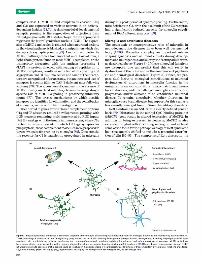
Neural development
Microglia
Adult neurogenesis
PCD [31]
-Phagocytosis [35]
(a)
(b)
Figure 3. Physiological roles of microglia. Schematic diagrams of the multiple postulate
These physiological functions include (a) regulating programmed cell death (PCD) durin
new-born cells, and (c) the surveillance, monitoring, and pruning of presynaptic termin
been demonstrated to be associated with a number of neurological and psychiatric dis
[85]. It is tempting to speculate that microglia could be involved in the pathogenesis of n
Key: blue, neuron; green, microglia; grey, dysfunctional microglia; red, synapses on de
214
during this peak period of synaptic pruning. Furthermore,mice deficient in C3, or in the a subunit of the C3 receptor,had a significantly reduced capacity for microglia engulf-ment of RGC afferent synapses [69].
Microglia and psychiatric disordersThe neurotoxic or neuroprotective roles of microglia inneurodegenerative diseases have been well documented(e.g., [1,78]). Microglia also play an important role inshaping synapses and neuronal circuits during develop-ment and neurogenesis, and survey the resting adult brain,as described above (Figure 3). If these microglial functionsare disrupted, one can predict that this will result indysfunction of the brain and in the emergence of psychiat-ric and neurological disorders (Figure 3). Hence, we pro-pose dual facets to microglial contributions to neuronaldysfunction: (i) alterations in microglia function in theuninjured brain can contribute to psychiatric and neuro-logical diseases, and (ii) challenged microglia can affect theprogression and/or outcome of an established neuronaldisease. It remains speculative whether alterations inmicroglia cause brain disease, but support for this scenariohas recently emerged from different hereditary disorders.
Rett syndrome is an ASD with a clearly defined geneticbasis [79]. Mutations in the methyl-CpG-binding protein 2(MECP2) gene result in altered expression of MeCP2. Inaddition to being expressed in neurons, MeCP2 is alsoexpressed in glial cells (including microglia) and at leastsome of the focus for the pathophysiology of Rett syndromehas consequently shifted to include a potential contribu-tion of glia [80–83]. The symptoms of Rett disease in the
Synap�c interac�ons
Neurological andpsychiatric disorders
- Re� syndrome [83,84]- OCD [85]
- Surveillance [64,65]
- Elimina�on/pruning [69]
- Phagocytosis [68]
- Synap�c plas�city [59]
??
TRENDS in Neurosciences
(c)
(d)
d physiological functions of microglia in forming and maintaining neuronal circuits.
g development, (b) regulation of neurogenesis, including the phagocytosis of adult
als and dendritic spines to maintain homeostasis of synapses. (d) Microglia have
orders, including Rett syndrome [83,84] and obsessive-compulsive disorder (OCD)
eurological disorders if any of these important physiological functions are altered.
ndrites; yellow, neural lineage cells.
Box 1. Outstanding questions
� Under what specific circumstances, and to what extent, can
microglia be supplied to the brain from circulating monocytes?
This question is important because it raises the possibility that
circulating monocytes may be a novel pathway to deliver
‘therapeutic microglia’ to treat neurological and psychiatric
disease.
� What are the signals and transduction mechanisms by which
microglia communicate with developing neurons and adult neural
progenitors to promote and/or regulate PCD?
� Do the same mechanisms mediate microglia–synapse interactions
during developmental pruning of synapses, and during synaptic
stripping following injury?
� What are the mechanisms and functional consequences of
microglia surveillance of synapses in uninjured adult CNS? How
do microglia choose a specific synapse to be eliminated, while
others are spared?
� How do microglia sense the different levels of electrical activity of
neuronal circuits with the necessary fine spatial resolution?
� Are the mechanisms by which microglia prune excitatory and
inhibitory synapses the same? Do microglia selectively survey
excitatory synapses in adult CNS and, if so, what may be the
function of such a selective interaction?
� Does microglia dysfunction result in disease? Determining the
mechanisms by which the known microglial hereditary patholo-
gies result in their cognitive symptoms will shed light on possible
broader contributions of microglial dysfunction to neurological
disorders.
Review Trends in Neurosciences April 2013, Vol. 36, No. 4
MeCP2 null mouse are ameliorated by irradiation andbone-marrow transplantation [84], a procedure known toreplenish the microglia population. The disease was alsoameliorated in a MeCP2lox/Lysmcre transgenic mouse, inwhich expression of MeCP2 was restored specifically inmyeloid cells, supporting the locus of effect to microglia. Itwas concluded that disease progression was inhibited byrestoring microglial phagocytic capacity to reduce the ac-cumulation of debris [84]. However, microglia–synapseinteractions may have also contributed to disease inhibi-tion. The time-window in which microglia could reducedisease progression (i.e., P28 but not P40) [84] would beconsistent with an effect on synapse maturation.
The pathogenesis of a mouse model of obsessive com-pulsive disorder (OCD) has also been linked to microglia. Aloss of function mutation in the transcription factor gene,Hoxb8, results in obsessive grooming and hair pulling inmice, and this has been associated with loss of Hox8b inmicroglia [85]. The phenotype was rescued by irradiationand bone-marrow transplantation, and was replicated in amicroglia-specific knockout of Hox8b [85]. Puzzlingly, theHox8b-deficient microglia (which accounted for about 40%of the adult microglia population) were traced to a hemato-poietic bone marrow-derived cell lineage which appearedlater in development (i.e., from P2) [85] instead of to theyolk-sac progenitors that have been shown to constitutethe adult resident microglia population (see above discus-sion). Hence, additional studies are needed to clarify theseseemingly contradictory findings.
In another example, mutations affecting a CSF (i.e.,CSF-1) have been linked to a late-onset neurodegenerativedisease known as hereditary diffuse leukoencephalopathywith spheroids (HDLS) [86], which is characterized bysymptoms that include depression, anxiety, aggres-siveness, and dementia [87]. CSF-1 is an important regu-lator of microglial proliferation and differentiation, and itwas proposed that the primary pathogenesis of the disor-der was dysfunction of microglial reactivity. Mutations inCSF-1 or its signaling pathways have been also observed insome other neurological disorders, including frontal tem-poral lobe dementia and Nasu–Hokala disease [86,87]. Thefunctional consequences of altered CSF-1 signaling inthese disorders, and selective rescue by normal microglia,have yet to be investigated.
Taken together, the results related to these hereditarydisorders suggest that microglial dysfunction may precipi-tate neuronal disease. Early-life activation of microglia byLPS, infection, or stress can result in cognitive disabilitiesthat can persist through to adulthood [88]. Depression,ASDs, bipolar disorder, and schizophrenia are known tobe associated with microglial activation [87,88], but whetherthis contributes to their pathogenesis is unknown. It hasbeen suggested that altered reactivity of microglia maycouple with other environmental and genetic factorsto precipitate schizophrenia and depression [88]. Wefurther speculate that altered homeostatic functions ofresting microglia may also contribute to neurological andpsychiatric disorders, either directly or via interactions withother environmental and/or genetic factors. In schizophre-nia, functional magnetic resonance imaging (fMRI) andpositron emission tomography (PET) have revealed both a
wider range of spontaneous cortical activity and a greatervariability in evoked cortical response patterns as comparedto control subjects [89,90]. At the morphological level,schizophrenia is associated with a decreased number ofsynapses in affected regions [51,91]. Spine morphologicalchanges have been also seen in schizophrenia [92,93] and indepression [94]. Decreases in the number of synaptic spineshave also been recently reported in major depressive disor-der [95]. It is interesting to speculate that microglial regu-lation of synapse number or morphology may contribute tosome of the psychiatric symptoms apparent in these dis-orders, although further studies are needed to address thispossibility.
Concluding remarksIn this review we have highlighted recent observationsregarding the CNS migration and proliferation of micro-glia, and the subsequent physiological roles that theseresident microglia mediate. In addition to their neuronalimmune function in reacting to infections and clearingdebris by phagocytosis, both resting and challenged micro-glia play an active role in neuronal circuit homeostasis.They contribute to PCD and synapse elimination duringmaturation of neuronal circuits and in response to neuro-nal injury. They also play a role in synapse and circuithomeostasis, by surveillance of synaptic elements in theadult brain and by actively participating in neurogenesis.Direct interactions between microglia and synaptic ele-ments have been shown in uninjured brain, and some keymolecules involved in these interactions include immunerecognition molecules such as complement proteins. Theability of microglia to release ATP, neurotrophic factors,and cytokines underlies their capacity to modify neuralcircuits in physiological and pathological states. Themicroglia–synapse interactions during development and
215
Review Trends in Neurosciences April 2013, Vol. 36, No. 4
in adult homeostasis help to fine-tune neural circuits tooptimize information processing and learning and memo-ry. Such important physiological roles suggest that dys-function of microglia may contribute to psychiatric andneurological disorders, and recent studies have identifiedhereditary disorders where microglial dysfunction hasbeen implicated in disease pathogenesis. Such findingssuggest that a paradigm shift is needed from consideringmicroglia as only contributing indirectly to disease pro-gression to potentially being actively involved in precipi-tating disease. However, much remains to be learnedabout the mechanisms and physiological consequencesof microglia–synapse circuit interactions (Box 1). Addres-sing these issues will require new approaches for selectivemodification of microglia to reveal exactly how microgliaaffect neuronal activity, information processing, and dis-ease pathogenesis.
Note added in proofA recent paper by Li et al. [96] confirmed and extended inzebrafish larvae the characterisation of resting microglia-neuron contacts that has been described previously inmouse brain [65]. Microglial processes made frequent brief(5–6 mins) contacts with neuronal somata in the optictectum of the larvae. Orientated movement towards neu-rons was activity-dependent and subsequent contacts wereassociated with an enlargment of the microglia process endinto a bulbous tip. Both these steps were reduced when thesmall Rho GTPase Rac and neuronal pannexin channelswere inhibited. Interestingly, microglia-neuron contactsresulted in a decrease of neuronal activity, suggesting ahomeostatic role of microglia in identifying hyperactiveneurons and then reducing their activity level.
AcknowledgmentsWe gratefully acknowledge Wai T. Wong, R. Douglas Fields, and OlenaBukalo for critical reading of the manuscript. This work was supported bya Japan Society for the Promotion of Science fellowship (to H.W.) and theJapan Science and Technology Agency for Core Research for EvolutionalScience and Technology (to J.N.).
References1 Kettenmann, H. et al. (2011) Physiology of microglia. Physiol. Rev. 91,
461–5532 Perry, V.H. et al. (2010) Microglia in neurodegenerative disease. Nat.
Rev. Neurol. 6, 193–2013 Prinz, M. et al. (2011) Heterogeneity of CNS myeloid cells and their
roles in neurodegeneration. Nat. Neurosci. 14, 1227–12354 Kaur, C. et al. (2001) Origin of microglia. Microsc. Res. Tech. 54, 2–95 Hanisch, U.K. (2002) Microglia as a source and target of cytokines. Glia
40, 140–1556 Lawson, L.J. et al. (1990) Heterogeneity in the distribution and
morphology of microglia in the normal adult mouse brain.Neuroscience 39, 151–170
7 Perry, V.H. et al. (1985) Immunohistochemical localization ofmacrophages and microglia in the adult and developing mousebrain. Neuroscience 15, 313–326
8 Prinz, M. and Mildner, A. (2011) Microglia in the CNS: immigrantsfrom another world. Glia 59, 177–187
9 Beers, D.R. et al. (2006) Wild-type microglia extend survival in PU.1knockout mice with familial amyotrophic lateral sclerosis. Proc. Natl.Acad. Sci. U.S.A. 103, 16021–16026
10 Ashwell, K. (1990) Microglia and cell death in the developing mousecerebellum. Brain Res. 55, 219–230
11 Chan, W.Y. et al. (2007) The origin and cell lineage of microglia: newconcepts. Brain Res. Rev. 53, 344–354
216
12 Ginhoux, F. et al. (2010) Fate mapping analysis reveals thatadult microglia derive from primitive macrophages. Science 330,841–845
13 Gomez Perdiguero, E. et al. (2013) Development and homeostasis of‘resident’ myeloid cells: the case of the microglia. Glia 61, 112–120
14 Schulz, C. et al. (2012) A lineage of myeloid cells independent ofMyb and hematopoietic stem cells. Science (New York, N.Y.) 336,86–90
15 Lawson, L.J. et al. (1992) Turnover of resident microglia in the normaladult mouse brain. Neuroscience 48, 405–415
16 Alliot, F. et al. (1999) Microglia derive from progenitors, originatingfrom the yolk sac, and which proliferate in the brain. Brain Res. 117,145–152
17 Ajami, B. et al. (2007) Local self-renewal can sustain CNS microgliamaintenance and function throughout adult life. Nat. Neurosci. 10,1538–1543
18 Zusso, M. et al. (2012) Regulation of postnatal forebrain amoeboidmicroglial cell proliferation and development by the transcriptionfactor runx1. J. Neurosci. 32, 11285–11298
19 Davoust, N. et al. (2008) From bone marrow to microglia: barriers andavenues. Trends Immunol. 29, 227–234
20 Gehrmann, J. et al. (1995) Microglia: intrinsic immuneffector cell of thebrain. Brain Res. Brain Res. Rev. 20, 269–287
21 Ladeby, R. et al. (2005) Microglial cell population dynamics in theinjured adult central nervous system. Brain Res. Brain Res. Rev. 48,196–206
22 Rappert, A. et al. (2004) CXCR3-dependent microglial recruitment isessential for dendrite loss after brain lesion. J. Neurosci. 24, 8500–8509
23 Bechmann, I. et al. (2005) Circulating monocytic cells infiltrate layersof anterograde axonal degeneration where they transform intomicroglia. FASEB J. 19, 647–649
24 Ajami, B. et al. (2011) Infiltrating monocytes trigger EAE progression,but do not contribute to the resident microglia pool. Nat. Neurosci. 14,1142–1149
25 Hulette, C.M. (1996) Microglioma, a histiocytic neoplasm of the centralnervous system. Mod. Pathol. 9, 316–319
26 de la Rosa, E.J. and de Pablo, F. (2000) Cell death in early neuraldevelopment: beyond the neurotrophic theory. Trends Neurosci. 23,454–458
27 Marin-Teva, J.L. et al. (1999) Naturally occurring cell death andmigration of microglial precursors in the quail retina during normaldevelopment. J. Comp. Neurol. 412, 255–275
28 Rakic, S. and Zecevic, N. (2000) Programmed cell death in thedeveloping human telencephalon. Eur. J. Neurosci. 12, 2721–2734
29 Bessis, A. et al. (2007) Microglial control of neuronal death and synapticproperties. Glia 55, 233–238
30 Marin-Teva, J.L. et al. (2011) Microglia and neuronal cell death.Neuron Glia Biol. 7, 25–40
31 Marin-Teva, J.L. et al. (2004) Microglia promote the death ofdeveloping Purkinje cells. Neuron 41, 535–547
32 Manto, M.U. and Jissendi, P. (2012) Cerebellum: links betweendevelopment, developmental disorders and motor learning. Front.Neuroanat. 6, 1
33 Wei, H. et al. (2012) Alteration of brain volume in IL-6 overexpressingmice related to autism. Int. J. Dev. Neurosci. 30, 554–559
34 Gould, E. and McEwen, B.S. (1993) Neuronal birth and death. Curr.Opin. Neurobiol. 3, 676–682
35 Sierra, A. et al. (2010) Microglia shape adult hippocampal neurogenesisthrough apoptosis-coupled phagocytosis. Cell Stem Cell 7, 483–495
36 Choi, S.H. et al. (2008) Non-cell-autonomous effects of presenilin 1variants on enrichment-mediated hippocampal progenitor cellproliferation and differentiation. Neuron 59, 568–580
37 Parent, J.M. et al. (1997) Dentate granule cell neurogenesis is increasedby seizures and contributes to aberrant network reorganization in theadult rat hippocampus. J. Neurosci. 17, 3727–3738
38 Curtis, M.A. et al. (2007) The effect of neurodegenerative diseases onthe subventricular zone. Nat. Rev. Neurosci. 8, 712–723
39 Jin, K. et al. (2001) Neurogenesis in dentate subgranular zone androstral subventricular zone after focal cerebral ischemia in the rat.Proc. Natl. Acad. Sci. U.S.A. 98, 4710–4715
40 Rodriguez, J.J. et al. (2008) Impaired adult neurogenesis in the dentategyrus of a triple transgenic mouse model of Alzheimer’s disease. PLoSONE 3, e2935
Review Trends in Neurosciences April 2013, Vol. 36, No. 4
41 Rodriguez, J.J. et al. (2009) Impaired cell proliferation in thesubventricular zone in an Alzheimer’s disease model. Neuroreport20, 907–912
42 Taffe, M.A. et al. (2010) Long-lasting reduction in hippocampalneurogenesis by alcohol consumption in adolescent nonhumanprimates. Proc. Natl. Acad. Sci. U.S.A. 107, 11104–11109
43 Inokuchi, K. (2011) Adult neurogenesis and modulation of neuralcircuit function. Curr. Opin. Neurobiol. 21, 360–364
44 Ziv, Y. et al. (2006) Immune cells contribute to the maintenance ofneurogenesis and spatial learning abilities in adulthood. Nat. Neurosci.9, 268–275
45 Rogers, J.T. et al. (2011) CX3CR1 deficiency leads to impairment ofhippocampal cognitive function and synaptic plasticity. J. Neurosci. 31,16241–16250
46 Vukovic, J. et al. (2012) Microglia modulate hippocampal neuralprecursor activity in response to exercise and aging. J. Neurosci. 32,6435–6443
47 Hua, J.Y. and Smith, S.J. (2004) Neural activity and the dynamics ofcentral nervous system development. Nat. Neurosci. 7, 327–332
48 Lichtman, J.W. and Colman, H. (2000) Synapse elimination andindelible memory. Neuron 25, 269–278
49 Mei, L. and Xiong, W.C. (2008) Neuregulin 1 in neural development,synaptic plasticity and schizophrenia. Nat. Rev. Neurosci. 9, 437–452
50 Palop, J.J. and Mucke, L. (2010) Amyloid-beta-induced neuronaldysfunction in Alzheimer’s disease: from synapses toward neuralnetworks. Nat. Neurosci. 13, 812–818
51 Penzes, P. et al. (2011) Dendritic spine pathology in neuropsychiatricdisorders. Nat. Neurosci. 14, 285–293
52 Rapin, I. and Tuchman, R.F. (2008) What is new in autism? Curr. Opin.Neurol. 21, 143–149
53 Moran, L.B. and Graeber, M.B. (2004) The facial nerve axotomy model.Brain Res. Brain Res. Rev. 44, 154–178
54 Jinno, S. and Yamada, J. (2011) Using comparative anatomy in theaxotomy model to identify distinct roles for microglia and astrocytes insynaptic stripping. Neuron Glia Biol. 7, 55–66
55 Kalla, R. et al. (2001) Microglia and the early phase of immunesurveillance in the axotomized facial motor nucleus: impairedmicroglial activation and lymphocyte recruitment but no effecton neuronal survival or axonal regeneration in macrophage-colonystimulating factor-deficient mice. J. Comp. Neurol. 436, 182–201
56 Blinzinger, K. and Kreutzberg, G. (1968) Displacement of synapticterminals from regenerating motoneurons by microglial cells. Z.Zellforsch. Mikrosk. Anat. 85, 145–157
57 Trapp, B.D. et al. (2007) Evidence for synaptic stripping by corticalmicroglia. Glia 55, 360–368
58 Yamada, J. et al. (2011) Differential involvement of perineuronalastrocytes and microglia in synaptic stripping after hypoglossalaxotomy. Neuroscience 182, 1–10
59 Pascual, O. et al. (2012) Microglia activation triggers astrocyte-mediated modulation of excitatory neurotransmission. Proc. Natl.Acad. Sci. U.S.A. 109, E197–E205
60 Li, X. et al. (2012) MEK is a key regulator of gliogenesis in thedeveloping brain. Neuron 75, 1035–1050
61 Tress, O. et al. (2012) Panglial gap junctional communication isessential for maintenance of myelin in the CNS. J. Neurosci. 32,7499–7518
62 Denk, W. et al. (1990) Two-photon laser scanning fluorescencemicroscopy. Science 248, 73–76
63 Denk, W. and Svoboda, K. (1997) Photon upmanship: why multiphotonimaging is more than a gimmick. Neuron 18, 351–357
64 Tremblay, M.E. et al. (2010) Microglial interactions with synapses aremodulated by visual experience. PLoS Biol. 8, e1000527
65 Wake, H. et al. (2009) Resting microglia directly monitor the functionalstate of synapses in vivo and determine the fate of ischemic terminals.J. Neurosci. 29, 3974–3980
66 Hirasawa, T. et al. (2005) Visualization of microglia in living tissuesusing Iba1-EGFP transgenic mice. J. Neurosci. Res. 81, 357–362
67 Feng, G. et al. (2000) Imaging neuronal subsets in transgenic miceexpressing multiple spectral variants of GFP. Neuron 28, 41–51
68 Paolicelli, R.C. et al. (2011) Synaptic pruning by microglia is necessaryfor normal brain development. Science 333, 1456–1458
69 Schafer, D.P. et al. (2012) Microglia sculpt postnatal neural circuits inan activity and complement-dependent manner. Neuron 74, 691–705
70 Nimmerjahn, A. et al. (2005) Resting microglial cells are highlydynamic surveillants of brain parenchyma in vivo. Science 308,1314–1318
71 Wu, L.J. and Zhuo, M. (2008) Resting microglial motility isindependent of synaptic plasticity in mammalian brain. J.Neurophysiol. 99, 2026–2032
72 Fontainhas, A.M. et al. (2011) Microglial morphology and dynamicbehavior is regulated by ionotropic glutamatergic and GABAergicneurotransmission. PLoS ONE 6, e15973
73 Corriveau, R.A. et al. (1998) Regulation of class I MHC gene expressionin the developing and mature CNS by neural activity. Neuron 21,505–520
74 Stevens, B. et al. (2007) The classical complement cascade mediatesCNS synapse elimination. Cell 131, 1164–1178
75 Huh, G.S. et al. (2000) Functional requirement for class I MHC in CNSdevelopment and plasticity. Science 290, 2155–2159
76 Cullheim, S. and Thams, S. (2007) The microglial networks of the brainand their role in neuronal network plasticity after lesion. Brain Res.Rev. 55, 89–96
77 Linda, H. et al. (2000) Ultrastructural evidence for a preferentialelimination of glutamate-immunoreactive synaptic terminals fromspinal motoneurons after intramedullary axotomy. J. Comp. Neurol.425, 10–23
78 Hanisch, U.K. and Kettenmann, H. (2007) Microglia: active sensor andversatile effector cells in the normal and pathologic brain. Nat.Neurosci. 10, 1387–1394
79 Van den Veyver, I.B. and Zoghbi, H.Y. (2000) Methyl-CpG-bindingprotein 2 mutations in Rett syndrome. Curr. Opin. Genet. Dev. 10,275–279
80 Ballas, N. et al. (2009) Non-cell autonomous influence of MeCP2-deficient glia on neuronal dendritic morphology. Nat. Neurosci. 12,311–317
81 Kifayathullah, L.A. et al. (2010) MeCP2 mutant protein is expressed inastrocytes as well as in neurons and localizes in the nucleus. Cytogenet.Genome Res. 129, 290–297
82 Lioy, D.T. et al. (2011) A role for glia in the progression of Rett’ssyndrome. Nature 475, 497–500
83 Maezawa, I. and Jin, L.W. (2010) Rett syndrome microglia damagedendrites and synapses by the elevated release of glutamate. J.Neurosci. 30, 5346–5356
84 Derecki, N.C. et al. (2012) Wild-type microglia arrest pathology in amouse model of Rett syndrome. Nature 484, 105–109
85 Chen, S.K. et al. (2010) Hematopoietic origin of pathological groomingin Hoxb8 mutant mice. Cell 141, 775–785
86 Rademakers, R. et al. (2012) Mutations in the colony stimulating factor1 receptor (CSF1R) gene cause hereditary diffuse leukoencephalopathywith spheroids. Nat. Genet. 44, 200–205
87 Blank, T. and Prinz, M. (2013) Microglia as modulators of cognition andneuropsychiatric disorders. Glia 61, 62–70
88 Beumer, W. et al. (2012) The immune theory of psychiatric diseases: akey role for activated microglia and circulating monocytes. J. Leukoc.Biol. 92, 959–975
89 Dierks, T. et al. (1999) Activation of Heschl’s gyrus during auditoryhallucinations. Neuron 22, 615–621
90 Silbersweig, D.A. et al. (1995) A functional neuroanatomy ofhallucinations in schizophrenia. Nature 378, 176–179
91 Lewis, D.A. et al. (2003) Altered cortical glutamate neurotransmissionin schizophrenia: evidence from morphological studies of pyramidalneurons. Ann. N. Y. Acad. Sci. 1003, 102–112
92 Garey, L.J. et al. (1998) Reduced dendritic spine density on cerebralcortical pyramidal neurons in schizophrenia. J. Neurol. Neurosurg.Psychiatry 65, 446–453
93 Glausier, J.R. and Lewis, D.A. (2012) Dendritic spine pathology inschizophrenia. Neuroscience http://dx.doi.org/10.1016/j.neuroscience.2012.04.044
94 Blugeot, A. et al. (2011) Vulnerability to depression: from brainneuroplasticity to identification of biomarkers. J. Neurosci. 31,12889–12899
95 Kang, H.J. et al. (2012) Decreased expression of synapse-relatedgenes and loss of synapses in major depressive disorder. Nat. Med. 18,1413–1417
96 Li, Y. et al. (2012) Reciprocal regulation between resting microglialdynamics and neuronal activity in vivo. Dev. Cell 23, 1189–1202
217