microcirculation in icu
TRANSCRIPT

Microcirculation Monitoring in Critical illness
Dr.Tarek Sabri
INTENSIVIST
Fujairah Hospital
UAE Nov 22- 2016

Agenda
• 1- Introduction.
• 2-Microcirulation.
• 3- Microcirculation in SEPSIS.
• 4- Microcirculation in SURGICAL Patient.
• 5- Microcirculation in various Critically ill patients .
• 6-Resuscitation effects on Microcirculation .
• 7-Sammary.
• 8-Conclusion. 2

1- INTRODUCTION
• Critical illness includes a wide range of disease states such as sepsis, high-risk surgery, cardiac arrest, and respiratory failure, and is associated with reduced tissue oxygenation related to a compromise in the cardiovascular system (CVS).
• Microcirculation involves the smallest branches of the CVS and plays an essential role in the transport of oxygen to the parenchymal cells needed to sustain organ function [1]
3

Introduction cont
• It is generally accepted that resuscitation procedures should aim to correct macro hemodynamic variables during critical illness, with the goal of improving tissue perfusion.
• However, although macrohemodynamic targets may be reached, it is often uncertain whether these procedures lead to a parallel improvement in the microcirculation[2]
• In this presentation, we will discuss the microcirculatory alterations in critical illness and the importance of hemodynamic coherence between the macrocirculation and microcirculation that occurs in response to resuscitation.
4

2- THE MICROCIRCULATION AND HEMODYNAMIC COHERENCE
• Microcirculation consists of a branching network of small blood vessels (<100µm diameter) that includes the arterioles, capillaries, and venules, and plays a vital role in the delivery of oxygen to tissue cells [3].
• The main mechanisms of oxygen transport are the convective flow of red blood cells (RBCs) and the passive diffusion of oxygen from the RBCs to the tissue cells [4].
5

MICROCIRCULATION Coherence CONT---
• As convective flow refers to the transport of oxygen-carrying RBCs to the capillaries, passive diffusion refers to the transport of oxygen from the RBCs in the capillaries to the tissue cells.
• Resuscitation procedures primarily target the correction of convective RBC flow under the assumption that hypovolemia is primarily associated with inadequate blood flow.
• However, convective and diffusive flows have an equal contributory effect on the transport of oxygen [4].
6

MICROCIRCULATION Coherence CONT-
• the normalization of systemic hemodynamic variables alone may increase convective flow but may not necessarily mean that adequate oxygen is being delivered to the tissues, especially if nonoxygen carrying resuscitation fluids are used and if areas of the microcirculation are obstructed [5].
7

MICROCIRCULATION Coherence CONT-
• Loss of hemodynamic coherence between the macrocirculation and the microcirculation can occur under various conditions of microcirculatory alterations (see Fig. 1)
• This led us to introduce the term of hemodynamic coherence [5] to describe the condition where resuscitation is successful if macrocirculatory resuscitation causes a parallel improvement in the perfusion and oxygen of the microcirculation.
8

Figure 1
9

MICROCIRCULATION Coherence CONT-
• where correction of systemic hemodynamic variables does not cause a parallel improvement in the condition of the microcirculation resulting in a lack of tissue perfusion despite apparently normalized systemic hemodynamic.
• Because loss of hemodynamic coherence does not improve surrogates of hypovolemia, such as lactate, oliguria, and strong ion difference, which are either related to microcirculatory dysfunction not being improved by targeting microcirculatory parameters or are being caused by other factors not related to conventional resuscitation procedures.
10

MICROCIRCULATION Coherence CONT-
• Clinician at the bedside will continue administrating inappropriate amounts of fluids and vasoactive drugs potentially causing harm.
• To identify this condition and assess the presence or absence of hemodynamic coherence, monitoring of the microcirculation is essential.
11

MICROCIRCULATION Coherence CONT-
• The microcirculation is controlled by many regulatory and compensatory systems including hormonal, neural, biochemical, and vascular control systems that all must be intact to respond adequately to systemic hemodynamic changes [6].
• However, these regulatory systems are often damaged in critically ill patients because of infection, inflammation, and regional ischemia or hypoxia, resulting in a loss of hemodynamic coherence between macrocirculation and microcirculation, and vulnerable microcirculatory units in organ beds that become shunted and hypoxic, manifesting clinically as a reduction in oxygen extraction [5,7].
12

MICROCIRCULATION CONT-
• Several studies have shown that the normalization of systemic hemodynamic variables does not always lead to parallel improvements in microcirculation and cell oxygenation, especially under specific conditions [8–13].
•
• Four types of microcirculatory alteration are associated with a loss of hemodynamic coherence between the macrocirculation and microcirculation (Fig. 1) [5&7].
13

MICROCIRCULATION CONT-
• alterations are associated with heterogeneity in the perfusion of the microcirculation, in which some capillaries are obstructed next to capillaries with flowing RBCs, and can be observed in states of inflammation, especially sepsis and reperfusion injury.
Type 1 : HETEROGENEITY
14

MICROCIRCULATION CONT-
• represents conditions of hemodilution, in which dilution of the blood causes a decrease in capillary hematocrit, resulting in increased diffusion distances between the oxygen-carrying RBCs and the tissue cells; this situation occurs mainly in cardiac surgery and also in sepsis when excessive nonoxygen carrying resuscitation fluids are given.
Type 2 ( HEMODILUTION)
15

MICROCIRCULATION CONT-
microcirculatory alterations occur when there is a vasoconstriction/tamponade of the microcirculation caused by the excessive use of vasopressors and/or increased venous pressure.
Type 3:-
CONSTRICTION/TAMPONAD
16

MICROCIRCULATION CONT-
•microcirculatory alteration is associated with tissue edema caused by damage to the endothelial cells and the loss of glycocalyx, capillary leakage because of compromised vascular barriers and fluid overload, all of which can be observed in sepsis, reperfusion injury, and surgery.
Type 4 EDEMA
17

MICROCIRCULATION CONT-
• Various methods can be used to visualize microcirculation. Sublingual microcirculation is the most commonly used area to visualize the microcirculation, and the use of this site to investigate the clinical effects of disease and therapy on the microcirculation is well established [14].
• Three generations of hand-held microscopes have been developed to monitor the sublingual microcirculation [15].
18

MICROCIRCULATION CONT-
• Orthogonal polarization spectral (OPS) and sidestream dark field (SDF) imaging methods are, respectively, the first-generation and second generation microcirculation-monitoring devices.
• These earlier devices had some limitations such as suboptimal optics and the lack of a direct computer control in the imaging modality, which is needed for direct bedside evaluation of the images. Recently, a third-generation lightweight device, the CytoCam-IDF device, was developed based on incident dark field imaging [16–18].
19

MICROCIRCULATION CONT- •
•The Cyto Cam-IDF device consists of a computer-controlled, high resolution image sensor in combination with a specifically designed microscope lens that provides better image quality, enabling the detection of more capillaries than previous generation devices [17,19,20].
• Although this device visualizes microcirculatory changes better than previous devices, it is a technique under continuous development, as clinical requirements provide new technical specification challenges.
• Pressure artifacts, the limited focus depth, the need to stabilize the microscope lens on the tissue surface and improved automatic image analysis are still limitations in need of technical development.
20

• CytoCam
• The CytoCam-IDF handheld microcirculation camera is offers a complete range of hardware and software for capturing images and analysis of the microcirculation.
21

22

• The perfusion of a tissue depends on the number, distribution, and diameters of the capillaries in combination with blood viscosity and driving pressure across the capillaries.
• There are two main hemodynamic principles governing how oxygen in red blood cells reaches the tissue cells; the first is the convection based on red blood cell flow, and the second is the diffusion distance oxygen must travel from the red blood cells in the capillaries to the parenchymal cells [19].
23

• Convection is quantified by measurement of flow in the microvessels, and diffusion is quantified by the density of the perfused microvessels (functional capillary density).
• Assessment can be don by measuring of microcirculatory parameters by using Software assisted analysis (AVA 3.0; Automated Vascular Analysis, Academic Medical Center, University of Amsterdam)
24

• The total vessel density (TVD; mm/mm2) is determine by using the AVA software.
• A semi quantitative analysis previously validated [23] and assisted by the AVA software is performed in individual vessels that distinguished among no flow (0), intermittent flow (1), sluggish flow (2), and continuous flow (3). A value was assigned for each vessel.
• The overall score, called the microvascular flow index (MFI), is the average of the individual values [24].
25

• The proportion of perfused vessels (PPV) was calculated as the number of vessels with flow values of 2 and 3 divided by the total number of vessels.
• Perfused vessel density (PVD) was determined as the total vessel density multiplied by the fraction of perfused vessels [22].
26

27

• Smaller SDF image in larger Cytocam-IDF image. This figure shows the field of view of SDF and Cytocam-IDF imaging superimposed on each other showing the larger field of view offered by the larger image sensor used by the later technique.
28
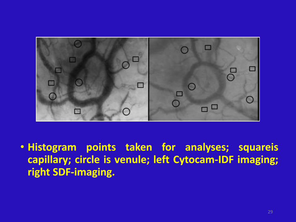
• Histogram points taken for analyses; squareis capillary; circle is venule; left Cytocam-IDF imaging; right SDF-imaging.
29

3- MICROCIRCULATORY ALTERATIONS IN SEPSIS
• Sepsis is one of the most common syndromes suffered by critically ill patients. It has recently been redefined as a life threatening form of organ dysfunction caused by a dysregulated host response to infection [21&&].
• Infections associated with sepsis trigger inflammation, and a resultant cytokine storm can lead to cardiovascular depression, which together causes cellular dysfunction that results in organ failure [22].
30

Micro-in Sepsis Cont
• Hemodynamic normalization can be achieved by the rapid administration of fluids and vasoactive drugs.
• Rivers’ study showed that early goal-directed therapy (EGDT) can improve survival rates in specific types of septic shock patients, which led to EGDT being recommended by Surviving Sepsis guidelines [23,24].
• However, recent multicenter trials were conducted in the United States [Protocolized Care for Early Septic Shock (ProCESS)] [25], Australasia [Australasian Resuscitation in Sepsis Evaluation (ARISE) trial] [26], and in the United Kingdom [Protocolized Management in Sepsis (ProMISe)] [27] that showed that this approach showed no clear benefits in terms of survival.
31

Micro-in Sepsis Cont
• Another large randomized trial,SEPSISPAM (Assessment of two levels of arterial pressure on survival in patients with septic shock study), included 776 septic shock patients and showed that higher mean arterial pressure (MAP) targets (80–85mmHg) in comparison with conventional targets (65–70mmHg) did not improve survival [28].
32

Micro-in Sepsis Cont
• The Transfusion Requirement in Septic Shock (TRISS) study compared lower versus higher hemoglobin levels in septic shock [29].
• They found that mortality at 90 days and the rates of ischemic events and use of life support were similar in both groups.
• The therapeutic end points of these studies were aimed at correcting macrohemodynamic variables such as blood pressure, heart rate, and cardiac output, but they did not evaluate whether correcting these systemic variables resulted in improved parenchymal perfusion, oxygenation, and microcirculation.
33

Micro-in Sepsis Cont
• It can be concluded that these large negative randomized control trial studies provide little insight into effective therapeutic strategies for sepsis from a hemodynamic perspective, indicating the need for information regarding the microcirculatory system and tissue perfusion.
34

Micro-in Sepsis Cont
• The pathogenesis of sepsis is defined at the level of the
microcirculation and parenchymal cells.
• Sepsis causes multifactorial microcirculatory alterations
including endothelial cell dysfunction associated with the
expression of adhesion molecules, increased leukocyte
adhesion, glycocalyx degradation, connexin uncoupling,
vascular leakage, micro-thrombi formation, altered local
perfusion pressures, and functional shunting of oxygen
transport [6]. 35

Micro-in Sepsis Cont
• Although various types of microcirculatory alterations have been observed in septic patients, from a hemodynamic perspective, the heterogeneity in microvascular perfusion (type 1 microcirculatory alteration) is characteristic, as it explains the origin of oxygen transport dysfunction in sepsis [7&].
• Even if the total blood flow to the organs is preserved, hypoxic zones can occur because of heterogeneity in microvascular blood flow [7&,9]. Thus, recruiting microcirculation is more complex than simply increasing the total blood flow to an organ.
36

Micro-in Sepsis Cont
• De Backer et al. [13] investigated microcirculatory alterations in 252 sepsis patients and found that early microcirculatory deteriorations (first 24h) were the strongest predictor of outcomes, more sensitive and specific than macrohemodynamic variable.
• Hernandez et al. [30] and Edul et al. [9] also showed that severe abnormalities in microcirculatory perfused vessel density were associated with organ dysfunction and mortality in septic shock patients.
37

Micro-in Sepsis Cont
• A current source of debate is the relationship between heart rate and sepsis.
• In a multicenter international observational trial with 530 mixed intensive care patients, Vellinga et al. [31] showed that tachycardia was the single most sensitive parameter for predicting outcomes but that if tachycardia was associated with microcirculatory alterations, an even worse outcome was the result.
• Recently, Morelli et al. [32] reported that the fast acting beta-blocker, esmolol, reduced heart rate and improved survival in septic patients. They explained that the expected increase in stroke volume in patients with beta-blocker-treated sepsis was associated with a decreasing heart rate.
38

Micro-in Sepsis Cont
• Aboab et al. [33] found that esmolol increased stroke volume by reducing heart rate in a porcine model of hypodynamic sepsis.
• Jacquet-Lagreze et al. [34] investigated the beneficial effects of the fast acting beta-blocker in improving microcirculation in aporcine model of hyperdynamic sepsis.
• They found that beta-blockers provided maintenance of sublingual and gut microcirculation during sepsis; however, they were not able to show that a reduction in heart rate was accompanied by an increase in stroke volume.
• Therefore, the hemodynamic role of beta-blockers in the treatment of sepsis remains unclear, as the microcirculatory findings are not explained by their macrohemodynamic changes [35]. 39

4- MICROCIRCULATORY ALTERATIONS IN SURGERY
• High-risk surgery associated with cardiac surgery, trauma, and hemorrhagic shock is another common cause of critical illness.
• Yu Chang and colleagues investigated the associations between surgical stress and microcirculatory dysfunction in patients post general and thoracic surgery.
• They concluded that early (first 24h) microcirculatory parameters (total and perfused vessel density) might be used as a predictor of surgical complications and outcomes in critically ill surgical patients.
40

Micro. In Surgery-- Cont.
• Maddison et al. [36] investigated sublingual microcirculatory alterations in patients with intra abdominal hypertension.
• They found that grades I and II intra abdominal hypertension (intra abdominal pressure from 12 to 18mmHg) was not associated with microcirculatory alterations.
41

Micro. In Surgery-- Cont.
• Cardiac surgery is characterized by a wide range of microcirculatory changes and reduced tissue oxygenation [37].
• Microcirculatory alterations occur not only as a result of underlying cardiac disease or cardiogenic shock but can also occur as a result of anesthesia, hypothermia, and hemodilution as well as the surgery itself [38,39].
• The hemodynamic effects of the nature of the cardiac surgery (e.g., off-pump or on-pump cardiac surgery) are still a source of controversy [40].
• De Backer et al. [39] showed that off-pump cardiac surgery was associated with a decrease in microcirculatory perfusion,
42

Micro. In Surgery-- Cont.
• Where as Atasever et al. [41] found that on-pump and off-pump cardiac surgeries caused specific and different types of sublingual microcirculation alterations.
• A recent study showed that off-pump surgery does not preserve postoperative microcirculatory parameters better than on-pump cardiac surgery [42].
• A consistent finding in cardiac surgery, however, is that hemodilution causes a reduction in functional capillary density, which can be corrected by blood transfusion [43–45].
43

Micro. In Surgery-- Cont.
• Microcirculatory dysfunction in patients with hemorrhagic shock has a similarly poor outcome as sepsis patients.
• In traumatic hemorrhagic shock patients, Tachon et al. [11] found that despite the successful restoration of systemic hemodynamic variables within hours, the restoration of sublingual microcirculation took up to 4 days.
• They found that the length of recovery of the microcirculation correlated with the severity of organ dysfunction.
44

Micro. In Surgery-- Cont.
• Stens et al. [46] investigated whether the hemodynamic optimization of systemic perfusion based on pulse pressure variation (PPV) and cardiac index (CI) improved the microcirculation in patients with abdominal surgery when compared with a MAP based strategy.
• They found that PPV and CI-based therapy was not associated with an improved microcirculatory perfusion compared with MAP-guided therapy.
• The outcomes may improve, however, when goal-directed therapy is aimed at correcting the microcirculation after major surgery [5&&].
45

5- MICROCIRCULATORY ALTERATIONS IN VARIOUS CLINICAL CONDITIONS IN
CRITICALLY ILL PATIENTS
• Cardiac arrest is one of the leading causes of death in critically ill patients and can cause microcirculatory deterioration.
• After cardiopulmonary resuscitation, therapeutic hypothermia is recommended to improve neurological outcomes [47].
• However, the optimal target therapeutic hypothermia level remains unknown.
46

Micro.in Critical illness Cont
• In an international trial, Nielsen et al. [48] investigated targeting the temperature management at 33 versus 36C after cardiac arrest.
• The authors found that hypothermia at a targeted temperature of 33 C was not more beneficial than a targeted temperature of 36 C. From a microcirculatory perspective.
47

Micro.in Critical illness Cont.
• Koopmans et al. [49] investigated the potential differences in microcirculatory alterations and vascular reactivity in comatose patients after cardiac arrest who were treated with a target temperature management of 33 C in comparison to patients treated with 36 C.
• They found that microcirculatory blood flow and vascular reactivity did not differ between the groups.
48

Micro.in Critical illness Cont
• Resuscitation guidelines also recommend keeping patients’ oxygen saturation levels at approximately 94% because of the side-effects of hyperoxia [50].
• Concerning the types of microcirculatory changes, hyperoxia falls into the type 3 microcirculatory alterations category [5&&], in which hyperoxia causes vasoconstriction and reductions in microvascular flow, as shown by Orbegozo Cortes et al. [51] in healthy volunteers.
49

Micro.in Critical illness Cont.
• Critically ill patients often have more than one chronic disease such as diabetes, cirrhosis, and chronic kidney disease.
• The microcirculatory effects of these comorbidities and age are important in hemodynamically stable patients.
• Reynolds et al. [52]investigated the effects of age ,diabetes mellitus, cirrhosis, and chronic kidney disease on sublingual microcirculatory flow.
• They showed that sublingual microcirculatory parameters did not significantly differ between healthy young volunteers, healthy older adults, and patients with diabetes, cirrhosis, and chronic renal failure
50

Micro.in Critical illness Cont
• Kanoore Edul et al. [53] evaluated sublingual microcirculation in patients with and without chronic arterial hypertension.
• They found that chronic arterial hypertension decreased vascular density but that the microcirculatory variables remained unchanged over a large age range.
• Dababneh et al. [54] compared microcirculatory alterations in patients with and without pulmonary hypertension. They found a lower microcirculatory flow index (MFI) in patients with pulmonary hypertension.
• Interestingly, George et al. [55] compared sublingual microcirculation between pregnant and non pregnant women and observed that pregnant women had a higher MFI compared with non pregnant women.
51

6-MICROCIRCULATORY EFFECTS OF RESUSCITATION THERAPIES
IN CRITICAL ILLNESS
• Fluid therapy is the initial approach when hypovolemia is suspected in critically ill patients.
• The afore mentioned early fluid resuscitation is important in restoring microcirculation [4]. However, fluid volume and fluid composition form are crucial aspects of effective volume therapy.
• Regarding their impact on the microcirculation from a physiological perspective, fluid therapy increases the convective flow in hypovolemia [56].
52

Microcirc.Effect of Resus. Cont
• Pranskunas et al. [57] assessed the changes in sublingual microcirculatory flow in patients with impaired organ perfusion based on clinical surrogates such as hyperlactate, tachycardia, hypotension, and oliguria because of fluid overload.
• They measured the MFI before and after fluid challenge, and they found that fluid administration only increased the MFI in patients with a low baseline MFI (<2.3).
• The correction of MFI caused a parallel response in the surrogates.
53

Microcirc.Effect of Resus. Cont
• However, in patients with surrogates but not with normal MFI, fluid was ineffective in correcting either variable.
• In all the conditions, however, fluids increased the stroke volume and were effective in correcting the surrogates.
• They concluded that MFI could be used to predict fluid response.
• Pottecher et al. [56] showed that increasing the intravascular volume by passive leg raising and intravenous volume administration improved sublingual microcirculatory perfusion in severe sepsis and septic shock patients.
54

Microcirc.Effect of Resus. Cont
• Ospina-Tascon et al. [58] explored the importance of the timing of fluid administration in septic patients and the authors found that early but not late fluid challenge can improve perfusion to the microcirculation.
• However, if too much fluid is given, hemodilution, capillary leakage, and tissue edema cause problems in oxygen transport.
• Sepsis guidelines recommend increasing central venous pressure up to 12mmHg for adequate volume therapy [24]. However, elevated central venous pressure may cause different adverse effects, in particular acute renal failure.
55

Microcirc.Effect of Resus. Cont
• From a microcirculatory perspective, elevated venous pressure can cause a type 3 microcirculatory alteration associated with tamponade of the microcirculation.
• Vellinga et al. [59] showed this effect when they compared critically ill patients with a central venous pressure higher than 12mmHg to those with a venous pressure lower than 12mmHg.
• They found that there was a significant reduction in microcirculatory flow in the high venous pressure group.
• Fluid therapy guided by optimizing stroke volume determined by the PiCCO technique was used in the PRISM (PiCCO-guided Resuscitation in Severe Malaria) trial in patients with malaria [60].
56

Microcirc.Effect of Resus. Cont
• PiCCO-guided resuscitation caused fluid overload and severe tissue edema.
• The normalized systemic hemodynamics but with altered type 4 microcirculation in this example shows the loss of hemodynamic coherence.
• This generalized peripheral edema did not resolve tissue hypovolemia or metabolic acidosis and increased acute renal failure, highlighting the importance of demonstrating hemodynamic coherence.
57

Microcirc.Effect of Resus. Cont
• Hanson et al. [61] evaluated rectal microcirculation in malaria patients who were resuscitated by stroke volume-guided fluid therapy.
• They found that although fluids were successful in correcting systemic hemodynamic variables, they had little effect on malaria-associated RBC sequestration, the primary pathology underlying malaria.
• Therefore, fluid administration targeting systemic hemodynamic parameters in this patient group was not successful in correcting for metabolic acidosis and resulted in adverse severe edema in the kidney, abdomen, and lungs.
58

Microcirc.Effect of Resus. Cont
• Types of fluids and their composition are also a highly controversial issue in resuscitation medicine. In a microcirculation study, Dubin et al. [62]
• They compared 6% hydroxy ethyl starch 130/0.4 to an isotonic saline solution for resuscitation in septic shock patients, targeting an improvement in MAP.
• They showed that fluid resuscitation with 6% hydroxy ethyl starch 130/0.4 required lower volumes to reach targeted blood pressures and caused a higher capillary density of flowing RBCs in sublingual microcirculation, with a higher flow being achieved with less volume than with the isotonic saline solution [9].
59

Microcirc.Effect of Resus. Cont
• In addition to resuscitation fluids, blood transfusions can have a positive as well as negative effect on patient outcomes [63,64].
• However, when applied physiologically, studies have shown that blood transfusions can lead to improved microcirculatory functional capillary density [44,45].
• In evaluating the effects of blood transfusion on critically ill patients, there is a large variability in the quality of blood, leuco-depletion, and age and storage solutions used. Therefore, a re-evaluation of the relative risks of hemodilution, anemia versus blood transfusion, is required [65].
60

Microcirc.Effect of Resus. Cont
• The effects of vasoactive compounds on the microcirculation have been extensively investigated in critically ill patients.
• Vasopressor agents are administered to achieve targeted systemic hemodynamic values with the expectation that this will augment oxygen delivery to the tissues [66].
• However, excessive vasopressor therapy can cause microcirculatory stasis (type 3) by severe vasoconstriction [67].
• For example, Boerma et al. [68] showed that the vasopressin analog terlipressin impaired sublingual microcirculation in a patient with catecholamine resistant septic shock.
• They also concluded in a review of the literature over the past 15 years that there were no beneficial effects of increasing MAP above 65 on microcirculatory perfusion [69].
61

Microcirc.Effect of Resus. Cont
• Xu et al. [70] evaluated the impact of increasing MAP levels to approximately 70 mmHg through norepinephrine administration in patients with chronic hypertension.
• They concluded that increasing the arterial blood pressure improved sublingual microcirculation independent of other tissue perfusion indicators such as lactate and urinary output.
62

Microcirc.Effect of Resus. Cont
• Dubin et al. [71] found that increases in MAP from 65 to 85 mmHg in septic shock patients resulted in decreased perfusion of the microcirculation, strongly dependent on the basal microcirculation.
• When basal microcirculation was normal at a MAP of 65 mmHg, increases in MAP worsened the microcirculation because of vasoconstriction (type 3 microcirculatory alteration).
• If, however, there was a slow flow in the baseline
microcirculation, increases in MAP improved microcirculatory flow parameters [71]. Similar results were found by Jhanji et al. [72].
63

Microcirc.Effect of Resus. Cont
• The vasoactive therapy most effective in promoting regional perfusion is vasodilatory therapy.
• Spronk et al.[73] showed that nitroglycerin administration improved sublingual microcirculatory perfusion in pressure-resuscitated septic shock patients.
• Boerma and Ince [69], however, were not able to reproduce this effect in fluid-resuscitated septic patients.
• Groeneveld and Lima [74], on the other hand, showed that increasing the doses of nitroglycerin was able to recruit microvascular perfusion in circulatory shock patients.
64

Microcirc.Effect of Resus. Cont
• In addition to vasoactive and fluid therapy, anti inflammatory therapy can also have positive effects on the microcirculation.
• Recombinant human activated protein C, which has an anti-inflammatory effect, was used in septic shock patients [75].
• Another anti-inflammatory drug, cortisol, was recommended in vasopressor refractory septic shock patients [24].
•
65

Microcirc.Effect of Resus. Cont
• Recently, an interesting study by Povoa et al. [76] investigated the effects of stress dose steroids with or without recombinant activated protein C therapy in septic shock patients. They found that there were no beneficial effects. From a microcirculatory perspective
• Donati et al. [77] showed that activated protein C treatment improved microcirculation in severe sepsis and septic shock patients. Buchele et al. [78] evaluated the effects of hydrocortisone on microcirculation in patients with septic shock. They found that hydrocortisone improved capillary perfusion.
66

7- SAMMARY
• Critical illness is associated with a wide range of diseases such as sepsis, high-risk surgery, cardiac arrest, and respiratory failure and is characterized by reduced tissue oxygenation caused by microcirculatory dysfunction.
• Optimal fluid therapy is the most important hemodynamic intervention in critically ill patients.
67

SAMMARY .Cont
• The main goals of fluid therapy are not only to maintain macrocirculation but also to recruit microcirculation.
• The loss of hemodynamic coherence between macrocirculation and microcirculation should always be kept in mind, and all therapeutic approach should aim to correct hemodynamic incoherence.
• This requires microcirculatory directed therapy to be considered.
68

SAMMARY .Cont
• Direct observations of the microcirculation are essential to monitor hemodynamic coherence.
• By doing so, a more physiological approach could prevent the unnecessary and inappropriate administration of large volumes of fluids.
• In the state of hypovolemia, colloid solutions are approximately three times more effective in volume expansion than crystalloids and improve the microcirculation more effectively than crystalloids.
• Apart from fluids, blood transfusions may improve microcirculatory parameters and, more importantly, transport oxygen more effectively than nonoxygen carrying fluids.
69

SAMMARY .Cont
• The new generation microcirculation monitoring device, the CytoCam-IDF, enables the clinical monitoring of sublingual microcirculation, and it can be easily used for the functional assessment of the hemodynamic state of the microcirculation.
• Direct visualization of the microcirculation at the bedside
should be integrated with monitoring systemic hemodynamic variables for the early diagnosis and treatment of critical illness.
• Establishing and monitoring hemodynamic coherence and
targeting not only the normalization of the macrocirculation but also that of microcirculation can be considered an essential component in the hemodynamic management of critically ill patients.
70

8- CONCLUSION
• In conclusion, we suggest that monitoring the microcirculation is important for determining hemodynamic coherence as a response to therapy and can provide the feedback needed to ensure good clinical outcomes. The introduction of a new generation of computer-controlled hand-held microscopes for monitoring the microcirculation now provides anew clinical approach to monitoring the determinants of tissue oxygenation.
71

THANK YOU
Mansoura- EGYPT
72

REFERENCES
• 1. Ince C. The microcirculation is the motor of sepsis. Crit Care 2005; 9 (Suppl 4):S13–S19.
• 2.DeBackerD,DurandA.Monitoringthemicrocirculationincriticallyillpatients. Best Pract Res Clin Anaesthesiol 2014; 28:441–451.
• 3. Gruartmoner G, Mesquida J, Ince C. Fluid therapy and the hypovolemic microcirculation. Curr Opin Crit Care 2015; 21:276–284.
• 4. Ince C. The rationale for microcirculatory guided fluid therapy. Curr Opin Crit Care 2014; 20:301–308.
73

• 5. Ince C. Hemodynamic coherence and the rationale for monitoring the microcirculation. Crit Care 2015; 19 (Suppl 3):S8. A comprehensive review that explains importance of hemodynamiccoherence and rationale for monitoring microcirculation to provide hemodynamic coherence.
• 6. De Backer D, Orbegozo Cortes D, Donadello K, Vincent JL. Pathophysiology of microcirculatory dysfunction and the pathogenesis of septic shock. Virulence 2014; 5:73–79.
• 7. & Ince C, Mik EG. Microcirculatory and mitochondrial hypoxia in sepsis, shock and resuscitation. J Appl Physiol (1985) 2016; 120:226–235. The mechanism of microcirculation and mitochondrial hypoxia in case of different clinical conditions.
• 8. De Backer D, Donadello K, Sakr Y, et al. Microcirculatory alterations in patients with severe sepsis: impact of time of assessment and relationship with outcome. Crit Care Med 2013; 41:791–799.
74

• 9. Edul VS, Enrico C, Laviolle B, et al. Quantitative assessment of the microcirculation in healthy volunteers and in patients with septic shock. Crit Care Med 2012; 40:1443–1448.
• 10. Trzeciak S, McCoy JV, Phillip Dellinger R, et al. Early increases in microcirculatory perfusion during protocol-directed resuscitation are associated with reduced multi-organ failure at 24h in patients with sepsis. Intensive Care Med 2008; 34:2210–2217.
• 11. Tachon G, Harrois A, Tanaka S, et al. Microcirculatory alterations in traumatic hemorrhagic shock. Crit Care Med 2014; 42:1433–1441.
• 12. Lima A,JansenTC,vanBommelJ,et al. Theprognosticvalue ofthe subjective assessment of peripheral perfusion in critically ill patients. Crit Care Med 2009; 37:934–938.
• 13. De Backer D, Ortiz JA, Salgado D. Coupling microcirculation to systemic hemodynamics. Curr Opin Crit Care 2010; 16:250–254.
• 14. Verdant CL, De Backer D, Bruhn A, et al. Evaluation of sublingual and gut mucosal microcirculation in sepsis: a quantitative analysis. Crit Care Med 2009; 37:2875–2881.
75

• 15. Bezemer R, Bartels SA, Bakker J, Ince C. Clinical review: Clinical imaging of the sublingual microcirculation in the critically ill – where do we stand? Crit Care 2012; 16:224.
• 16. Sherman H, Klausner S, Cook WA. Incident dark-field illumination: a new method for microcirculatory study. Angiology 1971; 22:295–303.
• 17. Aykut G, Veenstra G, Scorcella C, et al. Cytocam-IDF (incident dark field illumination) imaging for bedside monitoring of the microcirculation. Intensive Care Med Exp 2015; 3:40.
• 18. MikEG,JohannesT,FriesM.Clinicalmicrovascularmonitoring:abrightfuture without a future? Crit Care Med 2009; 37:2980–2981.
• 19. vanElterenHA,InceC,TibboelD,etal.Cutaneousmicrocirculationinpreterm neonates: comparisonbetweensidestream dark field(SDF) and incident dark field (IDF) imaging. J Clin Monit Comput 2015; 29:543–548.
• 20. Gilbert-Kawai E, Coppel J, Bountziouka V, et al. A comparison of the quality of image acquisition between the incident dark field and sidestream dark field video-microscopes. BMC Med Imaging 2016; 16:10.
• 21. && Singer M, Deutschman CS, Seymour CW, et al. The third international consensus definitions for sepsis and septic shock (Sepsis-3). JAMA 2016; 315:801–810. The new definition of sepsis.
76

• 22. Angus DC, van der Poll T. Severe sepsis and septic shock. N Engl J Med 2013; 369:840–851.
• 23. Rivers E, Nguyen B, Havstad S, et al. Early goal-directed therapy in the treatmentofseveresepsisandsepticshock.NEnglJMed2001;345:1368– 1377.
• 24. Dellinger RP, Levy MM, Rhodes A, et al. Surviving sepsis campaign: international guidelines for management of severe sepsis and septic shock. Intensive Care Med 2013; 39:165–228.
77

• 25. Pro CI, Yealy DM, Kellum JA, et al. A randomized trial of protocol-based care for early septic shock. N Engl J Med 2014; 370:1683–1693.
• 26. ARISE Investigators, ACTGroup.Peake SL, et al.Goal-directed resuscitation for patients with early septic shock. N Engl J Med 2014; 371:1496–1506.
• 27. Mouncey PR, Osborn TM, Power GS, et al. Protocolised Management In Sepsis (ProMISe): a multicentre randomised controlled trial of the clinical effectiveness and cost-effectiveness of early, goal-directed, protocolised resuscitation for emerging septic shock. Health Technol Assess 2015; 19: i–xxv, 1–150.
• 28. Asfar P, Meziani F, Hamel JF, et al. High versus low blood-pressure target in patients with septic shock. N Engl J Med 2014; 370:1583–1593.
• 29. Holst LB, Haase N, Wetterslev J, et al. Transfusion requirements in septic shock (TRISS) trial – comparing the effects and safety of liberal versus restrictive red blood cell transfusion in septic shock patients in the ICU: protocol for a randomised controlled trial. Trials 2013; 14:150.
• 30. Hernandez G, Boerma EC, Dubin A, et al. Severe abnormalities in microvascular perfused vessel density are associated to organ dysfunctions and mortality and can be predicted by hyperlactatemia and norepinephrine requirements in septic shock patients. J Crit Care 2013; 28:538.e9–538.e14.
78

• 31. Vellinga NA, Boerma EC, Koopmans M, et al. International study on microcirculatory shock occurrence in acutely ill patients. Crit Care Med 2015; 43:48–56
• 32. Morelli A, Donati A, Ertmer C, et al. Microvascular effects of heart rate control with esmolol in patients with septic shock: a pilot study. Crit Care Med 2013; 41:2162–2168.
• 33. Aboab J, Sebille V, Jourdain M, et al. Effects of esmolol on systemic and pulmonary hemodynamics and on oxygenation in pigs with hypodynamic endotoxin shock. Intensive Care Med 2011; 37:1344–1351.
• 34. Jacquet-Lagreze M, Allaouchiche B, Restagno D, et al. Gut and sublingual microvascular effect of esmolol during septic shock in a porcine model. Crit Care 2015; 19:241.
• 35.InceC.Tobetablockornottobetablock;thatisthequestion.CritCare2015; 19:339.
79

• 36. Maddison L, Karjagin J, Buldakov M, et al. Sublingual microcirculation in patients with intra-abdominal hypertension: a pilot study in 15 critically ill patients. J Crit Care 2014; 29:183.e1–183.e6.
• 37. Kara A, Akin S, Ince C. The response of the microcirculation to cardiac surgery. Curr Opin Anaesthesiol 2016; 29:85–93.
• 38. Bauer A, Kofler S, Thiel M, et al. Monitoring of the sublingual microcirculation in cardiac surgery using orthogonal polarization spectral imaging: preliminary results. Anesthesiology 2007; 107:939–945.
• 39. De Backer D, Dubois MJ, Schmartz D, et al. Microcirculatory alterations in cardiac surgery: effects of cardiopulmonary bypass and anesthesia. Ann Thorac Surg 2009; 88:1396–1403.
• 40. Koning NJ, Vonk AB, Meesters MI, et al. Microcirculatory perfusion is preservedduringoff-pumpbutnoton-pumpcardiacsurgery.JCardiothoracVasc Anesth 2014; 28:336–341.
80

• 41. Atasever B, Boer C, Goedhart P, et al. Distinct alterations in sublingual microcirculatory blood flow and hemoglobin oxygenation in on-pump and offpump coronary artery bypass graft surgery. J Cardiothorac Vasc Anesth 2011; 25:784–790.
• 42. Bienz M,Drullinsky D, Stevens LM, et al. Microcirculatory response during onpump versus off-pump coronary artery bypass graft surgery. Perfusion 2016; 31:207–215.
• 43. Yuruk K, Bartels SA, Milstein DM, et al. Red blood cell transfusions and tissue oxygenation in anemic hematology outpatients. Transfusion 2012; 52:641– 646.
• 44. Yuruk K, Almac E, Bezemer R, et al. Blood transfusions recruit the microcirculation during cardiac surgery. Transfusion 2011; 51:961–967.
• 45. AtaseverB,vanderKuilM,BoerC,et al.Redbloodcelltransfusion compared with gelatin solution and no infusion after cardiac surgery: effect on microvascular perfusion, vascular density, hemoglobin, and oxygen saturation. Transfusion 2012; 52:2452–2458.
81

• 46. Stens J, de Wolf SP, van der Zwan RJ, et al. Microcirculatory perfusion during different perioperative hemodynamic strategies. Microcirculation 2015; 22:267–275.
• 47. Soar J, Callaway CW, Aibiki M, et al. Part 4: Advanced life support: 2015 international consensus on cardiopulmonary resuscitation and emergency cardiovascular care science with treatment recommendations. Resuscitation 2015; 95:e71–e120.
• 48.NielsenN,WetterslevJ,CronbergT,etal.Targetedtemperaturemanagement at33degreesCversus36degreesCaftercardiacarrest.NEnglJMed2013; 369:2197–2206.
• 49. Koopmans M, Kuiper MA, Endeman H, et al. Microcirculatory perfusion and vascular reactivity are altered in post cardiac arrest patients, irrespective of target temperature management to 33 degrees C vs 36 degrees C. Resuscitation 2015; 86:14–18.
• 50. Neumar RW, Shuster M, Callaway CW, et al. Part 1: Executive Summary: 2015 American Heart Association Guidelines update for cardiopulmonary resuscitation and emergency cardiovascular care. Circulation 2015; 132:S315–S367.
82

• 51. Orbegozo Cortes D, Puflea F, Donadello K, et al. Normobaric hyperoxia alters the microcirculation in healthy volunteers. Microvasc Res 2015; 98:23–28.
• 52. Reynolds T, Vivian-Smith A, Jhanji S, Pearse RM. Observational study of the effects of age, diabetes mellitus, cirrhosis and chronic kidney disease on sublingual microvascular flow. Perioper Med (Lond) 2013; 2:7.
• 53. Kanoore Edul VS, Ince C, Estenssoro E, et al. The effects of arterial hypertension and age on the sublingual microcirculation of healthy volunteers and outpatients with cardiovascular risk factors. Microcirculation 2015; 22:485– 492.
• 54. Dababneh L, Cikach F, Alkukhun L, et al. Sublingual microcirculation in pulmonary arterial hypertension. Ann Am Thorac Soc 2014; 11:504–512.
• 55. George RB, Munro A, Abdo I, et al. An observational assessment of the sublingual microcirculation of pregnant and nonpregnant women. Int J Obstet Anesth 2014; 23:23–28.
• 56. Pottecher J, Deruddre S, Teboul JL, et al. Both passive leg raising and intravascular volume expansion improve sublingual microcirculatory perfusion in severe sepsis and septic shock patients. Intensive Care Med 2010; 36:1867–1874.
• 57. ranskunasA,KoopmansM,KoetsierPM,etal.Microcirculatorybloodflowas a tool to select ICU patients eligible for fluid therapy. Intensive Care Med 2013; 39:612–619. 58. Ospina-Tascon G, Neves AP, Occhipinti G, et al. Effects of fluids on microvascular perfusion in patients with severe sepsis. Intensive Care Med 2010; 36:949–955.
83

• 59. Vellinga NA, Ince C, Boerma EC. Elevated central venous pressure is associated with impairment of microcirculatory blood flow in sepsis: a hypothesis generating post hoc analysis. BMC Anesthesiol 2013; 13:17.
• 60. Hanson J, Anstey NM, Bihari D, et al. The fluid management of adults with severe malaria. Crit Care 2014; 18:642.
• 61. Hanson JP, Lam SW, Mohanty S, et al. Fluid resuscitation of adults with severe falciparum malaria: effects on acid-base status, renal function, and extravascular lung water. Crit Care Med 2013; 41:972–981.
• 62. Dubin A, Pozo MO, Casabella CA, et al. Comparison of 6% hydroxyethyl starch 130/0.4 and saline solution for resuscitation of the microcirculation during the early goal-directed therapy of septic patients. J Crit Care 2010; 25:659.e1–659.e8.
• 63. Kuduvalli M, Oo AY, Newall N, et al. Effect of peri-operative red blood cell transfusion on 30-day and 1-year mortality following coronary artery bypass surgery. Eur J Cardiothorac Surg 2005; 27:592–598.
• 64. Nakamura RE, Vincent JL, Fukushima JT, et al. A liberal strategy of red blood cell transfusion reduces cardiogenic shock in elderly patients undergoing cardiac surgery. J Thorac Cardiovasc Surg 2015; 150:1314–1320.
84

• 65. Vincent JL. Transfusion triggers: getting it right! Crit Care Med 2012; 40:3308–3309.
• 66. Shapiro NI, Angus DC. A review of therapeutic attempts to recruit the microcirculation in patients with sepsis. Minerva Anestesiol 2014; 80:225–235.
• 67. Krejci V, Hiltebrand LB, Sigurdsson GH. Effects of epinephrine, norepinephrine, and phenylephrine on microcirculatory blood flow in the gastrointestinal tract in sepsis. Crit Care Med 2006; 34:1456–1463.
• 68. Boerma EC, van der Voort PH, Ince C. Sublingual microcirculatory flow is impaired by the vasopressin-analogue terlipressin in a patient with catecholamine-resistant septic shock. Acta Anaesthesiol Scand 2005; 49:1387–1390
85

• 69. Boerma EC, Ince C. The role of vasoactive agents in the resuscitation of microvascular perfusion and tissue oxygenation in critically ill patients. Intensive Care Med 2010; 36:2004–2018.
• 70. Xu JY, Ma SQ, Pan C, et al. A high mean arterial pressure target is associated with improved microcirculation in septic shock patients with previous hypertension: a prospective open label study. Crit Care 2015; 19:130.
• 71. Dubin A, Pozo MO, Casabella CA, et al. Increasing arterial blood pressure with norepinephrine does not improve microcirculatory blood flow: a prospective study. Crit Care 2009; 13:R92.
• 72. Jhanji S, Stirling S, Patel N, et al. The effect of increasing doses of norepinephrine on tissue oxygenation and microvascular flow in patients with septic shock. Crit Care Med 2009; 37:1961–1966.
• 73. Spronk PE, Ince C, Gardien MJ, et al. Nitroglycerin in septic shock after intravascular volume resuscitation. Lancet 2002; 360:1395–1396.
86

• 74. Groeneveld ABJ, Lima A. Vasodilators in critical illness. Oxford University Press; 2016.
• 75. Bernard GR, Vincent JL, Laterre PF, et al. Efficacy and safety of recombinant human activated protein C for severe sepsis. N Engl J Med 2001; 344:699– 709.
• 76. PovoaP,SalluhJI,MartinezML,et al.Clinicalimpactofstressdosesteroidsin patients with septic shock: insights from the PROWESS-Shock trial. Crit Care 2015; 19:193.
• 77. Donati A, Damiani E, Botticelli L, et al. The aPC treatment improves microcirculation in severe sepsis/septic shock syndrome. BMC Anesthesiol 2013; 13:25.
• 78. Buchele GL, Silva E, Ospina-Tascon GA, et al. Effects of hydrocortisone on microcirculatory alterations in patients with septic shock. Crit Care Med 2009; 37:1341–1347.
87