microbubble super-localisation in human lower limb … · microbubble super-localisation in human...
TRANSCRIPT
Microbubble Super-Localisation
in Human Lower Limb Microvasculature
with Motion Correction
Sevan Harput, Kirsten Christensen-Jeffries, Yuanwei Li, Jemma Brown,
Paul Aljabar, Robert J. Eckersley, Christopher Dunsby and Meng-Xing Tang
Rotterdam
19-20 January 2017

Ideal Case:
Ultrafast plane wave imaging
Acquisition of RF or IQ data
No motion
Spatially isolated microbubbles
Practical Case:
Linear scan with a commercial scanner
Grey scale video. No RF data.
Motion
Multiple non-isolated microbubbles
Ultrasound Super-Resolution or Super-Localisation
Figure: Christensen-Jeffries et. al. “In Vivo Acoustic Super-Resolution
and Super-Resolved Velocity Mapping Using Microbubbles”, IEEE
Trans. Medical Imaging, Vol. 34, No. 2, 2015.
1mm 1mm

Ideal Case:
Ultrafast plane wave imaging
Acquisition of RF or IQ data
No motion
Spatially isolated microbubbles
Practical Case:
Linear scan with a commercial scanner
Grey scale video. No RF data.
Motion
Multiple non-isolated microbubbles
In-vivo Ultrasound Super-Resolution
Motion is an inherent part of
diagnostic imaging

Ideal Case:
Ultrafast plane wave imaging
Acquisition of RF or IQ data
No motion
Spatially isolated microbubbles
Practical Case:
Linear scan with a commercial scanner
Grey scale video. No RF data.
Motion
Multiple non-isolated microbubbles
Sub-optimal super-resolution imaging with a clinical scanner may still help
the practitioners to diagnose certain cases.
Imaging of Micro-circulation with a commercial scanner
Figure: Christensen-Jeffries et. al. “In Vivo Acoustic Super-Resolution
and Super-Resolved Velocity Mapping Using Microbubbles”, IEEE
Trans. Medical Imaging, Vol. 34, No. 2, 2015.
?

Human Lower Limb - Tibialis Anterior Muscle
Figure: http://is.muni.cz/do/1451/e-learning/kineziologie/elportal/img/tibialis_anterior.png
Figure: https://web.archive.org/web/20041209023150/http://education.yahoo.com/reference/gray/subjects/subject?id=160t

Focus: Peripheral artery disease (PAD) is a
type of atherosclerosis. It occurs most often in
the legs arteries and interferes with the blood
circulation in the limb.
Aim: To identify the presence of Claudication
and Diabetes using contrast enhanced
ultrasound imaging.
Peripheral Artery Disease and Intermittent Claudication
Patient Group Hypothesis
1 - Healthy VolunteersLarge difference in microvascular flow
before and after exercise
2 - ClaudicationSmall difference in microvascular flow
before and after exercise
3 - DiabetesAlmost no difference in microvascular flow
before and after exercise
Figure: http://www.adameducation.com

CEUS imaging of human lower limb

Motion Estimation1
(non-rigid registration)
Transformation Matrix
Spatio-temporal Filtering
B-mode
Contrast
Motion Correction(non-rigid)
Noise Threshold(depth-dependant)
MB detection2,3 Super-Localization2,3
Motion Estimation &Correction (rigid)
Motion Correction(rigid)
Transformation Matrix
1 Rueckert et al. "Nonrigid Registration Using Free-Form Deformations: Application to Breast MR Images", IEEE Trans Med Imag, 1999.2 Viessmann et. al. “Acoustic super-resolution with ultrasound and microbubbles.,” Phys. Med. Biol., 2013.3 Christensen-Jeffries et. al. “In Vivo Acoustic Super-Resolution and Super-Resolved Velocity Mapping Using MBs”, IEEE Trans Med Imag, 2015.

Rigid Motion Estimation & Correction
Figure: Christensen-Jeffries “Super-Resolution Ultrasound Imaging with Microbubbles”, PhD Thesis, 2016.
2D rigid sub-pixel cross-
correlation with a reference
frame.
Up-sampling with bicubic
interpolation from the original
pixel scale.

Error (regarding to the reference frame)
Mean SSD = 0.007 Mean SSD = 0.004

Maximum Intensity Projection of all frames
580 frames = 45 seconds

Maximum Intensity Projection of all frames
580 frames = 45 seconds

Super-Localisation
580 frames = 45 seconds
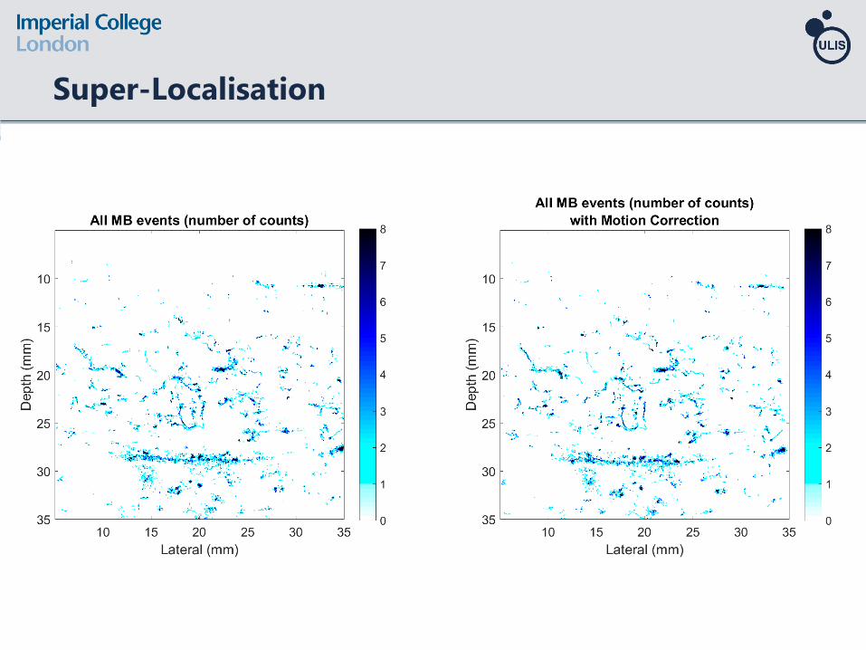
Super-Localisation
580 frames = 45 seconds

Effect of Motion Correction on Micro-vasculature

Effect of Motion Correction on Micro-vasculature

Motion Correction

Comparison - Micro-vessel thickness

Micro-vessel Thickness
Method MV #1 MV #2 MV #3 MV #4 MV #5 MV #6 ALL (µm)
Maximum Intensity Projection 769 562 1173 572 542 1136 792 ±293
Super-Localised MBs 227 172 151 157 153 126 164 ±34
Maximum Intensity Projection
with Motion Correction575 474 1015 520 520 1180 714 ±303
Super-Localised MBs
with Motion Correction115 91 90 80 108 87 95 ±13
λ = 250 µm (full bandwidth)
λ = 500 µm (second harmonic)

Healthy Volunteer
Before Exercise After Exercise
Total Events = 630k Total Events = 990k

Healthy Volunteer
Before Exercise After Exercise
Total Events = 630k Total Events = 990k

Preliminary Results – Healthy Volunteer
Number of Super-LocalisationsPeak Intensity
X X X X
<50%
Figure: Christensen-Jeffries “Super-Resolution Ultrasound Imaging with Microbubbles”, PhD Thesis, 2016.
4 healthy volunteers (V1, V2, V3, V4)
2 different days (A, B)
2 ultrasound scans to calculate relative change (baseline and after exercise)
A total of 16 ultrasound scans

In vivo super-localisation images were generated with a clinical scanner.
Two-stage motion correction was performed:
Rigid Motion Correction (to reduce the large movements)
Non-rigid Motion Correction (movement reduction in the first stage creates a
better starting point for the second stage, which eventually reduces the error)
Future work will be on 3D imaging to compensate for out of plane motion.
This technique has great potential when combined with motion correction.
Improvement in spatial resolution: ~5 times without motion correction
Improvement in spatial resolution: ~7 times with motion correction
Super-Localisation achieves better delineation of microvasculature from larger
vessels. Diffraction limited resolution may mask the details associated with PAD.
Summary