microbiota-associated risk factors for asymptomatic gut
TRANSCRIPT

RESEARCH Open Access
Microbiota-associated risk factors forasymptomatic gut colonisation with multi-drug-resistant organisms in a Dutchnursing homeQuinten R. Ducarmon1,2*† , Elisabeth M. Terveer1,2†, Sam Nooij1,2, Michelle N. Bloem1,2, Karuna E. W. Vendrik1,3,Monique A. A. Caljouw4, Ingrid M. J. G. Sanders1, Sofie M. van Dorp1,5, Man C. Wong1, Romy D. Zwittink1,2† andEd J. Kuijper1,2,3†
Abstract
Background: Nursing home residents have increased rates of intestinal colonisation with multidrug-resistantorganisms (MDROs). We assessed the colonisation and spread of MDROs among this population, determinedclinical risk factors for MDRO colonisation and investigated the role of the gut microbiota in providing colonisationresistance against MDROs.
Methods: We conducted a prospective cohort study in a Dutch nursing home. Demographical, epidemiologicaland clinical data were collected at four time points with 2-month intervals (October 2016–April 2017). To obtainlongitudinal data, faecal samples from residents were collected for at least two time points. Ultimately, twenty-seven residents were included in the study and 93 faecal samples were analysed, of which 27 (29.0%) were MDRO-positive. Twelve residents (44.4%) were colonised with an MDRO at at least one time point throughout the 6-monthstudy.
(Continued on next page)
© The Author(s). 2021 Open Access This article is licensed under a Creative Commons Attribution 4.0 International License,which permits use, sharing, adaptation, distribution and reproduction in any medium or format, as long as you giveappropriate credit to the original author(s) and the source, provide a link to the Creative Commons licence, and indicate ifchanges were made. The images or other third party material in this article are included in the article's Creative Commonslicence, unless indicated otherwise in a credit line to the material. If material is not included in the article's Creative Commonslicence and your intended use is not permitted by statutory regulation or exceeds the permitted use, you will need to obtainpermission directly from the copyright holder. To view a copy of this licence, visit http://creativecommons.org/licenses/by/4.0/.The Creative Commons Public Domain Dedication waiver (http://creativecommons.org/publicdomain/zero/1.0/) applies to thedata made available in this article, unless otherwise stated in a credit line to the data.
* Correspondence: [email protected]†Quinten R. Ducarmon, Elisabeth M. Terveer, Romy D. Zwittink and Ed J.Kuijper contributed equally to this work.1Department of Medical Microbiology, Leiden University Medical Center,Leiden, The Netherlands2Center for Microbiome Analyses and Therapeutics, Leiden University MedicalCenter, Leiden, The NetherlandsFull list of author information is available at the end of the article
Ducarmon et al. Genome Medicine (2021) 13:54 https://doi.org/10.1186/s13073-021-00869-z

(Continued from previous page)
Results: Univariable generalised estimating equation logistic regression indicated that antibiotic use in the previous2 months and hospital admittance in the previous year were associated with MDRO colonisation. Characterisationof MDRO isolates through whole-genome sequencing revealed Escherichia coli sequence type (ST)131 to be themost prevalent MDRO and ward-specific clusters of E. coli ST131 were identified. Microbiota analysis by 16S rRNAgene amplicon sequencing revealed no differences in alpha or beta diversity between MDRO-positive and negativesamples, nor between residents who were ever or never colonised. Three bacterial taxa (Dorea, Atopobiaceae andLachnospiraceae ND3007 group) were more abundant in residents never colonised with an MDRO throughout the6-month study. An unexpectedly high abundance of Bifidobacterium was observed in several residents. Furtherinvestigation of a subset of samples with metagenomics showed that various Bifidobacterium species were highlyabundant, of which B. longum strains remained identical within residents over time, but were different betweenresidents.
Conclusions: Our study provides new evidence for the role of the gut microbiota in colonisation resistance againstMDROs in the elderly living in a nursing home setting. Dorea, Atopobiaceae and Lachnospiraceae ND3007 groupmay be associated with protection against MDRO colonisation. Furthermore, we report a uniquely high abundanceof several Bifidobacterium species in multiple residents and excluded the possibility that this was due to probioticsupplementation.
Keywords: Gut microbiota, Multidrug-resistant organisms, Asymptomatic colonisation, Colonisation resistance,Bifidobacterium, Nursing home, Extended-spectrum beta-lactamase-producing Enterobacterales
BackgroundInfections caused by multidrug-resistant organisms(MDROs) are a rising threat to global health and caused~ 33,000 attributable deaths in Europe in 2015 [1]. Infec-tions with MDROs are usually preceded by asymptom-atic gut colonisation, and asymptomatically colonisedindividuals represent a potential transmission reservoir[2]. Nursing home residents are at increased risk forMDRO colonisation due to comorbidities resulting in in-creased healthcare contact and antibiotic use [3]. Inaddition, MDRO spread within a nursing home can befacilitated due to communal living, confined living spaceand incontinence of residents [4, 5]. This is similar tothe transmission dynamics of Clostridioides difficile. Theprevalence of MDROs and C. difficile varies betweennursing homes from different countries, but large differ-ences in prevalence can also be observed between differ-ent institutions in one country. For example, MDROprevalence ranges from 0 to 47% in various nursinghomes in the Netherlands [6–8] and from 0 to 75% inIreland [5]. C. difficile colonisation prevalence rangesfrom 0 to 17% in Dutch nursing homes [9, 10] and from0 to 10% in Germany [11]. These differences may reflectvariation in individual nursing home infection preven-tion and control practices, antimicrobial stewardship, in-frastructure, care load and presence of MDRO riskfactors such as incontinence, recent hospitalisation andcurrent antibiotic use. Colonisation resistance providedby the gut microbiome could contribute to preventingMDRO colonisation in the gut. The gut microbiome canprovide colonisation resistance through secretion ofantimicrobial products, nutrient competition, support of
epithelial barrier integrity, bacteriophage deploymentand immune activation. However, current knowledge onthe link between the microbiome and MDRO colonisa-tion is limited [12, 13]. In travellers, an increase of anti-microbial resistance genes and Escherichia coli relativeabundance in the microbiome were observed after acqui-sition and asymptomatic carriage of extended-spectrumbeta-lactamase (ESBL)-producing E. coli, but withoutclear differences in microbial community structure [14].An exception to the understudied role of the micro-biome in MDRO colonisation is vancomycin-resistantEnteroccocus (VRE). For example, it has recently beendemonstrated that a lantibiotic-producer, in this caseBlautia producta, could restore colonisation resistanceagainst VRE [15].To determine the prevalence and spread of MDROs in
a Dutch nursing home, and to elucidate the role of thegut microbiota and clinical risk factors herein, we con-ducted a four-point-prevalence study and analysed clin-ical data of residents and whole-genome sequencing(WGS) data of MDRO isolates, in combination with gutmicrobiota analysis through 16S rRNA gene ampliconsequencing. In addition, we conducted more in-depthmicrobiota analysis in a selection of samples throughmetagenomics in order to further investigate findingsfrom 16S rRNA gene amplicon analysis.
MethodsStudy designWe conducted a prospective cohort study in which resi-dents of a nursing home in the Netherlands were invitedto participate. The prevalence, dynamics and risk factors
Ducarmon et al. Genome Medicine (2021) 13:54 Page 2 of 17

of MDRO colonisation were studied in a non-outbreaksituation. Demographical, epidemiological and clinicaldata of four time points with a 2-month interval(October 2016 until April 2017) were collected. Micro-biota analysis was performed on stool samples collectedat the same four time points. Written informed consentwas obtained from the resident or corresponding proxy.Ethical approval was granted by the medical ethics com-mittee of the Leiden University Medical Center(No.P16.039). Sixty-four of 131 residents (49%) con-sented to participate. Data and corresponding faeceswere collected from 60 residents (94%). To make opti-mal use of the longitudinal data from this study, resi-dents were selected whom provided faeces at at leasttwo time points (n = 47). For this study, we included res-idents who gave consent for additional analyses, fromwhom faeces were cultured for MDROs at at least twotime points, and of which sufficient material was left formicrobiota profiling at at least two time points (n = 27residents). The prevalence of MDRO was not statisticallysignificant between the residents selected for microbiotaanalysis (12/27 residents and 27/93 time points) andthose not selected (10/30 residents and 12/61 time-points) (chi-squared test, p = 0.26).
Data and faeces collectionThe nursing home consisted of 131 beds divided overeight wards of various sizes (12–35 beds). The wardshad single en-suite rooms, except for three doublerooms for couples. All wards had a separate dining areawhere freshly prepared meals were served daily and resi-dents did not receive a specific diet or probiotics. Inaddition, the nursing home had a large communal recre-ation and shared physiotherapy area. Nursing staff wasdedicated to specific wards, but occasionally staff cross-covered wards. For each consenting resident, socio-demographic and the following MDRO risk factor datawere collected at each of the four time point using stan-dardised ECDC definitions: care load indicators (dis-orientation, mobility, incontinence), hospitalisation inthe previous 6 months, antibiotics (concomitant and inthe previous 6 months), comorbidities, presence of anindwelling urinary catheter or wounds and history ofpast MDRO colonisation [16].In addition, instructed caring staff collected fresh fae-
ces on the four time points and subsequently stored thesamples at 4 °C. Samples were transported within 72 h tothe laboratory (Leiden University Medical Center).
MDRO detectionFaecal samples were examined for multi-drug-resistantbacteria by culturing within 8 h after arrival at the la-boratory and the faeces and cultured MDROs were sub-sequently stored at − 20 °C [9]. Based on national
recommendations [17], the following micro-organismswere considered to be an MDRO: ESBL-producingEnterobacterales; Enterobacterales and Acinetobacterspp. resistant to both fluoroquinolones and aminoglyco-sides or carbapenemase-producing; carbapenemase-producing Pseudomonas aeruginosa; P. aeruginosaresistant to at least three of the following antibiotic clas-ses: fluoroquinolones, aminoglycosides, ceftazidime and/or piperacillin; trimethoprim/sulfamethoxazole-resistantStenotrophomonas maltophilia; or vancomycin-resistantenterococci (VRE). Faecal samples were enriched in 15ml of Tryptic Soy Broth (TSB) and incubated for 18 h at35 °C prior to plating on ChromID ESBL, ChromID VREand MacConkey tobramycin agars (BioMérieux, Marcyl’Etiole, France) for 48 h at 35 °C [9]. The twenty samplesof the first time point were re-cultured 2 years aftersampling, as these samples were initially enriched withTSB containing 8mg/L vancomycin and 0.25 mg/L cefo-taxime. The samples were stored in − 20 °C with gly-cerol. All morphological different aerobic Gram-negativebacteria and enterococci were identified by the BD Bru-ker matrix-assisted laser desorption ionisation-time offlight (MALDI-TOF) Biotyper (Microflex, Bruker Dal-tonics, Bremen, Germany). Phenotypic antibiotic suscep-tibility testing was performed with the VITEK2 system(card N199, BioMérieux) using the European Committeeof Antimicrobial Susceptibility Testing (EUCAST)breakpoints [18]. ESBL production was confirmed by adouble-disk method [19]. In addition, the faecal sampleswere screened for the presence of carbapenemase-producing Gram-negative bacteria [19]. The minimuminhibitory concentration (MIC) of Enterobacterales witha meropenem MIC > 0.25 mg/L was confirmed with anantibiotic gradient strip method (Etest, BioMérieux).Strains with an MIC > 0.25 mg/L were further investi-gated by an in-house multiplex PCR to detect the mostfrequently found carbapenemase genes (KPC, VIM,NDM, OXA-48 and IMP). Additionally, Clostridioidesdifficile was cultured and characterised as previously de-scribed [20].
Risk factor analysisData from 27 nursing home residents (93 samples intotal) were included for risk factor analysis. All analysescompared all MDRO-positive samples with all MDRO-negative samples, as extensive metadata was collected ateach time point for each individual resident. To accountfor the repeated measurements design, generalised esti-mating equations (GEE) logistic regressions (using thegeeglm() function in the geepack package) were per-formed with Resident number as cluster [21]. To identifyclinical factors associated with MDRO colonisation, uni-variable GEE logistic regression was performed usingvariables for which ten or more ‘events’ were recorded,
Ducarmon et al. Genome Medicine (2021) 13:54 Page 3 of 17

as previously recommended for logistic regression [22].Factors with a p-value < 0.05 were included in multivari-able GEE logistic regression analysis, as well as non-significant factors that were considered likely to influ-ence MDRO colonisation risk based on expert opinionand literature review. These factors were sex and currentuse of a urinary catheter. Lastly, we inspected possiblemulticollinearity between the variables included in themultivariable GEE logistic regression by computing vari-ance inflation factors. While opinions differ on when avariance inflation factor can be considered considerable,we used the stringent variance inflation factor value of2.5 here, as previously recommended, to obtain insightin possible multicollinearity [23].
Whole-genome sequencing of bacterial isolates and dataprocessingWGS analysis to characterise MDRO isolates was doneat GenomeScan B.V. (Leiden, the Netherlands). Genomesequences were determined using the Illumina HiSeq4000 platform (Illumina, San Diego, CA, USA) fromDNA prepared by the QIAsymphony DSP Virus/Patho-gen Midi Kit (Qiagen, Hilden, Germany) at Leiden Uni-versity Medical Center following manufacturer’srecommendations. Sequence libraries were preparedusing NEBNext® Ultra™ II DNA Library Prep Kit for 150-bp paired-end sequencing.Sequencing quality was evaluated with FastQC (ver-
sion 0.11.8) [24] and MultiQC (version 1.7) [25]. Readswere assembled using a hybrid assembly strategy, start-ing with SKESA (version 2.3.0) [26] using default param-eters for paired-end reads, followed by SPAdes (version3.13.1) [27] using default parameters while providingSKESA’s contigs with the ‘--untrusted-contigs’ param-eter. Assembly quality and length were checked aftereach step using QUAST (version 5.0.2) [28]. The scaf-folds produced by SPAdes were used for subsequentanalyses.To evaluate assembly quality, all scaffolds were blasted
(megablast version 2.9.0, parameters ‘-evalue 1e-10’ and‘-num_alignments 50’) [29, 30] against the NCBI BLASTnt database (from July 13, 2017) and taxonomically clas-sified using the Lowest Common Ancestor algorithmimplemented in Krona ktClassifyBLAST (version 2.7.1)[31]. Scaffolds classified as eukaryote were removed fromfurther analysis. The remaining non-eukaryotic scaffoldswere screened for the presence of antibiotic resistancegenes using staramr (version 0.5.1, https://github.com/phac-nml/staramr) and ABRicate (version 0.8.13, https://github.com/tseemann/abricate) against the ResFinderdatabase (from May 21, 2019) [32]. The same scaffoldswere also subjected to in silico multi-locus sequence typ-ing (MLST) and core-genome MLST using SeqSphere(version 6.0.2, Ridom GmbH, Münster, Germany) [33]
to determine Warwick sequence types (ST) and pairwiseallele distances using the built-in E. coli scheme. Next, apangenome analysis was conducted on the scaffoldsusing Roary (version 3.12.0) [34], for which the scaffoldswere annotated using Prokka (version 1.13.4) [35]. Fi-nally, a maximum-likelihood phylogenetic analysis wasgenerated with IQTree (version 1.6.10, parameters ‘-b500’ and ‘-m MFP’ for 500 bootstrap replicates and auto-matic model selection) [36] on the multiple sequencealignment of the core genomes generated by Roary. Theselected phylogenetic model based on the best BayesianInformation Criterion score was GTR+F+R2.All tools were run with default parameters unless
stated otherwise.
DNA extraction for gut microbiota analysesDNA was extracted from 0.1 g faeces (n = 93 samples)using the Quick-DNA™ Fecal/Soil Microbe Miniprep Kit(ZymoResearch, CA, USA) according to manufacturer’sinstructions with minor adaptations, as described previ-ously [37]. Beads were a mix of 0.1 and 0.5 mm size, andbead-beating was performed using a Precellys 24 tissuehomogeniser (Bertin Technologies, France) at 5.5 m/sfor three times 1 min with short intervals.
16S rRNA gene amplicon sequencingQuality control, library preparation and sequencing wereperformed by GenomeScan B.V. (Leiden, TheNetherlands) using the NEXTflex™ 16S V4 Amplicon-Seq Kit (BiooScientific, TX, USA) and the IlluminaNovaSeq6000 platform (paired-end, 150 bp). Raw readswere processed using the NG-Tax 0.4 pipeline with fol-lowing settings: forward read length of 120, reverse readlength of 120, ratio OTU abundance of 2.0, classify ratioof 0.9, minimum threshold of 1 × 10⁻⁷, identity level of100% and error correction of 98.5, using the Silva_132_SSU Ref database [38, 39]. Since a 100% identity levelwas used, amplicon sequence variants (ASVs) were ob-tained. The obtained ASV table was filtered for ASVswith less than 0.005% relative abundance [40]. ThreeZymoBiomics Microbial Community Standards (ZymoResearch, Irvine, CA, USA), two ZymoBiomics MicrobialCommunity DNA Standards (Zymo Research) and threenegative DNA extraction controls were included as posi-tive and negative controls for DNA extraction and se-quencing procedures.
Metagenomic sequencingTen faecal samples (two samples from five residents)and two positive controls were selected for metagenomicshotgun sequencing. Quality control, library preparationand sequencing were performed by GenomeScan B.V.(Leiden, The Netherlands) using the NEBNext® Ultra™ IIFS DNA Library Prep Kit (New England Biolabs,
Ducarmon et al. Genome Medicine (2021) 13:54 Page 4 of 17

Ipswich, Massachusetts, USA) and the Illumina Nova-Seq6000 platform (paired-end, 150 bp). Raw shotgun se-quencing reads were processed using the NGLess(v1.0.1) language and accompanying tools [41–45].NGLess is a domain-specific language especially de-signed for processing raw sequence data and designedfor enabling user-friendly computational reproducibility.Pre-processing of raw data was performed as previouslydescribed [41]. In short, raw sequence data was first pre-processed by performing quality-based trimming andreads with quality value below 25 were discarded,followed by discarding reads shorter than 45 bp. Second,reads were aligned to the human genome (hg19 refer-ence) and discarded if reads mapped with more than90% sequence identity and an alignment length of atleast 45 bp. Third, taxonomic profiling was performedusing the mOTUs2 (v2.5.1) tool using default parametersas previously described [44]. This profiler is based onten household, universal, single-copy marker gene fam-ilies and profiles bacterial species both with (ref-mOTUs) and without (meta-mOTUs) a sequenced refer-ence genome. A relative abundance table was obtainedas output.Next to the read-based analysis described above, we
used an assembly-based analysis pipeline, Jovian (versionv0.9.6.1) [46]. In short, the pipeline checks read quality,trims low-quality reads, removes reads derived from thehost organism (human) and de novo assembles reads intoscaffolds which are then taxonomically classified andquantified. These classifications were used to support theread-based results and scaffolds of selected species werecompared to one another using pyANI (version 0.2.10) tocalculate pairwise average nucleotide identities [47].
Positive and negative controls for gut microbiotaprofilingIncluded controls indicate good DNA extraction andsequencing performanceAn average of 24,095 reads (range 4841–68,057, median22,775 reads) was generated per sample for 16S rRNAgene amplicon sequencing (total n = 93), resulting in1042 ASVs after filtering on 0.005% abundance. Bothpositive DNA sequencing controls (n = 2) were highlysimilar to theoretical expectations (average fold change1.11), while DNA extraction controls (n = 3) were some-what less similar to theoretical expectation (average foldchange 1.81). One DNA extraction control showed alower than expected abundance (~ 12 fold) of Staphylo-coccus for unknown reasons (Additional file 1: Fig. S1A).Of the three included negative extraction controls, twodid not contain any reads post-filtering and one negativecontrol contained 21 reads, mostly from known contam-inants such as Delftia and Streptococcus, as previouslyobserved [37].
For metagenomic sequencing, the DNA extractioncontrol and sequencing control closely matched theoret-ical profiles and eight mOTUs were identified, apartfrom a small fraction of unassigned reads (Add-itional file 1: Fig. S1B).
Statistical analysis and visualisationsAnalyses and visualisations were performed in R (v3.6.1),using the following packages: phyloseq (v1.28.0), micro-biome (v1.6.0), Metalonda (v1.1.5), DESeq2 (v1.24.0),tidyverse packages (v1.2.1), pheatmap (v1.0.12) andggplot2 (v3.2.0) [48–54].
Community composition analysisPermutational multivariate analysis of variance (PERMANOVA) using Bray-Curtis dissimilarity was performedto test for differences in overall community composition.Prior to employing PERMANOVA testing, it was testedwhether groups had homogenous dispersions (homosce-dasticity) using the betadisper function, as violation ofthis statistical assumption can lead to erroneous conclu-sions regarding PERMANOVA results. No heteroscedas-ticity was observed between groups. To account for therepeated measurements design, we used ‘strata=Residentnumber’. Principal coordinates analysis (PCoA) based onBray-Curtis dissimilarity was made and 95% confidenceintervals were computed using the stat_ellipse function.Alpha diversity indices (observed ASVs/observed generaand Shannon index) were compared using independentt-tests or Wilcoxon rank sum tests. For calculatingintraindividual stability, Bray-Curtis dissimilarities be-tween all samples of a resident were calculated, and thiswas averaged to obtain a mean stability per resident.
Differential abundance analysisDifferential abundance analysis between groups(MDRO-positive samples versus MDRO-negative sam-ples) was performed at genus level using DESeq2 andstratified per time point. Genera had to be present in atleast 25% of samples to be included in the analysis. Tocorrect for false discovery rate, p-values were correctedusing the Benjamini-Hochberg procedure. Consideringthe low number of MDRO-positive samples per timepoint, adjusted p-values < 0.1 were included in visualisa-tion of results.
Time series modelling of alpha diversityLinear mixed models were applied to investigate thechanges in alpha diversity over time between the evercolonised versus never colonised groups using the lme4and lmerTest packages [55, 56]. Ever colonised was de-fined as having an MDRO-positive sample at at least onetime point during the study, while never colonised wasdefined as having no MDRO-positive sample during the
Ducarmon et al. Genome Medicine (2021) 13:54 Page 5 of 17

study. Resident number was included as a random inter-cept to control for inter-individual baseline differencesand repeated measurements design. The included fixedeffect was the interaction between ‘ever colonised’ andtimepoint (‘ever colonised’*timepoint). Models wereinspected for normally distributed residuals using qq-plots and p-values < 0.05 were considered significant.
Time series modelling of individual taxaTo identify temporal trends in differential abundance ofbacterial genera, the metagenomic longitudinal differen-tial abundance method (MetaLonDA) package was used[50]. Only residents with at least three available gutmicrobiota samples were included in this analysis (n = 24residents). Genera had to be present in at least 25% ofsamples to be included in the analysis. MetaLonDA iscapable of handling inconsistencies often observed inhuman microbiome studies (e.g. missing samples) andrelies on two main modelling components, the negativebinomial distribution for modelling read counts andsmoothing spline ANOVA for modelling longitudinalprofiles. The function metalondaAll was used with thefollowing settings: n.perm=1000, fit.method=“nbino-mial”, num.intervals=3, pvalue.treshold=0.05, adjust.-method=“BH”, norm.method=“median_ratio”. These
settings indicate that the function was run with 1000permutations using the median ratio method to normal-ise count data and fitting a negative binomial distribu-tion. P-values were corrected using the Benjamini-Hochberg procedure.
ResultsClinical risk factor analysis for MDRO colonisationMDRO colonisation among nursing home residents is highlyprevalent and dynamic over timeOf the 27 included residents, twelve (44.4%) were colo-nised by an MDRO at at least one time point; four(33.3%) were colonised at one time point and eight resi-dents (66.7%) at more than one time point during the 6-month study (Fig. 1). Of the 93 faecal samples, 27(29.0%) contained an MDRO. Fourteen samples (15.1%of all samples) from six different residents (22.2% of allresidents) were positive for ESBL-producing bacteria, ofwhich ten were E. coli, three Enterobacter cloacae andone Citrobacter non-koseri. The remaining thirteenMDRO isolates (14.0% of all samples) were both fluoro-quinolone- and aminoglycoside-resistant E. coli. Nocarbapenemase-producing Gram-negative bacteria, VREand Clostridioides difficile were cultured. As MDROs inthe current study are exclusively MDR Enterobacterales,
Fig. 1 Overview of MDRO status for all samples of each resident over time. Blue colour indicates a negative MDRO culture, while red indicates apositive MDRO culture. Prevalence per time point is shown in percentage. Resident numbers are preceded by either ‘R’ or ‘L’; these lettersindicate two physically separated buildings
Ducarmon et al. Genome Medicine (2021) 13:54 Page 6 of 17

we refer to MDR Enterobacterales as MDROs from hereonwards.
Clinical risk factors are only associated with MDROcolonisation in univariable analysisAnalysis of MDRO-status of faecal samples and clin-ical data using univariable GEE logistic regressionshowed several factors related to an increased risk ofMDRO colonisation, including bone fracture in med-ical history (p = 0.031, odds ratio (OR) 4.39, 95% con-fidence interval (CI) 1.14–16.95), antibiotic use in thepast 2 months (p = 0.039, OR 3.06, 95% CI 1.06–8.85)and hospital admittance in the last year (p = 0.043,OR 4.95, 95% CI 1.05–23.34). Based on expert opin-ion, we further included sex and present use of urin-ary catheter as variables in multivariable GEE logisticregression. After including all variables in a multivari-able GEE logistic regression only antibiotic use in thepast 2 months displayed a trend (p = 0.088, OR 2.84,95% CI 0.85–9.49), while hospital admittance in thepast year (p = 0.13, OR 3.78, 95% CI 0.69–20.70) andbone fracture in medical history (p = 0.35, OR 1.95,95% CI 0.48–8.00) became non-significant. Lastly,multicollinearity between the included variables wasassessed by computing variance inflation factors, butno considerable collinearity was observed (variance in-flation factors for all variables < 2).
WGS of bacterial isolatesAs most isolated MDRO strains were E. coli strains (22/27, 81.5%), we focused our analyses on this species. The22 isolates were derived from 11 residents and were ana-lysed by whole-genome analysis, including maximumlikelihood phylogeny of core genes, accessory genomeclustering, core-genome MLST and profiling of anti-biotic resistance genes.
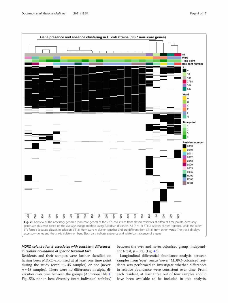
Genome-based clustering reveals a ward-specific E. coliST131 strainBased on pangenome analysis, we identified core andaccessory (non-core) genes, of which the accessorygenes (5057) were selected for clustering. Clusteringbased on presence/absence of these accessory genesshowed a clear cluster of ST131 strains (Fig. 2).Within the ST131 cluster, two separate clusters couldbe observed, one closely related cluster of twelve iso-lates belonging to three residents on ward A, and onecluster of four less related isolates from four residentsof four different wards. The isolates of three residentson ward A (R002, R003 and R004) have nearly identi-cal accessory genes, suggesting that they were colo-nised with the same strain. In addition, these isolateshave a nearly identical accessory genome over time,suggesting persistent colonisation of the same strain.
Clustering based on the maximum likelihood phylogenyof core genes also resulted in a clear clustering of ST131strains (data not shown). In addition, while the differencesare smaller than in the accessory genome, ST131 strainsfrom ward A still cluster separately from ST131 strainsfrom other wards. Lastly, a core-genome MLST confirmsclustering of ST131 strains on ward A (with up to two al-leles difference) and shows that ST131 isolates from otherwards are different (with more than 30 alleles difference)(Additional file 1: Fig. S2). These results support the hy-pothesis that an ST131 strain was spread across ward A.
Specific resistance genes are exclusive to certain wardsNext, the prevalence of antibiotic resistance genes wasdetermined. Based on resistance gene absence/presencein the genome, ST131 largely clustered together (Fig. 3),and again a cluster of ST131 belonging to residents ofone ward (ward A) was observed. These strains werecharacterised by presence of nine resistance genes (aac(6’)-Ib-cr, aadA5, bla-CTX-M-15, blaOXA-1, catB3,dfrA17, mph(A), sul1 and tet(a)). Three isolates belong-ing to ST131, 847 and 2786 from ward F clustered to-gether, and these three strains (from two residents)contained the rifampicin resistance gene arr-3, whichwas not detected in other strains.
Gut microbiota analysis using 16S rRNA gene ampliconsequencingA distinct gut microbiota between MDRO-positive andMDRO-negative samplesFirst, alpha diversity (using observed ASVs/genera andShannon index) was computed at both ASV andgenus level to compare MDRO-positive with MDRO-negative samples. To account for repeated measures,we stratified these alpha diversity analyses by timepoint. No significant differences in alpha diversities ateither level at any time point were observed (Add-itional file 1: Fig. S3). Beta diversity was also not sig-nificantly different between these samples (p = 0.12and R2 = 0.049) (Fig. 4a). To identify individual bacter-ial taxa associated with MDRO status, differentialabundance analysis was performed using DESeq2 ateach time point. Several taxa were more abundant inMDRO-negative samples on multiple timepoints,namely Atopobiaceae, Coprococcus_3, Dorea, Enorma,Holdemanella, Lachnospiraceae, Lachnospiraceae_ND3007_group, Phascolarctobacterium and Rumino-cocceae_UCG-014 (Additional file 1: Fig. S4,Additional file 2: Table S1). Only three taxa (Erysipe-latoclostridium, uncultured_Coriobacteriales and un-cultured_Ruminococcaceae) were more abundant inMDRO-positive samples at any time point.
Ducarmon et al. Genome Medicine (2021) 13:54 Page 7 of 17
MDRO colonisation is associated with consistent differencesin relative abundance of specific bacterial taxaResidents and their samples were further classified onhaving been MDRO-colonised at at least one time pointduring the study (ever, n = 45 samples) or not (never,n = 48 samples). There were no differences in alpha di-versities over time between the groups (Additional file 1:Fig. S5), nor in beta diversity (intra-individual stability)
between the ever and never colonised group (independ-ent t-test, p = 0.2) (Fig. 4b).Longitudinal differential abundance analysis between
samples from ‘ever’ versus ‘never’ MDRO-colonised resi-dents was performed to investigate whether differencesin relative abundance were consistent over time. Fromeach resident, at least three out of four samples shouldhave been available to be included in this analysis,
Fig. 2 Overview of the accessory genome (non-core genes) of the 22 E. coli strains from eleven residents at different time points. Accessorygenes are clustered based on the average linkage method using Euclidean distances. All (n = 17) ST131 isolates cluster together, while the otherSTs form a separate cluster. In addition, ST131 from ward A cluster together and are different from ST131 from other wards. The y-axis displaysaccessory genes and the x-axis isolate numbers. Black bars indicate presence and white bars absence of a gene
Ducarmon et al. Genome Medicine (2021) 13:54 Page 8 of 17

resulting in 45 samples from ever colonised residentsand 42 samples from never colonised residents. Threetaxa (Atopobiaceae, Dorea and Lachnospiraceae_ND3007_group) were consistently more abundant in‘never’ colonised residents throughout the 6-monthstudy period (Fig. 5, Additional file 1: Fig. S6). Thesetaxa were also identified to be more abundant inMDRO-negative samples compared to MDRO-positivesamples at two time points (Additional file 1: Fig. S4).Lastly, we looked for intra-individual changes in
pairs of samples of residents who either becameMDRO colonised or were MDRO decolonised duringthe study period. For this, samples were analysed ofan MDRO-negative sample prior to an MDRO-positive sample (n = 8 residents), and vice versa; anMDRO-positive sample followed by an MDRO-negative sample (n = 6 residents). Resident L10 couldbe included twice in the first comparison, but toavoid excessive impact of this resident on statisticalanalysis, it was included once. We then performedpaired analyses for each of the two groups. However,no differences in alpha or beta diversity were
observed, nor were any genera differentially abundantin any of the comparisons (data not shown).
Compositional profiles show very high abundance ofActinobacteria members Bifidobacterium and CollinsellaNext, we investigated the global microbiota profilesacross all residents without a focus on MDRO colonisa-tion. Compositional profiles at phylum and family levelshowed that the most abundant phylum in multiple resi-dents was Actinobacteria (Fig. 6a), which is in contrastto what is considered a ‘normal’ gut microbiota that gen-erally consists of ~ 90% Firmicutes and Bacteroidetes.Bifidobacterium and Collinsella were the Actinobacteriamembers with highest relative abundance (Fig. 6b).
Metagenome analysis using shotgun sequencing data often faecal samplesNot a single species, but several Bifidobacterium species arehighly abundant in residentsThe nursing home did not provide probiotics to theirresidents. However, the high abundance of Bifidobacter-ium in the residents’ stools suggested otherwise. Ten
Fig. 3 Heatmap of antibiotic resistance genes in the 22 E. coli isolates from eleven residents at different time points. Black boxes indicatepresence of a resistance gene, while white indicates absence of the resistance gene. Antibiotic resistance gene profiles are clustered byhierarchical clustering using Euclidian distances. Resident number, time, ward and time point are given as coloured annotations
Ducarmon et al. Genome Medicine (2021) 13:54 Page 9 of 17

stool samples from five residents with high Bifidobacter-ium and/or Collinsella relative abundance were furtherinvestigated by shotgun metagenomic sequencing, andtwo positive controls were included. The high relativeabundance of Bifidobacterium and Collinsella could beconfirmed and residents were colonised by seven highlyabundant Bifidobacterium species, namely B. adolescen-tis, B. angulatum, B. bifidium, B. breve, B. longum, B.pseudocatenulatum and B. ruminantium (Fig. 7a). Fromthese species, B. adolescentis, B. bifidum, B. breve and B.longum are the most commonly used species in probio-tics, although the others have been studied for probioticproperties as well [57].
Assembly-based method reveals that Bifidobacteriumlongum strains are (almost) identical within residents, butnot between residentsTo investigate whether Bifidobacterium longum strainswere identical between and within residents, we analysedthe strains using de novo assemblies. B. longum was se-lected because of its high relative abundance in multiplesamples, increasing the chance of recovering a full gen-ome from the respective metagenomes and because it is
commonly present in probiotics. Its genome size isabout 2.5Mb and contains a high GC content of ~ 60%.From samples of residents L001, L006 and L028, B.longum genomes larger than 2Mb could be recovered,indicating that (nearly) full genomes were successfullyobtained from the metagenome, but this was not thecase for L031 and R003 (Additional file 3: Table S2).While average nucleotide identities were high betweensamples, strains from the same individual were moreidentical to themselves than to strains from other resi-dents (Fig. 7b). This indicates that residents do not carrythe same B. longum strains. It should be noted that a fullB. longum genome could not be retrieved for all resi-dents. Lastly, B. longum genomes were compared to theNCBI reference genome (accession number NC_011593), the representative genome (NC_004307) and itsplasmid (NC_004943) and several other B. longumstrains (Fig. 7b) to provide insight in what levels of di-vergence are to be expected between strains. Comparingthese B. longum genomes from the NCBI database showsthat unrelated B. longum strains have an average nucleo-tide identity (ANI) of between 0.956 and 0.988. This fur-ther confirms that B. longum strains between the
Fig. 4 Bray-Curtis distance measures visualised by principle coordinates analysis (PCoA) for all (n = 93) faecal samples based on whether an MDROwas cultured (a) and by mean intraindividual stability (1 - Bray-Curtis dissimilarity) between ‘ever’ and ‘never’ colonised residents (b). Each dot inplot A represents a single sample, and ellipses indicate 95% confidence intervals
Ducarmon et al. Genome Medicine (2021) 13:54 Page 10 of 17

nursing home residents were different (maximum ANIbetween strains from different residents 0.99) and thatwithin residents strains were almost identical (ANI >0.994), in case a nearly full genome could be retrieved.
DiscussionWe present a unique study on asymptomatic gut MDRO(in this study MDR Enterobacterales) colonisation innursing home residents and performed a wide variety ofanalyses, namely clinical risk factor analysis, WGS ofMDRO isolates and 16S rRNA gene amplicon sequen-cing and metagenomic sequencing of the gut microbiota.We identify possible risk factors for MDRO colonisation,potential spread of MDROs within a ward and microbialsignatures associated with MDRO colonisation using16S rRNA gene amplicon sequencing. Many of theMDRO-associated microbial signatures are consistentover the 6-month time course of this study as shown bylongitudinal modelling. Additionally, the unexpectedlyhigh abundance of Bifidobacterium abundance in mul-tiple residents was further investigated using metage-nomic sequencing. We show that this high abundance isvery unlikely to be stemming from probiotic supplemen-tation, as Bifidobacterium species and B. longum strainsdiffered between residents.We observed a spread of E. coli ST131 within a ward,
but not between wards, as the ST131 seemed ward-specific. E. coli ST131 was the most commonly found STin our study, which is in line with previous results
showing that this ST is major driver of the currentworldwide spread of ESBL-producing E. coli [58, 59].This sequence type is associated with community-acquired infections and older age, and is frequently ob-served in nursing homes in countries throughout Europeand the USA [7, 60–62]. While ST131 outbreaks aregenerally seen among and between various nursinghomes, we concluded that spread of specific ST131strains was restricted within wards. However, previousstudies may have been limited by methods to character-ise ST131, as they characterise strains only with regularMLST (of a limited number of housekeeping genes). Byusing pangenome analysis, we investigated the geneticdifferences in detail, allowing for discrimination of theST131 strains between the wards. We conclude thatMDRO transmission within nursing home wards seemsto reflect that of household contacts [63]. This smallscale MDRO spread was observed in the samples of 27residents, one could hypothesise higher absolute num-bers of related strains if all nursing home residentswould have been screened. Not only strains can spread,plasmids are also able to move between different bacter-ial strains. For instance, three different E. coli ST typesfound at ward F contained arr-3, aadA16 and dfrA27.Considering that these three genes are usually encodedon a plasmid [64, 65], it is possible that they spread be-tween ST131 strains on ward F. However, definite con-clusions cannot be made based on these results, as onlythree MDRO strains were detected in ward F.
Fig. 5 Time intervals of significantly different bacterial genera between ever (n = 12) and never (n = 15) MDRO colonised residents. Each lineinterval represents a significant time interval, with significance being considered p < 0.05. Orange lines indicate higher abundance in the nevercolonised group, while blue indicates higher abundance in the ever colonised group. If no coloured line is observed, the respective genus is notsignificantly differentially abundant between specific time points
Ducarmon et al. Genome Medicine (2021) 13:54 Page 11 of 17

Novel microbial signatures of MDRO colonisationwere identified which could contribute to colonisationresistance against MDROs. Three taxa were consistentlymore abundant throughout the study in residents nevercolonised with an MDRO, namely Dorea, Lachnospira-ceae_ND3007_group and Atopobiaceae, and these taxawere also found to be more abundant in MDRO-negative samples at two time points. Increased relativeabundance of Dorea and the Lachnospiraceae family hasbeen shown to be associated with colonisation resistanceagainst Campylobacter infection [66]. The relative abun-dance of Dorea formicigenerans was identified as a po-tential pre-liver transplant marker for subsequentMDRO colonisation [67] but another report did notmention Dorea as either a protective taxon or a risk fac-tor [13]. While these results are conflicting, there is apossibility that different studies observed effects of dif-ferent Dorea species or strains, which could theoreticallyhave different or opposing effects on MDRO colonisa-tion. Lastly, as clinical variables were not evenly distrib-uted between compared groups, there is a possibilitythat observed differences in relative abundance of bac-terial taxa can partially be attributed to these confound-ing factors.We did not observe differences in alpha diversities be-
tween the different groups based on MDRO status. Thiscontrasts several reports where MDRO colonisation wasassociated with a reduced alpha diversity, although con-flicting evidence exists [13, 67, 68]. In addition, no
difference in beta diversity was observed between theever and never MDRO-colonised groups, nor betweenMDRO-positive and MDRO-negative samples. This con-tradicts findings in liver transplant patients and MDROcolonisation [67]. Conflicting results regarding diversitiesand microbial signatures could have multiple reasons.First, technical variation induced from the entire work-flow starting with sample collection and ending with useof different statistical tools. Second, different MDROtypes were studied between the various reports. In thecurrent study, we mainly observed multi-drug-resistantE. coli, while two other major studies investigatingMDROs and gut microbiota found a larger variety ofMDRO types [13, 67]. Considering that microbiome-mediated colonisation resistance is likely to be specificfor individual bacterial species and most likely even bac-terial strains, further studies should ideally focus on in-vestigating single MRDOs in relation to the gutmicrobiota. Third, geographical locations of the studiedcohorts were different, likely reflecting differences in gutmicrobiota composition due to varying dietary patternsand other cultural habits.An unexpectedly high relative abundance of Bifidobac-
terium was observed in several residents in differentwards. Such consistently high relative abundances have,to the best of our knowledge, not previously been de-scribed in adults or elderly. Incidental reports of an out-growth of Bifidobacterium species in elderly in a long-term care facility have been described [69]. Rowan et al.
Fig. 6 Compositional profiles at phylum level (a) and genus level (b) from 16S rRNA gene amplicon data of 27 residents at four time points.Other indicates the sum of all bacterial phyla or genera not specifically indicated in the legend. The y-axis displays relative abundance and the x-axis the study time point
Ducarmon et al. Genome Medicine (2021) 13:54 Page 12 of 17

observed a high relative abundance of Bifidobacteriumspecies in two out of eleven elderly subjects (> 15% rela-tive abundance at at least one time point; mainly B.longum, B. breve and B. adolescentis), although potentialexplanations were not discussed.It is known that in infancy the gut microbiota is largely
dominated by Bifidobacterium, but that this high abun-dance declines with ageing [70]. In addition, elderlymostly harbour B. longum, B.nucleatum, B. pseudonu-cleatum and B. adolescentis. While we found that thesespecies were indeed among the most abundant, highrelative abundances of B. angulatum, B. bifidus, B. breveand B. ruminantium were also observed. At first, wehypothesised that high Bifidobacterium relative abun-dance could be stemming from probiotic supplementa-tion used on a voluntary basis by the nursing homeresidents, despite knowing that probiotics generally donot colonise very successfully [71, 72]. By performingmetagenomic sequencing on a subset of samples, weshowed this was unlikely to be the case, as different Bifi-dobacterium species were observed between residents.In addition, using strain-resolved metagenomics, weshow that B. longum strains were different between
residents, but likely the same within residents. Our sec-ond hypothesis was related to dietary patterns of resi-dents that perhaps a very monotonous diet couldstimulate outgrowth of Bifidobacterium. However, resi-dents consumed fresh, daily prepared meals according toa normal Dutch diet. It is unclear what the reasons andconsequences of this high relative abundance of Bifido-bacterium are in our residents. In combination with theobservation that a high relative abundance of Bifidobac-terium is not associated with protection against MDROcolonisation, this suggests that probiotics based on theBifidobacterium species in our study may not effectivelyprotect against MDRO colonisation.This study has several limitations and strengths. First,
our sample size and number of MDRO-positive sampleswas limited, preventing the application of a more exten-sive epidemiological risk factor analysis. Sample size wasalso a limiting factor in differential abundance testingbetween MDRO-positive and MDRO-negative samplesper time point. Second, this study focused on a singlenursing home and we can therefore not be certain thatmicrobiota profiles are representative for residents ofother (Dutch) nursing homes. Especially in light of our
Fig. 7 Compositional plot based on metagenomes of ten faecal samples from five residents using mOTUs (a) and average nucleotide identitybetween assembled B. longum strains and reference sequences (b). Relative abundance is shown of the twenty most abundant bacterial speciesin all samples and different bacterial species are indicated by colours. ‘Other’ is the sum of the relative abundance of all species not listed in thecolour key. Numbers on the x-axis indicate the resident number and study time point. Average nucleotide identity of B. longum strains ascomputed by pyANI. The sequence labelled ‘NC_004307_REP’ in B is the representative genome on GenBank; the sequence with‘NC_004943_PLAS’ is its plasmid. The sequence with ‘NC_011593_REF’ is the B. longum reference genome
Ducarmon et al. Genome Medicine (2021) 13:54 Page 13 of 17

unique findings of high relative abundance of Bifidobac-terium species, profiling the gut microbiota across othernursing homes would be important. Third, some wardshad a very limited number of MDRO isolates, whichhampered making definite conclusions about MDROspread in those wards. Lastly, not all residents providedfaecal samples on all four time points.However, this study uses a unique combination of
analyses for in-depth understanding of MDRO spreadin a nursing home and the relation of MDRO colon-isation with residents’ microbiota. The longitudinalnature of our study setup allowed for (1) detection ofrobust associations between MDRO colonisation andspecific microbial taxa, (2) identifying whether colo-nising MDRO strains were identical over time and (3)comparing B. longum strains within and between resi-dents using strain-resolved metagenomics. In addition,the use of various statistical methods for identifyingmicrobial taxa associated with MDRO colonisationfurther strengthens our findings. Lastly, our finding ofhigh relative abundance of Bifidobacterium in mul-tiple residents warrants further investigation and con-firmation by other studies.
ConclusionsOur study provides new evidence regarding the gutmicrobiota’s potential in providing resistance againstMDRO colonisation in a nursing home. Several specifictaxa were identified which were consistently more abun-dant in residents never colonised with an MDROthroughout the 6-month study. Considering that most ofthe detected MDROs were E. coli strains belonging toST131, it may be especially interesting to test the poten-tially protective effect of these taxa against E. coli ST131.In addition, we report a uniquely high abundance of sev-eral Bifidobacterium species in multiple residents andexcluded the possibility that this was due to probioticsupplementation. While the reasons for, and conse-quences of this high relative abundance remain unclear,it does suggest that probiotics based on Bifidobacteriumspecies observed in our study are highly unlikely to pre-vent or eradicate MDRO colonisation in the gut of nurs-ing home residents.
AbbreviationsMDROs: Multidrug-resistant organisms; ESBL: Extended-spectrum beta-lactamase; VRE: Vancomycin-resistant Enteroccocus; WGS: Whole-genomesequencing; TSB: Tryptic soy broth; MALDI-TOF: Matrix-assisted laserdesorption ionisation-time of flight; EUCAST: European Committee ofAntimicrobial Susceptibility Testing; MIC: Minimum inhibitory concentration;GEE: Generalised estimating equations; ST: Sequence type; ASV: Ampliconsequence variant; PERMANOVA: Permutational multivariate analysis ofvariance; MetaLonDA: Metagenomic longitudinal differential abundancemethod; MLST: Multi-locus sequence typing; OR: Odds ratio; CI: Confidenceinterval; ANI: Average nucleotide identity
Supplementary InformationThe online version contains supplementary material available at https://doi.org/10.1186/s13073-021-00869-z.
Additional file 1: Figures S1-S6.
Additional file 2: Table S1. Results of DESeq2 analysis on MDRO-positive samples versus MDRO-negative samples per time point. Negativelog2FoldChanges in the table indicate that a genus is more abundant innon-MDRO-colonised samples.
Additional file 3: Table S2. Retrieved genome length for B. longum inbp from resident samples, and genome length of several referencesequences of B. longum. For L001, L006 and L028_2 (nearly) full B.longum genomes were obtained (length > 2 million bp).
AcknowledgementsWe would like to thank all participating nursing home residents, theirfamilies and the staff of the nursing home: Woonzorgcentra Haaglanden, theHague in the Netherlands.We would like to thank Renato Alves for his help and feedback regardingNGLess, Bastian Hornung for his guidance in analysis of MDRO isolates andJacco Wallinga for his advice on risk factor analysis. In addition, we wouldlike to thank all working members of CMAT for their valuable feedbackduring work discussions.
Authors’ contributionsQD analysed the epidemiological data and 16S rRNA gene amplicon data,processed and analysed metagenomic data, created figures and wrote draftsof the manuscript. ET was the principal investigator, supervised the culturingexperiments and wrote drafts of the manuscript. SN processed and analysedthe WGS data of MDRO isolates, processed and analysed metagenomic data,created figures and wrote drafts of the manuscript. MB performed practicalwork related to MDRO culturing and 16S rRNA gene amplicon sequencing.KV aided in the epidemiological analysis. MC designed the clinical study. ISperformed practical work related to MDRO culturing. SD and MW designedthe clinical study. RZ processed 16S rRNA gene amplicon data andsupervised microbiota analyses. EK designed and supervised the clinicalstudy. All authors read and approved the final manuscript.
FundingThis study was financially supported by RIVM with ‘Bijzonder resistentemicro-organismen en Clostridium difficile dragerschap bij verpleeghuisbew-oners:’ (2016–2017).
Availability of data and materialsThe sequencing data in this study are available at the European NucleotideArchive (ENA) under accession number PRJEB37898 (https://www.ebi.ac.uk/ena/browser/view/PRJEB37898) [73]. All data and R code necessary toreproduce analyses and figures from this manuscript can be found at https://github.com/qducarmon/nursing_home_MDRO [74].
Declarations
Ethics approval and consent to participateWritten informed consent was obtained from the resident or his/her proxy.Ethical approval was granted by the ‘Medisch Ethische Toetsings Commissie’of Leiden University Medical Centre (No.P16.039). The study was conductedin accordance with the guidelines of the Helsinki declaration. Sixty-four(49%) residents consented to participate. For this study, we included resi-dents who gave consent for additional analyses, from whom faeces was cul-tured for MDROs at at least two time points, and of which sufficient materialwas left for microbiota profiling at at least two time points (n = 27 residents).
Consent for publicationNot applicable.
Competing interestsET and EK are supported by an unrestricted grant from Vedanta BiosciencesInc. EK has performed research for Cubist, Novartis and Qiagen and hasparticipated in advisory forums of Astellas, Optimer, Actelion, Pfizer, Sanofi
Ducarmon et al. Genome Medicine (2021) 13:54 Page 14 of 17

Pasteur and Seres Therapeutics. The companies had no role in the study orwriting of the manuscript. The remaining authors declare that they have nocompeting interests.
Author details1Department of Medical Microbiology, Leiden University Medical Center,Leiden, The Netherlands. 2Center for Microbiome Analyses and Therapeutics,Leiden University Medical Center, Leiden, The Netherlands. 3Center forInfectious Disease Control, National Institute for Public Health and theEnvironment, Bilthoven, The Netherlands. 4Department of Public Health andPrimary Care, Leiden University Medical Center, Leiden, The Netherlands.5Department of Internal Medicine and Geriatrics, Onze Lieve VrouweGasthuis (OLVG Hospital), Amsterdam, The Netherlands.
Received: 17 December 2020 Accepted: 16 March 2021
References1. Cassini A, Högberg LD, Plachouras D, Quattrocchi A, Hoxha A, Simonsen GS,
Colomb-Cotinat M, Kretzschmar ME, Devleesschauwer B, Cecchini M,Ouakrim DA, Oliveira TC, Struelens MJ, Suetens C, Monnet DL, Strauss R,Mertens K, Struyf T, Catry B, Latour K, Ivanov IN, Dobreva EG, TambicAndraševic A, Soprek S, Budimir A, Paphitou N, Žemlicková H, Schytte OlsenS, Wolff Sönksen U, Märtin P, Ivanova M, Lyytikäinen O, Jalava J, Coignard B,Eckmanns T, Abu Sin M, Haller S, Daikos GL, Gikas A, Tsiodras S, KontopidouF, Tóth Á, Hajdu Á, Guólaugsson Ó, Kristinsson KG, Murchan S, Burns K,Pezzotti P, Gagliotti C, Dumpis U, Liuimiene A, Perrin M, Borg MA, de GreeffSC, Monen JCM, Koek MBG, Elstrøm P, Zabicka D, Deptula A, Hryniewicz W,Caniça M, Nogueira PJ, Fernandes PA, Manageiro V, Popescu GA, Serban RI,Schréterová E, Litvová S, Štefkovicová M, Kolman J, Klavs I, Korošec A, AracilB, Asensio A, Pérez-Vázquez M, Billström H, Larsson S, Reilly JS, Johnson A,Hopkins S. Attributable deaths and disability-adjusted life-years caused byinfections with antibiotic-resistant bacteria in the EU and the Europeaneconomic area in 2015: a population-level modelling analysis. Lancet InfectDis. 2019;19(1):56–66. https://doi.org/10.1016/S1473-3099(18)30605-4.
2. Cassone M, Mody L. Colonization with multi-drug resistant organisms innursing homes: scope, importance, and management. Curr Geriatr Rep.2015;4(1):87–95. https://doi.org/10.1007/s13670-015-0120-2.
3. Gorrie CL, Mirceta M, Wick RR, Judd LM, Wyres KL, Thomson NR, StrugnellRA, Pratt NF, Garlick JS, Watson KM, Hunter PC, McGloughlin SA, SpelmanDW, Jenney AWJ, Holt KE. Antimicrobial-resistant Klebsiella pneumoniaecarriage and infection in specialized geriatric care wards linked toacquisition in the referring hospital. Clin Infect Dis. 2018;67(2):161–70.https://doi.org/10.1093/cid/ciy027.
4. Giannella M, Tedeschi S, Bartoletti M, Viale P. Prevention of infections innursing homes: antibiotic prophylaxis versus infection control andantimicrobial stewardship measures. Expert Rev Anti-Infect Ther. 2016;14(2):219–30. https://doi.org/10.1586/14787210.2016.1132161.
5. Rooney PJ, O'Leary MC, Loughrey AC, McCalmont M, Smyth B, Donaghy P,Badri M, Woodford N, Karisik E, Livermore DM. Nursing homes as a reservoirof extended-spectrum beta-lactamase (ESBL)-producing ciprofloxacin-resistant Escherichia coli. J Antimicrob Chemother. 2009;64(3):635–41.https://doi.org/10.1093/jac/dkp220.
6. Verhoef L, Roukens M, de Greeff S, Meessen N, Natsch S, Stobberingh E.Carriage of antimicrobial-resistant commensal bacteria in Dutch long-term-care facilities. J Antimicrob Chemother. 2016;71(9):2586–92. https://doi.org/10.1093/jac/dkw183.
7. van der Donk CF, Schols JM, Driessen CJ, Hagenouw RG, Meulendijks A,Stobberingh EE. Prevalence and spread of multidrug resistant Escherichiacoli isolates among nursing home residents in the southern part of TheNetherlands. J Am Med Dir Assoc. 2013;14(3):199–203. https://doi.org/10.1016/j.jamda.2012.09.026.
8. van Dulm E, Tholen ATR, Pettersson A, van Rooijen MS, Willemsen I,Molenaar P, Damen M, Gruteke P, Oostvogel P, Kuijper EJ, Hertogh CMPM,Vandenbroucke-Grauls CMJE, Scholing M. High prevalence of multidrugresistant Enterobacteriaceae among residents of long term care facilities inAmsterdam, the Netherlands. Plos One. 2019;14(9):e0222200. https://doi.org/10.1371/journal.pone.0222200.
9. Terveer EM, Fallon M, Kraakman MEM, Ormond A, Fitzpatrick M, CaljouwMAA, Martin A, van Dorp SM, Wong MC, Kuijper EJ, Fitzpatrick F. Spread ofESBL-producing Escherichia coli in nursing home residents in Ireland and
the Netherlands may reflect infrastructural differences. J Hosp Infect. 2019;103(2):160–4. https://doi.org/10.1016/j.jhin.2019.05.003.
10. Verhoef L, Stobberingh E, Smid E, Kuijper EJ, De Greeff S, Heck M. Intestinalcarriage of resistant bacteria and Clostridium difficile in nursing homes in theNetherlands—a point prevalence study. Vienna: European Congress ofClinical Microbiology and Infectious Diseases; 2017.
11. Arvand M, Moser V, Schwehn C, Bettge-Weller G, Hensgens MP, Kuijper EJ.High prevalence of Clostridium difficile colonization among nursing homeresidents in Hesse, Germany. Plos One. 2012;7(1):e30183. https://doi.org/10.1371/journal.pone.0030183.
12. Ducarmon QR, Zwittink RD, Hornung BVH, van Schaik W, Young VB, KuijperEJ. Gut microbiota and colonization resistance against bacterial entericinfection. Microbiol Mol Biol Rev. 2019;83(3):e00007–19.
13. Araos R, Battaglia T, Ugalde JA, Rojas-Herrera M, Blaser MJ, D'Agata EMC.Fecal microbiome characteristics and the resistome associated withacquisition of multidrug-resistant organisms among elderly subjects. FrontMicrobiol. 2019;10:2260. https://doi.org/10.3389/fmicb.2019.02260.
14. Langelier C, Graves M, Kalantar K, Caldera S, Durrant R, Fisher M, Backman R,Tanner W, DeRisi JL, Leung DT. Microbiome and antimicrobial resistancegene dynamics in international travelers. Emerg Infect Dis. 2019;25(7):1380–3. https://doi.org/10.3201/eid2507.181492.
15. Kim SG, Becattini S, Moody TU, Shliaha PV, Littmann ER, Seok R, Gjonbalaj M,Eaton V, Fontana E, Amoretti L, Wright R, Caballero S, Wang ZMX, Jung HJ,Morjaria SM, Leiner IM, Qin W, Ramos RJJF, Cross JR, Narushima S, Honda K,Peled JU, Hendrickson RC, Taur Y, van den Brink MRM, Pamer EG.Microbiota-derived lantibiotic restores resistance against vancomycin-resistant enterococcus. Nature. 2019;572(7771):665–9. https://doi.org/10.1038/s41586-019-1501-z.
16. Point prevalence survey of healthcare-associated infections andantimicrobial use in European long-term care facilities. April–May 2013.Available at: https://ecdc.europa.eu/sites/portal/files/media/en/publications/Publications/healthcare-associated-infections-point-prevalence-survey-longterm-care-facilities-2013.pdf. ECDC. 2014. Accessed 30 Nov 2020.
17. RIVM. WIP richtlijn Bijzonder resistente micro-organismen. Verpleeghuizen,woonzorgcentra en voorzieningen voor kleinschalig wonen voor ouderen.http://www.rivm.nl/dsresource?objectid=513c8b7b-189c4bcd-a124-cdeb80af520a&type=org&disposition=inline2014. Accessed 30 Nov 2020.
18. EUCAST. Breakpoint tables for interpretation of MICs and zone diameters.version 73 http://www.eucast.org/clinical_breakpoints/. 2017. Accessed 30Nov 2020.
19. NVMM. Laboratory detection of highly resistant microorganisms (HRMO). Revision2017 https://www.nvmm.nl/media/1051/2012_hrmo_mrsa_esbl.pdf. 2012.Accessed 30 Nov 2020.
20. Terveer EM, Crobach MJ, Sanders IM, Vos MC, Verduin CM, Kuijper EJ.Detection of Clostridium difficile in feces of asymptomatic patientsadmitted to the hospital. J Clin Microbiol. 2017;55(2):403–11. https://doi.org/10.1128/JCM.01858-16.
21. Højsgaard S, Halekoh U, Yan J. The R Package geepack for generalizedestimating equations. J Stat Softw. 2005;15(2):1–11.
22. Peduzzi P, Concato J, Kemper E, Holford TR, Feinstein AR. A simulationstudy of the number of events per variable in logistic regressionanalysis. J Clin Epidemiol. 1996;49(12):1373–9. https://doi.org/10.1016/S0895-4356(96)00236-3.
23. Johnston R, Jones K, Manley D. Confounding and collinearity in regressionanalysis: a cautionary tale and an alternative procedure, illustrated bystudies of British voting behaviour. Qual Quant. 2018;52(4):1957–76. https://doi.org/10.1007/s11135-017-0584-6.
24. Andrews S. FastQC: a quality control tool for high throughput sequencedata. Available online at: http://wwwbioinformaticsbabrahamacuk/projects/fastqc. 2010. Accessed 12 Feb 2020.
25. Ewels P, Magnusson M, Lundin S, Käller M. MultiQC: summarize analysisresults for multiple tools and samples in a single report. Bioinformatics.2016;32(19):3047–8. https://doi.org/10.1093/bioinformatics/btw354.
26. Souvorov A, Agarwala R, Lipman DJ. SKESA: strategic k-mer extension forscrupulous assemblies. Genome Biol. 2018;19(1):153. https://doi.org/10.1186/s13059-018-1540-z.
27. Nurk S, Bankevich A, Antipov D, Gurevich A, Korobeynikov A, Lapidus A,et al. Assembling Genomes and Mini-metagenomes from Highly ChimericReads, eds Deng M, Jiang R, Sun F, Zhang X. Research in ComputationalMolecular Biology: 17th Annual International Conference, RECOMB 2013,Beijing, China. Springer, Berlin, Germany.
Ducarmon et al. Genome Medicine (2021) 13:54 Page 15 of 17
28. Gurevich A, Saveliev V, Vyahhi N, Tesler G. QUAST: quality assessment toolfor genome assemblies. Bioinformatics. 2013;29(8):1072–5. https://doi.org/10.1093/bioinformatics/btt086.
29. Altschul SF, Gish W, Miller W, Myers EW, Lipman DJ. Basic local alignmentsearch tool. J Mol Biol. 1990;215(3):403–10. https://doi.org/10.1016/S0022-2836(05)80360-2.
30. Camacho C, Coulouris G, Avagyan V, Ma N, Papadopoulos J, Bealer K,Madden TL. BLAST+: architecture and applications. BMC Bioinformatics.2009;10(1):421. https://doi.org/10.1186/1471-2105-10-421.
31. Ondov BD, Bergman NH, Phillippy AM. Interactive metagenomicvisualization in a web browser. BMC Bioinformatics. 2011;12(1):385. https://doi.org/10.1186/1471-2105-12-385.
32. Zankari E, Hasman H, Cosentino S, Vestergaard M, Rasmussen S, Lund O,Aarestrup FM, Larsen MV. Identification of acquired antimicrobial resistancegenes. J Antimicrob Chemother. 2012;67(11):2640–4. https://doi.org/10.1093/jac/dks261.
33. Jünemann S, Sedlazeck FJ, Prior K, Albersmeier A, John U, Kalinowski J,Mellmann A, Goesmann A, von Haeseler A, Stoye J, Harmsen D. Updatingbenchtop sequencing performance comparison. Nat Biotechnol. 2013;31(4):294–6. https://doi.org/10.1038/nbt.2522.
34. Page AJ, Cummins CA, Hunt M, Wong VK, Reuter S, Holden MTG, Fookes M,Falush D, Keane JA, Parkhill J. Roary: rapid large-scale prokaryote pangenome analysis. Bioinformatics. 2015;31(22):3691–3. https://doi.org/10.1093/bioinformatics/btv421.
35. Seemann T. Prokka: rapid prokaryotic genome annotation. Bioinformatics.2014;30(14):2068–9. https://doi.org/10.1093/bioinformatics/btu153.
36. Kalyaanamoorthy S, Minh BQ, Wong TKF, von Haeseler A, Jermiin LS.ModelFinder: fast model selection for accurate phylogenetic estimates. NatMethods. 2017;14(6):587–9. https://doi.org/10.1038/nmeth.4285.
37. Ducarmon QR, Hornung BVH, Geelen AR, Kuijper EJ, Zwittink RD. Towardstandards in clinical microbiota studies: comparison of three DNA extractionmethods and two bioinformatic pipelines. mSystems. 2020;5(1):e00547–19.
38. Ramiro-Garcia J, Hermes GDA, Giatsis C, Sipkema D, Zoetendal EG, SchaapPJ, et al. NG-Tax, a highly accurate and validated pipeline for analysis of 16SrRNA amplicons from complex biomes [version 1; referees: 2 approved withreservations, 1 not approved]. F1000Res. 2016;5:1791.
39. Quast C, Pruesse E, Yilmaz P, Gerken J, Schweer T, Yarza P, Peplies J,Glöckner FO. The SILVA ribosomal RNA gene database project: improveddata processing and web-based tools. Nucleic Acids Res. 2013;41(Databaseissue):D590–6. https://doi.org/10.1093/nar/gks1219.
40. Bokulich NA, Subramanian S, Faith JJ, Gevers D, Gordon JI, Knight R, MillsDA, Caporaso JG. Quality-filtering vastly improves diversity estimates fromIllumina amplicon sequencing. Nat Methods. 2013;10(1):57–9. https://doi.org/10.1038/nmeth.2276.
41. Coelho LP, Alves R, Monteiro P, Huerta-Cepas J, Freitas AT, Bork P. NG-meta-profiler: fast processing of metagenomes using NGLess, a domain-specificlanguage. Microbiome. 2019;7(1):84.
42. Kultima JR, Coelho LP, Forslund K, Huerta-Cepas J, Li SS, Driessen M, VoigtAY, Zeller G, Sunagawa S, Bork P. MOCAT2: a metagenomic assembly,annotation and profiling framework. Bioinformatics. 2016;32(16):2520–3.https://doi.org/10.1093/bioinformatics/btw183.
43. Kultima JR, Sunagawa S, Li J, Chen W, Chen H, Mende DR, et al. MOCAT: ametagenomics assembly and gene prediction toolkit. Plos One. 2012;7(10):e47656-e.
44. Milanese A, Mende DR, Paoli L, Salazar G, Ruscheweyh H-J, Cuenca M, et al.Microbial abundance, activity and population genomic profiling withmOTUs2. Nat Commun. 2019;10(1):1014.
45. Li H. Aligning sequence reads, clone sequences and assembly contigs withBWA-MEM. arXiv. 2013;preprint arXiv:1303.3997. Preprint at http://arxiv.org/abs/1303.3997.
46. Schmitz D, Nooij S, Verhagen R, Janssens T, Cremer J, Zwagemaker F, et al.Jovian, user-friendly metagenomics. GitHub. Online at https://github.com/DennisSchmitz/Jovian/. 2020. Accessed 17 Mar 2020.
47. Pritchard L, Glover RH, Humphris S, Elphinstone JG, Toth IK. Genomics andtaxonomy in diagnostics for food security: soft-rotting enterobacterial plantpathogens. Anal Methods. 2016;8(1):12–24. https://doi.org/10.1039/C5AY02550H.
48. Kolde R. pheatmap: Pretty Heatmaps. R package version 1.0.12. https://CRAN.R-project.org/package=pheatmap2019. Accessed 2 July 2020.
49. McMurdie PJ, Holmes S. phyloseq: an R package for reproducible interactiveanalysis and graphics of microbiome census data. Plos One. 2013;8(4):e61217.
50. Metwally AA, Yang J, Ascoli C, Dai Y, Finn PW, Perkins DL. MetaLonDA: aflexible R package for identifying time intervals of differentially abundantfeatures in metagenomic longitudinal studies. Microbiome. 2018;6(1):32.https://doi.org/10.1186/s40168-018-0402-y.
51. Lahti L, Shetty S. Tools for microbiome analysis in R. Microbiome packageversion 1.6.0; 2017.
52. Love MI, Huber W, Anders S. Moderated estimation of fold change anddispersion for RNA-seq data with DESeq2. Genome Biol. 2014;15(12):550.https://doi.org/10.1186/s13059-014-0550-8.
53. Wickham H. ggplot2: elegant graphics for data analysis. New York: SpringerVerlag; 2009. https://ggplot2.tidyverse.org.
54. Wickham H. tidyverse: easily install and load the ‘Tidyverse’. R packageversion 121. https://www.tidyverse.org/packages/. 2017. Accessed Mar 2020.
55. Bates D, Mächler M, Bolker BM, Walker SC. Fitting linear mixed-effectsmodels using lme4. J Stat Softw. 2015;67:1–48.
56. Kuznetsova A, Brockhoff PB, Christensen RHB. lmerTest package: Tests inlinear mixed effects models. J Stat Softw. 2017;82(13).
57. Fijan S. Microorganisms with claimed probiotic properties: an overview ofrecent literature. Int J Environ Res Public Health. 2014;11(5):4745–67. https://doi.org/10.3390/ijerph110504745.
58. Rogers BA, Sidjabat HE, Paterson DL. Escherichia coli O25b-ST131: apandemic, multiresistant, community-associated strain. J AntimicrobChemother. 2011;66(1):1–14. https://doi.org/10.1093/jac/dkq415.
59. Coque TM, Novais A, Carattoli A, Poirel L, Pitout J, Peixe L, Baquero F,Cantón R, Nordmann P. Dissemination of clonally related Escherichia colistrains expressing extended-spectrum beta-lactamase CTX-M-15. EmergInfect Dis. 2008;14(2):195–200. https://doi.org/10.3201/eid1402.070350.
60. Broussier M, Gbaguidi-Haoré H, Rachidi-Berjamy F, Bertrand X, Slekovec C.Prevalence, genetic diversity of and factors associated with ESBL-producingEnterobacterales carriage in residents of French nursing homes. J HospInfect. 2019;104(4):469–75.
61. Arvand M, Moser V, Pfeifer Y. Prevalence of extended-spectrum-β-lactamase-producing Escherichia coli and spread of the epidemic clonal lineage ST131in nursing homes in Hesse, Germany. J Antimicrob Chemother. 2013;68(11):2686–8. https://doi.org/10.1093/jac/dkt226.
62. Banerjee R, Johnston B, Lohse C, Porter SB, Clabots C, Johnson JR.Escherichia coli sequence type 131 is a dominant, antimicrobial-resistantclonal group associated with healthcare and elderly hosts. Infect ControlHosp Epidemiol. 2013;34(4):361–9. https://doi.org/10.1086/669865.
63. Hilty M, Betsch BY, Bögli-Stuber K, Heiniger N, Stadler M, Küffer M,Kronenberg A, Rohrer C, Aebi S, Endimiani A, Droz S, Mühlemann K.Transmission dynamics of extended-spectrum β-lactamase-producingEnterobacteriaceae in the tertiary care hospital and the household setting.Clin Infect Dis. 2012;55(7):967–75. https://doi.org/10.1093/cid/cis581.
64. Ma J, Zeng Z, Chen Z, Xu X, Wang X, Deng Y, Lü D, Huang L, Zhang Y, Liu J,Wang M. High prevalence of plasmid-mediated quinolone resistancedeterminants qnr, aac (6′)-Ib-cr, and qepA among ceftiofur-resistantEnterobacteriaceae isolates from companion and food-producing animals.Antimicrob Agents Chemother. 2009;53(2):519–24. https://doi.org/10.1128/AAC.00886-08.
65. Papagiannitsis CC, Kutilova I, Medvecky M, Hrabak J, Dolejska M.Characterization of the complete nucleotide sequences of IncA/C (2)plasmids carrying In809-like integrons from Enterobacteriaceae isolates ofwildlife origin. Antimicrob Agents Chemother. 2017;61(9).
66. Kampmann C, Dicksved J, Engstrand L, Rautelin H. Composition of humanfaecal microbiota in resistance to Campylobacter infection. Clin MicrobiolInfect. 2016;22(1):61.e1-.e8.
67. Annavajhala MK, Gomez-Simmonds A, Macesic N, Sullivan SB, Kress A, KhanSD, Giddins MJ, Stump S, Kim GI, Narain R, Verna EC, Uhlemann AC.Colonizing multidrug-resistant bacteria and the longitudinal evolution ofthe intestinal microbiome after liver transplantation. Nat Commun. 2019;10(1):4715. https://doi.org/10.1038/s41467-019-12633-4.
68. Araos R, Montgomery V, Ugalde JA, Snyder GM, D'Agata EMC. Microbialdisruption indices to detect colonization with multidrug-resistant organisms.Infect Control Hosp Epidemiol. 2017;38(11):1312–8. https://doi.org/10.1017/ice.2017.190.
69. Rowan-Nash AD, Araos R, D'Agata EMC, Belenky P. Antimicrobial resistancegene prevalence in a population of patients with advanced dementia isrelated to specific pathobionts. iScience. 2020;23(3):100905.
70. Arboleya S, Watkins C, Stanton C, Ross RP. Gut Bifidobacteria populations inhuman health and aging. Front Microbiol. 2016;7:1204.
Ducarmon et al. Genome Medicine (2021) 13:54 Page 16 of 17
71. Zmora N, Zilberman-Schapira G, Suez J, Mor U, Dori-Bachash M, BashiardesS, et al. Personalized gut mucosal colonization resistance to empiricprobiotics is associated with unique host and microbiome features. Cell.2018;174(6):1388–405.e21.
72. Maldonado-Gómez MX, Martínez I, Bottacini F, O'Callaghan A, Ventura M,van Sinderen D, et al. Stable engraftment of Bifidobacterium longumAH1206 in the human gut depends on individualized features of theresident microbiome. Cell Host Microbe. 2016;20(4):515–26. https://doi.org/10.1016/j.chom.2016.09.001.
73. Ducarmon QR, Terveer EM, Nooij S, Bloem MN, Vendrik KEW, Caljouw MAA,et al. Short-read sequencing data of MDRO isolates from residents of aDutch nursing home and short-read sequencing data of their gutmicrobiota with 16S rRNA gene amplicon and metagenomics sequencing.European Nucleotide Archive at EMBL-EBI under accession numberPRJEB37898: Available from: https://www.ebi.ac.uk/ena/browser/view/PRJEB37898; 2020. Accessed Aug 2020.
74. Ducarmon QR, Nooij S. GitHub repository with all necessary data andRMarkdown files to reproduce analyses and figures from this manuscript.GitHub. 2020. Available from: https://github.com/qducarmon/nursing_home_MDRO. Accessed 27 Jan 2021.
Publisher’s NoteSpringer Nature remains neutral with regard to jurisdictional claims inpublished maps and institutional affiliations.
Ducarmon et al. Genome Medicine (2021) 13:54 Page 17 of 17