microbiology: testing for bacteria linda wolf glencoe high school swrp teacher for 12 years
TRANSCRIPT

Microbiology:Testing for Bacteria
Linda Wolf
Glencoe High School
SWRP Teacher for 12 years

Pathogens Pathogens are organisms capable of
causing disease The following are some of the “bad guys”:
Protozoa: Giardia, Cryptosporidium Bacteria: Salmonella typhi, Legionella,
Shigella, Vibrio cholerae, Vibrio vulnificus Virus: Hepatitis, Polio

Sizes Bacteria are 2 - 4 µm Viruses are 0.02 - 0.09 µm
For reference: 106 microns (or micrometer, µm) = 1 meter 1000 µm = 1 mm

Testing for Pathogens Direct testing for pathogens is impractical Pathogens are usually found in low
numbers Can’t survive for very long outside the
warm confines of a human or animal body Too many methods are too sophisticated
and expensive

Indicator Bacteria Some bacteria can be good indicators of
human pollution – the source for most pathogens
Bacteria present in sewage pollution Survive longer than pathogens Easily detectable

Common Indicator Bacteria Total Coliforms Fecal Coliforms E. coli Enterococci
E. coli
Total Coliforms
Fecal Coliforms

Total coliforms Rod-shaped, gram negative bacteria Ferment lactose at 35°C Found in intestinal tracts of cold and
warm-blooded animals Group members: Escherichia, Klebsiella,
Enterobacter, Serratia, Citrobactera, Edwardsiella

Fecal coliforms Subset of Total coliform group Present in sewage and indicate possibility
of human pathogens Distinguished from Total coliform by
ability to ferment lactose at 44.5°C Group members: E. coli and Klebsiella (not
always fecal often associated with paper, textile & pulp waste)

Fecal coliforms Common in the intestines of both warm
and cold-blooded animals If fecal coliforms are present it is presumed
that human or animal excrement is present Diseases such as typhoid fever, hepatitis,
gastroenteritis, dysentery and ear infections can be contracted in water with high Fecal coliform levels

E. coli Escherichia coli is a specific species within
the Fecal coliform group Specific to intestines of mammals and
other warm blooded animals Only specific strains (i.e. O157:H7) are
pathogenic According to EPA, is the best indicator of
health risks from water contact recreation

Enterococci Survives in salt water More human specific Found primarily in the intestinal tract of
warm-blooded animals Used in some states as indicator organism
in estuarine and marine waters

Bacterial Measurement Membrane Filtration Methods
Quantify bacteria numbers by filtering water, growing bacteria, and counting
Most Probable Number Methods Estimate bacterial numbers based upon a
color change or amount of gas produced through a specific bacterial metabolic process

Membrane Filtration Known volume of water is filtered through
a filter (0.45 µm) that is capable of trapping all bacteria
Filter transferred to Petri dish containing growth media
Individual bacterial cells will grow on the filter into visible colonies in 24 hours

Membrane Filtration m-ColiBlue24 broth Due to the metabolism of the bacteria on
the media: Blue colonies indicate E. coli Red colonies indicate other Total coliform
bacteria E. coli turn blue from the action of β-
glucuronidase enzyme on 5-bromo-4-chloro-3-indolyl-Beta-B-glucuronide

Procedure Collect water in a sterile container Filter water within 6 hours* Place sample in cooler if taking to lab
*6 hours is standard holding time, but samples should definitely be filtered within 24 hours

Prepare plates Determine amount of water to filter
5 plates for each site For each site label one plate 0 mL for a
“blank”# plates Label Volume
1 0 mL 10 mL sterile
2 10 mL 10 mL sample
2 30 mL 30 mL sample
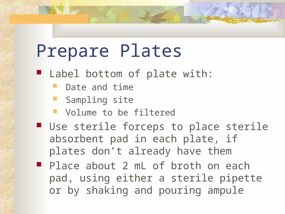
Prepare Plates Label bottom of plate with:
Date and time Sampling site Volume to be filtered
Use sterile forceps to place sterile absorbent pad in each plate, if plates don’t already have them
Place about 2 mL of broth on each pad, using either a sterile pipette or by shaking and pouring ampule

Filter Samples Sterilize forceps, place membrane filter on
filter holder Use sterile water for small samples of
water (1 mL) to wet the filter Pump until most of water is through filter Release pressure Sterilize forceps and place filter grid-side-
up on the absorbent pad
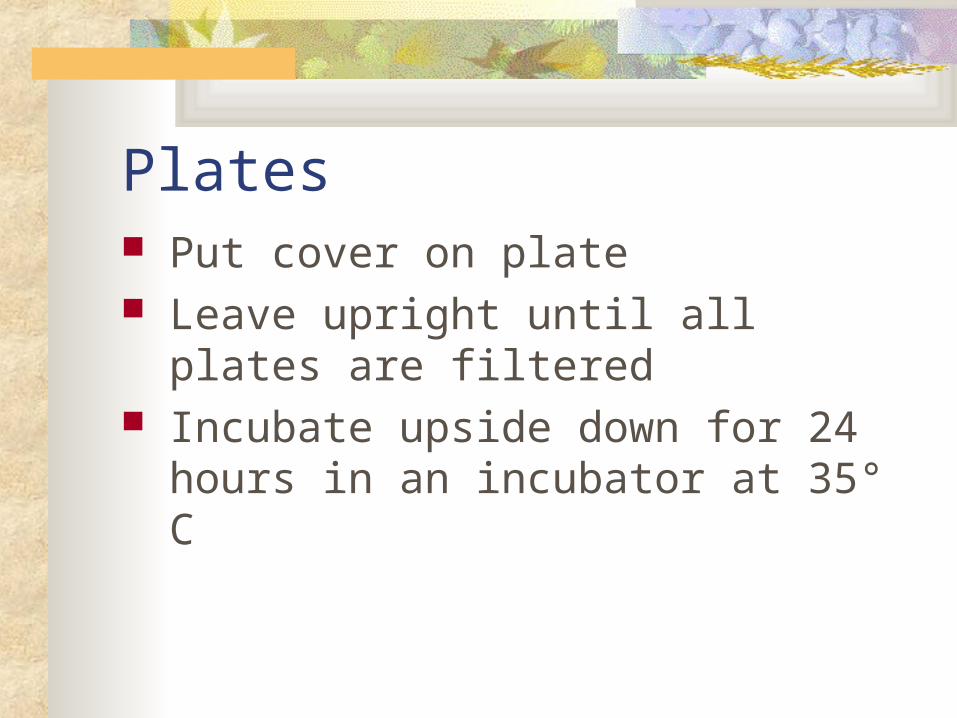
Plates Put cover on plate Leave upright until all plates are filtered Incubate upside down for 24 hours in an
incubator at 35° C

Calculating Results Count the blue and red colonies on each
plate Blue colonies are E. coli Red + Blue = Total Coliforms If there are greater than 200 colonies report
that plate as TNTC (Too numerous to count)

Most accurate platesThe best are when the colony counts are in the
range of: 20 – 80 colonies per plate for E. coli, and 50 – 200 for Total coliforms

Calculating Results Standard Units = CFU/100 mL (Colony Forming
Units) Average colony counts x 100 = CFU/100 ml Volume Filtered (mL) If fewer than 20, estimate CFU/ 100 ml using all
plates.Add total number of colonies and total volumeTotal colony counts x 100 = CFU/100 ml
Total mL filtered

Other problems If over 200, but colonies are clearly
countable, use the same general formula.
Conflicting colony counts:
go with the smaller sample size

0 mL filtered 1 mL filtered
10 mL filtered 30 mL filtered
24 colonies counted
6 colonies counted
0 colonies counted
67 colonies counted
The 10 mL plate would be used for calculating CFU/100 mL:
24 / 10 x 100 = 240 CFU/100 mL
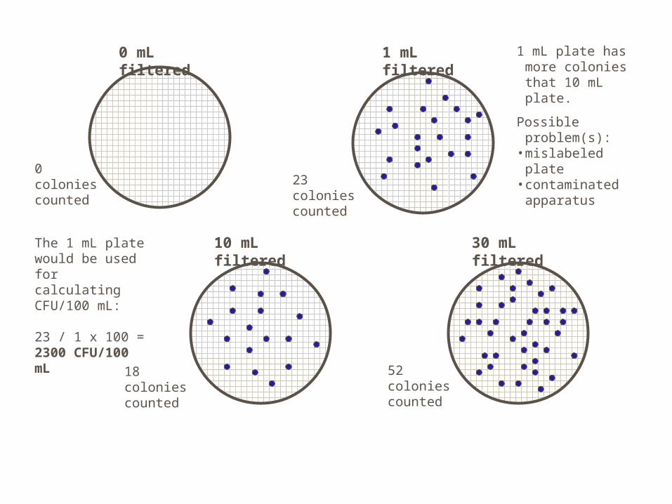
0 mL filtered
30 mL filtered
1 mL filtered
23 colonies counted
10 mL filtered
18 colonies counted
0 colonies counted
52 colonies counted
1 mL plate has more colonies that 10 mL plate.
Possible problem(s):• mislabeled plate• contaminated
apparatus
The 1 mL plate would be used for calculating CFU/100 mL:
23 / 1 x 100 = 2300 CFU/100 mL

0 mL filtered 0 mL filtered
1 mL filtered 30 mL filtered
23 colonies counted
6 colonies counted
7 colonies counted
Estimate or report as Too Numerous to Count (TNTC)
Possible problem: finger on filter or contaminated forceps
Possible problem: “sterile” water not sterilized
Possible problem: filter not wetted with sterilized water before filtering low volume sample – sample concentrated in one area of filter.
Example of plate with more than 200 colonies. Colonies could be counted or estimated, and results flagged as “estimate”.
The sterile water “blank” or 0 mL plate is a quality control measure – bacterial growth on the blank makes the other plate counts suspect.

Water Quality Standards In Oregon, based upon contact recreation 126 CFU/100 mL for 5 samples within a 30
day period 406 CFU/100 mL for a single sample