microbiology shellfish - aem.asm.org · yearling pacific (that is, japanese) oysters, crasso-strea...
TRANSCRIPT

R. R. COLWELL AND J. LISTON
VAN DER ZANT, W. C. AND NELSON, F. E. 1953 Proteolysisby Streptococcus lactis grown in milk with and withoutcontrolled pH. J. Dairy- Sci., 36, 1104-1111.
VAN DER ZANT, W. C. 1957 Proteolytic enzymes fromPseudomonas putrifaciens. I. Characteristics of an extra-cellular proteolytic enzyme system. Food Research, 22,151-163.
VAN HEYNINGEN, W. E. 1940 The proteinases of Clostridiumhistolyticurnm. Biochem. J., 34, 1540-1545.
VISWANATHA, T. AND LIENER, I. E. 1956 Isolation andproperties of a proteinase from Tetrahymena pyriformisW. Arch. Biochem. and Biophys., 61, 410-421.
WEIL, L. AND KOCHOLATY, W. 1937 Studies on the proteinaseof Clostridiunm histolyticumn. Biochem. J., 31, 1255-1267.
Microbiology of Shellfish
Bacteriological Study of the Natural Flora of Pacific Oysters (Crassostrea gigas)1'2
R. R. COLWELL AND J. LISTON
College of Fisheries, University of Washington, Seattle, Washington
Received for publication September 8, 1959
There is an extensive literature on the public healthaspects of shellfish bacteriology (Dodgson, 1928). Agreat deal of excellent work has been carried out onthe incidence and survival of such groups as the entericpathogens (Salmonella, Shigella) and related coliformindicator organisms in shellfish grown under variousconditions (Foote, 1895; Fabre-Domergue, 1912;Kelly and Arcisz, 1954). As a result of this work, thepractical conditions necessary for prevention andcontrol of shellfish-borne infection are now well es-tablished. However, there is an almost complete lackof information concerning the bacterial types notderived from sewage associated with shellfish.A comparison of the number of colonies obtained on
count plates prepared from shellfish incubated at roomtemperature (circa 20 to 25 C) and at 37 C indicatesthat nonmesophilic bacteria probably comprise thebulk of the bacterial population of shellfish. One type ofnoncoliform microorganism said to be peculiar toshellfish belongs to the group of large Spirochaetae.This type of microorganism was described by Fantham(1907) from mussels, Spirochaeta anodontae, and byDimitroff (1926) from oysters, Saprospira andCristispira. In some published reports concerning thepresence of coliform organisms in shellfish, casualreference has been made to the presence of other bac-teria. Thus Joseph (1914) described the occurrence ofspore bearing, asporogenous, pigmented, and non-pigmented bacteria in market oysters, Berry (1916),and Geiger et al. (1926) noted the presence of Proteus,Alcaligenes, and Pseudomonas fluorescens together with
I This work was supported in part by National Institutes ofHealth Grant No. E-2417 and by Initiative 171 Fund, Univer-sity of Washington, Seattle, Washington.
2 Contribution No. 66, College of Fisheries, University ofWashington, Seattle, Washington.
other common "water bacteria," also in market oysters.Eliot (1926) found that the green fluorescent, yellowpigmented, nonpigmented, and "vibrio" groups ofmicroorganisms rapidly increased in number during thespoilage of market oysters at 20 C. Tanikawa (1937)found that typical water bacteria of the generaAchromobacter, Pseudomonas, Flavobacterium, andAMicrococcus were of greatest importance in the spoilageof market oysters held at 0 C. The results of thesespoilage studies are remarkably similar to the bac-teriological findings for fin fishes held at similar tem-peratures (Shewan and Liston, 1956), and, in the lattercase, it has been quite well established that the spoilageorganisms are derived from the flora of the living fishwhich is predominantly composed of asporogenousgram-negative rods (Georgala, 1958). By analogy itseems not unreasonable to suspect that the spoilagebacteria in oysters are related to the normal bacterialpopulation present in the living animal.The purpose of this study was to determine the com-
position of the natural bacterial flora of oysters heldunder controlled natural conditions in various areas ofWashington. Coliform counts were carried out to ob-tain some information concerning the degree of pollu-tion of the environment, but the major portion of theinvestigation was concerned with noncoliform bacteria.
MATERIALS AND METHODS
Yearling Pacific (that is, Japanese) oysters, Crasso-strea gigas, were obtained from Purdy, Washington,and were placed in floating trays in three differentareas of Washington: fHood Canal, Oyster Bay, andWillapa Bay; a control group was maintained in thesalt-water aquarium at the College of Fisheries. Samplesof three oysters and 150 ml of seawater were takernfrom the aquarium weekly and from the floats every
[VOL. 8104
on February 16, 2019 by guest
http://aem.asm
.org/D
ownloaded from

MICROBIOLOGY OF SHELLFISH
third week. Most probable number (MPN) of coliformsand counts of Escherichia coli were established ac-
cording to the procedures described in Standard Methodsfor the Examination of Water, Sewage, and IndustrialWastes (APHA, 1955). The study extended over a
4j1-month period, from February to July 1959.Plating media and methods employed in the quanti-
tative determinations were as follows:MacLeod's maintenance medium (basal) from
MlacLeod et al. (1954) containing yeast extract, 0.5per cent; nutrient broth, 0.8 per cent; and Bacto-agar31.5 per cent in 1 L seawater.Basal medium plus glucose (basal + 1.0 per cent
glucose).Oyster agar (OA) modification of the medium of
Eyre (1923) consisting of 500 g of minced oyster meatextracted at 100 C for 30 min in 1 L sterile seawater,filtered, adjusted to pH 7.4, 15 g of Bacto-agar added,and sterilized 15 min at 120 C (15 pounds pressure).
Tryptone glucose extract (TGE).Nutrient agar (NA).Nutrient agar plus 0.5 per cent sodium chloride
(salt NA).Except where stated otherwise, the media were
obtained from Difco Laboratories in dehydrated formand made in accordance with the manufacturer'sdirections. The initial quantitative determinations forselection of the most appropriate medium of thoselisted above were done by means of surface plate counts.The basal medium plus glucose appeared to give themaximum count, but the colonies on the plates were
very mucoid and tended to fuse. The standard platecounts were thus carried out on the basal medium alone.
Colonies were picked at random from the countplates. The cultures obtained were subjected to purifi-cation procedures and the pure cultures were testedby a number of determinative methods. One hundredfifty-two cultures were maintained in seawater + 1per cent peptone or on basal agar slopes dependingupon how fastidious the organism was. Selective media(Difco, dehydrated) including the Enterococci Pre-sumptive, Ethyl Azide Violet Broth, Eosin-MethyleneBlue Agar, S S Agar, Brilliant Green Bile Broth, andTriple Sugar Iron Agar, were used to assist in theidentification of possible members of the Enterobac-teriaceae. Tests for identification and classification
were carried out according to the Manual of Micro-
biological Methods (SAB, 1957). Pure cultures of allthe organisms isolated in this study were streaked on
basal agar plates for determination of colonial mor-
phology and for tests of sensitivity to 0/129 vibriostat
compound (Shewan et al., 1954) and to 2, 5, 10 unitDifco penicillin discs. Other tests and media used wereas follows: litmus milk; seawater nutrient gelatin;
3Difco Laboratories, Inc., Detroit, Michigan.
lead acetate agar slopes; methyl red; Voges-Proskauer;nitrate broth; indole; urea agar slopes; Koser's citratebroth; Hugh and Liefson oxidative and fermentativemedium (Hugh and Liefson, 1953); lactose, glucose,maltose, mannitol, and sucrose fermentation tubes;and ammonia production. Temperature growth testsat 0, 25, and 37 C were carried out in a medium con-sisting of 0.5 per cent sodium chloride and 1 per centpeptone water. Routine tests and identification mediawere inoculated and incubated at 25 C (RT), but selectivemedia for enterobacteria were incubated at 37 C.
RESULTSThere was no significant difference between the
counts obtained in basal + 1 per cent glucose, basal,and oyster agar, which were always higher than thosein the other media. Basal medium seemed to support
TABLE 1Standard plate counts on several media*
Experiment 1 Experiment 2 Experiment 3
Medium 25 C 37 C 25 C 37 C 25 C 37 C
Total count per mlt
Basal agar.... 7,320 1,990 12,900 2,895 13,500 2,000Basal + 1%/
glucoseagar....... 10,500 1,395 14,000 2,895 14,500 3,395
Oyster agar .. 7,870 2,150 11,900 3,035 14,000 3,700Tryptone glu-
cose extractagar ....... 5,240 700 5,300 365 5,500 500
Nutrient agar. 1,170 400 1,170 395 1,500 500Nutrient agar+ 0.5%NaCl ....... 3,325 20 3,225 40 3,125 30
* See text for description.t Avg of duplicate platings.
TABLE 2Colifornm content of oysters and seawater examined at 3-week
inter vals
Time (weeks)
0 3 6 9 12
MPN coliforms per 100 ml sample
Aquarium oysters .............. 0 0 0 0 0Aquarium seawater (control). 0 0 0 0 0
Hood Canal oysters ............ 450 450 200 200Hood Canal seawater........... - 0 0 2 2
Oyster Bay oysters ............. 0 200 0 0Oyster Bay seawater ........... - 0 0 0 0
Willapa Bay oysters............ - 450 1100 20 20Willapa Bay seawater .......... 0 200 2 2
1960] 105
on February 16, 2019 by guest
http://aem.asm
.org/D
ownloaded from

R. R. COLWELL AND J. LISTON[vL
growth of marine and other bacteria as well as eitherbasal + glucose or oyster agar and was by far thesimplest to prepare (table 1). It was therefore chosenfor subsequent work.The MPN of coliforms in the seawater and oysters,
sampled at 3-week intervals throughout the period ofthe study, is given in table 2. Pollution as indicatedby MPN counts was light except during one briefperiod in the Willapa Bay area. Our results confirmedthat oysters tend to show higher coliform counts thanthe surrounding seawater. Figures 1 and 2 show the
o086
Q 4.rc 3(F)
2
t!1
L 8r.- 6
250C-37°C
Aquarium Oysters( Control)
_- 25°C4 -...37°C3 -- 2500-
2 - Hood Canal Oysters--- Hood Canal Seawater
0 I I
T2 3 4 5 6(ek 9TIME (Weeks)
Figure 1. Total viable count of bacteria per ml oyster fluidand per ml seawater for aquiaritum-held oysters (control) andHood Canal oysters.
10-8-
2500C2-1
370C -0 %'1370C2-
l0o
-Willapa Bay Oysters--Willapa Bay Seawater
8- -Oyster Bay Oysters6- --Oyster Bay Seawater
4- 250C4 370C3 - - 250C
2- __--- __-370C
total viable counts of bacteria per ml seawater and perml oyster body fluid sampled concurrently with theMPN determiniations.The group distribution of bacteria classified according
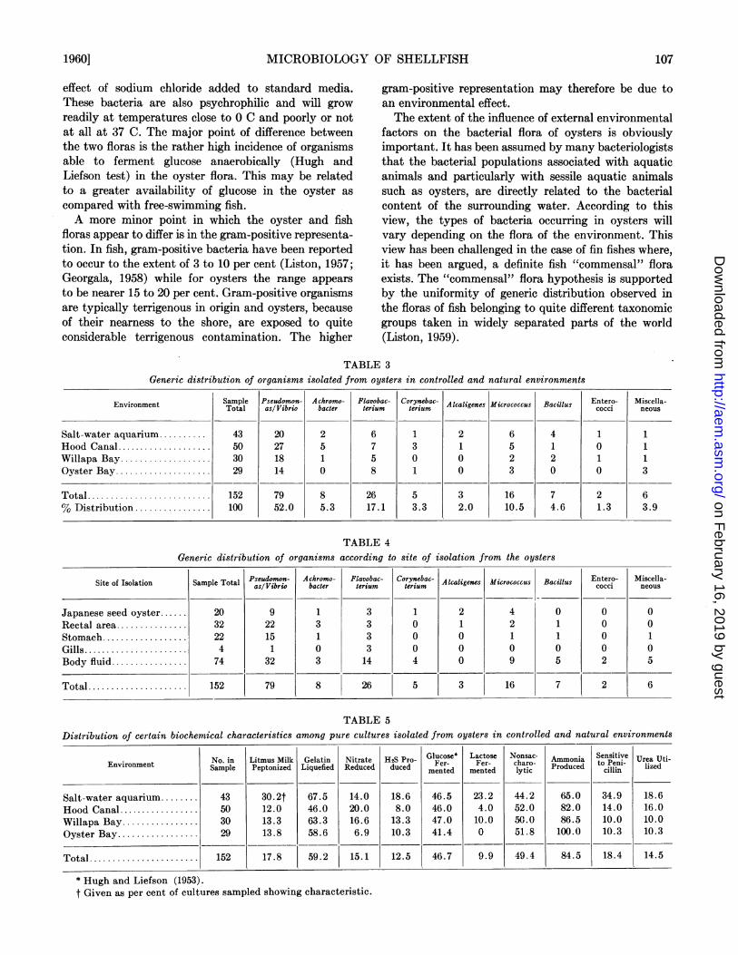
to genera, with the Pseudomonas and Vibrio groupscombined, is given in table 3. A similar distribution,but related to site of isolation in the oyster is given intable 4. Gram-negative nonsporing rods predominatedin the flora with Pseudomonas/Vibrio contributing themajor (circa 30 per cent) type and Flavobacteriumfollowing in importance. Gram-positive organisms con-stituted less than 20 per cent of the isolates. No realdifference appeared to exist in generic distribution oforganisms isolated from different sites in the oyster.
In table 5 are shown some of the more importantbiochemical properties of the organisms isolated, givenas percentages of the total number tested. It can beseeni that the biochemical abilities of the organismswithin the flora are similar in each environmental area.
Ill general, the bacterial population of ovsters isproteolytic in niatture, with only weak saccharolytic andreductive capacity. However, nearly 50 per cent ofthe organisms isolated were able to ferment glucoseanaerobically.
DIscussioNColiform bacteria, as calculated from the total viable
count and AWII'N values, never constituted more thani0.5 per cent of the total viable flora in our oyster sam-ples. The failure to isolate any of these organisms fromcount plates by our random selection procedure indi-cates that this is a true measure of the numericalinsignificance of these types in the oyster. Nevertheless,the results show once again the ability of the oyster toconcentrate coliform bacteria from seawater and under-lines the public health significance of oysters as potentialagents of enteric infection.The general composition of the bacterial flora of the
oysters studied by us is similar in outline to that indi-cated by the studies of Eliot (1926) and Tanikawa(1937). Gram-negative rod forms predominate whilegram-positive forms constitute a minor portion of theflora. The PseudomonasIVibrio group was found to bethe largest single group, and members of the Achromo-bacteriaceac next largest. This distribution of generictypes is very similar to that described by severalworkers for free swimming fish (Liston, 1957; Georgala,1938). There is, moreover, a striking resemblancebetwYeen physiological characters of the oyster flora andthat of the fish flora. In both cases proteolytic activity(as measured by litmus milk digestion and liquefactionof gelatin) is high while general saccharolytic activity islow. MIost of the organisms from both fish and shell-fish are characteristic marine types exhibiting a partialor complete salt dependence demonstrated, in thecase of the shellfish bacteria, by the growth stimulating
(F)
L.J
O i 2 3 4 5 6 7 8 9TIME (Weeks)
Fifpqre 2. Total viable count of bacteria per ml ovster fluidarid per ml seawater for Willapa Bayl and Oyster Bay samples.
[VOL. 8106
l<
CZ,
Q)i
-
I1
on February 16, 2019 by guest
http://aem.asm
.org/D
ownloaded from
MICROBIOLOGY OF SHELLFISH
effect of sodium chloride added to standard media.These bacteria are also psychrophilic and will grow
readily at temperatures close to 0 C and poorly or notat all at 37 C. The major point of difference betweenthe two floras is the rather high incidence of organismsable to ferment glucose anaerobically (Hugh andLiefson test) in the oyster flora. This may be relatedto a greater availability of glucose in the oyster as
compared with free-swimming fish.A more minor point in which the oyster and fish
floras appear to differ is in the gram-positive representa-tion. In fish, gram-positive bacteria have been reportedto occur to the extent of 3 to 10 per cent (Liston, 1957;Georgala, 1958) while for oysters the range appears
to be nearer 15 to 20 per cent. Gram-positive organismsare typically terrigenous in origin and oysters, becauseof their nearness to the shore, are exposed to quiteconsiderable terrigenous contamination. The higher
gram-positive representation may therefore be due toan environmental effect.The extent of the influence of external environmental
factors on the bacterial flora of oysters is obviouslyimportant. It has been assumed by many bacteriologiststhat the bacterial populations associated with aquaticanimals and particularly with sessile aquatic animalssuch as oysters, are directly related to the bacterialcontent of the surrounding water. According to thisview, the types of bacteria occurring in oysters willvary depending on the flora of the environment. Thisview has been challenged in the case of fin fishes where,it has been argued, a definite fish "commensal" floraexists. The "commensal" flora hypothesis is supportedby the uniformity of generic distribution observed inthe floras of fish belonging to quite different taxonomicgroups taken in widely separated parts of the world(Liston, 1959).
Generic distribution ofTABLE 3
organisms isolated from oysters in controlled and natural environments
Environment [ Sample Pseudomos- Achromo- Flavobac- Corynebac- Alcaligenes Micrococcus Bacillus cEntero- MisceloaEnvironment Total asl Vibrio bacder lerium teriumcoi neu
Salt-water aquarium .......... 43 20 2 6 1 2 6 4 1 1Hood Canal .................... 50 27 5 7 3 1 5 1 0 1Willapa Bay ................... 30 18 1 5 0 0 2 2 1 1Oyster Bay .................... 29 14 0 8 1 0 3 0 0 3
Total ....................... 152 79 8 26 5 3 16 7 2 6% Distribution ................ 100 52.0 5.3 17.1 3.3 2.0 10.5 4.6 1.3 3.9
TABLE 4Generic distribution of organisms according to site of isolation from the oysters
Pseudomon- Ackromo- Flavobac- Corynebac- Entero- Miscella-Site of Isolation Sample Total Alcaligenes Micrococcus Bacillus cocci neousasl Vibrio bacder terium teritumesMcocusBils cci nos
Japanese seed oyster.. 20 9 1 3 1 2 4 0 0 0Rectal area .32 22 3 3 0 1 2 1 0 0Stomach .22 15 1 3 0 0 1 1 0 1Gills .4 1 0 3 0 0 0 0 0 0Body fluid .74 32 3 14 4 0 9 5 2 5
Total ................... 152 79 8 26 5 3 16 7 2 6
TABLE 5Distribution of certain biochemical characteristics among putre cultures isolated from oysters in controlled and natural environments
No. in Litmus Milk Gelatin Nitrate H2S Pro- Glucose* Lactose Nonsac- Ammonia Sensitive Urea Uti-Environment Sample Peptonized Liquefied Reduced duced Fer- Fer- charo- Produced to enil- lized
Salt-water aquarium.43 30.2t 67.5 14.0 18.6 46.5 23.2 44.2 65.0 34.9 18.6Hood Canal .50 12.0 46.0 20.0 8.0 46.0 4.0 52.0 82.0 14.0 16.0Willapa Bay .30 13.3 63.3 16.6 13.3 47.0 10.0 -50.0 86.5 10.0 10.0Oyster Bay .29 13.8 58.6 6.9 10.3 41.4 0 51.8 100.0 10.3 10.3
Total .152 17.8 59.2 15.1 12.5 46.7 9.9 49.4 84.5 18.4 14.5
* Hugh and Liefson (1953).t Given as per cent of cultures sampled showing characteristic.
1960] 107
on February 16, 2019 by guest
http://aem.asm
.org/D
ownloaded from

R. R. COLWELL AND J. LISTONv
The bacterial floras of the oysters tested in this in-vestigation showed a high degree of similarity fromarea to area. Moreover, the similarity extended beyondthe taxonomic groupings to biochemical groupings.The same organisms with the same biochemical proper-ties were found in all areas. This general similaritywould be explained on the earlier hypothesis by theassumption that all the environments were similar.That this is not so can be seen from the coliform MPNresults (table 2) which indicate that the areas weresubject to different degrees of sewage pollution. More-over, the sites for the floats were selected to providedifferent water conditions at each place. It may beargued that the overriding environmental effects suchas salinity and so-called antibacterial effect of seawater(ZoBell, 1946) are similar in all areas, so that the bac-terial flora of the water is the same in all areas. Never-theless, the proximity of the oysters to the shore wouldpermit quite rapid transfer of terrigenous contaminantorganisms and there is evidence in the coliform countsthat even the more sensitive nonmarine types cansurvive for some time in seawater. Thus, the externalwater environment cannot exert the absolute controlover the bacterial flora that this theory requires.The assumption that the observed flora is in fact
typical of the oyster rather than of its environmentseems to be the simplest way of explaining the observa-tions. One cannot, of course, dismiss completely theeffect of environment on the oyster flora. Obviously,all microorganisms in the immediate vicinity of theoyster may gain access to it either fortuitously or as aresult of the feeding activity of the mollusc. It seemsprobable that only those organisms which are welladapted to the microenvironment provided by theoyster will establish themselves as a signiificant com-ponent of the oyster flora. Organisms such as the coli-form bacillus which are, in fact, ill-adapted for activegrowth under these conditions may survive and evenmultiply to a very small extenit in the oyster but willnever attain a numerically significant status in the totalbacterial population. The bacterial flora of the oysterwhich we suggest should be considered as a "com-mensal" flora, analogous to the "commensal" floras offish (or for that matter of land animals), is composedessentially of typical marine psychrophilic bacteriawell adapted physiologically to life within the micro-enivironimenit of the shellfish.The large Spirochaetes described by Dimitroff (1926)
and generally considered characteristic inhabitants ofshellfish were not observed in any of the oysters ex-amined in this survey. It is possible that our methodsof examination (phase contrast, dark field, and directilluminationi microscopy) were at fault. Another possi-bilty may be that the floatinig tray arranigement em-ployed in our studies in which the oysters were
under which the Spirillae described by Dimitroff couldnot survive.The high incidence of proteolytic bacteria and of
types capable of fermenting glucose in the naturalflora may be of practical significance in post-mortemspoilage of food oysters.
Eliot (1926) divided the spoilage process into threestages: acidity increase, abundant gas production, andproteolysis. He ascribed the major part of the spoilageto the "water forms" described as green fluorescentand yellow pigmented groups. Tanikawa (1937) showedthat Achromobacter, Pseudomonas, Flavobacterium, andMicrococcus were responsible for spoilage of oystermeat stored at 0 C. Thus it appears that the naturalflora of the oyster, as defined above, may be the majorfactor in spoilage. Methods of preservation should bedirected, therefore, to dealing with this flora.
ACKNOWLEDGMENTS
The field stations were designed, planted, and main-tained by D)r. A. K. Sparks and Mr. K. K. Chew, as
part of an experiment on oyster growth and mortality,and the authors are indebted to these gentlemen forpermitting us to use their facilities and for supplyingthe oysters used in our study.
SUMMARY
The study was undertaken to determine the com-
position of the bacterial flora of oysters held underapproximately natural conditions in three differentareas of Washington, with a control held in the salt-water aquarium at the College of Fisheries. MIostprobable number of coliform group organisms and totalviable counts for the 4/1-month period of study showedlittle or no apparent pollutioi.The group distribution of bacteria classified according
to genera showed that gram-negrativTe, asporogenous
rods of Psectdomo(nasIVibrio, and Flavobacterium, pre-
domiinated. GTram-positive organisms constituted lessthan 20 per cent of the isolates. No real differenceappeared to exist in generic distribution of organiismsisolated from differeint sites in the oyster or withinthe flora of oysters maintained in different areas ofWashington. Also, the biochemical abilities, proteolyticanid weakly saccharolytic, were similar for all organiismnsfrom each environmental area.
The "commensal" flora of the oyster was comparedwith that found oii free-swimming fish and discussedili terms of environmental factors. The relationiship of a
natural flora to post-mortem spoilage is also discussed.
REFEIRENCES
American Public Health Association 1955 Standard mlethodsfor the examlination of water, sewage, and industtrial wastes,10th ed. New ork, New York.
BERRY, F. 1916 The bacterial content of market oysters. J.suspenided clear of the sea bottoin, provided coniditions
[VOL. 8108
13acteriol., 1, 107-108.
on February 16, 2019 by guest
http://aem.asm
.org/D
ownloaded from

BACTERIAL SURVIVAL IN FROZEN FOOD
DIMITROFF, V. T. 1926 Spirochaetes in Baltimore marketoysters. J. Bacteriol., 12, 135-177.
DODGSON, R. W. 1928 Report on mussel purification. Minn.Agr. and Fisheries Invest. Ser. II, 10, 1-498.
ELIOT, C. 1926 Bacterial flora of the market oyster. Am. J.Hyg., 6, 755-776.
EYRE, J. W. H. 1923 Some notes on the bacteriology of theoyster (including description of two new species). J.Roy. Microseop. Soc., London, 1923 (4), 385-394.
FABRE-DOMERGUE, M. 1912 Bacterial purification of oystersby standing in filtered artificial seawater. Chem. Abstr.,6, 1045.
FANTHAM, H. B. 1907 Spirochaeta (Trypanosoma) balbianji(Certes) its movements, structure, and affinities; and onthe occurrence of Spirochaeta anodontae (Keysselitz) in theBritish mussel, Anodonta eygnea (Prelim. account). An-nual Magazine Natural History London, Ser. (7) 19, 493-501.
FOOTE, C. J. 1895 A bacteriological study of oysters withspecial reference to them as a source of typhoid infection.Medical News, 66, 320.
GEIGER, J. C., WARD, W. E., AND JACOBSON, M. A. 1926 Thebacterial flora of market oysters. J. Infectious Diseases,38, 273-280.
GEORGALA, D. L. 1958 The bacterial flora of the skin ofNorth Sea cod. J. Gen. Microbiol., 18, 84-91.
HUGH, R. AND LEIFSON, E. 1953 The taxonomic significance
of fermentative versus oxidative metabolism of carbohy-drates by various gram-negative bacteria. J. Bacteriol.,66, 24-26.
JOSEPH, M. 1914 Bacteriological findings in Baltimoreoysters. Bull. Johns Hopkins Hosp., 25, 128-131.
KELLY, C. B. AND ARCISZ, W. 1954 Survival of enteric or-ganisms in shellfish. Public Health Repts., 69, 1205-1210.
LISTON, J. 1957 The occurrence and distribution of bacterialtypes on flatfish. J. Gen. Microbiol., 16, 205-216.
LISTON, J. 1959 The bacterial flora of Pacific fish. Bac-teriol. Proc., 1959, 12.
MACLEOD, R. A., ONOFREY, E., AND NORRIS, M. E. 1954Nutrition and metabolism of marine bacteria. I. Survey ofnutritional requirements. J. Bacteriol., 68, 680-686.
SHEWAN, J. M. AND LISTON, J. 1956 Objective and subjectiveassessments of fish quality. Bull. Inst. Refrig. Suppl.Annexe, 1956-1, 137.
SHEWAN, J. M., HODGKISS, W., AND LISTON, J. 1954 Amethod for the rapid differentiation of certain non-patho-genic asporogenous bacilli. Nature, London, 173, 208.
Society of American Bacteriologists 1957 Manual of micro-biological methods. Biotech Publications, Geneva, NewYork.
TANIKAWA, E. 1937 Bacteriological examination of oystersstored at low temperatures. Zentr. Bakteriol. Parasitenk.Abt. 2, 97, 133-147.
ZoBELL, C. E. 1946 Marine microbiology. Chronica BotanicaCo., Waltham, M1assachusetts.
Survival of Salmonella typhimurium, Staphylococcus aureus, andStreptococcus faecalis Frozen in Simplified Food Substrates1'2
MARGY J. WOODBURN3 AND DOROTHY HUSSEMANN STRONG
School of Home Economics, University of Wisconsin, Madison, Wisconsin
Received for publication September 8, 1959
In nonsterile food products, including those pre-served by freezing, the possible survival of thosebacteria frequently implicated in food-borne illness hasimportant public health implications. Borgstrom (1955)in a review of the research in the area of the micro-biology of frozen foods stated that additional studiescould profitably be undertaken to elucidate moreclearly the relationships which may exist betweenbacterial survival and the substrate on which theorganism exists. Recently, the concept of the uniquenature and requirements of the bacterial cell which hasbeeni frozen was forwarded by Bretz and Hartsell (1959).
1 Published with the permission of the Director of the Wis-consin Agricultural Experiment Station, Madison, Wisconsin.
2 These data are a part of the thesis submitted to the Gradu-ate School at the University of Wisconsin in partial fulfillmentof the requirements for the Ph.D. degree by the senior auithorwho was a General Foods Fund Fellow in Home Economics.
3 Present address: School of Home Economics, Purdue Uni-versity, Lafayette, Indiana.
In the present. study, data were obtained on thenumbers of viable cells surviving freezing, frozenstorage, and thawing of a single species of each ofthree bacterial geinera generally acknowledged ascauses of food-borne illnesses. Four substrates wereprepared, each consistinig of a single food ingredientdissolved or suspended in buffer. To each was added aninoculum of viable cells of a single species. Bacterialcells suspended in buffer or in physiological salineserved as controls. Observations were made at threetemperatures of frozen storagfe and at intervals over a10-week period.
EXPERIMENTAL MIETHODSTen-milliliter quantities of the inoculated suspending
media were frozen in capped test tubes (16 by 125 mm)at - 11, -21, or -30 C. The tubes were subsequentlystored at the same temperature as had been used forfreezing. Duplicate samples were withdrawn after 24
1960] 109
on February 16, 2019 by guest
http://aem.asm
.org/D
ownloaded from