microbiology
DESCRIPTION
presentationTRANSCRIPT

Antigen Capture, Antigen Capture, Presentation Presentation
&& Recognition Recognition
Dr Maliha SumbulDr Maliha Sumbul

These photomicrographs show phagocytosis of antibody-coated pneumococci
Left: A neutrophil extends a pseudopod toward two pneumococci. Center: these bacteria have been engulfed (arrows),
and the neutrophil is beginning to engulf four more pneumococci at the upper right.
Right: Two pneumococci have escaped.
►Antigen presentationAntigen presentation is a process in is a process in the body's the body's immune system by which by which macrophages, , dendritic cells and other and other cell types capture cell types capture antigens and then and then enable their recognition by enable their recognition by T-cells

►The basis of adaptive immunity lies in the The basis of adaptive immunity lies in the capacity of immune cells to distinguish capacity of immune cells to distinguish between the between the body's own cellsbody's own cells and and infectious pathogensinfectious pathogens
► The host’s cells express “self” antigens that identify The host’s cells express “self” antigens that identify them as such. them as such.
► These antigens are different from those in bacteria These antigens are different from those in bacteria ("non-self" antigens) or in virally-infected host cells ("non-self" antigens) or in virally-infected host cells (“missing-self”). (“missing-self”).
THE MISSING – SELF HYPOTHESIS: the absence or THE MISSING – SELF HYPOTHESIS: the absence or altered expression of MHC class I molecules would altered expression of MHC class I molecules would render target cells susceptible to NK cell attackrender target cells susceptible to NK cell attack
► The ability of the The ability of the adaptive immune system to to survey for infection requires specialized pathways survey for infection requires specialized pathways of enabling recognition of pathogen-derived of enabling recognition of pathogen-derived antigens by T cellsantigens by T cells
► The First Molecular Basis of the "Missing Self" Hypothesis ► Francisco Borrego► 1 I still remember the day my advisor, Dr. Rafael Solana, came to me with the
article of Karlhofer et al. (1) in his hands and told me that that paper was the proof for the receptor inhibition model of the "missing self" hypothesis. I had just joined the Laboratory of Immunology in the University of Córdoba, in Spain, and my knowledge about NK cells was very basic. At that moment, I did not realize the whole significance of Yokoyama’s work, but as time went by, I fully understood the breakthrough that the manuscript represented.
► In 1990, Ljunggren and Kärre published in Immunology Today a very comprehensive review about the role of MHC class I Ags on NK cell recognition (2). According to the "missing self" hypothesis that they proposed, the absence or altered expression of MHC class I molecules would render target cells susceptible to NK cell attack. Klas Kärre adopted the concept of "missing self" while he was writing his Ph.D. thesis and found that it was easier to describe the common features of resistant target cells rather than susceptible ones (3). Four years earlier, in 1986, Kärre et al. published a seminal letter in Nature that showed in vivo that NK cells were able to reject tumor cells that have lost MHC class I expression (4). The finding that deficiency of MHC class I molecules constituted an alternative immune defense strategy was very provocative at the time. It was believed that NK cells worked like T cells by recognizing foreign Ags on the target cells, and it was very clear that NK cells were not MHC restricted. But what Kärre postulated was exactly the opposite in the sense that NK cells, like T cells, were strongly influenced by the expression of MHC class I molecules on the target cell (3). Following Kärre’s letter in Nature, many manuscripts were published about the role of MHC class I molecules in NK cell recognition. In vivo and in vitro experiments were reported both in the mouse and in human systems. In general, people in the field accepted that NK cell susceptibility was directly related to the absence of MHC class I expression on target cellsThe Journal of Immunology, 2006, 177: 5759-5760.
Copyright © 2006 by The American Association of Immunologists, Inc.

STEPS:STEPS:► Foreign protein or antigen is taken up by an Foreign protein or antigen is taken up by an
antigen-presenting cellantigen-presenting cell► The antigen is processed and displayed on an MHC The antigen is processed and displayed on an MHC
II molecule, which interacts with a T helper cellII molecule, which interacts with a T helper cell
► Whole foreign proteins are bound by membrane Whole foreign proteins are bound by membrane antibodies and presented to B lymphocytes which antibodies and presented to B lymphocytes which process and present antigen on MHC II to a process and present antigen on MHC II to a previously activated T helper cell, spurring the previously activated T helper cell, spurring the production of antigen-specific antibodies production of antigen-specific antibodies
OR

Antigen receptors:Antigen receptors:►B Lymphocytes: membrane bound antobodies can B Lymphocytes: membrane bound antobodies can
recognize wide variety of macromolecules – recognize wide variety of macromolecules – proteins, polysaccharides, lipids and nucleic acids proteins, polysaccharides, lipids and nucleic acids – therefore, against many types of microbial cell – therefore, against many types of microbial cell wall and soluble antigenswall and soluble antigens
►T Lymphocytes: TCR – can only see peptide T Lymphocytes: TCR – can only see peptide fragments of protein antigens and only when these fragments of protein antigens and only when these peptides are presented by specialized peptide peptides are presented by specialized peptide display molecules on host cellsdisplay molecules on host cells
Same microbe – different Same microbe – different actionaction
►Eg:Eg:►Virus – entered circulation and free in Virus – entered circulation and free in
blood – combated by antibodiesblood – combated by antibodies
►Virus infected host cell – antibodies no Virus infected host cell – antibodies no longer effective – necessary to longer effective – necessary to activate CTLs to kill infected cells and activate CTLs to kill infected cells and infectioninfection


MHC COMPLEX
► MHC class I molecules
► found on every nucleated cell of the body (and thus not on red blood cells).
► MHC (major histocompatibility complex) Class II molecules
► found only on a few specialized cell types, including macrophages, dendritic cells and B cells, all of which are professional antigen-presenting cells (APCs)
Responsive cellCD 8+ Responsive cell
CD 4+
ENDOGENOUS PATHWAY
EXOGENOUS PATHWAY

Professional APCsProfessional APCs►Ability of these cells to both display Ability of these cells to both display
antigens for T cells and provide the antigens for T cells and provide the additional signals needed to activate additional signals needed to activate naïve T cellsnaïve T cells

►The human leukocyte antigen system (HLA) is the name of the major histocompatibility complex (MHC) in humans
(genes reside on chromosome 6)

► The major HLA antigens are essential elements for immune function. Different classes have different functions:
► HLA class I antigens present peptides from inside the cell (including viral peptides if present). Foreign antigens attract killer T-cells (also called CD8 positive- or cytotoxic T-cells) that destroy cells
► ► HLA class II antigens present antigens from outside
of the cell to T-lymphocytes. These particular antigens stimulate T-helper cells to multiply, and these T-helper cells then stimulate antibody-producing B-cells to produce Antibodies to that specific antigen. Self-antigens are suppressed by suppressor T-cells
► HLA class III antigens encode components of the complement system
Endogenous pathway
Exogenous pathway

HLA have other roles:► They are important in disease defense
► They may be the cause of organ transplant rejections
►They may protect against or fail to protect (if down regulated by an infection) cancers
► They may mediate autoimmune disease (examples: type I diabetes, coeliac disease)
►Also, in reproduction, may be involved in mate selection

HLA and autoimmune diseases HLA allele Diseases with increased
riskRelative risk
HLA-B27 Ankylosing spondylitis 1214
Acute anterior uveitis 15HLA-DR3 Autoimmune hepatitis 14
Primary Sjögren syndrome 10Diabetes mellitus type 1 5
HLA-DR4 Rheumatoid arthritis 4Diabetes mellitus type 1 6
HLA-DR3 and-DR4 combined
Diabetes mellitus type 1 15
HLA-B47 21-hydroxylase deficiency 15

► Helper T cellsHelper T cells (CD4+) serve as managers, directing the immune response (CD4+) serve as managers, directing the immune response
They secrete chemicals called They secrete chemicals called lymphokineslymphokines that that
stimulate stimulate cytotoxic T cellscytotoxic T cells and and B cellsB cells to grow and divide, to grow and divide,
attract attract neutrophilsneutrophils, and enhance the ability of , and enhance the ability of macrophagesmacrophages
to engulf and destroy microbesto engulf and destroy microbes
Antigen recognitionAntigen recognition►Unlike Unlike B cells, T cells fail to recognize , T cells fail to recognize
antigens in the absence of antigen antigens in the absence of antigen presentation, with the important presentation, with the important exception of the exception of the superantigens
►The The T cell receptor is is restricted to to recognizing antigenic peptides only recognizing antigenic peptides only when bound to appropriate molecules when bound to appropriate molecules of the of the major histocompatibility complex (MHC), also known in humans as (MHC), also known in humans as Human leukocyte antigen (HLA)) (HLA))

Superantigens (SAgs)►a class of antigens which cause non-specific
activation of T-cells resulting in massive cytokine release.
►SAgs can be produced by pathogenic microbes (including viruses, mycoplasma, and bacteria) as a defense mechanism against the immune system
► Compared to a normal antigen-induced T-cell response where 0.001- 0.0001% of the body’s T-cells are activated, these SAgs are capable of activating up to 20% of the body’s T-cells

► The large number of activated T-cells generates a massive immune response which is not specific to any particular epitope on the SAg thus undermining one of the fundamental strengths of the adaptive immune system, that is, its ability to target antigens with high specificity
► More importantly, the large number of activated T-cells secrete large amounts of cytokines (the most important of which is TNF-alpha)
► TNF-alpha is particularly important as a part of the body's inflammatory response, and in normal circumstances (where it is released locally in low levels) helps the immune system defeat pathogens. However when it is systemically released in the blood and in high levels (due to mass T-cell activation resulting from the SAg binding), it can cause severe and life-threatening symptoms, including shock and multiple organ failure.
►An epitope is a portion of a molecule to which an antibody binds.► Epitopes can be composed of sugars, lipids or amino acids

►Diseases associated with superantigen production
►Toxic Shock Syndrome ►Kawasaki Disease ►Eczema ►Psoriasis ►Rheumatoid arthritis ►Diabetes mellitus ►Scarlet fever
►Most cells are capable of presenting Most cells are capable of presenting antigens and activating the adaptive antigens and activating the adaptive response. Some cells, however, are specially response. Some cells, however, are specially equipped to acquire and present antigen, equipped to acquire and present antigen, and to prime and to prime naive T cells. .
►Dendritic cells, B cells, and macrophagesDendritic cells, B cells, and macrophages play a major role in the innate response, and play a major role in the innate response, and also act as professional also act as professional antigen presenting cells (APC). These (APC). These professional APCs are equipped with special professional APCs are equipped with special immunostimulatory receptors that allow for immunostimulatory receptors that allow for enhanced activation of T cellsenhanced activation of T cells

►Several different types of T cell can be Several different types of T cell can be activated by professional APCs, and each activated by professional APCs, and each type of T cell is specially equipped to type of T cell is specially equipped to deal with different pathogens, whether deal with different pathogens, whether the pathogen is bacterial, viral or a toxinthe pathogen is bacterial, viral or a toxin
►The type of T cell activated, and The type of T cell activated, and therefore the type of response therefore the type of response generated, depends, in part, on the generated, depends, in part, on the context in which the antigen was first context in which the antigen was first encountered by the APCencountered by the APC

Intracellular antigens: MHC Class IIntracellular antigens: MHC Class I
► Intracellular antigens are mainly produced by viruses Intracellular antigens are mainly produced by viruses replicating within a host cell, though antigens here can replicating within a host cell, though antigens here can also derive from cytoplasmic bacteria or the host cell's also derive from cytoplasmic bacteria or the host cell's own proteinsown proteins
► The host cell digests cytoplasmic proteins by a specialized The host cell digests cytoplasmic proteins by a specialized enzyme complex, the enzyme complex, the proteasome into small into small peptides
► A specialized carrier, the Transporter associated with A specialized carrier, the Transporter associated with Antigen Processing (TAP) complex moves the peptide into Antigen Processing (TAP) complex moves the peptide into the the endoplasmic reticulum, allowing the antigenic peptide , allowing the antigenic peptide to be coupled to an to be coupled to an MHC Class I molecule and transported molecule and transported to the cell surfaceto the cell surface
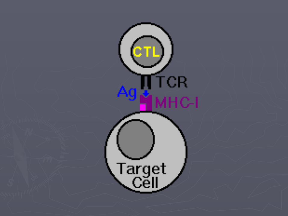
► MHC Class I molecules present antigen to CD8+ MHC Class I molecules present antigen to CD8+ cytotoxic T cells

► With the exception of some cell types (such as With the exception of some cell types (such as erythrocytes), Class I MHC is expressed by almost ), Class I MHC is expressed by almost all host cellsall host cells
► Cytotoxic T cells (also known as TC, killer T cell, or Cytotoxic T cells (also known as TC, killer T cell, or cytotoxic T-lymphocyte (CTL)) are a population of T cytotoxic T-lymphocyte (CTL)) are a population of T cells which are specialized for inducing the death of cells which are specialized for inducing the death of other cellsother cells
► Recognition of antigenic peptides through Class I by Recognition of antigenic peptides through Class I by CTLs leads to the killing of the target cell, which is CTLs leads to the killing of the target cell, which is infected by virus, intracytoplasmic bacterium, or infected by virus, intracytoplasmic bacterium, or are otherwise damaged or dysfunctionalare otherwise damaged or dysfunctional

Extracellular antigens: MHC Class IIExtracellular antigens: MHC Class II
►Dendritic cells (DCs) Dendritic cells (DCs) phagocytose exogenous exogenous pathogens, such as bacteria, parasites or toxins pathogens, such as bacteria, parasites or toxins in the tissues and then migrate, via in the tissues and then migrate, via chemotactic signals, to signals, to T cell enriched lymph nodes enriched lymph nodes
►During migration, DCs undergo a process of During migration, DCs undergo a process of maturation in which they lose phagocytic maturation in which they lose phagocytic capacity and develop an increased ability to capacity and develop an increased ability to communicate with T-cells in the lymph nodescommunicate with T-cells in the lymph nodes

► The DC uses The DC uses lysosome-associated -associated enzymes to digest pathogen-associated proteins into to digest pathogen-associated proteins into smaller peptidessmaller peptides
► In the lymph node, the DC will display these antigenic peptides on its surface by coupling In the lymph node, the DC will display these antigenic peptides on its surface by coupling them to MHC Class II moleculesthem to MHC Class II molecules
► This MHC:antigen complex is then recognized by T cells passing through the lymph nodeThis MHC:antigen complex is then recognized by T cells passing through the lymph node
► Exogenous antigens are usually displayed on Exogenous antigens are usually displayed on MHC Class II molecules, which interact with molecules, which interact with CD4+ CD4+ helper T cells

► CD4+ lymphocytes, or TH, are immune CD4+ lymphocytes, or TH, are immune response mediators, and play an important response mediators, and play an important role in establishing and maximizing the role in establishing and maximizing the capabilities of the adaptive immune responsecapabilities of the adaptive immune response
► Expression of Class II is more restricted than Expression of Class II is more restricted than Class I. Class I.
► High levels of Class II are found on High levels of Class II are found on dendritic cells, but can also be observed on , but can also be observed on activated activated macrophagesmacrophages and and B cellsB cells..
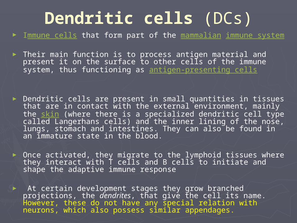
Dendritic cells (DCs)► Immune cells that form part of the mammalian immune system
► Their main function is to process antigen material and present it on the surface to other cells of the immune system, thus functioning as antigen-presenting cells
► Dendritic cells are present in small quantities in tissues that are in contact with the external environment, mainly the skin (where there is a specialized dendritic cell type called Langerhans cells) and the inner lining of the nose, lungs, stomach and intestines. They can also be found in an immature state in the blood.
► Once activated, they migrate to the lymphoid tissues where they interact with T cells and B cells to initiate and shape the adaptive immune response
► At certain development stages they grow branched projections, the dendrites, that give the cell its name. However, these do not have any special relation with neurons, which also possess similar appendages.

Basic model of how dendritic cells interact with CD4(+) T-cells


B-cells have the ability to capture and present antigen. Because this happens through the B-cell receptor, this is “specific” capture and presentation
This allows the efficient presentation of antigen to CD4(+) T cells by B-cells [B cells already have the ability to make antibody to that antigen]
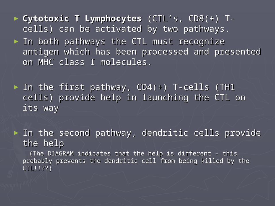
►Cytotoxic T Lymphocytes Cytotoxic T Lymphocytes (CTL’s, CD8(+) T-(CTL’s, CD8(+) T-cells) can be activated by two pathways. cells) can be activated by two pathways.
► In both pathways the CTL must recognize antigen In both pathways the CTL must recognize antigen which has been processed and presented on MHC which has been processed and presented on MHC class I molecules. class I molecules.
► In the first pathway, CD4(+) T-cells (TH1 cells) In the first pathway, CD4(+) T-cells (TH1 cells) provide help in launching the CTL on its wayprovide help in launching the CTL on its way
► In the second pathway, dendritic cells provide the In the second pathway, dendritic cells provide the help help
(The DIAGRAM indicates that the help is different – this probably (The DIAGRAM indicates that the help is different – this probably prevents the dendritic cell from being killed by the CTL!!??)prevents the dendritic cell from being killed by the CTL!!??)

a) How CD8(+) T-cells get activated b) How they kill target cells
1
2

► After activation, the CTL is now capable of After activation, the CTL is now capable of killing killing target cellstarget cells – that is, cells that are – that is, cells that are expressing the activating antigen in the expressing the activating antigen in the context of MHC class I.context of MHC class I.
► There are two killing mechanisms:There are two killing mechanisms:► The secretion of enzymes onto the target cell The secretion of enzymes onto the target cell
which induce permeability changes and which induce permeability changes and apoptosis – apoptosis – perforin perforin and and granzymesgranzymes
► The interaction of The interaction of FasFas on the CTL with on the CTL with FasLFasL on the target cell activates on the target cell activates caspases caspases within within the target cell and results in apoptosis.the target cell and results in apoptosis.

QUESTIONS QUESTIONS ??